

Submitted:
19 October 2026
Posted:
21 October 2026
You are already at the latest version
Abstract
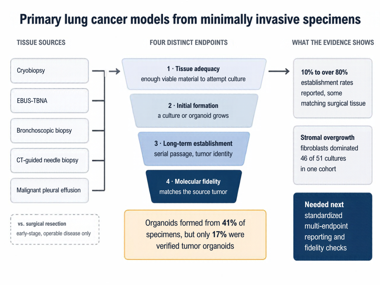
Primary cultures and organoids from lung cancer patients preserve tumor heterogeneity and enable patient-specific functional testing. Their generation has relied largely on surgical resection, which most patients never undergo because their disease is diagnosed too late. This review examines whether minimally invasive sources, namely cryobiopsy, endobronchial ultrasound-guided transbronchial needle aspiration, bronchoscopic biopsy, computed tomography (CT)-guided transthoracic needle biopsy, and malignant pleural effusion, can serve as alternatives. To compare outcomes across studies, we distinguish four endpoints: tissue adequacy, initial formation, long-term validated establishment, and molecular fidelity to the source tumor. Reported establishment rates from these sources range from 10% to over 80%, and some matched or exceeded surgical tissue within the same study. Yet these figures depend on how success is defined. In one cohort, organoids formed from 41% of specimens, but only 17% yielded verified tumor organoids. In another, cancer-associated fibroblasts dominated 46 of 51 cultures. Anatomical site, medium composition, and overgrowth by stromal or normal airway cells therefore explain outcomes better than acquisition route alone. Ex vivo drug responses have been linked to clinical outcomes, but prospective validation is lacking. Standardized multi-endpoint reporting and routine confirmation of molecular fidelity are needed before these models can guide therapy.
Keywords:
patient-derived organoids
; primary cell culture
; non-small cell lung cancer
; minimally invasive biopsy
; cryobiopsy
; CT-guided transthoracic biopsy
; EBUS-TBNA
; malignant pleural effusion
; tumor heterogeneity
; precision oncology
1. Introduction
Primary lung cancer cell lines derived from minimally invasive procedures constitute a valuable approach for studying tumor biology and supporting drug discovery [1,2]. Such models can better capture tumor heterogeneity and patient-specific responses than conventional cell lines, thereby more closely reflecting the complexity of clinical disease [3,4]. Patient-derived primary cultures address key limitations of established preclinical models, allowing assessment of morphological, molecular, and functional tumor characteristics to predict drug response and resistance mechanisms [4,5].
1.1. Lung Cancer: Epidemiology, Molecular Heterogeneity and Challenges in Treatment
Lung cancer remains the leading cause of cancer-related mortality worldwide and has a five-year survival rate of only 10-20% [6,7]. With the global cancer burden projected to reach 35 million new cases by 2050, a 77% increase over 2022, the need for reliable model platforms and improved therapeutic strategies is becoming increasingly important [6]. Its considerable molecular heterogeneity contributes to differences in tumor behavior and treatment response, complicating the development of effective therapeutic strategies [8]. Distinct lung cancer subtypes harbor different genetic mutations and activate divergent molecular pathways, further complicating therapeutic decision-making [9]. This heterogeneity may also promote acquired drug resistance. Under therapeutic pressure, genetically and phenotypically distinct tumor subclones undergo differential selection, ultimately limiting the durability of treatment responses in advanced disease [10,11]. Non-small cell lung cancer (NSCLC) accounts for approximately 85% of all cases [12,13], and the majority of patients present with advanced-stage disease unsuitable for surgical resection. Primary cell cultures have historically relied on resected specimens, which are available only for early-stage, operable disease. This reliance limits the applicability of such models to a small subset of patients, underscoring the need for alternative, minimally invasive tissue sources [14-16]. Conventional preclinical models, however, do not fully reflect this molecular and phenotypic diversity, as discussed in the following section.
1.2. Limitations of Conventional Preclinical Models
Established cell lines, while convenient for high-throughput screening, can undergo genetic and phenotypic changes during long-term culture, potentially altering molecular features associated with drug sensitivity and reducing their similarity to the original tumor [2,8,17]. Moreover, these models do not fully recapitulate the complex intercellular interactions and stromal components present in solid tumors, making it difficult to reproduce key aspects of their biology [2,4]. The limited physiological relevance of these conventional in vitro models may consequently reduce the predictive value of preclinical drug testing, particularly when interactions within the tumor microenvironment influence therapeutic response [8,18]. Patient-derived xenografts better preserve primary histology and genetic characteristics, but their high cost, long generation time, and logistical complexity make them less suitable for rapid and personalized preclinical drug testing [19,20].
1.3. Minimally Invasive Sampling as a Source of Primary Lung Cancer Cultures
Functional drug testing of primary tumor cells helps bridge the gap between molecular profiling and clinically relevant therapeutic outcomes [21]. Unlike immortalized lines, these cultures retain key genomic and phenotypic characteristics of the parental tumor. Furthermore, they provide patient-specific information on treatment responses and cellular behavior that may not be adequately captured by established models [4,22,23].
Material obtained through minimally invasive procedures, including endobronchial ultrasound (EBUS)-guided biopsies, provides access to tumor tissue from patients with advanced or inoperable disease while yielding viable cells suitable for patient-specific functional analyses [22,24]. Ex vivo drug sensitivity testing of patient-derived specimens complements molecular profiling by identifying patient-specific drug responses and therapeutic vulnerabilities, including in tumors refractory to genomics-guided targeted therapies [25-27]. Together, these observations support the central premise of this review. The feasibility of establishing primary cultures depends on the tissue-acquisition technique and anatomical sampling site, rather than simply on whether the tissue was obtained surgically or non-surgically. Evaluated on their own terms, minimally invasive approaches already extend primary culture-based research to patients who would otherwise be underrepresented in studies relying exclusively on surgical specimens.
The overall workflow for establishing primary lung cancer cultures from surgical and minimally invasive diagnostic specimens is summarized in Figure 1, illustrating how distinct tissue sources enter a common laboratory pipeline encompassing specimen processing, culture establishment, characterization, and biobanking.
1.4. Aim and Scope of This Review
This review addresses a question that arises directly from laboratory practice: how far apparent differences between surgically resected and minimally invasive tissue sources can be interpreted at all, given how inconsistently laboratory outcomes are defined and reported. Our position is that these differences are difficult to interpret unless tissue adequacy, initial formation, long-term establishment and molecular fidelity are reported as distinct endpoints, and that the available within-study evidence suggests anatomical site, specimen characteristics and culture conditions can substantially influence these outcomes. Because existing studies frequently conflate distinct laboratory milestones, we define four non-interchangeable endpoints: (1) sample adequacy, defined as sufficient viable material to attempt culture; (2) initial culture or organoid formation; (3) long-term validated establishment, typically requiring extended serial passage and confirmed tumor identity; (4) molecular or genomic fidelity to the source tumor. A study reporting success on one of these axes does not, by itself, provide evidence about the others, and comparisons across studies are made only where the same endpoint is being assessed. The emphasis throughout is on the laboratory determinants of culture establishment – specimen characteristics, dissociation strategy, medium composition and quality control – rather than on the technical performance of the sampling procedures themselves. Liquid-biopsy sources that do not yield tumor tissue or a cell-containing effusion, including circulating tumor cells and bronchoalveolar lavage, are outside the scope of this review.
This narrative review is based on a non-systematic literature search of PubMed/MEDLINE, Scopus and Wiley Online Library performed manually by the authors up to August 2026. Reference lists of relevant articles were additionally screened. Publications known to the authors served as sentinel records to check search retrieval, and all included studies were selected and evaluated by the authors. Studies were considered when they reported the establishment or characterization of primary lung cancer cultures or organoids from clinical specimens. No formal risk-of-bias assessment or quantitative synthesis was performed. Where studies reported overlapping patient cohorts or directly comparable protocols, their findings are presented together to support direct comparison; where definitions or protocols diverged, this is stated explicitly.
2. Non-Surgical Biopsy Sources for Primary Cell Culture
Surgical resection remains the most abundant single source of tumor material for primary culture, but its use is restricted to patients with early-stage, operable disease – a shrinking share of the total as lung cancer is increasingly diagnosed late [14-16]. Non-surgical biopsy techniques have therefore emerged as alternatives, making culture establishment possible across patients at all disease stages, including those for whom surgical resection is not an option. The five approaches considered below differ in invasiveness, typical tissue yield, and the patient populations they reach. None requires a thoracotomy or thoracoscopy, although most are performed under sedation or general anesthesia and carry procedure-specific risks.
A recurring assumption in this field holds that surgically obtained tissue is inherently better suited to primary culture than tissue obtained by biopsy. Direct within-study comparisons complicate this assumption: several studies [28-30] report non-surgical specimens achieving establishment rates that match or exceed those from resection, with success tracking the specific anatomical site sampled and the culture protocol used more closely than the route of acquisition itself. Section 9.1 examines this evidence in detail, alongside Supplementary Table S2. The practical implication for the sections that follow is straightforward: non-surgical techniques are best assessed on the tissue they deliver and the patients they reach, and the endpoint against which that tissue is judged should be stated explicitly.
2.1. Cryobiopsy Techniques
Cryobiopsy owes its role in this landscape to a straightforward physical advantage: freezing tissue to the probe, rather than crushing it between forceps jaws, yields larger specimens with better-preserved architecture. This advantage holds consistently across lung tumors and peripheral pulmonary lesions in meta-analytic comparisons with conventional forceps biopsy that assessed efficacy alongside procedural safety [31], and it translates into a usable culture-establishment rate: Park et al. generated lung cancer organoids from cryobiopsy specimens in 40.7% of cases [29]. Because the procedure is performed bronchoscopically rather than surgically, it reaches precisely the patients underrepresented in resection-based cohorts, such as the resection arm of Sachs et al. [32] – those with advanced or inoperable disease who would otherwise contribute no tissue to primary culture research at all. These properties also benefit culture-based applications [31,33,34], and Park et al. further showed that these specimens retained enough cellular viability and tumor content to support three-dimensional (3D) culture with high cancer-cell purity [29].
2.2. Endobronchial Ultrasound-Guided Transbronchial Needle Aspiration (EBUS-TBNA)
EBUS-guided transbronchial needle aspiration (EBUS-TBNA) occupies a different niche from cryobiopsy: it is already the standard of care for mediastinal staging in lung cancer [35], so the culture material it yields comes at little additional procedural cost to the patient, provided the extra tissue required for culture does not exceed what staging already involves. Guarize et al. demonstrated the principle directly, establishing primary NSCLC cultures from EBUS-TBNA specimens and using the resulting cells for microRNA expression profiling – evidence that a staging procedure can double as a tissue-acquisition procedure for research [36]. Because EBUS-TBNA is already performed routinely, including in patients with advanced or inoperable disease, tumor material for primary culture may in principle be obtained without any invasive step beyond standard clinical staging. Hwang et al. processed 100 EBUS-TBNA specimens from metastatic lymph nodes and 9 bronchoscopic or EBUS-TBNA specimens from primary tumors with the same protocol used for surgical tissue. Of the 25 organoids validated as tumor-derived, 13 originated from these small biopsies [30].
2.3. Bronchoscopic Biopsy Methods
Conventional forceps and transbronchial biopsy sit at the opposite end of the technical spectrum from cryobiopsy: smaller specimens and greater susceptibility to crush artifact, offset by far broader availability, since these tools require no specialized equipment and can be used regardless of surgical candidacy [37,38]. Their reach extends to culture work as well as diagnosis – Barbet et al. recovered viable primary airway epithelial cells suitable for ex vivo expansion from both forceps and cryobiopsy specimens – though direct evidence for establishing lung cancer cultures specifically from forceps material remains limited [39]. These technical limitations notwithstanding, forceps and transbronchial biopsy remain the most widely available, lowest-risk sampling methods in routine practice, which keeps them relevant as a potential tissue source. This combination defines the clearest evidence gap among the sources reviewed here. The most widely available bronchoscopic technique is also the one for which direct evidence of lung cancer culture establishment is weakest, and the available data come from mixed cohorts in which forceps material is not reported separately from needle aspiration [30]. Reporting tumor-culture outcomes for forceps-derived specimens as a distinct category would be a useful addition to the literature.
2.4. Transthoracic CT-Guided Needle Biopsy
Percutaneous image-guided transthoracic core needle biopsy and fine-needle aspiration represent established diagnostic modalities for peripheral pulmonary lesions that remain inaccessible via conventional or radial EBUS bronchoscopy [14,15]. Beyond routine cytopathological and molecular diagnostics, transthoracic needle biopsies provide viable tumor tissue sufficient for primary in vitro modeling [1]. Herreño et al. established primary two-dimensional (2D) lung cancer cell cultures from computed tomography (CT)-guided transthoracic needle biopsies with an initial success rate of 85.7% (12/14 specimens, of which only six were primary lung cancers), demonstrating that even needle core fragments yield proliferative malignant cells when processed promptly [1]. However, core needle biopsies from peripheral lesions are susceptible to small tissue volume, variable tumor cellularity, and contamination by non-neoplastic pulmonary parenchyma or necrotic tissue [1,14]. The use of gentle dissociation protocols and limitation of prolonged enzymatic exposure are critical to preserving viability in these small-caliber needle specimens before initiating 2D or 3D culture [1,40].
2.5. Malignant Pleural Effusions
Collection of malignant pleural effusion (MPE) requires no additional procedure, since therapeutic thoracentesis is already routine in advanced disease. This makes it arguably the least invasive source considered in this review and also the best supported: MPE-derived tumor cells expand readily in vitro and are used for molecular characterization and functional drug testing [41,42].
Roscilli et al. established 16 low-passage lung adenocarcinoma cultures from MPE, characterized them by next-generation sequencing, and tested them against clinically relevant chemotherapeutic agents [41]. Drug responses varied widely between cultures, and in all six patients with available clinical data the in vitro profile matched the clinical outcome, with a culture counted as concordant when it was sensitive to at least one drug of the administered regimen. Seo et al. extended this to 28 MPE-derived cell lines with integrated genomic, transcriptomic, and pharmacological analyses [42]. The lines retained key NSCLC drivers, including epidermal growth factor receptor (EGFR) alterations and anaplastic lymphoma kinase (ALK) fusions, while showing distinct molecular subtypes and heterogeneous drug responses, which indicates that MPE-derived cultures capture inter-patient diversity rather than a single tumor's biology.
MPE also supports 3D formats. Surina et al. derived both spheroids and organoids from eight patients and found that the two formats diverged in inflammatory and epithelial-mesenchymal transition signatures, so culture format, not only tissue source, shapes what a model captures [43]. Wang et al. showed that adding MPE supernatant improved organoid formation and maintenance, especially in otherwise difficult samples, and shifted responses to chemotherapy, implicating soluble pleural factors in both growth and functional phenotype [44].
Functional testing has already reached clinically oriented endpoints. In multicellular spheroids from 14 NSCLC patients, Yang et al. observed reduced viability after gefitinib exposure in spheroids from all five clinical EGFR-TKI responders and in none from the three treated non-responders. The readout was, however, a qualitative visual MTT assay performed after 28 days of spheroid formation, and the small cohort and single-drug focus limit generalization [45]. Lee et al. used MPE-derived organoids and a cancer organoid-based diagnosis reactivity prediction (CODRP) platform to assess EGFR tyrosine kinase inhibitor (TKI) sensitivity in advanced EGFR-mutant NSCLC within about ten days, with afatinib classifications matching clinical response in 13 of 14 patients. Including organoid growth characteristics improved agreement over area-under-the-curve analysis alone (10 of 14), although the classification cut-off was set in the same cohort [5].
One caveat is specific to this source: as a metastatic compartment, MPE may select for cells already adapted to pleural fluid, and establishment success still varies by sample and culture format [43]. Even so, among the five non-surgical sources reviewed here, MPE offers the most direct route from a routine clinical procedure to a functional, patient-specific model, and it is the source for which ex vivo drug responses have most often been compared with clinical outcomes rather than reported as establishment rates alone [5,28,41,45].
2.6. Competing Demands on Small Diagnostic Specimens
A consideration specific to minimally invasive material is that the specimen has a clinical purpose before it has a research purpose. Diagnosis, histological subtyping, PD-L1 immunostaining and molecular profiling draw on the same small sample, and in advanced disease they determine first-line treatment. Tissue for culture can therefore be taken only from what remains once diagnostic requirements are met. In practice, this means that the material available for culture is smaller, and more variable between patients, than the specimen descriptions in culture-focused studies suggest, and that a proportion of eligible patients will contribute no research material at all.
Several practical consequences follow. Additional dedicated passes or biopsies lengthen the procedure and, for transthoracic approaches, add procedural risk, so research sampling requires explicit patient consent and prospective agreement with the proceduralist rather than opportunistic use of residual material. Rapid on-site evaluation, where available, allows tumor content to be confirmed before a dedicated research pass is taken, which reduces the number of futile cultures and the number of additional passes required. Reported establishment rates from minimally invasive sources would be considerably easier to interpret if studies stated how many passes were allocated to research and whether diagnostic adequacy had been confirmed before the research specimen was taken.
3. Isolation and Culture Initiation Methods
The isolation and processing of tumor tissue are critical determinants of the successful establishment of primary lung cancer cultures. The optimal approach depends on the type and amount of available tissue, tumor composition, the proportion of stromal cells, and the intended downstream application. Mechanical dissociation, enzymatic digestion, explant culture, and direct processing of cell-containing fluids can therefore be considered complementary rather than interchangeable approaches [46,47]. The principal isolation strategies and their relationship to the type of starting material are summarized in Figure 2.
3.1. Mechanical Dissociation Techniques
Mechanical dissociation involves the physical disruption of tumor tissue to generate small tissue fragments or cell clusters while limiting exposure to proteolytic enzymes. Such approaches may involve shearing, pressing, or other forms of mechanical disruption followed by centrifugation to recover the cellular fraction [48]. Mechanical dissociation remains attractive when preservation of cell clusters and limitation of enzymatic exposure are important, although the degree of tissue disruption must be adapted to the characteristics of the specimen [46,48].
In a comparative study of primary NSCLC cultures derived from 51 surgical tumor specimens, Kahraman et al. evaluated explant, tumor-fluid, and enzymatic approaches and demonstrated substantial differences in culture kinetics and cellular composition [47]. The resulting cultures frequently contained both malignant cells and cancer-associated fibroblasts (CAFs), highlighting the influence of the isolation method and culture conditions on the cellular composition of primary cultures [47].
Mechanical dissociation can also be combined with selective enzymatic separation to improve the recovery of tumor cells. Seo et al. mechanically fragmented patient-derived lung tumor tissue into size-defined fractions and found that tissue fragments of approximately 70-100 μm generated the highest rate of proliferative clumps [49]. Subsequent treatment with dispase allowed preferential detachment of epithelial-like cancer cells from surrounding fibroblasts, whereas collagenase and trypsin-EDTA were less effective for selective separation. This study illustrates that mechanical dissociation and enzymatic digestion may be most effective when used sequentially rather than as isolated strategies [49].
While mechanical dissociation protocols have traditionally been developed and optimized using surgically resected tissue, they have since been successfully scaled down for smaller EBUS-TBNA biopsy specimens, as discussed further in Section 3.4 [40].
3.2. Enzymatic Digestion Approaches
Enzymatic digestion is widely used to facilitate the release of tumor cells from the extracellular matrix and can be performed using collagenase, dispase, trypsin, or combinations of proteolytic enzymes. However, the efficiency of enzymatic dissociation is strongly dependent on the composition of the tumor tissue and on the enzyme used, its concentration, and the duration of exposure. Excessive digestion may impair cell viability or alter the relative representation of different cellular populations [46]. Si et al. illustrated the feasibility of this approach, establishing primary lung cancer cultures from five of six surgically resected tumor specimens following collagenase digestion, filtration, and centrifugation [50].
More recent protocols have further demonstrated that the optimal dissociation strategy may depend on the tissue source. Ko et al. used collagenase/dispase digestion for surgically resected lung tumor tissue, whereas smaller EBUS-TBNA biopsy samples were treated with TrypLE followed by red blood cell lysis. Importantly, the authors observed that complete dissociation into single cells resulted in a lower rate of lung cancer organoid establishment, whereas maintaining small cell clusters improved organoid formation. Thus, excessive tissue dissociation may be counterproductive when subsequent 3D organization is required [40].
3.3. Direct Culture Methods
Direct culture approaches involve transferring tissue fragments or cell-containing material into culture conditions with limited initial processing. Their applicability depends strongly on the nature of the specimen. As with the MPE-derived cultures discussed above, this strategy is especially useful when tumor cells are already present in suspension; it is also applicable to selected tissue specimens maintained as explants. Lam et al. demonstrated the broader feasibility of this approach, establishing long-term lung cancer cell lines from both resected tumor tissue and malignant pleural or pericardial effusions [51].
Two early examples illustrate the approach. Sharifzadeh et al. established the Mehr-80 line from a cell-containing effusion after density-gradient separation and culture in RPMI-1640 with fetal bovine serum (FBS), maintaining it for more than a year and characterizing it morphologically, immunohistochemically and genetically [52].
Zheng et al. established eight primary lines from lung cancer specimens or pleural effusions using ACL4 medium for NSCLC and HITES medium for small cell lung cancer (SCLC), supplemented with 5% FBS, and maintained them beyond 25 passages [53]. Representative media and supplements used across protocols are summarized in Supplementary Table S1.
For solid tumor specimens, explant culture provides an alternative in which small tissue fragments are placed directly into culture conditions and allowed to generate cellular outgrowth. As noted above, Kahraman et al. also compared this approach with enzymatic and tumor-fluid methods, reporting that explant cultures required approximately 20-25 days for establishment, compared with 10-15 days for enzymatic cultures and 5-7 days for tumor-fluid cultures [47]. Direct and explant-based approaches may therefore be advantageous in selected settings but generally require longer establishment periods and can be associated with considerable stromal cell expansion.
3.4. Optimization Strategies
The successful establishment of primary lung cancer cultures depends not only on the isolation technique but also on pre-analytical handling and subsequent culture conditions. Important variables include the interval between specimen collection and processing, transport conditions, tissue quality, the proportion of viable tumor cells, and the composition of the initial culture medium [46]. Rapid processing and controlled transport conditions are especially important for small clinical specimens, for which loss of viable cells substantially affects culture establishment.
Ko et al. standardized processing for both surgical specimens and minimally invasive EBUS-TBNA biopsies: samples were transported to the laboratory on ice in cold Hanks' balanced salt solution (HBSS) containing antibiotics within one hour of collection, with the dissociation procedure adapted to the specimen type [40]. This tailoring of processing conditions to the amount and characteristics of the available tissue proved important for successful culture establishment.
Yılmaz et al. demonstrated a similar principle in surgical NSCLC tissue, combining mechanical fragmentation with subsequent collagenase/hyaluronidase digestion and adapting the processing procedure to the characteristics of the resected material. These findings further support the use of specimen-adapted rather than uniformly applied dissociation protocols [54].
Quality control is equally important because successful cell growth does not necessarily indicate that a culture accurately represents the original tumor. Assessment of tumor content, morphology, lineage markers, and, where appropriate, molecular characteristics is therefore necessary to distinguish tumor-derived cultures from normal epithelial or stromal outgrowth. Richter et al. emphasize the importance of characterizing primary cultures for their origin and purity [46]. Recent lung cancer 3D culture protocols have followed this principle, incorporating histological, immunohistochemical, and molecular quality-control steps [55].
Strocchi et al. illustrate this distinction directly: their two-step cancer tissue-originated spheroid (CTOS)/patient-derived organoid (PDO) protocol achieved initial formation in all processed samples (100%, 33/33). Of these, 25 were taken forward to PDO culture and 18 were cultured in the organoid media combinations evaluated in that study. Organoid expansion succeeded in only 61.1% (11/18) of cultures, and just 38.9% (7/18) met stringent quality-control criteria, with confirmed immunohistochemical and sequencing concordance [55]. Short-term culture success and long-term expansion should therefore be treated as distinct outcomes requiring separate quality-control criteria.
Taken together, current evidence supports a flexible, specimen-adapted approach to primary lung cancer culture initiation rather than a single standardized isolation protocol. Mechanical disruption, enzymatic digestion, explant culture, and direct processing may be combined or selectively applied according to tissue characteristics and the intended downstream model. Importantly, optimization should focus not only on maximizing cell yield but also on maintaining tumor representation, limiting unwanted stromal or normal epithelial overgrowth, and ensuring that the resulting culture remains biologically representative of the patient's tumor. For the smallest specimens, the practical options narrow considerably. Where only a few EBUS-TBNA or cryobiopsy fragments are available, short enzymatic digestion with red blood cell lysis and deliberate preservation of small cell clusters rather than complete single-cell dissociation has been applied successfully [40], whereas explant culture requires more tissue and a longer establishment period than such specimens usually allow [47]. Protocols reporting a single dissociation method for all specimen types are therefore difficult to transfer directly to minimally invasive material.
4. Two-Dimensional (2D) Culture Systems
Once tumor material has been isolated by the methods described above, it can be introduced into a range of culture formats, beginning with the simplest: 2D monolayer culture. This format has been widely used in lung cancer research because it provides a relatively simple, accessible, and reproducible experimental system. Established cell lines can be expanded extensively, providing a consistent source of material for molecular characterization and functional studies. Importantly, recurrent genetic and epigenetic alterations identified in primary tumors are also represented in these cell lines, supporting their continued use in studies of tumor biology and therapeutic mechanisms [56]. However, 2D models are subject to selection during culture and do not reproduce the full complexity of the original tumor microenvironment.
4.1. Monolayer Culture Methodologies
Traditional 2D monolayer cultures involve growing cells as a single layer on tissue culture plastic or extracellular-matrix-coated surfaces. For primary lung cancer cultures, the choice of surface coating can substantially influence cell attachment and subsequent proliferation. Seo et al., using the protocol described in Section 3.1, compared Matrigel-, gelatin-, and collagen-coated dishes and found that Matrigel provided the strongest attachment of tumor-derived clumps and the most effective conditions for subsequent proliferation [49]. The same study also showed that medium composition shaped the balance between tumor-cell and fibroblast growth – a topic explored further in Section 4.2.
4.2. Media Formulations and Culture Conditions
Media composition is a major determinant of primary lung cancer culture establishment and cellular selection. Historically, chemically defined media such as HITES and ACL4 contributed to the development of lung cancer cell culture systems, with HITES used primarily for SCLC and ACL4 for NSCLC [53,56]. Zheng et al. demonstrated the feasibility of this approach, establishing eight primary lung cancer cell lines using ACL4 or HITES supplemented with 5% FBS [53].
These findings indicate that primary lung cancer culture does not necessarily require a completely serum-free environment. Instead, the optimal formulation may depend on the stage of culture and the intended purpose. Seo et al., for example, used serum-free N2 medium during the purification phase to limit fibroblast proliferation, whereas a medium containing 10% FBS and supplemented with basic fibroblast growth factor (bFGF), epidermal growth factor (EGF), and insulin-like growth factor (IGF) was more effective for subsequent proliferation of purified lung cancer cells. Thus, media formulations can be used not only to support cell growth but also to influence the relative expansion of tumor and stromal populations [49]. A detailed comparison of representative culture formats, matrices, base media, and key supplements used across primary lung cancer culture protocols is provided in Supplementary Table S1, highlighting the diversity of conditions used for tumor-cell enrichment, culture establishment, and long-term expansion.
4.3. Advantages and Limitations
Two-dimensional cultures offer several practical advantages, including technical simplicity, relatively low cost, ease of microscopic observation, straightforward manipulation, and compatibility with standardized multi-well assays. Established lung cancer cell lines can also be expanded extensively, providing reproducible material for molecular studies and drug screening [56]. These characteristics make 2D systems particularly useful when large numbers of experimental conditions or compounds need to be compared under standardized conditions.
However, 2D cultures have important limitations in reproducing tumor biology. Growth on a rigid, planar surface does not reproduce the 3D architecture, extracellular-matrix organization, or spatial interactions characteristic of tumors. As discussed in Section 1.2, conventional 2D models also lack the complex stromal and inflammatory compartments present in vivo, and prolonged culture can select for subpopulations with altered phenotypic or genetic characteristics. Reviews of lung cancer models similarly emphasize that conventional 2D cultures do not adequately reproduce tumor heterogeneity, the surrounding microenvironment, and the 3D complexity of the original tumor [57,58].
An additional challenge in patient-derived cultures is the expansion of non-malignant stromal cells, which in the series of Kahraman et al. dominated most cultures (Section 9.1) [47]. Successful expansion of cells from a patient's tumor therefore does not indicate preferential expansion of malignant cells, and monitoring of culture composition is essential during prolonged 2D propagation. This matters most for small specimens, where the starting proportion of tumor cells is lowest and a single overgrowing fibroblast population can consume the entire sample.
4.4. Applications in Drug Screening
Despite these limitations, 2D primary cultures remain useful for functional drug testing, as the MPE-derived cultures of Roscilli et al. illustrate (Section 7.1) [41]. Monolayer culture also has the lowest material requirement of the formats reviewed here, which keeps it relevant for specimens too small to support matrix-embedded 3D culture.
2D cultures are also compatible with automated and real-time drug-response assays. Mansor et al. used xCELLigence Real-Time Cell Analysis to monitor the response of four patient-derived primary lung cancer cultures to different concentrations of cisplatin [59]. The system provided continuous, label-free measurements of cellular responses and allowed dose-dependent effects and differences in resistance behavior to be monitored over time. The authors proposed real-time cell analysis as a potentially useful high-throughput approach for evaluating drug cytotoxicity in primary lung cancer cells, although the findings are limited by the small number of patient-derived samples examined [59].
5. Three-Dimensional (3D) Culture Systems
Three-dimensional culture systems have become important models for primary lung cancer research because they can reproduce aspects of tumor architecture and cellular organization that are not captured by conventional 2D cultures. These models include organoids, spheroids, and CTOSs, which differ in their methods of establishment and in the extent to which they preserve characteristics of the original tumor. Their increasing use reflects the need for experimental models that provide greater biological complexity while remaining suitable for controlled in vitro studies [44,58].
5.1. Organoid Culture Techniques
Lung cancer organoids are self-organizing 3D structures that recapitulate key features of the original tumor, including aspects of cellular heterogeneity, genetic alterations, and histological architecture. The acronyms PDO (patient-derived organoid), LCO (lung cancer organoid) and PDTO (patient-derived tumor organoid) are used largely interchangeably in this literature; in the present review, each study is described using the term adopted by its authors. Werner et al. established and validated patient-derived NSCLC organoids as in vitro lung cancer models and assessed their similarity to the corresponding tumor specimens using histological, immunohistochemical, and genetic analyses [60]. Their findings demonstrated that patient-derived organoids can retain important characteristics of the original tumors, although the efficiency of organoid establishment varied among samples [60].
Organoid culture typically involves embedding dissociated tumor cells or small tissue fragments in an extracellular matrix, commonly Matrigel, and culturing them in specialized media supplemented with growth factors. Optimized culture conditions are important for supporting organoid formation and maintenance, with protocols adapted according to the tissue source and characteristics of the tumor [40].
Long-term expansion and maintenance of lung cancer organoids support longitudinal studies and biobanking applications. Werner reviewed the potential of primary lung cancer organoids for personalized medicine and emphasized that standardization of culture protocols and quality-control procedures remains important for their broader clinical application [58]. The establishment of lung cancer organoid biobanks from different clinical tumor sources has also been demonstrated: Wang et al. established 39 lung cancer organoid lines and five normal lung organoid lines from surgically resected tumor tissue and malignant pleural effusions [44]. As discussed in Section 2.5, MPE-derived organoids required supplementation with pleural effusion supernatant for successful long-term culture. These findings highlight the importance of adapting culture conditions to the tissue source when establishing and maintaining lung cancer organoid biobanks [44].
5.2. Spheroid and Tumorsphere Models
Spheroids and tumorspheres are 3D culture models generated through the aggregation or self-organization of cells under non-adherent or matrix-supported conditions. Several distinct spheroid formats have been described, including multicellular tumor spheroids, tissue-derived tumor spheres, and organotypic multicellular spheroids, which differ in their cellular source and degree of tumor microenvironmental reconstruction [61]. Compared with conventional monolayer cultures, these systems allow cells to establish 3D organization and cell-cell interactions that influence proliferation, gene expression, metabolism, and therapeutic responses. Mueggler et al. described an optimized two-step procedure for culturing human primary lung tumor cell spheroids, generating patient-derived spheroids (PDS) from both tumor and adjacent normal lung tissue [62]. These spheroids demonstrated reproducible 3D growth and were suitable for functional drug-response studies, including testing of cisplatin-based combinations.
Patient-derived spheroids can also preserve clinically relevant aspects of intratumoral heterogeneity. In spheroid cultures from lung adenocarcinoma, mutant subpopulations persisted and became enriched during prolonged culture with erlotinib in eight of 14 tumors, supporting the use of this model for investigating clonal selection and acquired resistance [63].
Organotypic formats additionally retain components of the tumor microenvironment. Patient-derived organotypic tumor spheroids (PDOTS) preserved tumor-infiltrating lymphoid and myeloid populations and reproduced features of sensitivity and resistance to programmed cell death protein 1 (PD-1) blockade [64], and heterotypic spheroids combining tumor cells and fibroblasts have been used to study tumor-stroma organization and cytotoxic lymphocyte-mediated tumor cell killing [65].
In contrast, tumorsphere assays are commonly used to enrich cancer stem cell (CSC)-like populations. Herreros-Pomares et al. demonstrated that NSCLC tumorspheres exhibited self-renewal, sustained proliferation, chemotherapy resistance, and increased tumorigenic potential, properties linked to a prognostic CSC gene score discussed further in Section 7.2 [66]. Recent findings further suggest that spheroid morphology may reflect distinct tumor phenotypes, with differences in cell aggregation associated with drug resistance and invasive capacity in lung cancer models [67].
5.3. Cancer Tissue-Originated Spheroids (CTOSs)
Cancer tissue-originated spheroids (CTOSs) are a 3D culture approach in which partially digested tumor tissue is maintained as multicellular spheroid structures rather than being completely dissociated into single cells. This strategy preserves cell-cell interactions and limits the extensive selection that may accompany complete tissue dissociation, allowing primary tumor cells to retain important characteristics of the original tumor [68,69].
The CTOS approach was applied to primary NSCLC by Endo et al., who processed 125 surgical tumor specimens and 18 pleural effusions. CTOSs were successfully established from 80% of the surgical specimens and from 44.4% (8/18) of the pleural effusions. Success in effusions depended on the presence of tumor fragments, as all eight effusions containing them yielded CTOSs. The resulting spheroids consisted predominantly of cancer cells and retained characteristics of the original tumors. The authors further demonstrated individual responses to the EGFR-TKI erlotinib and identified neuregulin 1 (NRG1), a ligand of human epidermal growth factor receptor 3 (HER3), as a prominent growth factor for CTOSs, demonstrating the potential of this model for studying both drug sensitivity and signaling mechanisms in primary lung cancer [68].
More recently, Strocchi et al. developed a two-step protocol combining short-term CTOS culture with subsequent establishment of long-term patient-derived organoids [55]. CTOSs were generated from partially digested lung adenocarcinoma tissue and successfully established from all 33 processed samples. As detailed in Section 3.4, only a subset of these cultures progressed to long-term, quality-controlled organoids [55]. This underscores that CTOSs provide an initial 3D culture stage rather than an automatically expandable model.
The potential of CTOSs for studying tumor-immune interactions has also been demonstrated by Reggiani et al. The authors established primary CTOSs from NSCLC surgical specimens and co-cultured them with autologous natural killer (NK) cells to investigate the effects of bromodomain and extra-terminal domain (BET) inhibitors on antitumor immunity. BET inhibitor treatment enhanced NK-cell cytotoxic activity against CTOSs and reduced CTOS growth and integrity, demonstrating the potential of CTOSs for functional evaluation of tumor-immune interactions and therapeutic strategies [70]. The format itself, however, carries a specimen requirement. Because CTOSs are recovered as partially digested tissue fragments rather than as single cells, they presuppose a specimen large enough to yield such fragments, which in practice has meant surgical resections or effusions containing visible tumor fragments [55,68]. Organoid and spheroid formats can start from a single-cell suspension and have been established from cryobiopsy and EBUS-TBNA material, which makes them the more realistic 3D option for small diagnostic specimens [29,30].
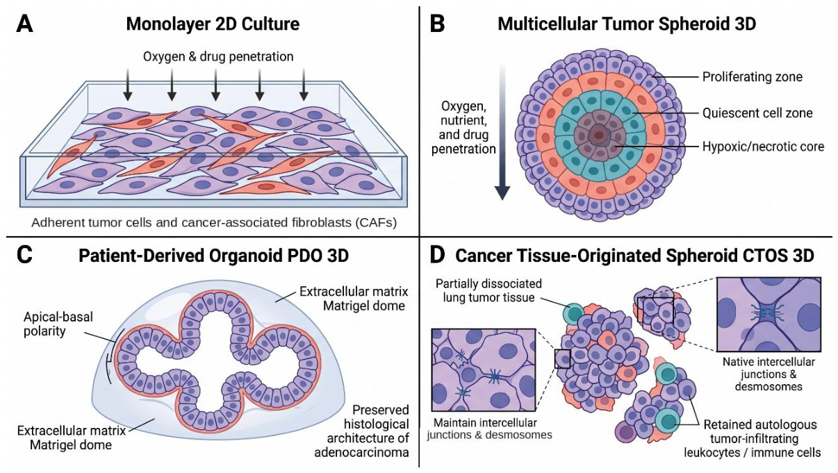
Figure 3.
Schematic representation of representative in vitro platforms derived from patient tissue: (A) Monolayer (2D): Adherent single-cell layer cultured on a rigid, planar plastic substrate, characterized by loss of apical-basal polarity and absence of native tissue architecture, frequently accompanied by rapid overgrowth of cancer-associated fibroblasts (CAFs). (B) Multicellular Spheroid (3D): Compact, non-lumenized spherical aggregate formed by self-aggregation under non-adherent conditions, establishing physiological diffusion gradients with an outer proliferating layer, an intermediate quiescent zone, and a core susceptible to hypoxia and necrosis. (C) Patient-Derived Organoid (PDO, 3D): Self-organizing polarized epithelial structure embedded within an extracellular matrix (ECM/Matrigel/BME dome), recapitulating native histological architecture, glandular lumen formation, and cell-matrix interactions of the parental adenocarcinoma. (D) Cancer Tissue-Originated Spheroid (CTOS, 3D): Multicellular cluster prepared by partial enzymatic dissociation, preserving native intercellular junctions, desmosomes, cell-cell signaling, and retained autologous tumor-infiltrating immune cells without complete single-cell disruption. (Abbreviations: 2D, two-dimensional; 3D, three-dimensional; CAF, cancer-associated fibroblast; CTOS, cancer tissue-originated spheroid; ECM, extracellular matrix; BME, basement membrane extract; PDO, patient-derived organoid). .
5.4. Comparative Analysis: 3D vs. 2D Systems
Direct comparisons between 2D and 3D culture systems indicate that the two approaches provide complementary advantages, extending the conclusion reached at the end of Section 4. Although 2D cultures offer technical simplicity, low cost, reproducibility and compatibility with high-throughput screening, their flat architecture does not adequately reproduce the spatial organization and cell-cell and cell-extracellular matrix (ECM) interactions characteristic of solid tumors. In contrast, 3D models more closely reproduce tumor architecture and provide spatially heterogeneous access to oxygen, nutrients and signaling molecules, which influence cellular morphology, signaling and drug sensitivity [69,71].
These differences are especially relevant to drug response. Three-dimensional cultures develop oxygen and nutrient gradients and heterogeneous cellular compartments, which tend to reduce drug penetration and sensitivity compared with conventional monolayers. Patient-derived 3D models additionally preserve aspects of tumor heterogeneity and patient-specific biology, making them useful for pharmacological studies and precision medicine. Differences in culture geometry also affect gene expression and cellular phenotypes, as demonstrated by comparative analyses of patient-derived spheroids and conventional cultures [62].
However, the greater biological complexity of 3D systems is accompanied by practical limitations. They are generally more technically demanding and time-consuming, often require specialized matrices or scaffolds, and can show greater variability in size, architecture and cellular composition. In lung cancer organoid models, differences in culture media and growth-factor formulations further contribute to this variability and may promote overgrowth of non-malignant cells, echoing the challenge with CAFs discussed in Section 4.3 [69,71].
6. Characterization Methods for Primary Lung Cancer Cells
Regardless of the culture format used – whether 2D or 3D, including CTOSs – validating the resulting cultures against the parental tumor is a necessary next step before they can be used in downstream applications. Detailed characterization of primary lung cancer cultures is necessary for confirming their fidelity to the original tumor and ensuring suitability for such applications. Because primary cultures may contain heterogeneous populations of malignant, stromal, and immune cells, complementary morphological, molecular, genetic, and functional approaches are required to assess culture quality and tumor representation [47,62]. Comparison with the parental tumor is especially important because cellular composition and phenotype may change during tissue processing, culture establishment, and subsequent expansion [72,73].
6.1. Morphological and Histological Assessment
Morphological evaluation is the first line of characterization for primary cultures. Light microscopy allows assessment of cell morphology, growth patterns, attachment, confluence, and potential contamination by stromal cells. In primary lung cancer cultures, morphological assessment also helps distinguish successfully established cultures from those showing progressive cell detachment or fibroblast overgrowth [72]. Kahraman et al. combined microscopic assessment with immunohistochemical analysis to evaluate malignant cell content and cancer-associated fibroblasts in primary NSCLC cultures [47].
Histological assessment provides additional information, notably for 3D cultures. Hematoxylin and eosin (H&E) staining of spheroids or organoid sections is used to evaluate cellular organization and morphology and to compare cultured models with the original tumor [62,73]. Such comparisons help determine whether relevant structural and differentiation features are retained after culture establishment.
6.2. Molecular Characterization
Immunohistochemistry and immunofluorescence provide essential molecular characterization of primary cultures and are used to confirm epithelial and tumor-cell identity. Cytokeratins such as cytokeratin 7 (CK7) and cytokeratin 19 (CK19) have been used to confirm epithelial characteristics in primary lung cancer cultures [50], while immunohistochemical markers such as thyroid transcription factor 1 (TTF-1) and Napsin A for adenocarcinoma and p40 and cytokeratin 5/6 (CK5/6) for squamous cell carcinoma support histological subtype classification [74]. In primary NSCLC cultures, Napsin A and p63 have also been used to characterize malignant epithelial populations and distinguish them from fibroblasts [47]. Molecular characterization is especially important when cultures contain multiple cell populations. Flow cytometry and immunostaining are used to determine the proportions of epithelial cells, fibroblasts, and immune cells within patient-derived spheroids [62].
Additional markers are selected according to the biological question and can include proteins associated with proliferation, epithelial-mesenchymal transition, cancer stemness, or immune-cell phenotypes [62,66]. For models designed to retain components of the tumor microenvironment, characterization of immune populations is notably important. Liu et al. demonstrated that patient-derived lung cancer organoids retained heterogeneous immune-cell populations and patient-specific features of the tumor immune microenvironment, which were subsequently investigated using single-cell approaches [73].
6.3. Genetic and Genomic Profiling
Genetic characterization is important for determining whether primary cultures retain the mutational characteristics of the original tumor. Next-generation sequencing is used to assess clinically relevant driver alterations, while karyotyping and copy-number analysis provide complementary information on genomic abnormalities [41,53]. Zheng et al., for example, identified EGFR kinase-domain mutations in primary NSCLC cell lines and additionally characterized their karyotypes and growth properties [53].
Molecular profiling is also used to compare cultured cells with the parental tumor and determine whether relevant genomic and transcriptomic characteristics are preserved. Roscilli et al. genotyped primary lung adenocarcinoma cultures derived from malignant pleural effusions by targeted next-generation sequencing and found highly heterogeneous, patient-specific mutation profiles [41]. The cultures were not compared with the parental tumor, and in xenografts derived from them the mutation pattern changed substantially in 7 of 10 cases, which illustrates that a characterized culture is not necessarily a stable one [41]. Broader approaches such as whole-exome sequencing and RNA sequencing provide additional information on mutations, copy-number alterations, and gene-expression profiles.
Single-cell RNA sequencing adds a further level of resolution, allowing heterogeneous populations within a culture to be characterized individually; its application to patient-derived lung cancer organoids is discussed in Section 8.1 [73].
6.4. Functional Validation Assays
Functional assays provide important validation of tumor-like behavior in primary cultures by determining whether the cells retain biologically relevant properties rather than merely expressing tumor-associated markers. Common approaches include proliferation and viability assays, apoptosis measurements, migration and invasion assays, and, where appropriate, assessment of self-renewal and tumorigenicity [62,66].
Several of these assays have already been applied to primary lung cancer models. Tumorsphere assays probe CSC-associated properties such as self-renewal and chemotherapy resistance (Section 7.2) [66]; metabolic drug-sensitivity assays have shown dose-dependent responses to cisplatin-containing combinations in patient-derived spheroids [62]; and tumorigenicity has been confirmed by xenograft formation in nude mice, both for cultures from resected tissue [50] and for those derived from malignant pleural effusions [41]. Xenograft assays provide functional evidence of malignant potential but should be interpreted alongside molecular and phenotypic characterization rather than used as standalone validation criteria.
For 3D cultures containing immune cells, functional validation also extends to immunotherapy responses, as in the anti-PD-1 experiments performed in patient-derived lung cancer organoids alongside single-cell analysis [73]. The feasibility of this characterization is itself constrained by specimen size. Immunohistochemistry on paraffin-embedded organoid pellets and targeted panel or Sanger sequencing have both been performed on material taken at the first or second passage, so a minimal quality-control set consisting of histology, lineage markers and confirmation of a known driver alteration is achievable even for cultures derived from small biopsies [30,55]. Whole-exome and RNA sequencing, flow cytometric quantification of cell populations and xenograft formation require substantially more material and are better regarded as optional extensions than as a validation standard for minimally invasive sources.
7. Clinical Applications and Translational Impact
Once validated by the methods described in Section 6, primary lung cancer cultures can be applied across a range of translational settings. Their ability to retain patient-specific molecular and cellular characteristics makes them particularly valuable for investigating therapeutic responses and mechanisms of treatment resistance in a way that conventional preclinical models often cannot [66,69].
7.1. Personalized Chemosensitivity Testing
Patient-derived cultures allow functional testing of therapeutic responses in a personalized manner. The MPE-derived cultures established by Roscilli et al. remain among the clearest examples: in vitro drug-response profiles correlated with clinical treatment response in a subset of patients, although the limited number of correlated cases warrants cautious interpretation [41].
Organoid-based drug screening platforms have further expanded this approach by allowing therapeutic responses to be evaluated in patient-specific 3D models. Patient-derived organoids have been investigated for testing conventional chemotherapy and targeted therapies and for assessing whether treatment responses observed ex vivo reflect patient-specific tumor characteristics [75,76]. Kim et al. established 83 tumor organoids from advanced lung adenocarcinoma at a success rate of 83% (83/100), 77 of them from malignant effusions, together with one normal-like organoid, and demonstrated that the organoids largely retained somatic alterations present in the corresponding tumors. Importantly, responses to targeted therapies in the organoids correlated with clinical outcomes in patients receiving TKIs, predicting progression-free survival correctly in 6 of 8 evaluable patients (75.0%), supporting their potential use for treatment-response prediction [77].
Building on the rapid-timeline approach discussed in Section 2.5, Jung et al. developed a microfluidic-based lung cancer organoid platform that enabled drug-sensitivity assessment using patient-derived tumor material within a clinically relevant timeframe [78]. Such approaches may allow several therapeutic options to be compared ex vivo, providing functional data that could complement molecular profiling in translational research settings [75,76].
However, the clinical implementation of patient-derived culture-, and organoid-based chemosensitivity testing remains subject to important limitations, including variability in culture establishment, the time required to generate sufficient material for testing, and the need for standardized drug-response assays. Larger prospective studies are therefore required to establish the predictive accuracy of these approaches and their broader relevance for functional precision oncology research [76,77].
7.2. Mechanistic Studies, Immunotherapy and Biomarker Discovery
Primary lung cancer cultures provide patient-specific models for investigating tumor biology and mechanisms of treatment response or resistance, information that is difficult to obtain from established cell lines alone [49,53]. Patient-derived 3D models extend this by preserving tumor organization and cellular interactions: spheroids have been used to compare drug responses between tumor and adjacent normal tissue [62], and cancer tissue-originated spheroids have been established together with tumor-infiltrating leukocytes [55]. Lung cancer 3D models incorporating stromal and immune components have correspondingly been proposed for studying tumor-microenvironment interactions, immune checkpoint inhibition and resistance [79,80], and such models are increasingly incorporated into functional precision oncology [81].
Beyond drug testing, primary cultures also serve as discovery platforms for prognostic markers. Herreros-Pomares et al. used tumorspheres from primary NSCLC cultures to enrich CSC-like populations and identified an associated gene signature including NANOG, NOTCH3, CD44, CDKN1A, SNAI1 and ITGA6. Expression of CDKN1A, SNAI1 and ITGA6 was incorporated into a CSC score associated with shorter overall survival in The Cancer Genome Atlas (TCGA) cohort and validated in an independent lung adenocarcinoma cohort [66].
Mechanistic studies of therapeutic resistance are notably relevant in lung cancer, where resistance can involve alterations in drug transport, DNA damage repair, apoptosis, cellular metabolism, phenotypic state, and interactions with the tumor microenvironment [82]. Patient-derived cultures and 3D models provide complementary experimental systems for investigating these processes in the context of patient-specific tumor characteristics. However, differences in culture conditions and selective expansion of particular cell populations may affect the phenotype observed in vitro and should therefore be considered when interpreting mechanistic and drug-response findings [79,81].
8. Recent Advances and Emerging Technologies (2021-2026)
The period from 2021 to 2026 has seen considerable advances in primary lung cancer culture technologies, particularly through the integration of single-cell and spatial analysis, microfluidic and high-throughput platforms, and the development of patient-derived culture biobanks. These innovations have improved the characterization of tumor heterogeneity and allowed more standardized and scalable experimental workflows [83].
8.1. Single-Cell Analysis
Single-cell RNA sequencing (scRNA-seq) resolves individual cell populations and states, capturing information that is obscured in bulk molecular analyses. In NSCLC, it has revealed extensive heterogeneity within tumor and immune compartments and identified cellular states associated with treatment response [84].
Applied to cultures, the same resolution measures fidelity directly: scRNA-seq confirmed high cancer-cell purity in cryobiopsy-derived lung cancer organoids [29]. Combining it with spatial and multi-omic methods is yielding increasingly detailed insight into cell-state transitions and tumor–microenvironment interactions in lung cancer [83,85].
For minimally invasive material, this analytical resolution is not an optional refinement. Where a culture is derived from a few fragments and cannot be split into parallel validation and experimental arms, single-cell profiling of the same culture provides tumor-purity information that a bulk assay cannot [29].
8.2. Microfluidic and High-Throughput Platforms
Microfluidic and microwell technologies have allowed miniaturization, better control of culture conditions and greater standardization of primary culture workflows. Park et al. screened cryobiopsy-derived lung cancer organoids on a micropillar platform [29], and organoid-on-chip systems provide controlled nutrient and drug delivery while incorporating components of the tumor microenvironment [20].
Microwell arrays have been used to generate hundreds of lung cancer organoids and assess drug responses, with agreement with patient-derived xenografts, tumor genetic alterations and clinical outcomes [21], and comparable microfluidic platforms combine organoid culture with drug-sensitivity testing in a single device [78]. For minimally invasive material, the attraction is practical: these formats require far fewer cells per tested condition than plate-based screening.
8.3. Biobanking
Biobanking provides standardized access to clinically annotated tumor tissue, blood and longitudinally collected specimens. Early lung cancer biobanking initiatives emphasized standardized collection, processing, storage and data management [86], and prospective serial biobanking has shown that cryopreserved tumor biopsies can be collected alongside blood samples during the course of advanced NSCLC, while highlighting the difficulty of repeated sampling and variable tumor cellularity [87].
9. Discussion
9.1. Success Rates and Technical Challenges
The comparisons that follow are organized around the four endpoints introduced in Section 1.4. Conflating these endpoints is a frequent source of apparently contradictory findings in this literature. A given tissue source or protocol may perform well on one endpoint and poorly on another. Such results are not mutually exclusive, as they address distinct experimental questions.
Tissue and sample adequacy.
Before establishment can be attempted, the specimen must yield sufficient viable cellular material. This endpoint is the one most directly shaped by acquisition technique. Cryobiopsy and EBUS-TBNA specimens are inherently much smaller than surgical resections. Nevertheless, several series have shown that such small-volume material can provide adequate cellularity for culture initiation. Acquisition route alone therefore does not predict downstream establishment success [29, 40].
Initial formation and comparative establishment rates.
Section 2 established that culture-establishment success depends on anatomical site and protocol rather than on surgical origin as such. The scale of this variability is made explicit in the cross-study synthesis in Supplementary Table S2 and visualized in Figure 4 and Figure 5.
Sachs et al. reported establishment of organoid lines from 87.5% (14/16) of primary NSCLC resections, compared with 27.8% (5/18) of pure tumoroid lines from needle biopsies of metastatic NSCLC [32]. Because the resection-derived lines were not purified from normal airway organoids, these two figures reflect different endpoints. Hwang et al., who applied a single protocol across specimen sources at one institution, observed a different ranking of sources (Figure 5) [30]. In a separate real-world cohort, Wang et al. established organoids more readily from malignant serous effusions (81.5%) than from solid tumor specimens, which included core needle biopsies and lymph node resections (57.7%) [28].
These figures cannot be compared directly, because each study defined success differently. Figure 4 highlights this by stratifying reported outcomes according to both endpoint stringency and tissue source. The differences also have implications beyond methodology. Minimally invasive sampling can provide material for culture-based research from patients who do not undergo resection. These procedures are not without burden. Bronchoscopic and endosonographic sampling is usually performed under sedation or general anesthesia, and needle biopsy and thoracentesis carry their own procedural risks. However, none of them requires thoracotomy or thoracoscopy, with the associated recovery period.
Figure 5 isolates the cleanest version of this comparison: the single protocol from Hwang et al. applied to four specimen sources from one institution, with establishment highest for primary tumor tissue obtained by bronchoscopy or EBUS-TBNA (33.3%), intermediate for surgical resection (23.1%), and lowest for EBUS-TBNA of metastatic lymph nodes (10.0%), whereas two MPE samples yielded no validated organoid lines; differences between sources were not statistically significant (p = 0.060) [30].
Together, Figure 4 and Figure 5 indicate that reported establishment success cannot be interpreted without reference to specimen characteristics, culture methodology, and the endpoint used to define success, and that acquisition route alone provides an insufficient basis for comparison. For example, Strocchi et al. obtained initial CTOS formation from all 33 processed samples (100%), yet only 7 of the 18 samples cultured in the evaluated organoid media (38.9%) met their stringent quality-control criteria [55]. Similarly, Yokota et al., applying a stricter definition of success, reported a long-term validated tumoroid rate of only 7% [89]. Likewise, in a cohort of 30 resected primary tumors and 35 patient-derived xenografts, Shi et al. obtained organoids from 88% (57/65) of models, yet only 15.4% (10/65) achieved long-term growth [90]. These findings are therefore not contradictory. Rather, they reflect distinct laboratory milestones, consistent with the broader picture presented in Section 2 and in this section.
Stromal overgrowth and normal-cell contamination.
Three technical factors account for much of the remaining variability once endpoint definitions are held constant. The first is stromal overgrowth: cancer-associated fibroblasts represented 80-94% of expanded cells across 46 of 51 primary NSCLC cultures in one series [47]. Consequently, successful culture expansion does not necessarily indicate enrichment of malignant cells and may occur even when tumor cells constitute only a small fraction of the culture. Selective culture conditions, early-passage experiments, and enrichment strategies can mitigate this, though each introduces its own selection pressure on the resulting cell population. A second and distinct form of contamination is overgrowth by non-malignant airway epithelium. Because the media that support the highest establishment rates were originally developed for long-term expansion of normal airway organoids [32], normal basal cells can outcompete tumor cells in the same well, and the resulting cultures are morphologically plausible but not malignant; minimal formulations designed to be less permissive to normal airway cells were introduced specifically to address this [4]. This problem is most pronounced for intrapulmonary lesions, where it has been quantified directly, as discussed below.
Culture medium, passage strategy and long-term stability.
The second factor is medium composition, which does not simply determine whether a culture grows but which cells within it survive. Across paired tumor and normal tissue from 71 resected NSCLC patients, a permissive medium maximized establishment and supported long-term expansion, while a minimal basal medium reduced normal-organoid overgrowth at the cost of expansion capacity – a direct illustration that culture conditions shape the biological identity of a model, not only whether it grows [91]. Strocchi et al. observed the same trade-off at the level of validated tumor identity. With a permissive organoid medium, 10 of 14 cultures grew (71%), but only 2 of these 10 (20%) passed quality control. A more selective formulation supported growth in fewer cases (6 of 11, 55%), but 5 of these 6 (83%) passed [55]. Because several samples were cultured in parallel in more than one medium, these medium-specific denominators overlap and sum to more than the 18 samples reported in Section 3.4 [55]. At the level of individual components, the permissive airway formulations combine Wnt pathway support through R-spondin-1 with Noggin, FGF7 and FGF10, the TGF-β/activin receptor-like kinase 5 (ALK5) inhibitor A83-01, the p38 inhibitor SB202190 and nicotinamide – a combination optimized for self-renewal of normal airway basal cells [32]. Formulations that withdraw or reduce these factors shift the balance toward tumor cells at the cost of overall growth [4], and counter-selection with the MDM2 inhibitor nutlin-3a exploits TP53 mutation to suppress TP53-wild-type normal airway organoids, at the cost of excluding tumors that are TP53 wild-type [55].
The third, related tension runs through CTOSs and other short-term formats: short-term cultures tend to preserve the original tumor more faithfully, while long-term expansion provides the material volume needed for repeated experiments [55,89]. The practical conclusion follows directly from the thesis of Section 2: a higher reported success rate is not, by itself, evidence of a better or more representative model. It has to be read against what counted as success and against how much of what grew was actually tumor.
Molecular and genomic fidelity to the source tumor.
This endpoint, separate from establishment rate altogether, concerns whether the resulting culture or organoid remains genomically and phenotypically representative of the tumor it was derived from. Dijkstra et al. found that organoid cultures were obtained from 24 of 58 NSCLC specimens (41%), but 14 of these proved to be normal airway organoids on copy-number profiling, p63 immunostaining and mutation testing, leaving 10 of 58 (17.2%) verified pure tumor organoids – a validated-fidelity rate less than half the raw formation rate reported for the same specimens [92]. In the same cohort, establishment did not differ significantly between core needle biopsies and surgical resections, and the authors recommended restricting organoid culture to extrapulmonary lesions unless rigorous steps are taken to prevent overgrowth by normal airway organoids, since overgrowth did not affect extrapulmonary metastases, for which they reported a 21% establishment rate from biopsy material [92]. Hwang et al. applied a comparable principle across surgical and non-surgical tissue sources by requiring immunohistochemical and sequencing concordance with the parental tumor before counting an organoid as established, which is why their reported rates are markedly more conservative than formation-only rates reported elsewhere for similar tissue volumes (Figure 5) [30]. Across all 163 specimens, 56 (34.4%) yielded organoids beyond passage 3, but only 25 (15.3%) were confirmed as tumor organoids. An equal number (25) were classified as benign organoids, which the authors attributed largely to overgrowth by normal epithelial cells [30]. For a laboratory deciding whether to invest in minimally invasive material, fidelity to the source tumor – not raw formation rate – is the endpoint that ultimately determines whether the resulting model is usable for downstream molecular or functional work.
9.2. Translational Considerations and Practical Challenges
Establishment rate is a laboratory metric. Clinical relevance, by contrast, depends on whether the resulting model predicts what happens in the patient. Although lung cancer organoids show considerable promise for personalized medicine, standardization of culture protocols and reproducibility remain real barriers to broader translational use [58]. On the predictive question, however, the evidence is encouraging: patient-derived organoids from advanced lung adenocarcinoma retained the major genomic features of the source tumor and matched clinical responses to targeted therapy [77].
Speed matters as much as accuracy for translational use, extending the rapid-timeline considerations already introduced in Section 7.1. Rapid organoid-based drug testing has delivered clinically relevant treatment-response data within roughly a week, depending on the culture system and sample characteristics [21], and organoid-based EGFR-targeted therapy testing has shown similar concordance with clinical response while cutting the time needed for functional assessment [5]. A turnaround measured in days, rather than weeks, is what would let functional testing compete with – rather than simply follow – standard molecular workup.
Realizing this potential at scale depends on standardization the field does not yet have. Culture protocols still differ in tissue source, medium composition, matrix, passage handling, and the criteria used to define successful establishment, and each of these choices contributes to variability between laboratories [58,91]. Quality-control procedures and standardized characterization are therefore not merely a procedural detail but a precondition for treating culture-based assays as reliable tools in functional precision oncology.
9.3. Future Directions
Four priorities follow from the picture assembled across this review. The first is the most direct: under the right conditions, non-surgical sources already match or even exceed surgical material (Section 2). Further optimizing cryobiopsy and related protocols, particularly to enrich for tumor cells without sacrificing the limited tissue these techniques yield, should therefore open primary culture research to more of the patients currently excluded from it [29,91].
The second priority is analytical. Single-cell sequencing, spatial transcriptomics, and multi-omics profiling can resolve what a bulk endpoint like 'establishment rate' cannot – which cells within a culture are tumor-derived, how faithfully they represent the original tumor's heterogeneity, and which features are retained from the tumor versus acquired during ex vivo expansion [73,83,84].
The third is operational: automating microwell and microfluidic workflows [21,29] is a precondition for the standardization that Section 9.2 identifies as currently missing, for multi-institution biobanking, and for scaling functional precision medicine beyond single-center studies.
Table 1. Reported concordance between primary-culture or organoid drug response and clinical treatment outcome across the five studies reviewed in Section 2.5, Section 7.1 and Section 9.3 that compared an ex vivo response against the observed patient outcome [5,28,41,45,77]. Values shown are the exact quantities reported by each source study; no concordance percentage is implied where the original publication did not report one. Entries marked 'Not reported' indicate that the source study did not state a turnaround time, generally because the cultures were analyzed retrospectively or the study focused on biological characterization rather than on point-of-care feasibility. (Abbreviations: 2D, two-dimensional; AUC, area under the curve; CODRP, cancer organoid-based diagnosis reactivity prediction; EGFR, epidermal growth factor receptor; MPE, malignant pleural effusion; MTT, 3-(4,5-dimethylthiazol-2-yl)-2,5-diphenyltetrazolium bromide; NSCLC, non-small cell lung cancer; PFS, progression-free survival; ROC, receiver operating characteristic; TKI, tyrosine kinase inhibitor).
The fourth priority is rigorous prospective validation. As summarized in Table 1, five studies have compared ex vivo drug response with clinical treatment outcome [5,28,41,45,77]. Three reported a formal accuracy estimate. Kim et al. predicted progression-free survival correctly in 6 of 8 evaluable patients (75.0%) [77]. Wang et al. reported concordance for 45 of 54 organoids (83.3%), with 84.0% sensitivity and 82.8% specificity [28]. Lee et al. classified afatinib response correctly in 13 of 14 patients (85.7% sensitivity, 100% specificity), but derived the classification cut-off in the same cohort [5]. The other two studies reported agreement without a formal statistic. Roscilli et al. found agreement in all 6 patients with available clinical data, but counted a culture as concordant when it was sensitive to at least one drug of the regimen [41]. Yang et al. found agreement in all 8 patients treated with an EGFR-TKI, using a qualitative, visually scored viability readout [45]. None of these studies tested whether the assay result changes treatment decisions [5,28,41,45,77]. While retrospective concordance has been documented more than once [5, 28,77], such concordance is fundamentally distinct from a prospective functional assay proven, in advance, to change a treatment decision [41].
Until that step is taken – and until turnaround time, feasibility, cost-effectiveness, and added value over currently available molecular characterization are assessed alongside predictive accuracy [58] – primary lung cancer cultures will remain a research tool whose clinical utility is plausible rather than demonstrated.
9.4. Points for Laboratory Practice and Future Research
Points for laboratory practice:
- Establishment rate alone should not determine the choice of tissue source: minimally invasive material (cryobiopsy, EBUS-TBNA, MPE) can match or exceed surgically resected tissue once the endpoint definition is held constant.
- The term "established" should therefore be qualified by the endpoint actually met, whether tissue adequacy, initial formation, long-term validated establishment, or confirmed molecular fidelity. These endpoints are frequently conflated in the literature, yet they are not interchangeable.
- Stromal overgrowth, normal-epithelial overgrowth, and medium composition are major technical determinants of whether an established culture remains tumor-predominant. Their relative contribution may vary with tissue volume, specimen cellularity, anatomical site, and acquisition technique.
- Confirmation of molecular or genomic concordance with the source tumor should be treated as a standard validation step for any primary culture or organoid intended for downstream functional or molecular work, rather than an optional characterization add-on.
Questions for future research
- Which combinations of dissociation method, medium formulation and passage strategy best preserve tumor-cell dominance across different minimally invasive tissue sources?
- What minimum tissue quantity or cellularity from cryobiopsy or EBUS-TBNA specimens reliably supports organoid establishment, and how does this threshold vary by anatomical sampling site?
- Can standardized, multi-endpoint reporting – tissue adequacy, initial formation, long-term validated establishment and molecular fidelity – be adopted across laboratories to allow direct, quantitative comparison of establishment protocols?
- Beyond retrospective concordance with treatment outcome, which quality-control metrics best predict whether a primary culture or organoid will remain a faithful model of the source tumor through extended passage?
10. Conclusions
This review examined how far primary lung cancer cultures remain tied to surgically resected tissue. Current evidence supports the feasibility of establishing organoid and spheroid models from cryobiopsy, EBUS-TBNA, conventional bronchoscopy, CT-guided transthoracic needle biopsy, and MPE, with reported establishment rates that in some within-study comparisons match or exceed those from resected tissue. These findings should be interpreted in the context of specimen characteristics, anatomical site, culture methodology, and endpoint definition. Minimally invasive approaches can also extend access to primary culture-based models to patients who would otherwise be underrepresented in studies relying on surgical specimens.
Cryobiopsy alone reaches roughly 40% organoid establishment from specimens substantially smaller than a surgical resection [29]. Three-dimensional formats – organoids, spheroids, and cancer tissue-originated spheroids – can preserve important aspects of tumor architecture and drug-response behavior, although much of this evidence still derives from surgically resected tissue [55,62]. Advanced characterization, including single-cell RNA sequencing, can further assess the extent to which these models retain features of the source tumor [73,84].
Beyond establishment feasibility, a smaller body of evidence links ex vivo drug responses from primary cultures or organoids to clinical treatment outcomes [5,28,41,45,77]. These findings suggest that some patient-derived models retain functionally relevant biology, but they do not establish prospective clinical utility. From a laboratory standpoint, the more immediate question is whether models meet the standardized, fidelity-confirmed criteria set out in Section 9.1, because clinical utility cannot be meaningfully assessed when establishment endpoints and tumor fidelity are not characterized and reported consistently.
Taken together, advances in single-cell and spatial omics, microfluidic and bioengineered platforms, high-throughput screening, and computational approaches are expanding the applications of primary culture systems [83]. For minimally invasive models, however, tissue accessibility and laboratory standardization remain complementary challenges. Consistent, multi-endpoint reporting and confirmation of molecular fidelity will be important for determining when models derived from minimally invasive material are directly comparable with those derived from surgical resection and for defining the contexts in which they may be used as alternatives.
Supplementary Materials
The following supporting information is available online: Table S1: Culture formats, matrices, and media used across primary lung cancer culture protocols; Table S2: Cross-study synthesis of reported primary lung cancer culture establishment outcomes.
Author Contributions
N.M.: Conceptualization; Investigation (literature search); Data Curation (selection of full-text records); Writing - Original Draft; Writing - Review & Editing. M.Z.: Conceptualization; Investigation (literature search); Data Curation (selection of full-text records); Writing - Original Draft; Writing - Review & Editing. L.M.: Writing - Review & Editing; Supervision. W.Z.: Data Curation (reference verification). R.M.M.: Conceptualization; Methodology; Investigation; Data Curation; Writing - Review & Editing; Supervision. All authors have read and agreed to the published version of the manuscript.
Funding
This research received no external funding. .
Institutional Review Board Statement
Not applicable.
Informed Consent Statement
Not applicable. .
Data Availability Statement
No new data were created or analyzed in this study. Data sharing is not applicable to this article.
Acknowledgments
During the preparation of this manuscript, the authors used GPT-5.5 (OpenAI) to organize subheadings and improve English grammar, Claude Sonnet 5 (Anthropic) and Canva to assist in preparing the schematic figures, and Claude Opus 5.5 (Anthropic) for language editing of selected passages. The authors reviewed and edited all output and take full responsibility for the content of this publication. .
Conflicts of Interest
The authors declare no conflicts of interest. .
Abbreviations
| 2D | two-dimensional (culture system) |
| 3D | three-dimensional (culture system) |
| ACL4 | chemically defined culture medium formulation used for non-small cell lung cancer cell cultures |
| ALK | anaplastic lymphoma kinase |
| BET (inhibitor) | bromodomain and extra-terminal domain (inhibitor) |
| bFGF | basic fibroblast growth factor |
| BME | basement membrane extract |
| CAF(s) | cancer-associated fibroblast(s) |
| CD44 | cluster of differentiation 44 (cell-surface glycoprotein, cancer stem cell marker) |
| CDKN1A | cyclin-dependent kinase inhibitor 1A (gene cancer stem cell-associated marker) |
| CK5/6, CK7, CK19 | cytokeratin 5/6, cytokeratin 7, cytokeratin 19 (epithelial markers) |
| CODRP | cancer organoid-based diagnosis reactivity prediction (platform) |
| CSC | cancer stem cell |
| CT | computed tomography |
| CTOS(s) | cancer tissue-originated spheroid(s) |
| EBUS | endobronchial ultrasound |
| EBUS-TBNA | endobronchial ultrasound-guided transbronchial needle aspiration |
| ECM | extracellular matrix |
| EGF | epidermal growth factor |
| EGFR | epidermal growth factor receptor |
| FBS | fetal bovine serum |
| FGF7, FGF10 | fibroblast growth factor 7, fibroblast growth factor 10 |
| H&E | hematoxylin and eosin (staining) |
| HBSS | Hanks' balanced salt solution |
| HER3 | human epidermal growth factor receptor 3 |
| HITES | serum-free medium formulation (hydrocortisone, insulin, transferrin, estradiol, selenium) used primarily for small cell lung cancer cultures |
| IGF | insulin-like growth factor |
| ITGA6 | integrin subunit alpha 6 (gene cancer stem cell-associated marker) |
| LCO | lung cancer organoid |
| MDM2 | mouse double minute 2 homolog |
| MPE | malignant pleural effusion |
| NANOG | Nanog homeobox (gene cancer stem cell-associated marker) |
| NOTCH3 | notch receptor 3 (gene cancer stem cell-associated marker) |
| NK cells | natural killer cells |
| NRG1 | neuregulin 1 (ligand of HER3) |
| NSCLC | non-small cell lung cancer |
| PD-1 | programmed cell death protein 1 |
| PDO(s) | patient-derived organoid(s) |
| PDOTS | patient-derived organotypic tumor spheroids |
| PDS(s) | patient-derived spheroid(s) |
| PDTO | patient-derived tumor organoid |
| RPMI-1640 | Roswell Park Memorial Institute 1640 (cell culture medium) |
| SCLC | small cell lung cancer |
| scRNA-seq | single-cell RNA sequencing |
| SNAI1 | snail family transcriptional repressor 1 (gene cancer stem cell-associated marker) |
| TCGA | The Cancer Genome Atlas |
| TGF-β | transforming growth factor beta |
| TKI | tyrosine kinase inhibitor |
| TP53 | tumor protein p53 (gene) |
| TTF-1 | thyroid transcription factor 1 |
References
- Herreño, A.M.; Fernández, M.J.; Rey, L.; Mejía, J.A.; Cañas, A.; Moreno, O.M.; Rojas, A.P. Primary lung cancer cell culture from transthoracic needle biopsy samples. Cogent Med. 2018, 5, 1503071. [Google Scholar] [CrossRef]
- Huo, K.G.; D’Arcangelo, E.; Tsao, M.S. Patient-derived cell line, xenograft and organoid models in lung cancer therapy. Transl. Lung Cancer Res. 2020, 9, 2214–2232. [Google Scholar] [CrossRef] [PubMed]
- Delom, F.; Begiristain, I.; Grenier, T.; Begueret, H.; Soulet, F.; Siegfried, G.; Khatib, A.M.; Robert, J.; Fessart, D. Patients Lung Derived Tumoroids (PLDTs) to model therapeutic response. Biochim. Biophys. Acta Mol. Cell Res. 2020, 1867, 118808. [Google Scholar] [CrossRef] [PubMed]
- Kim, M.; Mun, H.; Sung, C.O.; Cho, E.J.; Jeon, H.J.; Chun, S.M.; Jung, D.J.; Shin, T.H.; Jeong, G.S.; Kim, D.K.; et al. Patient-derived lung cancer organoids as in vitro cancer models for therapeutic screening. Nat. Commun. 2019, 10, 3991. [Google Scholar] [CrossRef] [PubMed]
- Lee, S.H.; Kim, K.; Lee, E.; Lee, K.; Ahn, K.H.; Park, H.; Kim, Y.; Shin, S.; Jeon, S.Y.; Hwang, Y.; et al. Prediction of TKI response in EGFR-mutant lung cancer patients-derived organoids using malignant pleural effusion. npj Precis. Oncol. 2024, 8, 111. [Google Scholar] [CrossRef] [PubMed]
- Bray, F.; Laversanne, M.; Sung, H.; Ferlay, J.; Siegel, R.L.; Soerjomataram, I.; Jemal, A. Global cancer statistics 2022: GLOBOCAN estimates of incidence and mortality worldwide for 36 cancers in 185 countries. CA Cancer J. Clin. 2024, 74, 229–263. [Google Scholar] [CrossRef] [PubMed]
- Allemani, C.; Matsuda, T.; Di Carlo, V.; Harewood, R.; Matz, M.; Nikšić, M.; et al. Global surveillance of trends in cancer survival 2000–14 (CONCORD-3): analysis of individual records for 37 513 025 patients diagnosed with one of 18 cancers from 322 population-based registries in 71 countries. Lancet 2018, 391, 1023–1075. [Google Scholar] [CrossRef] [PubMed]
- Liu, Y.; Zhou, Y.; Chen, P. Lung cancer organoids: Models for preclinical research and precision medicine. Front. Oncol. 2023, 13, 1293441. [Google Scholar] [CrossRef] [PubMed]
- Terai, H.; Yasuda, H.; Sato, T. Organoid-based precision cancer modeling: New frontier in lung cancer research. Cell Rep. 2025, 44, 116595. [Google Scholar] [CrossRef] [PubMed]
- Lim, Z.F.; Ma, P.C. Emerging insights of tumor heterogeneity and drug resistance mechanisms in lung cancer targeted therapy. J. Hematol. Oncol. 2019, 12, 134. [Google Scholar] [CrossRef] [PubMed]
- Tulpule, A.; Bivona, T.G. Acquired resistance in lung cancer. Annu. Rev. Cancer Biol. 2020, 4, 279–297. [Google Scholar] [CrossRef]
- Hynds, R.E.; Frese, K.K.; Pearce, D.R.; Grönroos, E.; Dive, C.; Swanton, C. Progress towards non-small-cell lung cancer models that represent clinical evolutionary trajectories. Open Biol. 2021, 11, 200247. [Google Scholar] [CrossRef] [PubMed]
- Kim, J.; Minna, J.D. Moving toward precision medicine with lung cancer organoids. Cell Rep. Med. 2023, 4, 100952. [Google Scholar] [CrossRef] [PubMed]
- Furuse, H.; Matsumoto, Y. Advances in lung cancer biopsy techniques and specimen collection methods. Haigan 2021, 61, 924–931. [Google Scholar] [CrossRef]
- Wang, L.; Miao, Y.; Kaneda, H.; Tani, Y.; Nakai, T. Application and advancement of bronchoscopic biopsy techniques for the diagnosis of pulmonary nodules. Respir. Endosc. 2024, 2, 122–127. [Google Scholar] [CrossRef]
- Park, J. Advanced bronchoscopic diagnostic techniques in lung cancer. Tuberc. Respir. Dis. 2024, 87, 145–158. [Google Scholar] [CrossRef] [PubMed]
- Song, Y.; Wang, Y.; Guan, A.; Xue, J.; Li, B.; Huang, Z.; Zheng, Z.; Liang, N.; Yang, Y.; Li, S. Footprints: Stamping hallmarks of lung cancer with patient-derived models, from molecular mechanisms to clinical translation. Front. Bioeng. Biotechnol. 2023, 11, 1132940. [Google Scholar] [CrossRef] [PubMed]
- Honkala, A.; Malhotra, S.V.; Kummar, S.; Junttila, M.R. Harnessing the predictive power of preclinical models for oncology drug development. Nat. Rev. Drug Discov. 2022, 21, 99–114. [Google Scholar] [CrossRef] [PubMed]
- Li, Y.; Gao, X.; Ni, C.; Zhao, B.; Cheng, X. The application of patient-derived organoid in the research of lung cancer. Cell Oncol. 2023, 46, 503–519. [Google Scholar] [CrossRef] [PubMed]
- Zeng, X.; Ma, Q.; Li, X.K.; You, L.T.; Li, J.; Fu, X.; You, F.M.; Ren, Y.F. Patient-derived organoids of lung cancer based on organoids-on-a-chip: Enhancing clinical and translational applications. Front. Bioeng. Biotechnol. 2023, 11, 1205157. [Google Scholar] [CrossRef] [PubMed]
- Hu, Y.; Sui, X.; Song, F.; Li, Y.; Li, K.; Chen, Z.; Yang, F.; Chen, X.; Zhang, Y.; Wang, X.; et al. Lung cancer organoids analyzed on microwell arrays predict drug responses of patients within a week. Nat. Commun. 2021, 12, 2581. [Google Scholar] [CrossRef] [PubMed]
- Yu, N.; Hwang, M.; Lee, Y.; Song, B.R.; Kang, E.H.; Sim, H.; Ahn, B.C.; Hwang, K.H.; Kim, J.; Hong, S.; et al. Patient-derived cell-based pharmacogenomic assessment to unveil underlying resistance mechanisms and novel therapeutics for advanced lung cancer. J. Exp. Clin. Cancer Res. 2023, 42, 37. [Google Scholar] [CrossRef] [PubMed]
- Kodack, D.P.; Farago, A.F.; Dastur, A.; Held, M.A.; Dardaei, L.; Friboulet, L.; Flotow, F.; Damon, L.J.; Lee, D.; Parks, M.; et al. Primary patient-derived cancer cells and their potential for personalized cancer patient care. Cell Rep. 2017, 21, 3298–3309. [Google Scholar] [CrossRef] [PubMed]
- Koga, T.; Ogawa, H.; Radulovich, N.; Pham, N.A.; Li, Q.; Nakajima, T.; Yasufuku, K.; Tsao, M.S. Endobronchial ultrasound-guided biopsy-derived lung cancer models: A platform for precision therapy. J. Thorac. Oncol. 2026, 21, 103893. [Google Scholar] [CrossRef] [PubMed]
- Dinić, J.; Jovanović Stojanov, S.; Dragoj, M.; Grozdanić, M.; Podolski-Renić, A.; Pešić, M. Cancer patient-derived cell-based models: Applications and challenges in functional precision medicine. Life 2024, 14, 1142. [Google Scholar] [CrossRef] [PubMed]
- Spiró, Z.; Amin, A.; Mair, M.J.; Pieber, T.R.; Prietl, B.; Spiegl-Kreinecker, S.; Stanzer, S.; Wöhrer, A.; Preusser, M.; Berghoff, A.S. Ex vivo drug screening on patient-derived tumor material to advance functional precision in oncology: An overview on current approaches and unresolved challenges. Cancer Treat. Rev. 2026, 143, 103072. [Google Scholar] [CrossRef] [PubMed]
- Fu, Y.; Khoo, B.L.; Lim, C.T. Advancements and challenges in culturing patient-derived cancer cells for personalized therapeutics. Front. Lab Chip Technol. 2025, 4, 1663420. [Google Scholar] [CrossRef]
- Wang, H.M.; Zhang, C.Y.; Peng, K.C.; Chen, Z.X.; Su, J.W.; Li, Y.F.; Li, W.F.; Gao, Q.Y.; Zhang, S.L.; Chen, Y.Q.; et al. Using patient-derived organoids to predict locally advanced or metastatic lung cancer tumor response: A real-world study. Cell Rep. Med. 2023, 4, 100911. [Google Scholar] [CrossRef] [PubMed]
- Park, D.; Lee, D.; Kim, Y.; Park, Y.; Lee, Y.J.; Lee, J.E.; Yeo, M.K.; Kang, M.W.; Chong, Y.; Han, S.J.; et al. Cryobiopsy: A breakthrough strategy for clinical utilization of lung cancer organoids. Cells 2023, 12, 1854. [Google Scholar] [CrossRef] [PubMed]
- Hwang, M.; Choe, J.; Shin, Y.J.; Seo, B.G.; Park, K.M.; Shin, S.H.; Jhun, B.W.; Yoo, H.; Jeong, B.H.; Jeon, K.; et al. Establishment of human lung cancer organoids using small biopsy and surgical tissues. Cancers 2025, 17, 2291. [Google Scholar] [CrossRef] [PubMed]
- Giri, M.; Huang, G.; Puri, A.; Zhuang, R.; Li, Y.; Guo, S. Efficacy and safety of cryobiopsy vs. forceps biopsy for interstitial lung diseases, lung tumors, and peripheral pulmonary lesions: An updated systematic review and meta-analysis. Front. Med. 2022, 9, 840702. [Google Scholar] [CrossRef] [PubMed]
- Sachs, N.; Papaspyropoulos, A.; Zomer-van Ommen, D.D.; Heo, I.; Böttinger, L.; Klay, D.; Weeber, F.; Huelsz-Prince, G.; Iakobachvili, N.; Amatngalim, G.D.; et al. Long-term expanding human airway organoids for disease modeling. EMBO J. 2019, 38, e100300. [Google Scholar] [CrossRef] [PubMed]
- Tang, Y.; Tian, S.; Chen, H.; Li, X.; Pu, X.; Zhang, X.; Zheng, Y.; Li, Y.; Huang, H.; Bai, C. Transbronchial lung cryobiopsy for peripheral pulmonary lesions. A narrative review. Pulmonology 2024, 30, 475–484. [Google Scholar] [CrossRef] [PubMed]
- Nishimatsu, K.; Matsumoto, Y.; Kashima, J.; Imabayashi, T.; Uchimura, K.; Furuse, H.; Masuda, K.; Shinno, Y.; Okuma, Y.; Yoshida, T.; et al. Concordance between cryobiopsy and forceps biopsy specimens in assessment of immunohistochemistry staining for non-small cell lung carcinoma. Transl. Lung Cancer Res. 2023, 12, 1245–1255. [Google Scholar] [CrossRef] [PubMed]
- Liam, C.K.; Lee, P.; Yu, C.J.; Bai, C.; Yasufuku, K. The diagnosis of lung cancer in the era of interventional pulmonology. Int. J. Tuberc. Lung Dis. 2021, 25, 6–15. [Google Scholar] [CrossRef] [PubMed]
- Guarize, J.; Bianchi, F.; Marino, E.; Belloni, E.; Vecchi, M.; Donghi, S.; Lo Iacono, G.; Casadio, C.; Cuttano, R.; Barberis, M.; et al. MicroRNA expression profile in primary lung cancer cells lines obtained by endobronchial ultrasound transbronchial needle aspiration. J. Thorac. Dis. 2018, 10, 408–415. [Google Scholar] [CrossRef] [PubMed]
- Jiang, L.; Xu, J.; Liu, C.; Gao, N.; Zhao, J.; Han, X.; Zhang, X.; Zhao, X.; Liu, Y.; Wang, W.; et al. Diagnosis of peripheral pulmonary lesions with transbronchial lung cryobiopsy by guide sheath and radial endobronchial ultrasonography: A prospective control study. Can. Respir. J. 2021, 2021, 6947037. [Google Scholar] [CrossRef] [PubMed]
- Brown, M.; Nguyen, P.; Jersmann, H.; Holmes, M.; Wong, M. Radial endobronchial ultrasound-guided transbronchial cryobiopsy versus forceps biopsy for the diagnosis of solitary pulmonary nodules: A prospective randomised trial. Open Respir. Med. J. 2023, 17, e18743064262132. [Google Scholar] [CrossRef] [PubMed]
- Barbet, K.; Schmitz, M.S.; Westhölter, D.; Kamler, M.; Rütten, S.; Thiebes, A.L.; Sitek, B.; Bayer, M.; Schedel, M.; Reuter, S.; et al. Bronchoscopic biopsies–A novel source for primary airway epithelial cells in respiratory research. Respir. Res. 2024, 25, 439. [Google Scholar] [CrossRef] [PubMed]
- Ko, Y.R.; Kim, J.M.; Kang, Y.; Kim, M.; Jang, S.J. A method for culturing patient-derived lung cancer organoids from surgically resected tissues and biopsy samples. Organoid 2022, 2, e19. [Google Scholar] [CrossRef]
- Roscilli, G.; De Vitis, C.; Ferrara, F.F.; Noto, A.; Cherubini, E.; Ricci, A.; Mariotta, S.; Giarnieri, E.; Giovagnoli, M.R.; Torrisi, M.R.; et al. Human lung adenocarcinoma cell cultures derived from malignant pleural effusions as model system to predict patients chemosensitivity. J. Transl. Med. 2016, 14, 61. [Google Scholar] [CrossRef] [PubMed]
- Seo, H.Y.; Kim, S.C.; Roh, W.; Shin, Y.K.; Kim, S.; Kim, D.W.; Kim, T.M.; Ku, J.L. Culture and multiomic analysis of lung cancer patient-derived pleural effusions revealed distinct druggable molecular types. Sci. Rep. 2022, 12, 6345. [Google Scholar] [CrossRef] [PubMed]
- Surina; Tanggis; Suzuki, T.; Hisata, S.; Fujita, K.; Fujiwara, S.; Liu, F.; Fukushima, N.; Suzuki, T.; Mato, N.; et al. Patient-derived spheroids and patient-derived organoids simulate evolutions of lung cancer. Heliyon 2023, 9, e13829. [Google Scholar] [CrossRef] [PubMed]
- Wang, L.; Yu, Y.; Fang, Y.; Li, Y.; Yu, W.; Wang, Z.; Lv, J.; Wang, R.; Liang, S. Malignant pleural effusion supports the establishment and maintenance of tumor organoid biobank with multiple patient-derived lung tumor cell sources. Exp. Hematol. Oncol. 2024, 13, 115. [Google Scholar] [CrossRef] [PubMed]
- Yang, T.M.; Fang, Y.H.; Lin, C.M.; Chen, M.F.; Lin, C.L. Spheroids generated from malignant pleural effusion as a tool to predict the response of non-small cell lung cancer to treatment. Diagnostics 2024, 14, 998. [Google Scholar] [CrossRef] [PubMed]
- Richter, M.; Piwocka, O.; Musielak, M.; Piotrowski, I.; Suchorska, W.M.; Trzeciak, T. From donor to the lab: A fascinating journey of primary cell lines. Front. Cell Dev. Biol. 2021, 9, 711381. [Google Scholar] [CrossRef] [PubMed]
- Kahraman, D.; İlhan, S.; Cangi, S.; Işık, A.F.; Bağcı, C.; Sağlam, E. Comparative assessment of primary cancer cell culture techniques and cellular composition analysis in non-small cell lung cancer. Pathol. Res. Pract. 2023, 248, 154580. [Google Scholar] [CrossRef] [PubMed]
- Dai, Y.; Guo, S.; Huang, S.; Zhang, J.; Zhang, Z. Summary of primary culture of human lung cancer cells. Austin J. Cancer Clin. Res. 2018, 5, 1083. [Google Scholar] [CrossRef]
- Seo, J.; Park, S.J.; Kim, J.; Choi, S.J.; Moon, S.H.; Chung, H.M. Effective method for the isolation and proliferation of primary lung cancer cells from patient lung tissues. Biotechnol. Lett. 2013, 35, 1165–1174. [Google Scholar] [CrossRef] [PubMed]
- Si, L.L.; Lv, L.; Zhou, W.H.; Hu, W.D. Establishment and identification of human primary lung cancer cell culture in vitro. Int. J. Clin. Exp. Pathol. 2015, 8, 6540–6546. [Google Scholar] [PubMed]
- Lam, D.C.; Girard, L.; Suen, W.S.; Chung, L.P.; Tin, V.P.; Lam, W.K.; Minna, J.D.; Wong, M.P. Establishment and expression profiling of new lung cancer cell lines from Chinese smokers and lifetime never-smokers. J. Thorac. Oncol. 2006, 1, 932–942. [Google Scholar] [CrossRef]
- Sharifzadeh, S.; Owji, S.M.; Pezeshki, A.M.; Malek-Hoseini, Z.; Kumar, P.V.; Ghayumi, S.M.; Ghaderi, A. Establishment and characterization of a human large cell lung cancer cell line with neuroendocrine differentiation. Pathol. Oncol. Res. 2004, 10, 225–230. [Google Scholar] [CrossRef] [PubMed]
- Zheng, C.; Sun, Y.H.; Ye, X.L.; Chen, H.Q.; Ji, H.B. Establishment and characterization of primary lung cancer cell lines from Chinese population. Acta Pharmacol. Sin. 2011, 32, 385–392. [Google Scholar] [CrossRef] [PubMed]
- Yılmaz, S.; Doğan Sarıkaya, M.; Atar, E.; Şen Bağcı, B.; Kenanoğlu, S.; Mutlu, N.; Yaşar, E.; Önal, Ö.; Canöz, Ö.; Özkul, Y. Primary culture of cancer cells derived from human lung tumors. Erciyes Univ. Fen. Bilim. Enst. Fen. Bilim. Derg. 2023, 39, 79–86. [Google Scholar]
- Strocchi, S.; Santandrea, G.; Zanetti, E.; Verna, G.; Cusenza, V.Y.; Nicoli, D.; Fantini, V.; Grieco, A.; Paci, M.; Ciarrocchi, A.; et al. A two-step protocol for isolation and maintenance of lung cancer primary 3D cultures. Cancers 2025, 17, 27. [Google Scholar] [CrossRef] [PubMed]
- Gazdar, A.F.; Gao, B.; Minna, J.D. Lung cancer cell lines: Useless artifacts or invaluable tools for medical science? Lung Cancer 2010, 68, 309–318. [Google Scholar] [CrossRef] [PubMed]
- Zhu, L.; Zhang, J.; Guo, Q.; Kuang, J.; Li, D.; Wu, M.; Mo, Y.; Zhang, T.; Gao, X.; Tan, J. Advanced lung organoids and lung-on-a-chip for cancer research and drug evaluation: A review. Front. Bioeng. Biotechnol. 2023, 11, 1299033. [Google Scholar] [CrossRef] [PubMed]
- Werner, J.M. Primary lung cancer organoids for personalized medicine–Are they ready for clinical use? Cancers 2021, 13, 4832. [Google Scholar] [CrossRef] [PubMed]
- Mansor, A.F.M.; Sedek, S.; Nordin, A.N.; Mohammad, M.; How, S.H. Application of real-time cell analysis biosensor technology for drug cytotoxicity studies in primary lung cancer cells. Biomed. Mater. Devices 2022, 1, 825–835. [Google Scholar] [CrossRef]
- Werner, R.S.; Jang, J.H.; Rechsteiner, M.; Kirschner, M.B.; Opitz, I. Establishment and validation of patient-derived non-small cell lung cancer organoids as in vitro lung cancer models. Organoids 2024, 3, 281–294. [Google Scholar] [CrossRef]
- Arora, S.; Singh, S.; Mittal, A.; Desai, N.; Khatri, D.K.; Gugulothu, D.; Lather, V.; Pandita, D.; Vora, L.K. Spheroids in cancer research: Recent advances and opportunities. J. Drug Deliv. Sci. Technol. 2024, 100, 106033. [Google Scholar] [CrossRef]
- Mueggler, A.; Pilotto, E.; Perriraz-Mayer, N.; Jiang, S.; Addeo, A.; Bédat, B.; Karenovics, W.; Triponez, F.; Serre-Beinier, V. An optimized method to culture human primary lung tumor cell spheroids. Cancers 2023, 15, 5576. [Google Scholar] [CrossRef] [PubMed]
- Banda, M.; McKim, K.L.; Myers, M.B.; Inoue, M.; Parsons, B.L. Outgrowth of erlotinib-resistant subpopulations recapitulated in patient-derived lung tumor spheroids and organoids. PLoS ONE 2020, 15, e0238862. [Google Scholar] [CrossRef] [PubMed]
- Jenkins, R.W.; Aref, A.R.; Lizotte, P.H.; Ivanova, E.; Stinson, S.; Zhou, C.W.; Bowden, M.; Deng, J.; Liu, H.; Miao, D.; et al. Ex vivo profiling of PD-1 blockade using organotypic tumor spheroids. Cancer Discov. 2018, 8, 196–215. [Google Scholar] [CrossRef] [PubMed]
- De Ridder, K.; Tung, N.; Werle, J.; Karpf, L.; Awad, R.M.; Bernier, A.; Ceuppens, H.; Salmon, H.; Goyvaerts, C. Novel 3D lung tumor spheroids for oncoimmunological assays. Adv. NanoBiomed Res. 2022, 2, 2100124. [Google Scholar] [CrossRef]
- Herreros-Pomares, A.; de-Maya-Girones, J.D.; Calabuig-Fariñas, S.; Lucas, R.; Martínez, A.; Pardo-Sánchez, J.M.; Alonso, S.; Blasco, A.; Guijarro, R.; Martorell, M.; et al. Lung tumorspheres reveal cancer stem cell-like properties and a score with prognostic impact in resected non-small-cell lung cancer. Cell Death Dis. 2019, 10, 660. [Google Scholar] [CrossRef] [PubMed]
- Oishi, K.; Matsumoto, K.; Hashimoto, S.; Uchida, F.; Hara, R.; Nishimuta, M.; Matsumoto, T.; Iwatake, M.; Tomoshige, K.; Doi, R.; et al. Spheroid morphology of lung cancer cell lines correlates with oncological profiles. Discov. Oncol. 2025, 16, 627. [Google Scholar] [CrossRef] [PubMed]
- Endo, H.; Okami, J.; Okuyama, H.; Kumagai, T.; Uchida, J.; Kondo, J.; Takehara, T.; Nishizawa, Y.; Imamura, F.; Higashiyama, M.; et al. Spheroid culture of primary lung cancer cells with neuregulin 1/HER3 pathway activation. J. Thorac. Oncol. 2013, 8, 131–139. [Google Scholar] [CrossRef] [PubMed]
- Vella, N.; Fenech, A.G.; Petroni Magri, V. 3D cell culture models in research: Applications to lung cancer pharmacology. Front. Pharmacol. 2024, 15, 1438067. [Google Scholar] [CrossRef] [PubMed]
- Reggiani, F.; Talarico, G.; Gobbi, G.; Sauta, E.; Torricelli, F.; Manicardi, V.; Zanetti, E.; Orecchioni, S.; Falvo, P.; Piana, S.; et al. BET inhibitors drive Natural Killer activation in non-small cell lung cancer via BRD4 and SMAD3. Nat. Commun. 2024, 15, 2567. [Google Scholar] [CrossRef] [PubMed]
- Kapałczyńska, M.; Kolenda, T.; Przybyła, W.; Zajączkowska, M.; Teresiak, A.; Filas, V.; Ibbs, M.; Bliźniak, R.; Łuczewski, Ł.; Lamperska, K. 2D and 3D cell cultures–A comparison of different types of cancer cell cultures. Arch. Med. Sci. 2018, 14, 910–919. [Google Scholar] [PubMed]
- Mezhevova, I.V.; Sitkovskaya, A.O.; Filippova, S.Y.; Shamova, T.V.; Timofeeva, S.V.; Gnennaya, N.V.; Novikova, I.A.; Kharagezov, D.A.; Milakin, A.G.; Leiman, I.A.; et al. Practical experience of a lung cancer primary cell culture collection creation at the National Medical Research Centre for Oncology. South Russ. J. Cancer 2022, 3, 14–25. [Google Scholar] [CrossRef]
- Liu, C.; Li, K.; Sui, X.; Zhao, T.; Zhang, T.; Chen, Z.; Wu, H.; Li, C.; Li, H.; Yang, F.; et al. Patient-derived tumor organoids combined with function-associated ScRNA-seq for dissecting the local immune response of lung cancer. Adv. Sci. 2024, 11, e2400185. [Google Scholar] [CrossRef] [PubMed]
- Kashima, J.; Kitadai, R.; Okuma, Y. Molecular and morphological profiling of lung cancer: A foundation for "next-generation" pathologists and oncologists. Cancers 2019, 11, 599. [Google Scholar] [CrossRef] [PubMed]
- Nagle, P.W.; Plukker, J.T.M.; Muijs, C.T.; van Luijk, P.; Coppes, R.P. Patient-derived tumor organoids for prediction of cancer treatment response. Semin. Cancer Biol. 2018, 53, 258–264. [Google Scholar] [CrossRef] [PubMed]
- Rossi, R.; De Angelis, M.L.; Xhelili, E.; Sette, G.; Eramo, A.; De Maria, R.; Cesta Incani, U.; Francescangeli, F.; Zeuner, A. Lung cancer organoids: The rough path to personalized medicine. Cancers 2022, 14, 3703. [Google Scholar] [CrossRef] [PubMed]
- Kim, S.Y.; Kim, S.M.; Lim, S.; Lee, J.Y.; Choi, S.J.; Yang, S.D.; Yun, M.R.; Kim, C.G.; Gu, S.R.; Park, C.; et al. Modeling clinical responses to targeted therapies by patient-derived organoids of advanced lung adenocarcinoma. Clin. Cancer Res. 2021, 27, 4397–4409. [Google Scholar] [CrossRef] [PubMed]
- Jung, D.J.; Shin, T.H.; Kim, M.; Sung, C.O.; Jang, S.J.; Jeong, G.S. A one-stop microfluidic-based lung cancer organoid culture platform for testing drug sensitivity. Lab Chip 2019, 19, 2854–2865. [Google Scholar] [CrossRef] [PubMed]
- Wieleba, I.; Wojas-Krawczyk, K.; Krawczyk, P.; Milanowski, J. Clinical application perspectives of lung cancers 3D tumor microenvironment models for in vitro cultures. Int. J. Mol. Sci. 2022, 23, 2261. [Google Scholar] [CrossRef] [PubMed]
- Roman, V.; Mihaila, M.; Radu, N.; Marineata, S.; Diaconu, C.C.; Bostan, M. Cell culture model evolution and its impact on improving therapy efficiency in lung cancer. Cancers 2023, 15, 4996. [Google Scholar] [CrossRef] [PubMed]
- Yu, J.Z.; Kiss, Z.; Ma, W.; Liang, R.; Li, T. Preclinical models for functional precision lung cancer research. Cancers 2025, 17, 22. [Google Scholar] [CrossRef] [PubMed]
- Chen, Z.; Qiang, M.; Dong, J.; Gong, K.; Zhang, X.; Huo, P.; Tang, M. Application and mechanistic research of novel therapeutic strategies in cisplatin-resistant small cell lung cancer. Ann. Med. 2026, 58, 2601401. [Google Scholar] [CrossRef] [PubMed]
- Huang, Q.; Li, Y.; Huang, Y.; Wu, J.; Bao, W.; Xue, C.; Li, X.; Dong, S.; Dong, Z.; Hu, S. Advances in molecular pathology and therapy of non-small cell lung cancer. Signal Transduct. Target. Ther. 2025, 10, 186. [Google Scholar] [CrossRef] [PubMed]
- Leader, A.M.; Grout, J.A.; Maier, B.B.; Nabet, B.Y.; Park, M.D.; Tabachnikova, A.; Chang, C.; Walker, L.; Lansky, A.; Le Berichel, J.; et al. Single-cell analysis of human non-small cell lung cancer lesions refines tumor classification and patient stratification. Cancer Cell 2021, 39, 1594–1609.e12. [Google Scholar] [CrossRef] [PubMed]
- Xu, Y.; Kuang, Y.; Guo, Y.; Lou, Y. Advances in translational lung cancer research in 2025: A narrative review. Transl. Lung Cancer Res. 2026, 15, 145. [Google Scholar] [CrossRef] [PubMed]
- Yu, K.; Zhang, J.; Li, X.; Xu, L.; Zhang, Y.; Xing, J.; Shao, J.; Zhu, L.; Liu, J.; Zhao, L.; et al. Establishment and management of a lung cancer biobank in Eastern China. Thorac. Cancer 2015, 6, 58–63. [Google Scholar] [CrossRef] [PubMed]
- Wessels, S.; Muley, T.; Christopoulos, P.; Meister, M.; Heinzmann-Groth, I.; Warth, A.; Herpel, E.; Hummler, S.; Klingmüller, U.; Kuon, J.; et al. Thorough serial biobanking in advanced NSCLC: Feasibility, challenges and perspectives. Transl. Lung Cancer Res. 2020, 9, 1000–1014. [Google Scholar] [CrossRef] [PubMed]
- Demircan Çeker, D.; Baysungur, V.; Evman, S.; Kolbaş, İ.; Gördebil, A.; Nalbantoğlu, S.M.; Tambağ, Y.; Kaçar, Ö.; Midi, A.; Aslanoğlu, H.; et al. LUNGBANK: A novel biorepository strategy tailored for comprehensive multiomics analysis and P-medicine applications in lung cancer. Turk. J. Biol. 2024, 48, 203–217. [Google Scholar] [CrossRef] [PubMed]
- Yokota, E.; Iwai, M.; Yukawa, T.; Yoshida, M.; Naomoto, Y.; Haisa, M.; Monobe, Y.; Takigawa, N.; Guo, M.; Maeda, Y.; et al. Clinical application of a lung cancer organoid (tumoroid) culture system. npj Precis. Oncol. 2021, 5, 29. [Google Scholar] [CrossRef] [PubMed]
- Shi, R.; Radulovich, N.; Ng, C.; Liu, N.; Notsuda, H.; Cabanero, M.; Martins-Filho, S.N.; Raghavan, V.; Li, Q.; Mer, A.S.; et al. Organoid cultures as preclinical models of non-small cell lung cancer. Clin. Cancer Res. 2020, 26, 1162–1174. [Google Scholar] [CrossRef] [PubMed]
- Acosta-Plasencia, M.; He, Y.; Martínez, D.; Orozco, J.P.; Carrasco, A.; Altuna-Coy, A.; Yang, T.; Díaz, T.; Molins, L.; Ramos, R.; et al. Selection of the most suitable culture medium for patient-derived lung cancer organoids. Cells Tissues Organs 2025, 214, 114–127. [Google Scholar] [CrossRef] [PubMed]
- Dijkstra, K.K.; Monkhorst, K.; Schipper, L.J.; Hartemink, K.J.; Smit, E.F.; Kaing, S.; de Groot, R.; Wolkers, M.C.; Clevers, H.; Cuppen, E.; et al. Challenges in establishing pure lung cancer organoids limit their utility for personalized medicine. Cell Rep. 2020, 31, 107588. [Google Scholar] [CrossRef] [PubMed]
Figure 1.
Workflow for establishment, characterization and biobanking of primary lung cancer cultures from surgical and minimally invasive diagnostic sources. Distinct specimen sources, including surgical resections and non-surgical materials such as cryobiopsy, EBUS-TBNA and malignant pleural effusion, can enter a common laboratory workflow involving specimen processing, two-dimensional (2D) or three-dimensional (3D) culture establishment, characterization and biobanking. Conceptual workflow synthesized from the studies reviewed in the present article.
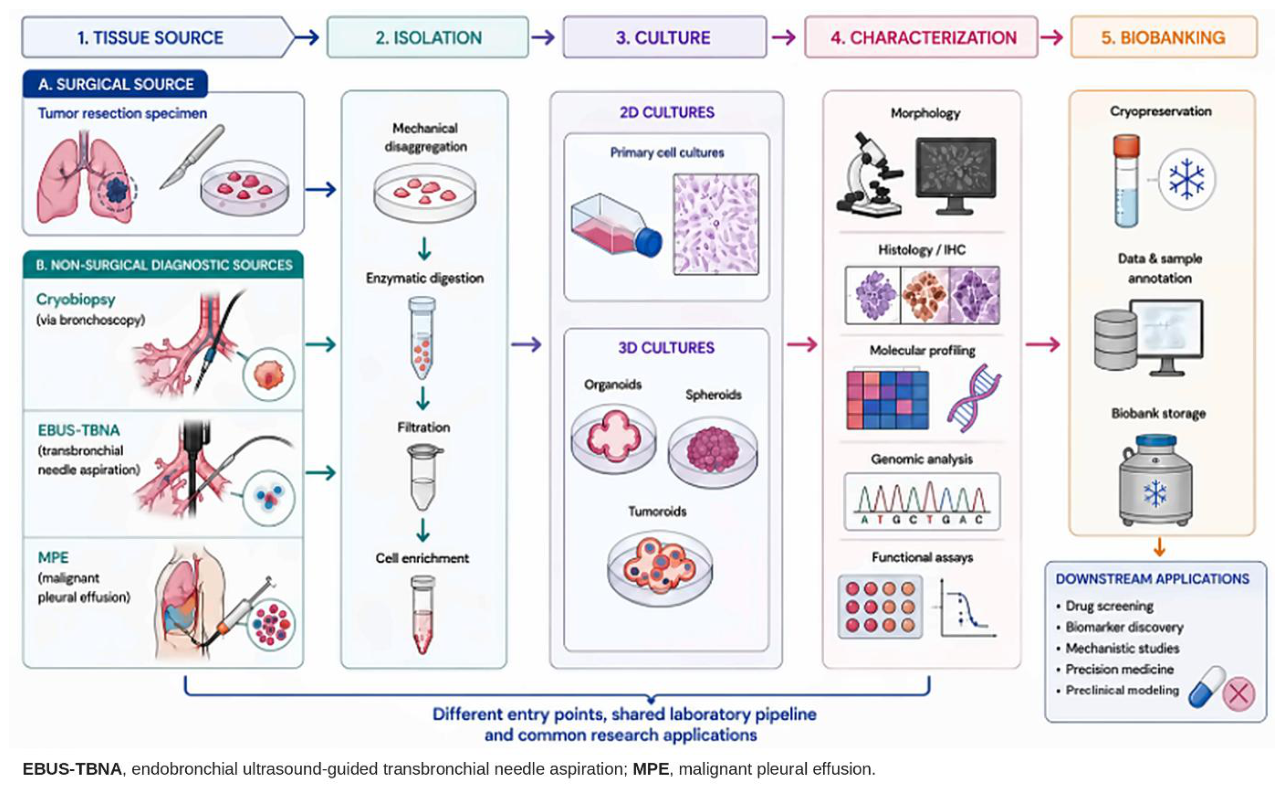
Figure 2.
Principal tissue isolation strategies used for the establishment of primary lung cancer cultures. Mechanical dissociation, enzymatic digestion, and direct processing or explant-based approaches represent complementary strategies that can be selected or combined according to specimen type, amount and cellular composition.
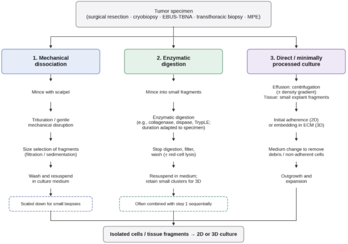
Figure 4.
Reported establishment outcomes of primary lung cancer cultures across studies, stratified by source category and endpoint stringency. The figure highlights the substantial heterogeneity in reported success rates and demonstrates that direct comparison of establishment outcomes requires consideration of tissue source, culture protocol, and the criteria used to define successful culture establishment. Data are based on the studies summarized in Supplementary Table S2. Bars show the reported rates and error bars show 95% Wilson confidence intervals; panel A shows formation or short-term expansion without confirmed tumor identity, and panel B shows long-term expansion and/or validated tumor identity. Studies without an extractable numerator and denominator are not plotted. The dashed interval in panel B marks the malignant pleural effusion arm of Hwang et al. (0/2), whose width reflects only the very small sample.
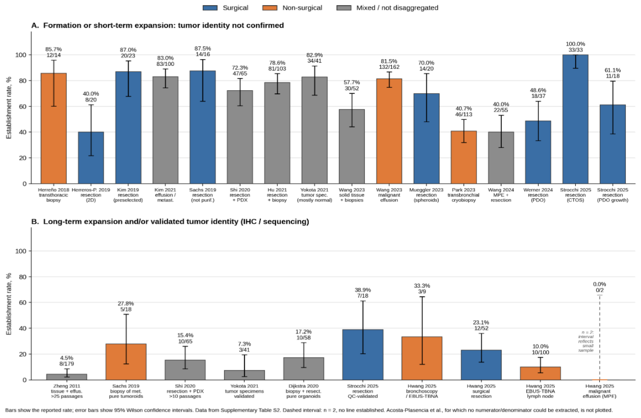
Figure 5.
Culture establishment success reported by Hwang et al. [30] for four specimen sources processed using the same culture protocol and a stringent definition of establishment (immunohistochemistry and sequencing concordance with the parental tumor). Primary tumor tissue obtained by bronchoscopy or endobronchial ultrasound-guided transbronchial needle aspiration (EBUS-TBNA) showed the highest establishment rate (33.3%), followed by surgical resection specimens (23.1%), whereas EBUS-TBNA samples from metastatic lymph nodes showed 10.0%, and two malignant pleural effusion samples yielded no validated organoid lines. Error bars show 95% Wilson confidence intervals; differences across acquisition methods were not statistically significant (p = 0.060, as reported by the original authors). The interval for the malignant pleural effusion arm (0/2) is drawn as a dashed line because its width reflects only the very small sample.
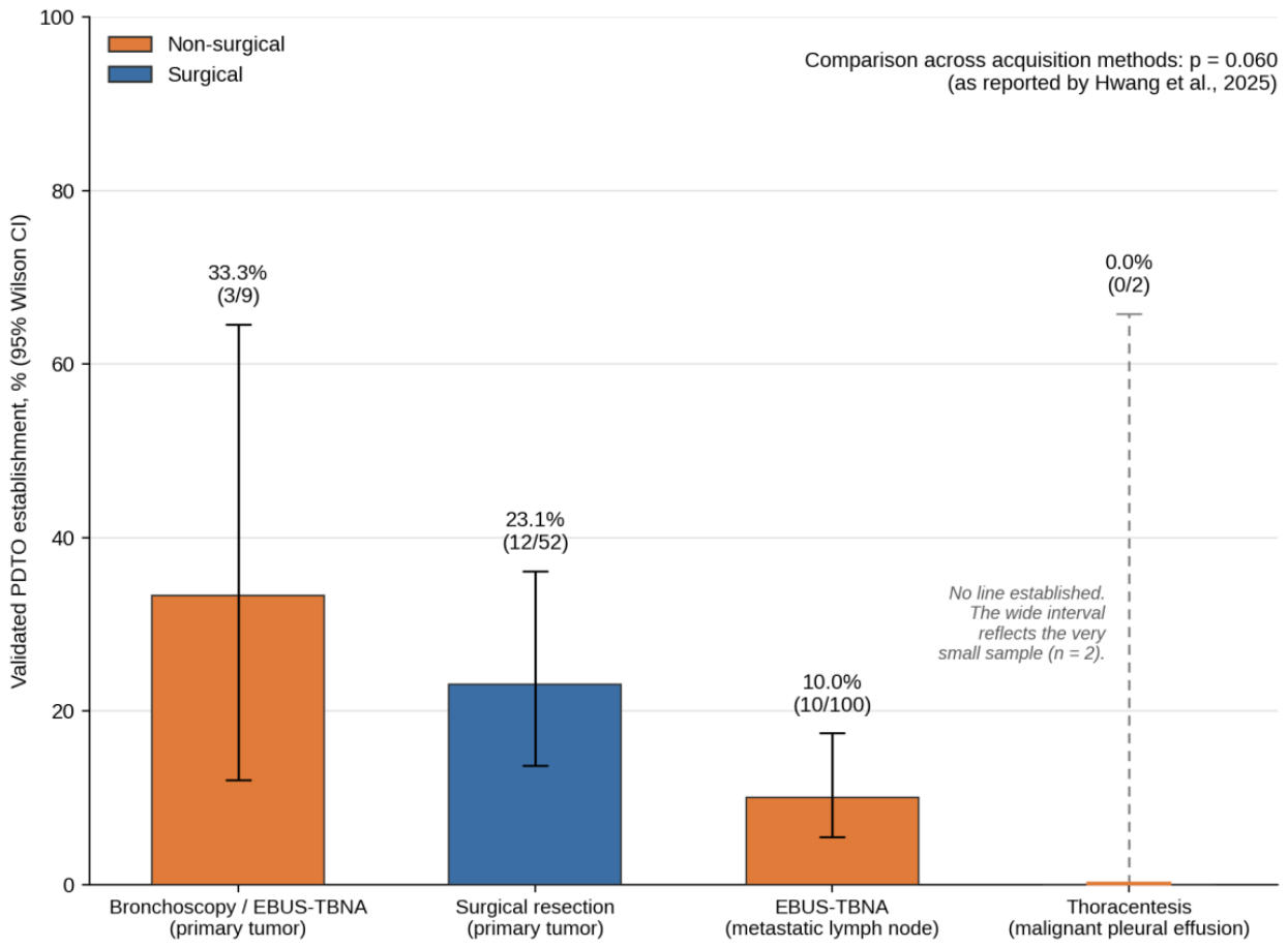

Table 1.
Evidence map: concordance between primary-culture/organoid drug response and clinical outcome.
Table 1.
Evidence map: concordance between primary-culture/organoid drug response and clinical outcome.
| Study | Tissue Source | Therapy Tested | Concordance with Clinical Outcome | Sample Size | Turnaround |
| Roscilli et al. [41] | MPE-derived 2D primary cultures (n = 16 lines) | Conventional chemotherapy | In vitro response matched clinical outcome in all 6 patients with available clinical data (concordance defined as sensitivity to at least one drug of the regimen) | 16 lines (clinical data for 6; 6/6 concordant) |
Not reported |
| Kim et al. [77] | Patient-derived organoids; advanced lung adenocarcinoma | Tyrosine kinase inhibitors (TKIs) | PFS predicted correctly in 6/8 evaluable patients (75.0% accuracy); viability decreased in 5/6 organoids from partial responders and increased in 2/3 from patients with stable disease | 9 patients with clinical follow-up (8 evaluable for PFS), from a biobank of 84 organoids | Not reported (retrospective) |
| Wang et al. [28] | Organoids from malignant serous effusions and tissue; advanced lung cancer | TKIs, chemotherapy and dual-targeted combinations | 45/54 concordant (83.3%); sensitivity 84.0%, specificity 82.8%; osimertinib ROC AUC 0.94 | 54 organoids from 36 patients | Not reported as such (organoids cultured 1–2 weeks before a 6-day assay) |
| Yang et al. [45] | MPE-derived spheroids; NSCLC | Gefitinib in vitro (patients received gefitinib, erlotinib or afatinib) | Viability reduced in all 5 responders and in none of the 3 treated non-responders (8/8); qualitative visual MTT readout, single 25 µM dose | 14 patients (8 treated with an EGFR-TKI) | ~5 weeks (28-day spheroid formation, 7-day drug exposure) |
| Lee et al. [5] | MPE-derived organoids; advanced EGFR-mutant NSCLC (CODRP) | EGFR-TKI | Afatinib: 13/14 concordant with the CODRP index (sensitivity 85.7%, specificity 100%) vs. 10/14 with AUC alone. Osimertinib: 6/6. Cut-off derived in the same cohort | 14 patients (6 for osimertinib) | ~10 days |
Disclaimer/Publisher’s Note: The statements, opinions and data contained in all publications are solely those of the individual author(s) and contributor(s) and not of MDPI and/or the editor(s). MDPI and/or the editor(s) disclaim responsibility for any injury to people or property resulting from any ideas, methods, instructions or products referred to in the content. |
© 2026 by the authors. Licensee MDPI, Basel, Switzerland. This article is an open access article distributed under the terms and conditions of the Creative Commons Attribution (CC BY) license.
Copyright: This open access article is published under a Creative Commons CC BY 4.0 license, which permit the free download, distribution, and reuse, provided that the author and preprint are cited in any reuse.