

Submitted:
18 October 2026
Posted:
20 October 2026
You are already at the latest version
Abstract
Background. Hepatocellular carcinoma (HCC) is a leading cause of cancer-related mortality, with limited survival rates in advanced stages. Systemic therapy using tyrosine kinase inhibitors such as lenvatinib is a standard treatment; however, therapeutic response may be suboptimal due to tumor hypoxia and an immunosuppressive tumor microenvironment. Hydrogen–oxygen (HHO) nanobubble therapy has emerged as a novel approach, although evidence remains limited. Case Presentation. A 55-year-old male was diagnosed with multifocal HCC following medical evaluation. Alpha-fetoprotein (AFP) and protein induced by vitamin K absence or antagonist-II (PIVKA-II) levels were 5,748.71 ng/mL and 14,433.16 mAU/mL, respectively. Abdominal CT demonstrated multiple hepatic lesions involving both lobes, with the largest measuring 15.7 × 10.35 × 13.93 cm. The patient received lenvatinib 10 mg once daily combined with 10 sessions of adjunctive intravenous HHO nanobubble therapy using escalating doses. After approximately one month, AFP decreased to 2,800 ng/mL (51.3%) and PIVKA-II to 2,102 mAU/mL (85.4%). Follow-up PET imaging demonstrated metabolically active hepatic lesions with necrotic components. Conclusions. This case describes a marked early biochemical response during combined treatment. However, the independent contribution of HHO nanobubble therapy cannot be determined from a single case without a comparator. Further controlled studies are required to assess safety and efficacy.
Keywords:
hepatocellular carcinoma
; lenvatinib
; hydrogen-oxygen nanobubbles
; alpha-fetoprotein
; PIVKA-II
; tumor hypoxia
1. Introduction
Hepatocellular carcinoma (HCC) is the most prevalent form of primary liver cancer; ranks as the sixth most common malignancy and the third leading cause of cancer-related death, with a five-year survival rate of around 15% [1,2,3]. The highest incidence rates are found in East Asia and sub-Saharan Africa, largely due to the high prevalence of hepatitis B infection [1,2]. Individuals over 50 years old have a higher risk, with the greatest incidence seen in those above 70 [1,3]. Men are more commonly affected than women, with a global male-to-female ratio of approximately 2:1 to 4:1 [2,3].
HCC can present with a wide range of clinical manifestations [1,3,4]. In the early stages, especially when the tumor is confined to the liver, patients are often asymptomatic [3]. As HCC advances, patients may experience nonspecific complaints such as constitutional symptoms, jaundice, anorexia, weight loss, fatigue, and upper abdominal discomfort [1]. On physical examination, findings may include hepatomegaly and ascites [1,3]. In more advanced disease, HCC may metastasize to organs such as the lungs, adrenal glands, peritoneum, and bones, leading to symptoms depending on the site involved [1,4]. Laboratory findings may show elevated liver enzymes and bilirubin, reflecting underlying liver dysfunction [3,4,5]. Alpha-fetoprotein (AFP) levels are often increased and can support the diagnosis, although this is not seen in all cases, with elevations present in up to 70% of patients [1,6]. Imaging modalities such as ultrasound, CT scan, and MRI are essential for diagnosis and staging, typically demonstrating liver lesions with characteristic radiologic features [3,4]. One of the pharmacological treatment options for HCC involves the use of tyrosine kinase inhibitors, including sorafenib, regorafenib, lenvatinib, and cabozantinib [4,5].
Nanobubbles are tiny gas-filled spherical structures with diameters below 1000 nm, typically ranging from tens to hundreds of nanometers [7]. They consist of gases such as oxygen or nitrogen dispersed in a liquid and stabilized at the gas–liquid interface [7,9]. Due to their unique characteristics, nanobubbles have attracted considerable interest across fields such as medicine, materials science, and environmental science [8,9]. As effective oxygen delivery systems must be compatible with biological environments, nanobubbles have emerged as promising tools to alleviate hypoxia-related stress responses [7,8,9].
Gas therapy is an emerging and promising approach that has gained increasing attention in antitumor treatment [8,10]. Bioactive gases such as molecular hydrogen (H₂) have been investigated for their ability to modulate processes relevant to tumor biology, including oxidative stress, tumor-promoting signaling, and immune regulation [10,11]. The proposed antitumor effects of hydrogen include reducing intracellular reactive oxygen species and reversing tumor-promoting and immunosuppressive phenotypes in cancer-associated fibroblasts [10]. Recently, hydrogen therapy has been widely explored in the development of nanomedicine-based antitumor strategies [8,10,11]. This case focuses on nanobubbles composed of hydrogen and oxygen as a potential adjunctive strategy alongside lenvatinib in HCC.
2. Case Presentation
On November 28, 2025, a 55-year-old man was first presented for a routine medical check-up without specific complaints. Initial laboratory tests revealed elevated liver enzymes, with AST at 59 U/L and ALT at 76 U/L. Tumor marker assessment demonstrated markedly elevated AFP of 5,748.71 ng/mL and PIVKA-II of 14,433.16 mAU/mL. Abdominal CT scan revealed hepatomegaly with multiple inhomogeneous nodules (solid with necrotic areas) involving both the right and left hepatic lobes. The largest lesion measured approximately 15.7 × 10.35 × 13.93 cm (CC × LL × AP), located in segments V–VI–VII–VIII of the liver. The diagnosis was considered advanced multifocal HCC based on the clinical, biochemical, and radiological findings.
Lenvatinib was administered orally at a dose of 10 mg once daily. Adjunctive intravenous HHO nanobubble therapy was administered 10 sessions with an escalating dose regimen ranging from 5 to 50 mL per session. The detailed HHO dosing schedule is presented in Table 1.
After 18 days of documented treatment with Lenvatinib therapy and 10 sessions of HHO nanobubble therapy, the patient underwent follow-up evaluation. The results showed AST 65 U/L and ALT 83 U/L, with a marked decline in tumor markers: AFP decreased to 2,800 ng/mL and PIVKA-II to 2,102 mAU/mL. A comparison of the baseline and follow-up laboratory findings is presented in Table 2. Follow-up PET scan results demonstrated multiple large lesions/masses with metabolic activity similar to the liver in segments VI–VII–VIII, with necrotic (ametabolic) components in segments VII and VIII. Multiple lesions/nodules were also identified in segment VIII of the liver with metabolic activity slightly higher than the liver. In addition, multiple lesions/nodules were identified in segments IV and II of the liver with metabolic activity similar to the liver, as shown in Figure 1.
3. Discussion
Tumor markers such as alpha-fetoprotein (AFP) play an essential role not only in diagnosis but also in monitoring treatment response and prognosis [1,4,6]. Recent studies have shown that an early decline in AFP levels (≥20–50%) during systemic therapy is associated with improved overall survival and better treatment response [6,12]. In a retrospective study involving 553 patients with unresectable hepatocellular carcinoma (uHCC) who received lenvatinib as first-line systemic therapy, a sharp-falling AFP trajectory, characterized by reductions consistently above 50%, was associated with better clinical outcomes [12]. Early AFP response in patients receiving lenvatinib is commonly defined as a reduction of >20% within 4 weeks, while greater declines (≥50%) are associated with improved clinical outcomes [6,12].
In contrast, the relationship between PIVKA-II dynamics and treatment response in systemic therapy remains less clear [13]. Some studies have reported no significant predictive value of PIVKA-II changes in patients receiving agents such as sorafenib or lenvatinib, while others have suggested that reductions greater than 50% may be associated with improved radiologic response and survival, particularly in immunotherapy settings [13]. Other studies suggest that PIVKA-II has been increasingly recognized as a valuable biomarker, with declining levels correlating with improved progression-free survival and reflecting tumor burden, vascular invasion, and biological aggressiveness [13,14,15].
In this case, the patient demonstrated a substantial early decline in tumor markers after approximately one month of therapy, with AFP decreasing by more than 50% and PIVKA-II by approximately 85%. This degree of reduction exceeds the commonly reported thresholds for meaningful biomarker response [6,12,13]. The magnitude of biomarker decline observed in this case was clinically notable; however, not all patients achieve significant early reductions in AFP with lenvatinib monotherapy, and data on PIVKA-II response remain limited [6,12,13]. This raises the possibility that adjunctive therapies may contribute to enhanced treatment response [7,10].
In this case, the patient also received HHO nanobubble therapy, which is hypothesized to exert anti-inflammatory and antioxidant effects through molecular hydrogen, potentially improving the tumor microenvironment and enhancing therapeutic efficacy [7,10,11]. In some studies, hydrogen not only demonstrated direct antitumor effects but also reduced pro-tumorigenic and immunosuppressive signaling in cancer-associated fibroblasts (CAFs) [10]. As a result, the tumor microenvironment shifted from an immunologically “cold” to a “hot” state, characterized by increased CD4⁺ and CD8⁺ T-cell infiltration, enhanced secretion of immunostimulatory factors, and activation of systemic antitumor immune responses [10].
Oxidative stress is increasingly recognized as a major contributor to hepatocellular carcinoma progression, promoting genomic instability, chronic inflammation, and resistance to therapy [16]. Excessive reactive oxygen species (ROS) may contribute to endothelial dysfunction, while abnormal tumor vasculature and impaired perfusion can limit tissue oxygen delivery, promoting a hypoxic tumor microenvironment [16,17]. Under hypoxic conditions, stabilization of hypoxia-inducible factor-1α (HIF-1α) promotes metabolic reprogramming toward glycolysis, angiogenesis, tumor progression, and resistance to anticancer treatment [7,17,18]. Consequently, targeting HIF-1α has become an area of interest to improve treatment response [18]. Molecular hydrogen has been reported to reduce intracellular ROS accumulation and suppress pro-inflammatory signaling pathways, thereby modulating the tumor microenvironment and enhancing antitumor immune responses [10,11]. In addition, oxygen nanobubbles function as oxygen delivery platforms capable of alleviating hypoxia, improving mitochondrial function, and restoring cellular bioenergetics [7,9]. Therefore, by reducing oxidative stress and improving oxygen availability, HHO therapy may help restore cellular metabolism, attenuate hypoxia-driven tumor progression, and enhance sensitivity to systemic treatment such as lenvatinib [7,10,11].
Oxygenation-based systems have the ability to selectively reprogram cellular metabolism [7]. This enables multiple therapeutic benefits, including tumor suppression in cancer by impairing adaptation to hypoxic conditions and the restoration of mitochondrial function in neurodegenerative diseases by promoting alternative energy pathways and reducing oxidative stress—thereby potentially slowing disease progression and supporting neuronal survival [7]. In an in vitro study, doxorubicin-loaded oxygen nanobubbles were able to reverse hypoxia and reduce HIF-1α activity in breast and cervical cancer cells [8,19].
Radiologically, the PET scan findings demonstrated metabolically active hepatic lesions with necrotic components. The presence of necrotic (ametabolic) areas may suggest treatment-related tumor changes, which is in line with the observed biochemical response. The concordance between tumor marker decline and imaging findings may support a favorable early therapeutic response.
Nanobubbles may enhance the effectiveness of cancer therapy through several key mechanisms [7,8,9]. They act as oxygen delivery platforms, helping to relieve tumor hypoxia, a major factor contributing to therapeutic resistance [7,8,9]. By improving oxygen availability, nanobubbles can restore mitochondrial function and normalize cellular metabolism within tumor cells [7,9]. The high stability of nanobubbles makes them particularly suitable for biomedical applications [7,8,9]. They have shown strong potential in areas such as drug and gene delivery, cancer immunotherapy and chemotherapy, as well as wound healing and tissue regeneration [7,8].
Overall, this case highlights a marked early biochemical response following systemic therapy, exceeding commonly reported thresholds for AFP and PIVKA-II reduction. While lenvatinib remains an evidence-based systemic treatment, the potential additive effect of adjunctive therapies warrants further investigation [4]. Larger studies are needed to better define the role of combined approaches and to establish the clinical significance of early biomarker dynamics in guiding treatment strategies for HCC.
This report has several limitations. First, the single-case design and absence of a comparator prevent determination of the independent effect of HHO nanobubble therapy. Second, the follow-up period was short, limiting assessment of progression-free survival and overall survival. Third, objective radiological response could not be quantitatively assessed using serial contrast-enhanced CT or MRI according to standardized HCC response criteria. Finally, the lack of detailed physicochemical characterization of the HHO nanobubble preparation and a fully standardized administration protocol limits the reproducibility and interpretation of the findings.
4. Conclusions
This case demonstrates a marked early biochemical response in a patient with advanced multifocal HCC treated with lenvatinib combined with adjunctive HHO nanobubble therapy. The substantial reduction in AFP and PIVKA-II levels, supported by radiological evidence of tumor necrosis, suggests a favorable treatment outcome that exceeds typical responses reported with lenvatinib alone. Although these findings are clinically noteworthy, the independent contribution of HHO therapy cannot be established from a single uncontrolled case. The findings therefore provide a hypothesis for further investigation rather than evidence of therapeutic synergy. Further controlled clinical studies are needed to confirm efficacy, establish safety, and define the role of HHO nanobubble-assisted therapy in the management of hepatocellular carcinoma.
Author Contributions
Conceptualization, G.J.S. and A.M.S.; methodology, G.J.S.; validation, G.J.S., A.M.S. and S.; investigation, G.J.S.; resources, G.J.S.; data curation, G.J.S.; writing—original draft preparation, G.J.S., A.M.S. and C.P.E.; writing—review and editing, G.J.S., A.M.S. and C.P.E.; visualization, G.J.S.; supervision, A.M.S. and S.; project administration, C.P.E. All authors have read and agreed to the published version of the manuscript.
Institutional Review Board Statement
The preparation and publication of this retrospective single-patient case report were conducted in accordance with the Declaration of Helsinki. Ethical review and approval were not required because the report was based solely on information documented during routine clinical care. No intervention was prospectively assigned for research purposes, and no additional procedures were performed solely for the preparation of this manuscript.
Informed Consent Statement
Written informed consent was obtained from the patient for publication of this paper and the anonymized clinical information contained herein.
Data Availability Statement
Relevant data supporting this case report are presented within the manuscript. Additional patient-level clinical records are not publicly available because of patient privacy and confidentiality restrictions.
Conflicts of Interest
The authors declare no conflict of interest.
Abbreviations
The following abbreviations are used in this manuscript:
| AFP | Alpha-fetoprotein |
| ALT | Alanine aminotransferase |
| AP | Anteroposterior |
| AST | Aspartate aminotransferase |
| CAFs | Cancer-associated fibroblasts |
| CC | Craniocaudal |
| CD4⁺ | Cluster of differentiation 4-positive |
| CD8⁺ | Cluster of differentiation 8-positive |
| CT | Computed tomography |
| H₂ | Molecular hydrogen |
| HCC | Hepatocellular carcinoma |
| HHO | Hydrogen–oxygen |
| HIF-1α | Hypoxia-inducible factor-1 alpha |
| LL | Laterolateral |
| MRI | Magnetic resonance imaging |
| PET | Positron emission tomography |
| PIVKA-II | Protein induced by vitamin K absence or antagonist-II |
| ROS | Reactive oxygen species |
| uHCC | Unresectable hepatocellular carcinoma |
References
- Amin, N.; Anwar, J.; Sulaiman, A.; Naumova, N.N.; Anwar, N. Hepatocellular carcinoma: a comprehensive review. Diseases 2025, 13(7), 207. [Google Scholar] [CrossRef]
- Li, Q.; Ding, C.; Cao, M.; Yang, F.; Yan, X.; He, S.; et al. Global epidemiology of liver cancer 2022: an emphasis on geographic disparities. Chin. Med. J. (Engl) 2024, 137(19), 2334–2342. [Google Scholar] [CrossRef]
- Llovet, J.M.; Kelley, R.K.; Villanueva, A.; Singal, A.G.; Pikarsky, E.; Roayaie, S.; et al. Hepatocellular carcinoma. Nat. Rev. Dis. Primers 2021, 7(1), 6. [Google Scholar] [CrossRef]
- Singal, A.G.; Llovet, J.M.; Yarchoan, M.; Mehta, N.; Heimbach, J.K.; Dawson, L.A.; et al. AASLD practice guidance on prevention, diagnosis, and treatment of hepatocellular carcinoma. Hepatology 2023, 78(6), 1922–1965. [Google Scholar] [CrossRef]
- Lu, C.H.; Kao, W.Y.; Wu, C.H.; Ting, W.Y.; Lu, C.H.; Chuang, K.I.; et al. Predicting survival outcomes in patients with hepatocellular carcinoma receiving lenvatinib by using the Up7-ALBI score. Liver Cancer 2025, 14(6), 704–717. [Google Scholar] [CrossRef]
- Liu, B.; Shang, X.; Shi, J.Y.; Cui, G.Z.; Li, X.; Wang, N.Y. Early alpha-fetoprotein response is associated with survival in patients with HBV-related hepatocellular carcinoma receiving lenvatinib. Front Oncol. 2022, 12, 807189. [Google Scholar] [CrossRef]
- Viafara Garcia, S.M.; Khan, M.S.; Haidar, Z.S.; Acevedo Cox, J.P. NanoBubble-mediated oxygenation: elucidating the underlying molecular mechanisms in hypoxia and mitochondrial-related pathologies. Nanomaterials 2023, 13(23), 3060. [Google Scholar] [CrossRef]
- Terlikowska, K.M.; Dobrzycka, B.; Terlikowski, S.J. Modifications of nanobubble therapy for cancer treatment. Int. J. Mol. Sci. 2024, 25(13), 7292. [Google Scholar] [CrossRef]
- Khan, M.S.; Hwang, J.; Lee, K.; Choi, Y.; Kim, K.; Koo, H.J.; et al. Oxygen-carrying micro/nanobubbles: composition, synthesis techniques and potential prospects in photo-triggered theranostics. Molecules 2018, 23(9), 2210. [Google Scholar] [CrossRef]
- Meng, X.; Liu, Z.; Deng, L.; Yang, Y.; Zhu, Y.; Sun, X.; et al. Hydrogen therapy reverses cancer-associated fibroblasts phenotypes and remodels stromal microenvironment to stimulate systematic anti-tumor immunity. Adv. Sci. (Weinh) 2024, 11(28), e2401269. [Google Scholar] [CrossRef]
- Artamonov, M.Y.; Martusevich, A.K.; Pyatakovich, F.A.; Minenko, I.A.; Dlin, S.V.; LeBaron, T.W. Molecular hydrogen: from molecular effects to stem cells management and tissue regeneration. Antioxidants 2023, 12(3), 636. [Google Scholar] [CrossRef]
- Zheng, Y.; Hu, J.; Mei, Y.; Xiang, Y.; Lu, H.; Zhu, J.; et al. Trajectories of α-fetoprotein and unresectable hepatocellular carcinoma outcomes receiving lenvatinib: a retrospective, multicenter cohort study. BMC Cancer 2025, 25(1), 1137. [Google Scholar] [CrossRef]
- Chen, S.C.; Ho, H.L.; Liu, C.A.; Hung, Y.P.; Chiang, N.J.; Chen, M.H.; et al. PIVKA-II as a surrogate biomarker for therapeutic response in non-AFP-secreting hepatocellular carcinoma. BMC Cancer 2025, 25(1), 199. [Google Scholar] [CrossRef]
- Zhang, D.; Liu, Z.; Yin, X.; Qi, X.; Lu, B.; Liu, Y.; et al. Prognostic value of PIVKA-II in hepatocellular carcinoma patients receiving curative ablation: a systematic review and meta-analysis. Int. J. Biol. Markers 2018, 33(3), 266–274. [Google Scholar] [CrossRef]
- Dong, L.; Qiu, X.; Gao, F.; Wang, K.; Xu, X. Protein induced by vitamin K absence or antagonist II: experience to date and future directions. Biochim Biophys. Acta Rev. Cancer 2023, 1878(6), 189016. [Google Scholar] [CrossRef]
- Liu, Y.; Hao, C.; Li, L.; Zhang, H.; Zha, W.; Ma, L.; et al. The role of oxidative stress in the development and therapeutic intervention of hepatocellular carcinoma. Curr. Cancer Drug Targets 2023, 23(10), 792–804. [Google Scholar] [CrossRef]
- Chen, Z.; Han, F.; Du, Y.; Shi, H.; Zhou, W. Hypoxic microenvironment in cancer: molecular mechanisms and therapeutic interventions. Signal Transduct. Target Ther. 2023, 8(1), 70. [Google Scholar] [CrossRef]
- Huynh, K.N.; Rao, S.; Roth, B.; Bryan, T.; Fernando, D.M.; Dayyani, F.; et al. Targeting hypoxia-inducible factor-1α for the management of hepatocellular carcinoma. Cancers 2023, 15(10), 2738. [Google Scholar] [CrossRef]
- Khan, M.S.; Hwang, J.; Lee, K.; Choi, Y.; Seo, Y.; Jeon, H.; et al. Anti-tumor drug-loaded oxygen nanobubbles for the degradation of HIF-1α and the upregulation of reactive oxygen species in tumor cells. Cancers 2019, 11(10), 1464. [Google Scholar] [CrossRef]
Figure 1.
Follow-up PET images after combined lenvatinib and adjunctive HHO nanobubble therapy.
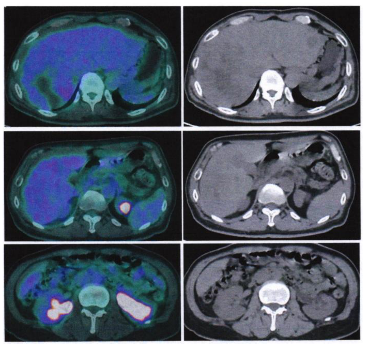

Table 1.
HHO dosing in patient therapy.
| Session | Date | HHO dose |
|---|---|---|
| 1 | 18 December 2025 | 5 mL |
| 2 | 19 December 2025 | 10 mL |
| 3 | 22 December 2025 | 15 mL |
| 4 | 23 December 2025 | 20 mL |
| 5 | 26 December 2025 | 30 mL |
| 6 | 27 December 2025 | 40 mL |
| 7 | 29 December 2025 | 50 mL |
| 8 | 30 December 2025 | 50 mL |
| 9 | 3 January 2026 | 50 mL |
| 10 | 5 January 2026 | 50 mL |
Table 2.
Comparison of baseline and follow-up laboratory evaluations.
| Parameter | Baseline | Follow-up | Change |
|---|---|---|---|
| AFP | 5,748.71 ng/mL | 2,800 ng/mL | Decrease of 51.3% |
| PIVKA-II | 14,433.16 mAU/mL | 2,102 mAU/mL | Decrease of 85.4% |
| AST | 59 U/L | 65 U/L | Increase |
| ALT | 76 U/L | 83 U/L | Increase |
Disclaimer/Publisher’s Note: The statements, opinions and data contained in all publications are solely those of the individual author(s) and contributor(s) and not of MDPI and/or the editor(s). MDPI and/or the editor(s) disclaim responsibility for any injury to people or property resulting from any ideas, methods, instructions or products referred to in the content. |
© 2026 by the authors. Licensee MDPI, Basel, Switzerland. This article is an open access article distributed under the terms and conditions of the Creative Commons Attribution (CC BY) license.
Copyright: This open access article is published under a Creative Commons CC BY 4.0 license, which permit the free download, distribution, and reuse, provided that the author and preprint are cited in any reuse.