

Submitted:
20 October 2026
Posted:
20 October 2026
You are already at the latest version
Abstract
Unresectable multifocal hepatocellular carcinoma (HCC) is generally managed with systemic therapy, including immune checkpoint inhibitors and molecular targeted agents. When adequate tumor control is achieved, conversion to surgical resection may become feasible. A 59-year-old man with untreated chronic hepatitis C was found to have seven HCC lesions during magnetic resonance imaging surveillance. Systemic therapy with atezolizumab (1200 mg/body) plus bevacizumab (15 mg/kg) was initiated for unresectable multifocal HCC. After three cycles, contrast-enhanced computed tomography showed tumor shrinkage, with a 28% reduction in the sum of target lesion diameters, corresponding to stable disease according to Response Evaluation Criteria in Solid Tumors v1.1. The future liver remnant volume was 39.2%. After two additional cycles of atezolizumab monotherapy as bridging therapy, the patient underwent extended right hepatectomy, including resection of segment 4 and partial resection of segment 2. Pathological examination confirmed R0 resection, with viable tumor cells and focal necrosis. The postoperative course was uneventful, and antiviral therapy for hepatitis C resulted in a sustained virological response. The patient remains recurrence-free. Systemic therapy with atezolizumab plus bevacizumab enabled successful conversion surgery for seven HCC lesions. This multimodal approach may represent a viable treatment option for carefully selected patients with initially unresectable multifocal HCC.
Keywords:
hepatocellular carcinoma
; atezolizumab
; bevacizumab
; conversion surgery
; immune checkpoint inhibitor
; multifocal hepatocellular carcinoma
1. Introduction
Hepatocellular carcinoma (HCC) is a major global health problem and one of the leading causes of cancer-related mortality worldwide, ranking sixth in incidence and third in cancer-related mortality [1,2]. The Barcelona Clinic Liver Cancer (BCLC) staging system guides treatment allocation: surgical resection or ablation is recommended for early-stage disease, transarterial chemoembolization for intermediate-stage disease, and systemic therapy for advanced-stage disease [3]. Surgical resection offers the best chance of cure, but most patients present with disease that precludes upfront resection because of multifocal involvement, vascular invasion, or extrahepatic spread [1].
In Japan, HCC with macrovascular invasion, extrahepatic metastasis, or multifocal intrahepatic disease involving four or more tumors is classified as unresectable and managed with systemic therapy [4]. Sorafenib was the first systemic agent to demonstrate a survival benefit for advanced HCC; subsequently, lenvatinib demonstrated non-inferior overall survival with superior progression-free survival and objective response rates in the phase III REFLECT trial [5]. The landmark phase III IMbrave150 trial demonstrated that atezolizumab—an anti-programmed death-ligand 1 antibody—plus bevacizumab—an anti-vascular endothelial growth factor (VEGF) antibody—significantly improved overall survival, progression-free survival, and objective response rate compared with sorafenib, establishing this combination as a first-line systemic therapy for unresectable HCC [6].
Growing evidence supports the concept of conversion surgery, whereby patients with initially unresectable HCC who achieve adequate tumor control with systemic therapy may become candidates for curative-intent resection. Most published reports of conversion surgery have involved lenvatinib-based regimens, whereas only a limited number of cases following atezolizumab plus bevacizumab therapy have been reported [7,8]. The ongoing multicenter phase II RACB trial (Resection After Combination therapy with atezolizumab and Bevacizumab) was designed specifically to evaluate the feasibility and efficacy of conversion surgery following atezolizumab plus bevacizumab therapy in Japan, and its short-term outcomes have recently been published [9,10]. Herein, we report a case of successful conversion surgery for seven HCC lesions following atezolizumab plus bevacizumab therapy.
2. Case Description
A 59-year-old man had been diagnosed with chronic hepatitis C virus (HCV) infection several years previously but had not received antiviral treatment. He presented to our outpatient hepatology clinic for routine surveillance, and follow-up magnetic resonance imaging revealed a dominant mass in the right hepatic lobe with multiple surrounding satellite lesions. Seven HCC lesions were identified.
Laboratory evaluation demonstrated markedly elevated serum tumor marker levels: alpha-fetoprotein, 10,077 ng/mL, and protein induced by vitamin K absence or antagonist-II, 29,606 mAU/mL. Viral serology was negative for hepatitis B (HB) surface antigen and positive for HB surface antibody and HB core antibody. Hepatitis B virus DNA was undetectable (<1.0 log IU/mL) by TaqMan assay. HCV antibody was positive, and the HCV RNA level was 4.8 log IU/mL. The indocyanine green retention rate at 15 minutes was 13.8%. Liver function was well preserved, with a Child–Pugh score of 5 (prothrombin activity >130%, serum albumin 3.8 g/dL, and total bilirubin 0.6 mg/dL), and there was no clinical evidence of ascites or hepatic encephalopathy.
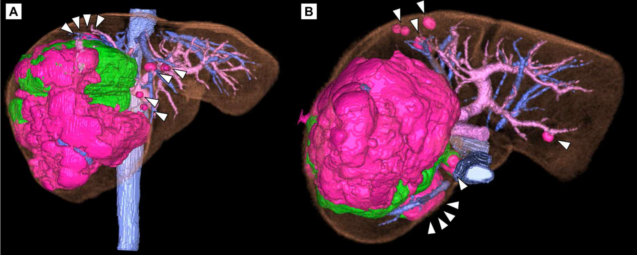
Contrast-enhanced computed tomography (CT) of the liver demonstrated a large, irregularly marginated mass predominantly occupying the right hepatic lobe and measuring 12 × 9 × 12 cm. The lesion showed heterogeneous arterial-phase enhancement followed by washout on portal venous- and delayed-phase imaging, consistent with HCC. In addition to the dominant tumor, multiple satellite lesions were distributed throughout both hepatic lobes: two lesions in segment 4 and one lesion each in segments 1r, 2, 5, and 7. All lesions exhibited imaging characteristics consistent with multifocal HCC (Figure 1 and Figure A1). Given the presence of four or more intrahepatic lesions involving both lobes, the patient was deemed ineligible for upfront surgical resection according to the Japan Society of Hepatology clinical practice guidelines [4].
Systemic therapy with atezolizumab (1200 mg/body) plus bevacizumab (15 mg/kg) every 3 weeks was initiated as first-line treatment. After three cycles, restaging contrast-enhanced CT demonstrated a reduction in the size of the dominant tumor to 8.9 × 7.6 × 7.2 cm. The sum of the longest diameters of the target lesions decreased by 28% according to Response Evaluation Criteria in Solid Tumors (RECIST) v1.1 criteria, corresponding to stable disease. Assessment using modified RECIST similarly indicated stable disease. Volumetric analysis estimated the future liver remnant at 39.2%. As most lesions were confined to the right hepatic lobe and no tumor progression was observed, the multidisciplinary team concluded that conversion surgery was oncologically and technically feasible (Figure 2 and Figure 3).
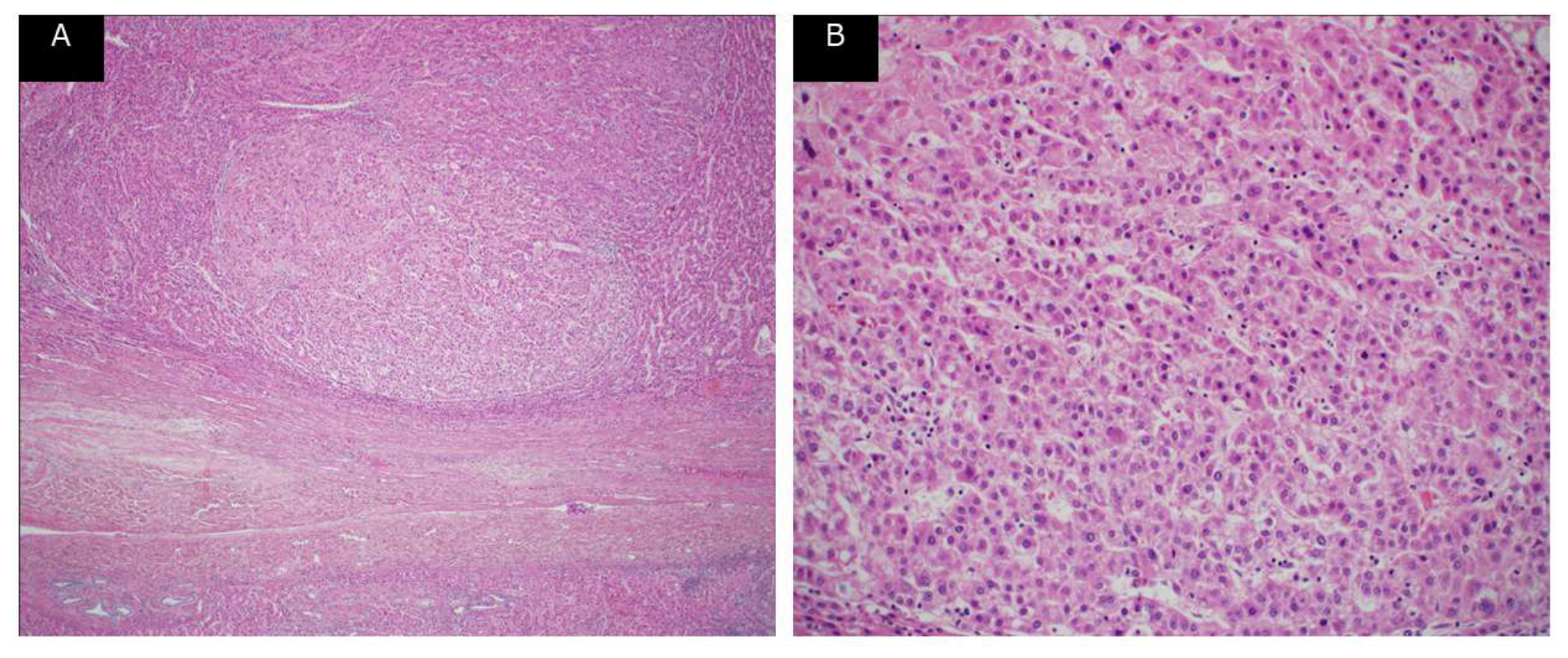
The patient received two additional cycles of atezolizumab monotherapy as bridging therapy. Bevacizumab was withheld for at least 6 weeks before surgery to minimize the risk of wound-healing complications and other bevacizumab-related adverse effects [11]. The patient subsequently underwent extended right hepatectomy, including resection of segment 4 and partial resection of segment 2. The operative time was 5 hours 52 minutes, and intraoperative blood loss was 223 mL. Pathological examination of the resected specimen revealed predominantly viable tumor cells with focal areas of necrosis; a pathological complete response was not achieved (Figure 4). All surgical margins were microscopically negative (R0 resection).
The postoperative course was uncomplicated, and the patient was discharged on postoperative day 8. Antiviral therapy for chronic hepatitis C was initiated postoperatively, resulting in a sustained virological response (SVR) at 9 months after surgery. At the time of this report, the patient remains recurrence-free with no evidence of disease.
3. Discussion
We report a rare case of successful conversion surgery enabling R0 hepatectomy for seven initially unresectable HCC lesions following systemic therapy with atezolizumab plus bevacizumab. This case illustrates the therapeutic potential of a multidisciplinary strategy combining immunotherapy-based systemic treatment with curative-intent surgical resection in carefully selected patients with advanced HCC.
HCC is among the most prevalent malignancies and the third leading cause of cancer-related death globally [1,2]. Although surgical resection remains the cornerstone of curative therapy for early-stage HCC, most patients present with disease beyond the threshold for resection. The BCLC staging system classifies multifocal disease with four or more lesions as intermediate-stage (BCLC B) or more advanced disease, for which systemic therapy is the primary treatment modality [3]. In Japan, clinical guidelines similarly restrict surgical resection to patients with three or fewer intrahepatic tumors without macrovascular invasion or extrahepatic spread [4].
The concept of conversion surgery—achieving R0 resection in patients initially deemed unresectable after a response to systemic therapy—has emerged as a promising strategy in HCC management [12,13,14,15]. Given the high objective response rate reported for atezolizumab plus bevacizumab in the IMbrave150 trial (approximately 30%), a growing number of patients may become candidates for conversion surgery [6]. The ongoing multicenter RACB study in Japan specifically evaluates atezolizumab plus bevacizumab as a platform for conversion to surgery. Short-term results demonstrated a resection rate of approximately 48% among enrolled patients and a 6-month progression-free survival rate of 59.1%, supporting the feasibility of this approach [9,10].
Before the widespread adoption of atezolizumab plus bevacizumab, conversion surgery was predominantly reported following lenvatinib therapy. Several case reports and small series have described successful R0 resection after lenvatinib-based regimens for initially unresectable HCC [16,17]. By contrast, reports of conversion surgery following atezolizumab plus bevacizumab remain limited. Hidaka et al. [7] described a case of HCC with portal vein tumor thrombus in which atezolizumab plus bevacizumab induced tumor regression, enabling R0 resection. Sato et al. [8] reported a case in which conversion hepatectomy after atezolizumab plus bevacizumab revealed a pathological complete response. Our case adds to this growing body of literature and further supports the real-world feasibility of this approach in patients with multifocal HCC.
Patient selection and the timing of surgery are critical determinants of outcome. In the present case, the decision to proceed with surgery was based on stable residual disease (no progression after initial treatment), the absence of extrahepatic spread, an adequate future liver remnant volume (39.2%), and preserved hepatic functional reserve (Child–Pugh score 5). Collectively, these factors supported the feasibility and safety of conversion surgery.
Immune-related adverse events and bevacizumab-specific toxicities require careful consideration when planning surgery after atezolizumab plus bevacizumab therapy. In the IMbrave150 trial, the most common bevacizumab-related adverse events included hypertension (23.7%) and proteinuria (18.8%) [6]. Bevacizumab inhibits VEGF-mediated angiogenesis and may impair wound healing and promote gastrointestinal complications [11]. In accordance with the RACB trial protocol, bevacizumab was discontinued at least 6 weeks before surgery in the present case, and no perioperative complications attributable to prior anti-VEGF therapy were observed [10].
Notably, the patient also had untreated chronic HCV infection, which was managed postoperatively with direct-acting antiviral therapy, resulting in SVR. Achievement of SVR reduces the risk of HCV-related HCC recurrence and may improve long-term prognosis [4]. Thus, integrating antiviral therapy into the postoperative management plan is an important aspect of comprehensive care for patients with HCV-related HCC.
Pathological examination in our case revealed predominantly viable tumor with focal necrosis, in contrast to the pathological complete response reported in another case of conversion surgery after atezolizumab plus bevacizumab [8]. The degree of pathological response may serve as a prognostic indicator; further studies are needed to clarify the relationship between radiological response, pathological findings, and long-term oncological outcomes.
4. Conclusions
This case highlights the potential role of atezolizumab plus bevacizumab as a bridge to conversion surgery in initially unresectable multifocal HCC. Accumulation of additional cases through prospective studies such as the RACB trial will be essential for establishing standardized criteria for patient selection and the optimal timing of surgery, as well as determining the long-term outcomes of this multimodal strategy.
Author Contributions
Conceptualization, H.T. (Takahata) and H.T. (Takahashi); data curation, H.T. (Takahata); data collection, S.M. (Mizukami), K.M. (Makino), and K.I.; clinical management, S.M. (Mizukami), K.I., and K.S.; pathological analysis, M.T. (Tanino); writing—original draft preparation, H.T. (Takahata); writing—review and editing, H.T. (Takahashi), M.T. (Tanino), and H.Y.; supervision, H.T. (Takahashi) and H.Y. All authors have read and agreed to the published version of the manuscript.
Funding
This research received no external funding.
Institutional Review Board Statement
This study was conducted in accordance with the Declaration of Helsinki. The requirements for ethical review and approval were waived for this study because it was a single case report.
Informed Consent Statement
Written informed consent was obtained from the patient to publish this paper.
Data Availability Statement
The original contributions presented in this study are included in the article. Further inquiries can be directed to the corresponding author.
Acknowledgments
We thank Angela Morben, DVM, ELS, from Edanz (https://jp.edanz.com/ac), for editing a draft of this manuscript.
Conflicts of Interest
The authors declare no conflicts of interest.
Abbreviations
The following abbreviations are used in this manuscript:
| AFP | Alpha-fetoprotein |
| BCLC | Barcelona Clinic Liver Cancer |
| CT | Computed tomography |
| HB HCC |
Hepatitis B Hepatocellular carcinoma |
| HCV | Hepatitis C virus |
| PIVKA-II | Protein induced by vitamin K absence or antagonist-II |
| RACB | Resection After Combination therapy with atezolizumab and Bevacizumab |
| RECIST | Response Evaluation Criteria in Solid Tumors |
| SVR | Sustained virological response |
| VEGF | Vascular endothelial growth factor |
Appendix A
Figure A1.
Pretreatment radiological findings of multifocal hepatocellular carcinoma. Three-dimensional reconstruction was performed for assessment of the tumor distribution and surgical planning. The reconstruction demonstrated the dominant tumor occupying most of the right hepatic lobe and six additional satellite lesions located in segments 1r, 2, 4 (two lesions), 5, and 7. The reconstruction was used to assess the spatial relationship between the tumors and intrahepatic vasculature and to plan the extent of hepatic resection.
Figure A1.
Pretreatment radiological findings of multifocal hepatocellular carcinoma. Three-dimensional reconstruction was performed for assessment of the tumor distribution and surgical planning. The reconstruction demonstrated the dominant tumor occupying most of the right hepatic lobe and six additional satellite lesions located in segments 1r, 2, 4 (two lesions), 5, and 7. The reconstruction was used to assess the spatial relationship between the tumors and intrahepatic vasculature and to plan the extent of hepatic resection.
References
- Bray, F.; Laversanne, M.; Sung, H.; Ferlay, J.; Siegel, R.L.; Soerjomataram, I.; Jemal, A. Global cancer statistics 2022: GLOBOCAN estimates of incidence and mortality worldwide for 36 cancers in 185 countries. CA Cancer J. Clin. 2024, 74, 229–263. [CrossRef]
- Singh, S.P.; Madke, T.; Chand, P. Global epidemiology of hepatocellular carcinoma. J. Clin. Exp. Hepatol. 2025, 15, 102446. [CrossRef]
- Reig, M.; Forner, A.; Rimola, J.; Ferrer-Fàbrega, J.; Burrel, M.; Garcia-Criado, Á.; Kelley, R.K.; Galle, P.R.; Mazzaferro, V.; Salem, R.; et al. BCLC strategy for prognosis prediction and treatment recommendation: The 2022 update. J. Hepatol. 2022, 76, 681–693. [CrossRef]
- Hasegawa, K.; Takemura, N.; Yamashita, T.; Watadani, T.; Kaibori, M.; Kubo, S.; Shimada, M.; Nagano, H.; Hatano, E.; Aikata, H.; et al. Clinical Practice Guidelines for Hepatocellular Carcinoma: The Japan Society of Hepatology 2021 version (5th JSH-HCC Guidelines). Hepatol. Res. 2023, 53, 383–390. [CrossRef]
- Kudo, M.; Finn, R.S.; Qin, S.; Han, K.-H.; Ikeda, K.; Piscaglia, F.; Baron, A.; Park, J.-W.; Han, G.; Jassem, J.; et al. Lenvatinib versus sorafenib in first-line treatment of patients with unresectable hepatocellular carcinoma: A randomised phase 3 non-inferiority trial. Lancet 2018, 391, 1163–1173. [CrossRef]
- Finn, R.S.; Qin, S.; Ikeda, M.; Galle, P.R.; Ducreux, M.; Kim, T.-Y.; Kudo, M.; Breder, V.; Merle, P.; Kaseb, A.O.; et al.; IMbrave150 Investigators. Atezolizumab plus bevacizumab in unresectable hepatocellular carcinoma. N. Engl. J. Med. 2020, 382, 1894–1905. [CrossRef]
- Hidaka, Y.; Tomita, M.; Desaki, R.; Hamanoue, M.; Takao, S.; Kirishima, M.; Ohtsuka, T. Conversion surgery for hepatocellular carcinoma with portal vein tumor thrombus after successful atezolizumab plus bevacizumab therapy: A case report. World J. Surg. Oncol. 2022, 20, 218. [CrossRef]
- Sato, S.; Aoki, T.; Matsumoto, T.; Shiraki, T.; Mori, S.; Iso, Y.; Nemoto, T.; Onishi, T.; Iijima, M.; Ishida, K. Pathological complete response of hepatocellular carcinoma confirmed by conversion hepatectomy following atezolizumab plus bevacizumab therapy: A case report and literature review. Clin. J. Gastroenterol. 2024, 17, 292–299. [CrossRef]
- Okuno, M.; Ishii, T.; Ichida, A.; Soyama, A.; Takemura, N.; Hirono, S.; Eguchi, S.; Hasegawa, K.; Sasaki, Y.; Uemura, K.; et al. Protocol of the RACB study: A multicenter, single-arm, prospective study to evaluate the efficacy of resection of initially unresectable hepatocellular carcinoma with atezolizumab combined with bevacizumab. BMC Cancer 2023, 23, 780. [CrossRef]
- Takayama, M.; Ishii, T.; Okuno, M.; Hasegawa, K.; Shimada, M.; Eguchi, S.; Takemura, N.; Maki, A.; Endo, I.; Tanabe, M.; et al. A multicenter prospective study to evaluate the efficacy of resection for initially unresectable hepatocellular carcinoma after atezolizumab combined with bevacizumab (the RACB study): Short-term outcomes. J. Clin. Oncol. 2025, 43 (Suppl. 4), 521. [CrossRef]
- Saif, M.W.; Mehra, R. Incidence and management of bevacizumab-related toxicities in colorectal cancer. Expert Opin. Drug Saf. 2006, 5, 553–566. [CrossRef]
- Yokoo, H.; Takahashi, H.; Hagiwara, M.; Iwata, H.; Imai, K.; Saito, Y.; Matsuno, N.; Furukawa, H. Successful hepatic resection for recurrent hepatocellular carcinoma after lenvatinib treatment: A case report. World J. Hepatol. 2020, 12, 1349–1357. [CrossRef]
- Akahoshi, K.; Shindoh, J.; Tanabe, M.; Ariizumi, S.; Eguchi, S.; Okamura, Y.; Kaibori, M.; Kubo, S.; Shimada, M.; Taketomi, A.; et al. Oncological resectability criteria for hepatocellular carcinoma in the era of novel systemic therapies: The Japan Liver Cancer Association and Japanese Society of Hepato-Biliary-Pancreatic Surgery Expert Consensus Statement 2023. Liver Cancer 2024, 13, 579–589. [CrossRef]
- Yokoo, H.; Mizukami, S.; Takahashi, H.; Takizawa, T.; Enomoto, K.; Makino, K.; Takahata, H.; Adachi, Y.; Imai, K. Surgical outcomes of lenvatinib treatment followed by liver resection for advanced hepatocellular carcinoma larger than 10 cm. Cancers (Basel) 2025, 17, 2818. [CrossRef]
- Shindoh, J.; Matsumura, M.; Komatsu, S.; Fukumoto, T.; Ichida, A.; Hasegawa, K.; Ishii, T.; Hatano, E.; Nakamura, M.; Ohtsuka, M. Prognostic factors and clinical significance of preoperative systemic therapy in patients with borderline resectable hepatocellular carcinoma: A JSHBPS project study 2023, Part 2. J. Hepatobiliary Pancreat. Sci. 2025, 32, 374–384. [CrossRef]
- Tomonari, T.; Sato, Y.; Tanaka, H.; Tanaka, T.; Taniguchi, T.; Sogabe, M.; Okamoto, K.; Miyamoto, H.; Muguruma, N.; Saito, Y.; et al. Conversion therapy for unresectable hepatocellular carcinoma after lenvatinib: Three case reports. Medicine (Baltimore) 2020, 99, e22782. [CrossRef]
- Shiraishi, J.; Itoh, S.; Tomino, T.; Yoshiya, S.; Nagao, Y.; Morita, K.; Kayashima, H.; Harada, N.; Ichiki, Y.; Yoshizumi, T. Curative surgery for multiple hepatocellular carcinomas after lenvatinib plus transarterial chemoembolization: A case report. J. Surg. Case Rep. 2023, 2023, rjad485. [CrossRef]
Figure 1.
Pretreatment radiological findings of multifocal hepatocellular carcinoma. (A, B) Axial contrast-enhanced computed tomography (CT) images showing the dominant tumor predominantly occupying the right hepatic lobe in the (A) arterial and (B) delayed phases. The tumor measured 12 × 9 × 12 cm and demonstrated heterogeneous arterial-phase enhancement with subsequent washout and a mosaic appearance. (C) Coronal arterial-phase CT image showing the extensive distribution of the dominant tumor within the right hepatic lobe. (D–H) Representative CT images showing six satellite lesions: (D) one in segment 7, (E) one in segment 1r, (F) two in segment 4, (G) one in segment 5, and (H) one in segment 2. The segment 7 lesion showed arterial-phase enhancement with subsequent washout, whereas the remaining satellite lesions were clearly visualized in the delayed phase. White dotted lines delineate the dominant tumor, and white arrowheads indicate the satellite lesions.
Figure 1.
Pretreatment radiological findings of multifocal hepatocellular carcinoma. (A, B) Axial contrast-enhanced computed tomography (CT) images showing the dominant tumor predominantly occupying the right hepatic lobe in the (A) arterial and (B) delayed phases. The tumor measured 12 × 9 × 12 cm and demonstrated heterogeneous arterial-phase enhancement with subsequent washout and a mosaic appearance. (C) Coronal arterial-phase CT image showing the extensive distribution of the dominant tumor within the right hepatic lobe. (D–H) Representative CT images showing six satellite lesions: (D) one in segment 7, (E) one in segment 1r, (F) two in segment 4, (G) one in segment 5, and (H) one in segment 2. The segment 7 lesion showed arterial-phase enhancement with subsequent washout, whereas the remaining satellite lesions were clearly visualized in the delayed phase. White dotted lines delineate the dominant tumor, and white arrowheads indicate the satellite lesions.
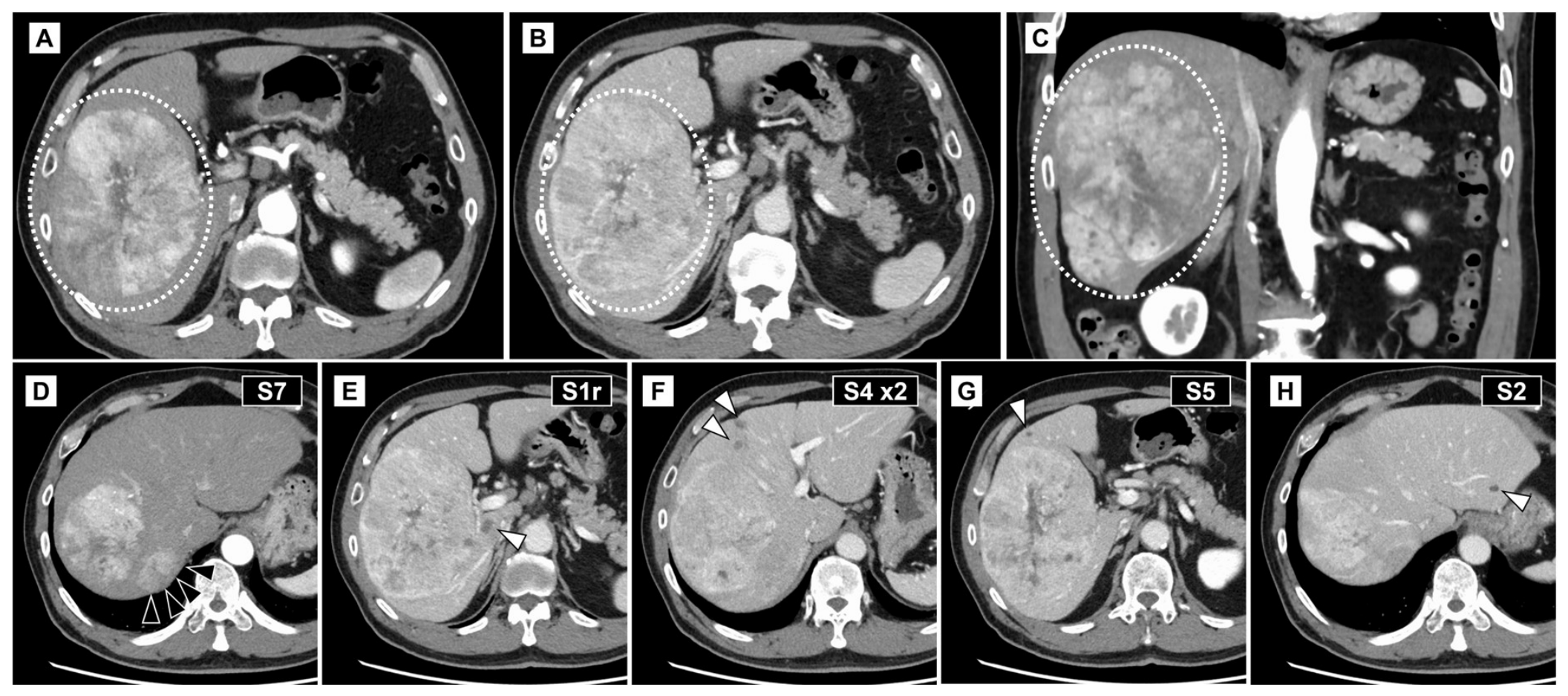
Figure 2.
Radiological response to atezolizumab plus bevacizumab. Contrast-enhanced CT images obtained before treatment (upper row) and after three cycles of atezolizumab plus bevacizumab (lower row). The dominant tumor in the right hepatic lobe decreased in size from 12 × 9 × 12 cm to 8.9 × 7.6 × 7.2 cm. The segment 7 and segment 4 satellite lesions became more hypoattenuating after treatment, suggesting treatment-related degenerative changes, whereas the segment 1r lesion showed no substantial interval change. White dotted lines delineate the dominant tumor, and white arrowheads indicate the satellite lesions.
Figure 2.
Radiological response to atezolizumab plus bevacizumab. Contrast-enhanced CT images obtained before treatment (upper row) and after three cycles of atezolizumab plus bevacizumab (lower row). The dominant tumor in the right hepatic lobe decreased in size from 12 × 9 × 12 cm to 8.9 × 7.6 × 7.2 cm. The segment 7 and segment 4 satellite lesions became more hypoattenuating after treatment, suggesting treatment-related degenerative changes, whereas the segment 1r lesion showed no substantial interval change. White dotted lines delineate the dominant tumor, and white arrowheads indicate the satellite lesions.
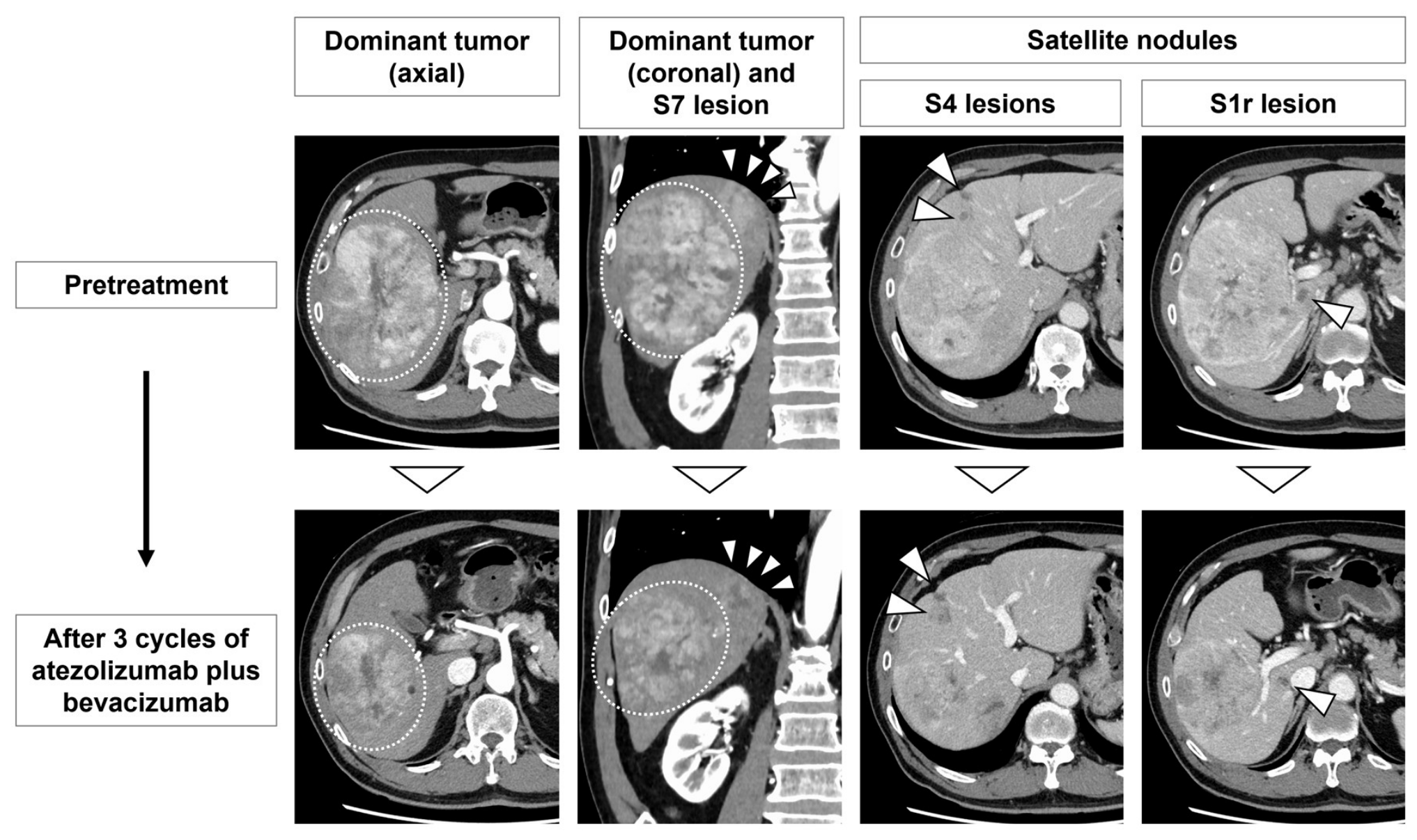
Figure 3.
Changes in serum tumor marker levels during atezolizumab plus bevacizumab therapy. The left y-axis shows PIVKA-II levels (mAU/mL; solid blue line), and the right y-axis shows AFP levels (ng/mL; dashed orange line). Filled arrowheads (▼) indicate cycles of atezolizumab (1200 mg/body) plus bevacizumab (15 mg/kg) combination therapy; open arrowheads (▽) indicate cycles of atezolizumab monotherapy. “CT” denotes the time point of restaging contrast-enhanced computed tomography. “Ope” indicates the day of surgical resection. AFP, alpha-fetoprotein; PIVKA-II, protein induced by vitamin K absence or antagonist-II.
Figure 3.
Changes in serum tumor marker levels during atezolizumab plus bevacizumab therapy. The left y-axis shows PIVKA-II levels (mAU/mL; solid blue line), and the right y-axis shows AFP levels (ng/mL; dashed orange line). Filled arrowheads (▼) indicate cycles of atezolizumab (1200 mg/body) plus bevacizumab (15 mg/kg) combination therapy; open arrowheads (▽) indicate cycles of atezolizumab monotherapy. “CT” denotes the time point of restaging contrast-enhanced computed tomography. “Ope” indicates the day of surgical resection. AFP, alpha-fetoprotein; PIVKA-II, protein induced by vitamin K absence or antagonist-II.
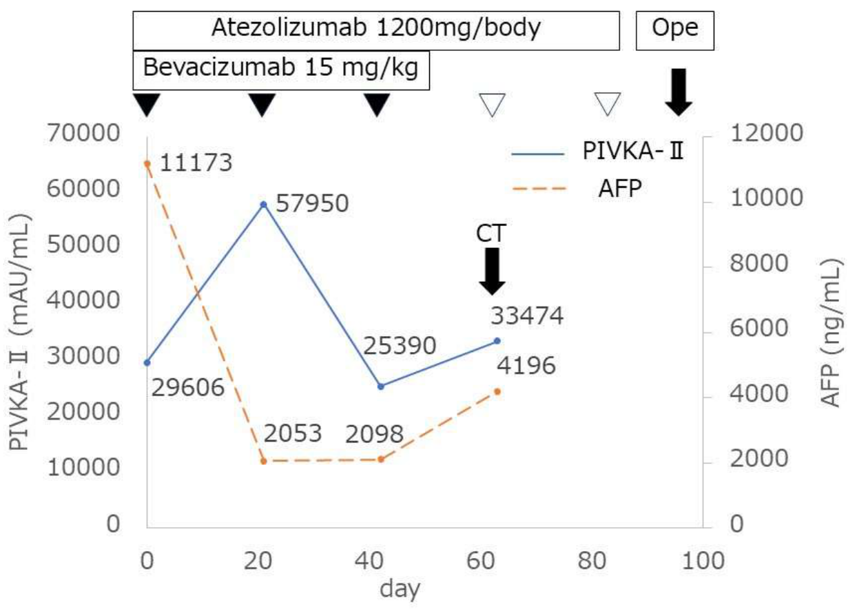
Figure 4.
Histopathological findings of the tumor. Hematoxylin and eosin staining of tumor tissue. (A) Low-power view (×40). (B) High-power view (×200). Approximately 10% of the tumor area showed necrotic changes, whereas the majority of the tumor remained viable.
Figure 4.
Histopathological findings of the tumor. Hematoxylin and eosin staining of tumor tissue. (A) Low-power view (×40). (B) High-power view (×200). Approximately 10% of the tumor area showed necrotic changes, whereas the majority of the tumor remained viable.
Disclaimer/Publisher’s Note: The statements, opinions and data contained in all publications are solely those of the individual author(s) and contributor(s) and not of MDPI and/or the editor(s). MDPI and/or the editor(s) disclaim responsibility for any injury to people or property resulting from any ideas, methods, instructions or products referred to in the content. |
© 2026 by the authors. Licensee MDPI, Basel, Switzerland. This article is an open access article distributed under the terms and conditions of the Creative Commons Attribution (CC BY) license (http://creativecommons.org/licenses/by/4.0/).
Copyright: This open access article is published under a Creative Commons CC BY 4.0 license, which permit the free download, distribution, and reuse, provided that the author and preprint are cited in any reuse.