

Submitted:
19 October 2026
Posted:
20 October 2026
You are already at the latest version
Abstract
Background/Objectives. To compare clinical and angiographic outcomes after staged bilateral ovarian and parauterine vein embolization using coils or vascular plugs in women with pelvic congestion syndrome. Methods. We retrospectively compared 97 coil-treated and 103 plug-treated premenopausal women with complete six-month follow-up after staged bilateral ovarian and parauterine embolization at two centres from February 2025 to March 2026. Device selection reflected anatomy, availability, and operator preference. Left-sided treatment preceded right-sided treatment by approximately one month. Before right-sided embolization, venography assessed filling of previously treated left parauterine veins. Pain was assessed with a continuous-line visual analogue scale (VAS), scored 0–10, at baseline and six months after bilateral completion. Comparisons were unadjusted. Results. Six-month VAS was 1.4 ± 0.7 with coils and 4.2 ± 1.1 with plugs (mean difference, −2.80 points; 95% confidence interval, −3.06 to −2.54; P < .001). Parauterine opacification occurred in 15/97 (15.5%) versus 77/103 (74.8%; risk difference, −59.3 percentage points; 95% confidence interval, −68.7 to −46.8; P < .001). Immediate technical success was reported in all patients. Combined angiographic intervals were 18.0 ± 2.5 versus 22.0 ± 3.7 minutes. Transient increased pelvic pain occurred in 33.0% versus 37.9%. No migration, ovarian vein rupture, or unplanned repeat embolization was reported through six months. Conclusions. Coil treatment was associated with lower six-month pain scores and less parauterine opacification during contralateral venography. Nonrandom device selection and unadjusted comparisons prevent conclusions about causal device superiority.
Keywords:
pelvic congestion syndrome
; pelvic venous disorders
; ovarian vein embolization
; coils
; vascular plugs
; visual analogue scale
Introduction
Pelvic congestion syndrome describes a symptomatic presentation within the broader spectrum of pelvic venous disorders. Chronic pelvic pain may coexist with dyspareunia, menstrual pain, and pelvic or extrapelvic varices, but these features overlap with other causes of pelvic pain. The Symptoms–Varices–Pathophysiology framework separates clinical presentation from venous anatomy and underlying mechanisms, an important distinction when comparing treatment strategies. [1] Current practice guidance and multidisciplinary consensus support integrating symptoms, imaging, and assessment for alternative causes when considering embolization. [2,3,4,5]
Diagnostic studies have evaluated venous diameter, pelvic varices, and dynamic Doppler findings, using different imaging routes and thresholds. [6,7] These differences complicate comparisons across treatment cohorts. A published diameter threshold can support clinical assessment, but it should not be treated as a universal definition of symptomatic disease or substituted for the criteria actually used in a retrospective series.
Early ovarian vein embolization studies reported technical feasibility and improvement in pelvic pain. [8,9] Longer follow-up and coil-specific series subsequently described sustained symptom responses, while also documenting recurrence and complications. [10,11,12] Later investigations assessed associated symptoms, patient satisfaction, and outcomes with different embolic agents or combinations of embolization and treatment for venous obstruction. [13,14,15] Randomized evidence supports studying pelvic vein occlusion as a treatment for selected women with chronic pelvic pain, although this does not resolve the comparative effects of individual devices. [16]
In a randomized comparison of fibered platinum coils and vascular plugs, Guirola and colleagues found no detected difference in clinical outcomes at one year, with procedural advantages for plugs. [17] The treated anatomy and the extent of embolization may also influence outcomes. Studies of distal sclerotherapy and pelvic venous plexus embolization suggest that treatment strategy deserves evaluation separately from the device selected. [18,19] These issues remain relevant to efforts to standardize pelvic venous research. [20]
We compared coils with vascular plugs in a two-centre retrospective cohort undergoing staged bilateral ovarian and parauterine vein embolization without sclerosant. The principal clinical outcome was pelvic pain six months after completion of bilateral treatment. A second objective was to describe contrast opacification of the previously treated left parauterine veins during right ovarian venography before the planned second embolization. Procedural measures and adverse events were also examined.
Materials and Methods
Design and Setting
We retrospectively reviewed records from 200 patients with completed six-month follow-up after bilateral treatment at Kilis Prof. Dr. Alaeddin Yavaşca State Hospital and Beyhekim Research and Training Hospital. Treatment began in February 2025, and the last planned bilateral treatment was completed in March 2026. The follow-up cutoff was 19 September 2026. The manuscript follows the STROBE framework for observational reporting. [21]
All otherwise eligible patients who completed six-month follow-up after the second procedure were included. Patients with incomplete follow-up were excluded from the analysis. The number of otherwise eligible patients excluded for incomplete follow-up was unavailable.
Patients were classified by the device actually received: coils (n = 97) or vascular plugs (n = 103). Device choice reflected availability, surgeon preference, and anatomical considerations. Coils were favored in tortuous veins with multiple tributaries on operator assessment. Two surgeons performed the procedures, and both used both devices. Each patient received the same device type on both sides.
The Istanbul Medipol University Non-Interventional Clinical Research Ethics Committee approved the study on 10 September 2026 (decision no. 1398). Written, signed research consent was obtained from all patients at both participating centres.
Patients and Diagnostic Assessment
The cohort comprised premenopausal women aged 18–45 years with pelvic pain lasting at least six months and a clinical diagnosis of pelvic congestion syndrome supported by abdominopelvic Doppler ultrasonography and CT venography. Obstetrics and Gynecology assessment excluded alternative gynecological causes, including pelvic inflammatory disease and endometriosis. Pregnancy, breastfeeding, menopause, a history of deep venous thrombosis, known iodinated contrast allergy, and renal impairment, defined as an estimated glomerular filtration rate (eGFR) <60 mL/min, were exclusion criteria. Failure of conservative treatment was not required.
The diagnostic criterion on transabdominal Doppler ultrasonography was an ovarian vein diameter >5 mm with reversed caudal flow. [3,5,6] CT venography criteria comprised an ovarian vein diameter >8 mm and at least four ipsilateral tortuous parauterine veins, with at least one measuring >4 mm. [3,7]
All patients underwent planned bilateral treatment. No patient had previously undergone pelvic vein embolization. CT venography showed no reported left renal vein or left common iliac vein compression.
Embolization Technique
Bilateral treatment was planned in two sessions, with the left side treated first in every patient and the right side treated approximately one month later. Local anesthesia and intravenous midazolam 1 mg were used. All venous access was ultrasound guided. The right femoral vein was used for left-sided treatment. Right-sided treatment used the right basilic vein in 198 patients and the right jugular vein in two patients because the basilic vein was narrow; one jugular access occurred in each device group.
The ovarian veins and their parauterine tributaries were treated. Medtronic Concerto coils were used in the coil group and INVAMED MultiBEAM embolization plugs in the plug group, including the parauterine targets. Devices were deployed at comparable anatomical levels according to operator assessment. No iliac vein embolization, sclerosant, or other injected embolic agent was used. Device oversizing was generally 20–40% according to the treating operator, rather than a fixed study protocol.
At completion, contrast was injected from a catheter positioned above, or proximal to, the embolized segment. Immediate technical success was defined as absence of contrast passage beyond that segment toward the distal pelvic veins. The two planned sessions were considered separately for technical assessment, while clinical outcomes were summarized per patient. Patients were discharged on the day of treatment and prescribed ibuprofen 200 mg twice daily for seven days after each session.
Venography at the Second Session
Before embolization of the right side, contrast was injected through the contralateral right ovarian vein to assess filling of the previously treated left parauterine veins. The reported protocol used manual injection of 2 mL total injectate, comprising 1 mL contrast agent and 1 mL saline, with observations at rest and during Valsalva. The same protocol was used in both groups and centres.
The treating surgeon assessed the images during the procedure. The investigator-defined endpoint was any visible contrast opacification of previously treated left parauterine veins; no contrast-retention duration threshold was applied. The presence or absence of visible filling of the previously treated left ovarian vein was also reported. No independent blinded adjudication was reported. This endpoint describes venous filling from a contralateral injection and was not classified as proven recanalization, passage through the implanted device, or clinical failure.
Clinical Outcomes and Procedural Measures
Pelvic pain was assessed using a continuous-line visual analogue scale and recorded on a 0–10 metric. [22] Baseline assessment preceded the first procedure. The only postoperative VAS measurement available for this analysis was obtained six months after the second procedure, when bilateral treatment had been completed. All 200 included patients had this assessment by the cohort selection rule. Early clinical review took place within approximately ten days; twelve-month follow-up was ongoing and was not included in the present analysis.
The principal clinical outcome of this retrospective analysis was six-month VAS. Additional outcomes were second-session parauterine opacification, immediate technical success, procedural and fluoroscopy intervals, recorded adverse events, and unplanned repeat embolization during six months after the second procedure. No pain-responder threshold was assigned from group means.
For each session, the recorded procedural interval began with the first angiographic acquisition and ended with the final contrast recording. The two intervals were summed for each patient before calculating group means and standard deviations. Machine-recorded fluoroscopy times were similarly summed over both sessions. This angiographic interval does not represent total room or venous-access time. Radiation-dose measurements were unavailable. Contrast-agent volumes before 1:1 dilution were available only as pooled side-specific means.
Early increased pelvic pain was counted after either session, with each patient counted once. Autonomic events were summarized by the reported number of affected patients. Migration, ovarian vein rupture, access-site complications, clinical venous thrombotic complications, fever, and unplanned repeat embolization were recorded as reported outcomes. The planned contralateral session was not considered repeat treatment for failure.
Statistical Analysis
The patient was the unit of analysis. Continuous variables are presented as mean ± standard deviation and categorical variables as counts and percentages. Analyses used investigator-supplied aggregate summaries. For six-month VAS and the reported procedural and fluoroscopy intervals, between-group mean differences and 95% confidence intervals were calculated using Welch independent-samples methods. Binary comparisons used two-sided Fisher exact tests, with absolute risk differences and Newcombe confidence intervals based on Wilson score limits. Differences are expressed as coils minus plugs. All comparisons were exploratory, without adjustment for multiple testing. Baseline characteristics were described without significance testing, and outcomes reported as absent in both groups or present in every patient were not tested.
The VAS comparison used investigator-confirmed full-cohort means, standard deviations, and group sizes. Individual paired scores were not extracted for this aggregate analysis, preventing estimation of the standard deviation of change, paired significance tests, responder proportions, or adjusted pain models. No missing values were imputed. Age, baseline pain, centre, and anatomical treatment-selection factors could not be jointly adjusted using the available marginal summaries. Calculations from rounded summaries provide approximate inferential estimates.
Calculations were performed using Python 3.12.14 with custom scripts. The aggregate inputs and reproducible analysis code are provided in Supplementary File S1.
Use of Generative Artificial Intelligence
OpenAI Codex (OpenAI; accessed October 2026) assisted with literature organization, manuscript drafting, and development of code for aggregate statistical calculations and figure preparation. Statistical results were computed from investigator-reported group summaries and event counts using the methods described above. No individual patient observations or clinical images were generated.
Results
Patient Characteristics
The cohort with complete six-month follow-up included 97 coil-treated and 103 plug-treated women (Figure 1). Kilis contributed 75 coil and 85 plug patients, and Beyhekim contributed 22 coil and 18 plug patients. Mean age was 34.6 ± 2.4 years in the coil group and 38.2 ± 2.1 years in the plug group. Overall parity was 3.7 ± 2.1; group-specific parity summaries were unavailable. All patients reported chronic pelvic pain, dyspareunia, and dysmenorrhea at baseline. Vulvar varices were reported in 15% overall, without group-specific counts (Table 1).
Procedural and Angiographic Outcomes
All patients completed the two planned sessions. Immediate technical success was reported at both sessions in all 97 coil and 103 plug patients. The combined angiographic interval was shorter with coils (18.0 ± 2.5 versus 22.0 ± 3.7 minutes; mean difference, −4.00 minutes; 95% CI, −4.88 to −3.12; P < .001). Combined fluoroscopy time was 7.4 ± 1.2 versus 8.9 ± 2.1 minutes (mean difference, −1.50 minutes; 95% CI, −1.97 to −1.03; P < .001). Across the pooled cohort, reported mean contrast-agent volume before dilution was 17.5 mL for left-sided treatment and 34 mL for right-sided treatment; device-group comparisons were unavailable (Table 2).
During right ovarian venography before the second embolization, previously treated left parauterine veins opacified in 15/97 coil patients (15.5%) and 77/103 plug patients (74.8%). The absolute difference was −59.3 percentage points (95% CI, −68.7 to −46.8; P < .001; Figure 2). The previously treated left ovarian vein did not opacify in either group. These observations describe the contralateral-injection finding and do not establish its route or mechanism.
Pain Outcomes
Baseline VAS was 7.2 ± 1.6 in the coil group and 7.4 ± 0.9 in the plug group. At six months after the second procedure, VAS was 1.4 ± 0.7 and 4.2 ± 1.1, respectively (Table 3; Figure 3). The unadjusted mean difference, coils minus plugs, was −2.80 points (95% CI, −3.06 to −2.54; Welch P < .001). Both group means were lower at follow-up than at baseline. Individual change-score variability and pain-responder proportions could not be determined from the available summaries.
Safety and Repeat Treatment
Transient autonomic symptoms, described by the treating team as vagal reactions with tachypnea and palpitations, occurred in 12/97 coil patients (12.4%) and 19/103 plug patients (18.4%; P = .248). Symptoms resolved within a few minutes of oxygen supplementation, without additional treatment or prolonged hospitalization. Hemodynamic measurements sufficient to characterize these episodes further were not available.
Temporary worsening of pelvic pain during the first week after either procedure occurred in 32/97 coil patients (33.0%) and 39/103 plug patients (37.9%; P = .555). Symptoms diminished over approximately ten days after the procedures, for which the seven-day ibuprofen regimen had been prescribed. No additional pain medication, unplanned hospital visit, or admission was reported for these episodes.
No device migration, ovarian vein rupture, access-site bleeding or hematoma, clinical venous thrombotic complication, or fever was reported in either group. No patient underwent unplanned repeat embolization within six months after completion of bilateral treatment. The overlap between autonomic symptoms and early pain was not available, so a composite proportion of patients with any adverse event was not calculated (Table 3).
Discussion
In this retrospective cohort of patients with complete six-month follow-up, coil treatment was associated with lower six-month pain scores and substantially less opacification of previously treated parauterine veins during contralateral ovarian venography than plug treatment. The unadjusted six-month VAS difference was −2.80 points. Coils were also associated with shorter recorded angiographic and fluoroscopy intervals, while immediate technical success was reported in both groups. These findings describe associations in patients whose device choice depended partly on anatomy; they do not establish that the device itself caused the clinical difference.
The results should be considered alongside the randomized comparison by Guirola and colleagues, which found no detected difference in one-year clinical outcomes between fibered coils and vascular plugs. [17] That trial does not establish clinical equivalence, and its procedural advantages for plugs contrast with the shorter coil intervals reported here. The devices differed from those used in our cohort, as did treated venous territories and treatment staging. Our duration measure excluded access and other nonangiographic steps. Consequently, the procedural results cannot be interpreted as a general time advantage for coils across techniques or centres.
Treatment of the distal venous reservoir may be clinically relevant, but evidence concerning treatment extent must be separated from evidence concerning device composition. Chen and colleagues evaluated proximal coil occlusion followed by distal sclerotherapy, whereas Karakaya and Ariturk compared pelvic plexus embolization with ovarian vein treatment alone. [18,19] Neither comparison isolates a coil-versus-plug effect. A prospective comparative series of plugs alone or plugs with polidocanol further illustrates how adjunct therapy can distinguish otherwise similar embolization strategies. [23] No injected embolic adjunct was used in the present cohort, although exact distal coverage was not quantified.
The venographic finding requires particular care in interpretation. Contrast introduced into one ovarian vein can cross the pelvis and opacify the contralateral periuterine plexus through communicating channels. [24] Thus, visualization of previously treated left parauterine veins during right-sided injection may reflect persistent collateral access to a venous reservoir. It does not by itself demonstrate recanalization of the treated ovarian vein or failure of a particular implant. The absence of visible left ovarian vein filling also does not establish complete occlusion under every flow condition. The study did not determine the pathway responsible for each positive image.
The any-opacification endpoint classified any visible filling as positive but lacked a validated clinical threshold. Manual injection, catheter position, Valsalva performance, and observation duration could influence the result. Although the reported injectate protocol was the same in both groups, injection pressure and rate were not quantified, and assessment by the treating surgeon could introduce observer bias. The association between this finding and individual pain outcomes could not be tested using separate aggregate summaries. Independent image review and linkage to patient-level outcomes would be needed before using it as a surrogate for clinical benefit.
The clinical interpretation is also limited by case mix. The coil group was younger, and coils were favored in tortuous veins with multiple tributaries. These anatomical features might affect both device choice and residual collateral filling, but their distribution was not systematically recorded. Comparable intended deployment levels therefore do not demonstrate equivalent anatomy or treatment extent. Both surgeons used both devices, which avoids complete separation of device and operator, but does not remove operator, centre, or treatment-selection confounding.
Transient early increased pelvic pain is compatible with a recognized postembolization inflammatory response. Aseptic inflammation of the treated venous wall, sometimes described as inflammatory phlebitis, is a plausible mechanism discussed in the embolization literature. [25] Phlebitis was not independently confirmed in this cohort, and the recorded pain episodes should not be treated as a formally adjudicated postembolization syndrome. The short course without additional treatment or hospital attendance is reassuring for these observed episodes. Routine ibuprofen use provides clinical context, but the study had no untreated comparison and cannot establish preventive efficacy of the prescribed regimen.
The absence of observed migration or rupture should be interpreted in relation to sample size and six-month follow-up. It does not exclude uncommon events or establish long-term device safety. Likewise, the lack of repeat embolization does not mean that every patient achieved satisfactory symptom relief. Earlier series evaluated pain, associated symptoms, recurrence, and longer follow-up, emphasizing that technical occlusion and clinical benefit are related but distinct outcomes. [10,12,15] The present six-month analysis cannot answer questions about twelve-month durability, reproductive outcomes, or changes in dyspareunia and dysmenorrhea.
Several additional limitations affect generalizability. Only patients who completed six-month follow-up were included. Exclusion of patients with incomplete follow-up may introduce selection bias, and the number and outcomes of those patients were unavailable. Diagnostic studies and current guidance emphasize interpreting venous diameter alongside clinical and other imaging findings. [2,4,5,6] Device burden, group-specific contrast volumes, and radiation dose were unavailable. Fluoroscopy duration alone cannot establish a reduction in radiation exposure. Pain inference was based on rounded aggregate summaries, and no adjusted or paired pain analysis was possible. Patient-level data would be needed to assess score distributions, individual responses, and confounding directly.
A subsequent comparison should document pretreatment anatomy and device selection, quantify distal treatment coverage, and assess venography independently. Verified individual pain measurements would permit adjustment for baseline pain, age, centre, and anatomical differences, as well as evaluation of clinically meaningful response. Standardized symptom and quality-of-life outcomes with longer follow-up would address the research priorities identified in recent pelvic venous consensus work. [20] The present observations provide a rationale for that investigation rather than sufficient evidence to recommend one device for all patients.
Conclusions
Among 200 women undergoing staged bilateral ovarian and parauterine vein embolization, coil treatment was associated with lower six-month VAS scores, less parauterine opacification on contralateral venography, and shorter recorded angiographic and fluoroscopy intervals. Both groups achieved reported immediate technical success. The retrospective selection process, anatomical confounding, and unadjusted analysis preclude a causal claim that coils provide superior clinical outcomes.
Funding
No funding was received for this study.
Institutional Review Board Statement
The study was approved by the Istanbul Medipol University Non-Interventional Clinical Research Ethics Committee, compliant with the Declaration of Helsinki, on 10 September 2026 (decision no. 1398).
Informed Consent Statement
Written informed consent for research participation was obtained from all patients at both participating centres.
Data Availability Statement
The underlying patient records are retained at the participating institutions and are available for verification. Requests for access may be directed to the corresponding author and are subject to institutional authorization and patient confidentiality safeguards.
Conflicts of Interest
The authors declare no conflicts of interest.
References
- Meissner, M.H.; Khilnani, N.M.; Labropoulos, N.; et al. The Symptoms-Varices-Pathophysiology classification of pelvic venous disorders: A report of the American Vein & Lymphatic Society International Working Group on Pelvic Venous Disorders. Phlebology 2021, 36(5), 342–360. [Google Scholar] [CrossRef] [PubMed]
- Kaufman, C.S.; Winokur, R.S.; Khilnani, N.M.; et al. The Society of Interventional Radiology Practice Guidance Document on Venous-Origin Chronic Pelvic Pain in Women. J. Vasc. Interv. Radiol. 2026, 37(6), 107954. [Google Scholar] [CrossRef] [PubMed]
- Antignani, P.L.; Lazarashvili, Z.; Monedero, J.L.; et al. Diagnosis and treatment of pelvic congestion syndrome: UIP consensus document. Int. Angiol. 2019, 38(4), 265–283. [Google Scholar] [CrossRef] [PubMed]
- Le Pennec, V.; Douane, F.; Brun, J.L.; et al. Endovascular management of pelvic congestion syndrome: An expert consensus statement from the French Society of Cardiovascular Imaging (SFICV), Interventional Radiology Federation (FRI), College of French Radiology Teachers (CERF), and French Society of Women’s Imaging (SIFEM). Diagn. Interv. Imaging 2025, 106(10), 356–366. [Google Scholar] [CrossRef] [PubMed]
- De Maeseneer, M.G.; Kakkos, S.K.; Aherne, T.; et al. Editor’s Choice — European Society for Vascular Surgery (ESVS) 2022 Clinical Practice Guidelines on the Management of Chronic Venous Disease of the Lower Limbs. Eur. J. Vasc. Endovasc. Surg. 2022, 63(2), 184–267. [Google Scholar] [CrossRef] [PubMed]
- Park, S.J.; Lim, J.W.; Ko, Y.T.; et al. Diagnosis of pelvic congestion syndrome using transabdominal and transvaginal sonography. AJR Am. J. Roentgenol. 2004, 182(3), 683–688. [Google Scholar] [CrossRef] [PubMed]
- Coakley, F.V.; Varghese, S.L.; Hricak, H. CT and MRI of pelvic varices in women. J. Comput Assist Tomogr. 1999, 23(3), 429–434. [Google Scholar] [CrossRef] [PubMed]
- Maleux, G.; Stockx, L.; Wilms, G.; Marchal, G. Ovarian vein embolization for the treatment of pelvic congestion syndrome: long-term technical and clinical results. J. Vasc. Interv. Radiol. 2000, 11(7), 859–864. [Google Scholar] [CrossRef] [PubMed]
- Venbrux, A.C.; Chang, A.H.; Kim, H.S.; et al. Pelvic congestion syndrome (pelvic venous incompetence): impact of ovarian and internal iliac vein embolotherapy on menstrual cycle and chronic pelvic pain. J. Vasc. Interv. Radiol. 2002, 13 2 Pt 1, 171–178. [Google Scholar] [CrossRef] [PubMed]
- Kim, H.S.; Malhotra, A.D.; Rowe, P.C.; Lee, J.M.; Venbrux, A.C. Embolotherapy for pelvic congestion syndrome: long-term results. J. Vasc. Interv. Radiol. 2006, 17 2 Pt 1, 289–297. [Google Scholar] [CrossRef] [PubMed]
- Kwon, S.H.; Oh, J.H.; Ko, K.R.; Park, H.C.; Huh, J.Y. Transcatheter ovarian vein embolization using coils for the treatment of pelvic congestion syndrome. Cardiovasc Interv. Radiol. 2007, 30(4), 655–661. [Google Scholar] [CrossRef] [PubMed]
- Laborda, A.; Medrano, J.; de Blas, I.; Urtiaga, I.; Carnevale, F.C.; de Gregorio, M.A. Endovascular treatment of pelvic congestion syndrome: visual analog scale (VAS) long-term follow-up clinical evaluation in 202 patients. Cardiovasc Interv. Radiol. 2013, 36(4), 1006–1014. [Google Scholar] [CrossRef] [PubMed]
- Nasser, F.; Cavalcante, R.N.; Affonso, B.B.; Messina, M.L.; Carnevale, F.C.; de Gregorio, M.A. Safety, efficacy, and prognostic factors in endovascular treatment of pelvic congestion syndrome. Int. J. Gynaecol. Obstet. 2014, 125(1), 65–68. [Google Scholar] [CrossRef] [PubMed]
- Senechal, Q.; Echegut, P.; Bravetti, M.; et al. Endovascular Treatment of Pelvic Congestion Syndrome: Visual Analog Scale Follow-Up. Front Cardiovasc Med. 2021, 8, 751178. [Google Scholar] [CrossRef] [PubMed]
- De Gregorio, M.A.; Guirola, J.A.; Alvarez-Arranz, E.; Sánchez-Ballestin, M.; Urbano, J.; Sierre, S. Pelvic Venous Disorders in Women due to Pelvic Varices: Treatment by Embolization: Experience in 520 Patients. J. Vasc. Interv. Radiol. 2020, 31(10), 1560–1569. [Google Scholar] [CrossRef] [PubMed]
- Hansrani, V.; Riding, D.; Seif, M.W.; Caress, A.L.; Payne, K.; Ghosh, J.; et al. Transvenous occlusion of incompetent pelvic veins to treat chronic pelvic pain in women: A randomised controlled trial. BJOG 2023, 130(11), 1362–1369. [Google Scholar] [CrossRef] [PubMed]
- Guirola, J.A.; Sánchez-Ballestin, M.; Sierre, S.; Lahuerta, C.; Mayoral, V.; De Gregorio, M.A. A Randomized Trial of Endovascular Embolization Treatment in Pelvic Congestion Syndrome: Fibered Platinum Coils versus Vascular Plugs with 1-Year Clinical Outcomes. J. Vasc. Interv. Radiol. 2018, 29(1), 45–53. [Google Scholar] [CrossRef] [PubMed]
- Chen, H.; Wu, Z.; Wu, Z.; Huang, Q.; Ye, M.; Sun, H.; Zhang, H.; Guo, P.; Zhuang, H. Proximal coil occlusion preceding distal sclerotherapy in patients with pelvic congestion syndrome: A multicenter, retrospective study. J. Vasc. Surg. Venous Lymphat Disord. 2023, 11(1), 149–155. [Google Scholar] [CrossRef] [PubMed]
- Karakaya, H.C.; Ariturk, C. Comparison of ovarian vein embolization alone with pelvic venous plexus embolization in the treatment of pelvic venous disorders at 12-month follow-up. J. Vasc. Surg. Venous Lymphat Disord. 2026, 14(4), 102502. [Google Scholar] [CrossRef] [PubMed]
- Khilnani, N.M.; Winokur, R.S.; Marvel, R.P.; Meissner, M.H. Pelvic Venous Disorders in Women: 5-Year Update from a Society of Interventional Radiology Foundation-Sponsored Research Consensus Panel. J. Vasc. Interv. Radiol. 2025, 36(1), 9–16. [Google Scholar] [CrossRef] [PubMed]
- von Elm, E.; Altman, D.G.; Egger, M.; Pocock, S.J.; Gøtzsche, P.C.; Vandenbroucke, J.P. STROBE Initiative. The Strengthening the Reporting of Observational Studies in Epidemiology (STROBE) statement: guidelines for reporting observational studies. PLoS Med. 2007, 4(10), e296. [Google Scholar] [CrossRef] [PubMed]
- Huskisson, E.C. Measurement of pain. Lancet 1974, 2(7889), 1127–1131. [Google Scholar] [CrossRef] [PubMed]
- De Gregorio, M.A.; Yamamoto-Ramos, M.; Fredes, A.; et al. A Comparative Study of a Small Series of Patients (50 Patients) with Pelvic Varicose Veins Treated with Plugs Alone or Plugs and Polidocanol. J. Clin. Med. 2023, 12(16), 5408. [Google Scholar] [CrossRef] [PubMed]
- Siqueira, F.M.; Monsignore, L.M.; Rosa-E-Silva, J.C.; et al. Evaluation of embolization for periuterine varices involving chronic pelvic pain secondary to pelvic congestion syndrome. Clinics (Sao Paulo) 2016, 71(12), 703–708. [Google Scholar] [CrossRef] [PubMed]
- Gavrilov, S.G.; Krasavin, G.V.; Mishakina, N.Y.; Efremova, O.I.; Zolotukhin, I.A. The Effect of Venoactive Drug Therapy on the Development and Severity of Post-Embolization Syndrome in Endovascular Interventions on the Gonadal Veins. J. Pers. Med. 2021, 11(6), 521. [Google Scholar] [CrossRef] [PubMed]
Figure 1.
Study cohort and treatment sequence The retrospective cohort included all 200 otherwise eligible women who completed six-month follow-up, grouped by the device received. Patients with incomplete follow-up were excluded; their number was unavailable. Left ovarian and parauterine embolization preceded the planned right-sided session by approximately one month. Six-month assessment was measured from completion of the second procedure. Follow-up completion among the included patients is a selection criterion and does not represent retention among all treated patients.
Figure 1.
Study cohort and treatment sequence The retrospective cohort included all 200 otherwise eligible women who completed six-month follow-up, grouped by the device received. Patients with incomplete follow-up were excluded; their number was unavailable. Left ovarian and parauterine embolization preceded the planned right-sided session by approximately one month. Six-month assessment was measured from completion of the second procedure. Follow-up completion among the included patients is a selection criterion and does not represent retention among all treated patients.
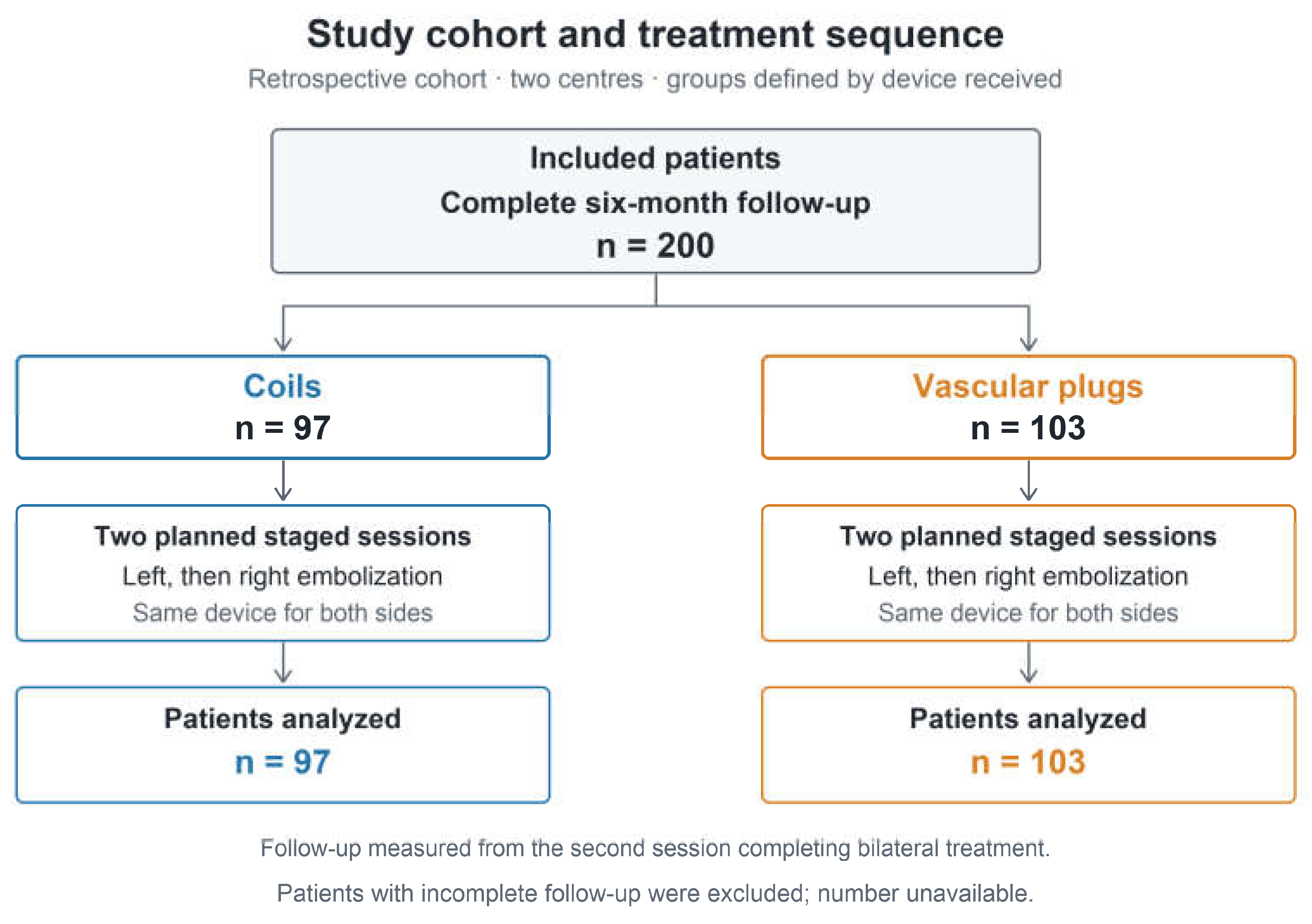
Figure 2.
Parauterine opacification at the second session. Points show the proportion with visible filling of previously treated left parauterine veins during right ovarian venography, before right-sided embolization. Error bars are Wilson 95% confidence intervals. Counts were 15/97 for coils and 77/103 for plugs. The unadjusted risk difference, coils minus plugs, was −59.3 percentage points (Newcombe 95% CI, −68.7 to −46.8; Fisher exact P < .001). The endpoint describes opacification and does not establish device recanalization or clinical failure.
Figure 2.
Parauterine opacification at the second session. Points show the proportion with visible filling of previously treated left parauterine veins during right ovarian venography, before right-sided embolization. Error bars are Wilson 95% confidence intervals. Counts were 15/97 for coils and 77/103 for plugs. The unadjusted risk difference, coils minus plugs, was −59.3 percentage points (Newcombe 95% CI, −68.7 to −46.8; Fisher exact P < .001). The endpoint describes opacification and does not establish device recanalization or clinical failure.
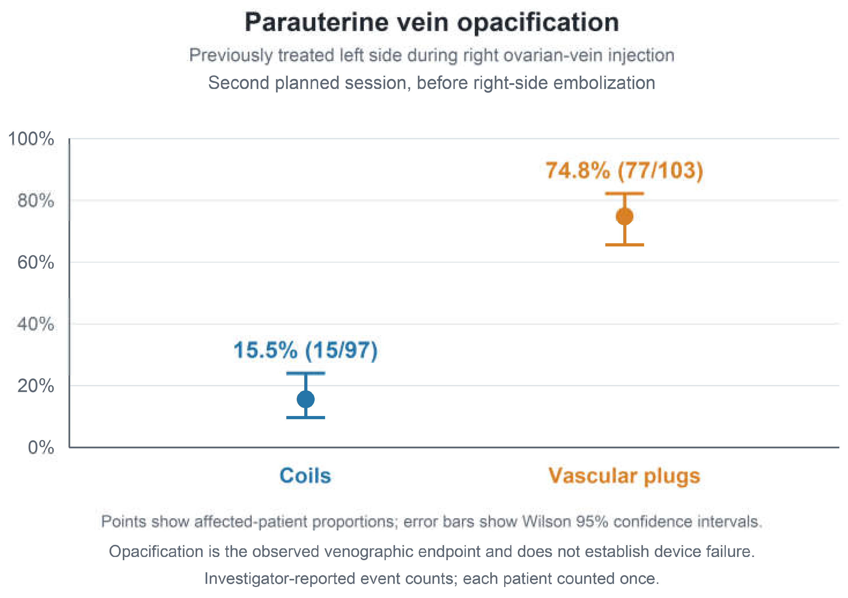
Figure 3.
Pain scores before treatment and at six months. Points show full-cohort VAS means, and error bars show standard deviations, for 97 coil and 103 plug patients. Baseline preceded the first procedure; follow-up was six months after the second procedure. The VAS was a continuous-line instrument scored from 0 to 10. The six-month unadjusted mean difference, coils minus plugs, was −2.80 points (Welch 95% CI, −3.06 to −2.54; P < .001). Error bars show dispersion within groups, not uncertainty in the between-group difference.
Figure 3.
Pain scores before treatment and at six months. Points show full-cohort VAS means, and error bars show standard deviations, for 97 coil and 103 plug patients. Baseline preceded the first procedure; follow-up was six months after the second procedure. The VAS was a continuous-line instrument scored from 0 to 10. The six-month unadjusted mean difference, coils minus plugs, was −2.80 points (Welch 95% CI, −3.06 to −2.54; P < .001). Error bars show dispersion within groups, not uncertainty in the between-group difference.
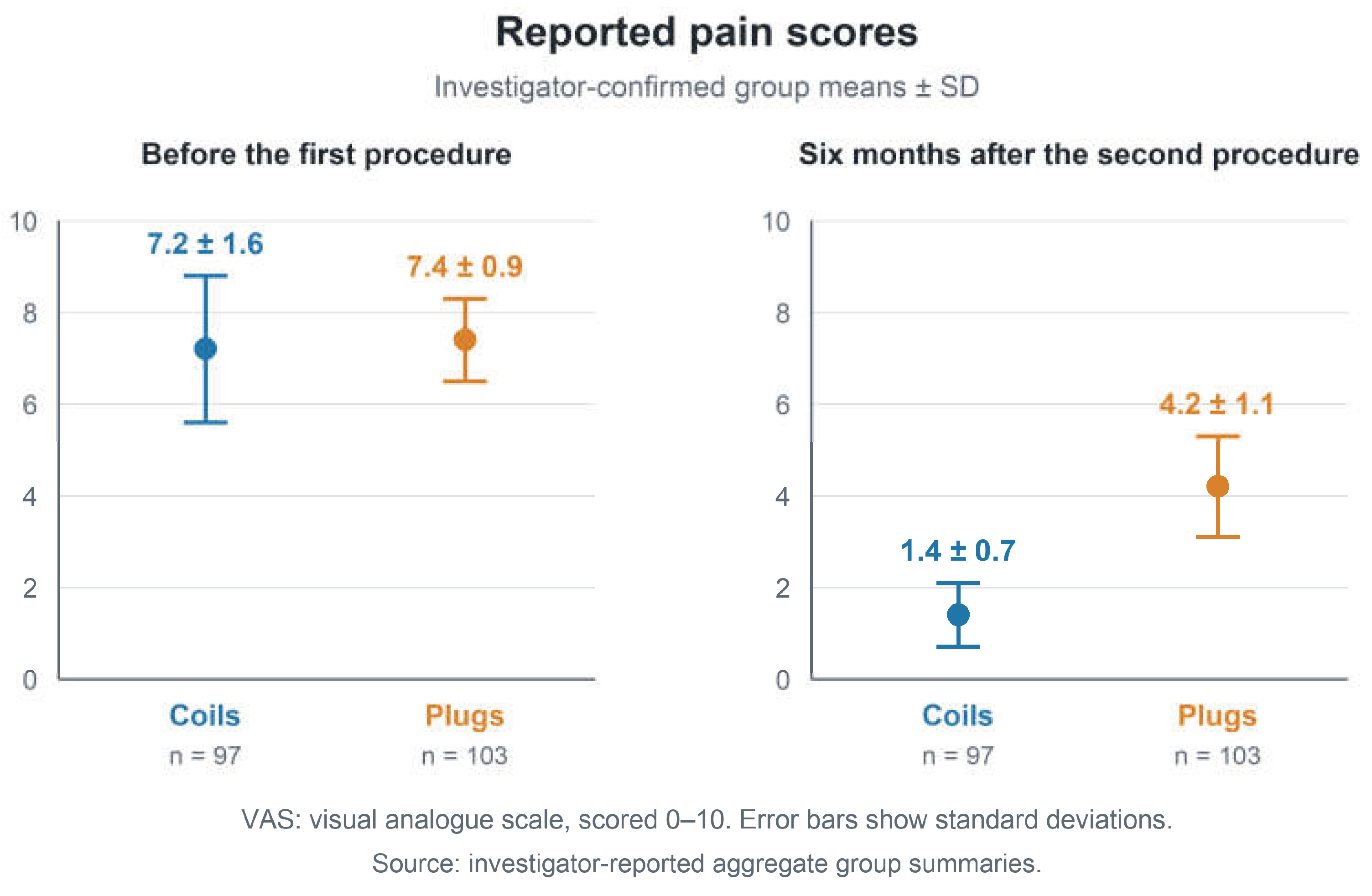

Table 1.
Baseline characteristics.
| Characteristic | Coils (n = 97) |
Plugs (n = 103) |
|---|---|---|
| Patients | 97 | 103 |
| Age, years | 34.6 ± 2.4 | 38.2 ± 2.1 |
| Kilis centre | 75 (77.3%) | 85 (82.5%) |
| Beyhekim centre | 22 (22.7%) | 18 (17.5%) |
| Premenopausal | 97 (100%) | 103 (100%) |
| Planned bilateral treatment | 97 (100%) | 103 (100%) |
| Chronic pelvic pain for at least 6 months | 97 (100%) | 103 (100%) |
| Dyspareunia | 97 (100%) | 103 (100%) |
| Dysmenorrhea | 97 (100%) | 103 (100%) |
| Prior pelvic vein embolization | 0 | 0 |
| Baseline VAS, 0–10 | 7.2 ± 1.6 | 7.4 ± 0.9 |
Values are mean ± SD or n (%). Percentages use the device-group denominator. Kilis: Kilis Prof. Dr. Alaeddin Yavaşca State Hospital; Beyhekim: Beyhekim Research and Training Hospital. Overall parity was 3.7 ± 2.1 and vulvar varices were reported in 15% overall; group-specific values were unavailable. BMI was not recorded. VAS: continuous-line visual analogue scale, scored 0–10. Baseline preceded the first procedure. Baseline significance tests were not performed.
Table 2.
Procedures and second session venography.
| Measure | Coils (n = 97) |
Plugs (n = 103) |
Difference (95% CI) and P |
|---|---|---|---|
| Device | Medtronic Concerto |
INVAMED MultiBEAM |
— |
| Right femoral access for the left session | 97 (100%) | 103 (100%) | — |
| Right basilic access for the right session | 96 (99.0%) | 102 (99.0%) | — |
| Right jugular access for the right session | 1 (1.0%) | 1 (1.0%) | — |
| Combined angiographic interval, min | 18.0 ± 2.5 | 22.0 ± 3.7 | −4.00 (−4.88 to −3.12) P < .001 |
| Combined fluoroscopy time, min | 7.4 ± 1.2 | 8.9 ± 2.1 | −1.50 (−1.97 to −1.03) P < .001 |
| Immediate technical success at both sessions | 97 (100%) | 103 (100%) | Not tested |
| Left parauterine opacification at second-session venography | 15 (15.5%) | 77 (74.8%) | −59.3 pp (−68.7 to −46.8) P < .001 |
| Visible filling of previously treated left ovarian vein | 0 | 0 | Not tested |
Values are mean ± SD or n (%). Differences are coils minus plugs: mean differences in minutes for timing outcomes and absolute risk difference in percentage points (pp) for parauterine opacification. Calculations use reported aggregate data; Welch tests and intervals apply to timing outcomes, Fisher exact testing and a Newcombe interval to opacification. Times are sums of both sessions per patient. The procedural interval runs from first angiographic acquisition to final contrast recording. It excludes other components of total procedure time. Venography preceded right-sided embolization. Any visible filling of previously treated left parauterine veins was positive after contralateral injection of 2 mL of 1:1 diluted contrast, assessed at rest and during Valsalva. This is not a validated device-failure endpoint. Pooled mean contrast-agent volume before dilution was 17.5 mL for the left and 34 mL for the right session. Group-specific summaries and radiation-dose measurements were unavailable.
Table 3.
Pain outcomes and safety.
| Outcome | Coils (n = 97) |
Plugs (n = 103) |
Difference (95% CI) and P |
|---|---|---|---|
| Six-month VAS, 0–10 | 1.4 ± 0.7 | 4.2 ± 1.1 | −2.80 points (−3.06 to −2.54) P < .001 |
| Six-month VAS assessment reported | 97 (100%) | 103 (100%) | — |
| Transient autonomic symptoms | 12 (12.4%) | 19 (18.4%) | −6.1 pp (−16.1 to 4.1) P = .248 |
| Transient early worsening of pelvic pain | 32 (33.0%) | 39 (37.9%) | −4.9 pp (−17.8 to 8.3) P = .555 |
| Device migration | 0 | 0 | Not tested |
| Ovarian vein rupture | 0 | 0 | Not tested |
| Access-site bleeding or hematoma | 0 | 0 | Not tested |
| Clinical venous thrombotic complications | 0 | 0 | Not tested |
| Fever | 0 | 0 | Not tested |
| Unplanned repeat embolization within 6 months | 0 | 0 | Not tested |
Values are mean ± SD or n (%). Six-month follow-up is measured from the second procedure. The VAS comparison is an unadjusted mean difference, coils minus plugs, with a Welch 95% CI and two-sided P value calculated from the reported group summaries. No paired-change analysis or responder proportion is supplied. Binary differences are coils minus plugs in percentage points (pp), with Newcombe 95% CIs and two-sided Fisher exact P values. All comparisons are exploratory. Early pelvic-pain episodes followed either session, with each patient counted once. Patients were prescribed ibuprofen 200 mg twice daily for seven days after each session. Symptoms diminished over approximately ten days, without additional analgesia or unplanned hospital attendance. Autonomic episodes included reported tachypnea and palpitations and resolved with oxygen in a few minutes. Overlap between these episodes and early pain is unknown; an overall adverse-event rate was not calculated. The planned second session is not a repeat intervention for treatment failure.
Disclaimer/Publisher’s Note: The statements, opinions and data contained in all publications are solely those of the individual author(s) and contributor(s) and not of MDPI and/or the editor(s). MDPI and/or the editor(s) disclaim responsibility for any injury to people or property resulting from any ideas, methods, instructions or products referred to in the content. |
© 2026 by the authors. Licensee MDPI, Basel, Switzerland. This article is an open access article distributed under the terms and conditions of the Creative Commons Attribution (CC BY) license (http://creativecommons.org/licenses/by/4.0/).
Copyright: This open access article is published under a Creative Commons CC BY 4.0 license, which permit the free download, distribution, and reuse, provided that the author and preprint are cited in any reuse.