

Submitted:
19 October 2026
Posted:
20 October 2026
You are already at the latest version
Abstract
Background: Youngswick osteotomy is an Austin-derived distal first-metatarsal decompression osteotomy intended to preserve first metatarsophalangeal joint motion in hallux limitus and hallux rigidus. An additional dorsal parallel cut permits shortening; plantar translation depends on cut geometry. Objective: To examine how operative geometry, procedural variation and concomitant surgery affect interpretation of the clinical evidence for Youngswick osteotomy. Methods: This critical narrative review drew on structured searches of PubMed, Ovid MEDLINE and Embase, and the Knowledge Network, supplemented by reference checking. Reports of Youngswick osteotomy were assessed separately from studies of other decompression procedures. Differences between studies prevented quantitative synthesis.Results: Clinical cohorts report improvement in pain, function and dorsiflexion, but evidence is predominantly observational and procedures and outcome measures vary. Frequent concomitant cheilectomy limits isolation of the osteotomy’s independent effect. Plantar-pressure findings suggest possible transfer loading, and symptom improvement can coexist with radiographic progression. Available direct comparisons do not establish superiority. Conclusion: Youngswick osteotomy remains an option for patients who wish to retain motion and have sufficient joint function to benefit from decompression. Selection is guided by clinical assessment rather than validated criteria. The best cut geometry, its comparative benefit and its ability to delay arthrodesis remain uncertain.
Keywords:
hallux limitus
; hallux rigidus
; Youngswick osteotomy
; metatarsal decompression
; cheilectomy
; first metatarsophalangeal joint
; joint-preserving surgery
1. Introduction and Scope
Hallux limitus and hallux rigidus (HL/HR) describe a spectrum of painful restriction at the first metatarsophalangeal joint (first MTPJ). Patients may have dorsal impingement with useful mid-range motion, or more diffuse cartilage loss and pain throughout movement, including at the sesamoid articulation. That distinction matters when choosing an operation: radiographic grade alone does not capture pain distribution, remaining motion, joint alignment, activity or the patient’s preference for retaining movement [1,2,3].
Operative options include cheilectomy, first-metatarsal decompression, proximal phalangeal osteotomy, interposition or resurfacing procedures, and arthrodesis. Arthrodesis remains the most predictable operative option for pain relief in advanced hallux rigidus; joint-preserving procedures seek to retain function where sufficient motion and articular surface remain [1,2,3].
Youngswick’s modification of the Austin chevron removes a dorsal wedge of bone so that the capital fragment can move proximally and plantarly [4]. It is often combined with cheilectomy. Shortening may relieve joint compression, while plantar translation changes the sagittal position of the metatarsal head; together these changes may permit more dorsiflexion before dorsal impingement [2,4,5]. Whether these intended mechanical effects explain clinical improvement has not been established. Figure 1 illustrates wedge removal, reorientation of the capital fragment and screw fixation.
Cheilectomy has long-term support in grade I–II and selected grade III disease [1,3,6]. A 2024 systematic review reported a 7.4% overall revision rate, although heterogeneous study populations limit comparison [6]. The unresolved question is whether Youngswick osteotomy adds clinically meaningful benefit beyond cheilectomy or alternative decompression procedures in selected patients [2,3,7].
Lewis et al.’s 2026 systematic review evaluated decompressive first-metatarsal osteotomies as a group [8]. The present critical narrative review addresses a narrower interpretative question: what can be attributed specifically to Youngswick osteotomy when cut geometry, achieved correction and accompanying procedures vary? It examines procedural identity, the distinction between intended and measured mechanical effects, and the extent to which published outcomes support clinical selection.
2. Method
This critical narrative review used structured literature searches without a registered systematic-review protocol. Searches of PubMed, Ovid MEDLINE and Embase, and the Knowledge Network were recorded on 23 September 2026. We considered original technical, clinical, comparative, biomechanical and cadaveric reports of Youngswick osteotomy and clearly described modifications. Reviews and studies of other decompression procedures provided context and were assessed separately. The Knowledge Network query was “Youngswick AND (osteotom* OR modif*)”. Results were checked manually because some records mentioned Youngswick only in their references. The broader Ovid query was “youngswick AND (osteotom* OR modif* OR Austin OR Chevron OR Duke OR Kaplan OR “hallux rigidus” OR “hallux limitus” OR Sagittal OR “first metatars*”)”. The PubMed query was “youngswick AND (osteotom* OR modif* OR Austin OR Chevron OR Duke OR Kaplan OR “hallux rigidus” OR “hallux limitus” OR Sagittal)”. The PubMed collection is available at https://www.ncbi.nlm.nih.gov/sites/myncbi/14MUDQpGpOxA5c/collections/67767119/public/.
Studies were grouped by procedural identity and design, with attention to disease grade, accompanying surgery, follow-up and outcome measurement. No quantitative synthesis or formal risk-of-bias assessment was undertaken. Search records do not permit reconstruction of database-specific yields, screening counts, separate MEDLINE and Embase strategies, or date and language restrictions. Independent screening and the direction of citation checking were not documented. These limitations restrict reproducibility and preclude a claim of comprehensive retrieval. Supplementary Table S1 records the available search information and its limitations.
In this review, Youngswick denotes an Austin/chevron-derived distal first-metatarsal osteotomy with an additional parallel dorsal cut and removal of a dorsal wedge. Youngswick-type denotes a reported modification with altered geometry or additional technical changes. Decompression osteotomy is the broader category of shortening and/or plantarflexion procedures intended to improve joint mechanics. Distal oblique osteotomy is a distinct contextual comparator unless the original report explicitly identifies a Youngswick modification [2,4,9,10].
3. Procedure and Biomechanical Rationale
3.1. Historical Development
Youngswick described the procedure in 1982 for metatarsus primus elevatus associated with hallux limitus [4]. Removing the interval between parallel dorsal cuts allows proximal capital-fragment translation; the plantar limb influences plantar displacement. Duke and Kaplan described a related Austin modification for shortening and plantarflexion by angulation of the chevron cut in both the sagittal and transverse planes [9]. Gerbert et al. demonstrated that plantar-arm orientation affects head displacement [11]. The final position therefore depends on cut geometry as well as fixation position.
3.2. Mechanical Rationale
Shortening is intended to decompress the first MTPJ, whereas cheilectomy removes a dorsal impingement source. These are related but distinct treatment targets. Cut geometry describes the intended correction; postoperative radiographic measurements describe achieved shortening and translation; plantar-pressure and motion studies examine functional effects. None alone establishes the mechanism of pain relief or a reduction in in-vivo joint contact force.
3.2.1. Metatarsal Length
A long first metatarsal has been implicated in HL/HR, but the association is inconsistent. Nilsonne found an index-plus pattern more often among 49 feet with HR than among 497 feet without HR or hallux valgus [12]. A longer metatarsal could bring the phalanx against the dorsal head earlier in propulsion, providing a rationale for shortening [2,4,7]. Coughlin and Shurnas, however, found no consistent association, and Slullitel et al. reported similar improvement after Youngswick osteotomy across metatarsal-index groups [13,14]. A long first metatarsal is therefore not a prerequisite for Youngswick osteotomy, although length remains relevant when planning shortening. Planned shortening should account for the joint, pain pattern, metatarsal parabola, and risk of transfer loading [7,14]. Figure 2A provides a radiographic example of a long first metatarsal.
3.2.2. Metatarsal Elevation
The historical rationale for plantar translation partly rests on metatarsus primus elevatus (MPE) as a contributor to HL/HR [4,15,16]. Its causal role remains contested. Meyer et al. found elevation in symptomatic and control radiographs [15]; Horton et al. found similar mean elevation in mild-to-moderate disease and controls [16]. Coughlin and Shurnas found no association with MPE, first-ray hypermobility or a long first metatarsal [13]. Elevation may be associated with, or follow, joint dysfunction. Plantar translation has a plausible mechanical rationale, but radiographic elevation alone is not a validated indication or predictor of Youngswick outcome. Excessive translation may alter plantar loading [2,13,16]. The corresponding lateral radiograph illustrates metatarsus primus elevatus (Figure 2B).
3.3. Geometry and Loading
Wedge dimensions, osteotomy orientation and final fragment positioning together determine achieved shortening and plantar translation [11]. Planning should distinguish residual joint compression from persistent dorsal osteophyte impingement. Excessive shortening may redistribute load to the lesser metatarsals, while excessive plantar translation may increase focal loading beneath the first ray. Bryant et al.’s pressure findings are considered in Section 4.1 [5].
Feldman’s geometric analysis of Green–Watermann osteotomy illustrates how orientation and radiographic templating can be used to anticipate displacement and preserve the metatarsal–sesamoid articulation [17]. This is a contextual geometric comparison rather than direct Youngswick evidence.
Bernbach reported follow-up of hallux limitus surgery in 1988 [18]. Later descriptions identify potential stability disadvantages of Green–Watermann relative to Youngswick [19,20], although Dickerson et al. reported no delayed union or non-union among 40 procedures in 32 questionnaire respondents [20]. These observations do not establish comparative clinical failure rates. Because limb orientation can blur the distinction between named operations [2], studies should report the actual cuts, fixation and achieved correction.
A cadaveric study of a modified distal oblique osteotomy also demonstrated that three-dimensional orientation affects head lowering and preservation of the sesamoid articulation [21]. Its displacement ratios and angular findings should not be transferred directly to Youngswick osteotomy.
3.4. Technical Modifications
Radovic et al. described a modification intended to change sagittal position when shortening was undesirable [10]. This technical report gives no comparative outcome estimate. It also illustrates a problem in reading the literature: procedures called “Youngswick” may have materially different geometry.
3.5. Assessment and Operative Principles
Preoperative assessment should locate the pain within the arc of motion. Predominantly dorsal pain at terminal dorsiflexion suggests impingement; mid-range pain raises concern about more extensive articular disease. Record motion, dorsal osteophytes, first-ray mobility, alignment, transfer lesions and lesser-metatarsal overload. Discuss footwear, activity, expectations for preserving movement and the possibility of later fusion. Weight-bearing anteroposterior, lateral and oblique radiographs help assess osteophytes, joint-space loss, sesamoids, metatarsal length, sagittal position and associated deformity [1,2,3,13].
The defining osteotomy comprises a distal chevron and an additional parallel dorsal cut; exposure, cheilectomy and other adjuncts should be documented separately [2,4,10,11]. Figure 3, Figure 4 and Figure 5 illustrate assessment, exposure and bone preparation. The principal steps are:
- Medial or dorsomedial exposure of the first MTPJ
- Capsulotomy and direct assessment of articular cartilage, dorsal osteophytes, and synovitis
- Dorsal cheilectomy where indicated, preserving viable articular surface and avoiding excessive resection
- Creation of a distal V-shaped chevron osteotomy with the apex directed distally
- Creation of a second dorsal cut parallel to the dorsal chevron limb and removal of the resulting dorsal wedge
- Controlled proximal and plantar translation of the capital fragment according to the planned osteotomy geometry
- Assessment of first-metatarsal shortening, plantar position, joint congruity, and passive MTPJ range of motion
- Fixation to maintain the intended position; screw choice and configuration vary with the fragment and surgeon
- Capsular closure without excessive tension, followed by final assessment of motion and alignment
3.6. Technical Considerations
Planning should balance decompression, stable fixation and preservation of first-ray loading [2,5,11,22]. Cheilectomy, medial eminence resection, cartilage fenestration and lateral translation are adjuncts rather than defining requirements of Youngswick osteotomy. The fenestration illustrated in Figure 4 should therefore be reported as an additional cartilage-directed intervention. Lateral translation for associated hallux valgus changes the correction and should be distinguished from sagittal decompression. Laboratory construct findings are considered in Section 4.2 [23].
3.7. Rehabilitation
Postoperative protocols vary with fixation and surgeon preference. Published descriptions include protected weight bearing and the introduction of first MTPJ motion as healing permits [2,10]. Comparative evidence does not define the best timing for mobilisation, footwear progression or return to sport.
4. Clinical Outcomes and Comparative Effectiveness
4.1. Plantar Pressure and Motion
At two years, Bryant et al. found improved dorsiflexion in 23 feet from 17 patients, alongside a shift in pressure towards the second metatarsal [5]. This small study illustrates the trade-off between improved movement and altered forefoot loading.
4.2. Laboratory Construct Mechanics
LaMar et al. tested 28 polyurethane sawbone models, divided equally between intact controls, Youngswick, sagittal V and modified Weil constructs [23]. Each osteotomy was fixed with a 2.7-mm cortical screw and a smooth wire before loading to failure. Sagittal V had significantly lower peak failure load and stiffness than modified Weil and intact controls. Youngswick did not differ significantly from the other groups, and failure energy did not differ significantly between groups. These laboratory results do not determine when clinical weight bearing can safely begin.
4.3. Longitudinal Outcomes
Slullitel et al. followed 61 patients with stage II–III disease for a mean of 54.8 months; the longest follow-up was 13 years [24]. Foot and Ankle Outcome Score domains improved, and no patient underwent first MTPJ fusion during follow-up. Radiographs nevertheless worsened in 49%. Symptom relief can therefore persist despite structural progression, but the series does not show that osteotomy prevents arthritis or avoids eventual fusion.
Ceccarini et al. reviewed 40 patients with grade II or III disease after an L-type distal shortening osteotomy with an additional parallel dorsal cut, considered here a Youngswick-type procedure [25]. At a mean follow-up of 35.4 months, median AOFAS score increased from 39 to 84 and reported motion from 14° to 56°; 90% reported good or excellent satisfaction. Two patients developed mild transfer metatarsalgia and one reported screw intolerance. All joints had more than 50% viable cartilage. Osteophyte removal and associated deformity correction were also performed, so the improvement cannot be attributed to the osteotomy alone.
4.4. Registry and Metatarsal Index Findings
The Swefoot registry included 115 Youngswick osteotomies and 181 cheilectomies [26]. At one year, mean SEFAS improvement was 12 and 10 points, respectively; 84% and 70% reported satisfaction. The two-point difference in mean improvement is descriptive and does not, by itself, establish a clinically important advantage. Both groups improved, but non-random procedure selection and potential differences in disease severity, deformity, concomitant hallux valgus and intraoperative cartilage findings prevent a causal comparison of effectiveness. In a separate series of 78 patients, Slullitel et al. found comparable outcomes in index-plus, index-plus-minus and index-minus feet at a mean 53 ± 17 months [14]. Mean visual analogue scale foot and ankle score rose from 56.4 to 84.1, and dorsiflexion from 20.4° to 37.3°; 97% would recommend the operation, while 6% reported postoperative metatarsal pain. These results further weaken the case for requiring a long first metatarsal before offering decompression.
4.5. Active Patients
In Ceccarini et al.’s retrospective sport study, 55 physically active patients with grade II disease were enrolled [27]. Return-to-sport results were available for 52 patients, of whom 49 returned (94.2%), at a mean follow-up of 79.3 months. The favourable result should be interpreted in the context of this selected cohort and the smaller outcome denominator. Xu et al.’s Youngswick–Akin series involved coexisting hallux valgus and HL/HR and therefore assessed a combined procedure [28].
4.6. Combined Cheilectomy and Decompression
Shields et al. reported outcomes in 94 patients after cheilectomy plus decompression osteotomy, with a mean 6.3-year follow-up [29]. Mean first MTPJ motion rose from 11.0° to 36.3°, and five patients later underwent fusion. Although this is not an isolated Youngswick cohort, the study offers a useful long-term benchmark and reports conversion to arthrodesis.
4.7. Direct Comparative Evidence
Xu et al. retrospectively compared 18 distal oblique and 15 Youngswick osteotomies in 29 patients with grade III–IV HR [30]. Pain, AOFAS scores, dorsiflexion and joint space improved in both groups over 12–46 months; no clear between-group difference or delayed/non-union was reported. The small, selected cohort cannot establish equivalence between these operations, or with fusion for end-stage disease. Viladot et al. prospectively compared Youngswick–Austin with distal oblique osteotomy in 46 patients (50 feet) with Regnauld grade II HR [31]. Both groups improved at a mean 42.7 months. In the Youngswick–Austin group, mean AOFAS score increased from 44.1 to 89.2 at 24 months and motion improved by 20.9°. Postoperative clinical scores and motion were comparable. Allocation was non-random, and a different surgeon performed each procedure, introducing potential selection and surgeon effects. Willingness to undergo the same operation again was 77.3% after Youngswick–Austin and 100% after distal oblique osteotomy, despite similar clinical scores and motion. The non-random allocation and surgeon differences limit conclusions about relative effectiveness. These comparisons are useful because the operations differ in cut geometry and capital-fragment movement. Neither study provides a firm basis for choosing one decompression technique over the other [30,31]. Distal oblique series provide context rather than Youngswick-specific estimates. Ronconi et al. reported improved motion and satisfaction after 30 procedures, but postoperative lesser-metatarsal pain included pre-existing symptoms and cannot be treated as incident transfer metatarsalgia [32]. Lundeen and Rose studied hallux valgus with functional hallux limitus using sliding oblique osteotomy [33]. The short follow-up and different clinical population limit its relevance to durable treatment of arthritic hallux rigidus. Youngswick and arthrodesis address partly overlapping clinical populations. Non-random selection based on arthritis severity and patient preferences makes comparisons particularly vulnerable to confounding by indication.
4.8. Contextual Decompression Evidence
Kilmartin compared phalangeal osteotomy with first-metatarsal decompression in matched patients with moderate HR [34]. Decompression increased peak hallux dorsiflexion during walking, but dissatisfaction and transfer metatarsalgia were more frequent; six patients needed later lesser-metatarsal surgery. Greater motion is therefore only one measure of success, and load transfer warrants explicit attention [22,34]. Schwab et al. followed 105 feet after a plantarflexory-shortening osteotomy, with at least seven years of radiographic follow-up [35]. Joint space and sagittal measures improved; in the clinical subset, total motion increased from 27.6° to 67.3° and dorsiflexion from 13.2° to 46.1°. This was a different operation, so it provides context for the decompression concept rather than evidence specific to Youngswick. Oloff and Jhala-Patel reported improvement in 28 feet with grade III–IV disease after decompression osteotomy, with satisfaction above 85% [36]. The small retrospective series suggests that some advanced joints may be salvageable, but cannot displace the more established role of arthrodesis when cartilage loss and pain are widespread [1,3,36,37].
Table 1 presents selected studies central to procedural classification, direct comparisons and interpretation of outcomes. It is an interpretative summary rather than an exhaustive inventory of every contextual report.
5. Patient Selection, Complications and Evidence Limitations
Youngswick osteotomy may be considered when the joint retains useful motion, decompression appears mechanically appropriate and the patient wishes to preserve movement [1,2,3]. These considerations reflect operative rationale and the patients selected in published cohorts. No study has established how reliably pain location, cartilage status, metatarsal morphology or sesamoid involvement predicts outcome.
Remaining cartilage deserves particular attention in grade III disease. Ceccarini et al. included only joints with more than 50% viable articular cartilage, including 28 patients classified as grade III [25]. That threshold was a cohort-selection criterion rather than a validated cut-off, but it reinforces the need to assess cartilage and pain distribution alongside radiographic grade.
For patients with diffuse pain, extensive cartilage loss, incongruity, sesamoid disease or little remaining motion, arthrodesis should be discussed [1,2,3,37]. Surgical suitability also depends on vascular status, absence of active infection and the ability to protect the osteotomy during healing. Patients choosing joint preservation should understand that symptoms may persist and fusion may eventually be needed.
Reported or plausible complications include delayed union, non-union, malunion, loss of position, hardware symptoms and altered forefoot loading [5,7,22,34]. Stiffness, residual impingement and continuing osteoarthritis may also affect recovery. The available studies do not support reliable pooled Youngswick-specific rates for these outcomes. Table 2 outlines potential complications and practical management considerations; Table 3 summarises factors to discuss when choosing treatment.
The proposed mechanisms and prevention measures are based on clinical reasoning and available evidence; they have not been tested as prevention protocols.
These considerations support shared decision-making rather than a validated treatment algorithm. Radiographic grade alone should not determine the operation.
5.1. Systematic Review Findings
Casais et al. reviewed seven studies comprising 444 feet treated with mixed shortening procedures [7]. Transfer metatarsalgia was reported in 5.8% and revision by second-metatarsal shortening in 1.8%; only one study was Level II. Roukis included four prospective studies and 93 heterogeneous periarticular osteotomies, with weighted mean follow-up of 18.6 months [22]. Revision followed 21/93 procedures (22.6%), including eight hardware removals and seven lesser-metatarsal operations. These are mixed-procedure estimates and should not be quoted as Youngswick-specific counselling rates. Lewis et al. included 30 studies and 1,293 feet; most studies had moderate-to-serious risk of bias, and cheilectomy accompanied surgery in 76.7% of studies [8]. The review therefore predominantly describes combined treatment. Differences in procedures, populations and follow-up prevent interpretation of these reviews as evidence of improving safety over time.
5.2. Limitations of the Evidence
The main attribution problem is concomitant cheilectomy: improvement after combined surgery does not quantify the osteotomy’s added benefit. Procedural labels also conceal variation in cuts, correction and additional surgery. Selection by cartilage status, arthritis severity and preference for motion creates confounding by indication, particularly in comparisons with arthrodesis. AOFAS scores combine patient and clinician assessments and are not purely patient-reported outcome measures; an increase in their total score does not establish a clinically important benefit. Future comparisons should prioritise validated patient-reported measures, prespecified clinically important differences and uncertainty around between-group estimates.
Follow-up completeness, outcome-specific denominators and unit of analysis require equal attention. Studies including bilateral feet should account for within-patient correlation; patient and foot counts should not be used interchangeably. Return-to-sport results available for 52 of 55 participants illustrate why outcome denominators must be explicit [27]. The Slullitel reports [14,24] and Ceccarini reports [25,27] share investigators and related clinical populations; independence cannot be assumed without matching recruitment periods and eligibility. They are treated here as separate reports, not added to produce a unique-patient total. Variable follow-up also limits interpretation of freedom from arthrodesis [24].
6. Conclusions and Research Priorities
Youngswick osteotomy is a mechanically plausible motion-preserving option with encouraging observational outcomes. Its clinical interpretation depends on the actual osteotomy geometry, achieved correction and accompanying procedures, rather than the operation’s name alone. The literature does not establish an optimal correction, validated selection criteria or an independent advantage over cheilectomy. Symptomatic improvement can coexist with radiographic progression and does not demonstrate prevention of later arthrodesis.
The most useful next study would compare cheilectomy alone with cheilectomy plus a standardised Youngswick osteotomy in clearly defined patient groups. Other decompression operations should be assessed separately [3,7,22,34,38]. Key outcomes and operative details should include:
- Standardised reporting of Coughlin-Shurnas grade, pain pattern, cartilage status, first-ray elevation, first-metatarsal length, amount of shortening, plantar translation, extent of cheilectomy, and fixation
- Use of validated patient-reported outcome measures with predefined minimum clinically important differences
- Dedicated assessment of transfer metatarsalgia, plantar-pressure redistribution, return to footwear, return to sport, and activity level
- Long-term reporting of conversion to first MTPJ arthrodesis and predictors of failure
- Separate analysis of isolated hallux rigidus and combined hallux valgus/hallux rigidus presentations
Supplementary Materials
The following supporting information can be downloaded at the website of this paper posted on Preprints.org.
Funding
No specific funding was received for preparation of this review.
Acknowledgments
Ben Yates contributed the clinical images.
Patient consent for publication: Consent for publication of the clinical images was obtained.
Data availability: The PubMed collection is publicly available at the link in the Methods. Complete database search logs and screening counts are not available.
Competing interests: No competing interests declared.
References
- Coughlin, M.J.; Shurnas, P.S. Hallux rigidus: grading and long-term results of operative treatment. J. Bone Jt. Surg. Am. 2003, 85-A(11), 2072–2088. [Google Scholar]
- Polzer, H.; Polzer, S.; Brumann, M.; Mutschler, W.; Regauer, M. Hallux rigidus: joint preserving alternatives to arthrodesis - a review of the literature. World J. Orthop. 2014, 5(1), 6–13. [Google Scholar] [CrossRef] [PubMed]
- McNeil, D.S.; Baumhauer, J.F.; Glazebrook, M.A. Evidence-based analysis of the efficacy for operative treatment of hallux rigidus. Foot Ankle Int. 2013, 34, 15–32. [Google Scholar] [CrossRef] [PubMed]
- Youngswick, F.D. Modifications of the Austin bunionectomy for treatment of metatarsus primus elevatus associated with hallux limitus. J. Foot Surg. 1982, 21, 114–116. [Google Scholar] [PubMed]
- Bryant, A.R.; Tinley, P.; Cole, J.H. Plantar pressure and joint motion after the Youngswick procedure for hallux limitus. J. Am. Podiatr. Med. Assoc. 2004, 94(1), 22–30. [Google Scholar] [CrossRef] [PubMed]
- Arceri, A.; Di Paola, G.; Mazzotti, A.; Zielli, S.O.; Artioli, E.; Langone, L.; Sgubbi, F.; Faldini, C. Reviewing evidence and patient outcomes of cheilectomy for hallux rigidus: a systematic review and meta-analysis. J. Clin. Med. 2024, 13(23), 7299. [Google Scholar] [CrossRef] [PubMed]
- Casais, I.; et al. Shortening first metatarsal osteotomies for hallux rigidus – Is there a place for them? A systematic review. Foot Ankle Surg. 2023, 29(1), 9–14. [Google Scholar] [CrossRef]
- Lewis, T.L.; Dellis, S.; Matheron, G.; Akinfala, M.; Nouri, A.; Lam, P.; et al. Decompressive first metatarsal osteotomy for the treatment of hallux rigidus: a systematic review. S1268-7731(26)00209-2; Foot Ankle Surg. Online ahead of print. 29 Jul 2026. [Google Scholar] [CrossRef] [PubMed]
- Duke, H.F.; Kaplan, E.M. A modification of the Austin bunionectomy for shortening and plantarflexion. J. Am. Podiatr. Med. Assoc. 1984, 74(5), 209–215. [Google Scholar] [CrossRef] [PubMed]
- Radovic, P.; Yadav-Shah, E.; Choe, K. Modified Youngswick procedure for hallux limitus. J. Am. Podiatr. Med. Assoc. 2007, 97(5), 420–423. [Google Scholar] [CrossRef] [PubMed]
- Gerbert, J.; Moadab, A.; Rupley, K.F. Youngswick-Austin procedure: the effect of plantar arm orientation on metatarsal head displacement. J. Foot Ankle Surg. 2001, 40(1), 8–14. [Google Scholar] [CrossRef] [PubMed]
- Nilsonne, H. Hallux rigidus and its treatment. Acta Orthop. Scand. 1930, 1(1-4), 295–303. [Google Scholar] [CrossRef]
- Coughlin, M.J.; Shurnas, P.S. Hallux rigidus: demographics, etiology, and radiographic assessment. Foot Ankle Int. 2003, 24(10), 731–743. [Google Scholar] [CrossRef] [PubMed]
- Slullitel, G.; López, V.; Seletti, M.; Calvi, J.P.; Bartolucci, C.; Pinton, G. Joint Preserving Procedure for Moderate Hallux Rigidus: Does the Metatarsal Index Really Matter? J. Foot Ankle Surg. 2016, 55(6), 1143–1147. [Google Scholar] [CrossRef] [PubMed]
- Meyer, J.O.; Nishon, L.R.; Weiss, L.; Docks, G. Metatarsus primus elevatus and the etiology of hallux rigidus. J. Foot Surg. 1987, 26(3), 237–241. [Google Scholar] [PubMed]
- Horton, G.A.; Park, Y.W.; Myerson, M.S. Role of metatarsus primus elevatus in the pathogenesis of hallux rigidus. Foot Ankle Int. 1999, 20(12), 777–780. [Google Scholar] [CrossRef] [PubMed]
- Feldman, K.A. The Green-Watermann procedure: geometric analysis and preoperative radiographic template technique. J. Foot Surg. 1992, 31(2), 182–185. [Google Scholar] [PubMed]
- Bernbach, M. Hallux limitus: follow-up study. In Reconstructive Surgery of the Foot and Leg: Update ’88; McGlamry, E.D., Ed.; Podiatry Institute: Tucker (GA), 1988; pp. 109–111. [Google Scholar]
- Freeman, B.L.; Hardy, M.A. Multiplanar phalangeal and metatarsal osteotomies for hallux rigidus. Clin. Podiatr. Med. Surg. 2011, 28(2), 329–344. [Google Scholar] [CrossRef] [PubMed]
- Dickerson, J.B.; Green, R.; Green, D.R. Long-term follow-up of the Green-Watermann osteotomy for hallux limitus. J. Am. Podiatr. Med. Assoc. 2002, 92(10), 543–554. [Google Scholar] [CrossRef] [PubMed]
- Asunción, J.; Poggio, D.; Pellegrini, M.J.; Melo, R.; Ríos, J. Evaluation of first metatarsal head declination through a modified distal osteotomy in hallux rigidus surgery. A cadaveric model. Foot Ankle Surg. 2015, 21(3), 187–192. [Google Scholar] [CrossRef] [PubMed]
- Roukis, T.S. Clinical outcomes after isolated periarticular osteotomies of the first metatarsal for hallux rigidus: a systematic review. J. Foot Ankle Surg. 2010, 49(6), 553–560. [Google Scholar] [CrossRef] [PubMed]
- LaMar, L.; Deroy, A.R.; Sinnot, M.T.; Haut, R.; Squire, M.; Wertheimer, S. Mechanical comparison of the Youngswick, sagittal V, and modified Weil osteotomies for hallux rigidus in a sawbone model. J. Foot Ankle Surg. 2006, 45(2), 70–75. [Google Scholar] [CrossRef] [PubMed]
- Slullitel, G.; Calvi, J.P.; Alvarez, V.; Gaitan, L.; Lopez, V. Youngswick osteotomy for treatment of moderate hallux rigidus: thirteen years without arthrodesis. Foot Ankle Surg. 2020, 26(8), 890–894. [Google Scholar] [CrossRef] [PubMed]
- Ceccarini, P.; Ceccarini, A.; Rinonapoli, G.; Caraffa, A. Outcome of distal first metatarsal osteotomy shortening in hallux rigidus grades II and III. Foot Ankle Int. 2015, 36(12), 1469–1474. [Google Scholar] [CrossRef] [PubMed]
- Cöster, M.E.C.; Montgomery, F.; Cöster, M.C. Patient-reported outcomes of joint-preserving surgery for moderate hallux rigidus: a 1-year follow-up of 296 patients from Swefoot. Acta Orthop. 2021, 92(1), 109–113. [Google Scholar] [CrossRef] [PubMed]
- Ceccarini, P.; Donantoni, M.; Nuzzo, G.; Carriero, B.; Ancillai, G.; Rinonapoli, G.; Caraffa, A. Return to sport after osteotomy in patients with hallux rigidus. Foot Ankle Surg. 2026, 32(2), 145–151. [Google Scholar] [CrossRef] [PubMed]
- Xu, W.; Huang, L.; Xu, C.; Wang, H.; Zhu, Y.; Ren, H.; et al. Effectiveness analysis of Youngswick-Akin osteotomy on moderate hallux valgus combined with mild to moderate hallux rigidus. Zhongguo Xiu Fu Chong Jian Wai Ke Za Zhi Article in Chinese. 2025, 39(10), 1256–1262. [Google Scholar] [CrossRef] [PubMed]
- Shields, J.; Gambhir, N.; Alben, M.; Kogan, A.; Greenberg, P.; Delmonte, R. Cheilectomy With Decompression Osteotomy for Treatment of Hallux Limitus and Rigidus: A Retrospective Study With 5-Year Outcomes. J. Foot Ankle Surg. 2023, 62(2), 282–285. [Google Scholar] [CrossRef] [PubMed]
- Xu, W.B.; Zhang, F.; He, P.P.; Zhu, Y.Z.; Yao, L.F.; Shen, C.C.; et al. Clinical study of Youngswick versus distal oblique osteotomy in the treatment of grade III and IV hallux rigidus. Zhonghua Yi Xue Za Zhi Article in Chinese. 2020, 100(27), 2144–2149. [Google Scholar] [CrossRef] [PubMed]
- Viladot, A.; Sodano, L.; Marcellini, L.; Zamperetti, M.; Hernandez, E.S.; Viladot Perice, R. Youngswick-Austin versus distal oblique osteotomy for the treatment of Hallux Rigidus. Foot (Edinb) 2017, 32, 53–58. [Google Scholar] [CrossRef] [PubMed]
- Ronconi, P.; Monachino, P.; Baleanu, P.M.; Favilli, G. Distal oblique osteotomy of the first metatarsal for the correction of hallux limitus and rigidus deformity. J. Foot Ankle Surg. 2000, 39(3), 154–160. [Google Scholar] [CrossRef] [PubMed]
- Lundeen, R.O.; Rose, J.M. Sliding oblique osteotomy for the treatment of hallux abducto valgus associated with functional hallux limitus. J. Foot Ankle Surg. 2000, 39(3), 161–167. [Google Scholar] [CrossRef] [PubMed]
- Kilmartin, T.E. Phalangeal osteotomy versus first metatarsal decompression osteotomy for the surgical treatment of hallux rigidus: a prospective study of age-matched and condition-matched patients. J. Foot Ankle Surg. 2005, 44(1), 2–12. [Google Scholar] [CrossRef] [PubMed]
- Schwab, A.; Varney, R.; Derner, R. Long term outcomes following a decompressional and plantarflexory osteotomy for hallux rigidus: a radiographical and clinical analysis. S1067-2516(26)00153-5; J Foot Ankle Surg. Online ahead of print. 5 Jun 2026. [Google Scholar] [CrossRef] [PubMed]
- Oloff, L.M.; Jhala-Patel, G. A retrospective analysis of joint salvage procedures for grades III and IV hallux rigidus. J. Foot Ankle Surg. 2008, 47(3), 230–236. [Google Scholar] [CrossRef] [PubMed]
- Acker, A.S.; Liles, J.; Easley, M.E. Arthrodesis for hallux rigidus. Foot Ankle Clin. 2024, 29(3), 507–520. [Google Scholar] [CrossRef] [PubMed]
- Del Vecchio, J.J.; Carvalho, P.; Ferraz Ferreira, G. Role of cheilectomy and minimally invasive surgery shortening osteotomies for hallux rigidus treatment. Foot Ankle Clin. 2025, 30(3), 541–559. [Google Scholar] [CrossRef] [PubMed]
Figure 1.
Schematic of the Youngswick procedure demonstrating dorsal wedge removal, reorientation of the capital fragment and fixation using a cannulated screw.
Figure 1.
Schematic of the Youngswick procedure demonstrating dorsal wedge removal, reorientation of the capital fragment and fixation using a cannulated screw.

Figure 2.
Preoperative weight-bearing radiographs. Morphology informs planning but does not alone validate an indication for decompression.
Figure 2.
Preoperative weight-bearing radiographs. Morphology informs planning but does not alone validate an indication for decompression.
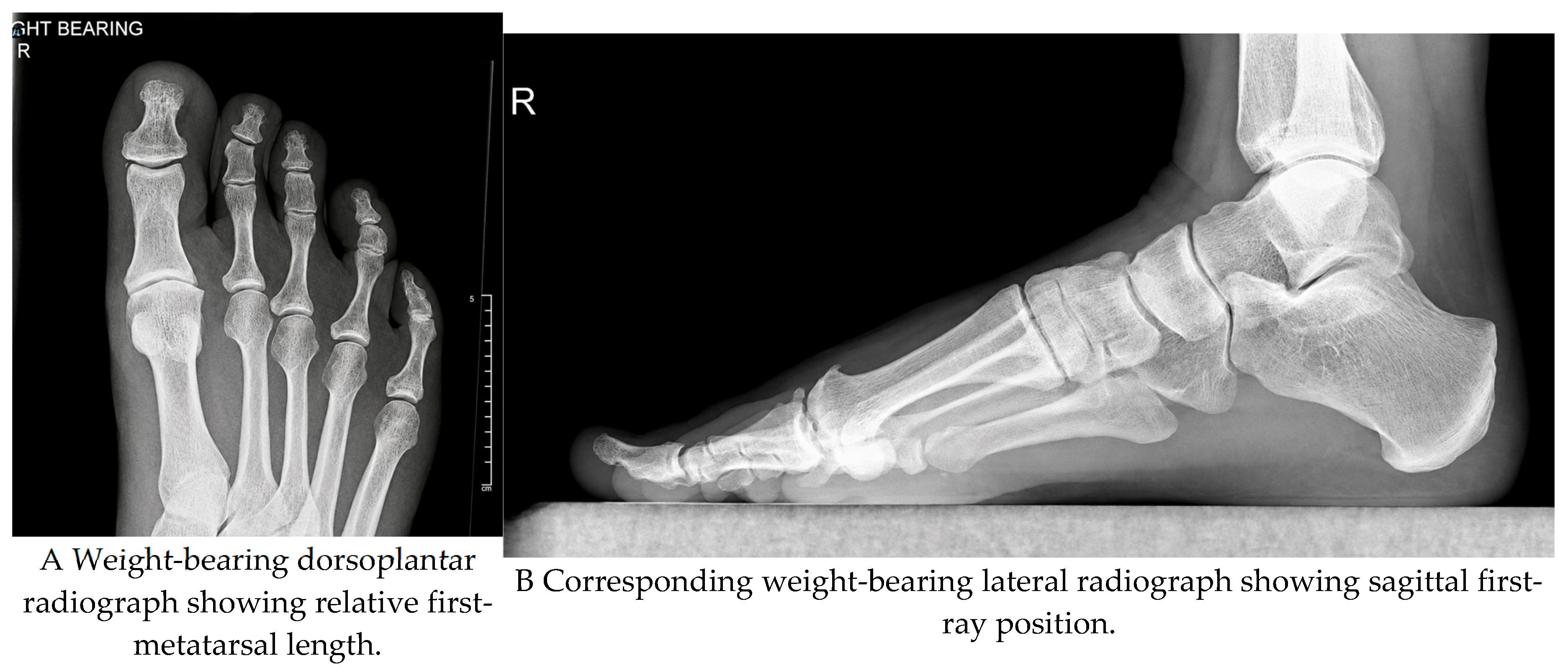
Figure 3.
Clinical assessment and joint exposure. Cheilectomy is an accompanying procedure and its contribution must be considered when interpreting outcomes.
Figure 3.
Clinical assessment and joint exposure. Cheilectomy is an accompanying procedure and its contribution must be considered when interpreting outcomes.
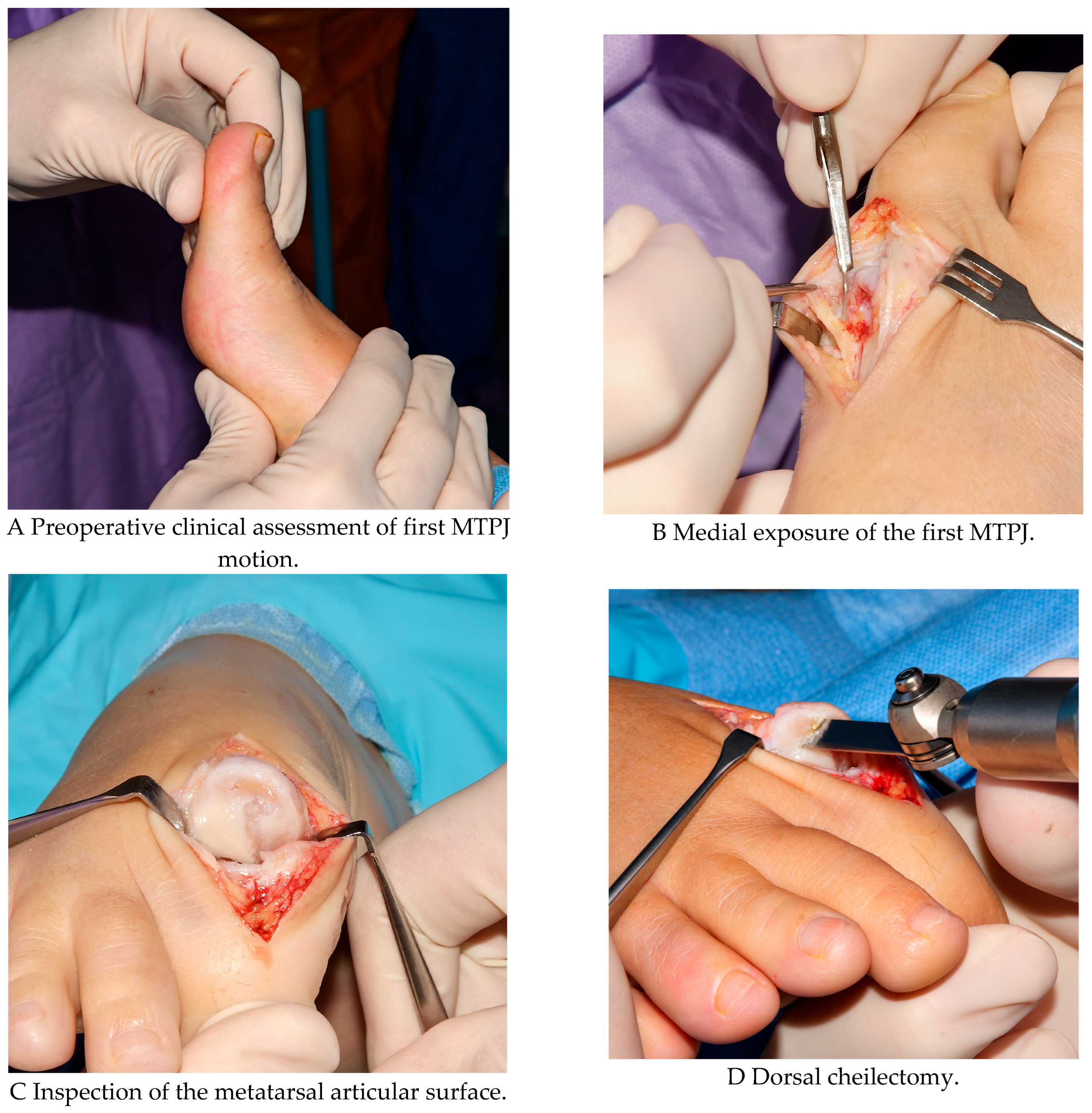
Figure 4.
Bone preparation and osteotomy construction. Fenestration and medial eminence resection are adjuncts; the parallel dorsal cut and wedge removal define the decompression component.
Figure 4.
Bone preparation and osteotomy construction. Fenestration and medial eminence resection are adjuncts; the parallel dorsal cut and wedge removal define the decompression component.
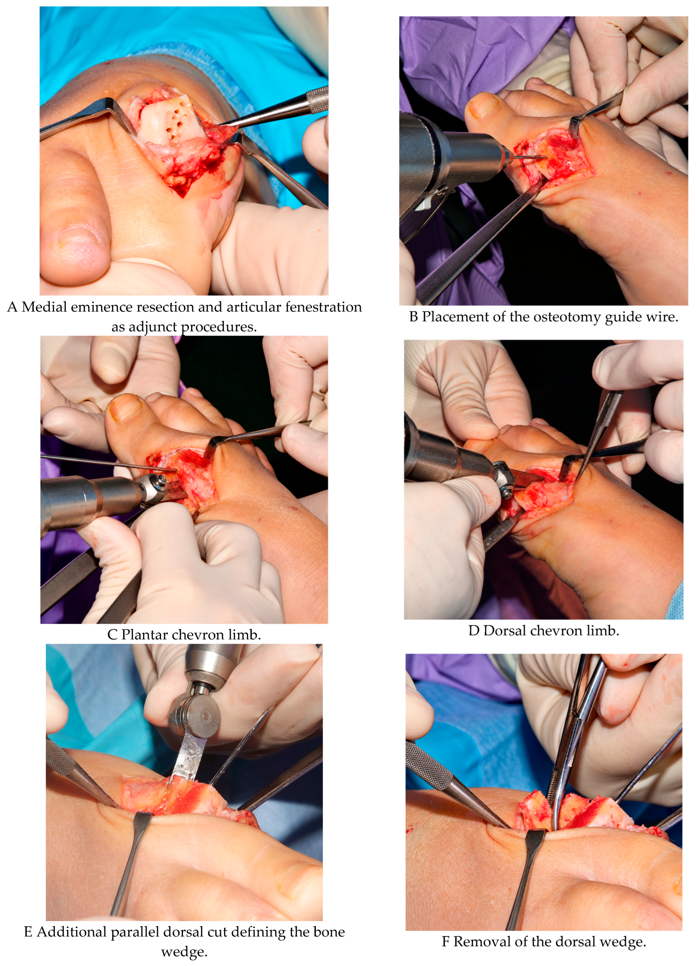
Figure 5.
Fragment positioning and additional correction. Lateral translation is an optional correction for associated deformity and does not imply that the operation is a Reverdin–Laird osteotomy.
Figure 5.
Fragment positioning and additional correction. Lateral translation is an optional correction for associated deformity and does not imply that the operation is a Reverdin–Laird osteotomy.
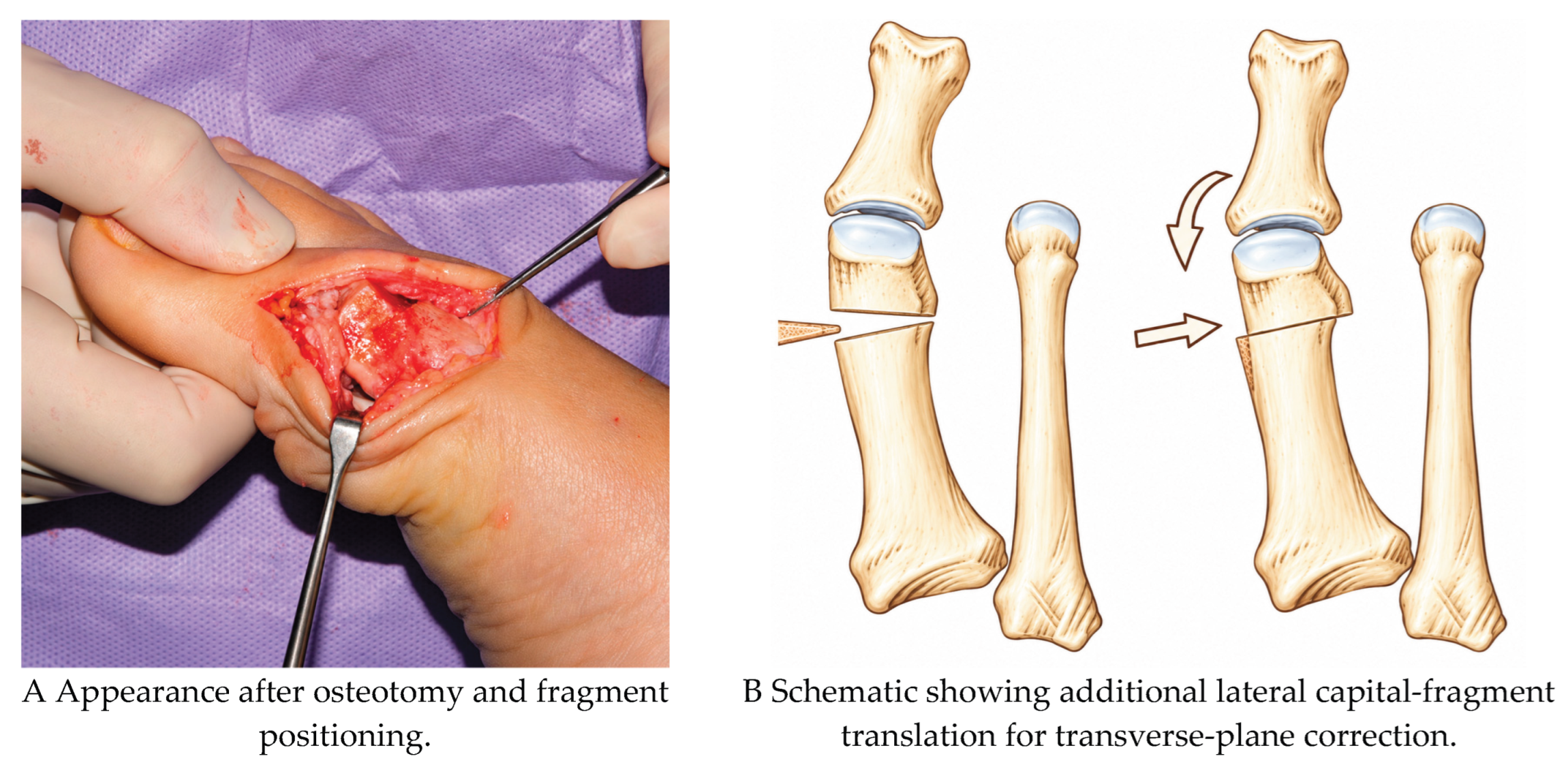

Table 1.
Selected studies and their relevance to the Youngswick evidence.
| Evidence category | Study | Design | Population | Follow-up | Principal findings | Main limitation |
| Direct Youngswick clinical | Bryant et al. 2004 [5] | Clinical + plantar pressure | 17 patients / 23 feet | 2 years | Improved dorsiflexion; increased 2nd-metatarsal pressure | Small cohort |
| Laboratory construct | LaMar et al. 2006 [23] | Sawbone mechanical comparison | Model study | N/A | After screw-and-wire fixation, sagittal V weaker/less stiff than modified Weil and controls; no significant Youngswick differences | No clinical outcomes |
| Direct Youngswick clinical | Slullitel et al. 2020 [24] | Retrospective series | 61 stage II-III patients | Mean 54.8 months; max 13 years | FAOS improved; no reported arthrodesis; 49% radiographic progression | Retrospective; cohort independence not established |
| Registry comparison | Cöster et al. 2021 [26] | Registry comparison | 115 Youngswick; 181 cheilectomy | 1 year | SEFAS +12 vs +10; satisfaction 84% vs 70% | Non-random selection/confounding |
| Youngswick-type clinical | Ceccarini et al. 2026 [27] | Retrospective observational | 55 included; 52 with return-to-sport results | Mean 79.3 months | 94.2% resumed sport; pain/function/ROM improved | Selected active cohort; cohort independence not established |
| Direct Youngswick clinical | Slullitel et al. 2016 [14] | Retrospective cohort | 78 stage II–III patients | Mean 53 months | Similar outcomes across metatarsal indices; ROM 20.4°→37.3°; 97% recommend | Non-randomized; cohort independence not established |
| Prospective comparison | Viladot et al. 2017 [31] | Prospective comparative, Level II | 46 patients / 50 grade II feet | Mean 42.7 months | Youngswick and distal oblique both improved AOFAS and ROM; comparable outcomes | Non-random allocation; different surgeons; willingness to repeat 77.3% vs 100% |
| Contextual combined decompression | Shields et al. 2023 [29] | Retrospective long-term cohort | 94 decompression + cheilectomy patients | Mean 6.3 years | ROM 11.0°→36.3°; 5 conversions to arthrodesis | Not isolated Youngswick |
| Contextual systematic review | Casais et al. 2023 [7] | Systematic review | 7 studies / 444 feet | 12–150 months across studies | Generally favorable outcomes; transfer metatarsalgia commonest complication | Predominantly Level III–IV evidence |
| Youngswick-type technical | Radovic et al. 2007 [10] | Technical modification | Hallux limitus / metatarsus primus elevatus | N/A | Modified Youngswick avoids obligatory shortening | Technical report; no comparative outcomes |
| Retrospective comparison | Xu et al. 2020 [30] | Retrospective comparative | 33 feet, grade III–IV | 12–46 months | Youngswick and distal oblique both improved pain, AOFAS, ROM and joint space | Small cohort; retrospective; Chinese-language article |
| Contextual systematic review | Roukis 2010 [22] | Systematic review | 4 prospective studies / 93 osteotomies | Weighted mean 18.6 months | AOFAS and motion improved; 22.6% revision overall | Not Youngswick-specific |
| Combined Youngswick–Akin | Xu et al. 2025 [28] | Combined-procedure clinical study | 43 patients; moderate hallux valgus + mild–moderate rigidus | 24-month outcome assessments | Combined deformity correction; does not isolate Youngswick effect | Concomitant Akin; not isolated Youngswick |
| Contextual systematic review | Lewis et al. 2026 [8] | Systematic review | 30 studies; 1,293 feet | Mean 44 months | Mixed procedures; Youngswick commonest; most studies moderate-to-serious risk of bias | Cheilectomy in 76.7% of studies; independent osteotomy effect uncertain |
| Youngswick-type clinical | Ceccarini et al. 2015 [25] | Retrospective case series | 40 grade II–III patients; >50% viable cartilage | Mean 35.4 months | Median AOFAS 39→84; motion 14°→56°; 90% good/excellent satisfaction; 2 transfer metatarsalgia cases | Selected cartilage-preserved joints; osteophyte removal; no control; cohort independence not established |
| Contextual cadaveric | Asunción et al. 2015 [21] | Cadaveric geometry study | 22 specimens; modified distal oblique osteotomy | N/A | Head lowering depended on inclination and lateral translation; sesamoid articulation compromised at 40° | Not Youngswick; no clinical outcomes |
| Contextual distal oblique | Ronconi et al. 2000 [32] | Uncontrolled clinical series | 26 patients / 30 distal oblique osteotomies | Mean 21 months | Dorsiflexion 22°→45°; 84% good/excellent satisfaction | Not Youngswick; short follow-up; associated procedures |
Table 2.
Potential complications and management considerations.
| Complication | Likely mechanism | Prevention | Management principle |
| Transfer metatarsalgia | Excessive shortening / altered load distribution | Plan to shorten conservatively; assess metatarsal parabola | Offloading; treat persistent mechanical cause if necessary |
| Persistent stiffness | Advanced disease, scarring, inadequate rehabilitation | Selection; controlled early ROM when safe | Rehabilitation; reassess joint pathology |
| Residual dorsal impingement | Inadequate cheilectomy or recurrent osteophytes | Adequate but conservative dorsal resection | Imaging and revision strategy based on pathology |
| Malunion/loss of position | Unstable construct or technical error | Precise cuts and stable fixation | Observation or revision according to symptoms/alignment |
| Progressive osteoarthritis | Underlying disease progression | Appropriate counselling and selection | Non-operative care or conversion to arthrodesis |
Table 3.
Considerations for shared decision-making.
| Consideration | Relevance to decompression | Discussion of arthrodesis |
| Joint function and mechanical target | Retained motion and plausible impingement/decompression target | Extensive destruction and limited recoverable motion |
| Pain and sesamoid involvement | Record terminal and mid-range pain; predictive value unvalidated | Diffuse pain or sesamoid disease may favour discussion of fusion |
| Patient priorities | Preference for retained motion; accept uncertain durability | Preference for predictable pain relief; discuss loss of joint motion |
| Metatarsal morphology | Length and elevation inform planning; neither is a prerequisite | Morphology alone does not determine choice |
| Surgical suitability | Assess circulation, infection, recovery capacity and adherence | The same suitability assessment applies before fusion |
Disclaimer/Publisher’s Note: The statements, opinions and data contained in all publications are solely those of the individual author(s) and contributor(s) and not of MDPI and/or the editor(s). MDPI and/or the editor(s) disclaim responsibility for any injury to people or property resulting from any ideas, methods, instructions or products referred to in the content. |
© 2026 by the authors. Licensee MDPI, Basel, Switzerland. This article is an open access article distributed under the terms and conditions of the Creative Commons Attribution (CC BY) license (http://creativecommons.org/licenses/by/4.0/).
Copyright: This open access article is published under a Creative Commons CC BY 4.0 license, which permit the free download, distribution, and reuse, provided that the author and preprint are cited in any reuse.