

Submitted:
19 October 2026
Posted:
20 October 2026
You are already at the latest version
Abstract
The vaginal microbiota represents a dynamic microbial ecosystem that contributes to mucosal homeostasis and protection against potentially pathogenic microorganisms. Vaginal dysbiosis, characterized by alterations in microbial composition and function, has increasingly been associated with gynecological, reproductive, obstetric, and puerperal conditions. This review summarizes current evidence on the composition and determinants of the vaginal microbiota, with an emphasis on Community State Types and major forms of dysbiosis, including bacterial vaginosis, vulvovaginal candidiasis, aerobic vaginitis, and viral infections. The associations between dysbiosis and sexually transmitted infections are discussed, with particular attention to the distinction between colonization, dysbiosis, and clinically relevant infection. Potential relationships between dysbiosis and infertility are examined, including tubal, cervical, and endometrial factors, as well as outcomes following assisted reproductive technologies. Evidence regarding obstetric and puerperal complications is critically evaluated. Finally, current therapeutic strategies are reviewed, ranging from etiological antimicrobial treatment to probiotics, defined live biotherapeutics, and emerging approaches such as vaginal microbiota transplantation. Although microbiota restoration represents a promising therapeutic concept, uncertainties remain regarding optimal strains, timing, diagnostic criteria, and clinically meaningful outcomes. Overall, vaginal dysbiosis should be regarded not simply as a local microbial imbalance but as a condition capable of modifying mucosal host-pathogen interactions throughout the female reproductive tract.
Keywords:
vaginal microbiota
; vaginal dysbiosis
; bacterial vaginosis
; vulvovaginal candidiasis
; aerobic vaginitis
; Lactobacillus
; human papillomavirus
; reproductive health
; pregnancy
; host-microbiome interactions
1. Introduction
This review summarizes current evidence on the composition and determinants of the vaginal microbiota, the mechanisms and clinical manifestations of vaginal dysbiosis, and its relevance to gynecological and obstetric outcomes, with particular emphasis on diagnostic and therapeutic approaches. Throughout this review, the terms ‘vaginal microbiota’ and ‘vaginal microbiome’ are used largely interchangeably, reflecting their frequent interchangeable use in the literature. Strictly speaking, the vaginal microbiota refers to the microorganisms inhabiting the vaginal environment, whereas the vaginal microbiome encompasses their collective genetic repertoire and functional potential.
2. Vaginal Microbiota
2.1. Community State Types
The female genital tract harbors a complex and dynamic microbial ecosystem comprising bacteria, archaea, fungi, viruses, and, to a lesser extent, protozoa. These microorganisms coexist in varying relative abundances and interact with the host through metabolic, immunological, and ecological mechanisms that contribute to the maintenance of the vaginal microenvironment and colonization resistance against potentially pathogenic microorganisms.
The composition and temporal dynamics of the vaginal microbiota are shaped by a complex interplay of host, hormonal, behavioral, reproductive, and environmental factors, including age, race and ethnicity, geographic region, parity and reproductive history, menstrual cycle phase, sexual activity, pregnancy, and contraceptive use. Major changes in vaginal physiology and microbial composition occur throughout the female life course, particularly during puberty, pregnancy, and menopause, whereas shorter-term fluctuations may accompany menstruation, sexual activity, and other physiological or behavioral factors. The menstrual cycle is particularly relevant because fluctuations in estrogen concentrations can influence vaginal epithelial maturation, glycogen availability, vaginal pH, and the relative abundance of Lactobacillus spp. [1,2,3,4,5,6,7,8,9,10].
The initial molecular characterization of the vaginal microbiome demonstrated differences in the distribution of microbial community types among women from different racial and ethnic backgrounds. Subsequent studies confirmed differences in the prevalence of Lactobacillus-dominated and Lactobacillus-depleted communities between women of African American and European ancestry. Importantly, these observations should not be interpreted as evidence that race itself is a direct biological determinant of the vaginal microbiome. Rather, race and ethnicity are social and demographic constructs that may reflect complex interactions among genetic background, geographic location, environmental exposures, socioeconomic conditions, behavioral factors, and access to health care.
During pregnancy, hormonal changes are associated with alterations in the vaginal microbiota, and studies have demonstrated associations between gestational age, maternal age, parity, and the abundance and composition of Lactobacillus-dominated communities. These findings underscore the importance of considering reproductive status and parity when comparing vaginal microbiome profiles across study populations [1,2,3,4,5,6,7,8,9,10].
Longitudinal studies have demonstrated associations between sexual activity, menstrual cycle phase, and changes in the composition and stability of vaginal microbial communities. Sexual activity may influence the vaginal microbiome through several mechanisms, including exposure to semen, alterations in the vaginal microenvironment, exchange of microorganisms between sexual partners, and behavioral factors associated with sexual activity [5,14].
Hormonal and non hormonal contraceptive methods have been associated with differences in vaginal microbial composition in observational and longitudinal studies. Thus, contraceptive use should be considered an important potential modifier of the vaginal microbiome without implying that contraception necessarily has a detrimental effect on vaginal microbial health [13,15,16].
Based on bacterial community composition, vaginal microbial communities have been classified into community state types (CSTs). The original molecular classification by Ravel et al. [4]. identified five major CSTs in reproductive-age women (Table 1). Four CSTs are dominated by individual Lactobacillus spp., whereas CST IV is characterized by a greater abundance and diversity of anaerobic bacteria and a comparatively low abundance of Lactobacillus spp. Specifically, CST I is dominated by Lactobacillus crispatus, CST II by Lactobacillus gasseri, CST III by Lactobacillus iners, and CST V by Lactobacillus jensenii. CST IV represents a heterogeneous, Lactobacillus-depleted community characterized by diverse anaerobic and facultatively anaerobic taxa. CST classification therefore provides a useful ecological framework for describing vaginal microbial community composition while acknowledging substantial interindividual and temporal variability [4,5].
Lactobacillus-dominated communities are generally associated with lactic acid production, which, together with other microbial metabolites and host factors, contributes to the antimicrobial properties of the vaginal microenvironment and may inhibit the growth or persistence of potentially pathogenic microorganisms. Among the Lactobacillus-dominated CSTs, L. crispatus has frequently been associated with relatively stable microbial communities and a lower prevalence of bacterial vaginosis BV-associated microbial profiles [2,6].
CST III, dominated by Lactobacillus iners, is often associated with a more heterogeneous microbial community and a less stable vaginal ecosystem, which may be linked to increased susceptibility to vaginal dysbiosis and genital tract infections.
However, Lactobacillus dominance should not be regarded as an absolute marker of vaginal health, as asymptomatic women may exhibit diverse microbial communities without clinical evidence of disease [2,3,6].
The vaginal microbiota may also influence susceptibility to sexually transmitted infections (STIs). Lactobacillus-depleted and anaerobe-rich communities have been associated with increased susceptibility to several STIs and with alterations in mucosal immune responses. In particular, Lactobacillus-deficient cervicovaginal bacterial communities have been associated with increased HIV acquisition among young South African women [7,8]. These findings support a potential role for the vaginal microbiome in modulating susceptibility to infection; however, the underlying mechanisms are complex and cannot be attributed solely to the presence or absence of a specific CST. The vaginal microbiota should therefore be regarded as a dynamic ecological system rather than as a fixed microbial composition.
2.2. Lactobacillosis
Vaginal lactobacillosis is an uncommon condition characterized by the presence of excessively long and morphologically abnormal Lactobacillus cells in the vaginal microbiota. This abnormal overgrowth may be associated with profuse, homogeneous, white vaginal discharge and persistent vulvovaginal discomfort. Lactobacillosis should be differentiated from cytolytic vaginosis, which is typically characterized by an increased abundance of lactobacilli, lysis of vaginal epithelial cells, and a low vaginal pH. Microscopic examination of a vaginal wet mount is particularly useful for identifying elongated lactobacilli and distinguishing lactobacillosis from other causes of abnormal vaginal discharge. Because this condition may mimic vulvovaginal candidiasis or bacterial vaginosis, accurate diagnosis is essential to avoid inappropriate antimicrobial treatment [11]. Although associations have been reported between L. iners-dominated microbiota and bacterial vaginosis or reduced microbiota stability, these observations do not establish a causal relationship with lactobacillosis [12]. Moreover, vaginal lactobacillosis is primarily defined by characteristic microscopic findings-particularly the presence of unusually elongated lactobacilli rather than by the identification of a specific Lactobacillus species.
2.3. Vaginal Dysbiosis. Relevance in Obstetrics and Gynecology
Vaginal dysbiosis refers to a disruption of the vaginal microbial ecosystem characterized by alterations in the composition, diversity, abundance, or functional activity of the vaginal microbiota, resulting in a shift away from microbial homeostasis. Although vaginal dysbiosis is often associated with the depletion or displacement of protective Lactobacillus spp., it is not defined solely by the absence of Lactobacillus dominance.
Vaginal dysbiosis is of particular relevance to obstetrics and gynecology because alterations in the vaginal microbial ecosystem may affect both local genital health and reproductive outcomes. In gynecology, vaginal dysbiosis is increasingly recognized as an important determinant of women’s health and has been associated with recurrent vaginal infections, impaired reproductive health, increased susceptibility to sexually transmitted infections, and altered mucosal immune responses [1,2,3,4,5,6,7,8,9,10].
During pregnancy, disturbances in the vaginal microbiota have been associated with ascending genital tract infection, preterm prelabor rupture of membranes (PPROM), and preterm birth. Although the underlying mechanisms are complex and causal relationships have not been established for all microbial alterations, increasing evidence supports an important interaction among the vaginal microbial ecosystem, the local immune response, and pregnancy outcomes. Recent systematic reviews and meta-analyses have particularly highlighted associations between Lactobacillus depletion or shifts in Lactobacillus species composition and an increased risk of preterm birth.
One of the most accessible parameters reflecting changes in the vaginal environment is vaginal pH. In a Lactobacillus-dominated vaginal ecosystem, the production of lactic acid contributes to the maintenance of an acidic environment, which represents an important component of the vaginal defense system. In reproductive-age women, a vaginal pH below 4.5 is generally considered characteristic of a physiologic vaginal environment, whereas an elevated pH is frequently observed in bacterial vaginosis and other conditions associated with a shift away from Lactobacillus dominance. However, vaginal pH is a nonspecific parameter and should not be regarded as a diagnostic test for a specific form of vaginal dysbiosis. Rather, it may serve as a simple indicator of an altered vaginal environment and, particularly during pregnancy, as a trigger for further clinical and microbiological assessment [19,20].
The potential obstetric relevance of vaginal pH monitoring has been illustrated by the so-called Thuringian model of preterm birth prevention. In 2016, the government of the Free State of Thuringia, in cooperation with the professional organization representing obstetricians and gynecologists, reestablished and promoted a statewide prevention program based on intravaginal pH self-monitoring beginning at more than 14 weeks of gestation, with the aim of facilitating the early detection and treatment of genital infections. By the end of 2018, more than 80% of pregnant women in Thuringia had participated in vaginal pH-monitoring. During this period, a reduction in early preterm birth (≤ 32+0 weeks of gestation) from 1.46% to 1.26% was reported [19,20].
Regular assessment of the vaginal environment may contribute to multifactorial strategies for the prevention of preterm birth. However, evidence supporting a causal effect of vaginal pH self-monitoring remains insufficient. Controlled evaluations have shown no significant reduction in preterm birth before 37 weeks of gestation, and the Thuringian data do not allow the contribution of individual preventive measures to be determined. Therefore, vaginal pH self-measurement cannot currently be considered an established universal screening strategy [19,20].
Nevertheless, the Thuringian experience highlights the potential clinical relevance of the vaginal environment as a modifiable component of the pathway linking microbial disturbances to adverse obstetric outcomes. Vaginal pH should therefore not be regarded as a diagnostic marker of dysbiosis per se, but rather as a simple and accessible indicator of changes within the vaginal ecosystem [19,20].
3. Bacterial Vaginosis
BV is the most common form of vaginal dysbiosis and is characterized by a shift from a Lactobacillus-dominated ecosystem toward a more diverse, polymicrobial community enriched in anaerobic and facultatively anaerobic bacteria. In molecular microbiome studies, BV is most frequently associated with CST IV, which comprises heterogeneous microbial communities containing diverse anaerobic taxa. Importantly, CST IV is not synonymous with clinically diagnosed BV, as not all women with a CST IV microbiota meet the clinical or Nugent criteria for BV.
BV may be asymptomatic or may present with homogeneous, thin, grayish-white vaginal discharge and a characteristic fishy odor, particularly after the addition of potassium hydroxide. Clinical diagnosis is based on the Amsel criteria, with at least three of four findings required: vaginal pH > 4.5, homogeneous thin discharge, clue cells on microscopy, and a positive amine or ‘whiff’ test [21,22,23,24,25].
Another laboratory-based approach for diagnosing BV is the assessment of the Nugent score, which is generally considered more objective and reproducible than the Amsel criteria. However, because accurate assessment requires trained personnel, the Nugent score is used less frequently in routine clinical practice [21,22,23,24,25].
The Nugent score is based on the microscopic quantification of three bacterial morphotypes: large gram-positive rods corresponding to protective lactobacilli, small gram-variable or gram-negative rods corresponding primarily to Gardnerella and Bacteroides species, which are increased in bacterial vaginosis, and curved gram-variable rods corresponding to Mobiluncus species, which are strongly associated with bacterial vaginosis.
A Nugent score of 7–10 is indicative of bacterial vaginosis, a score of 4–6 indicates intermediate microbiota, and a score of 0–3 indicates a Lactobacillus-predominant vaginal microbiota [21,22,23,24,25].
The pathogenesis of BV cannot be attributed to a single causative microorganism. Rather, BV represents a polymicrobial ecological shift in which the depletion or displacement of protective Lactobacillus spp. is accompanied by the expansion of a consortium of BV-associated bacteria. Gardnerella spp. are frequently detected and may contribute to disruption of the vaginal ecosystem; however, their presence alone is neither necessary nor sufficient for a diagnosis of BV. Other organisms, including Atopobium vaginae, Prevotella spp., Megasphaera spp., Sneathia spp., Mobiluncus spp., and Mycoplasma hominis, may also be present at increased abundance in BV-associated communities. Therefore, detection of an individual BV-associated bacterium should not be interpreted as evidence of a specific pathogen causing BV [21,22,23,24,25].
A key feature contributing to the persistence and recurrence of BV is the formation of polymicrobial biofilms, particularly on vaginal epithelial cells. Gardnerella spp. can form biofilms that facilitate the subsequent incorporation of other anaerobic microorganisms and may increase tolerance to antimicrobial treatment. Biofilm-associated persistence, together with incomplete restoration of a Lactobacillus-dominated microbiota, may contribute to the high recurrence rates observed after treatment [21,22,23,24,25].
Overall, BV should be understood not as a conventional infection caused by a single pathogen, but as a complex polymicrobial dysbiosis characterized by loss of Lactobacillus dominance, increased vaginal pH, expansion of BV-associated anaerobic communities, and, frequently, biofilm-associated persistence. This distinction is clinically important because the molecular detection of individual bacteria does not establish a specific infection. Clinical interpretation should instead integrate symptoms, vaginal pH, microscopy, Nugent or Amsel criteria, microbiological findings, and the individual clinical context [21,22,23,24,25].
3.1. Bacterial STIs and Their Association with Bacterial Vaginosis
Sexual activity is relevant to the epidemiology and recurrence of BV. Although BV is not classified as a STI and may occur in women without recent sexual activity, sexual exposure is associated with BV, and BV-associated bacterial communities may be exchanged between sexual partners. In male partners, BV-associated organisms may persist on the penile skin and within the urethral microbiota, potentially contributing to recurrence in the female partner. This has led to the concept of sexual re-exposure. However, this terminology should be used cautiously, as BV is not an STI in the conventional sense, and the clinical relevance of partner-associated bacterial exchange has historically remained uncertain. Recent evidence has strengthened the concept that the male partner’s microbiota may contribute to recurrence in at least some couples, suggesting that partner-directed treatment may become relevant in selected cases of recurrent BV.BV is also associated with an increased risk of acquiring sexually transmitted pathogens, including Trichomonas vaginalis, Chlamydia trachomatis, Neisseria gonorrhoeae, and Mycoplasma genitalium. Thus, BV and an STI may represent concomitant conditions rather than a single microbial disorder. In symptomatic women or those with relevant risk factors, appropriate molecular testing for specific STI pathogens should therefore be considered instead of attributing all vaginal symptoms to BV.
The interpretation of positive molecular tests for Ureaplasma spp. and Mycoplasma hominis requires particular caution. Ureaplasma parvum, Ureaplasma urealyticum, and M. hominis commonly colonize the urogenital tract, and their detection alone does not establish infection or explain vaginal symptoms. U. parvum, in particular, is frequently detected as asymptomatic carriage. Accordingly, the European STI Guidelines Editorial Board does not recommend routine testing or treatment for these organisms in asymptomatic individuals or as part of the routine evaluation of symptomatic women [26,27,28,29,30].
The interpretation of Ureaplasma spp. during pregnancy, however, requires additional consideration. Several observational studies and meta-analyses have reported associations between maternal Ureaplasma detection and adverse pregnancy outcomes, including preterm birth and premature rupture of membranes (PROM). A systematic review and meta-analysis including 53 studies found that U. urealyticum was associated with preterm birth (unadjusted OR, 1.96; 95% CI, 1.14-3.39) and PROM (OR, 9.87; 95% CI, 1.81-53.72), whereas U. parvum was associated with preterm birth (OR, 1.79; 95% CI, 1.28-2.52) and PROM (OR, 3.19; 95% CI, 1.25-8.15). A separate meta-analysis of 19 cohort studies including nearly 12,000 pregnant women reported associations between maternal Ureaplasma positivity and preterm birth (OR, 2.76; 95% CI, 1.63-4.68), PROM (OR, 2.19; 95% CI, 1.34-3.58), and chorioamnionitis (OR, 2.71; 95% CI, 2.02-3.64) [26,27,28,29,30].
These findings nevertheless require cautious interpretation. Ureaplasma spp. are common colonizers, and most available studies are observational, with substantial heterogeneity in study populations, sampling sites, diagnostic methods, and adjustment for potential confounders. Jonduo et al. [27].emphasized that only a small proportion of the included studies performed multivariable analyses and concluded that the available evidence does not establish a causal role for genital mycoplasmas in adverse pregnancy outcomes. Importantly, the authors also highlighted the need to evaluate Ureaplasma spp. and M. hominis within the broader vaginal microbiome rather than as isolated microorganisms.
This distinction is particularly relevant because the association between Ureaplasma spp. and adverse pregnancy outcomes may partly reflect a dysbiotic microbial environment. In studies stratified according to BV status, associations between genital mycoplasmas and preterm birth were stronger in the presence of BV for some organisms, particularly M. hominis and U. parvum. These findings do not establish that BV causes the association between Ureaplasma and preterm birth, but they support the concept that the clinical significance of Ureaplasma may depend on the composition and functional state of the surrounding microbial community. Thus, during pregnancy, Ureaplasma detection should not simply be categorized as either harmless colonization or definite infection. Rather, its potential significance should be interpreted in conjunction with gestational age, clinical findings, evidence of vaginal dysbiosis, other genital microorganisms, and signs of ascending infection [26,27,28,29,30].
M. hominis requires a similarly nuanced interpretation. It is frequently detected in BV-associated microbial communities and may represent part of the polymicrobial dysbiotic environment rather than an independent STI causing the clinical syndrome of BV. Its detection, particularly in the context of BV, should therefore not automatically be interpreted as a separate concomitant infection requiring targeted antimicrobial treatment.
In contrast, Mycoplasma genitalium represents a distinct clinical entity. Unlike M. hominis and the ureaplasmas, M. genitalium is an established sexually transmitted pathogen associated with urethritis, cervicitis, and pelvic inflammatory disease (PID). Infection in women may be asymptomatic; however, persistent or recurrent cervicitis or urethritis, or clinical features suggestive of PID, may warrant targeted testing in appropriate clinical settings. Importantly, M. genitalium has developed substantial antimicrobial resistance, particularly to macrolides. When treatment is indicated, resistance-guided therapy based on molecular macrolide-resistance testing should therefore be used whenever available [26,27,28,29,30].
3.2. Vulvovaginal Candidiasis and Its Association with Bacterial Vaginosis
Vulvovaginal candidiasis (VVC) represents a distinct form of vaginal dysbiosis characterized by the symptomatic overgrowth of Candida spp. in the vaginal environment. Candida spp. may be part of the normal vaginal microbiota without causing disease. Asymptomatic vaginal colonization is relatively common, and the detection of Candida in the absence of compatible clinical symptoms or signs does not constitute an indication for treatment. VVC may develop when host–microbe homeostasis is disturbed, with bacterial vaginosis potentially facilitating the transition from asymptomatic Candida colonization to symptomatic overgrowth. This distinction is particularly important because molecular detection or a positive culture alone cannot reliably differentiate colonization from clinically relevant infection. Microscopic examination may provide additional support for the diagnosis.
The most common causative species is Candida albicans, which accounts for the majority of symptomatic episodes. Typical clinical manifestations include vulvar pruritus, burning, erythema, edema, external dysuria, and a characteristic thick, white, adherent, or curd-like discharge. The discharge is usually odorless, in contrast to the malodorous discharge commonly associated with BV. On microscopy, C. albicans may be detected as budding yeast cells and, particularly in symptomatic infection, as pseudohyphae or hyphal forms. The ability of C. albicans to transition between yeast and filamentous forms is considered an important component of its pathogenic potential [31,32].
Non-albicans Candida species account for a smaller but clinically important proportion of VVC cases. Candida glabratais the most relevant non-albicans species and is encountered more frequently in recurrent or complicated VVC and in older women, including those in the perimenopausal and postmenopausal periods. The clinical presentation may be less typical than that of C. albicans, with less prominent inflammation and more nonspecific vulvovaginal symptoms. However, the nature of the vaginal discharge alone cannot reliably distinguish C. glabrata from other causes of vaginitis. Importantly, C. glabrata does not form pseudohyphae or true hyphae and may therefore be missed by conventional microscopy. The increasing recognition of non-albicans Candida species is clinically relevant because these organisms frequently show reduced susceptibility to conventional azole therapy. In particular, C. glabrata is less susceptible to fluconazole than C. albicans, and treatment failure may occur despite apparently adequate therapy [31,32].
Consequently, in cases of recurrent, persistent, or complicated VVC, vaginal culture with species identification should be performed. Subsequent treatment should be guided by the identified species, susceptibility pattern, clinical presentation, and patient-specific factors. Clinical symptoms of VVC may fluctuate during the menstrual cycle and are frequently reported to become more pronounced during the premenstrual phase. Hormonal fluctuations, changes in the vaginal microenvironment, and alterations in local epithelial and immune responses may contribute to this cyclical pattern. However, premenstrual worsening does not exclude other causes of vaginal dysbiosis and should therefore not be regarded as a pathognomonic diagnostic feature. Importantly, the severity and persistence of VVC symptoms are not determined solely by the fungal burden. The transition from asymptomatic Candida colonization to symptomatic infection involves complex interactions between the organism and the vaginal and vulvar epithelium, together with activation of the local innate immune response. In particular, hypha-associated factors of C. albicans can stimulate epithelial cells, induce the release of proinflammatory mediators, and promote the recruitment of immune cells. Clinical symptoms may persist for several days after the initiation of appropriate antifungal treatment, even when the fungal burden has already been substantially reduced. Because resolution of the epithelial inflammatory response may lag behind the microbiological response, gradual clinical improvement during the first few days of treatment does not necessarily indicate treatment failure. Conversely, persistent or worsening symptoms despite an appropriate treatment course should prompt diagnostic reassessment. The differential diagnosis includes non-albicans Candida species, antifungal resistance, mixed vaginitis, bacterial vaginosis, vulvar dermatoses, contact dermatitis, and other causes of vulvovaginal symptoms. In patients with recurrent, persistent, or complicated VVC, microbiological confirmation with species identification and, when indicated, antifungal susceptibility testing is therefore particularly important [31,32].
An important clinical challenge is the co-occurrence of VVC and bacterial vaginosis, commonly referred to as mixed vaginitis. In such cases, the vaginal pH may be elevated because of concomitant BV despite the presence of symptomatic Candida infection. In contrast, uncomplicated VVC is typically associated with a physiologically acidic vaginal pH (<4.5). The presence of an acidic environment therefore does not exclude symptomatic candidiasis. Thus, the traditional distinction between ‘acidic pH for Candida’ and ‘alkaline pH for BV’ remains clinically useful but should not be interpreted as an absolute rule. The relationship between BV-associated dysbiosis and Candida overgrowth appears to be complex. Rather than assuming that BV directly causes accelerated Candida proliferation, current evidence supports the concept of ecological interaction between the bacterial and fungal components of the vaginal microbiota. Alterations in Lactobacillus abundance, changes in microbial metabolites, epithelial interactions, and antimicrobial exposure may influence the balance between bacterial and fungal organisms. In particular, antibiotic treatment for bacterial vaginosis may predispose susceptible women to subsequent symptomatic VVC, illustrating that therapeutic manipulation of the bacterial component can itself alter the fungal component of the vaginal ecosystem [33,34,35,36,37,38].
Studies of the vaginal microbiota have demonstrated considerable heterogeneity among women with asymptomatic Candida colonization, VVC, and BV, indicating that the relationship between vaginal pH, bacterial community composition, and fungal colonization is more complex than a simple reciprocal model. From a diagnostic perspective, mixed vaginitis should therefore be considered when clinical findings are inconsistent with a single diagnosis-for example, when a woman presents with pruritus and curd-like discharge suggestive of VVC but also has an elevated vaginal pH, concomitant clue cells, or a positive amine test. Microscopy, vaginal pH measurement, and targeted microbiological testing can help distinguish isolated VVC, BV, and mixed infection [33,34,35,36,37,38].
3.2.1. Vulvovaginal Candidiasis and Adverse Pregnancy Outcomes
The relationship between VVC and adverse pregnancy outcomes differs from that observed for bacterial vaginosis. Although BV is consistently associated with an increased risk of preterm birth and premature rupture of membranes, current evidence does not demonstrate a comparable independent association between vaginal Candida infection and preterm birth. A systematic review and meta-analysis including 57 studies and 49,161 pregnant women with available preterm birth data found no significant association between vaginal yeast infection and preterm birth (summary OR, 1.01; 95% CI, 0.84-1.21). Similarly, a meta-analysis including 33,321 women found no significant association between asymptomatic vaginal Candida colonization and preterm birth (OR, 1.10; 95% CI, 0.99-1.22) [39,40].
The coexistence of BV and VVC, however, represents a clinically relevant form of mixed vaginitis and may reflect a more complex alteration of the vaginal ecosystem. From an obstetric perspective, this distinction is important because it illustrates that not all vaginal microbial alterations carry the same pregnancy-related risk. BV and other Lactobacillus-depleted dysbiotic states have stronger epidemiological links to ascending infection and preterm birth, whereas Candida colonization or VVC alone appears to have substantially less impact on these outcomes. The clinical significance of a mixed BV–Candida state therefore depends on the underlying bacterial dysbiosis, the presence of additional genital microorganisms, the degree of inflammation, and the individual clinical context) [39,40].
VVC at the time of vaginal birth creates a risk of vertical transmission, as the newborn may be exposed to Candida spp. while passing through the birth canal. In healthy, full-term infants, this exposure is generally not dangerous but may lead to localized conditions such as oral thrush, characterized by white patches in the mouth, or persistent fungal diaper dermatitis during the first few weeks of life. In preterm or very-low-birth-weight infants, however, the risk is considerably higher because their immature immune systems make them more vulnerable to neonatal invasive candidiasis, a serious systemic infection that may involve the bloodstream and internal organs. To mitigate these risks, symptomatic maternal yeast infections should be treated with pregnancy-compatible topical antifungal therapy before delivery) [41,42].
4. Aerobic Vaginitis, Group B Streptococcus, and Desquamative Inflammatory Vaginitis
Aerobic vaginitis (AV) represents a distinct form of vaginal dysbiosis that differs substantially from bacterial vaginosis. Both conditions are characterized by depletion of Lactobacillus-dominated microbiota and an elevated vaginal pH; however, AV is characterized by the predominance of aerobic or facultatively anaerobic microorganisms and, in contrast to BV, a pronounced local inflammatory response. Frequently detected organisms include Escherichia coli, Staphylococcus aureus, Streptococcus agalactiae (group B Streptococcus, GBS), and Enterococcus spp. Microscopically, AV is characterized by reduced lactobacillary flora, increased numbers of leukocytes and toxic leukocytes, altered bacterial morphotypes, and, in more severe cases, parabasal epithelial cells. Clinically, patients may present with vaginal erythema, burning, dyspareunia, and yellowish discharge [43,46].
The distinction between AV and BV is clinically important. Whereas BV is a polymicrobial, predominantly anaerobic dysbiosis that is generally associated with limited epithelial inflammation, AV is characterized by a more pronounced host inflammatory response, including increased local production of proinflammatory mediators [43,46]. AV should therefore be regarded as an inflammatory form of vaginal dysbiosis rather than simply an aerobic variant of BV. The most severe end of this inflammatory spectrum has been conceptually linked to desquamative inflammatory vaginitis (DIV).
4.1. Group B Streptococcus and Its Obstetric Relevance
Streptococcus agalactiae is a facultative Gram-positive organism that may asymptomatically colonize the gastrointestinal and genital tracts. Maternal vaginal-rectal GBS colonization occurs in approximately 10%-30% of pregnant women and may be transient, intermittent, or persistent. Importantly, maternal GBS colonization should be distinguished from GBS-associated aerobic vaginitis, as colonization alone does not imply vaginitis or dysbiosis [44,45].
The major obstetric relevance of GBS lies in its potential for vertical transmission during labor or after rupture of the membranes. Maternal genital tract colonization is the principal risk factor for early-onset neonatal GBS disease. Approximately 50% of colonized mothers transmit GBS to their newborns, and, in the absence of intrapartum antibiotic prophylaxis, approximately 1%-2% of exposed newborns develop early-onset GBS disease, which may manifest as neonatal sepsis, pneumonia, or meningitis. The implementation of systematic screening, treatment of asymptomatic maternal colonization, and intrapartum antibiotic prophylaxis has markedly reduced the incidence of early-onset GBS disease [44,45].
GBS is also clinically relevant to maternal obstetric morbidity. Although asymptomatic colonization should not be equated with infection, S. agalactiae can cause maternal urinary tract infection, intra-amniotic infection, and puerperal endometritis and has been associated with adverse pregnancy outcomes, including preterm birth. Thus, GBS represents an important example of how a normally colonizing microorganism can become clinically relevant under specific host and obstetric conditions.
The association between GBS and AV is particularly noteworthy because GBS is among the organisms frequently recovered from women who meet microscopic criteria for AV. However, the presence of GBS alone is insufficient to diagnose AV, and a positive GBS culture should not be interpreted as evidence of vaginal dysbiosis in the absence of corresponding clinical or microscopic findings. This distinction is particularly important during pregnancy, when GBS screening and management are directed primarily at preventing neonatal early-onset disease rather than treating asymptomatic maternal colonization [44,45].
4.2. Desquamative Inflammatory Vaginitis
DIV is an uncommon, chronic inflammatory vaginitis characterized by purulent discharge, vaginal irritation or burning, dyspareunia, diffuse vaginal erythema, and epithelial desquamation. Vaginal pH is typically >4.5, whereas microscopy demonstrates numerous inflammatory cells, including polymorphonuclear leukocytes, together with parabasal epithelial cells and an altered vaginal flora [47,48].
The relationship between DIV and AV remains incompletely defined. Some authors consider DIV to represent the severe inflammatory end of the AV spectrum, whereas others regard it as a distinct clinical entity. The absence of a clearly established infectious pathogen and the favorable response to topical anti-inflammatory therapy support an important immune-mediated component. Therefore, DIV should not simply be interpreted as an infection caused by organisms identified on vaginal culture. Microorganisms such as GBS may occasionally be recovered but do not necessarily represent the primary cause of the inflammatory syndrome [47,48].
For the purposes of understanding vaginal dysbiosis, AV and DIV are best considered inflammatory dysbiotic conditions that differ from the predominantly noninflammatory, anaerobe-rich ecosystem characteristic of BV. Their recognition is particularly relevant in patients with persistent vaginal symptoms despite conventional treatment for BV or candidiasis and in pregnant women, in whom inflammatory dysbiosis may be associated with adverse obstetric outcomes.
5. Vaginal Dysbiosis and Viral Infections
5.1. Human Papillomavirus Infection
The interaction between vaginal dysbiosis and viral infections is particularly relevant in the context of human papillomavirus (HPV) infection. Although most HPV infections are transient and undergo spontaneous clearance, a subset persists and may subsequently contribute to cervical intraepithelial neoplasia and cervical cancer. Increasing evidence suggests that the composition and functional characteristics of the vaginal microbiota may influence the natural history of HPV infection, particularly its persistence and progression [49,50].
Vaginal dysbiosis, characterized by reduced Lactobacillus dominance and increased microbial diversity, has been associated with both HPV acquisition and impaired viral clearance. A systematic review and meta-analysis including 15 predominantly prospective cohort studies and more than 101,000 women demonstrated that vaginal dysbiosis was associated with an increased risk of incident HPV infection (RR, 1.33; 95% CI, 1.18-1.50) and HPV persistence (RR, 1.14; 95% CI, 1.01-1.28). This association was also observed for oncogenic HPV types (RR, 1.18; 95% CI, 1.01-1.38). Importantly, dysbiosis was additionally associated with an increased risk of high-grade cervical lesions and cervical cancer. These findings should be interpreted cautiously, however, because HPV infection remains the necessary cause of cervical carcinogenesis, whereas the microbiome represents a potential modifying factor rather than an independent cause [49,50].
This association appears particularly relevant to the Lactobacillus-depleted vaginal environment characteristic of bacterial vaginosis and CST IV. In contrast, Lactobacillus-dominated microbial communities, particularly those characterized by L. crispatus, have generally been associated with more favorable HPV-related outcomes. A recent longitudinal systematic review found that Lactobacillus-dominated community states were consistently associated with HPV clearance and regression of cervical intraepithelial lesions, whereas Lactobacillus-depleted microbial states were more frequently associated with HPV persistence and unfavorable cervical outcomes [49,50].
Several mechanisms may contribute to this association. Lactobacillus-dominated vaginal communities maintain an acidic microenvironment and may contribute to mucosal barrier integrity and local immune homeostasis. In contrast, BV-associated microbial communities are characterized by increased microbial diversity, altered metabolite profiles, and increased abundances of organisms such as Gardnerella, Sneathia, Fusobacterium, Prevotella, and other anaerobic taxa. These changes may influence epithelial integrity as well as local inflammatory and immune responses, potentially affecting the host’s ability to control HPV infection. However, the available evidence remains predominantly observational, and the precise causal mechanisms linking vaginal dysbiosis to impaired HPV clearance remain incompletely understood [49,50].
The relationship between bacterial and viral pathogens may therefore be bidirectional and context dependent. HPV infection itself may alter the local epithelial and immune environment, whereas a dysbiotic vaginal microbiota may facilitate viral persistence. Importantly, the detection of BV-associated microorganisms in an HPV-positive woman should not be interpreted as evidence of a direct bacterial-viral coinfection. Rather, these findings may reflect the coexistence of bacterial and viral components within an altered vaginal ecosystem, in which bacterial community composition modifies the local environment in which viral infection is established and controlled.
The coexistence of bacterial dysbiosis and HPV is clinically relevant because persistent high-risk HPV infection represents the central prerequisite for cervical carcinogenesis. Thus, vaginal dysbiosis may represent one of several modifiable or potentially modifiable cofactors influencing the transition from transient HPV infection to persistent infection and, subsequently, cervical intraepithelial neoplasia [51]. Further longitudinal and mechanistic studies are required to determine whether restoration of a Lactobacillus-dominated microbiota can improve HPV clearance or reduce the progression of HPV-associated cervical lesions.
5.2. Dysbiosis and Other Viral Sexually Transmitted Infections
The interaction between vaginal dysbiosis and viral pathogens is not limited to HPV. Human immunodeficiency virus (HIV) provides another clinically important example of how the vaginal microbial environment may influence viral susceptibility. BV and Lactobacillus-depleted, highly diverse vaginal microbiota have been consistently associated with an increased risk of HIV acquisition. A meta-analysis of 23 studies including more than 30,000 women found that BV was associated with an approximately 60% increased risk of incident HIV infection (RR, 1.6; 95% CI, 1.2-2.1). More recent microbiome studies have further supported the association between nonoptimal vaginal bacterial communities and HIV susceptibility [52,53].
Several mechanisms have been proposed, including increased genital inflammation, disruption of mucosal barrier function, altered innate immunity, and changes in the local availability or activation of HIV target cells. BV-associated inflammation may recruit and activate CD4+ T-cells and other susceptible immune cells within the genital tract, potentially facilitating HIV acquisition following exposure. Thus, vaginal dysbiosis may create a local mucosal environment that provides less protection against viral pathogens [52,53].
In the context of this review, HPV and HIV illustrate two different but conceptually related consequences of vaginal dysbiosis: impaired control of an established viral infection, as demonstrated particularly by HPV persistence, and increased susceptibility to acquisition of a viral pathogen, as demonstrated for HIV. Together, these observations emphasize that vaginal dysbiosis should not be viewed solely as a local bacterial disorder but rather as a condition capable of influencing mucosal host-pathogen interactions more broadly.
6. Vaginal Dysbiosis, Infertility, and Assisted Reproductive Outcomes
The potential relationship between vaginal dysbiosis and female infertility has received increasing attention. Although a direct causal relationship has not been established, dysbiotic vaginal microbial communities may influence reproductive outcomes through interactions with the tubal, cervical, endometrial, and local immune environments.
6.1. Tubal Factor
An association between bacterial vaginosis and tubal-factor infertility has been reported in women undergoing assisted reproductive treatment. In a systematic review and meta-analysis of 12 studies including 2,980 women undergoing in vitro fertilization (IVF), BV or an abnormal vaginal microbiota was associated with a higher prevalence of tubal-factor infertility than a Lactobacillus-dominated or otherwise normal vaginal microbiota. However, this association does not establish that BV directly causes tubal infertility. Tubal pathology may itself alter the genital tract environment, whereas previous genital infections, sexual exposures, and other shared risk factors may contribute to both tubal disease and vaginal dysbiosis [54].
6.2. Cervical Factor
The cervix represents another potential interface between the vaginal microbiota and reproductive function. Alterations in the vaginal microbiota may influence cervical mucus characteristics, epithelial integrity, and local immune responses. A Lactobacillus-dominated environment may support a protective mucosal barrier, whereas BV-associated dysbiosis is characterized by increased microbial diversity, expansion of anaerobic organisms, altered microbial metabolites, and inflammatory signaling. These mechanisms provide biological plausibility for an influence of vaginal dysbiosis on sperm-cervical mucus interactions. However, direct evidence that vaginal dysbiosis independently impairs sperm transport through the cervix or constitutes a clinically relevant cervical factor in infertility remains limited. Vaginal dysbiosis should therefore currently be regarded as a potential modifying factor rather than an established cause of cervical-factor infertility [54,55,56,57].
6.3. Endometrial Factor
The endometrium represents a further potential interface between the vaginal microbiota and reproductive outcomes, particularly during implantation. BV-associated organisms and other dysbiosis-associated bacterial signatures have been detected in endometrial samples, raising the possibility of microbial continuity between the lower and upper genital tracts. Vaginal dysbiosis may potentially influence the endometrial environment through microbial translocation, altered metabolites, epithelial and immune signaling, or local inflammation. However, the detection of bacterial DNA in the endometrium does not necessarily demonstrate viable infection or establish a causal relationship with impaired endometrial receptivity. Thus, an ‘endometrial factor’ related to vaginal dysbiosis remains a plausible but insufficiently established mechanism [54,55,56,57].
6.4. Vaginal Dysbiosis and Assisted Reproductive Outcomes
The potential relevance of vaginal dysbiosis becomes particularly apparent in the context of assisted reproductive technologies (ART). An earlier systematic review and meta-analysis including 17 studies and 3,543 patients undergoing IVF found an increased risk of early pregnancy loss among women with vaginal dysbiosis (RR, 1.71; 95% CI, 1.29-2.27). In studies using molecular microbiome characterization, dysbiosis was additionally associated with a lower clinical pregnancy rate (RR, 0.55; 95% CI, 0.32-0.93). However, no significant effect on live birth was demonstrated, and the overall certainty of evidence was very low [54].
A more recent systematic review and meta-analysis including 25 studies and 6,835 patients undergoing IVF confirmed an association between vaginal dysbiosis and early reproductive failure. Dysbiosis was associated with an increased risk of early pregnancy loss (RR, 1.49; 95% CI, 1.15-1.94) and a lower clinical pregnancy rate (RR, 0.82; 95% CI, 0.70-0.95), but not with live birth (RR, 0.94; 95% CI, 0.76-1.16). These findings suggest that the potential effect of vaginal dysbiosis may be more apparent during implantation, establishment of clinical pregnancy, and early pregnancy maintenance than in determining the ultimate probability of live birth [55,56].
The composition of Lactobacillus-dominated communities may also be relevant. In particular, L. crispatus has repeatedly been associated with a more favorable reproductive environment. Recent systematic evidence suggests that Lactobacillus-dominated communities, particularly those enriched in L. crispatus, may be associated with higher clinical pregnancy and live birth rates and lower pregnancy-loss rates following ART. Nevertheless, the evidence remains heterogeneous because of differences in microbiome definitions, sampling methods and timing, patient characteristics, embryo-transfer protocols, and outcome definitions. More recent evidence published in 2025 further supports an association between the vaginal microbiome and assisted reproductive outcomes but does not yet establish causality.
6.5. Mechanistic Considerations
Taken together, the available evidence raises the hypothesis that vaginal dysbiosis may influence reproductive outcomes not primarily by impairing fertilization itself but by modifying the genital tract environment during the peri-implantation and early pregnancy periods. Potential interfaces include the tubal, cervical, and endometrial compartments, as well as the local immune system.
A Lactobacillus-depleted vaginal ecosystem with altered metabolic and immunologic activity may affect cervical barrier function, mucosal immune signaling, and potentially the microbial and inflammatory environment of the upper genital tract. These changes could, in turn, influence sperm transport, embryo implantation, and early pregnancy maintenance. This hypothesis may partly explain why associations between vaginal dysbiosis and IVF outcomes appear more consistent for clinical pregnancy and early pregnancy loss than for live birth [54,55,56,57].
Nevertheless, direct evidence linking vaginal dysbiosis to impaired endometrial receptivity or implantation remains limited. It remains unclear whether dysbiosis represents a causal factor, a marker of an underlying reproductive disorder, or one component of a broader host-microbiome interaction.
From a clinical perspective, current evidence does not justify routine microbiome screening or antimicrobial treatment of asymptomatic vaginal dysbiosis solely to improve IVF outcomes. Likewise, there is insufficient evidence to recommend universal manipulation of the vaginal microbiota before embryo transfer. The available data instead support further investigation of the vaginal microbiome as a potentially modifiable determinant of reproductive outcomes, particularly in women with recurrent implantation failure, recurrent early pregnancy loss, or repeated unsuccessful ART cycles [54,55,56,57].
Overall, vaginal dysbiosis should be regarded as one component of the complex reproductive environment rather than as an independent cause of infertility. Its potential effects may involve multiple interfaces including the vagina, fallopian tubes, cervix, endometrium, and local immune system and may become particularly relevant during assisted reproduction. Future longitudinal and mechanistic studies using standardized microbiome definitions and clinically meaningful endpoints, particularly live birth, are required to determine whether restoration of a Lactobacillus-dominated vaginal microbiota can improve reproductive outcomes.
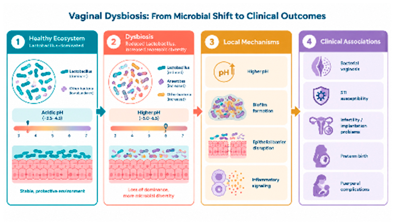
Figure 2.
Vaginal dysbiosis: transition from a Lactobacillus-dominated microbial community to dysbiosis, potential local mechanisms, and associated clinical outcomes. The figure presents conceptual associations and does not imply that every patient progresses through all stages.
Figure 2.
Vaginal dysbiosis: transition from a Lactobacillus-dominated microbial community to dysbiosis, potential local mechanisms, and associated clinical outcomes. The figure presents conceptual associations and does not imply that every patient progresses through all stages.
7. Vaginal Dysbiosis and Puerperal Complications
The clinical relevance of vaginal dysbiosis extends beyond pregnancy and delivery into the puerperium. Postpartum endometritis is a predominantly polymicrobial infection of the upper genital tract, with organisms frequently originating from the vaginal and cervical microbial communities. Accordingly, alterations in the vaginal microbiota, particularly bacterial vaginosis, have been investigated as potential risk factors for postpartum uterine infection [58].
The biological plausibility of this association lies in the close proximity between the vaginal microbial reservoir and the upper genital tract during labor and delivery. A Lactobacillus-depleted, anaerobe-enriched vaginal ecosystem may increase exposure of the cervix and uterine cavity to potentially pathogenic microorganisms and facilitate ascending infection. Nevertheless, postpartum endometritis is multifactorial, and established obstetric risk factors including cesarean delivery, prolonged rupture of membranes, prolonged labor, multiple vaginal examinations, and intra-amniotic infection remain clinically more important than vaginal dysbiosis alone [58].
Importantly, puerperal infections further illustrate that vaginal dysbiosis should not be understood as a single-pathogen disorder. Postpartum endometritis is typically polymicrobial and involves combinations of aerobic and anaerobic organisms. The vaginal microbiota may therefore serve as a microbial reservoir from which opportunistic organisms can ascend under favorable conditions rather than representing a single causative source. This concept is consistent with the broader view of vaginal dysbiosis as an ecological disturbance that may modify susceptibility to ascending infection rather than act as an isolated disease entity.
8. Therapeutic Implications: Can the Vaginal Microbiota Be Restored?
Treatment of vaginal dysbiosis should extend beyond the eradication of microorganisms associated with disease. Although antimicrobial therapy remains the cornerstone of treatment for clinically established bacterial and fungal vaginitis, eliminating dysbiosis-associated microorganisms does not necessarily result in the immediate restoration of a Lactobacillus-dominated vaginal ecosystem. This distinction is particularly relevant in recurrent disease, in which persistence of an altered microbial community may contribute to treatment failure and recurrence.
8.1. Antimicrobial Therapy: Treating the Underlying Condition
The first therapeutic step should remain etiologic and evidence-based anti-infective treatment [58]. BV should be treated with established antimicrobial regimens, VVC with appropriate antifungal therapy, and AV according to its clinical and microscopic characteristics. Treatment should target clinically relevant disease rather than individual microorganisms detected in the absence of corresponding clinical or microscopic findings.
This distinction is important because microbiological testing may identify organisms that represent normal colonization or components of a dysbiotic community rather than independent pathogens. Treatment should therefore be guided by the overall clinical, microscopic, and microbiological context rather than by attempts to eradicate every detected microorganism.
Recurrence remains a major challenge, particularly in BV, and may reflect persistence of biofilm-associated organisms, incomplete restoration of Lactobacillus dominance, re-exposure, or a combination of microbial and host-related factors. Recurrent BV may require repeated or suppressive antimicrobial regimens in selected patients.
Thus, antimicrobial therapy should be regarded primarily as treatment of the dysbiotic state and not necessarily as a means of restoring the vaginal ecosystem.
8.2. Microbiota Restoration: Probiotics as an Adjunctive Strategy
This therapeutic gap has stimulated interest in probiotics and other microbiome-directed approaches. Selected Lactobacillus strains may compete with dysbiosis-associated microorganisms, produce lactic acid and other antimicrobial metabolites, support epithelial barrier function, and promote restoration of a Lactobacillus-dominated environment.
The most promising evidence currently concerns BV. In a randomized controlled trial, women treated with vaginal metronidazole were subsequently randomized to receive Lactobacillus crispatus CTV-05 (Lactin-V) or placebo. BV recurrence at 12 weeks occurred in 30% of women receiving Lactin-V compared with 45% of those receiving placebo (RR, 0.66; 95% CI, 0.44-0.87), with a beneficial effect persisting to some extent through 24 weeks [59].
These findings support an important therapeutic concept: microbiota restoration may be most effective after reduction of the dysbiotic microbial burden rather than as a replacement for antimicrobial treatment. However, current evidence does not support a universal probiotic regimen for BV. Studies differ substantially in the Lactobacillus species and strains used, dosage, route of administration, and treatment duration. The CDC currently does not recommend available probiotic products as established adjunctive or replacement therapy for BV, although defined live biotherapeutic approaches such as Lactin-V have shown promising results and remain under development.
For VVC, the evidence is considerably less established. Probiotics may improve short-term clinical and microbiological outcomes when combined with antifungal therapy, but the certainty of the evidence remains low, and probiotics should not replace appropriate antifungal treatment. Thus, probiotics should currently be regarded as potential adjuncts rather than substitutes for etiologic therapy [60].
This treatment sequence should not be interpreted as evidence that every patient receiving antimicrobial therapy requires probiotic treatment. Rather, it represents a conceptual framework in which treatment of clinically relevant dysbiosis precedes or accompanies attempts at ecological restoration.
Whether probiotics should be administered concomitantly with antimicrobial therapy or sequentially after its completion remains unresolved. Both strategies have been studied, and the optimal timing is uncertain. The strongest evidence for a defined vaginal live biotherapeutic currently derives from administration of L. crispatus CTV-05 after antimicrobial treatment.
8.3. Beyond Probiotics
The next generation of therapies is moving beyond conventional probiotics toward defined, strain-specific, and microbiome-directed interventions. These approaches include live biotherapeutic products containing selected Lactobacillus strains, strategies targeting BV-associated biofilms, modulation of microbial metabolites, and potentially individualized interventions based on microbiome composition.
Importantly, ‘restoring the microbiota’ should not be equated simply with increasing Lactobacillus abundance. Different Lactobacillus species and strains may possess distinct metabolic and immunologic properties, and the vaginal ecosystem cannot be adequately characterized by the abundance of a single bacterial genus.
A particularly innovative approach is vaginal microbiota transplantation (VMT), in which microbiota obtained from a screened donor are transferred to a recipient with the aim of replacing a dysbiotic microbial community with a more favorable ecological state. Although early studies have generated considerable interest, VMT remains experimental and is not an established clinical treatment [61,62].
8.3.1. Why Does VMT Require Particular Caution?
VMT differs fundamentally from the administration of a defined probiotic strain. A probiotic introduces one or several selected microorganisms with relatively well-defined characteristics, whereas VMT transfers a complex microbial community of incompletely characterized composition. The transplanted material may therefore contain not only potentially beneficial Lactobacillus spp. but also other bacteria, fungi, viruses, bacteriophages, mobile genetic elements, and antimicrobial resistance determinants. Even rigorous donor screening cannot completely eliminate the possibility of transferring microorganisms or microbial characteristics whose long-term clinical relevance remains unknown [61,62].
The consequences of transferring an entire foreign microbiome may consequently be broader than those of administering a defined microbial strain. It remains unclear how a transplanted community will interact with the recipient’s existing microbiota, epithelial environment, immune system, hormonal milieu, and reproductive tract. Long-term engraftment and ecological stability are also insufficiently characterized. Recent reviews continue to emphasize the lack of standardized donor-screening criteria, procedural protocols, and long-term safety data [61,62].
Therefore, VMT should currently be regarded as an experimental microbiome-restoration strategy requiring rigorous donor screening, standardized protocols, regulatory oversight, and long-term follow-up. It should not presently be considered a routine therapeutic option for vaginal dysbiosis.
8.3.2. Limitations of Current Microbiome-Directed Therapy
Despite the rapidly expanding literature, several limitations prevent the routine implementation of microbiome-directed therapy. Studies differ substantially in their definitions of dysbiosis; microbiological sampling and sequencing methods; probiotic strains, dosage, and routes of administration; treatment duration; and clinical endpoints. Moreover, restoration of Lactobacillus abundance does not necessarily demonstrate restoration of the entire ecological and functional vaginal microbiome.
Microbiome composition is also dynamic. Hormonal status, menstruation, pregnancy, sexual activity, antimicrobial exposure, age, and other host factors may influence the vaginal microbial community. Consequently, a single microbiome measurement may not adequately represent an individual’s long-term microbial state.
Finally, although some microbiome-directed interventions have improved surrogate outcomes such as Lactobacillus colonization or BV recurrence, evidence for clinically meaningful endpoints including fertility, pregnancy outcomes, preterm birth, neonatal outcomes, and long-term reproductive health remains limited.
Future therapeutic strategies will therefore likely move from empiric probiotic supplementation toward precision microbiome medicine, in which microbial composition, functional characteristics, host environment, and clinical phenotype are considered together.
8.3.3. Physical and Energy-Based Therapies
In addition to antimicrobial and probiotic approaches, physical rehabilitation and energy-based interventions have been investigated as nonpharmacologic strategies for improving the vaginal and pelvic environment.
Vaginal carbon dioxide (CO₂) and erbium-doped yttrium aluminum garnet (Er: YAG) laser therapies induce controlled thermal or microablative effects that may promote epithelial remodeling, collagen synthesis, vascularization, and restoration of tissue elasticity and hydration. Clinical studies have reported improvements in vaginal dryness, dyspareunia, burning, and other symptoms of genitourinary syndrome of menopause. However, systematic reviews of randomized controlled trials indicate that the certainty of the evidence remains low, with several sham-controlled studies failing to demonstrate clinically meaningful superiority over placebo or established therapies [63].
Similarly, electrical stimulation and other pelvic floor rehabilitation techniques may improve neuromuscular activation, pelvic floor muscle contraction, and urinary continence, particularly when used as adjuncts to pelvic floor muscle training. Current evidence, however, does not demonstrate that electrical stimulation leads to an improvement in vaginal dysbiosis [64].
Importantly, these interventions should therefore be regarded as supportive or symptom-directed therapies rather than microbiome-restoring treatments. Robust evidence that laser therapy, electrical stimulation, or other energy-based modalities directly normalize vaginal microbial communities is currently lacking.
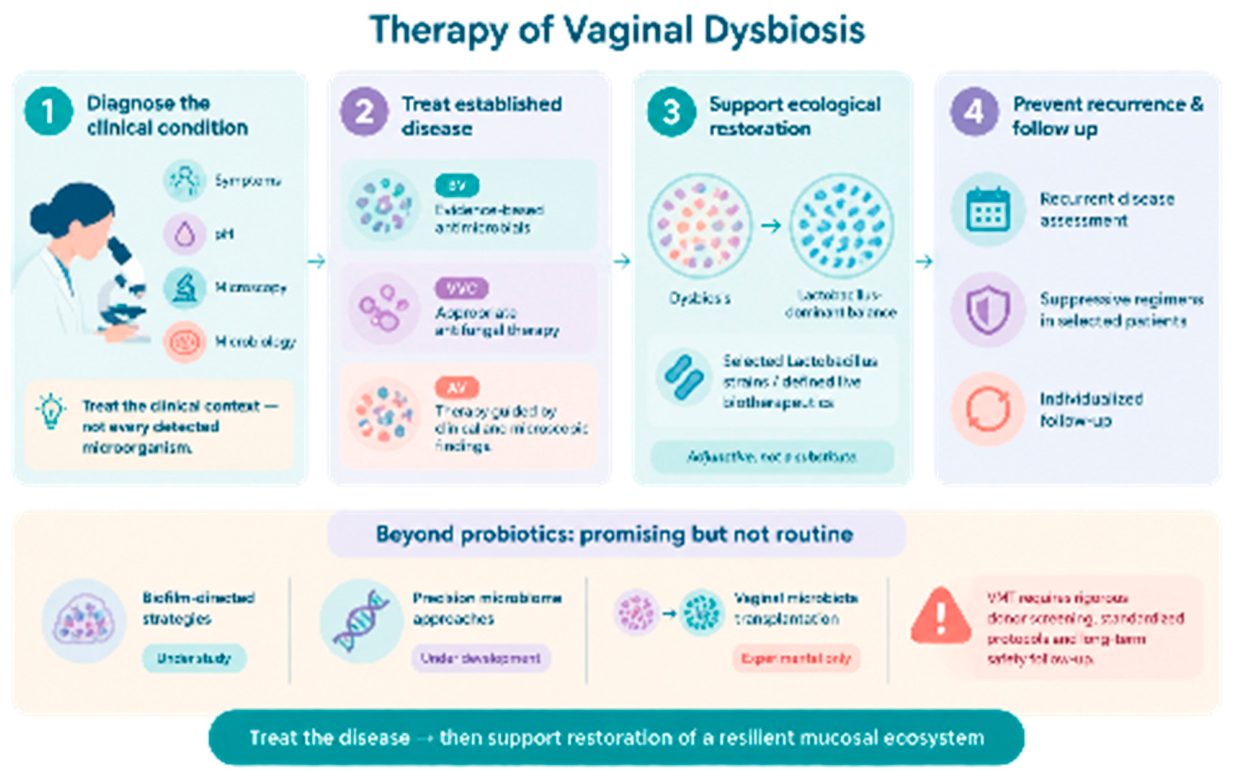
Figure 3.
Therapeutic approach to vaginal dysbiosis: diagnose the clinical condition, treat established disease, support ecological restoration, and prevent recurrence.
Figure 3.
Therapeutic approach to vaginal dysbiosis: diagnose the clinical condition, treat established disease, support ecological restoration, and prevent recurrence.
9. Conclusions and Future Perspectives
Vaginal dysbiosis should not be viewed solely as a local bacterial disorder but as a condition capable of modifying mucosal host–pathogen interactions. Alterations in the vaginal microbial ecosystem may influence epithelial integrity, local immune responses, microbial persistence, and susceptibility to ascending infection, with potential consequences ranging from recurrent vaginitis to reproductive, obstetric, and puerperal outcomes. However, the strength of the evidence varies among clinical conditions, and causality remains to be established for many of these associations. Importantly, the detection of an individual microorganism should not automatically be interpreted as evidence of infection; its clinical significance depends on the surrounding microbial community, host response, symptoms, and overall clinical context.
Future research should move beyond descriptive microbiome profiling toward a better understanding of microbial function and host-microbiome interactions. Antimicrobial treatment remains essential for established infections, whereas microbiota restoration with selected probiotics or defined live biotherapeutics represents a promising adjunctive strategy, particularly for recurrent dysbiosis. However, the optimal strains, timing, dosage, and long-term clinical benefits remain insufficiently defined. More complex approaches, such as vaginal microbiota transplantation, remain experimental because they involve the transfer of an incompletely characterized microbial ecosystem. Their long-term effects on recipients and subsequent host–microbiome interactions are not yet known. Future studies should therefore focus on standardized diagnostic approaches, longitudinal assessments, clinically meaningful outcomes, and safe, targeted strategies for precision microbiome restoration.
Take-Home Message: Vaginal dysbiosis should not be viewed solely as a local bacterial disorder but as a condition capable of modifying mucosal host–pathogen interactions. The future of vaginal dysbiosis management lies not simply in eliminating microorganisms but in understanding and restoring a resilient mucosal ecosystem.
Author Contributions
Conceptualization, D. Vlasakova and co-authors; writing original draft preparation, D. Vlasakova and co-authors; writing review and editing, all authors. All authors have read and agreed to the published version of the manuscript.
Funding
The authors received no external funding for this work.
Institutional Review Board Statement
Not applicable. This manuscript is a narrative review and does not report research involving human participants, human samples, or animals.
Informed Consent Statement
Not applicable.
Data Availability Statement
No new data were generated or analyzed in this study. All information discussed is based on the cited literature.
Use of Artificial Intelligence
Artificial intelligence AI-based tools were used solely to assist with language editing, grammar correction, stylistic refinement of the manuscript, and the preparation and visual enhancement of figures. The authors reviewed and critically evaluated all AI-assisted outputs and take full responsibility for the accuracy, originality, and scientific content of the manuscript.
Conflicts of Interest
The authors declare no conflict of interest.
References
- Nunn, K.L.; Forney, L.J. Unraveling the dynamics of the human vaginal microbiome. Yale J. Biol. Med. 2016, 89, 331–337. [Google Scholar]
- Smith, S.B.; Ravel, J. The vaginal microbiota, host defence and reproductive physiology. J. Physiol. 2017, 595, 451–463. [Google Scholar] [CrossRef]
- Ma, B.; Forney, L.J.; Ravel, J. Vaginal microbiome: Rethinking health and disease. Annu. Rev. Microbiol. 2012, 66, 371–389. [Google Scholar] [CrossRef]
- Ravel, J.; Gajer, P.; Abdo, Z.; et al. Vaginal microbiome of reproductive-age women. Proc. Natl. Acad. Sci. USA 2011, 108 (Suppl. 1), 4680–4687. [Google Scholar] [CrossRef]
- Gajer, P.; Brotman, R.M.; Bai, G.; et al. Temporal dynamics of the human vaginal microbiota. Sci. Transl. Med. 2012, 4, 132ra52. [Google Scholar] [CrossRef]
- Chee, W.J.Y.; Chew, S.Y.; Than, L.T.L. Vaginal microbiota and the potential of Lactobacillus derivatives in maintaining vaginal health. Microorganisms 2020, 8, 1069. [Google Scholar]
- Fettweis, J.M.; Brooks, J.P.; Serrano, M.G.; et al. Differences in vaginal microbiome in African American women versus women of European ancestry. Microbiology 2014, 160, 2272–2282. [Google Scholar] [CrossRef]
- Roachford, O.S.; Alleyne, A.T.; Nelson, K.E. Insights into the vaginal microbiome in a diverse group of women of African, Asian and European ancestries. PeerJ 2022, 10, e14449. [Google Scholar] [CrossRef]
- Kervinen, K.; Holster, T.; Saqib, S.; et al. Parity and gestational age are associated with vaginal microbiota composition in term and late-term pregnancies. EBioMedicine 2022, 81, 104107. [Google Scholar] [CrossRef]
- Prince, A.L.; Chu, D.M.; Seferovic, M.D.; et al. The vaginal microbiota of pregnant women varies with gestational age, maternal age, and parity. mBio 2017, 8, e01597-17. [Google Scholar] [CrossRef]
- Horowitz, B.J.; Mårdh, P.-A.; Nagy, E.; Rank, E.L. Vaginal lactobacillosis. Am. J. Obstet. Gynecol. 1994, 170, 857–861. [Google Scholar] [CrossRef]
- Zheng, N.; Guo, R.; Yao, Y.; Jin, M.; Pan, L.; Huang, Y. Contribution of Lactobacillus iners to Vaginal Health: A Systematic Review. Front. Cell. Infect. Microbiol. 2021, 11, 792011. [Google Scholar] [CrossRef]
- van de Wijgert, J.H.H.M.; Verwijs, M.C.; Turner, A.N.; Morrison, C.S. Hormonal contraception decreases bacterial vaginosis but oral contraception may increase vaginal candidiasis: A systematic review and meta-analysis. PLoS ONE 2013, 8, e73055. [Google Scholar] [CrossRef]
- Brotman, R.M.; Klebanoff, M.A.; Nansel, T.R.; et al. A longitudinal study of vaginal douching and bacterial vaginosis: A randomized trial. Am. J. Obstet. Gynecol. 2010, 202, 437.e1–437.e8. [Google Scholar]
- Madden, T.; Grentzer, J.M.; Secura, G.M.; Allsworth, J.E.; Peipert, J.F. Risk of bacterial vaginosis in users of the intrauterine device: A longitudinal study. Sex. Transm. Dis. 2012, 39, 217–222. [Google Scholar] [CrossRef]
- Achilles, S.L.; Austin, M.N.; Meyn, L.A.; et al. Impact of contraceptive initiation on vaginal microbiota. Am. J. Obstet. Gynecol. 2018, 218, 622.e1–622.e10. [Google Scholar] [CrossRef]
- Gudnadottir, U.; Debelius, J.W.; Du, J.; et al. The vaginal microbiome and the risk of preterm birth: A systematic review and network meta-analysis. Sci. Rep. 2022, 12, 7926. [Google Scholar] [CrossRef]
- Zhou, Q.; Yu, Y.; Zhou, J.; Liu, J.; Gao, J. Relationship of Lactobacillus vaginal microbiota changes and the risk of preterm birth: A systematic review and meta-analysis. J. Womens Health 2024, 33, 2–12. [Google Scholar] [CrossRef]
- Hoyme, U.B.; Hesse, M. Reduced incidence of early preterm birth in the State of Thuringia following an intravaginal pH self-monitoring screening program. Arch. Gynecol. Obstet. 2020, 302, 1–4. [Google Scholar] [CrossRef]
- Bitzer, E.M.; Schneider, A.; Wenzlaff, P.; Hoyme, U.B.; Siegmund-Schultze, E. Self-testing of vaginal pH to prevent preterm delivery: A controlled trial. Dtsch. Arztebl. Int. 2011, 108, 81–86. [Google Scholar] [CrossRef]
- Schwebke, J.R.; Muzny, C.A.; Josey, W.E. Role of Gardnerella vaginalis in the pathogenesis of bacterial vaginosis: A conceptual model. J. Infect. Dis. 2014, 210, 338–343. [Google Scholar] [CrossRef]
- Muzny, C.A.; Kardas, P. A narrative review of current challenges in the diagnosis and management of bacterial vaginosis. Sex. Transm. Dis. 2020, 47, 441–446. [Google Scholar] [CrossRef]
- Swidsinski, A.; Mendling, W.; Loening-Baucke, V.; et al. Adherent biofilms in bacterial vaginosis. Obstet. Gynecol. 2005, 106, 1013–1023. [Google Scholar] [CrossRef]
- Bradshaw, C.S.; Morton, A.N.; Hocking, J.; et al. High recurrence rates of bacterial vaginosis over the course of 12 months after oral metronidazole therapy and factors associated with recurrence. J. Infect. Dis. 2006, 193, 1478–1486. [Google Scholar] [CrossRef]
- Vodstrcil, L.A.; Plummer, E.L.; Fairley, C.K.; et al. Male-partner treatment to prevent recurrence of bacterial vaginosis. N. Engl. J. Med. 2025, 392, 1425–1435. [Google Scholar] [CrossRef]
- Horner, P.; Donders, G.; Cusini, M.; et al. Should we be testing for urogenital Mycoplasma hominis, Ureaplasma parvum and Ureaplasma urealyticum in men and women? J. Eur. Acad. Dermatol. Venereol. 2018, 32, 1845–1851. [Google Scholar] [CrossRef]
- Jonduo, M.E.; Vallely, L.M.; Wand, H.; et al. Adverse pregnancy and birth outcomes associated with Mycoplasma hominis, Ureaplasma urealyticum and Ureaplasma parvum: A systematic review and meta-analysis. BMJ Open 2022, 12, e062990. [Google Scholar] [CrossRef]
- Mitra, A.; MacIntyre, D.A.; Lee, Y.S.; et al. Maternal Ureaplasma exposure during pregnancy and the risk of preterm birth and bronchopulmonary dysplasia: A meta-analysis. Arch. Gynecol. Obstet. 2022, 306, 1863–1872. [Google Scholar] [CrossRef]
- Jensen, J.S.; Cusini, M.; Gomberg, M.; et al. 2021 European guideline on the management of Mycoplasma genitalium infections. J. Eur. Acad. Dermatol. Venereol. 2022, 36, 641–650. [Google Scholar] [CrossRef]
- Workowski, K.A.; Bachmann, L.H.; Chan, P.A.; et al. Sexually transmitted infections treatment guidelines, 2021: Mycoplasma genitalium infection. MMWR Recomm. Rep. 2021, 70(No. RR-4), 1–13. [Google Scholar] [CrossRef]
- Farr, A.; Effendy, I.; Frey Tirri, B.; et al. Guideline: Vulvovaginal candidosis (AWMF 015/072, S2k level). Mycoses 2021, 64, 583–602. [Google Scholar] [CrossRef]
- Mendling, W.; Brasch, J. Guideline on vulvovaginal candidosis. Mycoses 2012, 55 (Suppl. 3), 1–13. [Google Scholar] [CrossRef]
- Sobel, J.D.; Mitchell, C.; Calkins, B.; et al. Mixed vaginitis due to bacterial vaginosis and candidiasis. J. Low. Genit. Tract. Dis. 2021, 25, 154–160. [Google Scholar]
- Zhang, Z.; et al. Vaginal microbiota of asymptomatic bacterial vaginosis and vulvovaginal candidiasis: Are they different from normal microbiota? J. Infect. Public Health 2019. [Google Scholar] [CrossRef]
- Donders, G.G.G.; Bellen, G.; Ausma, J.; et al. The effect of antifungal treatment on the vaginal flora of women with vulvo-vaginal yeast infection with or without bacterial vaginosis. Eur. J. Clin. Microbiol. Infect. Dis. 2011, 30, 59–63. [Google Scholar] [CrossRef]
- Donders, G.G.G.; Bellen, G.; Rezeberga, D. Vaginal pH and microbiota during fluconazole maintenance treatment for recurrent vulvovaginal candidosis. Diagn. Microbiol. Infect. Dis. 2020, 97, 115024. [Google Scholar] [CrossRef]
- Naglik, J.R.; Moyes, D.L. Epithelial cell innate response to Candida albicans. Adv. Dent. Res. 2011, 23, 50–55. [Google Scholar] [CrossRef]
- Moyes, D.L.; Wilson, D.; Richardson, J.P.; et al. Candida albicans yeast and hyphae are discriminated by the innate immune system. PLoS Pathog. 2016, 12, e1005722. [Google Scholar] [CrossRef]
- Roberts, C.L.; et al. Vulvovaginal yeast infections during pregnancy and perinatal outcomes: A systematic review and meta-analysis. BMC Womens Health 2023, 23, 76. [Google Scholar] [CrossRef]
- Gigi, V.; et al. Asymptomatic vaginal Candida colonization and adverse pregnancy outcomes: A systematic review and meta-analysis. Am. J. Obstet. Gynecol. MFM 2021, 3, 100163. [Google Scholar] [CrossRef]
- Bliss, J.M.; Basile, L.A.; Clancy, C.J.; et al. Vertical and horizontal transmission of Candida albicans in very low birth weight infants using DNA fingerprinting techniques. Pediatr. Infect. Dis. J. 2008, 27, 231–235. [Google Scholar] [CrossRef]
- Weimer, K.E.D.; Smith, P.B.; Puia-Dumitrescu, M.; Aleem, S. Invasive fungal infections in neonates: A review. Pediatr. Res. 2022, 91, 404–412. [Google Scholar] [CrossRef]
- Donders, G.G.G.; Vereecken, A.; Bosmans, E.; et al. Definition of a type of abnormal vaginal flora that is distinct from bacterial vaginosis: Aerobic vaginitis. BJOG 2002, 109, 34–43. [Google Scholar] [CrossRef]
- Donders, G.G.G.; Bellen, G.; Rezeberga, D. Aerobic vaginitis in pregnancy. BJOG 2011, 118, 1163–1170. [Google Scholar] [CrossRef]
- American College of Obstetricians and Gynecologists. Prevention of Group B Streptococcal Early-Onset Disease in Newborns. ACOG Committee Opinion No. 797. Obstet. Gynecol. 2020, 135, e51–e72. [Google Scholar] [CrossRef]
- Han, C.; Wu, W.; Fan, A.; et al. Diagnostic and therapeutic advancements for aerobic vaginitis. Arch. Gynecol. Obstet. 2015, 291, 251–257. [Google Scholar] [CrossRef]
- Reichman, O.; Akins, R.; Guise, J.M. Desquamative inflammatory vaginitis. Best Pract. Res. Clin. Obstet. Gynaecol. 2014, 28, 1042–1050. [Google Scholar] [CrossRef]
- Sobel, J.D.; Reichman, O. Desquamative inflammatory vaginitis. Best Pract. Res. Clin. Obstet. Gynaecol. 2014, 28, 1042–1050. [Google Scholar] [CrossRef]
- Brusselaers, N.; Shrestha, S.; van de Wijgert, J.; Verstraelen, H. Vaginal dysbiosis and the risk of human papillomavirus and cervical cancer: A systematic review and meta-analysis. Am. J. Obstet. Gynecol. 2019, 221, 9–18. [Google Scholar] [CrossRef]
- Norenhag, J.; et al. The vaginal microbiota, human papillomavirus and cervical dysplasia: A systematic review and network meta-analysis. BJOG 2020, 127, 171–180. [Google Scholar] [CrossRef]
- Houvessou, G.M.; Alfane, N.W.A.; Mahoche, M. Dynamic, transition and variation of cervicovaginal microbiome and HPV infection and cervical dysplasia and cancer: A systematic review. Infect. Agents Cancer 2026, 21, 7. [Google Scholar] [CrossRef]
- Atashili, J.; Poole, C.; Ndumbe, P.M.; Adimora, A.A.; Smith, J.S. Bacterial vaginosis and HIV acquisition: A meta-analysis of published studies. AIDS 2008, 22, 1493–1501. [Google Scholar] [CrossRef]
- Gosmann, C.; Anahtar, M.N.; Handley, S.A.; et al. Lactobacillus-deficient cervicovaginal bacterial communities are associated with increased HIV acquisition in young South African women. Immunity 2017, 46, 29–37. [Google Scholar] [CrossRef]
- Haahr, T.; Jensen, J.S.; Thomsen, L.; et al. Reproductive outcome of patients undergoing in vitro fertilisation treatment and diagnosed with bacterial vaginosis or abnormal vaginal microbiota: A systematic PRISMA review and meta-analysis. BJOG 2019, 126, 200–209. [Google Scholar] [CrossRef]
- Haahr, T.; et al. The association between vaginal dysbiosis and reproductive outcomes in sub-fertile women undergoing IVF treatment: A systematic PRISMA review and meta-analysis. Reprod. Biomed. Online 2021. [Google Scholar] [CrossRef]
- Maksimovic Celicanin, M.; Haahr, T.; Humaidan, P.; Skafte-Holm, A. Vaginal dysbiosis—the association with reproductive outcomes in IVF patients: A systematic review and meta-analysis. Curr. Opin. Obstet. Gynecol. 2024, 36, 155–164. [Google Scholar] [CrossRef]
- Samama, M.; Ueno, J.; Veiga, E.C.A.; et al. Vaginal microbiome and its relationship with assisted reproduction: A systematic review and meta-analysis. Life 2025, 15, 1382. [Google Scholar] [CrossRef]
- Workowski, K.A.; Bachmann, L.H.; Chan, P.A.; et al. Sexually transmitted infections treatment guidelines, 2021: Bacterial vaginosis. MMWR Recomm. Rep. 2021, 70(No. RR-4), 1–13. [Google Scholar] [CrossRef]
- Cohen, C.R.; et al. Randomized trial of Lactin-V to prevent recurrence of bacterial vaginosis. N. Engl. J. Med. 2020, 382, 1906–1915. [Google Scholar] [CrossRef]
- Zahedifard, T.; et al. The role of probiotics in the treatment of vulvovaginal candidiasis: A systematic review and meta-analysis. Eur. J. Health Sci. 2024, 10, 81–94. [Google Scholar] [CrossRef]
- Lev-Sagie, A.; Goldman-Wohl, D.; Cohen, Y.; et al. Vaginal microbiome transplantation in women with intractable bacterial vaginosis. Nat. Med. 2019, 25, 1500–1504. [Google Scholar] [CrossRef]
- Vijay, S.; et al. The evolving facets of vaginal microbiota transplantation: Reinvigorating the unexplored frontier amid complex challenges. Arch. Microbiol. 2024, 206, 370. [Google Scholar] [CrossRef]
- Pessoa, L.L.M.N.; de Souza, A.T.B.; Sarmento, A.C.A.; et al. Laser therapy for genitourinary syndrome of menopause: Systematic review and meta-analysis of randomized controlled trials. Rev. Bras. Ginecol. Obstet. 2024, 46, e-rbgo38. [Google Scholar] [CrossRef]
- Zhang, Y.; Yang, H.; Zhang, C.; Lin, L.; Yang, W.; Xiong, G.; Gao, G. The impact of pelvic floor electrical stimulation on vaginal microbiota and immunity. Front. Cell. Infect. Microbiol. 2022, 12, 1006576. [Google Scholar] [CrossRef]

| Community State Type | Predominant bacterial species | Microbial characteristics and potential clinical relevance |
|---|---|---|
| CST I | Lactobacillus crispatus | Lactobacillus-dominated community, generally associated with lactic acid production, an acidic vaginal environment, and comparatively high microbial stability. |
| CST II | Lactobacillus gasseri | Lactobacillus-dominated community associated with lactic acid production and an acidic vaginal environment. |
| CST III | Lactobacillus iners | Common Lactobacillus-dominated community that may be comparatively dynamic and may occur during transitions between different microbial states. |
| CST IV | Diverse anaerobic and facultatively anaerobic bacteria | Lactobacillus-depleted and highly diverse community, frequently associated with BV-associated microbial profiles, but not synonymous with clinical BV. |
| CST V | Lactobacillus jensenii | Lactobacillus-dominated community associated with lactic acid production and an acidic vaginal environment. |
Disclaimer/Publisher’s Note: The statements, opinions and data contained in all publications are solely those of the individual author(s) and contributor(s) and not of MDPI and/or the editor(s). MDPI and/or the editor(s) disclaim responsibility for any injury to people or property resulting from any ideas, methods, instructions or products referred to in the content. |
© 2026 by the authors. Licensee MDPI, Basel, Switzerland. This article is an open access article distributed under the terms and conditions of the Creative Commons Attribution (CC BY) license.
Copyright: This open access article is published under a Creative Commons CC BY 4.0 license, which permit the free download, distribution, and reuse, provided that the author and preprint are cited in any reuse.