

Submitted:
18 October 2026
Posted:
20 October 2026
You are already at the latest version
Abstract
Background: The cornea is a transparent, avascular, and highly specialized structure that serves as the eye’s primary refractive surface, making it essential for normal vision. Corneal ulcers, commonly resulting from trauma or infection, are a leading cause of ocular morbidity and blindness in dogs. Surgical intervention is often required for deep or non-healing ulcers. Conjunctival grafts help restore corneal integrity but may prolong recovery. Canine amniotic membrane (AM) possesses an-ti-inflammatory, antimicrobial, and epithelialization-promoting properties that may enhance corneal wound healing. Methods: Twelve clinical canine cases with corneal ulcers were randomly allocated into two Groups (n = 6 each). Group A underwent bulbar conjunctival pedicle grafting, while Group B received canine amniotic membrane grafts prepared from placentas collected during elective cae-sarean sections. The amniotic membrane was applied in three layers and stabilized with a third-eyelid flap and partial tarsorrhaphy. Postoperative assessment included physiological pa-rameters, detailed ophthalmic examinations, tear production tests, fluorescein dye uptake, and rel-evant biochemical analyses. Results: Complete healing was achieved in five of six dogs in Group A and in all six dogs in Group B. Physiological and biochemical values did not differ significantly between Groups; however, Group B exhibited more rapid epithelialization, reduced postoperative complications, and overall better clinical recovery. Conclusion: Both surgical approaches were effec-tive in managing canine corneal ulcers, but the canine amniotic membrane graft provided superior healing, faster epithelial restoration, and improved clinical outcomes.
Keywords:
canine corneal ulcer
; canine amniotic membrane graft
; Interleukin
; corneal epithelialization
1. Introduction
The eye is a remarkable organ that reflects both inner feelings and systemic health. It is situated in the orbital cavity, where the cornea and sclera meet at the limbus. The cornea is a transparent, avascular structure that forms the anterior one-sixth of the eyeball. It serves as the principal refractive surface, maintains intraocular pressure, and protects against environmental insults [1]. Corneal sensitivity helps prevent injury and is essential for maintaining corneal transparency. Keratitis, or inflammation of the cornea, is a common ocular disease that can lead to blindness or loss of the eye [2]. It may be classified as ulcerative or non-ulcerative. Corneal ulcers may be non-infectious (e.g., due to erosions or trauma) or infectious (e.g., bacterial, viral, or fungal), although secondary infection may occur following epithelial damage [3]. Based on the extent of tissue loss, ulcers are categorized as superficial, descemetoceles, or perforated [4]. Vision loss may result from corneal opacification or perforation. Diagnosis of ulcerative keratitis relies on clinical examination, cytology, and microbiological analysis [2]. Schirmer’s Tear Test is used to diagnose keratoconjunctivitis sicca [5], while fluorescein and Rose Bengal staining are employed to detect corneal ulcers and epithelial defects [6]. An accurate diagnosis is essential to guide appropriate therapy. Corneal ulcers ≤2 mm in diameter can be managed conservatively; however, larger defects typically require surgical intervention [7]. Various techniques have been reported to be effective in treating corneal ulcers in canine patients, whether used individually or in combination [8]. Medical management generally includes topical and systemic antibiotics alongside supportive therapies, while surgery becomes necessary when significant corneal tissue loss occurs. Surgical options include penetrating keratoplasty, lamellar corneal grafts, conjunctival grafting techniques, corneoscleral transposition, and the use of synthetic or biological grafts, such as human amniotic membrane patches, canine amniotic membrane grafts, porcine small intestinal submucosal grafts, and equine pericardial grafts [9,10]. Conjunctival pedicle graft surgery helps restore corneal integrity, prevents disease progression and infection, relieves pain, improves ocular appearance, and may avert enucleation [11]. It is widely used in ophthalmology for various ocular surface reconstruction procedures, including the management of persistent epithelial defects, non-healing corneal ulcers, corneal perforations, descemetoceles, bullous keratopathy, and corneal disorders associated with limbal stem cell deficiency. It is also employed in the treatment of pterygium, conjunctival reconstruction, management of corneoscleral melts and perforations, and as an adjunct in several glaucoma surgeries [12]. Amniotic membrane (AM), the innermost layer of the placenta, promotes epithelialization and possesses anti-fibrotic, anti-inflammatory, anti-angiogenic, and antimicrobial properties [13]. Human AM grafts are used in various corneal disorders, including burnt corneas [14], limbal stem cell deficiency [15], bullous keratopathy [16], deep corneal ulcers [17], neurotrophic corneal ulcers [18], descemetoceles and corneal perforations, infectious corneal ulcers, and defects after the excision of ocular surface neoplasia [19]. Canine AM shares a similar structure, including an epithelium, a thick basement membrane, and stroma, and may offer comparable benefits in ocular surface reconstruction. Cryopreserved AM can be used after thawing by keeping the bottle at room temperature for 10 minutes and remains suitable for clinical use for up to 6 months after preparation, highlighting its practicality in veterinary ophthalmology [20].
In addition to its recognized advantages, AM transplantation presents several practical limitations. These include limited mechanical strength in deep stromal defects, potential early biodegradation, and reduced efficacy in severely infected or highly vascularized corneas [21]. The procedure also requires strict preparation, sterilisation, and storage protocols to maintain membrane integrity and minimise contamination risk [22]. Although generally well tolerated, AM grafts may cause transient inflammation, graft displacement, premature dissolution, or, rarely, immune-mediated rejection when allogenic tissue is used [23]. Clinical success may further depend on membrane orientation, fixation technique, and postoperative conditions [24]. Despite these constraints, AM remains a valuable biomaterial for ocular surface reconstruction due to its biocompatibility and wound-modulating properties. Nevertheless, comparative studies are still needed to clarify its performance relative to other grafting options in veterinary ophthalmology [25]. Hypothesis of the study was that allogeneic canine amniotic membrane grafting would result in faster corneal epithelialization, fewer postoperative complications, and improved clinical healing outcomes compared with bulbar conjunctival pedicle grafting in dogs with corneal ulcers, without inducing significant changes in systemic physiological or biochemical parameters. Accordingly, the present study was undertaken to comparatively evaluate bulbar conjunctival pedicle grafts and allogeneic canine amniotic membrane grafts for the surgical management of corneal ulcers in dogs.
2. Materials and Methods
2.1. Animals and Experimental Design
This study included 12 clinical cases of corneal ulcers in client-owned canines, regardless of age, body weight, or breed, at the Veterinary Clinical Complex, Bihar Veterinary College, Patna (India). All cases were subjected to surgical management. Each patient underwent a detailed ophthalmic examination to confirm the diagnosis of corneal ulceration. The cases were randomly divided into two equal Groups based on the surgical technique employed. A bulbar conjunctival pedicle graft was applied in Group A, and a canine amniotic membrane graft was applied in Group B.
2.2. Preparation, Preservation and Storage of Amniotic Membrane
The amniotic membrane was collected from an elective canine caesarean section. The placenta was decontaminated to eliminate potential surface pathogens and repeatedly rinsed with antibiotic and antimycotic solutions. Dulbecco’s Modified Eagle’s Medium (DMEM) was used to remove blood clots. Any residual chorion or blood clots attached to the amniotic membrane were carefully removed from the epithelial layer using round-ended forceps. The membrane was uniformly spread, with the epithelial/basement membrane side facing upward and free of folds or tears, onto a sterile nitrocellulose membrane. It was then trimmed along the edges of the nitrocellulose membrane, allowing the amniotic membrane to adhere firmly. The adhered amniotic membrane, together with the supporting filter paper, was placed into a preservative medium and stored at −80 °C until further use. The preservative medium generally comprised a 1:1 (v/v) mixture of sterile glycerol (sterilized by autoclaving) and EMEM with 3.3% L-glutamine, 25 µg/mL gentamicin, 50 U/mL penicillin, 100 µg/mL ciprofloxacin, and 0.5 mg/mL amphotericin B. These membranes can be used for up to six months post-preparation, following thawing at room temperature for 10 minutes [26].
All clinical cases received preoperative ocular antisepsis with antibiotic (0.6% Besifloxacin eye drops) and extraocular asepsis with 0.5% diluted povidone-iodine, administered two to three times at 15-minute intervals for 1 hour just before surgery. The interval between diagnosis and surgery ranged from 1 to 3 days, depending on the severity and stabilization of the corneal ulcer. Before surgical intervention, supportive therapy including topical lubricants, anti-inflammatory agents for pain control, and systemic antibiotics when indicated, was used to optimize corneal surface conditions and general health before graft placement. A systemic antibiotic—Amoxicillin and Sulbactam at a dosage of 12 mg/kg body weight (Amoxirum Forte, Virbac, Mumbai, India)—was administered to prevent systemic infection. Surgery was performed under general anaesthesia.
2.3. Surgical Procedure
Surgery was performed under general anaesthesia. For induction, all animals were premedicated with glycopyrrolate (0.01 mg/kg, intramuscularly), butorphanol (0.2 mg/kg, intramuscularly), and diazepam (0.3 mg/kg, intravenously). Anaesthesia was induced using propofol at 3–6 mg/kg, administered intravenously to effect, until endotracheal intubation was achieved. Anaesthesia was then maintained with 1–2% isoflurane. Following induction, animals were positioned in lateral recumbency with the affected eye uppermost. Soft bedding material was placed under the head to provide gentle elevation and support. In Group A, a bulbar conjunctival pedicle graft was applied (Figure 1), whereas in Group B, an amniotic membrane graft was used (Figure 2). For the application of the bulbar conjunctival pedicle graft, the affected eye was gently retracted from the bony orbit using digital pressure to adequately expose the bulbar conjunctiva. The recipient ulcer margins were prepared by thoroughly debriding necrotic tissue and infected stromal layers by a corneal curette and ophthalmic scalpel. Depending on the location of the ulcer, conjucntival pedicle grafts were harvested from the superior, inferior, or lateral bulbar conjunctiva, whichever site was closest to the lesion—to minimize conjunctival pedicle graft travel distance.
After identifying the location of the ulcer, an incision of appropriate length was made on the bulbar conjunctiva, approximately 1–2 mm from the limbus. The conjunctival pedicle graft was undermined by blunt dissection using Stevens’s tenotomy scissors. A second
Incise on was then made at the base on either side of the graft, depending on which side allowed easier translocation or rotation of the graft to the recipient ulcer site. The conjunctival pedicle graft thus created was extended to cover the ulcer, reaching its margins. The graft was secured to the cornea using a simple interrupted suture pattern with 5-0 polyglactin 910, absorbable suture material.
In Group B, the ulcer bed was similarly prepared for the application of the amniotic membrane graft. The membrane was trimmed to a size approximately 2–3 mm larger than the ulcer dimensions. As the amniotic membrane consists of two surfaces—epithelial and stromal—the stromal side was placed in contact with the epithelial surface of the cornea, serving as the graft interface. The membrane was transplanted in three layers over the corneal ulcer to ensure full coverage of the defect. Before placement, the dry amniotic membrane was moistened by instilling a few drops of sterile saline solution to facilitate adherence to the corneal surface.
Following graft application in both Groups, a third eyelid flap was sutured to the lateral margins of the upper eyelid. To protect the eye from self-trauma, temporary partial tarsorrhaphy was performed using non-absorbable nylon suture and maintained for 14 days. The third eyelid flap sutures were removed on day 21. On the same day, the surgical outcome was evaluated, and the total duration required for ulcer healing was recorded.
2.4. Postoperative Care
The post-operative regimen for both Groups included parenteral administration of antibiotics, Amoxicillin and Sulbactam (12 mg/kg, intramuscularly) for 7 days—and a non-steroidal anti-inflammatory drug (Meloxicam, 0.2 mg/kg, intramuscularly) for 3 days. In addition, topical antibiotic eye drops (Besifloxacin) and a topical antihistaminic solution (0.2% Olopatadine) were instilled four times daily for 14 days. All animals were discharged on the same day of surgery once fully recovered from anesthesia, and postoperative evaluations, including assessment of graft absorption, were performed during scheduled outpatient follow-up visits until complete graft absorption was confirmed.
2.5. Evaluation and Data Collection
Physiological parameters, including rectal temperature (°C), respiratory rate (breaths per minute), heart rate (beats per minute), and mucous membrane color, were recorded before surgery, on postoperative days 7 and 45, and on the day of complete graft absorption.
Ophthalmic examinations—including menace response (MR), palpebral reflex, pupillary light reflex (PLR), cotton ball test (tracking reflex), fluorescein dye test (FDT), and Schirmer’s tear test—were conducted preoperatively and postoperatively at the time of complete clinical recovery. The menace response was assessed by performing a visually threatening gesture (e.g., waving a hand) toward the eye to elicit a blink reaction. The palpebral reflex was evaluated by gently tapping the medial and lateral canthus to induce a blink response. The pupillary light reflex was tested using a pen torch or bright light source to observe pupillary constriction in response to light. The cotton ball test involved dropping a small cotton ball approximately 20–30 cm in front of either eye and recording the dog’s visual tracking response. The fluorescein dye test was performed using commercially available sterile strips impregnated with fluorescein dye. The dye stained only the corneal defect where the pre-corneal tear film disruption was present. Schirmer’s tear test was used to assess tear production. Sterile, absorbent Whatman No. 41 filter paper strips (5 mm × 40 mm) were placed in the ventral conjunctival cul-de-sac of the lower eyelid for exactly one minute, and the length of the moistened strip was recorded. In addition to clinical evaluations, biochemical analysis of T-helper cytokine levels was performed. TH1 (interleukin-2, IL-2) and TH2 (interleukin-4, IL-4) concentrations were measured. IL-2 and IL-4 were measured in blood serum. Peripheral blood was collected into plain tubes, allowed to clot, centrifuged, and the serum was stored at −80 °C. Cytokine concentrations were quantified using commercial, canine-specific sandwich ELISA kits (Geno Biosciences Private Limited, Delhi, India) according to the manufacturer’s protocols; samples were run in duplicate and concentrations (pg/mL) calculated from kit standards.
The time to complete clinical recovery was also recorded.
2.6. Statistical Analysis
The means and standard errors of various parametric observations were calculated and compared between Groups using the independent samples t-test. One-way analysis of variance (ANOVA) was employed to evaluate the effects of parametric variables across different time intervals [27]. Non-parametric data were analyzed using the Related Samples Wilcoxon Signed-Rank Test, as described by Scheffé [28], with statistical analysis performed using SPSS software, version 29.0 (IBM Corp., Armonk, NY, USA).
3. Results
3.1. Physiological Parameters
Physiological parameters, including rectal temperature, respiratory rate, and heart rate, did not show any statistically significant changes across the observation periods in either Group (Table 1).
3.2. Ocular Function and Healing
Menace reflex scores, pupillary light reflex, and cotton ball test results showed statistically significant improvement at complete recovery in both Groups when compared to their respective baseline values. In contrast, palpebral reflex scores exhibited non-significant improvements (p > 0.05) at complete recovery in both Groups relative to baseline (Table 2). Schirmer’s Tear Test (STT) values demonstrated no statistically significant change at the time of complete recovery in either compared to baseline measurements (Table 2). Observations from the fluorescein dye test (FDT) before surgery revealed that five out of six eyes tested positive in both Group A and Group B, while one eye in each Group tested negative. Postoperatively, in Group A, two eyes tested positive and four tested negative for FDT. In Group B, one eye tested positive and five eyes tested negative following surgery (Table 3).
3.3. Culture and Antibiotic Sensitivity Test (CST)
All twelve cases were subjected to bacterial culture and antibiotic sensitivity testing prior to surgery. Bacterial growth on nutrient agar was observed in seven samples (58.3%). Among the positive cultures, Staphylococcus spp. were isolated in three cases (42.8%), Streptococcus spp. in two cases (28.6%), and Enterococcus spp. and Bacillus spp. in one case each (14.2%). Most bacterial isolates demonstrated sensitivity to gentamicin, norfloxacin, ciprofloxacin, amikacin, and azithromycin (Table 4).
3.3. Cytokine Analysis
The level of interleukin-2 (IL-2) showed a statistically significant increase in both Groups on 7th day following surgical management of corneal ulcers when compared to their respective baseline values. Furthermore, IL-2 levels remained significantly decreased in both Groups on day 45 and at complete recovery compared to day 7 (Figure 3, Table 5). In contrast, interleukin-4 (IL-4) levels demonstrated a significant increase in both Groups on day 7 compared to baseline (Figure 4, Table 6). However, IL-4 levels subsequently declined and were not significantly different from baseline on day 45 and at the time of complete recovery.
3.4. Healing Time
Complete healing time in Group A was 9.67 weeks whereas in Group b was 7.67 weeks (Figure 5, Table 7). By the 45th post-operative day, five corneal ulcers in Group A had nearly healed, with reduced edema and good graft neovascularization. One eye developed adhesions to the opposite limbus, particularly in cases where the ulcer affected a larger area. In Group B, Vision was restored in all six eyes, with five achieving full transparency by 45 days, while one eye showed opacity and neovascularization but healed completely by 60 days.
4. Discussion
The physiological parameters exhibited no statistically significant changes throughout the observation periods in either Group and remained within the normal physiological range. This stability indicates that these systemic metrics were not adversely affected by the presence of corneal ulcers or their surgical management. The present findings are consistent with those reported by Aiello et al. [29] and Kaushal et al. [30].
The menace response test evaluates the function of the optic nerve (cranial nerve II, sensory) and the facial nerve (cranial nerve VII, motor). This reflex results in closure of the palpebral fissure and head to turn away the noxious stimulus. Neuro-ophthalmological examination is considered one of the most refined and precise components of the clinical assessment, as it enables accurate diagnosis and informs the development of an appropriate treatment plan [31]. Similar observations have been reported by Martin [32], and Beranek and Vit [33]. Despite observable improvements in clinical signs, no statistically significant differences were noted in palpebral reflex scores before and after treatment in either Group. This suggests that the treatment did not have a measurable impact on the palpebral reflex response. The clinical significance of the palpebral reflex may be limited, as Ofri [34] reported that this reflex can still be present in blind animals, thereby questioning its reliability as an indicator of visual function.
Pupillary light reflex (PLR) scores showed statistically significant improvement at the time of complete recovery in both Groups following surgical management of corneal ulcers. However, Group B exhibited greater improvement compared to Group A. Felchle and Urbanz [35] also reported that the PLR test is a reliable method for assessing the integrity of corneal transparency, as well as the functional status of the retina, optic nerve, and iris sphincter muscle. Cotton ball test scores similarly demonstrated significant improvement in both Groups post-treatment, with identical responses observed. This indicates that both Groups showed comparable visual recovery. Martin [32] also emphasized that the cotton ball test, involving the introduction of a cotton ball into the visual field, is a simple and commonly used method to evaluate vision. Schirmer’s Tear Test (STT) values showed no statistically significant change at complete recovery in either Group when compared to baseline, aligning with the findings reported by Korittum et al. [36]. Fluorescein sodium, an external ophthalmic dye, served as an effective diagnostic tool for detecting corneal ulcers and conjunctival surface defects [37]. Due to its hydrophilic nature, fluorescein readily stains exposed corneal stroma, whereas intact corneal epithelium, being lipophilic, resists staining [38]. In one case, a new positive result was observed after treatment. This was initially diagnosed as a descemetocele-type corneal ulcer, which typically does not retain fluorescein stain due to the absence of exposed stroma [39].
Most bacterial isolates demonstrated sensitivity to norfloxacin, gentamicin, amikacin, clindamycin, ciprofloxacin, and ceftriaxone—antibiotics commonly employed in the management of ocular infections [40]. Streptococcus spp., known for their aggressive pathogenicity, often cause purulent infections that can result in severe keratitis and rapid deterioration of corneal integrity. In the present study, their sensitivity to azithromycin, clindamycin, ciprofloxacin, enrofloxacin, and ampicillin aligns with previous findings [41]. Enterococcus spp., although typically part of the normal gastrointestinal flora, may act as opportunistic pathogens at extraintestinal sites, including the ocular surface. Their susceptibility to ampicillin, ciprofloxacin, gentamicin, norfloxacin, enrofloxacin, and tetracycline suggests several viable therapeutic options [42]. Bacillus spp., often regarded as environmental contaminants, can persist under harsh conditions due to their spore-forming capacity, allowing survival even on the ocular surface. The observed sensitivity of Bacillus isolates to gentamicin, clindamycin, ampicillin, and ciprofloxacin is consistent with previous reports [43].
Cytokines such as interleukin-2 (IL-2) and interleukin-4 (IL-4) play a crucial role in the healing of corneal ulcers. In the present study, IL-2 levels exhibited statistically significant changes in both Groups following surgical intervention when compared to baseline values. Notably, Group A showed higher IL-2 levels on day 7 compared to Group B. These findings suggest that surgical management of corneal ulcers has a marked impact on the systemic inflammatory response, as reflected by IL-2 concentrations. IL-2 is primarily involved in T-cell proliferation and plays a key role in modulating the immune response.
Similarly, IL-4 concentrations showed significant changes in both Groups over the study period, although values remained within the normal physiological range. This indicates that surgical intervention also influences IL-4-mediated inflammatory pathways. IL-4 is associated with the differentiation of immune cells and may contribute to anti-inflammatory effects. Johnson [44] reported that immune cell differentiation may support anti-inflammatory responses. In the context of corneal ulcers, IL-4 may aid in reducing inflammation and facilitating tissue repair. The postoperative changes in IL-2 and IL-4 observed in this study likely reflect a systemic inflammatory response rather than a localized ocular effect, as cytokines were measured from peripheral blood. The significant rise on day 7 is consistent with the normal acute-phase immune activation caused by surgical stress and tissue manipulation [45]. The conjunctival pedicle graft technique used in Group A is more invasive than the amniotic membrane graft, which may further contribute to elevated systemic cytokine levels [46]. The conjunctival pedicle graft technique required extensive conjunctival tissue dissection, mobilisation, and creation of a donor site, whereas amniotic membrane transplantation did not involve tissue harvesting. This increased surgical manipulation may augment the systemic stress response and contribute to higher postoperative cytokine levels. Surgical stress is influenced not only by the type of procedure but also by the duration of surgery and anesthetic exposure; although operative time was not consistently recorded in the present study, conjunctival pedicle graft procedures are generally more time-consuming than amniotic membrane grafting, which may further exacerbate the systemic inflammatory response. The return of cytokine values toward baseline by day 45 supports the view that these changes represent a transient systemic response rather than an effect of the graft material itself.
In Group A, bulbar conjunctival pedicle grafting was performed on six eyes, followed by third eyelid covering and partial temporary tarsorrhaphy. Partial tarsorrhaphy serves to protect the cornea from further trauma while permitting the administration of topical medications and allowing for periodic assessment of the lesion [47].
All six eyes exhibited intact grafts with proper adherence to the ulcer bed. Notable neovascularization was observed in all grafts, promoting enhanced vascular supply to the healing cornea. Corneal edema was present in all treated eyes, primarily localized around suture entry points, likely due to endothelial penetration. Despite this, the overall healing outcome was satisfactory, with only mild to moderate edema observed. By postoperative day 45, five of the six corneal ulcers had nearly healed, demonstrating reduced edema and robust graft neovascularisation. One eye developed adhesions to the opposite limbus, particularly in cases where the ulcer affected a larger corneal area. The procedure demonstrated an 83% success rate, consistent with the findings of Wilkie and Whittaker [48]. In one case, graft trimming was required on day 45, although the remaining graft continued to cover the ulcer bed effectively. Five of the six treated eyes regained vision with minimal scarring, while one eye did not fully recover normal visual function. The average duration ± standard error for the restoration of corneal transparency was 9.67 ± 0.67 weeks. Postoperative complications included corneal edema in six cases and localized scarring and pigmentation in one eye. Bulbar conjunctival pedicle grafting proved effective in managing corneal ulcers by preserving corneal integrity, reducing the risk of vision-threatening complications, and replacing lost stromal tissue. Compared to procedures such as lamellar corneoscleral transposition or full-thickness corneal grafting, the pedicle graft offers superior vascular support to the healing cornea [49]. Additionally, because the graft covers only a limited area of the normal cornea, it allows for optimal visualization of the surgical site, enabling early detection and timely management of postoperative complications [49].
In Group B, a three-layered canine allogenic amniotic membrane (AM) graft was applied to six eyes with corneal ulcers. The first layer was sutured to the corneal surface at the limbus, followed by the placement of two additional layers. The remaining postoperative protocol was identical to that of Group A, and the eyes were monitored for healing outcomes. Complete epithelialization was achieved within 21 days in all cases. However, no significant clinical changes were observed at 21 days in Group B following grafting procedures [50]. Therefore, postoperative assessments were performed on days 7, 45, and at complete recovery to capture key stages of corneal healing and graft integration. The stromal side of the AM promotes corneal healing by facilitating epithelial cell migration, enhancing basal epithelial adhesion, supporting cell differentiation, and inhibiting apoptosis—factors that collectively contribute to reduced healing time [51]. The success of the technique was evaluated based on the restoration of vision and corneal transparency. Vision was restored in all six eyes. Five of the six eyes achieved full corneal transparency by day 45, while one eye exhibited corneal opacity and neovascularization but healed completely by day 60. The average time ± standard error for restoration of normal corneal transparency was 7.67 ± 0.34 weeks, which was significantly shorter (p < 0.05) than the recovery time observed with bulbar conjunctival pedicle grafting. By nine weeks post-transplantation, no signs of neovascularization, opacity, scarring, photophobia, or visual impairment were observed in any of the treated dogs. Furthermore, no cases of allograft rejection were noted, in accordance with the findings of Vongsakul et al. [52]. Similarly, Kalpravidh et al. [53] reported the restoration of normal corneal transparency within 35 days following the use of canine amniotic membrane, with minimal complications such as neovascularization and scarring. The findings of Ollivier et al. [54] further support the present results, indicating that AM grafts effectively reduce corneal scarring and enhance clarity. However, a commonly observed intraoperative complication was the unintended adhesion of the stromal side of the AM to the corneal epithelium. This study has several limitations. Cytokines were measured only in peripheral blood, reflecting systemic rather than local ocular responses. The small sample size and limited evaluation intervals (days 7, 45, and complete recovery) may have missed intermediate changes. Differences in ulcer size and surgical invasiveness between Groups could also have influenced outcomes. Future studies with larger cohorts, more frequent monitoring, and local cytokine analysis are recommended.
5. Conclusions
Both bulbar conjunctival pedicle grafting and allogeneic canine amniotic membrane grafting were effective surgical techniques for the management of corneal ulcers in dogs, resulting in significant improvement in functional ocular reflexes and satisfactory corneal healing. Tear production remained unchanged, and postoperative fluorescein dye testing confirmed progressive epithelial repair in both groups. Transient increases in IL-2 and IL-4 during the early postoperative period reflected a temporary inflammatory response that subsided with healing. However, allogeneic amniotic membrane grafting was associated with faster epithelialization, a shorter overall healing time, improved corneal transparency, and superior visual outcomes, with fewer postoperative complications compared to bulbar conjunctival pedicle grafting. These findings indicate that, while both procedures are clinically reliable, allogeneic canine amniotic membrane grafting represents a more advantageous option for the surgical management of canine corneal ulcers.
Funding Sources
This research was supported by institutional funds provided by Bihar Animal Sciences University. No external funding was received.
Supplementary Materials
The following supporting information can be downloaded at the website of this paper posted on Preprints.org.
Author Contributions
Conceptualization, R.K.; and A.A.; Methodology, R.K.; Software, B.K.R.K.C, A.k, A.A..; Validation, R.K., A.A. and P.S; Formal Analysis, B.K.; Investigation, B.K..; Resources, P.S..; Data Curation, B.K..; Writing – Original Draft Preparation, R.K..; Writing – Review & Editing, R.T.; Visualization, B.K..; Supervision, R.K.; Project Administration, R. K.; Funding Acquisition, N.K.S.
Institutional Review Board Statement
The animal study protocol was approved by the Institutional Animal Ethics Committee (IAEC) of Bihar Veterinary College, Patna (protocol code IAEC/BVC/2024/12, and date of approval 4–5 January 2024).
Informed Consent Statement
Informed consent was obtained from all subjects involved in the study.
Data Availability Statement
The original contributions presented in this study are included in the article. Further inquiries can be directed to the corresponding author.
Acknowledgments
Author’s express sincere gratitude to the Vice-Chancellor, Bihar Animal Sciences University, Patna, for their valuable support for this study. Their encouragement and insights were instrumental in the successful completion of this work.
Conflicts of Interest
The authors declare that there are no financial or non-financial conflicts of interest related to this study.
Abbreviations
The following abbreviations are used in this manuscript:
| STT | Schirmer’s Tear Test |
| PLR | Pupillary light reflex |
| FDT | Fluorescein dye test |
| IL | Interleukin |
References
- Nautscher, N.; Bauer, A.; Steffl, M.; Amselgruber, W.M. Comparative morphological evaluation of domestic animal cornea. Vet. Ophthalmol. 2016, 19, 297–304. [Google Scholar] [CrossRef]
- Ollivier, F.J. Bacterial corneal diseases in dogs and cats. Clin. Tech. Small Anim. Pract. 2003, 18, 193–198. [Google Scholar] [CrossRef]
- Whitley, R.D. Canine and feline primary ocular bacterial infections. Vet. Clin. N. Am. Small Anim. Pract. 2000, 30, 1151–1167. [Google Scholar] [CrossRef]
- Singh, S.; Kumar, A.; Mahajan, S.K.; Singh, T.; Anand, A.; Kaur, M. Comparison of different surgical techniques for the management of corneal ulcers in 24 dogs. Indian J. Vet. Surg. 2014, 35, 144–146. [Google Scholar]
- Hartley, C.; Williams, D.L.; Adams, V.J. Effect of age, gender, weight, and time of day on tear production in normal dogs. Vet. Ophthalmol. 2006, 9, 53–57. [Google Scholar] [CrossRef]
- Williams, D.L. Immunopathogenesis of keratoconjunctivitis sicca in the dog. Vet. Clin. N. Am. Small Anim. Pract. 2008, 38, 251–268. [Google Scholar] [CrossRef]
- Deshmukh, R.; Stevenson, L.J.; Vajpayee, R. Management of corneal perforations: An update. Indian J. Ophthalmol. 2020, 68, 7–14. [Google Scholar] [CrossRef]
- Gelatt, K.N.; Brooks, D.E. Surgery of the cornea and sclera. In Ophthalmic Surg.; 2011; pp. 191–236. [Google Scholar] [CrossRef]
- Gelatt, K.N.; Gelatt, J.P. Corneal and intraocular procedures. In Handb. Small Anim. Ophthalmic Surg., 2nd ed.; Gelatt, K.N., Gelatt, J.P., Eds.; Pergamon: New York, 1995; pp. 127–178. [Google Scholar]
- Hofmeister, E. H.; Mosunic, C. B.; Torres, B. T.; Ralph, A. G.; Moore, P. A.; Read, M. R. Effects of ketamine, diazepam, and their combination on intraocular pressures in clinically normal dogs. Am. J. Vet. Res. 2000, 67(7), 1136–39. [Google Scholar]
- Zemba, M.; Stamate, A.C.; Tataru, C.P.; Branisteanu, D.C.; Balta, F. Conjunctival flap surgery in the management of ocular surface disease. Exp. Ther. Med. 2020, 3412–3416. [Google Scholar] [CrossRef]
- Jirsova, K.; Jones, G.L.A. Amniotic membrane in ophthalmology: Properties, preparation, storage and indications for grafting—A review. Cell Tissue Bank. 2017, 18, 193–204. [Google Scholar] [CrossRef]
- Rafii, B.A.; Aghayan, H.R.; Arjmand, B.; Javadi, M.A. Amniotic membrane transplantation. J. Ophthalmic Vis. Res. 2007, 2, 58–75. [Google Scholar]
- Meller, D.; Pires, R.T.; Mack, R.J.; Figueiredo, F.; Heiligenhaus, A.; Park, W.C.; Tseng, S.C. Amniotic membrane transplantation for acute chemical or thermal burns. Ophthalmology 2000, 107, 980–989. [Google Scholar] [CrossRef]
- Tseng, S.C.; Prabhasawat, P.; Barton, K.; Gray, T.; Meller, D. Amniotic membrane transplantation with or without limbal allografts for corneal surface reconstruction in patients with limbal stem cell deficiency. Arch. Ophthalmol. 1998, 116, 431–441. [Google Scholar] [CrossRef]
- Pires, R.T.; Tseng, S.C.; Prabhasawat, P.; Puangsricharern, V.; Maskin, S.L.; Kim, J.C.; Tan, D.T. Amniotic membrane transplantation for symptomatic bullous keratopathy. Arch. Ophthalmol. 1999, 117, 1291–1297. [Google Scholar] [CrossRef]
- Solomon, A.; Rosenblatt, M.; Monroy, D.; Ji, Z.; Pflugfelder, S.C.; Tseng, S.C. Suppression of interleukin-1α and interleukin-1β in human limbal epithelial cells cultured on the amniotic membrane stromal matrix. Br. J. Ophthalmol. 2001, 85, 444–449. [Google Scholar] [CrossRef]
- Chen, H.J.; Pires, R.T.F.; Tseng, S.C.G. Amniotic membrane transplantation for severe neurotrophic corneal ulcers. Br. J. Ophthalmol. 2000, 84, 826–833. [Google Scholar] [CrossRef]
- Hick, S.; Demers, P.E.; Brunette, I.; La, C.; Mabon, M.; Duchesne, B. Amniotic membrane transplantation and fibrin glue in the management of corneal ulcers and perforations: A review of 33 cases. Cornea 2005, 24, 369–377. [Google Scholar]
- Lee, S.H.; Tseng, S.C.G. Amniotic membrane transplantation for persistent epithelial defects with ulceration. Am. J. Ophthalmol. 1997, 123, 303–312. [Google Scholar] [CrossRef]
- Dua, H.S.; Gomes, J.A.; King, A.J.; Maharajan, V.S. The amniotic membrane in ophthalmology. Surv. Ophthalmol. 2004, 49, 51–77. [Google Scholar] [CrossRef]
- Meller, D.; Tseng, S.C.G. Conjunctival epithelial cell differentiation on amniotic membrane. Invest. Ophthalmol. Vis. Sci. 1999, 40, 878–886. [Google Scholar]
- Ricci, F.; Missiroli, F.; Ciotti, M.; Nucci, C. Immunologic reactions after amniotic membrane transplantation: Review of literature and clinical considerations. Ocul. Surf. 2014, 12, 1–12. [Google Scholar]
- Baradaran-Rafii, A.; Aghayan, H.R.; Arjmand, B.; et al. Amniotic membrane transplantation: Indications and outcomes. Oman J. Ophthalmol. 2011, 4, 110–115. [Google Scholar]
- Gallo, J.E.; Vieira, A.C.; Gallo, G.R. Use of amniotic membrane in veterinary ophthalmology. Vet. Ophthalmol. 2012, 15, 2–12. [Google Scholar] [CrossRef]
- Madhavan, H.N.; Priya, K.; Malathi, J.; Joseph, P.R. Preparation of amniotic membrane for ocular surface reconstruction. Indian J. Ophthalmol. 2002, 50, 227–231. [Google Scholar]
- Ross, A.; Willson, V. L. Basic and advanced statistical tests: Writing results sections and creating tables and figures; Springer, 2018; pp. 13–16. [Google Scholar]
- Scheff, S.W. Chapter 8 - nonparametric statistics. In Fundamental Statistical Principles for the Neurobiologist; Scheff, S.W., Ed.; Academic Press, 2016; pp. 157–182. [Google Scholar] [CrossRef]
- Aiello, S.E.; Moses, M.A.; Allen, D.G. (Eds.) The Merck Veterinary Manual, 11th ed.; Merck & Company, Inc.: White Station, NJ, USA, 2016; p. 3325. [Google Scholar]
- Kaushal, S.; Jadon, N.S.; Kandpal, M.; Saini, R.; Kumar, A. Studies on the diagnosis and management of corneal ulcers in dogs. Int. J. Contemp. Res. Multidiscip. 2024, 3, 155–161. [Google Scholar] [CrossRef]
- Rucker, J.C.; Kennard, C.; Leigh, R.J. The neuro-ophthalmological examination. In Handbook of Clinical Neurology; Elsevier: Amsterdam, The Netherlands, 2011; Volume 102, pp. 71–94. [Google Scholar]
- Martin, C.L. Evaluation of patients with decreased vision or blindness. Clin. Tech. Small Anim. Pract. 2001, 16, 62–70. [Google Scholar] [CrossRef]
- Beránek, J.; Vít, P. Corneal ulceration in dogs: Retrospective study of 100 cases. Acta Vet. Brno 2007, 76, 409–416. [Google Scholar] [CrossRef]
- Ofri, R. Cataracts: Causes and classification. Sci. Proc. Companion Anim. Program. 2008, 1, 171–172. [Google Scholar] [CrossRef]
- Felchle, L.; Urbanz, J.L. Examining the anterior segment of the eye in small animals. Vet. Med. 2001, 96, 792–800. [Google Scholar]
- Korittum, A.S.; Kassem, M.M.; Adel, A.; Gaith, A.A.; El-Habashi, N. Effect of human amniotic membrane transplantation in reconstruction of canine corneal wound. Alex. J. Vet. Sci. 2019, 60, 1544–1548. [Google Scholar] [CrossRef]
- Singh, N.P.; Jhirwal, S.K.; Gahlot, T.K. Management of corneal ulcer. Res. Rev. J. Vet. Sci. 2016, 2, 3–5. [Google Scholar]
- Morreale, R.J. Corneal diagnostic procedures. Clin. Tech. Small Anim. Pract. 2003, 18, 145–151. [Google Scholar] [CrossRef]
- Mitchell, N. Ophthalmic emergencies. Vet. Irel. J. 2024, 9, 427–430. [Google Scholar]
- Siqueira, J. F.; Rôças, I. N.; Ricucci, D. Biofilm formation on the ocular surface: Role in disease and implications for therapy. Eye Contact Lens Sci. Clin. Pract. 2021, 47(3), 1–8. [Google Scholar]
- Mowat, F.M.; Creer, A.; Dawson, C. Streptococcal keratitis in a dog: A case report and literature review. J. Small Anim. Pract. 2017, 58(11), 672–675. [Google Scholar]
- Carattoli, A. Plasmids and the spread of resistance. Int. J. Med. Microbiol. 2018, 308(3), 1–12. [Google Scholar]
- Kalorey, D.R.; Warke, S.R.; Kurkure, N.V. Bacillus species as contaminants in different environmental sources. Asian J. Anim. Vet. Adv. 2010, 5(1), 1–4. [Google Scholar]
- Johnson, A.C. Cytokine regulation of ocular surface inflammation and wound healing. Vet. Ophthalmol. 2011, 14, 111–118. [Google Scholar] [CrossRef]
- Sutherland, M.A.; Niekamp, S.R.; Rodriguez-Zas, S.L.; Salak-Johnson, J.L. Impacts of chronic stress and surgical procedures on immune responses in livestock. J. Anim. Sci. 2006, 84(7), 1771–1783. [Google Scholar]
- Barros, P.S.; Safatle, A.M.V.; Godoy, C.A. Amniotic membrane transplantation for the reconstruction of the ocular surface in domestic animals. Vet. Ophthalmol. 2005, 8(5), 307–313. [Google Scholar] [CrossRef]
- Moura-Coelho, N.; Medel, R.; Cunha, J. P.; Amich, N.; Papa, R.; Manero, F.; Güell, J. L. Tarsorrhaphy–principles and clinical applications. J. EuCornea 2022, 11(1), 1–8. [Google Scholar] [CrossRef]
- Wilkie, D.A.; Whittaker, C. Surgery of the cornea. Vet. Clin. N. Am. Small Anim. Pract. 1997, 27, 1097–1207. [Google Scholar] [CrossRef]
- Gelatt, K.N.; Brooks, D.E. Surgery of the cornea and sclera. In Ophthalmic Surgery; Gelatt, K.N., Ed.; Lippincott Williams & Wilkins: Baltimore, MD, USA, 2011; pp. 191–236. [Google Scholar]
- Wilkie, D.A.; Whittaker, C. Surgical management of corneal disease in dogs and cats. Clin. Tech. Small Anim. Pract. 2005, 20, 102–111. [Google Scholar]
- Tourinho, F.S.V.; Barbosa, F.L.; Alves, M.R.; Cunha, R.M. The use of amniotic membrane in ophthalmology. Arq. Bras. Oftalmol. 2004, 67, 677–684. [Google Scholar] [CrossRef]
- Vongskakul, S.; Tuntivanich, P.; Sirivaidyapong, S.; Kalpravidh, M. Canine amniotic membrane transplantation for ocular surface restoration of created deep corneal ulcers in dogs. Thai J. Vet. Med. 2009, 39, 135–144. [Google Scholar] [CrossRef]
- Kalpravidh, M.; Tuntivanich, P.; Vongsakul, S.; Sirivaidyapong, S. Canine amniotic membrane transplantation for corneal reconstruction after excision of dermoids in dogs. Vet. Res. Commun. 2009, 33, 1003–1012. [Google Scholar] [CrossRef]
- Ollivier, F.J.; Kallberg, M.E.; Plummer, C.E.; Barrie, K.P.; O’Reilly, S.; Taylor, D.P.; Gelatt, K.N.; Brooks, D.E. Amniotic membrane transplantation for corneal surface reconstruction after excision of corneolimbal squamous cell carcinomas in nine horses. Vet. Ophthalmol. 2006, 9(6), 404–413. [Google Scholar] [CrossRef]
Figure 1.
a. Clinical case of corneal ulcer, b. Application of conjunctival pedicle graft, c 45th day post surgery, d. After complete recovery(65th day).
Figure 1.
a. Clinical case of corneal ulcer, b. Application of conjunctival pedicle graft, c 45th day post surgery, d. After complete recovery(65th day).
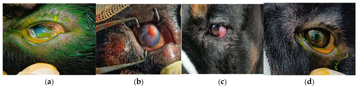
Figure 2.
a. Clinical case of corneal ulcer, b. Suturing of amniotic membrane graft, c.Application of first laye of AM d. Application of third layer of AM e 45th day post surgery, f. After complete recovery(55th day).
Figure 2.
a. Clinical case of corneal ulcer, b. Suturing of amniotic membrane graft, c.Application of first laye of AM d. Application of third layer of AM e 45th day post surgery, f. After complete recovery(55th day).

Figure 3.
Mean ± SE values of Interleukin-2 recorded in both Groups at different intervals.
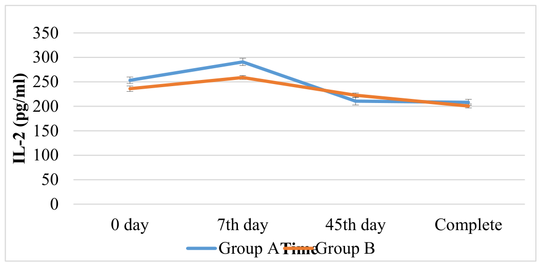
Figure 4.
Mean ± SE values of Interleukin-4 recorded in both Groups at different intervals.
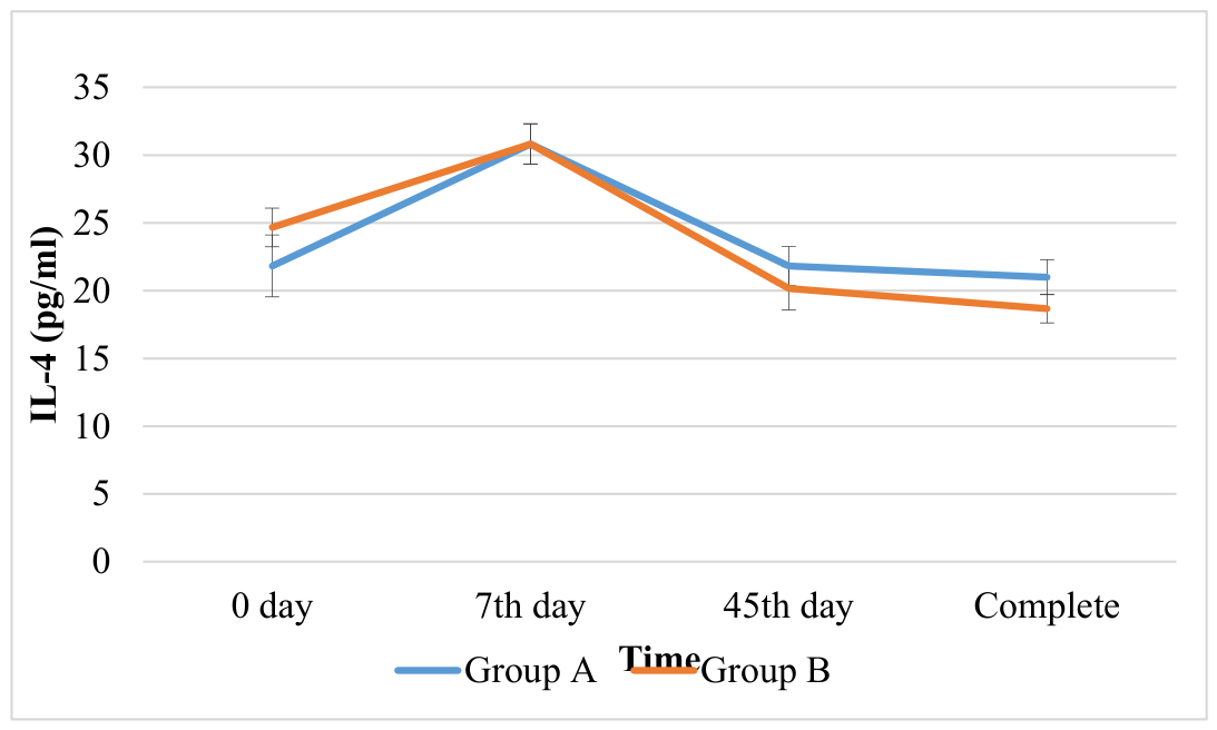
Figure 5.
Mean ± SE value of Healing time.
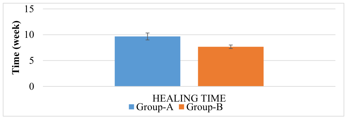

Table 1.
Average values of rectal temperature (°C), respiratory rate (RR/min) and heart rate (HR/min) recorded in Groups A and B at different intervals.
Table 1.
Average values of rectal temperature (°C), respiratory rate (RR/min) and heart rate (HR/min) recorded in Groups A and B at different intervals.
| Groups | 0 Day | 7 Day | 45 Day | |
|---|---|---|---|---|
| Temperature | Group A | 38.71±0.17 | 38.68±0.10 | 38.78±0.13 |
| Group B | 38.91±0.11 | 38.86±0.15 | 38.76±0.11 | |
| Respiratory rate | Group A | 30.17±2.13 | 29.33±1.92 | 28.00±1.86 |
| Group B | 31.00±1.15 | 30.17±1.16 | 31.50±1.30 | |
| Heart rate | Group A | 121.50±5.17 | 117.67±6.39 | 115.67±4.45 |
| Group B | 121.67±4.99 | 119.67±4.99 | 124.00±4.13 |
Table 2.
Mean ± S.E. Visual function test and Schirmer’s Tear Test score in Groups A and B, before and after surgery.
Table 2.
Mean ± S.E. Visual function test and Schirmer’s Tear Test score in Groups A and B, before and after surgery.
| Visual function test | Groups | Before | After |
|---|---|---|---|
| Menace reflex | Group A | 1.50a±0.23 | 2.50b±0.23 |
| Group B | 1.50a±0.23 | 2.83b±0.17 | |
| Palpebral reflex | Group A | 2.50±0.23 | 2.83±0.17 |
| Group B | 2.67±0.21 | 3.00±00 | |
| Pupillary light reflex | Group A | 1.50a±0.23 | 2.50b±0.23 |
| Group B | 1.50a±0.23 | 2.83b±0.17 | |
| Cotton ball test | Group A | 1.17a±0.31 | 2.33b±0.21 |
| Group B | 1.17a±0.40 | 2.33b±0.21 | |
| Schirmer’s Tear Test (STT) | Group A | 18.17±3.12 | 16.50±1.41 |
| Group B | 13.33±1.78 | 15.00±1.37 |
a, b Superscript indicates significant difference (p<0.05) in between the Groups.
Table 3.
Fluorescein dye test before and after surgery.
| Groups | Case no | FDT before | FDT after |
|---|---|---|---|
| A | 1 | PRESENT | ABSENT |
| A | 2 | PRESENT | ABSENT |
| A | 3 | PRESENT | ABSENT |
| A | 4 | PRESENT | PRESENT |
| A | 5 | PRESENT | ABSENT |
| A | 6 | ABSENT | PRESENT |
| B | 7 | PRESENT | ABSENT |
| B | 8 | ABSENT | PRESENT |
| B | 9 | PRESENT | ABSENT |
| B | 10 | PRESENT | ABSENT |
| B | 11 | PRESENT | ABSENT |
| B | 12 | PRESENT | ABSENT |
Table 4.
Antibiotic sensitivity test.
| No. of sample | Organism | Sensitive Antibiotics |
|---|---|---|
| 3 (42.8%) | Staphylococcus spp. | Norfloxacin, Gentamycin, Amikacin, Clindamycin, Ciprofloxacin, Ceftriaxone |
| 2 (28.6%) | Streptococcus spp. | Azithromycin, Clindamycin, Ciprofloxacin, Enrofloxacin, Ampicillin |
| 1 (14.2%) | Enterococci spp. | Ampicillin, Ciprofloxacin, Gentamycin, Norfloxacin, Enrofloxacin, Tetracycline |
| 1 (14.2%) | Bacillus spp. | Gentamycin, Clindamycin, Ampicillin, Ciprofloxacin |
Table 5.
Mean ± SE values of Interleukins-2 recorded in both groups at different intervals.
| Group | 0 day | 7th day | 45th day | Complete |
|---|---|---|---|---|
| Group A | 253.33a±6.67 | 290.83bA±7.40 | 210.67c±7.84 | 208.17c±6.03 |
| Group B | 236.00a±5.63 | 259.00bB±3.94 | 222.67c±4.53 | 200.67c±4.15 |
Table 6.
Mean ± SE values of Interleukins-4 recorded in both groups at different intervals.
| Group | 0 day | 7th day | 45th day | Complete |
|---|---|---|---|---|
| Group A | 21.83a±2.29 | 30.83b±1.49 | 21.83a±1.42 | 21.00a±1.29 |
| Group B | 24.67a±1.43 | 30.83b±1.49 | 20.17a±1.60 | 18.67a±1.05 |
Table 7.
Mean ± SE value of Healing time.
| Group | Healing Time (weeks) |
|---|---|
| Group A | 9.67A±0.67 |
| Group B | 7.67B±0.34 |
Disclaimer/Publisher’s Note: The statements, opinions and data contained in all publications are solely those of the individual author(s) and contributor(s) and not of MDPI and/or the editor(s). MDPI and/or the editor(s) disclaim responsibility for any injury to people or property resulting from any ideas, methods, instructions or products referred to in the content. |
© 2026 by the authors. Licensee MDPI, Basel, Switzerland. This article is an open access article distributed under the terms and conditions of the Creative Commons Attribution (CC BY) license.
Copyright: This open access article is published under a Creative Commons CC BY 4.0 license, which permit the free download, distribution, and reuse, provided that the author and preprint are cited in any reuse.