

Submitted:
16 October 2026
Posted:
20 October 2026
You are already at the latest version
Preprints on COVID-19 and SARS-CoV-2
Abstract
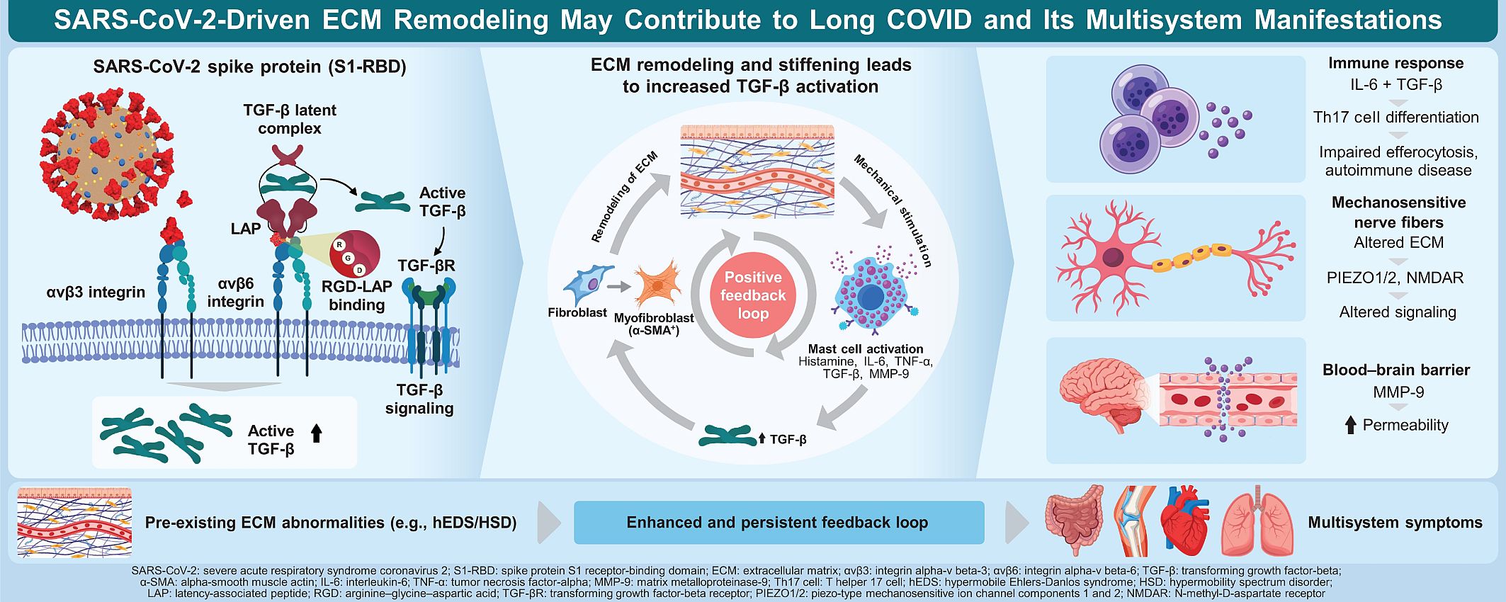
Long coronavirus disease (long COVID) and hypermobility disorders (e.g., hypermobile Ehlers-Danlos syndrome) often share comorbidities, including mast cell activation syndrome, small fiber neuropathy, and myalgic encephalomyelitis/chronic fatigue syndrome. The relationship between long COVID and hypermobility disorders, as well as the extracellular matrix (ECM), has been reported, with evidence suggesting that ECM alterations may underlie these associations. The current hypothesis posits that ECM alterations may become partly self-perpetuating, with the severe acute respiratory syndrome coronavirus 2 spike protein as a possible initial trigger. The proposed mechanism involves interactions among the spike protein, integrins, latent transforming growth factor β, mast cells, interleukin-6, and matrix metalloproteinase-9 (MMP-9). Possible feedback loops, such as those between latent transforming growth factor β and a stiffened ECM, increased sympathetic activity and a pro-inflammatory state, insulin resistance and a pro-inflammatory state, inflammation and mast cell migration, and increased blood–brain barrier permeability via MMP-9 and inflammation, are highlighted. The impact of the modified ECM on the nervous system, as described through PIEZO and N-methyl-D-aspartate receptors, is also discussed, alongside evidence for increased differentiation of T-helper 17 cells and their potentially disruptive effects on efferocytosis. This new hypothesis may have important implications for clinical practice and future research.
Keywords:
extracellular matrix
; long COVID
; hypermobility disorders
; inflammation
; hypothesis
1. Introduction
Long coronavirus disease (long COVID) and hypermobility disorders share overlapping features and potential underlying mechanisms. Both present complex clinical pictures that require multimodal treatment approaches; thus, identifying and understanding these underlying pathological mechanisms are important. Numerous studies on long COVID and hypermobility disorders link both conditions through the extracellular matrix (ECM) [1,2,3]. Comorbidities associated with hypermobility disorders can overlap with those of long COVID, including musculoskeletal pain, dysautonomia with or without small fiber neuropathy (SFN), cognitive disorders, myalgic encephalomyelitis (ME)/chronic fatigue syndrome (CFS), and mast cell activation syndrome (MCAS). Furthermore, hypermobility spectrum disorders (HSDs) and hypermobile Ehlers-Danlos syndrome (hEDS), the most common hypermobility conditions, as well as long COVID, can present with symptoms resembling fibromyalgia, which is characterized by chronic musculoskeletal and myofascial pain, sleep disturbances, fatigue, and neuropsychiatric disorders (e.g., anxiety and depression) [1,4]. Similarly, HSD, hEDS, and long COVID may present with post-exertional malaise; fatigue; musculoskeletal, cardiovascular (e.g., postural orthostatic tachycardia syndrome [POTS]), gastrointestinal, urogenital, immunological, pulmonary, otolaryngologic, dermatologic, reproductive, and neuropsychiatric symptoms; and multiorgan systemic dysfunction [1,2,3]. Logarbo et al. [3] indicated that hEDS/HSD should be considered in cases of persistent long COVID, as they observed the co-occurrence of these conditions in some cases.
HSD and hEDS are hereditary disorders that can substantially impair quality of life. However, their complex multisystemic manifestations often lead to delayed or missed diagnosis. Unlike HSD, hEDS is diagnosed according to specific criteria, including a positive Beighton score, characteristic systemic features and/or a positive family history, and chronic musculoskeletal pain and/or joint instability. Increasing evidence suggests that HSD and hEDS may not represent distinct entities, but rather lie along a phenotypic continuum and share a similar underlying pathophysiology [5]. Historically, HSD and hEDS have been considered connective tissue disorders characterized by joint hypermobility and/or instability, among other features. These characteristics highlight the potential role of the fascial system ECM in the pathogenesis of these conditions [5]. Dysregulation of fibroblast-to-myofibroblast transformation has been reported in HSD and hEDS, with a marked increase in myofibroblasts in the deep fascia. This dysregulation may contribute to persistent ECM densification and dysfunction in patients with hEDS/HSD. ECM densification is accompanied by increased matrix viscosity, reduced interfascial glide, and impaired force transmission, potentially resulting in the irritation of free nerve endings [5].
Fascial myofibroblasts have been proposed to play a mechanistic role in long COVID [1]. Fascial myofibroblasts and the ECM form a dynamic network that regulates structural tissue integrity, and severe acute respiratory syndrome coronavirus 2 (SARS-CoV-2) can interact with the ECM [6]. Furthermore, the SARS-CoV-2 spike (S) glycoprotein triggers a transcriptional response associated with ECM reorganization and latent transforming growth factor β (TGF-β) signaling, which could lead to barrier dysfunction and vascular leak [6]. In addition, the receptor-binding domain of the S protein within its S1 region (S1-RBD) binds to recombinant human αvβ3 and αvβ6 integrins. Thus, S1-RBD functions as an αv-selective integrin agonist and can dysregulate the ECM [7]. Enhanced αvβ3 engagement can stimulate fibroblast contractility, thereby increasing the stiffness of the soft, nonlinear elastic ECM [8]. Similarly, integrin αvβ6 has been shown to bind and activate latent TGF-β1 [9].
Serum matrix metalloproteinase-9 (MMP-9) levels are elevated in patients with long COVID, and cultured human microglia stimulated with the spike protein exhibit significantly increased MMP-9 release [10]. Increased MMP-9 levels can promote vascular inflammation and increase blood–brain barrier (BBB) permeability. MMP-9 can also influence tight junctions and disrupt neuronal connectivity, playing a vital role in neurodegenerative disorders [10]. In patients with long COVID, elevated MMP-9 levels are associated with worse disease severity [10]. MMP-9 levels in skin fibroblasts are also elevated in vitro in patients with hEDS/HSD [11]. Furthermore, elevated MMP-9 levels may be associated with autism [12,13]. The possibility of co-occurring autism should also be considered in individuals with fibromyalgia [14]. Additionally, individuals with higher sensory reactivity scores on the Ritvo Autism & Asperger Diagnostic Scale are more likely to experience long COVID symptoms lasting longer than 12 weeks [15]. Moreover, autism can be a comorbidity of hEDS/HSD [16]. In one study, in the hEDS/HSD group, autistic individuals were more likely to have co-occurring physical and mental health conditions, including attention-deficit/hyperactivity disorder, depression, anxiety, dysautonomia/POTS, ME/CFS, and MCAS, than non-autistic individuals [17].
Taken together, these findings suggest that an ECM-based model may provide a plausible explanation for the overlapping pathophysiological features commonly observed in long COVID, hypermobility disorders, and related conditions. This paper outlines a new integrated model that explains how these conditions and their comorbidities might be interconnected. Additionally, the model uncovers potential pathological mechanisms to inform future research.
2. Hypothesis
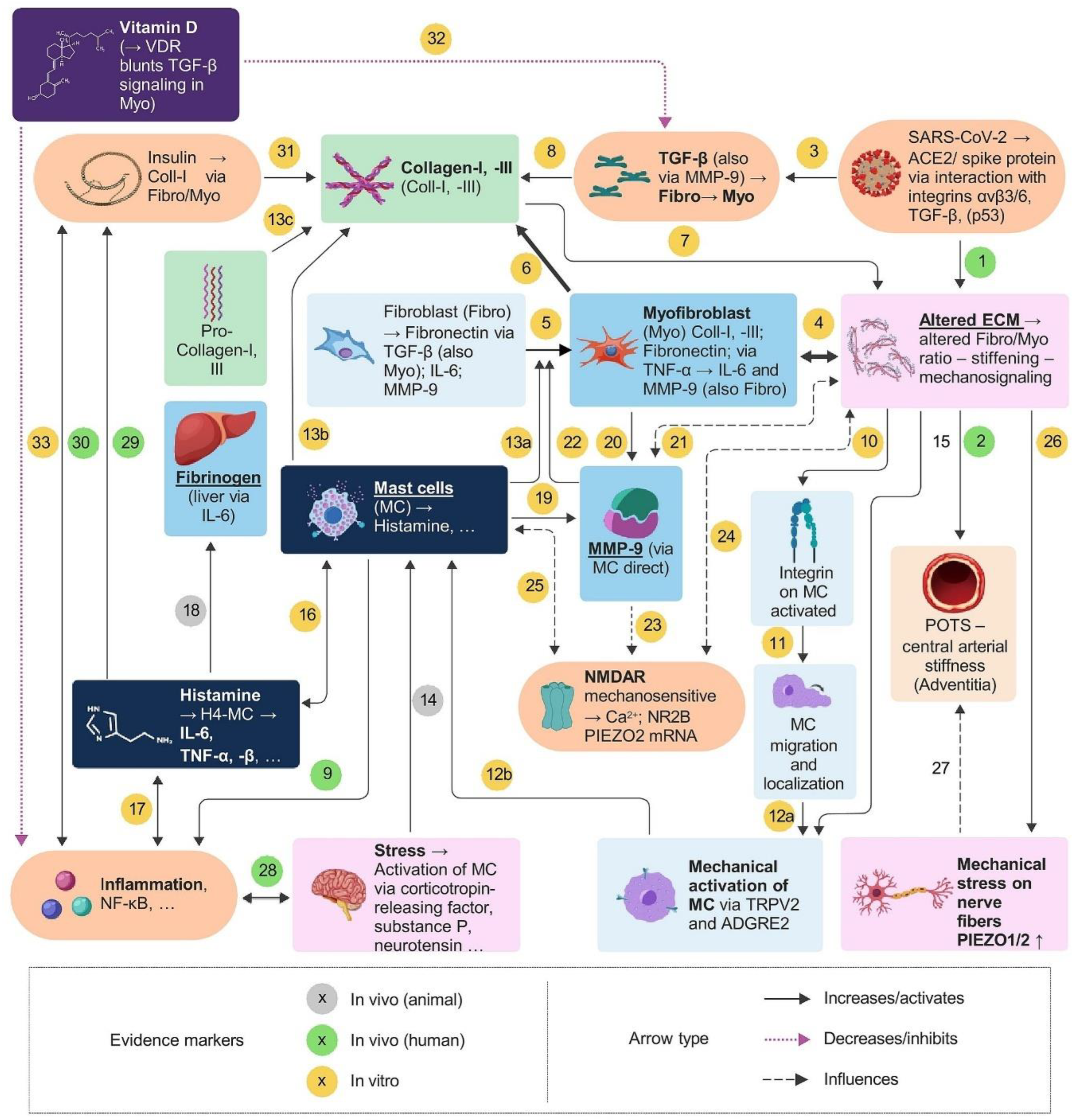
This section is divided into three subsections. The first subsection focuses on how SARS-CoV-2 can alter the ECM (Part 1a) and on how the ECM is altered in patients with hEDS/HSD (Part 1b) (Figure 1). The second subsection integrates the potentially important roles of mast cells and MMP-9 into the hypothesis (Figure 2). The third subsection discusses the possible roles of the N-methyl-D-aspartate receptor (NMDAR), PIEZO1, PIEZO2, insulin, and vitamin D (Figure 3).
2.1. Role of ECM in the Pathophysiology of SARS-CoV-2 and hEDS/HSD
2.1.1. Part 1a: SARS-CoV-2 Influences the ECM Through Multiple Pathways
Long COVID affects a broad spectrum of patients, including those with mild initial symptoms. The persistence of SARS-CoV-2 and its viral fragments has been suggested as a potential contributor to long COVID [18,19]. A 2020 bioinformatic study identified potential links between SARS-CoV-2 binding to angiotensin-converting enzyme 2 (ACE2) and the integration of ACE2 into a molecular network involving TGF-β and connective tissue growth factor. Specifically, mRNA levels of TGF-β and connective tissue growth factor are elevated in lung epithelial cells following SARS-CoV-2 infection [20]. The ACE2 receptor facilitates SARS-CoV-2 entry into host cells and plays a role in the pulmonary fibrosis following COVID-19 [20]. ACE2 mRNA is also expressed at relatively high levels in the normal, uninfected small intestine, suggesting that the small intestine may be susceptible to SARS-CoV-2 infection [20].
A 2022 in vitro study [6] using transcriptomic profiling of human pulmonary microvascular endothelial cells treated with soluble trimeric SARS-CoV-2 spike protein identified an upregulation of the TGF-β signaling pathway. Furthermore, the authors noted that the TGF-β signaling axis was required for spike protein-mediated dysfunction of endothelial and epithelial barriers. TGF-β is a mediator of cellular proliferation, differentiation (e.g., myofibroblast differentiation), and migration, and plays an important role in regulating vascular permeability and inflammatory responses [6,21]. TGF-β signaling may also promote tissue fibrosis, contributing to the vascular fibrosis and stiffness observed in long COVID [21,22]. This vascular stiffness may, in turn, contribute to the co-occurrence of POTS in patients with long COVID and hEDS, as increased central arterial stiffness is a feature of POTS (Figure 1, arrow 2) [23].
Norris et al. [7] demonstrated that the adhesive Arg-Gly-Asp (RGD) motif of the S1-RBD region binds to recombinant human integrins αvβ6 and αvβ3, offering a possible mechanism by which SARS-CoV-2 can upregulate the TGF-β pathway. Integrin αvβ6 antibodies reduce fibrosis and TGF-β activity [24]. Notably, integrin αvβ6 is expressed only in epithelial tissue. Upon activation of TGF-β1 and TGF-β3 by integrin αvβ6, TGF-β may directly signal myofibroblasts to promote further ECM production (Figure 1, arrows 3 and 4) [24]. Integrin αvβ3 can activate TGF-β1 and is involved in cardiac and skin fibrosis, as well as in aortic atherosclerosis [24,25,26]. Integrin αvβ3 is typically expressed at low levels in most normal tissues, such as intestinal, vascular, and smooth muscle tissues [26]. High integrin αvβ3 expression is mainly observed in bone, mid-cycle endometrium, placenta, sites of inflammation, and invasive tumors [26]. High integrin αvβ3 levels can also be found in mature osteoclasts, activated macrophages, a small subset of neutrophils, angiogenic endothelial cells, and migrating smooth muscle cells [26].
Angiogenic stimuli, such as tumor necrosis factor alpha (TNF-α) and vascular endothelial growth factor (VEGF), produced by endothelial cells, induce integrin αvβ3 expression on sprouting endothelial buds and promote the formation of new blood vessels [26]. TGF-β1 was previously shown to induce dose-dependent fibronectin synthesis during the early phase of cultured intestinal fibroblast-to-myofibroblast transformation, with peak production at 2 days in the fibroblast phase and continued production in the myofibroblast phase (Figure 1, arrow 5) [27]. A similar pattern was observed in cultured lung fibroblasts, with fibronectin production peaking at 2 days [28]. However, this experiment lasted only 2 days. Collectively, these results indicate that in long COVID, TGF-β activation, and subsequent fibroblast-to-myofibroblast differentiation and myofibroblast maintenance, may lead to changes in the ECM of certain tissues, resulting in increased stiffness (Figure 1, arrows 4 and 5) [29].
Importantly, a stiff ECM may promote TGF-β activation, thereby creating a positive feedback loop (Figure 1, arrow 4) [29] that can perpetuate inflammation and fibrosis. Mechanotransduction pathway enzymes/proteins (e.g., integrins, focal adhesion kinase, Rho-associated kinase, and Yes-associated protein) play a key role in these processes. Integrins are transmembrane receptors that serve as mechanosensors [30]. Integrins (e.g., αvβ6) can bind to the RGD motif within the latency-associated protein (LAP) and, upon mechanical force transmission, promote the activation of TGF-β from its latent complex. Latent TGF-β binding protein (LTBP) binds to matrix proteins, such as fibrillin, proteoglycans, and fibronectin [21,29]. An extremely acidic environment may facilitate the activation of latent TGF-β [21].
A 2026 myotonometry study of four women with long COVID identified a reduction in creep (i.e., the reduced magnitude of fascial deformation over time under a constant load) in the superficial and deep fascia (Figure 1, arrow 1) [31]. The time-dependent viscoelasticity of the ECM characterizes creep behavior [32]. Fibrotic tissue is characterized by an altered ECM and a buildup of components such as fibrillar collagen type I, mediated by myofibroblasts (Figure 1, arrows 6–8) [33,34]. ECM stiffness can increase in fibrotic tissue, thereby reducing creep [35]. Regarding COVID and an altered ECM, one study reported that bleomycin-induced lung injury in mice led to perivascular fibrosis and ECM densification (i.e., increased ECM viscosity and changes in gliding) [36]. Similar to ECM stiffness, ECM densification influences creep.
2.1.2. Part 1b: Altered ECM in hEDS/HSD Patients
Several studies of patients with hEDS/HSD have reported fascial thickening, reduced interfascial glide, altered viscoelasticity, and densification of the deep fascia, particularly in the sternocleidomastoid muscle, iliotibial tract, thoracolumbar fascia, and fascia iliaca [5,39,40]. In general, higher ECM stiffness is not commonly observed in patients with hEDS/HSD. By contrast, their tendons exhibit reduced stiffness and excessive elongation [5]. In one study, the strain index in regions with myofascial pain was higher in patients with non-hEDS than in patients with hEDS and pain or in patients with non-hEDS without back or knee pain [39]. This indicated greater fascial softening with relative muscle stiffening in patients with non-hEDS. Typically, myofascial pain involves softening of the fascia along with stiffening of the underlying muscles. However, this pattern does not apply to hEDS, where fascial stiffness in relation to muscle occurs in regions of myofascial pain instead [39]. Despite being preliminary, these data emphasize significant fascial features in hEDS [39]. Tissue-level alterations, such as excessive tendon elongation, can impair force transmission and increase the risk of subluxation and/or dislocation, leading to repetitive microtrauma [5].
Alterations in molecular pathways in hEDS/HSD include activation of Wnt/β-catenin signaling; upregulation of miR-224, miR-378-3p, and the prolactin receptor; reduction of NR4A nuclear receptor expression; and downregulation of miR-23a, which is associated with increased Wnt/β-catenin pathway activation [5]. Activation of canonical Wnt signaling is required for TGF-β-mediated fibrosis [41]. These changes can contribute to an inflammatory state and myofibroblast persistence, thereby leading to ECM alterations (Figure 1, arrow 4) [5]. At the cellular level, pathological dysregulation of fibroblast-to-myofibroblast transformation in hEDS/HSD, as mediated by upregulation of integrin αvβ3, has been reported (Figure 1, arrow 5). The αvβ3 integrin-ILK complexes and the Snail1/Slug transcription factors are involved in this fibroblast-to-myofibroblast transition in hEDS/HSD (Figure 1, arrow 5) [11].
In post-COVID-19 conditions, increased Snail expression is required for pulmonary fibrosis [42]. The spike protein can trigger an integrin αvβ3 response, which can lead to Snail upregulation through various signaling pathways [7,42]. Snail can promote the expression of α-smooth muscle actin (α-SMA) with or without prior TGF-β activation, driving the transformation of fibroblasts into myofibroblasts. In patients with hEDS/HSD, 60–80% of fibroblasts express α-SMA, indicating a shift toward a pro-fibrotic ECM [5]. Furthermore, TGF-β, interleukin (IL)-6, ECM-derived danger-associated molecular patterns, fragmented fibronectin, collagen type I, and tenascin are upregulated [5]. Elastin is mislocalized to the cytoplasm rather than the ECM, potentially impairing elastogenesis [5]. Overall, a disorganized ECM is observed in hEDS/HSD. A unique fragment pattern consisting of 52 kDa fibronectin and 45 kDa collagen type I fragments was recently identified in the plasma of patients with hEDS/HSD, offering hope for a diagnostic blood test for hEDS/HSD [43].
The ECM can be divided into different structural entities, including (i) deep fascia that contains densely packed collagen bundles and elastin and (ii) thin gliding layers of loose connective tissue between individual layers of deep fascia [44]. The lamina propria is a loose connective tissue layer in direct proximity to the epithelial structures of the digestive tract and surrounding blood and lymph vessels. This loose connective tissue contains cells, such as fibroblasts and myofibroblasts, that can influence the structure and, consequently, the mechanical properties of the ECM [45]. These layers are of special interest to the hypothesis regarding hEDS/HSD, as they may be sites where densification and/or increased stiffness resulting from trauma and/or inflammation occur. The observed tendon elongation and reduced stiffness in hEDS/HSD fascia can increase the risk of traumatic injury and subsequent inflammation; this can then affect the mechanical properties of this loose connective tissue layer and help explain the observed reduced interfascial glide in hEDS/HSD.
Collectively, these findings indicate an altered ECM with potentially increased stiffness in localized areas, especially those affected by inflammation, as observed in long COVID. This altered ECM may create a positive feedback loop, as increased tissue stiffness can enhance TGF-β activation (Figure 1, arrows 4–8). A 2015 review of eight studies found elevated TGF-β levels in CFS in five (63%) studies [46]. Patients with long COVID, as well as patients with hEDS/HSD, frequently experience CFS.
2.2. Part 2: Incorporating the Potentially Important Roles of Mast Cells and MMP-9 into the Hypothesis
Mast cells are a type of white blood cell that plays a critical role in immune responses, including inflammation and allergic reactions (Figure 2, arrow 9). Mature mast cells are distributed throughout the collagenous connective tissue, between collagen bundles and elastic fibers, and near blood vessels and nerves. Mast cells are also detected in the superficial fascia, where they play modulatory roles in thermoregulation, exteroception, and pain modulation [47]. They recognize appropriate chemotactic stimuli, such as TGF-β1–3 and TNF-α, and respond by migrating. Activated mast cells can produce chemoattractants, leading to the accumulation of mast cells and other hematopoietic cells at sites of inflammation [48]. This can result in a positive feedback loop between inflammation and mast cell migration.
MCAS is observed in both patients with hEDS/HSD and long COVID [49,50]. MCAS is associated with an increased number of mast cells, which may form clusters due to reduced cell death. This can affect multiple organs and lead to clinical symptoms, including flushing, hypotension, urticaria, angioedema, headache, vomiting, diarrhea, and anaphylaxis [49]. Common triggers in MCAS include weather changes, heat, pressure, rubbing, exercise, stress, and dietary factors (e.g., certain medications, including opiates) [50].
Furthermore, mast cells can express integrin αvβ3 on their surface, which can interact with fibrin and platelet endothelial cell adhesion molecule on endothelial cells [51]. SARS-CoV-2 interacts with recombinant human αvβ3 and αvβ6 integrins via its S1-RBD. Consequently, SARS-CoV-2 may directly interact with mast cells via these integrins. Integrin αvβ3 on the mast cell membrane functions as a true mechanosensor and can respond to stiffness [52]. Mast cells use integrin-mediated pathways to sense mechanical properties, guiding them toward regions of varying stiffness. Mechanical stimuli can alter the microscopic structure of collagen fibers, muscle bundles, nerve fibers, and vascular tracts within the ECM, enabling these structures to bind to integrins on the mast cell membrane (Figure 2, arrow 10) [53,54]. Under mechanical stimulation, integrins can transition from a bent to an extended conformation, thereby enhancing their interaction with ECM ligands (Figure 2, arrow 11) [54].
Mechanical stimulation has been shown to increase both mast cell density and degranulation rates in humans and various disease models (Figure 2, arrow 12a+b) [54,55]. Mast cell responses to mechanical signals promote fibrosis, and mechanical stretch-induced mast cell degranulation and TGF-β activation are attenuated by sodium cromoglycate, a mast cell stabilizer (Figure 2, arrow 13a+b) [54,56]. Mechanoreceptors on mast cell membranes include transient receptor potential vanilloid channel 2 (TRPV2) and adhesion G protein-coupled receptor E2 (ADGRE2). TRPV2 is highly sensitive to physical stimuli, including mechanical stress, shear forces, and thermal changes [54]. Loratadine, an antihistamine, has been shown to reduce calcium influx triggered by TRPV2. Meanwhile, ADGRE2 is activated by vibration; in human skin mast cells, this activation results in rapid histamine release (Figure 2, arrow 12b) [54].
The mast cell Mas-related G-protein-coupled receptor member X2 (MRGPRX2) is mainly expressed by chymase+ cells, a specific subtype of human mast cells in connective tissues [54]. MRGPRX2 activation by substance P from mechanosensitive neurons can result in immunoglobulin E (IgE)-independent pseudo-allergic reactions (Figure2, arrow 14) [54,57,58]. In addition, activation of the mast cell receptor MRGPRX2 can result in the secretion of tryptase and chymase, among other substances. Tryptase can promote the transformation of fibroblasts into myofibroblasts, and chymase can contribute to the synthesis and secretion of collagen types I and III (Figure 2, arrow 13a, b, c) [54,56]. In an altered ECM, as in patients with long COVID and/or hEDS/HSD, mechanical stimulation of mast cells may occur more quickly and frequently (Figure 2, arrow 15).
Mast cells themselves can respond to histamine via their H4 receptor and then release inflammatory proteins, including TGF-β, TNF-α, tumor necrosis factor beta (TNF-β), IL-6, and IL-8 (Figure 2, arrows 16 and 17) [59]. IL-6 is also synthesized by macrophages and monocytes in response to other inflammatory cytokines, including TNF-β and IL-11 [60]. Increased IL-6 expression may contribute to pain in SFN when cutaneous nociceptors are damaged. In one study, incubation of murine sensory dorsal root ganglion neurons, cultured for 1 day as a model of SFN, increased action potential firing rates in response to IL-6 exposure [61]. The study also showed higher IL-6 and IL-8 expression in fibroblasts of patients with SFN, and their keratinocytes had higher netrin-1 expression. This is of interest because SFN is a comorbidity in both long COVID and hEDS/HSD.
IL-6, which is produced rapidly and transiently in response to infection and tissue injury, can trigger rapid induction of various acute-phase proteins mainly in the liver, including C-reactive protein, serum amyloid A, fibrinogen, haptoglobin, and α1-antichymotrypsin (Figure 2, arrow 18) [60,62]. Plasma fibrinogen and haptoglobin have been identified as promising biomarkers for assessing disease severity and mortality risk in long COVID [63]. Fibrinogen is cleaved by thrombin, forming fibrin. Fibrin binds to the SARS-CoV-2 spike protein, forming pro-inflammatory blood clots that drive systemic thromboinflammation and neuropathology in COVID-19 [64]. After infection, fibrin promotes neuroinflammation and neuronal loss, suppresses natural killer cells, and activates macrophages in the lungs [64]. Furthermore, fibrin(ogen) can contribute to matrix stiffness through its deposition, cross-linking, and interactions with other ECM components [65].
IL-6, together with TGF-β, can induce the differentiation of T helper 17 (Th17) cells [66]. Although Th17 cells mainly play protective roles, dysregulated Th17 cells can lead to inappropriate inflammatory responses, resulting in autoimmune diseases (e.g., inflammatory bowel disease) [67]. An enhanced Th17-associated immune response has been reported in both long COVID and hypermobility disorders [68,69]. IL-17A signaling was shown to inhibit efferocytosis in synovial macrophages via a STAT3/ADAM17/MERTK-mediated pathway in a rat model of rheumatoid arthritis [70]. Impaired efferocytosis appears to be associated with the pathogenesis of various inflammatory diseases [71].
Mast cells, as well as myofibroblasts and fibroblasts, can produce MMP-9 when stimulated by TNF-α or other stimuli, and MMP-9 is involved in inflammation and tissue remodeling and repair (Figure 2, arrows 19 and 20) [72,73]. MMP-9 cleaves a wide range of ECM substrates, including collagen and gelatin (Figure 2, arrow 21) [73]. It can cause vascular inflammation and increase BBB permeability, thereby disrupting neural connectivity [10]. MMP-9 plays a crucial role in the onset and progression of central nervous system disorders, including neuropathic pain [74], not only through an inflammatory feedback loop that worsens pain, but also through its influence on neuron–glia interactions [74]. For example, nerve growth factor released by glial cells can be degraded by MMP-9 [74].
Pregabalin, a gamma-aminobutyric acid analog used to treat neuropathic pain, epilepsy, and generalized anxiety disorder, has been shown to reduce VEGF and MMP-9 levels in vitro [75]. Furthermore, in vitro studies have demonstrated that recruiting MMP-9 to the fibroblast surface can activate latent TGF-β and trigger fibroblast transformation into myofibroblasts (Figure 2, arrow 22) [76]. There may also be an interplay between TGF-β and MMP-9; this may involve a bidirectional positive feedback loop that varies by tissue type and context, as observed in idiopathic pulmonary fibrosis [73]. MMP-9 levels are elevated in long COVID and hEDS/HSD [5,10]. In long COVID, increased serum levels of MMP-9 are associated with disease severity [10]. Elevated MMP-9 levels may also contribute to autism, a comorbidity of hEDS/HSD. A relationship between autism and long COVID has also been reported [12,13,15].
2.3. Part 3: Potential Roles of NMDAR, PIEZO1, PIEZO2, Insulin, and Vitamin D in the Hypothesis
MMP-9 is a physiological regulator of NMDARs. NMDARs function as glutamate-gated Ca2+-permeable channels and require glycine as a co-agonist. Synaptic plasticity and memory are NMDAR-dependent. In vitro, MMP-9 enzymatic activity in hippocampal cells increases lateral diffusion and, consequently, the surface mobility of the NR1 subunit of NMDARs via an integrin-β1-dependent signaling pathway (Figure 3, arrow 23) [77]. Additionally, NMDARs have been shown to function as mechanosensitive receptors [78]. Therefore, they could theoretically respond to changes in the ECM, such as increased stiffness or densification (Figure 3, arrow 24). In fibromyalgia, NMDARs exhibit increased activity [79]. This finding suggests a potential role for NMDARs in the pathophysiology of hEDS/HSD and long COVID, as both conditions can resemble fibromyalgia. In addition, NMDAR dysfunction has been proposed in autism [80,81].
Homocysteine, a sulfur-containing amino acid and a product of methionine metabolism, is a recognized NMDAR agonist and contributes to neurotoxicity [82]. Polymorphisms of the MTHFR gene (e.g., C677T) in relation to autism, hEDS, and long COVID have also been proposed [83,84,85].
Recent studies have shown that NMDARs are not only present in the central nervous system, but also in peripheral tissues. Their role in atherosclerosis development has also been investigated [86]. Furthermore, evidence suggests that NMDAR blockers attenuate bleomycin-induced pulmonary fibrosis in mice, possibly by inhibiting NMDAR-mediated senescence of bone marrow mesenchymal stem cells through the Wnt/β-catenin signaling pathway [87].
In murine mast cells, glutamate induces upregulation of glutamate receptors, including ionotropic (NMDAR1, NMDAR2A, and NMDAR2B) and metabotropic (mGluR2 and mGluR7) types, at both the mRNA and protein levels (Figure 3, arrow 25) [88]. Glutamate also induces gene expression in mast cells, including upregulation of proinflammatory components (e.g., IL-6 and chemokine (C-C motif) ligand 2) [88]. These findings suggest functional glutamate-glutamate receptor signaling in mast cells [88]. However, the direct effects of homocysteine on human mast cells have not been sufficiently studied to date. Nevertheless, the expression of functional NMDARs on mast cells suggests that homocysteine could act directly on these cells via NMDAR-mediated signaling pathways.
Unlike NMDARs, whose role as mechanosensitive channels has only recently been recognized, piezo-type mechanosensitive ion channel components 1 and 2 (PIEZO1 and PIEZO2) are classic mechanosensitive channels involved in mechanotransduction (Figure 3, arrow 26). PIEZO1 is involved in regulating development and maintaining functional homeostasis. It is activated by poking, stretching, shear stress, local membrane tension, and myosin II-mediated traction force. PIEZO1 is expressed in various cell types, including epithelial cells, endothelial cells, microglia, astrocytes, musculoskeletal cells, and lymphatic and immune cells. Meanwhile, PIEZO2 primarily mediates mechanosensation for touch, tactile pain, proprioception, and sensing of mechanical states in internal organs. It is mainly activated by poking [89]. Both PIEZO1 and PIEZO2 are nonselective cation channels that slightly favor Ca2+ over Na2+ [89]. A 2024 study in rats showed that PIEZO1 detected substrate stiffness at the growth cone and regulated axon regrowth by activating the downstream Ca2+–CaMKII–focal adhesion kinase–actin signaling cascade [90]. PIEZO1 knockdown in adult rat DRG neurons promoted axon regeneration and accelerated the recovery of sensory function after sciatic nerve injury. Furthermore, PIEZO1 inhibition enhanced growth on relatively soft substrates, whereas PIEZO1 activation impaired growth on relatively stiff substrates.
This presents an intriguing research approach, particularly in SFN and dysautonomia, which can occur in both long COVID and hEDS/HSD. One study proposed PIEZO1 as a potential therapeutic target for COVID-19 and long COVID, particularly for endothelial dysfunction [91]. PIEZO1 inhibition may prevent the disruption of intracellular calcium homeostasis in endothelial cells. PIEZO1 and PIEZO2 are also involved in arterial baroreceptor activity and in the acute regulation of blood pressure [92]. Therefore, they may also influence the pathophysiology of POTS, a potential comorbidity of long COVID and hEDS/HSD (Figure 3, arrow 27). Clinically, POTS manifests as orthostatic tachycardia without substantial orthostatic hypotension, with symptoms worsening in the upright position and improving in the recumbent position. Cardiovagal baroreflex function is altered in POTS, although this study has not found significant changes in cerebral autoregulation [93]. Notably, there may be a connection between NMDAR subunit 2B (NR2B), which is associated with nociceptive signaling, and PIEZO2 upregulation [94].
Chronic inflammatory processes, such as those seen in long COVID and hEDS/HSD, can affect neuronal stress networks through inflammatory signaling, thereby contributing to sustained elevation of sympathetic activity [5,95,96]. Increased sympathetic activity can, in turn, lead to a pro-inflammatory state via nuclear factor kappa-light-chain-enhancer of activated B-cell (NF-κB) activation and other mechanisms, creating a bidirectional positive feedback loop that can maintain this state (Figure 3, arrow 28) [96]. Stress mediators include corticotropin-releasing factor and the neuropeptides neurotensin and substance P [96,97]. These mediators can contribute to mast cell degranulation (Figure 2 and Figure 3, arrow 14) and cause microvascular inflammation and injury [97]. This inflammation and tissue injury can then manifest as cardiovascular diseases and BBB dysfunction [97]. A dysfunction of the blood-brain barrier is discussed in connection with the development of cerebrospinal fluid (CFS) leaks as a clinical symptom of hEDS [98]. Spontaneous CSF leakage can manifest as positional headache, nausea, vertigo, neck stiffness/pain, tinnitus, visual disturbances, fatigue, cognitive changes, orthostatic hypotension, CSF otorrhea and rhinorrhea, metallic taste, extreme weakness, and pain [98].
Inflammation can mediate insulin resistance via NF-κB, which is activated by TNF-α and IL-6 (Figure 3, arrow 29) [99,100]. Insulin resistance can also promote inflammation, thus creating a potential positive feedback loop between insulin resistance and inflammation (Figure 3, arrow 30) [99,100,101]. A 2022 study found that patients with POTS had reduced insulin sensitivity and signs of increased sympathetic activity, particularly during orthostatic stress [102]. Insulin has been shown to increase collagen production in cultured human embryonic lung fibroblasts, particularly collagen types I and III (Figure 3, arrow 31) [103]. Importantly, a prospective observational study identified a link between long COVID and insulin resistance [104].
The active vitamin D metabolite 1,25-dihydroxyvitamin D3 (hereinafter referred to as vitamin D) can act as an anti-fibrotic agent by inhibiting NF-κB activation and TGF-β/SMAD signaling in myofibroblasts via the vitamin D receptor (VDR) (Figure 3, arrows 32 and 33) [105,106]. The TGF-β/SMAD pathway is the canonical TGF-β signaling pathway and is involved in fibrotic diseases [21]. Vitamin D may contribute to mast cell stabilization, and its deficiency may promote their activation and possibly cause increased degranulation [107]. A 2023 study found that low vitamin D levels were more prevalent among patients with long COVID than among the general population, though there was no correlation between vitamin D levels and symptom prevalence or severity [108]. It should be noted that a normal vitamin D level does not necessarily indicate normal function. For example, single-nucleotide polymorphisms can alter vitamin D receptors, thereby impairing normal signal transduction. A 2025 study found that vitamin D deficiency was significantly associated with overall long COVID signs and symptoms [109].
Vitamin D supplementation can lead to a significant reduction in ME/CFS symptoms among patients with vitamin D deficiency or insufficiency in cases where ME/CFS is caused by post-vaccination syndrome or post-acute sequelae of SARS-CoV-2 infection [110]. In a mouse model of experimental autoimmune encephalomyelitis, vitamin D, acting through VDR, ameliorated symptoms by suppressing Th17 cytokines and reducing the expression of IL-17 and IL-17F [111]. IL-17F expression is increased in patients with long COVID [68]. Vitamin D exerts regulatory effects through the widespread expression of VDR in key brain regions, profoundly influencing the complex physiological processes underlying sleep regulation and potential interactions with melatonin secretion pathways [112]. Individuals with vitamin D insufficiency (<20 ng/mL) exhibit prolonged sleep latency and reduced sleep efficiency [112]. VDR directly regulates the transcriptional activity of arylalkylamine N-acetyltransferase, a rate-limiting enzyme in melatonin synthesis [112].
3. Discussion
Although this hypothesis captures only a fraction of biological functions and relationships, it may help explain the pathological mechanisms underlying long COVID, hEDS/HSD, and their comorbidities in certain subgroups. Given the feedback loops described above, the disease process can theoretically persist even after the original trigger has been removed, particularly when underlying conditions are pre-existing or sufficiently severe. One possible pathway would begin with infection by SARS-CoV-2, which interacts with integrins αvβ6 and αvβ3 via the spike protein. This leads in part to a direct interaction with the ECM, particularly in endothelial and epithelial tissues (Figure 3, arrow 1), and the upregulation of active TGF-β (Figure 3, arrow 3), resulting in the conversion of fibroblasts into myofibroblasts (Figure3, arrow 5). This is followed by myofibroblast-mediated remodeling of the ECM, including the production of collagen types I and III (Figure3, arrow 6), driving increased stiffness (Figure 3, arrow 4), which in turn can activate additional TGF-β (Figure 3, arrow 4; feedback loop 1). A stiffer ECM can trigger mast cell migration via integrins (Figure 3, arrows 10 and 11) and mechanoreceptors, leading to mast cell activation and histamine release (Figure 3, arrows 12 and 13). This can then lead to the release of IL-6, IL-8, TNF-α, TNF-β, and TGF-β1 by mast cells via the H4 receptor (Figure 3, arrows 13a+b and 16). TGF-β1–3 and TNF-α act as chemotactic stimuli for mast cells, prompting further migration (Figure 3, arrows 16 and 17; feedback loop 2).
In theory, this feedback loop can contribute to the development of MCAS. Mast cell activation can result in an inflammatory response (Figure 3, arrow 17). The resulting inflammation can activate the sympathetic nervous system, thereby inducing stress (Figure 3, arrow 28) and triggering the release of substance P, which is also be secreted by mechanosensitive neurons. Activation of the mast cell receptor MRGPRX2 via substance P can in turn trigger the release of tryptase and chymase. Tryptase can promote the conversion of fibroblasts into myofibroblasts (Figure 3, arrow 13a), while chymase can contribute to the synthesis and secretion of collagen types I and III (Figure 3, arrow 13b, c). What has been described so far could constitute a major feedback loop in and of itself and can thus be self-perpetuating, with the altered mechanical properties of the ECM serving as a key element.
Inflammation and stress can reinforce each other in both directions (Figure 3, arrow 28, feedback loop 3). Inflammation, which is primarily systemic in this context, can promote insulin resistance and vice versa (Figure 3, arrow 30, feedback loop 4). Meanwhile, insulin can promote collagen production, particularly collagen types I and III (Figure 3, arrow 31). Activated mast cells, fibroblasts, and myofibroblasts can produce MMP-9 when stimulated by TNF-α and other factors (Figure 3, arrows 19 and 20). Elevated levels of MMP-9 and fibrinogen have been observed in patients with long COVID. Elevated MMP-9 levels are also observed in patients with hEDS/HSD and autism. IL-6 is secreted by mast cells and other inflammatory cells, stimulating the liver to increase fibrinogen synthesis and release (Figure 3, arrow 18). In addition to its ECM-modulating properties, MMP-9 can promote vascular inflammation and increase BBB permeability, thereby disrupting neural connections and contributing to neuropathic pain.
Furthermore, changes in the ECM can influence the properties of the mechanosensitive channels PIEZO1 and PIEZO2 (Figure 3, arrow 26), which in turn can affect nerve regeneration, SFN, and dysautonomia. PIEZO1 and PIEZO2 are also involved in arterial baroreceptor activity and, in addition to central arterial stiffening (Figure 3, arrow 2), may contribute to the development of POTS. Notably, there may be a partially reciprocal interaction between NMDARs and the structures described above, including an altered ECM, MMP-9, and mast cells (Figure 3, arrows 23–25). This is particularly relevant given that homocysteine is an NMDAR agonist and that a predisposition due to MTHFR mutations has been suggested for long COVID, hEDS, and autism. Severe damage, such as infection, disrupted efferocytosis, or pre-existing harm, may amplify one or more of these feedback loops, resulting in the persistence of the pathological system, either partially or entirely.
Preliminary data suggest that, in hEDS myofascial pain is associated with higher fascial stiffness (in relation to muscle) than in non-hEDS myofascial pain. One hypothetical explanation is that, in hEDS, the already altered ECM structure may lead to a more pronounced inflammatory response as a result of repetitive trauma. This triggers the cascade described above for long COVID, including its feedback loops. This may explain why hEDS/HSD can co-occur with long COVID, given that the tissue is pre-damaged. An initial means to test this hypothesis would be to analyze the mechanical properties of the ECM. A myotonometric study of long COVID patients has provided preliminary evidence of reduced creep in the superficial and deep fascia. For example, magnetic resonance elastography of the small intestine combined with a biopsy of the corresponding tissue and histopathological examination would be a feasible first approach in long COVID patients with gastrointestinal involvement. Furthermore, histopathological examination of the loose connective tissue layer between fascial layers, as well as the lamina propria, in patients with hEDS/HSD can provide additional insights into the pathophysiological mechanisms underlying this hypothesis.
This hypothesis has limitations. Each arrow in the figures represents one or more published studies and synthesizes evidence from different experimental models (in vivo, in vitro, and in silico). Further studies are needed to refine this hypothesis and to assess the proposed effects appropriately.
Author Contributions
Conceptualization, R.D. and R.S.; writing—original draft preparation, R.D.; writing—review and editing, R.D.; visualization, R.D.; funding acquisition, R.S. Both authors have read and agreed to the published version of the manuscript.
Funding
The open access publication fees for this article will be covered by the Technical University of Munich upon acceptance.
Institutional Review Board Statement
Not applicable.
Informed Consent Statement
Not applicable.
Data Availability Statement
No datasets were generated or analyzed.
Acknowledgments
The authors used Grammarly solely for language editing. All scientific content, interpretations, and conclusions were developed and verified by the authors. The authors gratefully acknowledge Editage for English language editing and assistance with the preparation of the graphical abstract and illustrations.
Conflicts of Interest
The authors declare no conflict of interest.
Abbreviations
The following abbreviations are used in this manuscript:
| ACE2 | angiotensin-converting enzyme 2 |
| ADGRE2 | adhesion G protein-coupled receptor E2 |
| α-SMA | alpha-smooth muscle actin |
| BBB | blood–brain barrier |
| CFS | chronic fatigue syndrome |
| COVID | coronavirus disease |
| CSF | cerebrospinal fluid |
| ECM | extracellular matrix |
| hEDS | hypermobile Ehlers-Danlos syndrome |
| HSDs | hypermobility spectrum disorders |
| IgE | immunoglobulin E |
| IL | interleukin |
| LAP | latency-associated protein |
| LTBP | latent TGF-β binding protein |
| MCAS | mast cell activation syndrome |
| ME | myalgic encephalomyelitis |
| MMP-9 | matrix metalloproteinase-9 |
| MRGPRX2 | Mas-related G-protein-coupled receptor member X2 |
| NF-κB | nuclear factor kappa-light-chain-enhancer of activated B-cell |
| NMDAR | N-methyl-D-aspartate receptor |
| PIEZO1/2 | piezo-type mechanosensitive ion channel components 1 and 2 |
| POTS | postural orthostatic tachycardia syndrome |
| RBD | receptor-binding domain |
| RGD | arginine–glycine–aspartic acid |
| S1-RBD | receptor-binding domain of the viral spike (S) protein within its S1 region |
| SARS-CoV-2 | severe acute respiratory syndrome coronavirus 2 |
| SFN | small fiber neuropathy |
| TRPV2 | transient receptor potential vanilloid channel 2 |
| TGF-β | transforming growth factor β |
| TNF-α | tumor necrosis factor alpha |
| TNF-β | tumor necrosis factor beta |
| VEGF | vascular endothelial growth factor |
| VDR | vitamin D receptor |
References
- Plaut, S. “Long COVID-19” and Viral “Fibromyalgia-Ness”: Suggesting a Mechanistic Role for Fascial Myofibroblasts (Nineveh, the Shadow Is in the Fascia). Front. Med. 2023, 10, 952278. [Google Scholar] [CrossRef] [PubMed]
- Ganesh, R.; Munipalli, B. Long COVID and Hypermobility Spectrum Disorders Have Shared Pathophysiology. Front. Neurol. 2024, 15, 1455498. [Google Scholar] [CrossRef] [PubMed]
- Logarbo, B.P.; Yang, M.; Longo, M.T.; Kingry, C.; Courseault, J. Long COVID and the Diagnosis of Underlying Hypermobile Ehlers-Danlos Syndrome and Hypermobility Spectrum Disorders. PM&R 2024, 16, 935–937. [Google Scholar] [CrossRef] [PubMed]
- Fairweather, D.; Bruno, K.A.; Darakjian, A.A.; Bruce, B.K.; Gehin, J.M.; Kotha, A.; Jain, A.; Peng, Z.; Hodge, D.O.; Rozen, T.D.; et al. High Overlap in Patients Diagnosed with Hypermobile Ehlers-Danlos Syndrome or Hypermobile Spectrum Disorders with Fibromyalgia and 40 Self-Reported Symptoms and Comorbidities. Front. Med. 2023, 10, 1096180. [Google Scholar] [CrossRef] [PubMed]
- Wang, T.J.; Stecco, A.; Hakim, A.J.; Schleip, R. Fascial Pathophysiology in Hypermobility Spectrum Disorders and Hypermobile Ehlers–Danlos Syndrome: A Review of Emerging Evidence. IJMS 2025, 26, 5587. [Google Scholar] [CrossRef] [PubMed]
- Biering, S.B.; Gomes de Sousa, F.T.; Tjang, L.V.; Pahmeier, F.; Zhu, C.; Ruan, R.; Blanc, S.F.; Patel, T.S.; Worthington, C.M.; Glasner, D.R.; et al. SARS-CoV-2 Spike Triggers Barrier Dysfunction and Vascular Leak via Integrins and TGF-β Signaling. Nat. Commun. 2022, 13, 7630. [Google Scholar] [CrossRef] [PubMed]
- Norris, E.G.; Pan, X.S.; Hocking, D.C. Receptor-Binding Domain of SARS-CoV-2 Is a Functional Av-Integrin Agonist. J. Biol. Chem. 2023, 299, 102922. [Google Scholar] [CrossRef] [PubMed]
- Fiore, V.F.; Wong, S.S.; Tran, C.; Tan, C.; Xu, W.; Sulchek, T.; White, E.S.; Hagood, J.S.; Barker, T.H. Avβ3 Integrin Drives Fibroblast Contraction and Strain Stiffening of Soft Provisional Matrix during Progressive Fibrosis. JCI Insight 2018, 3, e97597. [Google Scholar] [CrossRef] [PubMed]
- Munger, J.S.; Huang, X.; Kawakatsu, H.; Griffiths, M.J.; Dalton, S.L.; Wu, J.; Pittet, J.F.; Kaminski, N.; Garat, C.; Matthay, M.A.; et al. The Integrin Alpha v Beta 6 Binds and Activates Latent TGF Beta 1: A Mechanism for Regulating Pulmonary Inflammation and Fibrosis. Cell 1999, 96, 319–328. [Google Scholar] [CrossRef] [PubMed]
- Kempuraj, D.; Tsilioni, I.; Aenlle, K.K.; Klimas, N.G.; Theoharides, T.C. Long COVID Elevated MMP-9 and Release from Microglia by SARS-CoV-2 Spike Protein. Transl. Neurosci. 2024, 15, 20220352. [Google Scholar] [CrossRef] [PubMed]
- Zoppi, N.; Chiarelli, N.; Binetti, S.; Ritelli, M.; Colombi, M. Dermal Fibroblast-to-Myofibroblast Transition Sustained by Avß3 Integrin-ILK-Snail1/Slug Signaling Is a Common Feature for Hypermobile Ehlers-Danlos Syndrome and Hypermobility Spectrum Disorders. Biochim. Et. Biophys. Acta (BBA) —Molecular Basis Dis. 2018, 1864, 1010–1023. [Google Scholar] [CrossRef] [PubMed]
- Lord, J.R.; Mashayekhi, F.; Salehi, Z. How Matrix Metalloproteinase ( MMP )- 9 (Rs3918242) Polymorphism Affects MMP-9 Serum Concentration and Associates with Autism Spectrum Disorders: A Case-Control Study in Iranian Population. Dev. Psychopathol. 2022, 34, 882–888. [Google Scholar] [CrossRef] [PubMed]
- Hu, C.; Li, H.; Cui, J.; Li, Y.; Zhang, F.; Li, H.; Luo, X.; Hao, Y. Integrative Analysis Identifies IL-6/JUN/MMP-9 Pathway Destroyed Blood-Brain-Barrier in Autism Mice via Machine Learning and Bioinformatic Analysis. Transl. Psychiatry 2025, 15, 239. [Google Scholar] [CrossRef] [PubMed]
- Johnston, H.; Bacon, A.M. Autistic Traits in Fibromyalgia Are Associated with Hypersensitivity and Central Sensitisation. Psychol. Health Med. 2025, 1–17. [Google Scholar] [CrossRef] [PubMed]
- Raw, R.K.; Rees, J.; Pearson, A.; Chadwick, D.R. Neurodivergence as a Risk Factor for Post-Covid-19 Syndrome 2023. [CrossRef]
- Casanova, E.L.; Baeza-Velasco, C.; Buchanan, C.B.; Casanova, M.F. The Relationship between Autism and Ehlers-Danlos Syndromes/Hypermobility Spectrum Disorders. JPM 2020, 10, 260. [Google Scholar] [CrossRef] [PubMed]
- Crompton, C.J.; Efthimiou, T.N.; Dockrell, D.M.; Berg, K.M. Health Experiences and Outcomes of Autistic and Non-Autistic Adults with Hypermobile Ehlers-Danlos Syndrome and Hypermobility Spectrum Disorder. BMC Med. 2026, 24, 193. [Google Scholar] [CrossRef] [PubMed]
- Liu, S.; Guo, Y.; Wang, F.-S. Viral Persistence in Long COVID: Research Advances and Treatment Strategies. Infect. Dis. Immun. 2025, 5, 272–288. [Google Scholar] [CrossRef] [PubMed]
- Rong, Z.; Mai, H.; Ebert, G.; Kapoor, S.; Puelles, V.G.; Czogalla, J.; Hu, S.; Su, J.; Prtvar, D.; Singh, I.; et al. Persistence of Spike Protein at the Skull-Meninges-Brain Axis May Contribute to the Neurological Sequelae of COVID-19. Cell Host Microbe 2024, 32, 2112–2130.e10. [Google Scholar] [CrossRef] [PubMed]
- Xu, J.; Xu, X.; Jiang, L.; Dua, K.; Hansbro, P.M.; Liu, G. SARS-CoV-2 Induces Transcriptional Signatures in Human Lung Epithelial Cells That Promote Lung Fibrosis. Respir. Res. 2020, 21, 182. [Google Scholar] [CrossRef] [PubMed]
- Deng, Z.; Fan, T.; Xiao, C.; Tian, H.; Zheng, Y.; Li, C.; He, J. TGF-β Signaling in Health, Disease and Therapeutics. Sig Transduct. Target Ther. 2024, 9, 61. [Google Scholar] [CrossRef] [PubMed]
- Kamdar, A.; Sykes, R.; Thomson, C.R.; Mangion, K.; Ang, D.; Lee, M.A.; Van Agtmael, T.; Berry, C. Vascular Fibrosis and Extracellular Matrix Remodelling in Post-COVID 19 Conditions. Infect. Med. 2024, 3, 100147. [Google Scholar] [CrossRef] [PubMed]
- Pugh, G.E.; Thomas, K.N.; Fan, J.-L.; Fisher, J.P. Central Arterial Stiffness, Flow-Mediated Dilation, and Venous Function in Postural Orthostatic Tachycardia Syndrome. Am. J. Physiol.-Heart Circ. Physiol. 2026, 330, H89–H99. [Google Scholar] [CrossRef] [PubMed]
- Conroy, K.P.; Kitto, L.J.; Henderson, N.C. Av Integrins: Key Regulators of Tissue Fibrosis. Cell Tissue Res. 2016, 365, 511–519. [Google Scholar] [CrossRef] [PubMed]
- Jenkins, W.S.; Vesey, A.T.; Vickers, A.; Neale, A.; Moles, C.; Connell, M.; Joshi, N.V.; Lucatelli, C.; Fletcher, A.M.; Spratt, J.C.; et al. In Vivo Alpha-V Beta-3 Integrin Expression in Human Aortic Atherosclerosis. Heart 2019, 105, 1868–1875. [Google Scholar] [CrossRef] [PubMed]
- Wilder, R.L. Integrin Alpha V Beta 3 as a Target for Treatment of Rheumatoid Arthritis and Related Rheumatic Diseases. Ann. Rheum. Dis. 2002, 61, ii96–ii99. [Google Scholar] [CrossRef] [PubMed]
- Brenmoehl, J.; Miller, S.N.; Hofmann, C.; Vogl, D.; Falk, W.; Schölmerich, J.; Rogler, G. Transforming Growth Factor-Β1 Induces Intestinal Myofibroblast Differentiation and Modulates Their Migration. WJG 2009, 15, 1431. [Google Scholar] [CrossRef] [PubMed]
- Roberts, C.J.; Birkenmeier, T.M.; McQuillan, J.J.; Akiyama, S.K.; Yamada, S.S.; Chen, W.T.; Yamada, K.M.; McDonald, J.A. Transforming Growth Factor Beta Stimulates the Expression of Fibronectin and of Both Subunits of the Human Fibronectin Receptor by Cultured Human Lung Fibroblasts. J. Biol. Chem. 1988, 263, 4586–4592. [Google Scholar] [CrossRef]
- Wells, R.G.; Discher, D.E. Matrix Elasticity, Cytoskeletal Tension, and TGF-β: The Insoluble and Soluble Meet. Sci. Signal. 2008, 1. [Google Scholar] [CrossRef] [PubMed]
- Tiskratok, W.; Chuinsiri, N.; Limraksasin, P.; Kyawsoewin, M.; Jitprasertwong, P. Extracellular Matrix Stiffness: Mechanotransduction and Mechanobiological Response-Driven Strategies for Biomedical Applications Targeting Fibroblast Inflammation. Polymers 2025, 17, 822. [Google Scholar] [CrossRef] [PubMed]
- Sinhorim, L.; Duarte, L.; França, M.E.; Albuquerque, N.; Amorim, M.; do Nascimento, I.; Schleip, R.; Santos, G. Poster: Effects of Myofascial Reorganization on Viscoelastic Property in Women with Long COVID: A Myotonometry Study. J. Bodyw. Mov. Ther. 2026, 47, 711. [Google Scholar] [CrossRef]
- Courbot, O.; Elosegui-Artola, A. The Role of Extracellular Matrix Viscoelasticity in Development and Disease. npj Biol. Phys. Mech. 2025, 2, 10. [Google Scholar] [CrossRef] [PubMed]
- Mulsow, J.J.W.; Watson, R.W.G.; Fitzpatrick, J.M. O??Connell, P.R. Transforming Growth Factor-?? Promotes Pro-Fibrotic Behavior by Serosal Fibroblasts via PKC and ERK1/2 Mitogen Activated Protein Kinase Cell. Signal. Ann. Surg. 2005, 242, 880–889. [Google Scholar] [CrossRef] [PubMed]
- Cambria, E.; Blazeski, A.; Ko, E.C.; Thai, T.; Dantes, S.; Barbie, D.A.; Shelton, S.E.; Kamm, R.D. A Vascular Microphysiological Model of Lung Fibrosis Reveals That Myofibroblasts and IPF Patient-Derived Fibroblasts Impair Angiogenesis and Vasculogenesis. Adv. Funct. Mater. 2026, 36, e15610. [Google Scholar] [CrossRef]
- Grant, C.A.; Twigg, P.C.; Tobin, D.J. Static and Dynamic Nanomechanical Properties of Human Skin Tissue Using Atomic Force Microscopy: Effect of Scarring in the Upper Dermis. Acta Biomater. 2012, 8, 4123–4129. [Google Scholar] [CrossRef] [PubMed]
- Xia, J.; Wang, W.Y.; Jacobs, K.A.; Maniar, K.; Lin, D.; Jarman, E.H.; Matera, D.L.; Loesel, K.; Davidson, C.D.; Hiraki, H.L.; et al. Perivascular Matrix Densification Dysregulates Angiogenesis and Activates Pro-Inflammatory Endothelial Cells. Adv. Sci. 2026, e10448. [Google Scholar] [CrossRef] [PubMed]
- Singh, N.; Bharara Singh, A. S2 Subunit of SARS-nCoV-2 Interacts with Tumor Suppressor Protein P53 and BRCA: An in Silico Study. Transl. Oncol. 2020, 13, 100814. [Google Scholar] [CrossRef] [PubMed]
- Bao, Y.-N.; Yang, Q.; Shen, X.-L.; Yu, W.-K.; Zhou, L.; Zhu, Q.-R.; Shan, Q.-Y.; Wang, Z.-C.; Cao, G. Targeting Tumor Suppressor P53 for Organ Fibrosis Therapy. Cell Death Dis. 2024, 15, 336. [Google Scholar] [CrossRef] [PubMed]
- Wang, T.J.; Stecco, A. Fascial Thickness and Stiffness in Hypermobile Ehlers-Danlos Syndrome. Am. J. Med. Genet. Pt C 2021, 187, 446–452. [Google Scholar] [CrossRef] [PubMed]
- Wang, T.J.; Stecco, A.; Schleip, R.; Stecco, C.; Pirri, C. Change in Gliding Properties of the Iliotibial Tract in Hypermobile Ehlers–Danlos Syndrome. J. Ultrasound 2023, 26, 809–813. [Google Scholar] [CrossRef] [PubMed]
- Akhmetshina, A.; Palumbo, K.; Dees, C.; Bergmann, C.; Venalis, P.; Zerr, P.; Horn, A.; Kireva, T.; Beyer, C.; Zwerina, J.; et al. Activation of Canonical Wnt Signalling Is Required for TGF-β-Mediated Fibrosis. Nat. Commun. 2012, 3, 735. [Google Scholar] [CrossRef] [PubMed]
- Ramanan, R.S.; Kesavan, K.V.; Ravikumar, V. Pulmonary Fibrosis in Post-COVID-19: Epithelial-Mesenchymal Transition as a Key Mechanism and Target for Therapy. Inflammopharmacol 2026, 34, 79–89. [Google Scholar] [CrossRef] [PubMed]
- Ritelli, M.; Chiarelli, N.; Cinquina, V.; Bertini, V.; Piantoni, S.; Caproli, A.; Della Pina, S.E.L.; Franceschini, F.; Zarattini, G.; Gandy, W.; et al. Bridging the Diagnostic Gap for Hypermobile Ehlers-Danlos Syndrome and Hypermobility Spectrum Disorders: Evidence of a Common Extracellular Matrix Fragmentation Pattern in Patient Plasma as a Potential Biomarker. Am. J. Med. Genet. Pt A 2025, 197, e63857. [Google Scholar] [CrossRef] [PubMed]
- Pratt, R.L. Hyaluronan and the Fascial Frontier. IJMS 2021, 22, 6845. [Google Scholar] [CrossRef] [PubMed]
- Mifflin, R.C.; Pinchuk, I.V.; Saada, J.I.; Powell, D.W. Intestinal Myofibroblasts: Targets for Stem Cell Therapy. Am. J. Physiol.-Gastrointest. Liver Physiol. 2011, 300, G684–G696. [Google Scholar] [CrossRef] [PubMed]
- Blundell, S.; Ray, K.K.; Buckland, M.; White, P.D. Chronic Fatigue Syndrome and Circulating Cytokines: A Systematic Review. Brain Behav. Immun. 2015, 50, 186–195. [Google Scholar] [CrossRef] [PubMed]
- Fede, C.; Petrelli, L.; Pirri, C.; Tiengo, C.; De Caro, R.; Stecco, C. Detection of Mast Cells in Human Superficial Fascia. IJMS 2023, 24, 11599. [Google Scholar] [CrossRef] [PubMed]
- Draber, P. Mast Cell Chemotaxis—Chemoattractants and Signaling Pathways. [CrossRef]
- Özdemir, Ö.; Kasımoğlu, G.; Bak, A.; Sütlüoğlu, H.; Savaşan, S. Mast Cell Activation Syndrome: An up-to-Date Review of Literature. World J. Clin. Pediatr. 2024, 13. [Google Scholar] [CrossRef] [PubMed]
- Sumantri, S.; Rengganis, I. Immunological Dysfunction and Mast Cell Activation Syndrome in Long COVID. Asia Pac. Allergy 2023, 13, 50–53. [Google Scholar] [CrossRef] [PubMed]
- Pastwińska, J.; Żelechowska, P.; Walczak-Drzewiecka, A.; Brzezińska-Błaszczyk, E.; Dastych, J. The Art of Mast Cell Adhesion. Cells 2020, 9, 2664. [Google Scholar] [CrossRef] [PubMed]
- Driscoll, T.P.; Bidone, T.C.; Ahn, S.J.; Yu, A.; Groisman, A.; Voth, G.A.; Schwartz, M.A. Integrin-Based Mechanosensing through Conformational Deformation. Biophys. J. 2021, 120, 4349–4359. [Google Scholar] [CrossRef] [PubMed]
- Kaltenbach, L.; Martzloff, P.; Bambach, S.K.; Aizarani, N.; Mihlan, M.; Gavrilov, A.; Glaser, K.M.; Stecher, M.; Thünauer, R.; Thiriot, A.; et al. Slow Integrin-Dependent Migration Organizes Networks of Tissue-Resident Mast Cells. Nat. Immunol. 2023, 24, 915–924. [Google Scholar] [CrossRef] [PubMed]
- Li, H.; Guo, Z.; Xiangdong, Q. Role of Mechanical Stimulus in Mast Cell Activation. Digit Med. 2024, 10. [Google Scholar] [CrossRef]
- Shimbori, C.; Upagupta, C.; Bellaye, P.-S.; Ayaub, E.A.; Sato, S.; Yanagihara, T.; Zhou, Q.; Ognjanovic, A.; Ask, K.; Gauldie, J.; et al. Mechanical Stress-Induced Mast Cell Degranulation Activates TGF-Β1 Signalling Pathway in Pulmonary Fibrosis. Thorax 2019, 74, 455–465. [Google Scholar] [CrossRef] [PubMed]
- Atiakshin, D.; Buchwalow, I.; Tiemann, M. Mast Cells and Collagen Fibrillogenesis. Histochem Cell Biol. 2020, 154, 21–40. [Google Scholar] [CrossRef] [PubMed]
- Wang, N.; Wang, J.; Zhang, Y.; Hu, S.; Zhang, T.; Wu, Y.; Sun, X.; Zhang, T.; Yang, S.; He, L. Substance P-induced Lung Inflammation in Mice Is Mast Cell Dependent. Clin. Exp. Allergy 2022, 52, 46–58. [Google Scholar] [CrossRef] [PubMed]
- Aitella, E.; Romano, C.; Ginaldi, L.; Cozzolino, D. Mast Cells at the Crossroads of Hypersensitivity Reactions and Neurogenic Inflammation. IJMS 2025, 26, 927. [Google Scholar] [CrossRef] [PubMed]
- Jemima, E.A.; Prema, A.; Thangam, E.B. Functional Characterization of Histamine H4 Receptor on Human Mast Cells. Mol. Immunol. 2014, 62, 19–28. [Google Scholar] [CrossRef] [PubMed]
- Obeagu, E.I. An Insight of Interleukin -6 and Fibrinogen: In Regulating the Immune System. J. Biomed. Sci. 2022, 11. [Google Scholar]
- Kreß, L.; Hofmann, L.; Klein, T.; Klug, K.; Saffer, N.; Spitzel, M.; Bär, F.; Sommer, C.; Karl, F.; Üçeyler, N. Differential Impact of Keratinocytes and Fibroblasts on Nociceptor Degeneration and Sensitization in Small Fiber Neuropathy. Pain 2021, 162, 1262–1272. [Google Scholar] [CrossRef] [PubMed]
- CASTELLt, J.V.; Andust, T. Interleukin-6 and the Acute Phase Response. 1990, 265. [Google Scholar] [CrossRef] [PubMed]
- Maqsood, K.; Ahmad, S.; Saeed, A.; Roohi, N. Plasma Protein Biomarkers for Long COVID-19: Predictors of Symptom Severity and Mortality Risk. Clin. Chim. Acta 2025, 575, 120350. [Google Scholar] [CrossRef] [PubMed]
- Ryu, J.K.; Yan, Z.; Montano, M.; Sozmen, E.G.; Dixit, K.; Suryawanshi, R.K.; Matsui, Y.; Helmy, E.; Kaushal, P.; Makanani, S.K.; et al. Fibrin Drives Thromboinflammation and Neuropathology in COVID-19. Nature 2024, 633, 905–913. [Google Scholar] [CrossRef] [PubMed]
- Liao, C.; Lv, W.; Huang, J.; Diallo, M.; Tian, Z. Fibrinogen in Extracellular Matrix Remodeling: Functional Switching, Source Heterogeneity, and Biomaterial Translation. Front. Cell Dev. Biol. 2026, 14, 1874458. [Google Scholar] [CrossRef] [PubMed]
- Korn, T.; Bettelli, E.; Oukka, M.; Kuchroo, V.K. IL-17 and Th17 Cells. Annu. Rev. Immunol. 2009, 27, 485–517. [Google Scholar] [CrossRef] [PubMed]
- Park, E.; Ciofani, M. Th17 Cell Pathogenicity in Autoimmune Disease. Exp. Mol. Med. 2025, 57, 1913–1927. [Google Scholar] [CrossRef] [PubMed]
- Almulla, A.F.; Thipakorn, Y.; Zhou, B.; Vojdani, A.; Maes, M. Immune Activation and Immune-Associated Neurotoxicity in Long-COVID: A Systematic Review and Meta-Analysis of 103 Studies Comprising 58 Cytokines/Chemokines/Growth Factors. Brain Behav. Immun. 2024, 122, 75–94. [Google Scholar] [CrossRef] [PubMed]
- Elizondo Solis, C.V.; Montemayor Zarazúa, A.P.; Ilizaliturri Guerra, O.; Silva Luna, K.; Arana Guajardo, A.C.; Garcia Pompermayer, M.R.; Purón González, E.; Lozano Plata, L.I.; Garza Elizondo, M.A. POS1438 MOLECULAR PATHWAYS ALTERED IN THE HYPERMOBILITY SPECTRUM DISORDER. Ann. Rheum. Dis. 2025, 84, 1448–1449. [Google Scholar] [CrossRef]
- Samarpita, S.; Srivastava, S.; Srikanth, M.; Miriam Jose, A.; Rithvik, A.; Rasool, M. IL-17A/IL-17RA Interaction Blockade Sensitizes Synovial Macrophages to Efferocytosis and PD-L1 Signaling via Rewiring STAT-3/ADAM17/MERTK Axis in Rheumatoid Arthritis Animal Model. Int. Immunopharmacol. 2024, 136, 112343. [Google Scholar] [CrossRef] [PubMed]
- Ge, Y.; Huang, M.; Yao, Y. Efferocytosis and Its Role in Inflammatory Disorders. Front. Cell Dev. Biol. 2022, 10, 839248. [Google Scholar] [CrossRef] [PubMed]
- Di Girolamo, N.; Indoh, I.; Jackson, N.; Wakefield, D.; McNeil, H.P.; Yan, W.; Geczy, C.; Arm, J.P.; Tedla, N. Human Mast Cell-Derived Gelatinase B (Matrix Metalloproteinase-9) Is Regulated by Inflammatory Cytokines: Role in Cell Migration. J. Immunol. 2006, 177, 2638–2650. [Google Scholar] [CrossRef] [PubMed]
- Wang, Y.; Jiao, L.; Qiang, C.; Chen, C.; Shen, Z.; Ding, F.; Lv, L.; Zhu, T.; Lu, Y.; Cui, X. The Role of Matrix Metalloproteinase 9 in Fibrosis Diseases and Its Molecular Mechanisms. Biomed. Pharmacother. 2024, 171, 116116. [Google Scholar] [CrossRef] [PubMed]
- Jia, S.Y.; Tang, X.-J.; Su, Y.; Liu, Y.-N.; Ming, Z.; Lin, J.-Y. The Role and Mechanisms of Matrix Metalloproteinase-9 in Peripheral Neuropathic Pain. Front. Mol. Neurosci. 2025, 18, 1647316. [Google Scholar] [CrossRef] [PubMed]
- Hajighasemi, F.; Darouni, L. Inhibition of VEGF and MMP-9 Production by Pregabalin in Human Immunocompetent Cells in Vitro. Eur. Respir. J. 2020, 56. [Google Scholar] [CrossRef]
- Dayer, C.; Stamenkovic, I. Recruitment of Matrix Metalloproteinase-9 (MMP-9) to the Fibroblast Cell Surface by Lysyl Hydroxylase 3 (LH3) Triggers Transforming Growth Factor-β (TGF-β) Activation and Fibroblast Differentiation. J. Biol. Chem. 2015, 290, 13763–13778. [Google Scholar] [CrossRef] [PubMed]
- Michaluk, P.; Mikasova, L.; Groc, L.; Frischknecht, R.; Choquet, D.; Kaczmarek, L. Matrix Metalloproteinase-9 Controls NMDA Receptor Surface Diffusion through Integrin Β1 Signaling. J. Neurosci. 2009, 29, 6007–6012. [Google Scholar] [CrossRef] [PubMed]
- Belin, S.; Maki, B.A.; Catlin, J.; Rein, B.A.; Popescu, G.K. Membrane Stretch Gates NMDA Receptors. J. Neurosci. 2022, 42, 5672–5680. [Google Scholar] [CrossRef] [PubMed]
- Littlejohn, G.; Guymer, E. Modulation of NMDA Receptor Activity in Fibromyalgia. Biomedicines 2017, 5, 15. [Google Scholar] [CrossRef] [PubMed]
- Lee, S.; Moon, H.; Kim, E. NMDAR Dysfunction in Autism Spectrum Disorders: Lessons Learned from 10 Years of Study. Curr. Opin. Neurobiol. 2025, 92, 103023. [Google Scholar] [CrossRef] [PubMed]
- Dangmann, R. A Hypothesis to Explain the Potential Influence of Hormones on the Severity of Autism Spectrum Conditions in Women. Med. Hypotheses 2023, 178, 111136. [Google Scholar] [CrossRef]
- Deep, S.N.; Seelig, S.; Paul, S.; Poddar, R. Homocysteine-Induced Sustained GluN2A NMDA Receptor Stimulation Leads to Mitochondrial ROS Generation and Neurotoxicity. J. Biol. Chem. 2024, 300, 107253. [Google Scholar] [CrossRef] [PubMed]
- Petrova, B.; Syphurs, C.; Culhane, A.J.; Chen, J.; Chen, E.; Cotsapas, C.; Esserman, D.; Montgomery, R.R.; Kleinstein, S.H.; Smolen, K.K.; et al. MTHFR Allele and One-Carbon Metabolic Profile Predict Severity of COVID-19. Proc. Natl. Acad. Sci. U.S.A. 2025, 122, e2509118122. [Google Scholar] [CrossRef] [PubMed]
- Courseault, J.; Umar, M.; Bordnick, P.; Simons, J.; Volic, M.; Stock, A.; Bix, G. Prevalence of MTHFR Polymorphisms in Patients With Hypermobile Ehlers-Danlos Syndrome and Hypermobile Spectrum Disorders in a US Hypermobility Clinic. ACR Open Rheumatol. 2024, 6, 399–402. [Google Scholar] [CrossRef] [PubMed]
- Sener, E.F.; Oztop, D.B.; Ozkul, Y. MTHFR Gene C677T Polymorphism in Autism Spectrum Disorders. Genet. Res. Int. 2014, 2014, 698574. [Google Scholar] [CrossRef] [PubMed]
- Cai, J.; Luo, Z.; Zhang, M.; Zhao, Z.; Ren, L.; Cheng, H. N-Methyl-D-Aspartate Receptors: Structure, Signaling and Roles in Atherosclerosis. J. Transl. Med. 2026, 24, 1037. [Google Scholar] [CrossRef] [PubMed]
- Huang, P.; Zhou, Y.; Li, X.-H.; Zhang, Y.-N.; Cheng, H.-P.; Fu, J.-F.; Liu, W.; Yue, S.; Luo, Z.-Q. N-Methyl-D-Aspartate Receptor Blockers Attenuate Bleomycin-Induced Pulmonary Fibrosis by Inhibiting Endogenous Mesenchymal Stem Cells Senescence. Ann. Transl. Med. 2022, 10, 642–642. [Google Scholar] [CrossRef] [PubMed]
- Alim, M.A.; Grujic, M.; Ackerman, P.W.; Kristiansson, P.; Eliasson, P.; Peterson, M.; Pejler, G. Glutamate Triggers the Expression of Functional Ionotropic and Metabotropic Glutamate Receptors in Mast Cells. Cell Mol. Immunol. 2021, 18, 2383–2392. [Google Scholar] [CrossRef] [PubMed]
- Xiao, B. Mechanisms of Mechanotransduction and Physiological Roles of PIEZO Channels. Nat. Rev. Mol. Cell Biol. 2024, 25, 886–903. [Google Scholar] [CrossRef] [PubMed]
- Lei, M.; Wang, W.; Zhang, H.; Gong, J.; Cai, H.; Wang, Z.; Zhu, L.; Yang, X.; Wang, S.; Ma, C. Piezo1 Regulates Stiffness-Dependent DRG Axon Regeneration via Modifying Cytoskeletal Dynamics. Adv. Sci. 2024, 11, 2405705. [Google Scholar] [CrossRef] [PubMed]
- Zhang, X.; Liu, J.; Deng, X.; Bo, L. Understanding COVID-19-Associated Endothelial Dysfunction: Role of PIEZO1 as a Potential Therapeutic Target. Front. Immunol. 2024, 15, 1281263. [Google Scholar] [CrossRef] [PubMed]
- Zeng, W.-Z.; Marshall, K.L.; Min, S.; Daou, I.; Chapleau, M.W.; Abboud, F.M.; Liberles, S.D.; Patapoutian, A. PIEZOs Mediate Neuronal Sensing of Blood Pressure and the Baroreceptor Reflex. Science 2018, 362, 464–467. [Google Scholar] [CrossRef] [PubMed]
- Yeh, S.-J.; Lung, C.-W.; Jan, Y.-K.; Lee, L.-L.; Wang, Y.-C.; Liau, B.-Y. The Relationship between Cardiovagal Baroreflex and Cerebral Autoregulation in Postural Orthostatic Tachycardia Disorder Using Advanced Cross-Correlation Function. Sci. Rep. 2024, 14, 25158. [Google Scholar] [CrossRef] [PubMed]
- Ni, K.; Zhang, W.; Ni, Y.; Mao, Y.-T.; Wang, Y.; Gu, X.-P.; Ma, Z.-L. Dorsal Root Ganglia NR2B-mediated Epac1-Piezo2 Signaling Pathway Contributes to Mechanical Allodynia of Bone Cancer Pain. Oncol. Lett. 2021, 21, 338. [Google Scholar] [CrossRef] [PubMed]
- Davis, H.E.; McCorkell, L.; Vogel, J.M.; Topol, E.J. Long COVID: Major Findings, Mechanisms and Recommendations. Nat. Rev. Microbiol. 2023, 21, 133–146. [Google Scholar] [CrossRef] [PubMed]
- Tong, R.L.; Kahn, U.N.; Grafe, L.A.; Hitti, F.L.; Fried, N.T.; Corbett, B.F. Stress Circuitry: Mechanisms behind Nervous and Immune System Communication That Influence Behavior. Front. Psychiatry 2023, 14, 1240783. [Google Scholar] [CrossRef] [PubMed]
- Papadopoulou, P.; Theoharides, T.C. Neuroimmune Regulation of Microvascular Inflammation: The Heart–Brain Axis, Mast Cells, and the Protective Role of Flavonoids—A Comprehensive Review. Biomedicines 2026, 14, 1073. [Google Scholar] [CrossRef] [PubMed]
- Severance, S.; Daylor, V.; Petrucci, T.; Gensemer, C.; Patel, S.; Norris, R.A. Hypermobile Ehlers-Danlos Syndrome and Spontaneous CSF Leaks: The Connective Tissue Conundrum. Front. Neurol. 2024, 15, 1452409. [Google Scholar] [CrossRef] [PubMed]
- Shoelson, S.E. Inflammation and Insulin Resistance. J. Clin. Investig. 2006, 116, 1793–1801. [Google Scholar] [CrossRef] [PubMed]
- Plomgaard, P.; Bouzakri, K.; Krogh-Madsen, R.; Mittendorfer, B.; Zierath, J.R.; Pedersen, B.K. Tumor Necrosis Factor- Induces Skeletal Muscle Insulin Resistance in Healthy Human Subjects via Inhibition of Akt Substrate 160 Phosphorylation. [CrossRef] [PubMed]
- Shimobayashi, M.; Albert, V.; Woelnerhanssen, B.; Frei, I.C.; Weissenberger, D.; Meyer-Gerspach, A.C.; Clement, N.; Moes, S.; Colombi, M.; Meier, J.A.; et al. Insulin Resistance Causes Inflammation in Adipose Tissue. J. Clin. Investig. 2018, 128, 1538–1550. [Google Scholar] [CrossRef] [PubMed]
- Breier, N.C.; Paranjape, S.Y.; Scudder, S.; Mehr, S.E.; Diedrich, A.; Flynn, C.R.; Okamoto, L.E.; Hartmann, B.; Gasbjerg, L.S.; Shibao, C.A. Worsening Postural Tachycardia Syndrome Is Associated With Increased Glucose-Dependent Insulinotropic Polypeptide Secretion. Hypertension 2022, 79. [Google Scholar] [CrossRef] [PubMed]
- Goldstein, R.H.; Poliks, C.F.; Pilch, P.F.; Smith, B.D.; Fine, A. Stimulation of Collagen Formation by Insulin and Insulin-Like Growth Factor I in Cultures of Human Lung Fibroblasts*. Endocrinology 1989, 124, 964–970. [Google Scholar] [CrossRef] [PubMed]
- Man, D.E.; Andor, M.; Buda, V.; Kundnani, N.R.; Duda-Seiman, D.M.; Craciun, L.M.; Neagu, M.N.; Carlogea, I.-S.; Dragan, S.-R. Insulin Resistance in Long COVID-19 Syndrome. JPM 2024, 14, 911. [Google Scholar] [CrossRef] [PubMed]
- Potter, J.J.; Liu, X.; Koteish, A.; Mezey, E. 1,25-dihydroxyvitamin D3 and Its Nuclear Receptor Repress Human α1 (I) Collagen Expression and Type I Collagen Formation. Liver Int. 2013, 33, 677–686. [Google Scholar] [CrossRef] [PubMed]
- Shany, S.; Sigal-Batikoff, I.; Lamprecht, S. Vitamin D and Myofibroblasts in Fibrosis and Cancer: At Cross-Purposes with TGF-β/SMAD Signaling. AR 2016, 36, 6225–6234. [Google Scholar] [CrossRef] [PubMed]
- Mehrani, Y.; Morovati, S.; Tieu, S.; Karimi, N.; Javadi, H.; Vanderkamp, S.; Sarmadi, S.; Tajik, T.; Kakish, J.E.; Bridle, B.W.; et al. Vitamin D Influences the Activity of Mast Cells in Allergic Manifestations and Potentiates Their Effector Functions against Pathogens. Cells 2023, 12, 2271. [Google Scholar] [CrossRef] [PubMed]
- Hikmet, R.G.; Wejse, C.; Agergaard, J. Effect of Vitamin D in Long COVID Patients. IJERPH 2023, 20, 7058. [Google Scholar] [CrossRef] [PubMed]
- Matangkha, K.; Punyahotara, V.; Rintra, J.; Sittiprapaporn, P. Association Between Vitamin D Levels and Long COVID Signs and Symptoms. Med. Sci. 2025, 13, 199. [Google Scholar] [CrossRef] [PubMed]
- Kodama, S.; Nakata, M.; Konishi, N.; Yoshino, M.; Fujisawa, A.; Naganuma, M.; Kobayashi, Y.; Hirai, Y.; Kitagawa, A.; Miyokawa, M.; et al. Vitamin D in Myalgic Encephalomyelitis/Chronic Fatigue Syndrome After COVID-19 or Vaccination: A Randomized Controlled Trial. Nutrients 2026, 18, 521. [Google Scholar] [CrossRef] [PubMed]
- Chang, S.H.; Chung, Y.; Dong, C. Vitamin D Suppresses Th17 Cytokine Production by Inducing C/EBP Homologous Protein (CHOP) Expression. J. Biol. Chem. 2010, 285, 38751–38755. [Google Scholar] [CrossRef] [PubMed]
- Cai, Z.; Rui, S.; Huang, N.; Feng, F.; Luo, Y. The Role of Vitamin D in Sleep Regulation: Mechanisms, Clinical Advances, and Future Directions. Front. Nutr. 2025, 12, 1595813. [Google Scholar] [CrossRef] [PubMed]
Figure 1.
Illustration of the mechanisms through which SARS-CoV-2 may alter the extracellular matrix (ECM), the ECM changes observed in patients with hEDS/HSD, and potential structural links to postural orthostatic tachycardia syndrome (POTS). Latent transforming growth factor β (TGF-β) promotes fibroblast (Fibro) differentiation into myofibroblasts (Myo) and collagen type I (Coll-I) production. Both activated fibroblasts and myofibroblasts produce fibronectin in response to TGF-β. A possible positive feedback loop between an altered ECM (stiffened local areas, especially in inflamed regions) and enhanced TGF-β activation is shown (arrow 4). Arrows represent different types of interactions and are distinguished by color, line style, and shape, as indicated in the figure key. The numbers assigned to the arrows correspond to the respective descriptions and references in the main text.
Figure 1.
Illustration of the mechanisms through which SARS-CoV-2 may alter the extracellular matrix (ECM), the ECM changes observed in patients with hEDS/HSD, and potential structural links to postural orthostatic tachycardia syndrome (POTS). Latent transforming growth factor β (TGF-β) promotes fibroblast (Fibro) differentiation into myofibroblasts (Myo) and collagen type I (Coll-I) production. Both activated fibroblasts and myofibroblasts produce fibronectin in response to TGF-β. A possible positive feedback loop between an altered ECM (stiffened local areas, especially in inflamed regions) and enhanced TGF-β activation is shown (arrow 4). Arrows represent different types of interactions and are distinguished by color, line style, and shape, as indicated in the figure key. The numbers assigned to the arrows correspond to the respective descriptions and references in the main text.
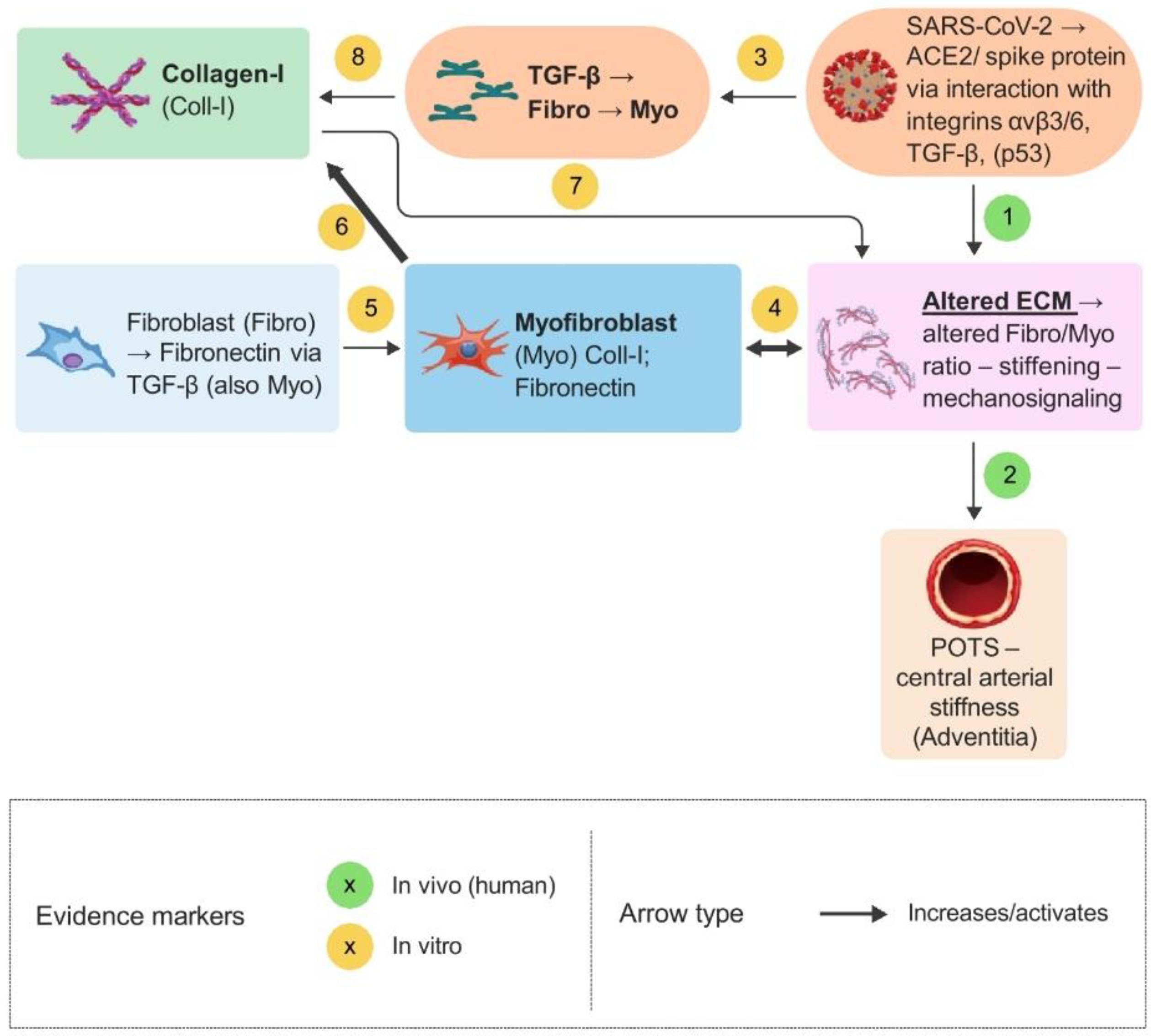
Figure 2.
Influence of the altered ECM on the potentially important roles of mast cells (MCs) and matrix metalloproteinase 9 (MMP-9) within the hypothesis. Possible feedback loops involving activated mast cells and inflammation (arrows 16 and 17) are shown. The influence of mast cell mediators (e.g., interleukin-6 (IL-6) and MMP-9) on the ECM is also shown. This can form a major feedback loop, especially since the mast cell mediator tumor necrosis factor α (TNF-α) can also induce IL-6 and MMP-9 production in fibroblasts and myofibroblasts. Arrows represent different types of interactions and are distinguished by color, line style, and shape, as indicated in the figure key. The numbers assigned to the arrows correspond to the respective descriptions and references in the main text.
Figure 2.
Influence of the altered ECM on the potentially important roles of mast cells (MCs) and matrix metalloproteinase 9 (MMP-9) within the hypothesis. Possible feedback loops involving activated mast cells and inflammation (arrows 16 and 17) are shown. The influence of mast cell mediators (e.g., interleukin-6 (IL-6) and MMP-9) on the ECM is also shown. This can form a major feedback loop, especially since the mast cell mediator tumor necrosis factor α (TNF-α) can also induce IL-6 and MMP-9 production in fibroblasts and myofibroblasts. Arrows represent different types of interactions and are distinguished by color, line style, and shape, as indicated in the figure key. The numbers assigned to the arrows correspond to the respective descriptions and references in the main text.
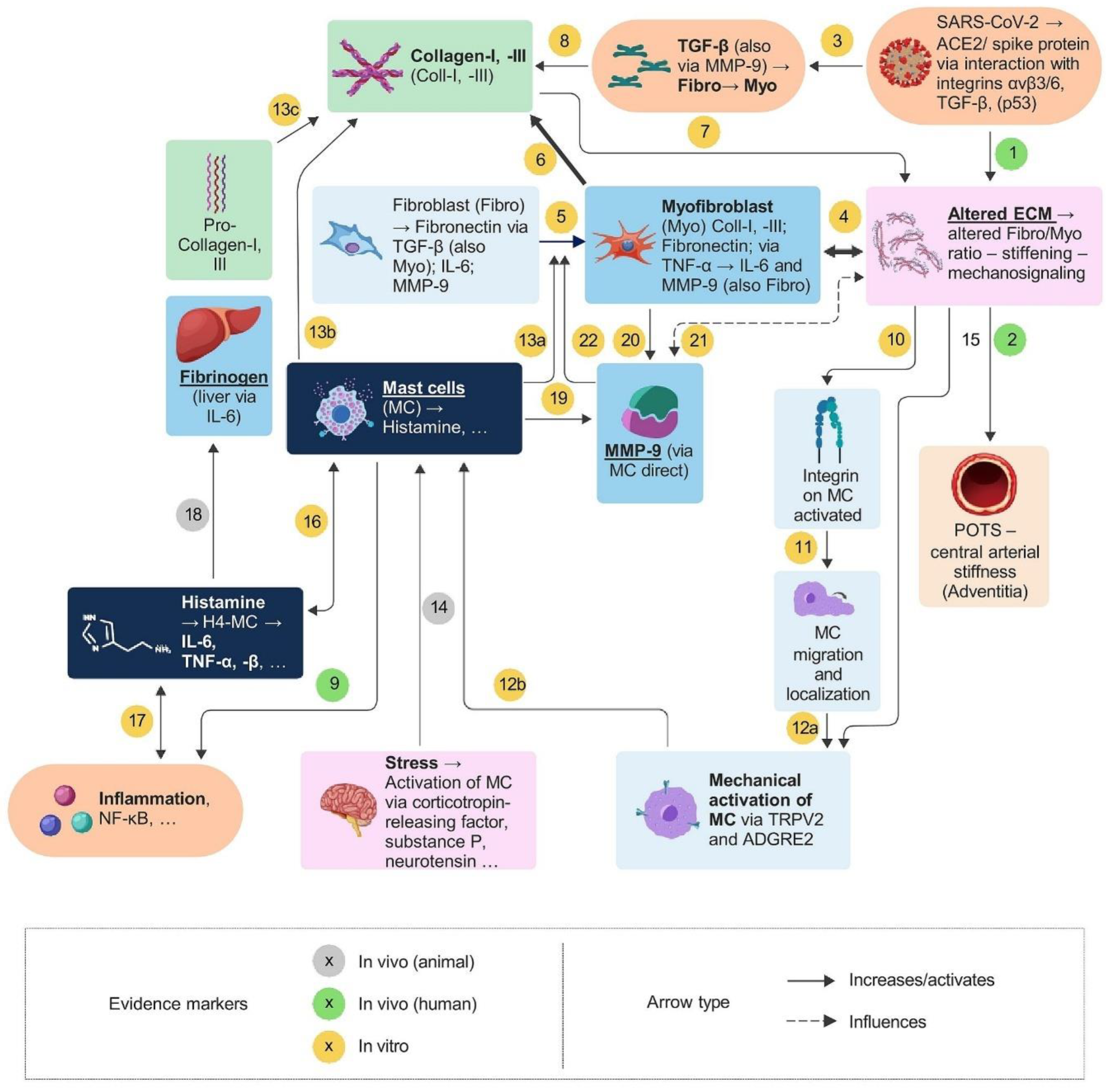
Figure 3.
Integration of the nervous system, including the N-methyl-D-aspartate receptor (NMDAR), PIEZO1, PIEZO2, as well as stress and insulin, within the hypothesis and its possible interactions with vitamin D. The possible positive feedback loops between chronic stress and inflammation and between inflammation and insulin resistance are shown (arrows 28 and 30). Arrows represent different types of interactions and are distinguished by color, line style, and shape, as indicated in the figure key. The numbers assigned to the arrows correspond to the respective descriptions and references in the main text.
Figure 3.
Integration of the nervous system, including the N-methyl-D-aspartate receptor (NMDAR), PIEZO1, PIEZO2, as well as stress and insulin, within the hypothesis and its possible interactions with vitamin D. The possible positive feedback loops between chronic stress and inflammation and between inflammation and insulin resistance are shown (arrows 28 and 30). Arrows represent different types of interactions and are distinguished by color, line style, and shape, as indicated in the figure key. The numbers assigned to the arrows correspond to the respective descriptions and references in the main text.
Disclaimer/Publisher’s Note: The statements, opinions and data contained in all publications are solely those of the individual author(s) and contributor(s) and not of MDPI and/or the editor(s). MDPI and/or the editor(s) disclaim responsibility for any injury to people or property resulting from any ideas, methods, instructions or products referred to in the content. |
© 2026 by the authors. Licensee MDPI, Basel, Switzerland. This article is an open access article distributed under the terms and conditions of the Creative Commons Attribution (CC BY) license (http://creativecommons.org/licenses/by/4.0/).
Copyright: This open access article is published under a Creative Commons CC BY 4.0 license, which permit the free download, distribution, and reuse, provided that the author and preprint are cited in any reuse.