

Submitted:
16 October 2026
Posted:
20 October 2026
You are already at the latest version
Abstract
Renal abnormalities are increasingly recognized in transfusion-dependent β-thalassemia, but evidence on intrarenal Doppler indices remains limited. This single-center case–control study compared the resistive index (RI) and pulsatility index (PI) in 44 patients aged 7–57 years and 30 apparently healthy controls aged 7–45 years. Three interlobar arterial measurements from each kidney were averaged for each participant. Group differences were evaluated using Welch’s t-test, and receiver operating characteristic analysis assessed discrimination between thalassemia and control status. Mean RI was higher in patients than controls (0.6789 ± 0.0500 versus 0.5943 ± 0.0440; mean difference, 0.0846; 95% confidence interval, 0.0626–0.1066). Mean PI was also higher (1.1655 ± 0.1824 versus 0.9197 ± 0.1290; mean difference, 0.2458; 95% confidence interval, 0.1736–0.3180). Both differences were significant (p < 0.001), with Hedges’ g values of 1.76 and 1.49, respectively. Areas under the curve were 0.895 for RI and 0.865 for PI. These findings indicate altered intrarenal hemodynamics in transfusion-dependent β-thalassemia. Without an independent kidney-injury reference standard or adjustment for potential confounders, they do not establish either index as a diagnostic marker of kidney injury.
Keywords:
transfusion-dependent β-thalassemia
; resistive index
; pulsatility index
; renal Doppler ultrasonography
; intrarenal hemodynamics
1. Introduction
β-Thalassemia is an inherited disorder of hemoglobin synthesis caused by reduced or absent production of β-globin chains. The resulting imbalance between α- and β-globin synthesis leads to precipitation of excess unpaired α-globin chains, oxidative membrane injury, ineffective erythropoiesis and chronic hemolytic anemia. Patients with the most severe phenotype require regular lifelong red blood cell transfusions and are currently classified as having transfusion-dependent thalassemia.
Regular transfusion therapy suppresses ineffective erythropoiesis and improves survival but inevitably causes progressive iron accumulation. In the absence of physiological mechanisms for active iron excretion, transfusional iron is deposited in the liver, heart, endocrine organs and other tissues. When the binding capacity of transferrin is exceeded, non-transferrin-bound iron and labile intracellular iron promote reactive oxygen species formation, lipid peroxidation, mitochondrial dysfunction and tissue fibrosis. Experimental hemosiderosis models have demonstrated associations between renal iron accumulation, oxidative stress and structural renal injury [1].
Renal involvement in transfusion-dependent β-thalassemia was previously considered uncommon. Increasing life expectancy has revealed a broad spectrum of glomerular and tubular abnormalities, including glomerular hyperfiltration, albuminuria, proteinuria, hypercalciuria, phosphaturia, hyperuricosuria, increased urinary excretion of β2-microglobulin and N-acetyl-β-D-glucosaminidase, and elevated serum cystatin C [2].
Renal dysfunction in β-thalassemia is likely multifactorial. Chronic anemia and tissue hypoxia may alter renal perfusion and promote hyperfiltration, whereas iron deposition and oxidative stress may affect glomerular, tubular and vascular structures. Iron-chelation therapy reduces iron burden but can also influence kidney function. Deferasirox has been associated with increases in serum creatinine, altered renal hemodynamics and proximal tubular abnormalities [3,4,5].
Conventional serum creatinine may have limited sensitivity for early kidney dysfunction in patients with β-thalassemia. Low muscle mass, altered creatinine generation and hyperfiltration may result in apparently normal or low serum creatinine concentrations despite underlying abnormalities. Consequently, cystatin C, urinary albumin, β2-microglobulin, neutrophil gelatinase-associated lipocalin, kidney injury molecule-1 and N-acetyl-β-D-glucosaminidase have been evaluated as potential early markers [3,4,6,7,8,9,10,11,12].
Renal Doppler ultrasonography provides a non-invasive assessment of intrarenal blood-flow waveforms. The resistive index (RI) is calculated from peak systolic and end-diastolic velocities, whereas the pulsatility index (PI) additionally incorporates time-averaged velocity. These indices have been investigated in relation to renal functional reserve, histological injury and kidney outcomes [13,14,15,16,17,18].
RI is not a direct measure of isolated renal vascular resistance. Its interpretation depends on renal parenchymal characteristics and systemic hemodynamics, including age, blood pressure and vascular properties [15]. Cardiovascular adaptation to chronic anemia may therefore affect renal Doppler waveforms independently of structural kidney injury.
Evidence regarding intrarenal Doppler indices in thalassemia remains limited and heterogeneous. Basut et al. reported lower resting RI values in subjects with β-thalassemia minor than in healthy controls, attributing this finding to reduced blood viscosity and vascular resistance [19]. Conversely, a pilot study in patients with β-thalassemia major suggested an association between iron burden and impaired Doppler-assessed renal functional reserve [13]. These observations indicate that the Doppler phenotype may vary according to disease severity, transfusion dependency, iron load, anemia and chelation exposure.
This study compared intrarenal RI and PI between patients with transfusion-dependent β-thalassemia and apparently healthy controls and assessed their ability to distinguish the groups. We hypothesized that thalassemia would be associated with altered intrarenal Doppler indices. Higher indices were observed in the patient group, although their relationship to independently established kidney injury could not be determined.
2. Materials and Methods
A single-center observational case–control study was conducted at UMHAT “St. Marina”, Varna, Bulgaria, between September 2018 and August 2019. The study included 44 patients with β-thalassemia major, currently classified clinically as transfusion-dependent β-thalassemia. Their ages ranged from 7 to 57 years. Eighteen participants were children and 26 were adults. The diagnosis was established on the basis of clinical history and hemoglobin electrophoresis. The control group consisted of 30 apparently healthy participants aged 7–45 years, including 13 children and 17 adults. Controls had no known hereditary anemia requiring regular transfusion therapy and no reported renal, cardiovascular, endocrine or malignant disease.
Patients were eligible when they had a confirmed diagnosis of transfusion-dependent β-thalassemia and provided written informed consent. Exclusion criteria were diabetes mellitus, known chronic glomerular disease, uncontrolled arterial hypertension, acute urinary tract infection, pregnancy, active malignant disease, current exposure to known nephrotoxic medications, administration of iodinated contrast material during the preceding month, acute febrile or inflammatory illness, known renal artery stenosis and obstructive uropathy.
Renal ultrasonography was performed with a 3.5–5 MHz transducer. Participants were examined in the supine position and instructed to take a deep breath and hold it during Doppler assessment. B-mode imaging assessed renal size, parenchymal echogenicity, corticomedullary differentiation, structural abnormalities and urinary tract obstruction. Pulsed-wave Doppler measurements were obtained from the interlobar arteries of both kidneys. Three measurements per kidney were obtained from reproducible waveforms containing consecutive cardiac cycles. Values were averaged to produce one RI and one PI value per participant. The resistive index was calculated as RI = (peak systolic velocity − end-diastolic velocity) / peak systolic velocity. The pulsatility index was calculated as PI = (peak systolic velocity − end-diastolic velocity) / time-averaged maximum velocity.
The primary outcome was the difference in mean intrarenal RI between patients with β-thalassemia and healthy controls. Secondary outcomes were the difference in mean PI and the ability of RI and PI to discriminate patients with β-thalassemia from healthy controls. No independent biochemical, functional, histological or longitudinal definition of kidney injury was used as a reference standard. Consequently, the ROC analysis evaluates discrimination between β-thalassemia and control status and must not be interpreted as direct diagnostic accuracy for structural kidney injury.
Continuous variables are presented as mean ± standard deviation (SD). Group means are presented with 95% confidence intervals calculated using the t distribution. Between-group differences were assessed using Welch’s t-test and are reported with 95% confidence intervals. Standardized differences are expressed as Hedges’ g, using the pooled SD with a small-sample correction. The participant was the unit of analysis. Receiver operating characteristic (ROC) analysis evaluated discrimination between thalassemia and control status. Areas under the curve (AUCs) are reported with standard errors and 95% CIs. The reported comparisons were unadjusted. RI and PI AUCs were interpreted descriptively; no formal paired comparison is reported. A two-sided p value < 0.05 was considered statistically significant. Statistical analyses were performed using jamovi, version 2.6.44.
3. Results
3.1. Study Population
The study included 74 participants: 44 patients with transfusion-dependent β-thalassemia (18 children and 26 adults; age range, 7–57 years) and 30 apparently healthy controls (13 children and 17 adults; age range, 7–45 years).
3.2. Intrarenal Doppler Indices
Both intrarenal Doppler indices were significantly higher in patients with transfusion-dependent β-thalassemia than in healthy controls. Mean RI was 0.6789 ± 0.0500 in the β-thalassemia group and 0.5943 ± 0.0440 in the control group. The absolute mean difference was 0.0846 (95% CI 0.0626–0.1066; Welch’s t = 7.68; p < 0.001). The standardized difference was large (Hedges’ g = 1.76).
Mean PI was 1.1655 ± 0.1824 in patients with β-thalassemia and 0.9197 ± 0.1290 in controls. The absolute mean difference was 0.2458 (95% CI 0.1736–0.3180; Welch’s t = 6.79; p < 0.001). The standardized difference was also large (Hedges’ g = 1.49). The group comparisons are summarized in Table 1 and Figure 1 and Figure 2.
3.3. Receiver Operating Characteristic Analysis
RI discriminated between thalassemia and control status with an AUC of 0.895 (standard error, 0.036; 95% CI, 0.824–0.966; p < 0.001). The corresponding AUC for PI was 0.865 (standard error, 0.042; 95% CI, 0.782–0.948; p < 0.001) (Figure 3).
The AUC was numerically higher for RI than for PI. This descriptive difference does not establish statistical superiority because no formal paired comparison of the correlated ROC curves was performed.
4. Discussion
Patients with transfusion-dependent β-thalassemia had higher intrarenal RI and PI than apparently healthy controls, with large standardized between-group differences. These findings indicate altered intrarenal arterial flow patterns but do not establish structural kidney injury or identify its underlying cause. Chronic anemia, tissue hypoxia, iron overload, chelation exposure and cardiovascular adaptation may contribute to the observed differences. These factors were not evaluated in adjusted analyses, and their individual contributions cannot be separated in this study.
Renal dysfunction in β-thalassemia is increasingly recognized as a clinically relevant complication. Early studies described increased urinary excretion of calcium, phosphate, uric acid and low-molecular-weight proteins, indicating predominantly proximal tubular dysfunction [2,6]. Subsequent investigations demonstrated glomerular hyperfiltration, albuminuria, proteinuria and abnormalities in serum cystatin C, urinary β2-microglobulin, N-acetyl-β-D-glucosaminidase and neutrophil gelatinase-associated lipocalin [3,4,7,8,9,10,11,12].
The pathogenic mechanisms are likely interrelated. Chronic anemia and tissue hypoxia can produce systemic vasodilation and increased cardiac output, thereby altering renal perfusion. Increased renal plasma flow and glomerular hyperfiltration may initially preserve or increase estimated glomerular filtration rate (eGFR) but may expose the glomerulus to sustained hemodynamic stress. Iron accumulation may simultaneously cause oxidative mitochondrial and cellular injury in tubular, glomerular and vascular structures [1].
Iron-chelation therapy further complicates interpretation. Deferasirox reduces iron overload but may alter renal hemodynamics and is associated in some patients with increases in serum creatinine or proximal tubular abnormalities. Piga et al. observed mild and largely reversible changes in renal hemodynamic parameters during deferasirox treatment [5]. Other studies have identified elevated tubular injury markers or declining eGFR in subsets of treated patients [3,4]. Consequently, chelation regimen and degree of iron overload should be considered potential determinants of renal Doppler indices.
Basut et al. reported lower RI in β-thalassemia minor than in healthy controls and suggested that reduced blood viscosity and vascular resistance might contribute [19]. Differences in disease severity, transfusion dependency, iron burden and treatment exposure may explain the divergent findings; however, this hypothesis was not tested in the present study. Nalesso et al. investigated Doppler-assessed renal functional reserve in β-thalassemia major and reported an inverse association with ferritin concentrations [13]. Their pilot findings suggest that dynamic RI measurements may provide complementary physiological information to resting measurements.
The patient-group mean RI of 0.6789 remained below the commonly used adult upper reference value of approximately 0.70. A higher group mean does not imply that every participant had an abnormal value. Moreover, a single threshold should be interpreted cautiously across the pediatric and adult age range, particularly because RI is associated with age [15]. RI has been associated with kidney histopathology and long-term renal outcomes in other populations. Ikee et al. identified relationships between RI, kidney function and histological injury, while a large study by Chen et al. demonstrated an association between interlobar RI and tubulointerstitial damage [16,17]. Higher RI has also been associated with faster deterioration of kidney function in chronic renal disease [18]. These studies establish the biological relevance of RI but do not justify extrapolating prognostic conclusions to the present cross-sectional β-thalassemia cohort.
The ROC results describe discrimination between thalassemia and control status within this sample. They do not quantify diagnostic accuracy for kidney injury, which was not independently defined. The numerically higher AUC for RI does not establish superiority over PI without a formal paired comparison.
Validation against albuminuria, proteinuria, cystatin C, measured or estimated glomerular filtration rate, and urinary tubular biomarkers is needed. Such studies could establish whether Doppler indices add clinically useful information beyond systemic hemodynamics and established kidney assessments.
Renal Doppler ultrasonography is non-invasive and requires neither ionizing radiation nor contrast administration. These features make it suitable for further investigation as an adjunct to renal assessment in transfusion-dependent β-thalassemia. The present findings do not support treatment decisions based on RI or PI alone; interpretation requires clinical context, kidney-function measures, blood pressure, cardiovascular status, iron burden and chelation exposure.
This single-center, cross-sectional study had a small sample and included both children and adults. The age ranges differed between groups, and the reported analyses were unadjusted. Confounding by age, blood pressure, heart rate, pulse pressure, hemoglobin, ferritin, transfusion timing, splenectomy status and chelation regimen may therefore have influenced the observed differences.
The ROC analyses classified thalassemia status rather than kidney injury. They therefore cannot establish diagnostic performance for kidney injury or provide validated thresholds for clinical use. A formal paired comparison would be needed to evaluate differences between the RI and PI AUCs.
The cross-sectional design precludes conclusions about subsequent kidney-function decline. Prospective studies with standardized acquisition, reproducibility assessment, fuller clinical characterization and longitudinal outcomes are required.
5. Conclusions
Intrarenal RI and PI were higher in patients with transfusion-dependent β-thalassemia than in apparently healthy controls. Both indices distinguished the study groups, but neither was validated as a diagnostic marker of kidney injury or a predictor of kidney-function decline. Their potential clinical value requires evaluation alongside established renal and systemic hemodynamic measures.
Author Contributions
Conceptualization, L.S.; methodology, L.S. and R.S.; validation, A.P. and M.B-P.; formal analysis, M.B.-P. and A.P.; investigation, L.S. and M.B.-P.; resources, L.S. and R.S.; data curation, M.B.-P. and A.P.; writing—original draft preparation, L.S. and M.B.-P.; writing—review and editing, L.S., M.B.-P., A.P. and R.S.; visualization, R.S. and A.P.; supervision, A.P.; project administration, L.S. and M.-B-P. All authors have read and agreed to the published version of the manuscript.
Funding
This research received no external funding.
Institutional Review Board Statement
The study was conducted in accordance with the Declaration of Helsinki, and approved by the Research Ethics Committee (REC) at the Medical University of Varna (approval no. 76, 9 August 2018).
Informed Consent Statement
Written informed consent was obtained from adult participants and from the parents or legal guardians of participants younger than 18 years. Age-appropriate assent was obtained when applicable.
Data Availability Statement
I The data presented in this study are available on request from the corresponding author due to participant privacy and confidentiality considerations.
Acknowledgments
During the preparation of this manuscript, the authors used OpenAI Codex (GPT-6) to assist in generating figures based on the study’s reported summary statistics and ROC results. The authors reviewed, edited, and approved the figures and take full responsibility for the content of this publication.
Conflicts of Interest
The authors declare no conflicts of interest.
Abbreviations
The following abbreviations are used in this manuscript:
| AUC | Area under the receiver operating characteristic curve |
| CI | Confidence interval |
| eGFR | Estimated glomerular filtration rate |
| PI | Pulsatility index |
| REC | Research Ethics Committee |
| RI | Resistive index |
| ROC | Receiver operating characteristic |
| SD | Standard deviation |
| UMHAT | University Multiprofile Hospital for Active Treatment |
References
- Zhou, X.J.; Laszik, Z.; Wang, X.Q.; Silva, F.G.; Vaziri, N.D. Association of renal injury with increased oxygen free radical activity and altered nitric oxide metabolism in chronic experimental hemosiderosis. Lab Invest. 2000, 80, 1905–1914. [Google Scholar] [CrossRef] [PubMed]
- Sumboonnanonda, A.; Malasit, P.; Tanphaichitr, V.S.; Ong-ajyooth, S.; Sunthornchart, S.; Pattanakitsakul, S.; et al. Renal tubular function in beta-thalassemia. Pediatr. Nephrol. 1998, 12, 280–283. [Google Scholar] [CrossRef] [PubMed]
- Annayev, A.; Karakaş, Z.; Karaman, S.; Yalçıner, A.; Yılmaz, A.; Emre, S. Glomerular and tubular functions in children and adults with transfusion-dependent thalassemia. Turk. J. Haematol. 2018, 35, 66–70. [Google Scholar] [CrossRef] [PubMed]
- Badeli, H.; Baghersalimi, A.; Eslami, S.; Saadat, F.; Hassanzadeh Rad, A.; Basavand, R.; et al. Early kidney damage markers after deferasirox treatment in patients with thalassemia major: a case-control study. Oxid. Med. Cell Longev. 2019, 2019, 5461617. [Google Scholar] [CrossRef] [PubMed]
- Piga, A.; Fracchia, S.; Lai, M.E.; Cappellini, M.D.; Hirschberg, R.; Habr, D.; et al. Deferasirox effect on renal haemodynamic parameters in patients with transfusion-dependent beta thalassaemia. Br. J. Haematol. 2015, 168, 882–890. [Google Scholar] [CrossRef] [PubMed]
- Aldudak, B.; Karabay Bayazit, A.; Noyan, A.; Ozel, A.; Anarat, A.; Sasmaz, I.; et al. Renal function in pediatric patients with beta-thalassemia major. Pediatr. Nephrol. 2000, 15, 109–112. [Google Scholar] [CrossRef] [PubMed]
- Economou, M.; Printza, N.; Teli, A.; Tzimouli, V.; Tsatra, I.; Papachristou, F.; Athanassiou-Metaxa, M. Renal dysfunction in patients with beta-thalassemia major receiving iron chelation therapy either with deferoxamine and deferiprone or with deferasirox. Acta Haematol. 2010, 123, 148–152. [Google Scholar] [CrossRef] [PubMed]
- Quinn, C.T.; Johnson, V.L.; Kim, H.Y.; Trachtenberg, F.; Vogiatzi, M.G.; Kwiatkowski, J.L.; et al. Renal dysfunction in patients with thalassaemia. Br. J. Haematol. 2011, 153, 111–117. [Google Scholar] [CrossRef] [PubMed]
- Ali, B.A.; Mahmoud, A.M. Frequency of glomerular dysfunction in children with beta thalassaemia major. Sultan Qaboos Univ. Med. J. 2014, 14, e88–e94. [Google Scholar] [CrossRef] [PubMed]
- Şen, V.; Ece, A.; Uluca, Ü.; Söker, M.; Güneş, A.; Kaplan, İ.; et al. Urinary early kidney injury molecules in children with beta-thalassemia major. Ren. Fail. 2015, 37, 607–613. [Google Scholar] [CrossRef] [PubMed]
- Behairy, O.G.; Abd Almonaem, E.R.; Abed, N.T.; Abdel Haiea, O.M.; Zakaria, R.M.; AbdEllaty, R.I.; et al. Role of serum cystatin-C and beta-2 microglobulin as early markers of renal dysfunction in children with beta thalassemia major. Int. J. Nephrol. Renov. Dis. 2017, 10, 261–268. [Google Scholar] [CrossRef] [PubMed]
- Mahmoud, A.A.; Elian, D.M.; Abd El Hady, N.M.; Abdallah, H.M.; Abdelsattar, S.; Khalil, F.O.; Abd El Naby, S.A. Assessment of subclinical renal glomerular and tubular dysfunction in children with beta thalassemia major. Children 2021, 8, 100. [Google Scholar] [CrossRef] [PubMed]
- Nalesso, F.; Rigato, M.; Cirella, I.; Protti, M.P.; Zanella, R.; Rossi, B.; et al. The assessment of renal functional reserve in β-thalassemia major patients by an innovative ultrasound and Doppler technique: a pilot study. J. Clin. Med. 2022, 11, 6752. [Google Scholar] [CrossRef] [PubMed]
- Petersen, L.J.; Petersen, J.R.; Talleruphuus, U.; Ladefoged, S.D.; Mehlsen, J.; Jensen, H.A. The pulsatility index and the resistive index in renal arteries: associations with long-term progression in chronic renal failure. Nephrol. Dial. Transplant. 1997, 12, 1376–1380. [Google Scholar] [CrossRef] [PubMed]
- Ponte, B.; Pruijm, M.; Ackermann, D.; Vuistiner, P.; Eisenberger, U.; Guessous, I.; et al. Reference values and factors associated with renal resistive index in a family-based population study. Hypertension 2014, 63, 136–142. [Google Scholar] [CrossRef] [PubMed]
- Ikee, R.; Kobayashi, S.; Hemmi, N.; Imakiire, T.; Kikuchi, Y.; Moriya, H.; et al. Correlation between the resistive index by Doppler ultrasound and kidney function and histology. Am. J. Kidney Dis. 2005, 46, 603–609. [Google Scholar] [CrossRef] [PubMed]
- Chen, Q.; He, F.; Feng, X.; Luo, Z.; Zhang, J.; Zhang, L.; et al. Correlation of Doppler parameters with renal pathology: a study of 992 patients. Exp. Ther. Med. 2014, 7, 439–442. [Google Scholar] [CrossRef] [PubMed]
- Radermacher, J.; Ellis, S.; Haller, H. Renal resistance index and progression of renal disease. Hypertension 2002, 39, 699–703. [Google Scholar] [CrossRef] [PubMed]
- Basut, F.; Keşkek, Ş.Ö.; Gülek, B. Better renal resistive index profile in subjects with beta thalassemia minor. Med. Princ. Pract. 2018, 27, 367–371. [Google Scholar] [CrossRef] [PubMed]
Figure 1.
Mean intrarenal resistive index in patients with transfusion-dependent β-thalassemia and healthy controls. Error bars represent 95% confidence intervals of the group means. The between-group mean difference was 0.0846 (95% CI 0.0626–0.1066; p < 0.001; Hedges’ g = 1.76).
Figure 1.
Mean intrarenal resistive index in patients with transfusion-dependent β-thalassemia and healthy controls. Error bars represent 95% confidence intervals of the group means. The between-group mean difference was 0.0846 (95% CI 0.0626–0.1066; p < 0.001; Hedges’ g = 1.76).
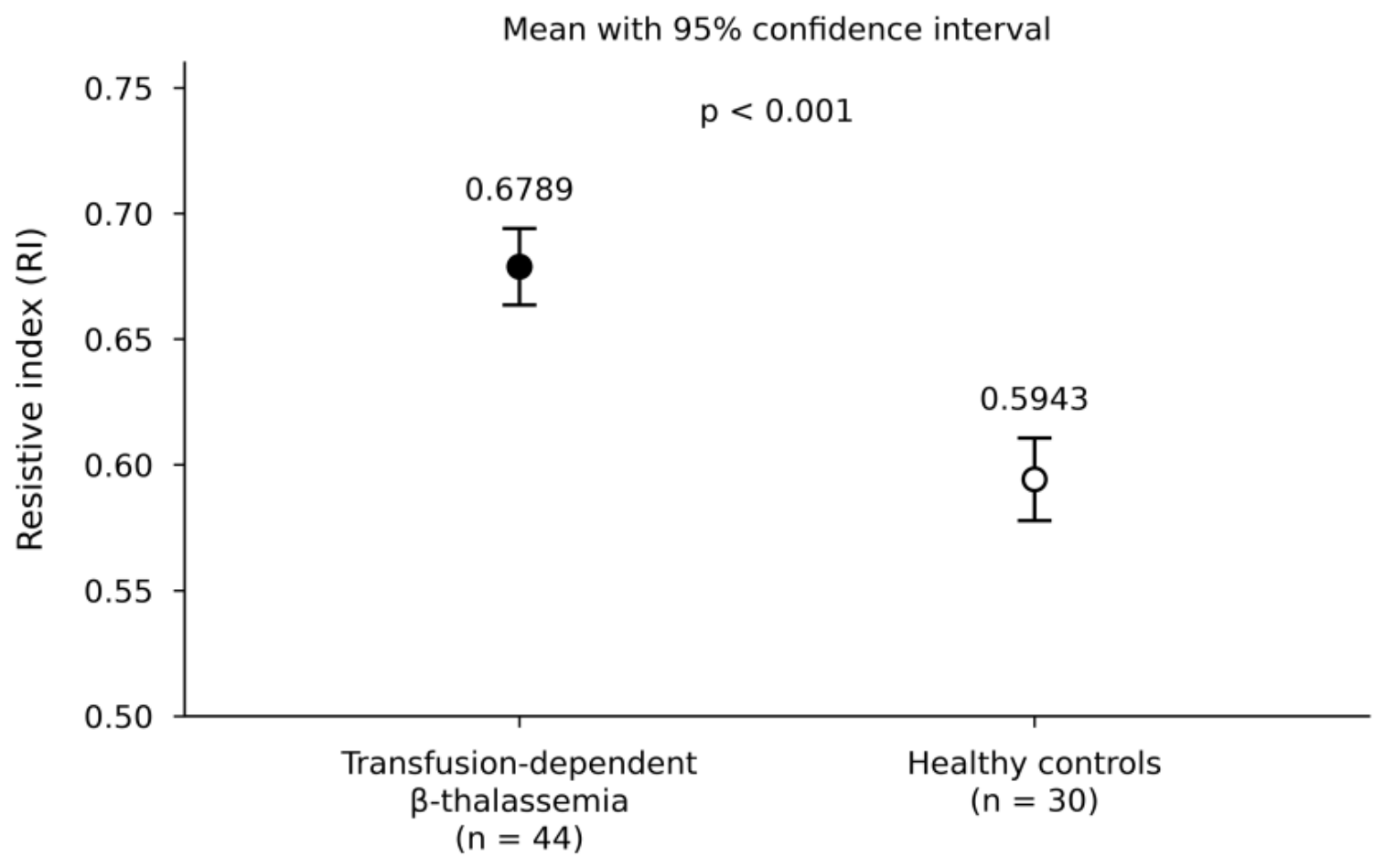
Figure 2.
Mean intrarenal pulsatility index in patients with transfusion-dependent β-thalassemia and healthy controls. Error bars represent 95% confidence intervals of the group means. The between-group mean difference was 0.2458 (95% CI 0.1736–0.3180; p < 0.001; Hedges’ g = 1.49).
Figure 2.
Mean intrarenal pulsatility index in patients with transfusion-dependent β-thalassemia and healthy controls. Error bars represent 95% confidence intervals of the group means. The between-group mean difference was 0.2458 (95% CI 0.1736–0.3180; p < 0.001; Hedges’ g = 1.49).
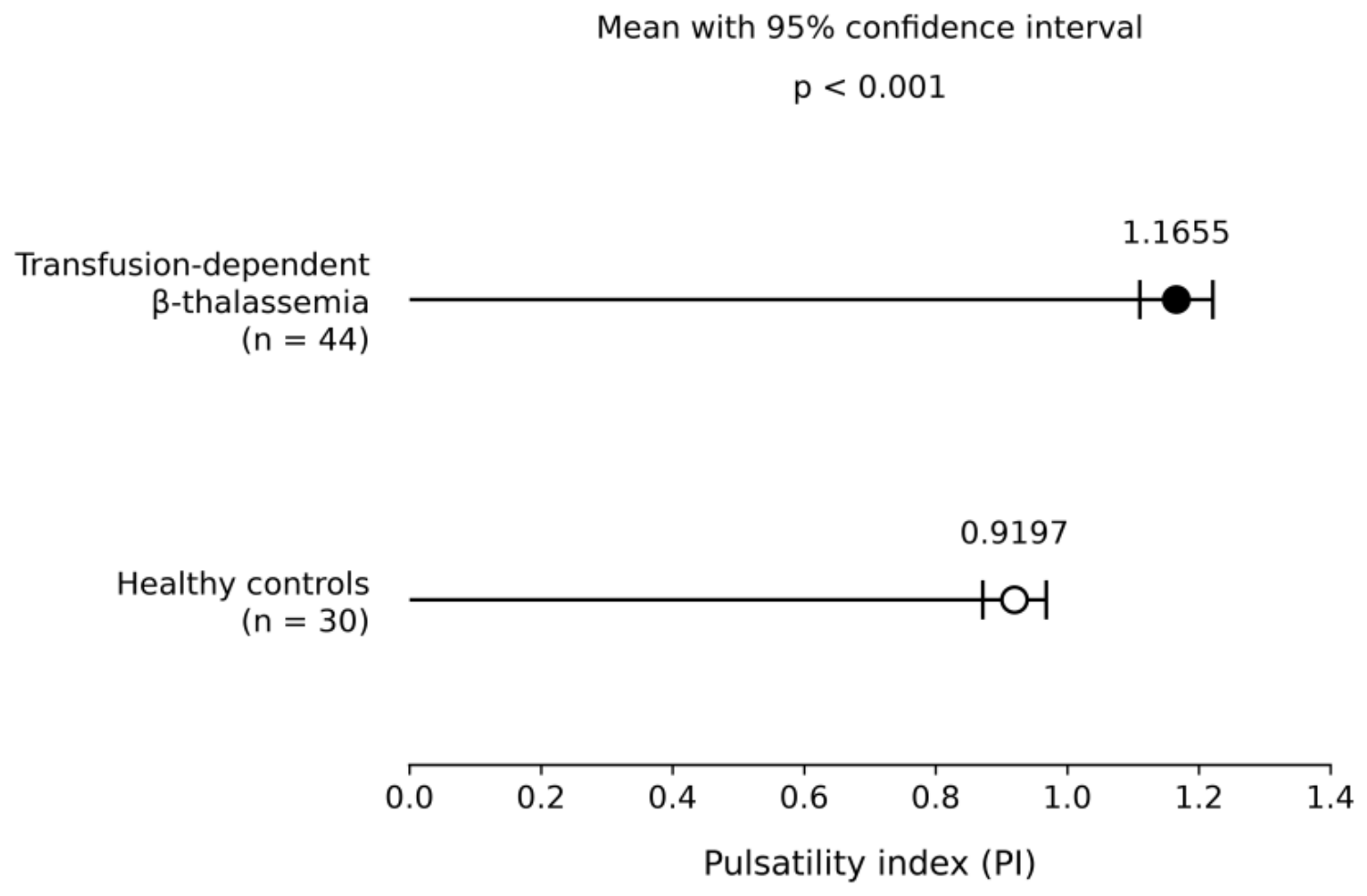
Figure 3.
Areas under the receiver operating characteristic curves for the resistive index and pulsatility index, with 95% confidence intervals. The classification outcome is thalassemia versus control status, not independently confirmed kidney injury.
Figure 3.
Areas under the receiver operating characteristic curves for the resistive index and pulsatility index, with 95% confidence intervals. The classification outcome is thalassemia versus control status, not independently confirmed kidney injury.
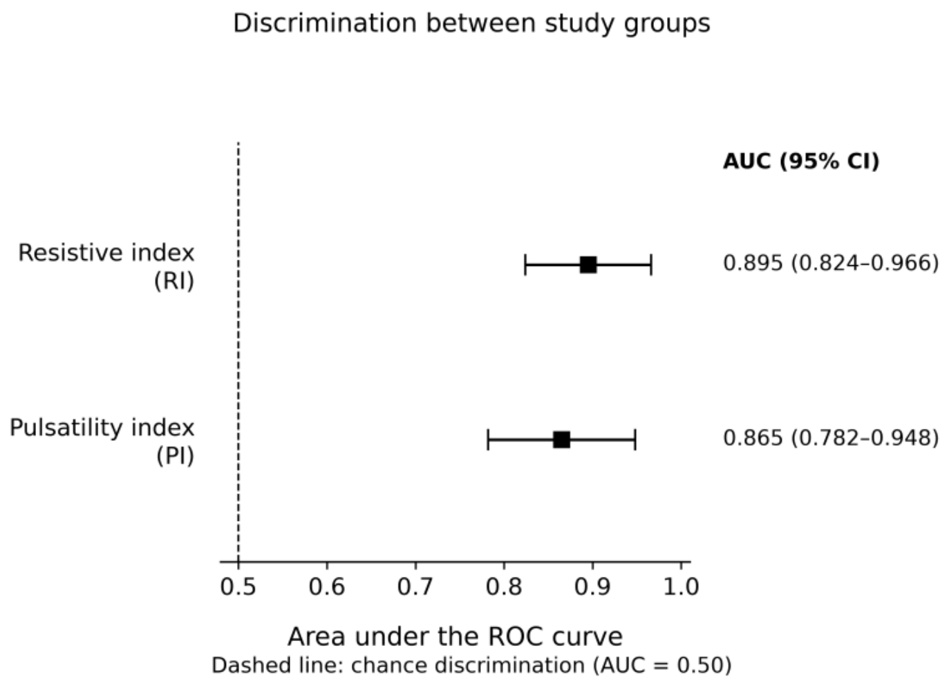

Table 1.
This is a table. Tables should be placed in the main text near the first time they are cited.
Table 1.
This is a table. Tables should be placed in the main text near the first time they are cited.
| Index | Thalassemia (n = 44) |
Controls (n = 30) |
|---|---|---|
| RI | 0.6789 ± 0.0500 | 0.5943 ± 0.0440 |
| PI | 1.1655 ± 0.1824 | 0.9197 ± 0.1290 |
Values are mean ± SD. Differences are thalassemia minus control values; CIs and p values are from Welch’s t-test. RI, resistive index; PI, pulsatility index; SD, standard deviation; CI, confidence interval.
Disclaimer/Publisher’s Note: The statements, opinions and data contained in all publications are solely those of the individual author(s) and contributor(s) and not of MDPI and/or the editor(s). MDPI and/or the editor(s) disclaim responsibility for any injury to people or property resulting from any ideas, methods, instructions or products referred to in the content. |
© 2026 by the authors. Licensee MDPI, Basel, Switzerland. This article is an open access article distributed under the terms and conditions of the Creative Commons Attribution (CC BY) license (http://creativecommons.org/licenses/by/4.0/).
Copyright: This open access article is published under a Creative Commons CC BY 4.0 license, which permit the free download, distribution, and reuse, provided that the author and preprint are cited in any reuse.