

Submitted:
12 October 2026
Posted:
19 October 2026
You are already at the latest version
Preprints on COVID-19 and SARS-CoV-2
Abstract
Epigenetic age acceleration (EAA) is defined as the difference when an epigenetic clock shows a higher age than the birth certificate. EAA is associated with neurological disease, amongst many other diseases. A series of studies, indexed in Medline, identify that SARS-CoV-2 infection, COVID-19 and long COVID are all associated with EAA. The main mechanism of SARS-CoV-2 to induce pathogenesis in the organism is via the spike protein S2 subunit (SP2). Through the Toll Like Receptor 2 (TRL2) and MyD88-mediated response, the spike protein, via the canonical pathway, activates the nuclear factor kappa-light-chain-enhancer of activated B cells (NF-κB), which initiates the pro-inflammatory response and thereafter promotes inflammation. In turn, NF-κB mediates EAA. The SARS-CoV-2 infection (through its spike protein) causes methylation and demethylation profile alterations in DNA and telomere attrition, which associates with COVID-19 severity. Moreover, the post COVID-19 syndrome is associated with EAA and telomere attrition. Alteration of the methylome, i.e. the methylation and demethylation of the DNA and the nucleosome during EAA, contributes to cellular senescence, which is linked to autoimmunity, and, furthermore, to cancer. In this paper, we investigate the role of NF-κB activation to cause and promote EAA. We identify that nucleosome alterations can occur not only with COVID-19 but also following administration of the mRNA vaccines coding for the SARS-CoV-2 spike protein (post COVID vaccine syndrome; PCVS). We therefore analyse in depth the mechanisms that implicate NF-κB to cause methylome and nucleosome alterations and contribute to EAA. We identify the critical role of long non-coding RNAs (lncRNAs), which regulate NF-κB activation through scaffolds, decoys, and feedback loops, in modulating type I interferon expression, premature aging and senescence. Moreover, we point to the consequences that the NF-κB-mediated EAA and inflammaging can have on the overall health of the human population. The mRNAs in the vaccine and the transcribed spike protein persist in time, promoting a chronic activation of NF-κB and other pathogenic pathways.
Keywords:
epigenetic age acceleration
; long COVID
; SARS-CoV-2 spike protein
; chronic NF-κB activation
; long noncoding RNAs
; methylome
; inflammaging
1. Introduction
The advancement of aging of DNA primarily occurs due to the epigenetic dysregulation of the methylome (methylation and demethylation of DNA), and the histone modification alterations (methylation, acetylation, phosphorylation and others), which act cooperatively with the alteration of methylations of the DNA [1,2]. In addition, the non-coding RNAs (ncRNAs) have substantial contributory-regulatory roles in the epigenetic changes that occur during aging [3]. Hence, the structure of the nucleosome with aging becomes altered, and a differential gene expression that promotes disease occurs [1,4].
In this context, the epigenetic age acceleration (EAA) of the DNA has important deteriorating health implications that signal for various diseases, including neurodegeneration, autoimmunity, and cancer [5]. For cancer incidence and mortality, it is estimated that, for one year of DNA age acceleration, the risk of developing cancer increases by 6% and the risk of dying from cancer increases by 17% during the next 5 years [5,6].
The dysregulated activation of the interferon signalling pathway [7], encompassing the cytokine family of proteins, the mitogen-activating protein kinase (MAPK), and the phosphoinositide 3-kinase MAPK (PI3K)-protein kinase B (AKT)-mechanistic target of rapamycin (mTOR) cascades, causes EAA and is linked to aging-related diseases (inflammatory diseases, autoimmunity and cancer [8]).
Remarkably, in COVID-19 patients, immune dysregulation is the causative factor of EAA [9]. Undoubtedly, the main pathogenic mechanism by which SARS-CoV-2 infection causes immune dysregulation encountered in COVID-19 is by the spike protein (SP) [10,11]. SP interactions cause the upregulation of cytokine levels (e.g., cytokine storm) that match the severity and mortality of COVID-19 [12,13]. Similarly, long COVID pathology (autoimmunity, neuroinflammation) is characterised by an upregulated level of peripheral and central cytokines [14].
One of the main reasons for the cytokine level upregulation caused by SP to produce high or low levels of inflammation is through its interaction with the Toll-Like Receptor 2 (TLR2) in a MyD88-dependent manner [15]. The TLR2/MyD88 mediation corresponds to a potent activation of the nuclear factor kappa-light-chain-enhancer of activated B cells (NF-κB) in cells. MyD88 is an essential adaptor to the TLRs that, once stimulated by an external stimulus, e.g., SP, through a series of phosphorylation and ubiquitination events, activates the IKK complex, thereby inactivating the IκB NF-κB inhibitors by phosphorylation. The disabling of IκB inhibitors allows for the NF-κB family of proteins to enter the nucleus and perform their significant transcription factor roles for the immune system that also, however, promote inflammation [16,17]. The TLR2/MyD88-dependent signalling involves the canonical part of NF-κB activation that transforms monocytes into M1-like inflammatory macrophages and allows the expression of pro-inflammatory cytokines (interleukin (IL)-1, IL-2, IL-6, IL-8 IL-12 and TNF-α) [18]. A continuously activated or/and, a dysregulated form of NF-κB activation can lead to epigenetic age acceleration (EAA), premature aging, and the development of age-related diseases, including cancer [19,20]. Moreover, the activated NF-κB regulates aging-dependent ncRNAs that further provoke the inflammatory conditions, senescence, and aging [21].
Moreover, the meta-analysis study of M. Shekhar Patil et al. [22] examined six research studies on the epigenetic alterations in Long COVID, with symptoms persisting 12 weeks or longer after initial infection, and post-acute COVID-19 syndrome (PACS), with symptoms persisting over four weeks after initial infection. The researchers concluded that DNA methylation alterations of both conditions relate to immune, metabolism and autonomic nervous system dysfunctions.
Due to the strong relationships between SP persistence and implications in Long COVID chronic pathology [23], the long-term persistence of SP in human blood monocytes in a) Long-COVID after SARS-COV-2 infection [24] and b) post-COVID-19 vaccine syndrome (PCVS) [25], we reexamine the role of chronic NF-κB activation to promote EAA, reevaluate the scientific evidence of COVID-19 to be a causative factor for EAA, and finally evaluate the impact that SP can have on premature aging and age-associated diseases, whether it is derived from the vaccine or the disease-infection.
2. Methods
In order to investigate the role of SARS-CoV-2 spike protein (specifically, SP1) on epigenetic age acceleration, we used the Medline/PubMed search engine. The following key words / searches were made: 1) “SARS-CoV-2, COVID-19, epigenetic age acceleration”. This gave 24 results, https://pubmed.ncbi.nlm.nih.gov/?term=SARS%20CoV-2%2C%20covid%2019%20epigenetic%20age%20acceleration&page=3
2) “Long COVID, epigenetic age acceleration”, that gave 6 results.
and 3) “mRNA vaccines, COVID 19 epigenetic memory”, that gave 5 results.
Out of search 1, we included 16 / 24 studies that were relevant to SARS-CoV-2 infection, COVID-19, Long COVID, and EAA. Out of search 2, we did not include any additional studies -- 4/6 studies in search 2 were already included during search 1. Out of search 3, we included 2/5 studies that were found to be relevant to epigenetic change induction after the mRNA COVID-19 vaccination. A total of 14 research studies and 4 review studies were included for evaluation. During the literature investigation, the G Farkas et al. research study, a study that is clearly related to our topic but was missed during the PubMed searches, was also included for evaluation [26].
4) We also used the Google Scholar search engine after the Medline searches, using the keywords: epigenetic age acceleration, COVID-19, Long COVID: https://scholar.google.com/scholar?start=30&q=epigenetic+age+acceleration+covid+19+long+covid&hl=el&as_sdt=0,5&as_ylo=2022&as_vis=1
The search gave 54,300 results. However, all research studies revealed that were relevant to the topic of current investigation overlapped with the Medline searches. Out of this search, only the meta-analysis study of M. Shekhar Patil et al. [22] on epigenetic changes in PACS and Long COVID was included.
3. Evidence that COVID-19, Long COVID and SARS-CoV-2 Spike Protein Can Cause Methylome and Nucleosome Alterations that Associate with Epigenetic Age Acceleration
In Table 1, the research studies that have investigated the impact of SARS-CoV-2 infection, COVID-19, Long COVID, and mRNA vaccines (coding for the SARS-CoV-2 spike protein) on epigenetic regulation and modifications that can confer epigenetic age acceleration (or epigenetic memory due to the vaccination) are included. Although epigenetic memory does not necessarily mean an EAA, there are environmental conditions where epigenetic memory is closely linked to biological aging and EAA [27], and therefore these studies were included, as they also assessed epigenetic regulation events on histone modifications. A total of 15 research studies, found in PubMed, are summarized in Table 1.
3.1. Studies Using Epigenetic Aging Tools to Evaluate EAA in Association with COVID-19
An epigenetic clock is a biomarker that measures aging by using machine learning to track patterns of cytosine methylation at specific CpG sites in DNA. They track "biological" age, which can then be compared to chronological age [43]. The epigenetic aging tools used to evaluate EAA in the research studies listed in Table 1 have shown diverging results across the different studies, such as in the J Franzen et al., P García-delaTorre et al., and J Bohlin et al. studies [28,34,42]. This is plausibly due to variable sensitivities of the epigenetic clocks used. Even in the same study [26], DNAmAge showed accelerated aging, whereas five other epigenetic clocks showed decelerating aging in the patients’ DNA. Nevertheless, the studies that showed EAA, especially when next generation estimators of DNA methylation were used, as in the MK Gupta and R Vadde study [9], may be convincing for EAA and for the methylome alterations occurring.
More importantly, these epigenetic aging estimators correlated with immune dysregulation and inflammatory processes. The immune dysregulation may be linked to demethylation of promoters mediated by the activity of NF-κB [44,45] The LHA Ventura et al. study investigated the immunosenescence profile in COVID-19 patients and found an exceptional number of inflammatory mediators in their plasma. Amongst these, there was an elevated expression of the chemokine CXCL9 [39]. This chemokine’s elevation is characteristic for inflammaging (a process of aging due to inflammation [40]). Studies link this chemokine to cardiac aging and poor vascular function, indicating for multimorbidity conditions [46]. The expression of CXCL9 is under the regulation of activated NF-κB signalling that follows a complicated molecular route of the canonical pathway stimulation [47]. The COVID-19 patients in the LHA Ventura et al. study that were shown to have an accelerated immunosenescence phenotype also showed an EAA during their methylome measurements [39].
Furthermore, the L. Calzari et al. group, during their Long COVID study in 2024, identified a large number of dysregulated genes, actually 42 CpG sites, mainly within CpG islands and gene promoters, that showed significant methylation differences (False Discovery Rate (FDR) < 0.05) [34]. The researchers suggested that an epigenetic drift occurs in patients’ methylomes. Apart from the fact that the epigenetic drift may confuse the measurement of EAA with epigenetic clocks [48], as also happened in the MK Gupta and R Vadde [9], the G Farkas et al. [26], the P García-delaTorre et al. [35], and other studies presented in Table 1, this kind of epigenetic change can be attributed to the NF-κB overactivation, which can play an important role in the progress of aging [19].
3.2. Specific Genes that Have Been Found to be Dysregulated
Ann epigenetic drift creates progressive methylations or demethylations at certain gene loci that associate with aging and disease occurrence, such as the emergence of cancer [49]. Specifically, the dysregulated genes identified in the L. Calzari et al. study [34] are linked to glutamate metabolism, indicating a trend for neurological dysfunction in the Long COVID patients [50]. Amongst the genes that were found to be affected by the significant alterations in the methylome were GLUD1, ATP1A3, and ARRB2.
GLUD1 (Glutamate Dehydrogenase 1) encodes a mitochondrial matrix enzyme that converts glutamate to α-ketoglutarate and ammonia [51]. ATP1A3 (ATPase Na+/K+ Transporting Subunit α 3) builds the α-3 subunit of the Na+/K+ ATPase pump. ATP1A3 is critical for neuronal cell signaling and for maintenance of electrochemical stability, enabling cell excitation and action potential propagation [52]. ARRB2 (beta-arrestin 2) is a major regulatory protein that desensitizes G-protein coupled receptors (GPCRs) and also regulates a diverse array of cellular functions [53]. The normal function of these genes is essential for the preservation of health.
Dysfunction of glutamate dehydrogenase is linked to the pathogenesis of diverse cellular processes, including ammonia metabolism, acid-base equilibrium, redox homeostasis lipid biosynthesis, and lactate production. [54]. The production of α-ketoglutarate cooperates with the activation of NF-κB, to regulate glucose levels, and this cooperation is implicated in the development of brain cancer [55]. The α3 Na+/K+ ATPase ion pump is implicated primarily in cardiological and renal functions and disease [56]. Immunological – cytokine dysfunction during NF-κΒ signalling associate with loss of this ATPase normal function in neurological disorders related to mitochondrial dysfunction [57]. beta-Arrestin 2, but not beta-arrestin 1, is required for lysophosphatidic acid (LPA)-induced NF- κB activation and IL-6 expression [58]. In summary, all three genes found to be dysregulated in Long COVID participate in the NF-κB inflammatory signalling and are involved in energy and mitochondrial dysfunction.
The N. Cheung study [36] is in accordance with the L. Calzari et al. study [34], as the researchers in both studies found an epigenetic anomaly (increased methylation) in the NMDA receptor gene promoter, indicating for a glutamatergic aging signature, i.e., defective glutamatergic neurotransmission [59].
3.3. A Role for Telomere Shortening
The W. Xu et al. study also found telomere attrition (shortening) in the chromosomes of patients with severe COVID-19 [30]. Severe COVID-19 is a condition notably described with cytokine storm caused by overactivation of NF-κB inflammatory pathways [60].
A similar telomere attrition was found in the patients who were deceased from COVID-19 in the Y. Bejaoui et al. study [37]. Importantly, in this study [37], the researchers measured an increased rate of telomere shortening that was accompanied with an increased EAA only in the deceased-from-severe-COVID-19 patients, indicating a rapidly progressing aging machinery due to a severe and deadly systemic infection. Additionally, telomere shortening occurred in severe COVID-19 patients in the W. Xu et al. study [30], (although epigenetic clocks were unable to show an increase in EAA). Biological age of patients was shown to be increased in the post-COVID patients in the study by A. Mongelli et al. [29],). This study concluded that telomere shortening resulted in a several year increase in premature aging [29]. These studies are all listed in Table 1.
3.4. Studies Involving the SARS-CoV-2 mRNA Vaccines
Apart from the methylome abnormalities found in the epigenetic dysregulation studies that link EAA with NF-κB activation, listed in Table 1, the studies on mRNA coding for SARS-CoV-2 spike protein remarkably reveal more on the epigenetic modification remodelling of the nucleosome occurring by the activation of NF-κB. The researchers of the A. Simonis et al. study found that SARS-CoV-2 spike protein (as coded by the mRNA COVID-19 vaccines) induced NF-κB activation and was responsible for the lysine acetylation (H3K27ac) of histones at promoters of human monocyte-derived macrophages [41], Moreover, the acetylation modifications of histones were persistent for a six-month period after the second spike protein stimulation of NF-kB. Although the researchers found this to be important for the immunological epigenetic memory of the immune cells, this finding also underlies a continuous activation of the canonical NF-κΒ pathway for the six-month period, as the acetylation of histones is a reversible process [61].
The Y. Yamaguchi et al. research study also refers to a boosted cytokine and chemokine response after the second mRNA vaccination (i.e. induction of NF-kB activation by the SARS-CoV-2 spike protein) [32]. These responses were accompanied by epigenetic changes in the monocytes tested, as the research showed. However, most importantly, there was a particular elevation in the type 1 interferon response. In this way, NF-κB cross talks with the interferon response.
The induction of type 1 interferon results in an increase in histone methylations, particularly the trimethylation of H3 Lysine 4 (H3K4me3). Such a histone modification enforces the transcription of genes that encode the inflammatory mediators shown in the study. The type 1 interferons potentiate the activity of TNF by altering chromatin structure and thus preventing the silencing of NF-κB target genes that encode the inflammatory mediators [62]. Thus, the type 1 interferon response shown in the Y. Yamaguchi et al. study, apart from an activated NF-κB response, hides a nucleosome (chromatin) alteration and thus a change in the epigenome of monocytes [32].
Of special concern regarding the vaccine vs the disease is that the lipid nanoparticles by themselves have also been shown by AE Zelkoski et al. to activate NF-κB even in the absence of mRNA cargo [63]. These authors wrote in the abstract: “Activation induced by empty LNPs was similar to that induced by LNPs containing mRNA, indicating that LNPs may provide the majority of innate stimulation for the mRNA vaccine platform.” Activation was largely dependent on TLR4 through the MyD88 pathway, and it depended on the ionizable cationic lipids in the vaccine formulation.
In summary, the research studies described in Table 1 underscore an immunological dysfunction-driven alteration of the methylome and nucleosome by an activated NF-κB canonical pathway that can result in EAA and premature aging, mainly via the SARS-CoV-2 spike protein stimulation, either by infection or by vaccination.
Considering the investigations summarised in Table 1, and the involvement of NF-κB in EAA, the following section(s) describe the effects that NF-κB activation has on EAA, premature aging, and senescence.
4. The Activation of NF-κB and Cellular Senescence
NF-κB is a family of potent transcription factors, namely p65/relA, relB, c-rel, p50, and p52, that stimulate immune system maturation, initiate the pro-inflammatory response, and, finally, induce, establish and perpetuate cytokine-mediated chronic inflammation [64,65]. The NF-κB protein complex acts as a master switch for inflammation. As we age, it becomes chronically activated due to accumulated cellular stress, DNA damage, and metabolic waste. Amongst the cell’s activities that are strongly influenced by the transcription of NF-κB factors are the cell cycle, and, thus, cell survival and proliferation [66]. The pathological consequences of prolonged activation of NF-κB have been established [67] and have recently been reevaluated [68]. The pathological involvement of an aberrant activation of NF-κB to inflammatory diseases, uncontrolled cell proliferation, cancer and metastasis are indisputable. However, amongst the important biological consequences of NF-κB activation is its contribution to aging.
Aging is an inevitable continuous biological process that however comprises numerous regulatory factors that influence its progress. Amongst these are the epigenetic modifications, telomere damage, and, generally, genomic instability [69,70]. In the early study of A.S. Adler et al. [71], the researchers, by blocking the transcriptional activity of NF-κB for 2 weeks, were able to reverse the epidermis characteristics of chronologically aged mice to that of young mice. These results accompanied the even earlier experimental results of D. Bernard et al. [72], which showed that the Rel/NF-κB members cause cellular senescence by inducing the expression of manganese superoxide dismutase (MnSOD) (an NF-κB target gene) and thus elevating cellular oxidative stress. Later experimental results, however, reviewed in the V.A. Garcia-Garcia et al. [73] and M. Haga et al. [74] studies, reveal how NF-κB causes the expression of genes that lead to premature aging and a senescence-associated secretory phenotype (SASP). Through the tumour necrosis factor receptor (TNFR) and the TLR stimulation and activation of the canonical pathway of NF-κB (which involves IKK activation), the expression of corresponding NF-κB cytokine target genes and the development of chronic inflammation leads to cellular senescence. The enhanced transcription of IL-6 and TNF-α, however by the activated NF-κB, provides an extra positive loop to enhance inflammation in healthy people above 50-60 years of age as the IL-6 and TNF-α levels are extremely low in most young individuals, and this can promote even more cellular senescence. Beyond the findings describing the potent influence of NF-κB active transcriptional activity on inflammation and premature aging, alterations in the DNA methylation and demethylation patterns and the histone modifications occur due to the NF-κB transcriptional activities.
4.1. NF-κB Activation Alters the Nucleosome Composition and, In extension, Can Contribute to Epigenetic Age Acceleration
How the NF-κB transcriptional activity affects the composition of the methylome towards a methylated or a demethylated state is revealed in the D.J. Jarome et al. study on the hippocampus [45]. Apart from inflammation, the role of NF-κB in synaptic plasticity, memory formation, and beyond, on the development of neurodegenerative disease has been exploited [75]. How it turns on and off corresponding target genes to achieve its tasks on neural activity has been determined in the Jarome et al. study [45]. Central to this process is the expression of Gadd45 proteins and the brain-derived neurotrophic factor (BDNF) gene expression [76]. The growth arrest and DNA damage-inducible protein 45 α (Gadd45α), occupancy at the BDNF P1 promoter associates with reduced methylation in this promoter [77]. Consequently, the demethylation of BDNF gene exons induces the expression of the BDNF gene. Therefore, the Jarome et al. study on the hippocampus [45] has revealed an important finding for NF-κB activity. It has a direct impact through p65/RelA and cRel transcription factors on inducing an active demethylation of certain DNA regions that promote the expression of BDNF.
How this is physically achieved is described in the following studies. The Gadd45α proteins interact physically with thymine DNA glycosylase (TDG), which readily excises oxidised 5-methylcytosine regions containing 5-formylcytosine (5fC) and 5-carboxylcytosine (5caC), intermediates of 5-methylcytosine (5mC) [78]. This interaction enhances the demethylation process operated by the ten eleven translocation (TET) enzymes which demethylate the DNA [79]. The Gadd45α mediated demethylation is TDG- and TET-dependent, and the final outcome is the incorporation of unmethylated cytosines in the previously TDG excised regions [80].
Furthermore, the induction of active demethylation or hypomethylation by the NF-κB activity is described in studies on cancer, implicating inflammation as a mediator for neoplastic disease. The hypomethylation or enhanced demethylation in stem cells has important positive developmental implications during embryogenesis [81]. However, in cancer, the stemness genes (the same genes of stem cells), have the ability to drive and propagate cancer [82]. In a human colorectal cancer cell study the p65 transcription factor of NF-κB was found to physically interact with the TET enzymes (particularly with TET3) and with the DNA methyltransferase 3 beta (DMT3b), during the presence of TNF-α [83]. These physical interactions of TETs and DMT3b with p65 were found to alter the methylation status of MYC and NANOG stemness genes, causing hypomethylation in their promoter regions. Promoter hypomethylation activates these oncogenic genes [84]. These hypomethylations were markedly induced by TNF-α. In an earlier study of the same group of authors, it was found that, additionally, the POU5F1 stemness gene becomes hypomethylated by the activity of NF-κB.
The hypomethylation of these stemness genes opens the way for induction of malignancy, as these genes will be turned on by hypomethylation of their promoters and will let the cancer cells self-renew and propagate malignancy and metastasis [85]. The DMT3b physical interaction with the NF-κB transcription factor however and its biological consequences were not able to be clearly determined with experimental results in the H. Zand et al. study [83]. Nevertheless, earlier studies show more about the NF-κB and DMT3b physical interaction, and these implicate the tumor necrosis factor receptor (TNFR)-associated factor 1 (TRAF1) promoter demethylation [86].
Markedly, the expression of TRAF1 is linked to cancers such as B-cell related cancers and non-Hodgkin's lymphoma, to autoimmune disease and viral infections (for review, please refer to the M. Edilova et al. study [87]). The R. Pacaud et al. study, back in 2014, showed that another methyltransferase, the DNA (cytosine-5)-methyltransferase 3-like protein (DNMT3L) interacts with the DMT3b and p65 transcription factor of NF-κB forming a complex [86]. This complex regulates methylation on the TRAF1 promoter. Importantly, this methylation process involves also a demethylation phase catalysed by TET3. As both DMT3b and TET3 interact physically and seem to be directed by the NF-κB p65 to be specific and sensitive for expression regions like the TRAF1 promoter, it is considered that the activation of NF-κB plays a potent role in the methylation and / or demethylation (alteration of the methylome) of the DNA.
4.2. The Impact of NF-κB Activity on the Alteration of the Histone Modification Profile
The alteration of the histone modification profile by methylation, acetylation, and other modifications is an epigenetic regulator of the aging process. Histone modifications, by reorganising and remodelling chromatin, alter the expression of genes, and histone variant accumulation has a negative influence on lifespan and contributes to senescence (for a review see [88]).
The acetylation profile of histones is regulated by histone acetyl transferases (HATs) and histone deacetylases (HDACs). The HATs transfer an acetyl group from acetyl-coenzyme A (acetyl-CoA) to the ε-amino group of lysine residues located in the N-terminal of core histones of the nucleosome [89]. Although this acetylation is reversible by the action of HDACs, it is considered as heritable epigenetic information. The N-terminal–tail regions of histones that sustain acetylation extend out of the nucleosome, and, by acetylation, their otherwise positive charge is neutralised. It is believed that, by this neutralisation, the positive charge of histones and negative charge of the DNA attraction becomes weaker, and, therefore, the nucleosome becomes less packed and rigid [90,91]. The weakening of nucleosome attractions by histone acetylation enhances the assembly of transcription factors to the DNA and therefore induces transcription [92]. On the other hand, the deacetylation by the HDACs represses gene transcription by disabling the access of transcription factors to the DNA. These two processes are in balance to regulate the normal function of gene transcription [93].
Activated NF-κB has an important role in histone modifications, and, in particular, NF-κB alters the acetylation profile of histones by promoting the acetylation of histone lysines by HATs [94]. Neurodevelopmental studies on the hippocampus give more data for the NF-κB induction of histone acetylation. In this study, it was found that, during acquisition of long-lasting memory in the animals tested, the hippocampal histone H3 acetylation was increased. The researchers were able to decrease the H3 acetylation by inhibiting the activity of NF-κB. Moreover, NF-κB induced the H3 acetylation at the NF-κB target promoter regions of the Camk2d gene [95]. This particular H3 acetylation was reversed when NF-κB was inhibited [95].
Furthermore, NF-κB interacts intensely with the p300 and CBP acetyltransferases, which are strong transcriptional co-activators [96]. The same acetylating machinery that acetylates histones potentiates the transcriptional activity of NF-κB. The RelA transcription factor of NF-κB gets acetylated at its 218, 221 and 310 lysine residues by the P300 and CBP HATs, and this modification enhances, on one hand, the efficiency of RelA for DNA binding, and, on the other hand, it disables the RelA assembly with the NF-κB inhibitor IκBα [97]. In other words, the activation of transcription target genes by NF-κB is interrelated with the HATs.
In addition, the potent contribution of NF-κB to histone modifications, this time on the methylation of certain histones, is revealed in the K. Lin et al. study on glioblastoma cancer [98]. These researchers have shown that chronic activation of the canonical pathway of NF-κB alters the methylation profile of histones of the nucleosome towards the expression and redistribution of H3K27me3 instead of H3K4me3, H3K9me3 and H3K36me3. The H3 histones (H3.1, H3.2, and H.3.3) together with H2 and H4 variants constitute the backbone of the nucleosome and have a major role in epigenetic regulation and disease progression [99]. Specifically, the triple methylation of H3K27 to H3K27me3, catalysed by histone methyltransferase enhancer of Zeste homologue 2 (EZH2), represses transcription. Amongst these repressed genes are tumour suppressor genes that deactivate cancer [100].
Nevertheless, increases or decreases in H3K27me3 nucleosome content are implicated in many cancers (for review see the C.A. Day et al. study [101]). In the K. Lin et al. glioblastoma study, apart from the enrichment with H3K27me3 in gene loci by NF-κB, it was found that the NF-κB transcription factors target and increase the expression of EZH2 [98]. EZH2, together with NF-κB, cooperate to induce the progression of glioblastoma, and this cooperation results to the worst clinical outcomes. In conclusion, it should be mentioned that an increase of H3K27me3 associates with aging and a decreased life-span in a drosophila model [102], and alterations in the H3K27me3 profile occur with aging [103].
The NF-κB activity mediates both methylation and acetylation of histones. These modifications have the propensity to alter the nucleosome and the gene expression, although this is in a reversible manner [104]. NF-κB, however, also synergises physically with the methyltransferases and HATs, to exert its pluripotent nucleosome modifications and to extend the epigenetic alterations and biological activities encountered in both health and disease.
4.3. The Activation of NF-κB and the Aging-Associated lncRNAs that Regulate Aging
The long non-coding RNAs (lncRNAs) regulate the progress of aging and indeed are implicated in age-related diseases involving multiple organs (for review see the J. He et al. study [105]). The NF-κB activation cascade is intergraded with ancient, age-related lncRNAs that are evolutionary conserved. These are called NF-κB-modulating aging-related lncRNAs (NFKBMARLs) after the pioneering study of D. Cai and J.D.J. Han [21]. In particular, NF-κBMARL-1, is an ancient lncRNA, existing well before humans appeared, and it is induced by NF-κB during inflammation and aging. D. Cai and J.D.J. Han have shown that NFκBMARL-1 regulates the transcription of nuclear factor of kappa light polypeptide gene enhancer in B-cells inhibitor, zeta (NFKBIZ) [21]. Although the gene product, IκB-ζ, shares sequence homology with the NF-κB inhibitor IκB, it works as a transcriptional coactivator to complement the NF-κB-mediated cytokine response and express IL-1, IL-6 and other important inflammatory mediators in an MyD88-dependent pathway [106,107]. Dysregulation of IκB-ζ transcriptional mediation, e.g., in chronic inflammation, is implicated in autoimmunity and cancer (e.g., colorectal carcinogenesis and glioblastoma progression [108,109]). The strong modulation of aging and disease by NFKBMARLs [21] constitutes a multifaced role for the canonical MyD88-dependent activation pathway of NF-κB, where, in chronicity (continuous stimulation) it can be a serious contributing factor to premature aging [19].
5. Beyond a Chronic Activation of NF-κB; Other Pathogenic Pathways Involved
The recent extensive meta-analysis study of W. Dornas et al. has identified that, in Long COVID or Post-acute sequelae of Covid-19 (PASC), elevated cytokine profiles exist even after six months post COVID-19 [110]. The researchers have split the persisting elevated cytokine - inflammatory responses into two time frame periods, i.e., one before or until six months post COVID-19 and one beyond the six months post COVID-19. Markedly, there were differences in the elevated cytokine profiles between these two periods. For both time periods, the IL-6, MCP-1/CCL2, TNF-α, and IFN-γ remained elevated, whereas, the IL-1β, IL-8, IP-10/CXCL10, IL-2, and IL-10 showed increased levels beyond the six-month period post COVID-19.
The over-six-months elevated cytokine profile signifies both an acute and chronic inflammatory response mediated by more than one inflammatory pathway. However, analysing the pathways involved, these are interconnected, i.e., these pathways involve a) the chronic activation of NF-κB and b) the stimulation of the TNFR to produce TNF-α [111,112]. The TNFR signalling, although a distinct type of stimulation, crosstalks with the augmentation of the NF-κB activation [113]. Apart from the direct SP stimulation of the TLR2 receptor, to trigger NF-κB, the SP directly stimulates the TNFR to sustain both acute and chronic inflammatory responses [114]. Furthermore, the more-than-six-months elevated INF-γ response signifies more. The expression of IFN-γ is mediated by the stimulation of IFN cell surface gamma receptor (IFN-γR), leading to regulation of multiple genes involved in chronic inflammation [115]. This pathway of immune regulation is distinct from the NF-κΒ pathway. However, it co-operates with the augmentation of NF-κB [116]. Both mechanisms of inflammatory activation, IFN and NF-κB, go through the elevation of endoplasmic reticulum oxidative stress which activates the pancreatic endoplasmic reticulum kinase (PERK) pathway of the unfolded protein response (UPR) and contributes to amyloidosis [117].
Furthermore, explaining the cytokine profile of the Long COVID patients beyond six months , in the W. Dornas et al. study, the activated expression of cytokines (IL1β, IL8 and IL10) and the chemokine (IP-10/CXCL10), indicate a low-grade but chronic (persistent) dual activation of NF-κB and IFN pathways in time [110]. The IFN-activated chemokine C-X-C motif chemokine ligand 10 (CXCL10) increases with age and is characteristically elevated during inflammaging [118]. Similarly, in the L.H.A. Ventura et al. study [39] listed in Table 1, another age-related biomarker, the chemokine CXCL9, indicative of inflammaging, that also showed increased EAA, was detected in post COVID-19 patients. A similar persistent elevation of CXCL10 has been detected in patients that had mild omicron COVID-19, post eight months post infection [119]. However, this persistent-in-time activation of NF-κB and IFN pathways may hide the robustness of SP, or segments of it, to remain for a long time period in the human organism and persist in the stimulation and activation of the pathogenic mechanisms.
5.1. Inflammaging, Immuno-Senescence, or Finally Senescence?
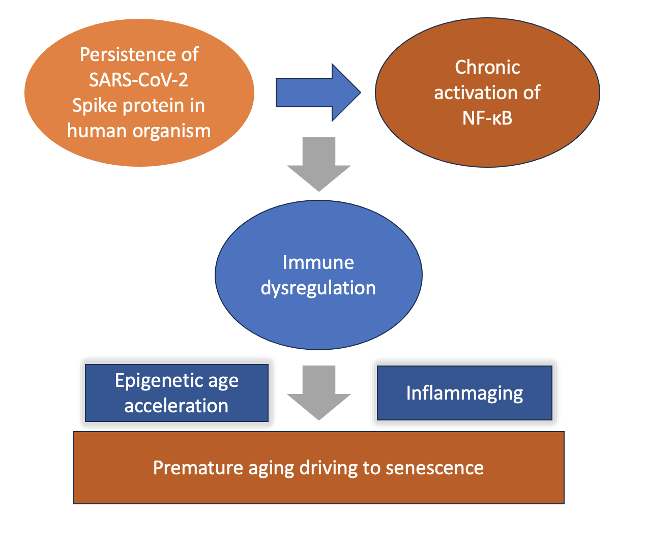
Inflammaging is a particular state of a rapidly aging immune system, where senescent cells, cells that, due to aging, are no longer able to divide (an irreversible cell cycle arrest), secrete factors to induce senescence in other normal cells [120]. The senescent cell-secreted factors comprise a mixture of cytokines (e.g., IL-1, IL-6, IL-8), chemokines such as the CXCL family, and other inflammatory factors, including IFN-γ. This is called the senescence associated secretory phenotype (SASP). During progression of inflammaging to senescence, there is excessive DNA damage and shortening of telomeres. Chronic low-grade inflammation (inflammaging) generates ROS and releases pro-inflammatory cytokines like IL-6 and TNF-α, which inhibit telomerase activity (TERT) and directly damage telomeric sequences, accelerating erosion [121]. Figure 1 illustrates schematically the processes by which COVID-19 infection, and, more specifically the SARS-CoV-2 spike protein, can induce EAA and inflammaging.
In the recent study of Z. Liu et al. [8], researchers were able to closely match interferon related inflammaging (including raised, CXCL9 and CXCL10) with certain age acceleration clocks, depicting that immune dysregulation due to microbial pathogens accelerates age. One of the epigenetic clocks used to show acceleration of aging was GrimAge. Similarly, in the P. García-delaTorre et al. [35] and M.K. Gupta and R. Vadde [9] COVID-19 studies, the GrimAge model scored a high score for EAA. While it remained relatively inconclusive whether the severe COVID-19 and Long-COVID interferon-related inflammaging resulted finally in immunosenescence in the tissues tested in the research studies included in Table 1, a continuous activation of NF-κB by chronic activation of pattern recognition receptors (PRRs, including the TLRs), by neighbouring senescent cells with defective proteostasis, that secrete DAMPs and commit apoptosis, can result in immunosenescence [122]. The study of L.H.A. Ventura et al. [39], listed in Table 1, identified immunosenescence in the CD4+ and CD8+ lymphocytes in the blood of COVID-19 patients. Immunosenescence is a deadly stage of immune aging occurring in late stages of diseases like cancer, in elderly patients [123]. Thus, an important question is raised. For how long does the SP persist in the human organism and what are the health consequences that have been recorded in peer reviewed scientific literature that describe the time duration of SP and in relation to disease? EAA due to inflammaging is a condition that can affect all human tissues and results in multimorbidity [124]. Moreover, the A. Mongelli et al. study [29], (listed in Table 1) has identified a subgroup of Long COVID patients (56 ± 12.8 years) that had a markedly high rate of EAA in their blood. The increase in chronological age can render the Long COVID individuals affected by premature aging more susceptible to age-related diseases, with the vivid examples of neurodegenerative and cardiovascular disease [125,126].
5.2. The Persistence of SARS CoV-2 and COVID-19 mRNA Vaccine-Coded Spike Protein
What is worrisome about the association of SP with chronic activation of NF-κB (and other pathogenic pathways involved), progression of aging to inflammaging and the overall acceleration of aging that may involve senescence, is the long-time persistence of SP in the human organism. An early study of B.K. Patteson et al. in 2022 [127] indicated that, in patients that had developed Long COVID, the S1 subunit of SP persisted for 15 months after infection in the non-classical (CD14Lo, CD16+) subset of monocytes in previously severe COVID-19 and Long COVID patients. Moreover, a later study of B.K. Patterson et al. in 2025 focused on the post-COVID-19 vaccine syndrome (PCVS), a condition that develops after the mRNA COVID-19 vaccination and has cardiological, neurological, inflammatory and other presentations similar to Long COVID [128]. The researchers showed that a particular elevated cytokine and chemokine profile was characteristic of the PCVS patients. Amongst these, the IL-4, and IL-8 elevations are characteristic of an active NF-κB transcriptional regulation of its target cytokine genes and indicate a chronic inflammatory condition [129]. Particularly the elevation of IL-4 is indicative of an endogenous DAMPs - TLR-mediated stimulation of NF-κB activation that is responsible for an extensive epigenetic reprogramming [130]. Furthermore, the elevation of chemokine CCL5 expression is a typical consequence of the type I interferon response involved in the pathogenesis of vascular diseases [131].
These biomarker elevations indicate a chronic inflammation mediated by IFN and NF-κB pathways in PCVS patients. Also, the IL-4, IL-8 and CCL5 elevations closely match findings of the Y. Yamaguchi et al. study listed in Table 1 [32]. In this study, the inflammatory mediators that were identified after the second mRNA vaccination highlighted a particular elevation of the type 1 IFN response. As analysed in earlier sections, this pinpoints epigenetic modifications (methylations) of histones that can cause remodelling of the nucleosome.
What is a particularly important point to raise in the PCVS B.K. Patterson et al. study is the persistence of the mRNA-coded SP in the CD16+ monocytes in the patients’ blood [25]. The S1 subunit of SP persisted in this monocyte subset for up to 245 days post vaccination. Moreover, the persistence of the SP S1 subunit correlated with the immunological profile of a chronic, NF-κB / IFN, and TNF-α mediated inflammation (as analysed above). Although no studies exist that have measured EAA in PCVS patients, the studies from the Long COVID patients predict that there would be a considerable acceleration of biological age also in the PCVS patients. However, specific studies using various epigenetic clocks are needed to unravel this important issue.
Referring to another important issue regarding the distribution of mRNA vaccine that codes SP in human tissues, although it was preliminarily suggested that it should remain for a relatively short period of time at neighbouring parts of injection [132], it seems that this is not the case. A.J. Krauson et al. have investigated the distribution of the mRNA vaccine in patients that suffered from myocardial injury (an autopsy study) [133]. The mRNA vaccine was detected in the auxiliary nodes and cardiac tissue of deceased patients from cardiac injury after 30 months of vaccination. Moreover, the finding that the mRNA vaccine was detected only in heart tissue of patients that died 30 months post vaccination and not in the heart tissues of patients that died more than 30 days from vaccination may underline an inflammaging process in the cardiac cells of deceased patients. The cardiac tissue in deceased patients had healing presentation of myocardial injury at the time of vaccination. Inflammaging can cause acute myocardial complications like plaque rupture to induce deadly thrombotic incidences and myocardial infarction [134].
A remarkable case of a PCVS patient that shows an extensive distribution and time persistence of SP has recently been described [135]. Although a single case study, it is important to be analysed. The patient suffered from multi-organ dysfunction and numerous clinical manifestations, including pulmonary emboli, myocarditis, and cognitive impairment, amongst many other, equally important symptoms. After excluding numerous etiological factors (autoimmunity, malignancy, neurologic, rheumatologic, amongst many others), the patient was diagnosed as suffering from Long COVID after myocarditis. Whilst the patient remained negative for the presence of SARS-CoV-2 nucleocapsid throughout all clinical investigation (spanning 809-1,433 days post the mRNA vaccination), the patient had markedly increased anti-spike antibodies (4,553 U/ml) at 1,433 days post the final mRNA vaccination. At 852-day post vaccination, the S1 segment of SP was detected in both classical and non-classical monocytes. At 1,173 days post vaccination, the wild type SARS CoV-2 spike protein (presumably from previous infection) was identified in the plasma and in circulating exosomes. Detectable SP was also found in macrophages, endothelial cells and nerve fibres after a skin biopsy performed at 1364 days post vaccination. Spike protein detection was accompanied with systemic inflammation. To the best of our knowledge, this is the longest time period of persistence of SP ever recorded in current medical scientific literature. Moreover, the structural variation whole genome analysis performed at 1,277 days post vaccination revealed genomic instability particularly affecting the EGFR, MYC, ERBB2, and ETV6/RUNX genes. This finding is specifically indicative for an NF-kB mediation of methylome alteration, as analysed in depth in Section 4.
Moreover, although the DNA methylome in the blood or in other tissues where SP was detected was not tested for EAA, it can be assumed and suggested that the patient suffered from myocarditis due to inflammaging of immune cells that was inflicted by the persistence of SP. A dysregulated immune signalling like that of inflammaging, consisting of the cytokine and chemokine profile of chronic inflammation, harms the heart tissue and causes acute myocarditis [136]. Therefore, inflammaging of the heart tissue (although it was not able to be detected during the clinical case), cannot be excluded [137].
A paradox that needs further explanation, however, involves how the SP persistence triggers the type I interferon response and expression of INF-α and β. During a SARS CoV-2 infection, the SP associates with the interferon regulatory factor 3 (IRF3) and supresses the expression of type I interferons. Nevertheless, the levels of NF-κB are not supressed and remain unchanged [138]. During the activation of NF-κB through the TLR/Myd88 pathway (activated by both SP and the cationic lipid nanoparticles), the NF-κB-associated lncRNAs [139], namely, LETHE, PACER, NKILA and THRIL, regulate positively or negatively the NF-κB transcription potential and interaction with the p300 and CBP acetyltransferases (described in Section 4.2). The interaction of NF-κB transcription factors with the p300/ CBP acetyltransferases, which are also strong transcriptional co-activators of NF-κB, enhance the expression of INF-α and β [140].
In this way a chronic-persistent stimulation by SP /NF-κB activation can also mediate the expression of type I interferons. The process of activation of type I interferon expression, involves primarily a synergistic transcriptional activation (called enhanceosome assembly) of the IFNβ gene promoter that requires the active participation of NF-κB (p65) amongst other transcriptional factors (IRF1 or ATF2/c-Jun) and CBP/p300 co-activators [141].
In the following section, the main molecular events due to inflammaging and progress to senescence are described. These drive the emergence of age-related diseases.
6. Consequences of the COVID-19, Long COVID and Plausibly mRNA COVID Vaccines-Induced Inflammaging to the General Public Health
6.1. A False DNA Repair and the Possibilities for Genomic Integration of Foreign Material.
As age progresses, there is an accumulation of DNA damage that has not been adequately repaired. Through complex cascades, NF-κB is activated through type I interferons and TNF to induce inflammation that feeds the loss of DNA damage repair mechanisms. Senescence- induced phenomena and nucleosome epigenetic deformations lead to telomere attritions and further complicate the inflammatory response. The "false DNA repair response" is driven by persistent DNA Damage Response signaling. When a cell enters senescence, it experiences unresolved double-strand breaks. The cell continually tries, but fails, to fix these lesions. This chronic state of repair failure initiates a profound crosstalk with the innate immune system, fundamentally driven by type I IFN signaling as a protagonist [142].
The detection of foreign nucleic acids such as exogenous viral RNA retroelements activates the type I IFN signalling [143]. The self and non-self-recognition (foreign nucleic acid exclusion) involves a delicate cellular sensing system where type I interferon in finally triggered to induce an antiviral immune response. During this foreign nucleic acid exclusion sensing system NF-κB is also activated [144]. However, it is the continuously activated NF-κB transcription during age-accumulated DNA damage, that, apart from inducing a type I IFN-related inflammatory response, creates defects in the sensing of exclusion of foreign DNA incorporation into the genome, and this has to do with the endogenous retroelements, like LINE-1.
LINE-1 (Long Interspersed Nuclear Element-1) is an active, autonomous retrotransposon that generates complementary DNA (cDNA) as part of a "copy and paste" mechanism called target-primed reverse transcription (TPRT) [145]. During inflammaging, there is an upregulation of LINE-1 transcripts which initiates a rapid cDNA amplification which has lost the sense of specificity to exclude foreign DNA into the genome [146]. Continuously fed stress-induced premature senescence (SIPS) and secretion of SASP immunogens further stages aging, and senescence proceeds to enhance retrotransposition that ultimately alters the deletion or integration sites of the genome [147,148].
It must be emphasized that the risk for genomic instability by the upregulation of retrotransposome transcription is finely regulated by DNA methylation epigenetic control [148] that can be severely dismantled by NF-κB active demethylation and alteration of the methylome during chronic inflammation in ways that are described in Section 4.
Thus, this may plausibly be the reason that plasmid DNA elements of SP gene sequences: S1-S3 segments, ori1/ori2 and SV40 enhancer sequences may increase cancer risk. These remnants of vaccine mRNA were found to be present for a long-time in patient’s tissues (as in the case that was described in a previous section [133]) Furthermore, a similar age-related inflamed microenvironment and loss of DNA repair and genomic stability in the cell drives mutagenesis and cancer (for review see the J. Kay et al. study [149]).
6.2. Ribosomal Frameshifting and Immune Dysregulation During Aging
Lessons learned from the type I diabetes autoimmune disorder indicate that, during inflammation, the B cells construct and express polypeptides (called neoantigens), due to loss of translational fidelity cross-check mechanisms [150]. These polypeptides are derived from noncanonical translation initiation sites within long noncoding RNA. However, aging contributes more due to the frameshifting translational errors. During aging, the ribosome-associated quality control (RQC) that modulates misfolded nascent polypeptide clearance by degradation is impaired [151,152]. The K.C. Stein et al. study has shown that aging results in proteostasis collapse [153]. This is because ribosome pausing during translation is increased, and, thereby, the RQC gets overwhelmed. Proteostasis collapse and subsequent ribosomal mistranslation due to aging increases the chances of wrong sequence polypeptides becoming translated [154]. Under normal conditions, the mRNA slippage on ribosomes is controlled and occurs only when it is needed [155]. When proteostasis is impaired, there is a loss of recognition in error-prone translation. This is particularly important for the pathogenesis of neurodegenerative disorders [156].
It can thus plausibly be suggested that the effects of N1-methylpseudouridine incorporation into COVID-19 mRNA vaccines that results in ribosomal frameshifting and the generation of mis-quality control (RQC) recombinant polypeptides can also be attributed to age-related miscalculations of translation occurring during the generation of immunogenicity and immune dysregulation factors in the cell [157]. The authors of the study have suggested that these recombinant polypeptides were probably generated by ribosome stalling that can also be true for ribosome pausing due to aging, as is described in the aforementioned studies.
6.3. Further Contribution to Amyloidogenesis and Neurodegenerative Diseases
Loss of proteostasis control due to aging can result in misfolding and aggregation of nascent polypeptides during a mis-regulated RQC as suggested in the previous section [151]. An IFN and NF-κB chronic activation and cross talk would mean that the unfolded protein response that would otherwise inhibit NF-κB and avoid amyloidogenesis would no longer be protective [158,159]. Particularly the activated IFN-γ would further enforce the activation of NF-κB, through the elevation of stress and PERK signalling. This effect has been shown in oligodendrocytes depicting the IFN-NF-κB mediated development of immune-mediated demyelinating diseases [160]. Thus, the effects of hypermethylation near the glutamatergic / N-methyl-D-aspartate (NMDA) genes revealing glutamatergic signatures of aging in post COVID (after 6 months of infection [36]), study listed in Table 1, can be worrisome for the development of neurodegeneration.
7. Limitations
The deduction of interferon-related inflammaging is based on the immunological profile of COVID-19 and Long COVID patients that have shown an acceleration of epigenetic age and the impacts on their methylome and nucleosome, presumably due to activation of NF-κB. Due to similar immunological profiles in PCVS patients and data revealing a long-term persistence of SP in the organism indicating a chronic NF-κB/IFN/TNF mediated inflammatory response, the study highlights the need for epigenetic age acceleration measurements using similar age clocks in the PCVS group of patients to reveal whether similar age acceleration as is seen with Long COVID and severe COVID-19 patients is also occurring in PCVS. Finally, only the Medline/ PubMed search engine was used in this study to identify research studies on EAA, COVID-19, and Long COVID. This was because the NIH library hosts and indexes the highest impact factor journals in biomedical research. Complementary search with the Google Scholar search engine did not identify other research studies besides the ones included for investigation and discussion using the Medline engine.
8. Conclusions
The TLR2/MyD88-dependent chronic activation of NF-κB through the canonical pathway by the SARS-CoV-2 spike protein has the potential to alter the methylome and nucleosome and cause epigenomic age acceleration, premature aging, and probably senescence. However, to what extent an NF-κB activation can produce, through the epigenetic changes occurring, cellular senescence and premature aging, seems to be based on its chronic stimulation and the expression of senescence-associated genes like TNF-α along with the decrease in IκBα activity (NF-κB inhibitors).
TNF-α, augments the activation of NF-κB. The persistence of spike protein in blood after COVID-19 and Long COVID accelerates aging. Plausibly due to similar immune dysregulation, mRNA COVID-19 vaccination coded SP may also accelerate aging, and NF-κB activation of TLR4 by the cationic lipid nanoparticles may also contribute to the pathology. In severe COVID-19 and Long COVID patients, the chronic inflammatory marks found show the tissues tested suffered from interferon-related inflammaging. The similar chronic interferon-related inflammation markers in PCVS patients, where the mRNA coded SP persists in immune cells, indicate that these patients may also suffer from inflammaging. Thus, the resulting deaths in vaccinated patients with myocardial injuries can also plausibly be due to inflammaging.
Some Long COVID patients show an even faster age acceleration. Depending on the current aging signature of an individual when the NF-κB pathway is activated through Long COVID, and the degree of prior immune defects, there can be an increased rate of EAA following Long COVID. Thus, an already existing premature aging after contracting COVID-19 can be an extra contributing factor for EAA and inflammaging, and this seems to also count for mRNA-coded SP to further reactivate NF-κB and other associated pathogenic pathways.
Within this context, immune dysregulation due to SP-mediated extensive NF-κB, IFN and TNF activation may be the primary cause of EAA and immune-related aging. The effects of Long COVID on accelerating the epigenetic age of patients can be attributed mainly to the chronicity of NF-κB activation due to persistence of SP stimulation. NF-kB activation has an extreme potential to alter the methylome and nucleosome and cause aging. Studies with larger population samples than the ones discussed on Long COVID and PCVS are needed to prove long-time persistence of SP in general population.
Spike protein encoded by COVID-19 mRNA vaccines, by contributing to chronic NF-κB activation (and alteration of the methylome and nucleosome) can contribute to the already altered DNA and histone modifications linked to EAA and the aging process, in a similar way to immune dysregulation occurring in severe COVID-19 and Long COVID patients. Due to the similar immunological profile between Long COVID and PCVS patients indicating chronic NF-κΒ / IFN/ TNF mediated inflammation, and the considerable persistence of vaccine-derived SP (S1) in PCVS patients’ blood, specific studies to measure EAA in PCVS patients, as have been done in Long COVID patients, are urgently needed. Induction of inflammaging in the general population can vastly contribute to age-related disease pathogenesis.
Generative AI
The authors declare that generative AI was not used in the creation of this manuscript.
Disclosures
The authors declare that there are no conflicts of interest to disclose.
Author Contributions
A.M.K. wrote the first draft. S.S. and A.M.K. both contributed substantially to multiple iterations of refinements and to figure creation.
Acknowledgments
This research was funded in part by Quanta Computer, Inc., in Taoyuan City, Taiwan, under grant number 6950759.
References
- la Torre, A.; Lo Vecchio, F.; Greco, A. Epigenetic Mechanisms of Aging and Aging-Associated Diseases. Cells 2023, 12(8), 1163. [Google Scholar] [CrossRef] [PubMed]
- Nawaz, K.; Cziesielski, M.J.; Mariappan, K.G.; Cui, G.; Aranda, M. Histone modifications and DNA methylation act cooperatively in regulating symbiosis genes in the sea anemone Aiptasia. BMC Biol. 2022, 20(1), 265. [Google Scholar] [CrossRef] [PubMed]
- Ugalde, A.P.; Roiz-Valle, D.; Moledo-Nodar, L.; Caravia, X.M.; Freije, J.M.P.; López-Otín, C. Noncoding RNA Contribution to Aging and Lifespan. J. Gerontol. A Biol. Sci. Med. Sci. 2024, 79(4), glae058. [Google Scholar] [CrossRef] [PubMed]
- Wang, K.; Liu, H.; Hu, Q.; Wang, L.; Liu, J.; Zheng, Z.; et al. Epigenetic regulation of aging: implications for interventions of aging and diseases. Sig Transduct. Target Ther. 2022, 7, 374. [Google Scholar] [CrossRef] [PubMed]
- Tuminello, S.; Ashebir, Y.A.; Schroff, C.; Ramaswami, S.; Durmus, N.; Chen, Y.; et al. Epigenetic aging acceleration among World Trade Center-exposed community members. Sci. Rep. 2025, 15, 33942. [Google Scholar] [CrossRef] [PubMed]
- Zheng, Y.; Joyce, B.T.; Colicino, E.; Liu, L.; Zhang, W.; Dai, Q.; et al. Blood epigenetic age may predict cancer incidence and mortality. EBioMedicine 2016, 5, 68–73. [Google Scholar] [CrossRef] [PubMed]
- Wen, C.; Lan, Q.; Wang, Y.; Ni, Y.; Wong, A.S.T.; Liu, D. Interferon signaling pathways in health and disease. Mol. Biomed. 2025, 6(1), 135. [Google Scholar] [CrossRef] [PubMed]
- Liu, Z.; Ziogas, A.; Zhang, Y.; Gupta, M.K.; Föhse, K.; Taks, E.; et al. Interferon-related inflammaging links epigenetic age acceleration to multimorbidity. Cell Genom. 2026, 6(6), 101218. [Google Scholar] [CrossRef] [PubMed]
- Gupta, M.K.; Vadde, R. DNA methylation-based epigenetic clocks highlight immune-driven aging acceleration in COVID-19 across diverse populations. Biogerontology 2025, 27(1), 12. [Google Scholar] [CrossRef] [PubMed]
- Grimm, L.; Onyeukwu, C.; Kenny, G.; Parent, D.M.; Fu, J.; Dhingra, S.; et al. Immune Dysregulation in Acute SARS-CoV-2 Infection. Pathog. Immun. 2023, 7(2), 143–170. [Google Scholar] [CrossRef] [PubMed]
- Almehdi, A.M.; Khoder, G.; Alchakee, A.S.; Alsayyid, A.T.; Sarg, N.H.; Soliman, S.S.M. SARS-CoV-2 spike protein: pathogenesis, vaccines, and potential therapies. Infection 2021, 49(5), 855–876. [Google Scholar] [CrossRef] [PubMed]
- Ahmad, R.; Haque, M. Surviving the Storm: Cytokine Biosignature in SARS-CoV-2 Severity Prediction. Vaccines 2022, 10(4), 614. [Google Scholar] [CrossRef] [PubMed]
- Niu, C.; Liang, T.; Chen, Y.; Zhu, S.; Zhou, L.; Chen, N.; et al. SARS-CoV-2 spike protein induces the cytokine release syndrome by stimulating T cells to produce more IL-2. Front Immunol. 2024, 15, 1444643. [Google Scholar] [CrossRef] [PubMed]
- Low, R.N.; Low, R.J.; Akrami, A. A review of cytokine-based pathophysiology of Long COVID symptoms. Front Med. 2023, 10, 1011936. [Google Scholar] [CrossRef] [PubMed]
- Khan, S.; Shafiei, M.S.; Longoria, C.; Schoggins, J.W.; Savani, R.C.; Zaki, H. SARS-CoV-2 spike protein induces inflammation via TLR2-dependent activation of the NF-κB pathway. Elife 2021, 10, e68563. [Google Scholar] [CrossRef] [PubMed]
- Sun, S.C.; Ley, S.C. New insights into NF-kappaB regulation and function. Trends Immunol. 2008, 29(10), 469–78. [Google Scholar] [CrossRef] [PubMed]
- Israël, A. The IKK complex, a central regulator of NF-kappaB activation. Cold Spring Harb. Perspect. Biol. 2010, 2(3), a000158. [Google Scholar] [CrossRef] [PubMed]
- Yu, M.; Zhou, H.; Zhao, J.; Xiao, N.; Roychowdhury, S.; Schmitt, D.; et al. MyD88-dependent interplay between myeloid and endothelial cells in the initiation and progression of obesity-associated inflammatory diseases. J. Exp. Med. Erratum in: J Exp Med. 2014 May 5;211(5):1003. Ransohoff, Richard M [added]. 2014, 211(5), 887–907. [Google Scholar] [CrossRef] [PubMed]
- Tabata, S.; Matsuda, K.; Soeda, S.; Nagai, K.; Izumi, Y.; Takahashi, M.; et al. NF-κB dynamics-dependent epigenetic changes modulate inflammatory gene expression and induce cellular senescence. FEBS J. 2024, 291(22), 4951–4968. [Google Scholar] [CrossRef] [PubMed]
- García-García, V.A.; Alameda, J.P.; Page, A.; Casanova, M.L. Role of NF-κB in ageing and age-related diseases: lessons from genetically modified mouse models. Cells 2021, 10(8), 1906. [Google Scholar] [CrossRef] [PubMed]
- Cai, D.; Han, J.J. Aging-associated lncRNAs are evolutionarily conserved and participate in NFκB signaling. Nat. Aging 2021, 1(5), 438–453. [Google Scholar] [CrossRef] [PubMed]
- Shekhar Patil, M.; Richter, E.; Fanning, L.; Hendrix, J.; Wyns, A.; Barrero Santiago, L.; et al. Epigenetic changes in patients with post-acute COVID-19 symptoms (PACS) and long-COVID: A systematic review. Expert Rev. Mol. Med. 2024, 26, e29. [Google Scholar] [CrossRef] [PubMed]
- de Melo, B.P.; da Silva, J.A.M.; Rodrigues, M.A.; Palmeira, J.D.F.; Saldanha-Araujo, F.; Argañaraz, G.A.; et al. SARS-CoV-2 spike protein and long COVID-part 1: impact of spike protein in pathophysiological mechanisms of long COVID syndrome. Viruses Erratum in: Viruses. 2025 Dec 19;18(1):2. doi: 10.3390/v18010002. 2025, 17(5), 617. [Google Scholar] [CrossRef] [PubMed]
- Patterson, B.K.; Francisco, E.B.; Yogendra, R.; Long, E.; Pise, A.; Rodrigues, H.; Hall, E.; et al. Persistence of SARS-CoV-2 S1 protein in CD16+ monocytes in post-acute sequelae of COVID-19 (PASC) up to 15 months post-infection. Front Immunol. Erratum in: Front Immunol. 2026 Apr 23;17:1832958. doi: 10.3389/fimmu.2026.1832958. 2022, 12, 746021. [Google Scholar] [CrossRef] [PubMed]
- Patterson, B.K.; Yogendra, R.; Francisco, E.B.; Guevara-Coto, J.; Long, E.; Pise, A.; et al. Detection of S1 spike protein in CD16+ monocytes up to 245 days in SARS-CoV-2-negative post-COVID-19 vaccine syndrome (PCVS) individuals. Hum. Vaccin Immunother. 2025, 21(1), 2494934. [Google Scholar] [CrossRef] [PubMed]
- Farkas, G.; Mahdaouy, Z.E.; Babszky, G.; Jokai, M.; Torma, F.; Gu, Y.; et al. Associations of epigenetic aging and COVID- 19: A 3-year longitudinal study. Geroscience 2025, 47(3), 4889–4898. [Google Scholar] [CrossRef] [PubMed]
- Choi, E.Y.; Ailshire, J.A. Ambient outdoor heat and accelerated epigenetic aging among older adults in the US. Sci. Adv. 2025, 11(9), eadr0616. [Google Scholar] [CrossRef] [PubMed]
- Franzen, J.; Nüchtern, S.; Tharmapalan, V.; Vieri, M.; Nikolić, M.; Han, Y.; et al. Epigenetic clocks are not accelerated in COVID-19 patients. Int. J. Mol. Sci. 2021, 22(17), 9306. [Google Scholar] [CrossRef] [PubMed]
- Mongelli, A.; Barbi, V.; Gottardi Zamperla, M.; Atlante, S.; Forleo, L.; Nesta, M.; et al. Evidence for biological age acceleration and telomere shortening in COVID-19 survivors. Int. J. Mol. Sci. 2021, 22(11), 6151. [Google Scholar] [CrossRef] [PubMed]
- Xu, W.; Zhang, F.; Shi, Y.; Chen, Y.; Shi, B.; Yu, G. Causal association of epigenetic aging and COVID-19 severity and susceptibility: A bidirectional Mendelian randomization study. Front Med. 2022, 9, 989950. [Google Scholar] [CrossRef] [PubMed]
- Guo, Y.; Zhang, Y.; Hu, Y. COVID-19 subgroups may slow down biological age acceleration. J. Infect. 2023, 86(1), 66–117. [Google Scholar] [CrossRef] [PubMed]
- Yamaguchi, Y.; Kato, Y.; Edahiro, R.; Søndergaard, J.N.; Murakami, T.; Amiya, S.; et al. Consecutive BNT162b2 mRNA vaccination induces short-term epigenetic memory in innate immune cells. JCI Insight 2022, 7(22), e163347. [Google Scholar] [CrossRef] [PubMed]
- Calzari, L.; Zanotti, L.; Inglese, E.; Scaglione, F.; Cavagnola, R.; Ranucci, F.; et al. Role of epigenetics in the clinical evolution of COVID-19 disease. Epigenome-wide association study identifies markers of severe outcome. Eur. J. Med. Res. 2023, 28(1), 81. [Google Scholar] [CrossRef] [PubMed]
- Calzari, L.; Dragani, D.F.; Zanotti, L.; Inglese, E.; Danesi, R.; Cavagnola, R.; et al. Epigenetic patterns, accelerated biological aging, and enhanced epigenetic drift detected 6 months following COVID-19 infection: insights from a genome-wide DNA methylation study. Clin. Epigenet. Erratum in: Clin Epigenetics. 2024 Nov 14;16(1):158. doi: 10.1186/s13148-024-01764-1. 2024, 16(1), 112. [Google Scholar] [CrossRef] [PubMed]
- García-delaTorre, P.; Rivero-Segura, N.A.; Sánchez-García, S.; Becerril-Rojas, K.; Sandoval-Rodriguez, F.E.; Castro-Morales, D.; et al. GrimAge is elevated in older adults with mild COVID-19 an exploratory analysis. Geroscience 2024, 46(4), 3511–3524. [Google Scholar] [CrossRef] [PubMed]
- Cheung, N. DNA Methylation at Core N-Methyl-D-aspartate (NMDA) Receptor genes reveals a glutamatergic signature of aging in Post-COVID whole blood with implications for Long-COVID neuropsychiatric sequelae. Cureus 2026, 18(7), e112902. [Google Scholar] [CrossRef] [PubMed]
- Bejaoui, Y.; Humaira Amanullah, F.; Saad, M.; Taleb, S.; Bradic, M.; Megarbane, A.; et al. Epigenetic age acceleration in surviving versus deceased COVID-19 patients with acute respiratory distress syndrome following hospitalization. Clin. Epigenet. 2023, 15(1), 186. [Google Scholar] [CrossRef] [PubMed]
- Barha, C.K.; Liu-Ambrose, T.; Inkster, A.M.; Falck, R.S.; Burma, J.S.; Kirkland, S.; et al. Epigenetic age predicts depressive symptoms during the COVID-19 pandemic in the Canadian Longitudinal Study on Aging: importance of biological sex. Aging 2025, 17(11), 2759–2777. [Google Scholar] [CrossRef] [PubMed]
- Ventura, L.H.A.; Torres, L.; Camatta, G.C.; Zamame, J.; Coelho, M.M.; Ramalho-Pinto, C.H.; et al. Immunosenescence profile is associated with increased susceptibility to severe COVID-19. Aging Cell 2025, 24(7), e70077. [Google Scholar] [CrossRef] [PubMed]
- Seo, D.H.; Corr, M.; Patel, S.; Lui, L.Y.; Cauley, J.A.; Evans, D.; et al. Chemokine CXCL9, a marker of inflammaging, is associated with changes of muscle strength and mortality in older men. Osteoporos. Int. 2024, 35(10), 1789–1796. [Google Scholar] [CrossRef] [PubMed]
- Simonis, A.; Theobald, S.J.; Koch, A.E.; Mummadavarapu, R.; Mudler, J.M.; Pouikli, A.; et al. Persistent epigenetic memory of SARS-CoV-2 mRNA vaccination in monocyte-derived macrophages. Mol. Syst. Biol. 2025, 21(4), 341–360. [Google Scholar] [CrossRef] [PubMed]
- Bohlin, J.; Lee, Y.; Caspersen, I.H.; Hayman Robertson, A.; Page, C.M.; Gjessing, H.K.; et al. Longitudinal study of genome-wide DNA methylation in individuals with and without post-acute symptoms following SARS-CoV-2 infection. Epigenomics 2026, 18(4), 411–419. [Google Scholar] [CrossRef] [PubMed]
- Levy, J.J.; Diallo, A.B.; Saldias Montivero, M.K.; Gabbita, S.; Salas, L.A.; Christensen, B.C. Insights to aging prediction with AI based epigenetic clocks. Epigenomics 2025, 17(1), 49–57. [Google Scholar] [CrossRef] [PubMed]
- Dabbah-Krancher, G.; Snow, A.L. Mistuned NF-κB signaling in lymphocytes: lessons from relevant inborn errors of immunity. Clin. Exp. Immunol. 2023, 212(2), 117–128. [Google Scholar] [CrossRef] [PubMed]
- Jarome, T.J.; Butler, A.A.; Nichols, J.N.; Pacheco, N.L.; Lubin, F.D. NF-κB mediates Gadd45β expression and DNA demethylation in the hippocampus during fear memory formation. Front Mol. Neurosci. 2015, 8, 54. [Google Scholar] [CrossRef] [PubMed]
- Sayed, N.; Huang, Y.; Nguyen, K.; Krejciova-Rajaniemi, Z.; Grawe, A.P.; Gao, T.; et al. An inflammatory aging clock (iAge) based on deep learning tracks multimorbidity, immunosenescence, frailty and cardiovascular aging. Nat. Aging Erratum in: Nat Aging. 2021 Aug;1(8):748. doi: 10.1038/s43587-021-00102-x. 2021, 1, 598–615. [Google Scholar] [CrossRef] [PubMed]
- Veluthakal, R.; Oh, E.; Ahn, M.; Chatterjee Bhowmick, D.; Thurmond, D.C. Syntaxin 4 mediates NF-κB signaling and chemokine ligand expression via specific interaction with IκBβ. Diabetes 2021, 70(4), 889–902. [Google Scholar] [CrossRef] [PubMed]
- El Khoury, L.Y.; Gorrie-Stone, T.; Smart, M.; Hughes, A.; Bao, Y.; Andrayas, A.; et al. Systematic underestimation of the epigenetic clock and age acceleration in older subjects. Genome Biol. 2019, 20(1), 283. [Google Scholar] [CrossRef] [PubMed]
- Issa, J.P. Aging and epigenetic drift: a vicious cycle. J. Clin. Invest. 2014, 124(1), 24–9. [Google Scholar] [CrossRef] [PubMed]
- Prusiner, S.B. Disorders of glutamate metabolism and neurological dysfunction. Annu Rev. Med. 1981, 32, 521–42. [Google Scholar] [CrossRef] [PubMed]
- Kapoor, R.R.; Flanagan, S.E.; Fulton, P.; Chakrapani, A.; Chadefaux, B.; Ben-Omran, T.; et al. Hyperinsulinism-hyperammonaemia syndrome: novel mutations in the GLUD1 gene and genotype-phenotype correlations. Eur. J. Endocrinol. 2009, 161(5), 731–5. [Google Scholar] [CrossRef] [PubMed]
- Smith, R.S.; Florio, M.; Akula, S.K.; Neil, J.E.; Wang, Y.; Hill, R.S.; et al. Early role for a Na+,K+-ATPase (ATP1A3) in brain development. Proc. Natl. Acad. Sci. U S A 2021, 118(25), e2023333118. [Google Scholar] [CrossRef] [PubMed]
- Smith, J.S.; Rajagopal, S. The β-Arrestins: Multifunctional Regulators of G Protein-coupled Receptors. J. Biol. Chem. 2016, 291(17), 8969–77. [Google Scholar] [CrossRef] [PubMed] [PubMed Central]
- Plaitakis, A.; Kalef-Ezra, E.; Kotzamani, D.; Zaganas, I.; Spanaki, C. The glutamate dehydrogenase pathway and its roles in cell and tissue biology in health and disease. Biology 2017, 6(1), 11. [Google Scholar] [CrossRef] [PubMed]
- Wang, X.; Liu, R.; Qu, X.; Yu, H.; Chu, H.; Zhang, Y.; et al. α-Ketoglutarate-activated NF-κB signaling promotes compensatory glucose uptake and brain tumor development. Mol. Cell 2019, 76(1), 148–162.e7. [Google Scholar] [CrossRef] [PubMed]
- Gao, Y.; Xu, Y.; Bai, F.; Puri, R.; Tian, J.; Liu, J. Factors that influence the Na/K-ATPase signaling and function. Front Pharmacol. 2025, 16, 1639859. [Google Scholar] [CrossRef] [PubMed]
- Zhang, X.; Lee, W.; Bian, J.S. Recent advances in the study of Na+/K+-ATPase in neurodegenerative diseases. Cells 2022, 11(24), 4075. [Google Scholar] [CrossRef] [PubMed]
- Sun, J.; Lin, X. Beta-arrestin 2 is required for lysophosphatidic acid-induced NF-kappaB activation. Proc. Natl. Acad. Sci. U S A 2008, 105(44), 17085–90. [Google Scholar] [CrossRef] [PubMed]
- Segovia, G.; Porras, A.; Del Arco, A.; Mora, F. Glutamatergic neurotransmission in aging: a critical perspective. Mech. Ageing Dev. 2001, 122(1), 1–29. [Google Scholar] [CrossRef] [PubMed]
- Muhammad, M.; Hassan, T.M.; Baba, S.S.; Radda, M.I.; Mutawakkil, M.M.; Musa, M.A.; AbuBakar, S.; Loong, S.K.; Yusuf, I. Exploring NFκB pathway as a potent strategy to mitigate COVID-19 severe morbidity and mortality. J. Public Health Afr. 2022, 13(3), 1679. [Google Scholar] [CrossRef] [PubMed]
- Sterner, D.E.; Berger, S.L. Acetylation of histones and transcription-related factors. Microbiol. Mol. Biol. Rev. 2000, 64(2), 435–59. [Google Scholar] [CrossRef] [PubMed]
- Park, S.H.; Kang, K.; Giannopoulou, E.; Qiao, Y.; Kang, K.; Kim, G.; et al. Type I interferons and the cytokine TNF cooperatively reprogram the macrophage epigenome to promote inflammatory activation. Nat. Immunol. 2017, 18(10), 1104–1116. [Google Scholar] [CrossRef] [PubMed]
- Zelkoski, A.E.; Lu, Z.; Sukumar, G.; Dalgard, C.; Said, H.; Alameh, M.G.; et al. Ionizable lipid nanoparticles of mRNA vaccines elicit NF-κB and IRF responses through toll-like receptor 4. npj Vaccines 2025, 10(1), 73. [Google Scholar] [CrossRef] [PubMed]
- Liu, T.; Zhang, L.; Joo, D.; Sun, S.C. NF-κB signaling in inflammation. Signal Transduct. Target Ther. 2017, 2, 17023. [Google Scholar] [CrossRef] [PubMed]
- Tilstra, J.S.; Clauson, C.L.; Niedernhofer, L.J.; Robbins, P.D. NF-κB in Aging and Disease. Aging Dis. 2011, 2(6), 449–65. [Google Scholar] [PubMed]
- Oeckinghaus, A.; Ghosh, S. The NF-kappaB family of transcription factors and its regulation. Cold Spring Harb. Perspect. Biol. 2009, 1(4), a000034. [Google Scholar] [CrossRef] [PubMed]
- Verma, I.M. Nuclear factor (NF)-kappaB proteins: therapeutic targets. Ann. Rheum. Dis. 2004, 63 Suppl 2(Suppl 2), ii57–ii61. [Google Scholar] [CrossRef] [PubMed]
- Mao, H.; Zhao, X.; Sun, S.C. NF-κB in inflammation and cancer. Cell Mol. Immunol. 2025, 22(8), 811–839. [Google Scholar] [CrossRef] [PubMed]
- Guo, J.; Huang, X.; Dou, L.; Yan, M.; Shen, T.; Tang, W.; et al. Aging and aging-related diseases: from molecular mechanisms to interventions and treatments. Signal Transduct. Target Ther. 2022, 7(1), 391. [Google Scholar] [CrossRef] [PubMed]
- Rossiello, F.; Jurk, D.; Passos, J.F.; d'Adda di Fagagna, F. Telomere dysfunction in ageing and age-related diseases. Nat. Cell Biol. 2022, 24(2), 135–147. [Google Scholar] [CrossRef] [PubMed]
- Adler, A.S.; Sinha, S.; Kawahara, T.L.; Zhang, J.Y.; Segal, E.; Chang, H.Y. Motif module map reveals enforcement of aging by continual NF-kappaB activity. Genes Dev. 2007, 21(24), 3244–57. [Google Scholar] [CrossRef] [PubMed]
- Bernard, D.; Gosselin, K.; Monte, D.; Vercamer, C.; Bouali, F.; Pourtier, A.; et al. Involvement of Rel/nuclear factor-kappaB transcription factors in keratinocyte senescence. Cancer Res. 2004, 64(2), 472–81. [Google Scholar] [CrossRef] [PubMed]
- García-García, V.A.; Alameda, J.P.; Page, A.; Casanova, M.L. Role of NF-κB in ageing and age-related diseases: lessons from genetically modified mouse models. Cells 2021, 10(8), 1906. [Google Scholar] [CrossRef] [PubMed]
- Haga, M.; Okada, M. Systems approaches to investigate the role of NF-κB signaling in aging. Biochem J. 2022, 479(2), 161–183. [Google Scholar] [CrossRef] [PubMed]
- Marwarha, G.; Ghribi, O. Nuclear Factor Kappa-light-chain-enhancer of Activated B Cells (NF-κB) - a Friend, a Foe, or a Bystander - in the Neurodegenerative Cascade and Pathogenesis of Alzheimer's Disease. CNS Neurol. Disord. Drug Targets 2017, 16(10), 1050–1065. [Google Scholar] [CrossRef] [PubMed]
- Mizuno, K.; Dempster, E.; Mill, J.; Giese, K.P. Long-lasting regulation of hippocampal Bdnf gene transcription after contextual fear conditioning. Genes Brain Behav. 2012, 11(6), 651–9. [Google Scholar] [CrossRef] [PubMed]
- Feng, Y.; Wang, Z.; Wei, W.; Zhang, T.; Li, Z.; Chen, J.; Liu, K. Gadd45α is involved in regulating activity-dependent and exon-specific BDNF expression in postmitotic cortical neurons. Neuroreport 2021, 32(13), 1147–1152. [Google Scholar] [CrossRef] [PubMed]
- He, Y.F.; Li, B.Z.; Li, Z.; Liu, P.; Wang, Y.; Tang, Q.; et al. Tet-mediated formation of 5-carboxylcytosine and its excision by TDG in mammalian DNA. Science 2011, 333(6047), 1303–7. [Google Scholar] [CrossRef] [PubMed]
- Li, Z.; Gu, T.P.; Weber, A.R.; Shen, J.Z.; Li, B.Z.; Xie, Z.G.; et al. Gadd45a promotes DNA demethylation through TDG. Nucleic Acids Res. 2015, 43(8), 3986–97. [Google Scholar] [CrossRef] [PubMed]
- Maiti, A.; Drohat, A.C. Thymine DNA glycosylase can rapidly excise 5-formylcytosine and 5-carboxylcytosine: potential implications for active demethylation of CpG sites. J. Biol. Chem. 2011, 286(41), 35334–35338. [Google Scholar] [CrossRef] [PubMed]
- Kar, S.; Parbin, S.; Deb, M.; Shilpi, A.; Sengupta, D.; Rath, S.K.; et al. Epigenetic choreography of stem cells: the DNA demethylation episode of development. Cell Mol. Life Sci. 2014, 71(6), 1017–32. [Google Scholar] [CrossRef] [PubMed]
- Nicolis, S.K. Cancer stem cells and "stemness" genes in neuro-oncology. Neurobiol. Dis. 2007, 25(2), 217–29. [Google Scholar] [CrossRef] [PubMed]
- Zand, H.; Hosseini, S.A.; Cheraghpour, M.; Alipour, M.; Sedaghat, F. TNF-α-Induced NF-κB Alter the Methylation Status of Some Stemness Genes in HT-29 Human Colon Cancer Cell. Adv. BioMed Res. 2024, 13, 114. [Google Scholar] [CrossRef] [PubMed]
- Bott, A.J.; Peng, I.C.; Fan, Y.; Faubert, B.; Zhao, L.; Li, J.; et al. Oncogenic Myc induces expression of glutamine synthetase through promoter demethylation. Cell Metab. 2015, 22(6), 1068–77. [Google Scholar] [CrossRef] [PubMed]
- Galassi, C.; Manic, G.; Esteller, M.; Galluzzi, L.; Vitale, I. Epigenetic regulation of cancer stemness. Signal Transduct. Target Ther. 2025, 10(1), 243. [Google Scholar] [CrossRef] [PubMed]
- Pacaud, R.; Sery, Q.; Oliver, L.; Vallette, F.M.; Tost, J.; Cartron, P.F. DNMT3L interacts with transcription factors to target DNMT3L/DNMT3B to specific DNA sequences: role of the DNMT3L/DNMT3B/p65-NFκB complex in the (de-)methylation of TRAF1. Biochimie 2014, 104, 36–49. [Google Scholar] [CrossRef] [PubMed]
- Edilova, M.I.; Abdul-Sater, A.A.; Watts, T.H. TRAF1 Signaling in Human Health and Disease. Front Immunol. 2018, 9, 2969. [Google Scholar] [CrossRef] [PubMed]
- Yi, S.J.; Kim, K. New Insights into the Role of Histone Changes in Aging. Int. J. Mol. Sci. 2020, 21(21), 8241. [Google Scholar] [CrossRef] [PubMed]
- Loidl, P. Histone acetylation: facts and questions. Chromosoma 1994, 103(7), 441–9. [Google Scholar] [CrossRef] [PubMed]
- Luger, K.; Mäder, A.W.; Richmond, R.K.; Sargent, D.F.; Richmond, T.J. Crystal structure of the nucleosome core particle at 2.8 A resolution. Nature 1997, 389(6648), 251–60. [Google Scholar] [CrossRef] [PubMed]
- Luger, K.; Richmond, T.J. The histone tails of the nucleosome. Curr. Opin. Genet Dev. 1998, 8(2), 140–6. [Google Scholar] [CrossRef] [PubMed]
- Vettese-Dadey, M.; Grant, P.A.; Hebbes, T.R.; Crane-Robinson, C.; Allis, C.D.; Workman, J.L. Acetylation of histone H4 plays a primary role in enhancing transcription factor binding to nucleosomal DNA in vitro. EMBO J. 1996, 15(10), 2508–18. [Google Scholar] [CrossRef]
- Kuo, M.H.; Allis, C.D. Roles of histone acetyltransferases and deacetylases in gene regulation. Bioessays 1998, 20(8), 615–26. [Google Scholar] [CrossRef]
- Rahman, I.; Marwick, J.; Kirkham, P. Redox modulation of chromatin remodeling: impact on histone acetylation and deacetylation, NF-kappaB and pro-inflammatory gene expression. Biochem Pharmacol. 2004, 68(6), 1255–67. [Google Scholar] [CrossRef] [PubMed]
- Federman, N.; de la Fuente, V.; Zalcman, G.; Corbi, N.; Onori, A.; Passananti, C.; et al. Nuclear factor κB-dependent histone acetylation is specifically involved in persistent forms of memory. J. Neurosci. 2013, 33(17), 7603–14. [Google Scholar] [CrossRef] [PubMed]
- Ogryzko, V.V.; Schiltz, R.L.; Russanova, V.; Howard, B.H.; Nakatani, Y. The transcriptional coactivators p300 and CBP are histone acetyltransferases. Cell 1996, 87(5), 953–9. [Google Scholar] [CrossRef] [PubMed]
- Chen, L.F.; Mu, Y.; Greene, W.C. Acetylation of RelA at discrete sites regulates distinct nuclear functions of NF-kappaB. EMBO J. 2002, 21(23), 6539–48. [Google Scholar] [CrossRef] [PubMed]
- Lin, K.; Gao, W.; Chen, N.; Yang, S.; Wang, H.; Wang, R.; et al. Chronic inflammation pathway NF-κB cooperates with epigenetic reprogramming to drive the malignant progression of glioblastoma. Int. J. Biol. Sci. 2022, 18(15), 5770–5786. [Google Scholar] [CrossRef] [PubMed]
- Di Liegro, C.M.; Schiera, G.; Schirò, G.; Di Liegro, I. Involvement of the H3.3 histone variant in the epigenetic regulation of gene expression in the nervous system, in both physiological and pathological conditions. Int. J. Mol. Sci. 2023, 24(13), 11028. [Google Scholar] [CrossRef] [PubMed]
- Yamagishi, M.; Kuze, Y.; Kobayashi, S.; Nakashima, M.; Morishima, S.; Kawamata, T.; et al. Mechanisms of action and resistance in histone methylation-targeted therapy. Nature 2024, 627(8002), 221–228. [Google Scholar] [CrossRef] [PubMed]
- Day, C.A.; Hinchcliffe, E.H.; Robinson, J.P. H3K27me3 in diffuse midline glioma and epithelial ovarian cancer: opposing epigenetic changes leading to the same poor outcomes. Cells 2022, 11(21), 3376. [Google Scholar] [CrossRef] [PubMed]
- Ma, Z.; Wang, H.; Cai, Y.; Wang, H.; Niu, K.; Wu, X.; et al. Epigenetic drift of H3K27me3 in aging links glycolysis to healthy longevity in Drosophila. Elife 2018, 7, e35368. [Google Scholar] [CrossRef] [PubMed]
- Signal, B.; Phipps, A.J.; Giles, K.A.; Huskins, S.N.; Mercer, T.R.; Robinson, M.D.; et al. Ageing-related changes to H3K4me3, H3K27ac, and H3K27me3 in purified mouse neurons. Cells 2024, 13(16), 1393. [Google Scholar] [CrossRef] [PubMed]
- Bhatt, D.; Ghosh, S. Regulation of the NF-κB-Mediated Transcription of Inflammatory Genes. Front Immunol. 2014, 5, 71. [Google Scholar] [CrossRef] [PubMed]
- He, J.; Tu, C.; Liu, Y. Role of lncRNAs in aging and age-related diseases. Aging Med. 2018, 1(2), 158–175. [Google Scholar] [CrossRef] [PubMed]
- Cowland, J.B.; Muta, T.; Borregaard, N. IL-1beta-specific up-regulation of neutrophil gelatinase-associated lipocalin is controlled by IkappaB-zeta. J. Immunol. 2006, 176(9), 5559–66. [Google Scholar] [CrossRef] [PubMed]
- Matsuo, S.; Yamazaki, S.; Takeshige, K.; Muta, T. Crucial roles of binding sites for NF-kappaB and C/EBPs in IkappaB-zeta-mediated transcriptional activation. Biochem J. 2007, 405(3), 605–15. [Google Scholar] [CrossRef] [PubMed]
- Gautam, P.; Maenner, S.; Cailotto, F.; Reboul, P.; Labialle, S.; Jouzeau, J.Y.; et al. Emerging role of IκBζ in inflammation: Emphasis on psoriasis. Clin. Transl. Med. 2022, 12(10), e1032. [Google Scholar] [CrossRef] [PubMed]
- Willems, M.; Dubois, N.; Musumeci, L.; Bours, V.; Robe, P.A. IκBζ: an emerging player in cancer. Oncotarget 2016, 7(40), 66310–66322. [Google Scholar] [CrossRef] [PubMed]
- Dornas, W.; Reis, J.P.; Belilo, T.E.; Vaz, L.G.; Nasser, H.H.; de Souza Maia, M.L.; et al. Persistent inflammatory cytokine signature in long Covid-19 patients: a meta-analysis. Inflammopharmacology 2026, 34(1), 1–16. [Google Scholar] [CrossRef] [PubMed]
- Yoshimura, T. The chemokine MCP-1 (CCL2) in the host interaction with cancer: a foe or ally? Cell Mol. Immunol. 2018, 15(4), 335–345. [Google Scholar] [CrossRef] [PubMed]
- Thompson, W.L.; Van Eldik, L.J. Inflammatory cytokines stimulate the chemokines CCL2/MCP-1 and CCL7/MCP-3 through NFkB and MAPK dependent pathways in rat astrocytes [corrected]. Brain Res. Erratum in: Brain Res. 2009 Oct 27;1295:230. 2009, 1287, 47–57. [Google Scholar] [CrossRef] [PubMed]
- Verstrepen, L.; Bekaert, T.; Chau, T.L.; Tavernier, J.; Chariot, A.; Beyaert, R. TLR-4, IL-1R and TNF-R signaling to NF-kappaB: variations on a common theme. Cell Mol. Life Sci. 2008, 65(19), 2964–78. [Google Scholar] [CrossRef] [PubMed]
- Wang, W.; Ye, L.; Ye, L.; Li, B.; Gao, B.; Zeng, Y.; et al. Up-regulation of IL-6 and TNF-alpha induced by SARS-coronavirus spike protein in murine macrophages via NF-kappaB pathway. Virus Res. 2007, 128(1-2), 1–8. [Google Scholar] [CrossRef] [PubMed]
- Tau, G.; Rothman, P. Biologic functions of the IFN-gamma receptors. Allergy 1999, 54(12), 1233–51. [Google Scholar] [CrossRef] [PubMed]
- Lin, Y.; Jamison, S.; Lin, W. Interferon-γ activates nuclear factor-κ B in oligodendrocytes through a process mediated by the unfolded protein response. PLoS ONE 2012, 7(5), e36408. [Google Scholar] [CrossRef] [PubMed]
- Cooley, C.B.; Ryno, L.M.; Plate, L.; Morgan, G.J.; Hulleman, J.D.; Kelly, J.W.; et al. Unfolded protein response activation reduces secretion and extracellular aggregation of amyloidogenic immunoglobulin light chain. Proc. Natl. Acad. Sci. U S A 2014, 111(36), 13046–51. [Google Scholar] [CrossRef] [PubMed]
- Bradburn, S.; McPhee, J.; Bagley, L.; Carroll, M.; Slevin, M.; Al-Shanti, N.; et al. Dysregulation of C-X-C motif ligand 10 during aging and association with cognitive performance. Neurobiol. Aging 2018, 63, 54–64. [Google Scholar] [CrossRef] [PubMed]
- Vacharathit, V.; Pluempreecha, M.; Manopwisedjaroen, S.; Srisaowakarn, C.; Srichatrapimuk, S.; Sritipsukho, P.; et al. Persistent IP-10/CXCL10 dysregulation following mild Omicron breakthrough infection: Immune network signatures across COVID-19 waves and implications for mRNA vaccine outcomes. Clin. Immunol. 2025, 278, 110507. [Google Scholar] [CrossRef] [PubMed]
- Coppé, J.P.; Desprez, P.Y.; Krtolica, A.; Campisi, J. The senescence-associated secretory phenotype: the dark side of tumor suppression. Annu Rev. Pathol. 2010, 5, 99–118. [Google Scholar] [CrossRef] [PubMed]
- Kordinas, V.; Ioannidis, A.; Chatzipanagiotou, S. The telomere/telomerase system in chronic inflammatory diseases. cause or effect? Genes 2016, 7(9), 60. [Google Scholar] [CrossRef] [PubMed]
- Karpuzoglu, E.; Holladay, S.D.; Gogal, R.M., Jr. Inflammaging: triggers, molecular mechanisms, immunological consequences, sex differences, and cutaneous manifestations. Front Immunol. 2025, 16, 1704203. [Google Scholar] [CrossRef] [PubMed]
- Gao, J.; Yuan, Y.; Wang, X.; He, L.; Li, L. Impact of immunosenescence on immune-related adverse events in elderly patients with cancer. Aging Med. 2025, 8(1), e70000. [Google Scholar] [CrossRef] [PubMed]
- Liu, Z.; Ziogas, A.; Zhang, Y.; Gupta, M.K.; Föhse, K.; Taks, E.; et al. Interferon-related inflammaging links epigenetic age acceleration to multimorbidity. Cell Genom. 2026, 6(6), 101218. [Google Scholar] [CrossRef] [PubMed]
- García-Domínguez, M. Pathological and Inflammatory Consequences of Aging. Biomolecules 2025, 15(3), 404. [Google Scholar] [CrossRef] [PubMed]
- Ferrucci, L.; Fabbri, E. Inflammageing: chronic inflammation in ageing, cardiovascular disease, and frailty. Nat. Rev. Cardiol. 2018, 15(9), 505–522. [Google Scholar] [CrossRef] [PubMed]
- Patterson, B.K.; Francisco, E.B.; Yogendra, R.; Long, E.; Pise, A.; Rodrigues, H.; et al. Persistence of SARS CoV-2 S1 Protein in CD16+ Monocytes in Post-Acute Sequelae of COVID-19 (PASC) up to 15 Months Post-Infection. Front Immunol. Erratum in: Front Immunol. 2026 Apr 23;17:1832958. doi: 10.3389/fimmu.2026.1832958. 2022, 12, 746021. [Google Scholar] [CrossRef] [PubMed]
- Patterson, B.K.; Yogendra, R.; Francisco, E.B.; Guevara-Coto, J.; Long, E.; Pise, A.; et al. Detection of S1 spike protein in CD16+ monocytes up to 245 days in SARS-CoV-2-negative post-COVID-19 vaccine syndrome (PCVS) individuals. Hum. Vaccin Immunother. 2025, 21(1), 2494934. [Google Scholar] [CrossRef] [PubMed]
- Qazi, B.S.; Tang, K.; Qazi, A. Recent advances in underlying pathologies provide insight into interleukin-8 expression-mediated inflammation and angiogenesis. Int. J. Inflam. 2011, 2011, 908468. [Google Scholar] [CrossRef] [PubMed]
- Qian, C.; Cao, X.; Hoffmann, J.A. Toll-like receptors in innate immunity and inflammation: from fundamental biology to clinic insights. Immun. Inflamm. 2026, 2(1), 22. [Google Scholar] [CrossRef] [PubMed]
- Nakano, M.; Fujii, T.; Hashimoto, M.; Yukawa, N.; Yoshifuji, H.; Ohmura, K.; et al. Type I interferon induces CX3CL1 (fractalkine) and CCL5 (RANTES) production in human pulmonary vascular endothelial cells. Clin. Exp. Immunol. 2012, 170(1), 94–100. [Google Scholar] [CrossRef] [PubMed]
- Boros, L.G.; Kyriakopoulos, A.M.; Brogna, C.; Piscopo, M.; McCullough, P.A.; Seneff, S. Long-lasting, biochemically modified mRNA, and its frameshifted recombinant spike proteins in human tissues and circulation after COVID-19 vaccination. Pharmacol. Res. Perspect. 2024, 12(3), e1218. [Google Scholar] [CrossRef] [PubMed]
- Krauson, A.J.; Casimero, F.V.C.; Siddiquee, Z.; Stone, J.R. Duration of SARS-CoV-2 mRNA vaccine persistence and factors associated with cardiac involvement in recently vaccinated patients. npj Vaccines 2023, 8(1), 141. [Google Scholar] [CrossRef] [PubMed]
- Puspitasari, Y.M.; Ministrini, S.; Schwarz, L.; Karch, C.; Liberale, L.; Camici, G.G. Modern Concepts in Cardiovascular Disease: Inflamm-Aging. Front Cell Dev. Biol. 2022, 10, 882211. [Google Scholar] [CrossRef] [PubMed]
- Hulscher, N.; Schmidt, V.; Morz, M.; Rogers, C.; von Ranke, N.; Zhang, W.; et al. Persistence of vaccine mRNA, plasmid DNA, spike protein, and genomic dysregulation over 3.5 years post-COVID-19 mRNA vaccination. Med. Res. Arch. 2026, 14(6). [Google Scholar] [CrossRef]
- Soulaidopoulos, S.; Tousoulis, D.; Sagris, M.; Aghayan, S.; Platanias, K.; Giannakodimos, A.; et al. Inflammatory mechanisms in myocarditis-recent therapeutic strategies. Biomolecules 2025, 15(10), 1475. [Google Scholar] [CrossRef] [PubMed]
- Spray, L.; Richardson, G.; Haendeler, J.; Altschmied, J.; Rumampouw, V.; Wallis, S.B.; et al. Cardiovascular inflammaging: Mechanisms, consequences, and therapeutic perspectives. Cell Rep. Med. 2025, 6(9), 102264. [Google Scholar] [CrossRef] [PubMed]
- Freitas, R.S.; Crum, T.F.; Parvatiyar, K. SARS-CoV-2 spike antagonizes innate antiviral immunity by targeting interferon regulatory factor 3. Front Cell Infect. Microbiol. 2022, 11, 789462. [Google Scholar] [CrossRef] [PubMed]
- Gupta, S.C.; Awasthee, N.; Rai, V.; Chava, S.; Gunda, V.; Challagundla, K.B. Long non-coding RNAs and nuclear factor-κB crosstalk in cancer and other human diseases. Biochim Biophys. Acta Rev. Cancer 2020, 1873(1), 188316. [Google Scholar] [CrossRef] [PubMed]
- Valadkhan, S.; Gunawardane, L.S. lncRNA-mediated regulation of the interferon response. Virus Res. 2016, 212, 127–36. [Google Scholar] [CrossRef] [PubMed]
- Merika, M.; Williams, A.J.; Chen, G.; Collins, T.; Thanos, D. Recruitment of CBP/p300 by the IFN beta enhanceosome is required for synergistic activation of transcription. Mol. Cell 1998, 1(2), 277–87. [Google Scholar] [CrossRef] [PubMed]
- Yu, Q.; Katlinskaya, Y.V.; Carbone, C.J.; Zhao, B.; Katlinski, K.V.; Zheng, H.; et al. DNA-damage-induced type I interferon promotes senescence and inhibits stem cell function. Cell Rep. 2015, 11(5), 785–797. [Google Scholar] [CrossRef] [PubMed]
- Volkman, H.E.; Stetson, D.B. The enemy within: endogenous retroelements and autoimmune disease. Nat. Immunol. 2014, 15(5), 415–22. [Google Scholar] [CrossRef] [PubMed]
- Schlee, M.; Hartmann, G. Discriminating self from non-self in nucleic acid sensing. Nat. Rev. Immunol. 2016, 16(9), 566–80. [Google Scholar] [CrossRef] [PubMed]
- Mendez-Dorantes, C.; Burns, K.H. LINE-1 retrotransposition and its deregulation in cancers: implications for therapeutic opportunities. Genes Dev. 2023, 37(21-24), 948–967. [Google Scholar] [CrossRef] [PubMed]
- De Cecco, M.; Ito, T.; Petrashen, A.P.; Elias, A.E.; Skvir, N.J.; Criscione, S.W.; et al. L1 drives IFN in senescent cells and promotes age-associated inflammation. Nature Erratum in: Nature. 2019 Aug;572(7767):E5. doi: 10.1038/s41586-019-1350-9. 2019, 566(7742), 73–78. [Google Scholar] [CrossRef] [PubMed]
- Maxwell, P.H. What might retrotransposons teach us about aging? Curr. Genet 2016, 62(2), 277–82. [Google Scholar] [CrossRef] [PubMed]
- Cardelli, M. The epigenetic alterations of endogenous retroelements in aging. Mech. Ageing Dev. 2018, 174, 30–46. [Google Scholar] [CrossRef] [PubMed]
- Kay, J.; Thadhani, E.; Samson, L.; Engelward, B. Inflammation-induced DNA damage, mutations and cancer. DNA Repair (Amst) 2019, 83, 102673. [Google Scholar] [CrossRef] [PubMed]
- Thomaidou, S.; Slieker, R.C.; van der Slik, A.R.; Boom, J.; Mulder, F.; Munoz-Garcia, A.; et al. Long RNA sequencing and ribosome profiling of inflamed β-cells reveal an extensive translatome landscape. Diabetes 2021, 70(10), 2299–2312. [Google Scholar] [CrossRef] [PubMed]
- Carmody, P.J.; Zimmer, M.H.; Kuntz, C.P.; Harrington, H.R.; Duckworth, K.E.; Penn, W.D.; et al. Coordination of -1 programmed ribosomal frameshifting by transcript and nascent chain features revealed by deep mutational scanning. Nucleic Acids Res. 2021, 49(22), 12943–12954. [Google Scholar] [CrossRef] [PubMed]
- Pechmann, S.; Willmund, F.; Frydman, J. The ribosome as a hub for protein quality control. Mol. Cell 2013, 49(3), 411–21. [Google Scholar] [CrossRef] [PubMed]
- Stein, K.C.; Morales-Polanco, F.; van der Lienden, J.; Rainbolt, T.K.; Frydman, J. Ageing exacerbates ribosome pausing to disrupt cotranslational proteostasis. Nature 2022, 601(7894), 637–642. [Google Scholar] [CrossRef] [PubMed]
- Ben-Zvi, A.; Miller, E.A.; Morimoto, R.I. Collapse of proteostasis represents an early molecular event in Caenorhabditis elegans aging. Proc. Natl. Acad. Sci. U S A 2009, 106(35), 14914–9. [Google Scholar] [CrossRef] [PubMed]
- Atkins, J.F.; Björk, G.R. A gripping tale of ribosomal frameshifting: extragenic suppressors of frameshift mutations spotlight P-site realignment. Microbiol. Mol. Biol. Rev. 2009, 73(1), 178–210. [Google Scholar] [CrossRef] [PubMed]
- Akbergenov, R.; Wolfer, D.P.; Gillingham, D.; Shcherbakov, D. Error-prone translation as a driver of proteostasis collapse and neurodegeneration Neural Regeneration Research 2026, 21(10), 4594–4602. [CrossRef] [PubMed]
- Mulroney, T.E.; Pöyry, T.; Yam-Puc, J.C.; Rust, M.; Harvey, R.F.; Kalmar, L.; et al. N1-methylpseudouridylation of mRNA causes +1 ribosomal frameshifting. Nature 2024, 625(7993), 189–194. [Google Scholar] [CrossRef] [PubMed]
- Kitamura, M. Control of NF-κB and inflammation by the unfolded protein response. Int. Rev. Immunol. 2011, 30(1), 4–15. [Google Scholar] [CrossRef] [PubMed]
- Cooley, C.B.; Ryno, L.M.; Plate, L.; Morgan, G.J.; Hulleman, J.D.; Kelly, J.W.; et al. Unfolded protein response activation reduces secretion and extracellular aggregation of amyloidogenic immunoglobulin light chain. Proc. Natl. Acad. Sci. U S A 2014, 111(36), 13046–51. [Google Scholar] [CrossRef] [PubMed]
- Lin, Y.; Jamison, S.; Lin, W. Interferon-γ activates nuclear factor-κ B in oligodendrocytes through a process mediated by the unfolded protein response. PLoS ONE 2012, 7(5), e36408. [Google Scholar] [CrossRef] [PubMed]
Figure 1.
Chronic activation of NF-κB, type I interferon, IFN-γ and TNF-α in COVID-19, Long COVID and plausibly in PCVS alter the methylome and the nucleosome and produce epigenetic age acceleration that leads to inflammaging.
Figure 1.
Chronic activation of NF-κB, type I interferon, IFN-γ and TNF-α in COVID-19, Long COVID and plausibly in PCVS alter the methylome and the nucleosome and produce epigenetic age acceleration that leads to inflammaging.
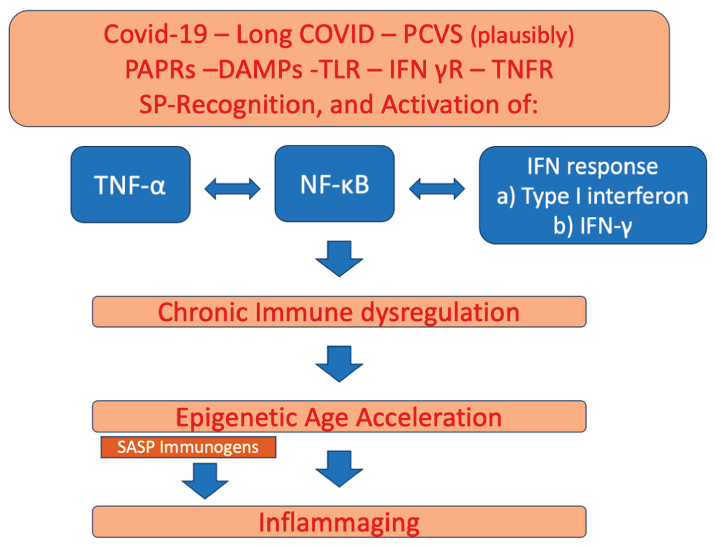

Table 1.
The research studies that show epigenetic age alteration due to COVID-19, Long COVID, and/or COVID-19 mRNA vaccines.
Table 1.
The research studies that show epigenetic age alteration due to COVID-19, Long COVID, and/or COVID-19 mRNA vaccines.
| Scope of Study / Year | Methods of Investigation | Main Findings |
| J Franzen et al. 2021. [28]. Tested the association of COVID-19 with EAA. |
Identification for four age-related signatures was performed in various tissues including blood. Additionally, age acceleration in three age related gene regions was tested. | Chronological age of COVID-19 patients matched their biological age. No telomere attrition in lymphocytes was evident. The biomarkers used were not reliable to predict for severeness of COVID-19 in older patients. |
| A Mongelli et al. 2021 [29]. Tested the association of post-COVID syndrome with biological age acceleration and telomere shortening. |
Pyrosequencing was used on previously defined CpG islands as suitable for biological age determination. A telomere length quantification was performed. | A consistent increase in biological age in post-COVID syndrome was recorded. A remarkable +5.25 years above the range of normality was recorded in the post-COVID group. A remarkable telomere shortening was recorded in the post-COVID group as well. |
| W Xu et al., 2022 [30]. Tested the association of COVID-19 severeness with biological aging and telomere attrition. |
A bidirectional Mendelian randomization (MR) study. Epigenetic clocks and telomere length measurement were tested to indicate aging. | Severe COVID-19 caused telomere shortening and reversed (slowed down) the epigenetic clocks that show EAA indicating for alteration of DNA methylome-nucleosome modifications [31]. |
| Y Yamaguchi et al. 2022 [32]. Tested the effect of mRNA vaccination coding for SARS-CoV-2 spike protein on epigenetic regulation. | Used mass cytometry, RNA-Seq, and ATAC-Seq analysis to identify upregulation of IFN-stimulated genes. Also, transcription factor-binding motif analysis was used to identify enriched IFN regulatory factors and PU.1 motifs in accessible chromatin regions in blood monocytes. |
An upregulation (especially after the second mRNA vaccination) of IFN-stimulated genes, and enrichment of IFN regulatory motifs and PU.1 regions was identified. The second mRNA vaccination caused epigenetic changes in monocytes. |
| L Calzari et al. 2023 [33]. Tested the effect of severe and mild COVID-19 cases on epigenomic aging signatures. | Investigated the genome-wide differences in DNA methylation between severe and mild prognosis COVID-19 patients. | The epigenetic age signature at hospital admission predicted risk for severe outcome. EAA associated with severe prognosis after SARS-CoV-2 infection. |
| L.Calzari et al. 2024 [34]. Tested the effects of Long COVID on premature aging and EAA. |
A genome-wide DNA methylation pattern was analysed in 96 patients, 6 months after COVID-19. | Identification of 42 CpG sites with significant methylation differences located in CpG islands and gene promoters. Dysregulated genes associated with glutamate/glutamine metabolism were identified in Long COVID. These included the glutamate dehydrogenase GLUD1 gene, the ATP1A3 gene of ATPase pump, and the ARRB2 gene of beta-arrestin-2. Using Horvath's epigenetic clock identified a slight but significant age acceleration in post-COVID-19 group. A high number of changes in DNA methylation and histone modifications suggested epigenetic drift effect in the post COVID-19 group |
| P García-delaTorre et al. 2024 [35]. Tested the association between mild COVID-19 and epigenetic changes. |
Used various epigenetic clocks (HorvathAge, HannumAge, GrimAge, PhenoAge, SkinAge, and DNAmTL) to estimate EAA. In addition, immunological profile was correlated with epigenetic estimators. | All epigenetic age estimations differed significantly from the chronological age. However, only GrimAge was elevated. In addition, the intrinsic epigenetic age acceleration (IEAA) and the extrinsic epigenetic age acceleration (EEAA) were accelerated in all patients. Immunological and DNA damage estimators showed that EAA associated with immunological dysfunction and inflammation. |
| N Cheung 2026 [36]. Tested post COVID association with hyper-hypomethylation of cytosine-phosphate-guanine (CpG) regions and showed age related methylome modifications that link post COVID syndrome with immune remodelling, biological age and neuropsychiatric vulnerability. |
Used 94 post-COVID blood samples and equivalent negative controls. CpG gene mapping was performed to show age-related differentially methylated positions. |
Hypermethylation was found at various canonical aging loci. Specifically, age associated hypermethylation was identified near glutamatergic / N-methyl-D-aspartate (NMDA) genes, revealing glutamatergic signatures of aging in post COVID (after six months of infection). |
| Y Bejaoui et al. 2023 [37]. Tested the effect of EAA and telomere attrition on the outcome (deceased and surviving) of COVID-19 patients. | Evaluated DNA methylation age and telomere attrition acceleration in skin and blood samples of 87 severe COVID-19 patients and compared dynamic epigenetic changes until surviving (recovery) or death. | Higher EAA was detected in deceased vs recovered patients. Telomere attrition acceleration was higher in deceased vs recovered patients. |
| CK Barha et al. 2025 [38]. Tested the association of EAA and depressive symptoms. before and during the COVID-19 pandemic. |
Evaluated EAA (using EAA clocks that detect methylation patterns) and the effect on depressive symptomatology during COVID-19. | Younger epigenetic age, i.e. less EAA, associated with depressive symptoms was oberved in females only, during the first periods of COVID-19. |
| G Farkas et al. 2025 [26] Assessed the effect of COVID-19 on premature aging and EAA. |
Used eight epigenetic clocks, SkinBloodClock, DNAmAge DNAmAgeSkinBlood, DNAmAgeHannum, DNAmFitAge, PhenoAge, DNAmGrimAge, and DNAmTL, to measure the methylome and indicate epigenetic aging in a three-year period after COVID-19. | The DNAmAge showed accelerated aging. Five out of eight clocks (DNAmAgeSkinBlood, DNAmAgeHannum, DNAmFitAge, PhenoAge, and DNAmTL) showed slow aging. Females had a higher increase in DNAmAge acceleration, while with the SkinBloodClock, and DNAmTL clocks, slowed aging was observed. The methylation of promoters was either increased, CSTL1, which encodes Cystatin-like 1, or decreased, H1 FNT genes, which encodes testis-specific histone H1 family member N (H1fnt). The set of DNAmGrimAge (DNAmGrimAge, DNAmGrimAge2, and DNAmFitAge) showed that the 3-year period after contracting COVID-19 significantly increased the progress of aging. |
| MK Gupta and R Vadde. 2025 [9]. Assessed the impact of COVID-19 on the rate of epigenetic aging. |
Used various epigenetic clocks to measure the methylation-based aging markers in healthy individuals and COVD-19 patients. Next-generation epigenetic clocks (PCGrimAge) associated with immune dysregulation -inflammation and EAA and first-generation clocks (Hannum2013) were used to measure EAA. | The next generation, immune dysregulation, PCGrimAge epigenetic clock showed the strongest correlation with the chronological age of COVID-19 patients. Other next generation clocks (PCGrimAge, DunedinPACE, and ZhangY2017) also showed EAA, mostly in older and female patients. First generation clocks showed a reduction in EAA, raising concerns for their sensitivity in measuring infection-related EAA. Immune dysregulation due to COVID-19 was found to be the primary cause of EAA. |
| LHA Ventura et al. 2025 [39]. Assessed the immunosenescence phenotype in COVID-19 patients and epigenetic age acceleration. |
Analysed the immunological profile of COVID-19 patients at early stages of infection. Used epigenetic clocks to measure DNA methylation. | Immunological profile resembled the typical profile of senescence. The CXC motif chemokine ligand 9 (CXCL9), an age related proinflammatory protein, was identified [40]. CD8+ and CD4+ lymphocytes showed EAA. |
| A Simonis et al. 2025 [41]. Assessed the epigenetic memory conferred in blood monocytes/macrophages by the mRNA vaccines for COVID-19. |
Analysed the cytokine expression and the alteration epigenetic modifications on histones by the restimulation of the immune system by the SARS-CoV-2 spike protein coded by the mRNA vaccines. | A significant and persistent, histone acetylation-modification, H3K27ac, was identified at promoter regions and preserved for six months. The cytokine response was due to the activation of NF-κB; result concluded by using NF-κB inhibitors that abolished the expression of cytokines like IL-1β. |
| J Bohlin et al. 2026 [42]. Assessed that rate of EAA in Long COVID and non-Long COVID patients following previous SARS-CoV-2 infection. |
This longitudinal study analysed EAA using third generation epigenetic clocks to measure DNA methylation in blood of patients with Long COVID and non-Long COVID. Study performed a sex-stratified analysis of the X chromosome in males. | No evidence of persistent EAA was recorded in Long COVID compared to non-Long COVID patients. |
Disclaimer/Publisher’s Note: The statements, opinions and data contained in all publications are solely those of the individual author(s) and contributor(s) and not of MDPI and/or the editor(s). MDPI and/or the editor(s) disclaim responsibility for any injury to people or property resulting from any ideas, methods, instructions or products referred to in the content. |
© 2026 by the authors. Licensee MDPI, Basel, Switzerland. This article is an open access article distributed under the terms and conditions of the Creative Commons Attribution (CC BY) license (http://creativecommons.org/licenses/by/4.0/).
Copyright: This open access article is published under a Creative Commons CC BY 4.0 license, which permit the free download, distribution, and reuse, provided that the author and preprint are cited in any reuse.