

Submitted:
16 October 2026
Posted:
19 October 2026
You are already at the latest version
Abstract
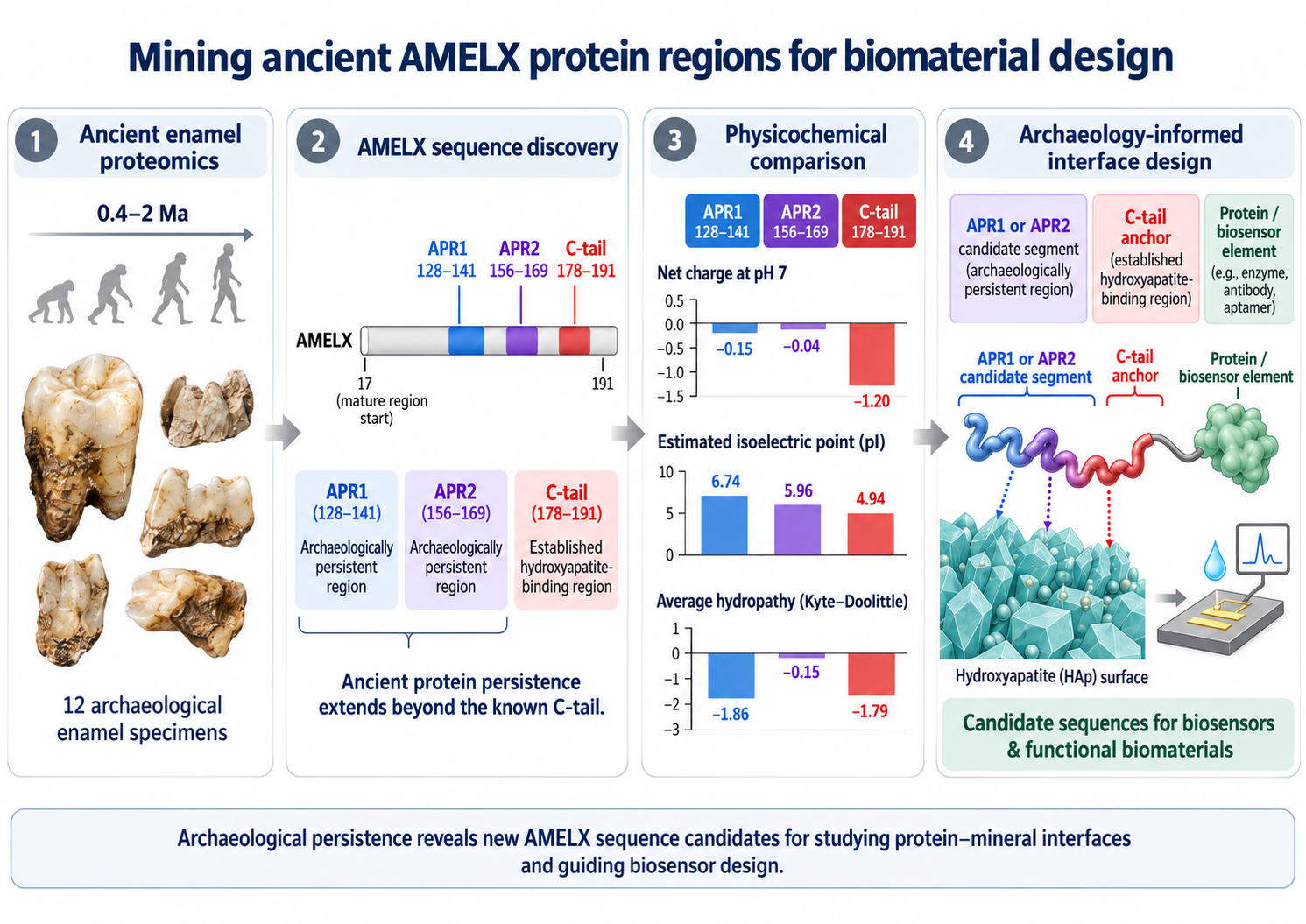
Improving the stability of protein–mineral interfaces while preserving biological activity is a major challenge in biosensor and functional biomaterials design. Ancient tooth enamel offers a unique opportunity to identify protein sequences that have persisted in mineralized environments for millions of years. Here we investigate whether archaeological protein evidence can guide the selection of new peptide candidates for biomaterials engineering. We analyzed amelogenin (AMELX) peptide coverage from 12 ancient enamel specimens dating from approximately 0.4 to 2 million years ago. The data reveal, beyond the established C-terminal hydroxyapatite-binding region, two new peptides regions, archaeologically persistent regions (APR1 and APR2), with substantial archaeological coverage (82.7% and 100%, respectively) and different chemical and physicochemical properties from the C-tail, including differences in electrical charge, isoelectric point, and hydropathy. These findings identify additional AMELX sequences for investigating protein–mineral interactions and suggest that archaeological proteomics can guide the selection of new peptide motifs and candidate protein-mineral interfaces whose potential to improve biosensor durability can be evaluated experimentally.
Keywords:
amelogenin
; AMELX
; hydroxyapatite
; ancient enamel proteomics
; peptides
; biomineralization
; protein immobilization
; biosensors
1. Introduction
The past ten years have seen the emergence of biosensors engineering for health monitoring or non-invasive diagnostic, due to the advancement in nanoelectronics and microelectronics. Biosensors leverage the properties of biological components, bioreceptors, like enzyme or antibody, to quickly identify the presence of biological markers in each sample and turn that interaction into a measurable signal. Bioreceptors can be immobilized on solid supports, including mineral surfaces, to ensure the stability of the biosensor, the proper molecular orientation and an efficient transduction of the measured signal (see Figure 1).
The way the protein binds to the substrate is therefore critical both for the functioning of the sensor and for its duration. If the binding is too weak, the protein can easily wash away. On the contrary, if the binding is too strong it restricts the protein’s conformation or blocks access to its active site, and consequently its biological activity may be hindered or modified. These observations suggest that, in our quest for stable and sustainable biosensors, the challenge is to find a substrate that allows the molecule to bind relatively strongly, while letting the molecule retain useful activity, be accessible to its target and allow for repeated use [1,2,3,4]. Today one of the bottlenecks is the fact that bioreceptors are fragile and rapidly denature losing their functional structure at high temperatures. The key is therefore to find the right mineral surface and protein binding. Among these, tooth enamel constitutes an interesting candidate due to its robust and long-lasting structure. There are several specialized proteins involved in the formation of enamel and in regulating mineral growth, including amelogenin (AMELX), enamelin and ameloblastin. Each of these proteins interact strongly with hydroxyapatite, a calcium-phosphate mineral, Ca₁₀(PO₄)₆(OH)₂, and the main responsible for making enamel hard.
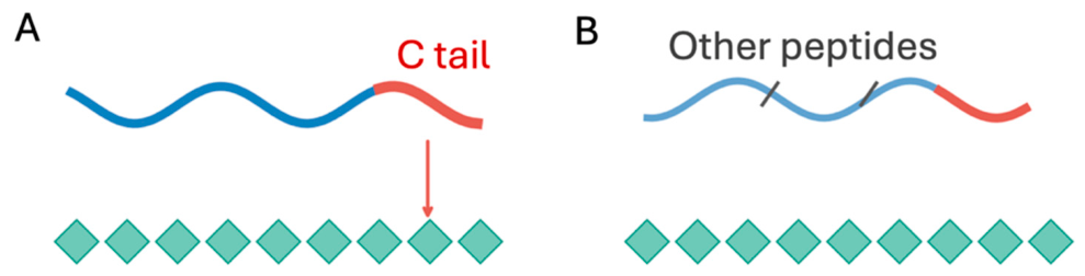
In this study we will focus on the interaction of the protein AMELX with hydroxyapatite, since AMELX is a major enamel matrix protein whose interactions with hydroxyapatite have been extensively investigated. The canonical human AMELX reference sequence used in this study contains 191 amino acids that can be divided in different fragments. Among these different groups, experimental works have shown that the C-terminal region binds with the hydroxyapatite helping enamel mineralization, making C-terminal region a useful reference for biomaterials engineering [5,6]. Whether other regions of the 191 amino-acid protein can equally bind and be considered viable source for biomaterials engineering is still an open question. Identifying these regions opens new opportunities for engineering of protein-mineral interfaces for use in a variety of fields including biosensors. While studies of these proteins can tell us which regions interact strongly with the mineral, we do not have a full understanding of which molecular properties contribute to the long preservation of proteins within mineralized tissues. Ancient proteins constitute a unique dataset to address this question and a unique natural record of molecular persistence, well beyond what can be achieved in a modern laboratory. Archaeological proteomics therefore provides an opportunity to identify persistent protein sequences whose properties may inform the design of new, stable, protein-mineral interfaces [7,8,9]. In the specific case, thanks to the strong mineral binding of AMLEX with hydroxyapatite, and its protection from water and degradation, there is a large dataset of ancient proteins to study and eventually to use to predict new protein-mineral structures with potential applications in biosensor design [7,8,9].
2. Materials and Methods
The data used in this study were obtained from publicly released work, namely an independent set of samples from the previously assembled AMELX hydroxyapatite-binding preservation pilot workbook and compiled from public PRIDE proteomics outputs [10,11,12,13,14,15]. Specifically, the data are extracted from three publicly available proteomics dataset, including 12 fossil tooth enamel specimens from PRIDE projects: PXD068897 (six specimens from ~ 0.40 million years) [10,11], PXD014342 (two specimens from ~0.85-1.77 million years) [12,13], and PXD040221 (four specimens from ~ 2.00 million years) [14,15]. Peptide identifications were obtained from the published proteomics datasets and processed using the sequence-mapping procedure described below. Differences in the original studies’ identification criteria and analytical methods were considered limitations of the cross-dataset comparison. Figure 3 summarizes the age distribution of the archaeological specimens used in this study, while Table 1 presents their archaeological origins, estimated ages, and source information [10,11,12,13,14,15].
These specimens belong to three extinct hominin species, Homo erectus, Homo antecessor, and Paranthropus robustus, recovered from archaeological sites in China, Spain, Georgia, and South Africa, as shown in the table below, and their estimated ages range from approximately 400,000 to 2 million years [10,12,14].
Data: The data here presented have the following limitations. 1) The sampling is based on three public datasets; 2) the data are obtained from different degradation experiments; 3) Protein uniqueness was evaluated against the canonical human AMELX sequence [16], leaving species-specific variations, modified residues, and unverified cross-species uniqueness unaddressed without validation against taxon-specific proteomes. To address these constraints, future research must examine individual spectral assignments, re-map sequences against appropriate species references, evaluate study-level sensitivity, and experimentally verify mineral binding and enzymatic functionality.
Computation: The calculations were performed using Biopython 1.86 [17], specifically the ProteinAnalysis module and the net electrical charge at pH 7 was estimated using the Henderson–Hasselbalch equation and the pKa values implemented in Biopython's IsoelectricPoint module. The model accounts for ionizable amino acid side chains (D, E, C, Y, H, K, and R), as well as the free N- and C-terminal groups. The isoelectric point (pI), defined as the pH at which the peptide has zero net electrical charge, was estimated by iteratively identifying the pH at which the calculated charge approaches zero. Average hydropathy was calculated using the Kyte–Doolittle scale [18], which assigns a hydropathy value to each amino acid; these values were averaged across the 14 residues of each peptide. More negative hydropathy values indicate greater average hydrophilicity.
It is important to point out that the calculations treat each region as an isolated unmodified 14-amino acid peptide with free terminal groups, while in any given protein the three peptides group interact. These calculations are useful for comparing candidate peptide sequences, but they do not account for their structural context within intact AMELX or their interactions with hydroxyapatite.
3. Results
To uncover which parts of the AMELX have survived in ancient enamel, the first step is sequence mapping, which consist in comparing the recovered fragments of the AMELX protein, peptides, with recent aminoacid sequences found in modern AMELX (UniProtQ99217) [16]. Each peptide sequence detected in the ancient tooth is then compared with the exact same sequence in the known AMELX protein and assigned a specific position within the reference AMELX chain. The assigned position numbers therefore represent the reference chain and not the absolute number in the ancient specimen. The comparison between the reference chain and archaeological specimens suggest that there are three main regions that appear to persist (see Figure 4). Each region contains 14 aminoacids:
178-191 (AWPSTDKTKREEVD), a human C-terminal sequence homologous to experimentally studied C-terminal regions; 128–141 (QPQPVQPQPHQPMQ), identified in earlier exploratory screening; and 156–169 (PPLPPMFPMQPLPP), which appeared repeatedly in ancient-only inspection. This characterization allows us to directly compare their composition and preservation using the same sequence length. We define Archaeologically Persistent Regions (APRs) segments of the AMELX sequence that show peptide coverage across the ancient enamel species. We identify two regions APR1, covering residues 128-141 and APR2, covering residues 156-169. Figure 4 illustrates the positions of APR1 (128-141), APR2 (156-169) and the C-terminal region (178-191) within the canonical AMELX sequence [16]. The internal regions APR1 and APR2 are located upstream of the established C-terminal hydroxyapatite-binding region.
We note that this coding of ancient peptides is applied only if at least one accepted mapped peptide overlapped that position. The equal length (14-residue) keeps the sequence composition counts directly comparable.
The result presented here emerge from the comparison of 12 ancient teeth over the three peptide chains 128-141, 156-169 and 178-191. For each region we calculated three measurements: 1) the mean residue coverage, which represents how much of that region was detected across all 12 specimens, corresponding to a total of 168 possible detections (14 x 12); 2) Specimens with any coverage, which tells how many of the 12 ancient specimens contained at least one amino acid position from that region; 3) Complete region covered, which tells us how many specimens covered all 14 amino acid positions. It is important to note that we count a full coverage, even if several smaller overlapping peptides cover all 14 positions, and not the entire region survived as one intact piece. Overlapping peptides and specimens from a common dataset do not represent independent replication of a binding event. [7,9]
To make sure that a given sequence motifs is not counted more than once, we repeated peptide mapping with an exact-match, unique-reference-position filter. We checked all 2,397 deep-time peptide entries against the 191-residue AMELX reference: 2,124 entries matched at exactly one position, 273 had no exact match, and none matched more than one position. The source workbook mapping counts agreed with this independent recalculation for every entry. We rebuilt the residue-by-specimen detection matrix from the 2,124 unique-reference matches and compared it cell by cell with the original pilot matrix: all 2,292 cells (12 specimens × 191 positions) agreed.
To investigate why certain AMELX regions remain detectable in ancient enamel, we compared their amino acid composition. For each of the three 14-amino-acid regions, we counted amino acids with different chemical properties, including proline, glutamine, acidic and basic residues, and histidine. Table 3 summarizes the amino acid sequences of APR1, APR2, and the C terminal region, together with the number of prolines, glutamine, acidic, basics, and histidine residues, in each region. These amino acids can influence protein structure, stability and interactions with mineral surfaces. By comparing their distribution, we aimed to identify chemical characteristics that might contribute to the long-term preservation of AMELX. However, amino acid composition alone cannot tell us how strongly a region binds to hydroxyapatite or whether mineral binding explains its survival.
In Figure 5 we summarize data from the 12 specimens covering the period spanning approximately 400,000 to 2 million years ago, to understand whether other regions of the aminoacid chain could be potentially useful for biosensors. When compared with the standard amino acid sequence found in modern enamel, we identify the peptide regions that have survived over millions of years.
The distribution of detected amino acid positions across the 12 archaeological specimens is shown in panel a, where each row represents one specimen (from Table 1), and each colored segment indicates a portion of the AMELX sequence covered by at least one identified peptide. The results show that only some region of the entire amino acid chains is detected and persist over time, while other are absent. While many of these regions corresponds to the C-terminal mineral binding, the data reveal also the persistence over millions of years of other pieces of the amino acid chain. These regions correspond to the peptides with amino acid positions from APR1 (128-141) and APR 2 (156-169).
In panel b we show the frequency by which each amino acid position was detected. Interestingly we find that all 14 positions of APR2 appears in were covered by identified peptides in all 12 specimens, whereas at least part of APR1 was detected in 11 of the 12 specimens (91.7%), which is higher than the C-terminal region (82%). Note that, although the coverage of APR1 appears higher than the C-terminal region, if we consider the recovery of the full 14 amino acid sequence, the C-terminal region has all 14 positions covered in more specimens (6/12) than APR1 (5/12), as summarized in Table 2. In contrast, the regions between 50 and 140 have little to no peptide coverage. Table 2 summarizes the three coverage measurements for each selected region: mean residue coverage, the number of specimens with any detected positions, and the number of specimens with all 14 positions covered.
These results suggest that APR2 and APR1 are viable candidates for protein-mineral interaction alongside the widely studied C-terminal, although their binding properties have not been fully established. The first step is to understand why these peptides remain detectable in ancient enamel.
There are two possible scenarios. The first is that they create some additional bonding with the mineral surface, while the second is that the composition of the amino acid or the structural environment contribute to their resistance to degradation. Differences in peptide extraction and detection may also influence the observed patterns.
It is important to notice that recovery of a protein sequence does not necessarily indicate stronger mineral binding, since preservation may also depend on amino acid chemistry, protection within the enamel structure, or differences in peptide detection. Our results therefore identify APR1 and APR2 as promising candidates for further investigation rather than confirming their mineral-binding strength. To explore possible explanations for their persistence, we need to examine their amino acid composition and compare it with that of the established C-terminal binding region.
In order to understand whether the chemical properties of the amino acid are responsible for the long-term preservation of AMELX in ancient enamel, we compared the amino acid composition of the selected regions with their frequency of detection in archaeological specimens. In this way we can compare regions with different chemical characteristics and see whether they show similar patterns of preservation, to eventually obtain some clues about their interaction with hydroxyapatite. Table 3 summarizes the amino acid sequences and the distribution of the selected residue types across APR1, APR2, and the C-terminal region.
Among the 14 amino acids that make the peptides APR1, APR2, and C-tail, proline, glutamine, acidic and basic residues, and histidine were selected to highlight chemical differences potentially relevant to peptide conformation and mineral interactions.
Figure 6 shows the distribution of these amino acids in standard AMELX protein sequence. As expected, each peptides have its own dominant aminoacid, revealing a marked chemical contrast between the different peptides. In C-tail, the dominant one are the Acidic D/E amino acids, in APR2 proline (P), and in APR1 glutamine (Q). Moreover, it is interesting to observe that neither APR1 and APR2 shows any acid D/E, or basic K/R amino acids.
This contrast matters because the internal regions do not simply reproduce the charged C-tail motif. Proline often constrains peptide backbone conformations, glutamine side chains can participate in hydrogen bonding, and charged side chains may interact with calcium or phosphate groups. However, residue counts alone do not identify the orientation of a peptide at a hydroxyapatite surface or distinguish intrinsic resistance to cleavage from mineral-mediated protection. Strong recovery is compatible with several explanations.
To better understand the chemical bonding between peptides and hydroxyapatite, in Figure 7 we calculate the chemical properties of each region. To do so we perform a sequence-based computational analysis of each individual peptides. The calculations characterize only the intrinsic sequence properties of isolated peptides and do not model their three-dimensional structures or adsorption within the hydroxyapatite. Figure 7A shows that APR1 and APR2 have approximately neutral estimated net charges at pH 7 (−0.15 and −0.04, respectively), whereas the C tail is more negatively charged (−1.20).
Figure 7B shows corresponding differences in estimated isoelectric point, with APR1 having the highest value (6.74), followed by APR2 (5.96) and the C-tail (4.94).
These results indicate that the three peptides have different ionization properties, potentially influencing their electrostatic interactions with hydroxyapatite under different pH conditions. However, neither the net charge nor the isoelectric point independently determines mineral-binding strength.
Another interesting observation results from the comparison of the average hydropathy shown in panel c, which is a measure of the tendency of amino acids to interact with water. The results show that both APR1 and C-tail are quite hydrophilic while APR2 has an average hydropathy closer to zero and is less hydrophilic than APR1 and the C tail. This suggests that APR2 might have different hydration and conformational properties, influencing its behavior at the hydroxyapatite interface. Indeed, when a peptide approaches hydroxyapatite, it encounters a mineral surface surrounded by water molecules. In order to attach the peptide needs to reorganize some of the interactions with water. The fact that APR 2 was detected across all 14 residue positions in all 12 archaeological specimen and it shows to be enriched in proline (8 out of 14 aminoacids) as shown in Figure 6, suggests that its distinctive composition may influence its hydration and conformational behavior, potentially contributing to its long-term persistence. However, the relationship between these properties and archaeological preservation remains to be established.
4. Discussion
One of the main challenges in biosensors engineering today is achieving stable protein immobilization while maintaining its biological activity. Most biosensors to date are built on the assumption that long-term protein preservation depends primarily on the specific mineral binding interactions, while the role of molecular properties responsible for long-term protein stability is poorly investigated. The current study has revealed the persistence of APR1 and APR2 peptides in ancient enamel, in addition to the most studied C-tail. These peptides show different chemical and physiochemical properties with respect to the C-tail peptide, raising the important question whether long-term protein preservation depends primarily on specific mineral-binding interactions, or whether different chemical and structural properties provide alternative pathways to molecular persistence. Even if the study does not provide direct evidence of which region binds hydroxyapatite most strongly, the new chemical properties identified, warrant further investigations [5,6,7].
These findings open opportunities to investigate alternative protein–mineral interfaces for biosensor applications. One possible strategy is illustrated in Figure 8, where the established C-tail serves as a mineral-binding anchor connected through a flexible linker to APR1 or APR2, and the additional sequence could provide further mineral contacts, modify the conformation of the immobilized construct, or influence its susceptibility to degradation. This hybrid design could combine a known attachment motif with an additional region selected for different physicochemical characteristics, where for example the function of mineral attachment and protection from degradation do not need to be performed by the same peptide sequence.
Although the results presented here open new opportunities, it is important to point out that future investigations are needed to compare APR1, APR2, the C-tail for functional biomaterials design. Indeed, previous studies have argued that stronger adsorption alone does not necessarily produce a better sensing interface, as several other factors could contribute to higher efficiency, including enzyme immobilization [1], the potential of inorganic mineral supports [2], the influence of attachment geometry on enzyme function [3], and the importance of stable enzyme loading, substrate accessibility, and sustained signal generation in biosensors [4].
Combining these studies with molecular simulations could provide new information on potential interaction sites and conformational arrangements. In summary, these results have shown the potential of combining archaeological protein evidence with physicochemical characterization and functional testing, to provide a new route toward designing stable and effective protein–mineral interfaces for biosensors.
In summary, these results have shown the potential of combining archaeological protein evidence with physicochemical characterization and functional testing, to provide a new route toward designing stable and effective protein–mineral interfaces for biosensors
5. Conclusions
Archaeological protein evidence offers a promising, largely unexplored resource for identifying molecular candidates for functional biomaterials. In this paper we analyzed fragments of AMELX protein which binds with hydroxyapatite mineral, recovered from ancient enamel from four different geographical areas dating from 400,000 million years to 2 million years ago. We have identified two internal AMELX regions, APR1 and APR2, in almost each one of the studied archaeological peptide samples. A comparison with the well-studied C-terminal peptides has revealed different physicochemical properties which might be appealing for the design of novel biosensors. Moreover, the results suggest that different chemical and structural properties may contribute to long-term protein persistence, although the underlying mechanisms remain unresolved, although future molecular simulations and experimental studies will be required to determine the binding properties of these sequences and assess their effects on biosensor performance.
This work establishes a new framework for designing peptide-based interfaces for biomaterials and biosensor applications, combining archaeologically persistent sequences with established mineral-binding motifs. More broadly, this work establishes a framework for using archaeological proteomics to generate and prioritize candidates for the development of stable protein–mineral interfaces.
Author Contributions
R.B. ideated the research and wrote the manuscript. A.S. edited the manuscript.
Funding
This research received no external funding.
Institutional Review Board Statement
Not applicable to the present secondary analysis of publicly archived proteomic datasets.
Informed Consent Statement
Not applicable to this secondary analysis of public archaeological proteomics data.
Data Availability Statement
Acknowledgments
We thank Prof. Lanzara for useful comments and discussions.
Conflicts of Interest
The authors declare that have no conflict of interest.
Abbreviations
The following abbreviations are used in this manuscript:
| AMELX | amelogenin |
| APR | Archaeologically Persistent Regions |
References
- Sheldon, R.A.; van Pelt, S. Enzyme immobilization in biocatalysis: Why, what and how. Chem. Soc. Rev. 2013, 42, 6223–6235. [Google Scholar] [CrossRef] [PubMed]
- Hartmann, M.; Kostrov, X. Immobilization of enzymes on porous silicas—benefits and challenges. Chem. Soc. Rev. 2013, 42, 6277–6289. [Google Scholar] [CrossRef] [PubMed]
- Rodrigues, R.C.; Ortiz, C.; Berenguer-Murcia, Á.; Torres, R.; Fernández-Lafuente, R. Modifying enzyme activity and selectivity by immobilization. Chem. Soc. Rev. 2013, 42, 6290–6307. [Google Scholar] [CrossRef] [PubMed]
- Sun, G.; Wei, X.; Zhang, D.; Huang, L.; Liu, H.; Fang, H. Immobilization of Enzyme Electrochemical Biosensors and Their Application to Food Bioprocess Monitoring. Biosensors 2023, 13, 886. [Google Scholar] [CrossRef] [PubMed]
- Friddle, R.W.; Battle, K.; Trubetskoy, V.; Tao, J.; Salter, E.A.; Moradian-Oldak, J.; De Yoreo, J.J.; Wierzbicki, A. Single-Molecule Determination of the Face-Specific Adsorption of Amelogenin's C-Terminus on Hydroxyapatite. Angew. Chem. Int. Ed. 2011, 50, 7541–7545. [Google Scholar] [CrossRef] [PubMed]
- Tao, J.; Hanson, E.; Dohnalkova, A.C.; Buchko, G.W.; Jin, B.; Shaw, W.J.; Tarasevich, B.J. Changes in the C-Terminal, N-Terminal, and Histidine Regions of Amelogenin Reveal the Role of Oligomer Quaternary Structure on Adsorption and Hydroxyapatite Mineralization. Front. Physiol. 2022, 13, 1034662. [Google Scholar] [CrossRef] [PubMed]
- Demarchi, B.; et al. Protein sequences bound to mineral surfaces persist into deep time. eLife 2016, 5, e17092. [Google Scholar] [CrossRef] [PubMed]
- Cappellini, E.; Welker, F.; Pandolfi, L.; et al. Early Pleistocene enamel proteome from Dmanisi resolves Stephanorhinus phylogeny. Nature 2019, 574, 103–107. [Google Scholar] [CrossRef] [PubMed]
- Hendy, J. Ancient protein analysis in archaeology. Sci. Adv. 2021, 7, eabb9314. [Google Scholar] [CrossRef] [PubMed]
- Fu, Q.; Wu, Z.; Bennett, E.A.; Xing, S.; Ji, Q.; Dong, Z.; Rao, H.; et al. Enamel proteins from six Homo erectus specimens across China. Nature 2026, 655, 141–147. [Google Scholar] [CrossRef] [PubMed]
- Archive, P.R.I.D.E. Proteomics dataset PXD068897. ProteomeXchange Consortium. Available online: https://www.ebi.ac.uk/pride/archive/projects/PXD068897 (accessed on 12 October 2026).
- Welker, F.; Ramos-Madrigal, J.; Gutenbrunner, P.; et al. The dental proteome of Homo antecessor. Nature 2020, 580, 235–238. [Google Scholar] [CrossRef] [PubMed]
- Archive, P.R.I.D.E. Proteomics dataset PXD014342. ProteomeXchange Consortium. Available online: https://www.ebi.ac.uk/pride/archive/projects/PXD014342 (accessed on 12 October 2026).
- Madupe, P.P.; Koenig, C.; Patramanis, I.; et al. Enamel proteins reveal biological sex and genetic variability in southern African Paranthropus. Science 2025, 388, 969–973. [Google Scholar] [CrossRef] [PubMed]
- Archive, P.R.I.D.E. Proteomics dataset PXD040221. ProteomeXchange Consortium. Available online: https://www.ebi.ac.uk/pride/archive/projects/PXD040221 (accessed on 12 October 2026).
- Consortium, UniProt. AMELX_HUMAN (Q99217), canonical human amelogenin sequence. UniProtKB. Available online: https://www.uniprot.org/uniprotkb/Q99217/entry (accessed on 12 October 2026).
- Cock, P.J.A.; Antao, T.; Chang, J.T. Biopython: Freely available Python tools for computational molecular biology and bioinformatics. Bioinformatics 2009, 25, 1422–1423. [Google Scholar] [CrossRef]
- Kyte, J.; Doolittle, R.F. A simple method for displaying the hydropathic character of a protein. J. Mol. Biol. 1982, 157, 105–132. [Google Scholar] [CrossRef]
Figure 1.
Schematic of a biosensor showing biological recognition, signal transduction and processing.
Figure 1.
Schematic of a biosensor showing biological recognition, signal transduction and processing.
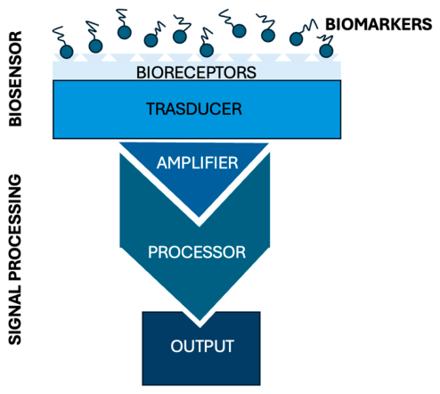
Figure 3.
Age distribution of the 12 archaeological enamel specimens analyzed in this study. A) Number of specimens by estimated age. B) Ages of individual specimens.
Figure 3.
Age distribution of the 12 archaeological enamel specimens analyzed in this study. A) Number of specimens by estimated age. B) Ages of individual specimens.
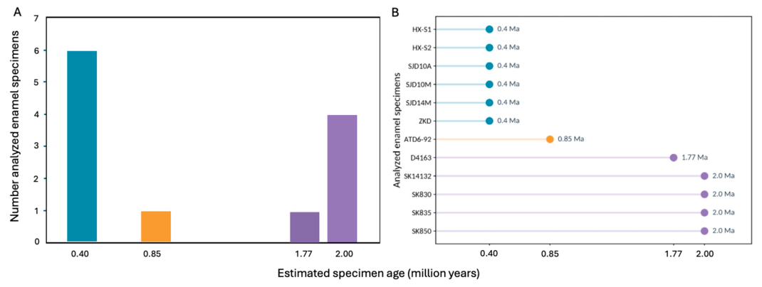
Figure 4.
Position of APR1, APR2, and the C-tail within AMELX.

Figure 5.
Archaeological AMELX residue coverage. A) Residue detection across 12 ancient enamel specimens; b) Detection frequency at each amino acid position. Colored regions identify APR1, APR2 and the C-tail.
Figure 5.
Archaeological AMELX residue coverage. A) Residue detection across 12 ancient enamel specimens; b) Detection frequency at each amino acid position. Colored regions identify APR1, APR2 and the C-tail.
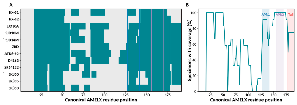
Figure 6.
Chemical composition of selected AMELX regions. A) Counts of selected amino acid types. B) Amino acid sequences colored by chemical category.
Figure 6.
Chemical composition of selected AMELX regions. A) Counts of selected amino acid types. B) Amino acid sequences colored by chemical category.
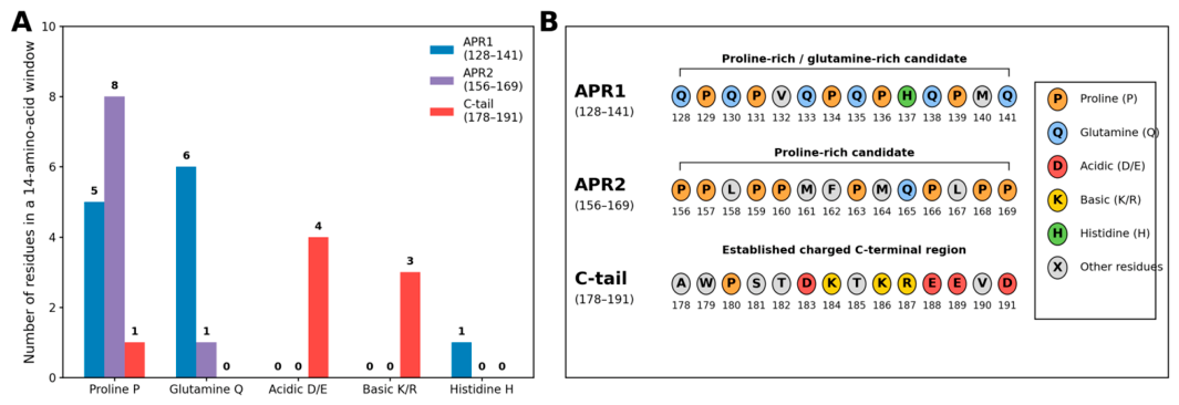
Figure 7.
Calculated physicochemical properties of APR1, APR2 and the C-tail. A) Net charge at pH 7. B) Isoelectric point. C) Average hydropathy. Calculations describe isolated peptides, not mineral-binding affinity.
Figure 7.
Calculated physicochemical properties of APR1, APR2 and the C-tail. A) Net charge at pH 7. B) Isoelectric point. C) Average hydropathy. Calculations describe isolated peptides, not mineral-binding affinity.
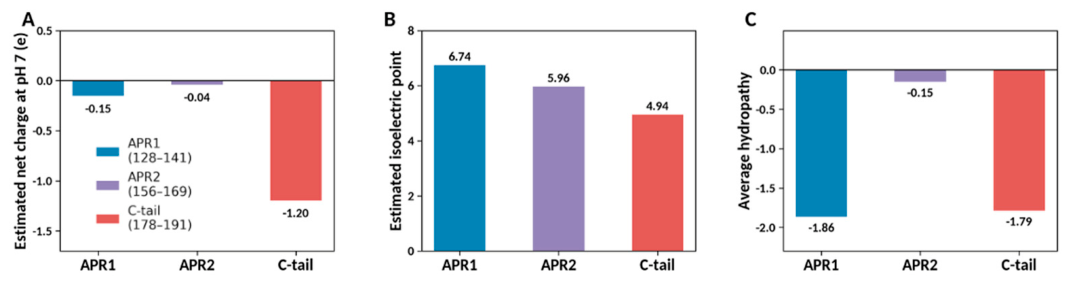
Figure 8.
Proposed archaeology-informed protein-mineral interfaces. A) Established C-tail binding motif. B, C) Candidate APR1 and APR2 regions; D) Hypothetical hybrid combining the C-tail with an internal region through a flexible linker.
Figure 8.
Proposed archaeology-informed protein-mineral interfaces. A) Established C-tail binding motif. B, C) Candidate APR1 and APR2 regions; D) Hypothetical hybrid combining the C-tail with an internal region through a flexible linker.
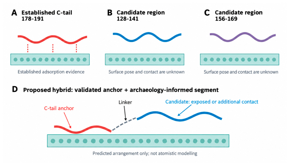

Table 1.
This is a table. Tables should be placed in the main text near to the first time they are cited.
Table 1.
This is a table. Tables should be placed in the main text near to the first time they are cited.
| Specimen [ref] | Species | Archaeological site and context | Tooth / fossil description | Age (Ma) |
|---|---|---|---|---|
| HX-S1 [10] | Homo erectus | Hexian, Anhui, China (layer 4) | Tooth; Hexian sample 1 | ~0.41 |
| HX-S2 [10] | Homo erectus | Hexian, Anhui, China (layer 4) | Tooth; Hexian sample 2 | ~0.41 |
| SJD10A [10] | Homo erectus | Sunjiadong, Henan, China (layer 4) | Tooth 12SJD1#10-45 | ~0.40 |
| SJD10M [10] | Homo erectus | Sunjiadong, Henan, China (layer 4) | Tooth 12SJD1#10-42 | ~0.40 |
| SJD14M [10] | Homo erectus | Sunjiadong, Henan, China (layer 5) | Tooth 12SJD1#14-22 | ~0.40 |
| ZKD [10] | Homo erectus | Zhoukoudian, Beijing, China (layers 8–9) | Tooth PA69 | ~0.42 |
| ATD6-92 [12] | Homo antecessor | Gran Dolina, Atapuerca, Spain (TD6.2) | Lower left molar fragment (M1/M2) | 0.772–0.949 |
| D4163 [12] | Homo erectus | Dmanisi, Georgia (layer B1) | Fragmented right upper first molar | 1.76–1.78 |
| SK14132 [14] | Paranthropus robustus | Swartkrans, South Africa (Member 1) | Isolated postcanine tooth fragment | 1.8–2.2 |
| SK830 [14] | Paranthropus robustus | Swartkrans, South Africa (Member 1) | Left lower fourth premolar | 1.8–2.2 |
| SK835 [14] | Paranthropus robustus | Swartkrans, South Africa (Member 1) | Left upper third molar | 1.8–2.2 |
| SK850 [14] | Paranthropus robustus | Swartkrans, South Africa (Member 1) | Right lower third premolar | 1.8–2.2 |
Table 2.
Archaeological peptide coverage of APR1, APR2 and the C-terminal region across 12 ancient enamel specimens.
Table 2.
Archaeological peptide coverage of APR1, APR2 and the C-terminal region across 12 ancient enamel specimens.
| AMELX window | Mean residue coverage | Specimens with any coverage | Specimens with all 14 positions covered |
| 128–141 | 82.7% | 11/12 | 5/12 |
| 156–169 | 100% | 12/12 | 12/12 |
| 178–191 (C-tail) | 73.2% | 10/12 | 6/12 |
Table 3.
Amino acid sequences and composition APR1, APR2 and the C-terminal region of AMELX. P = proline; Q=glutamine; D/E = acidic residues; K/R = basic residues; H=histidine. Sequences and residue counts are based on the canonical human AMELX [16].
Table 3.
Amino acid sequences and composition APR1, APR2 and the C-terminal region of AMELX. P = proline; Q=glutamine; D/E = acidic residues; K/R = basic residues; H=histidine. Sequences and residue counts are based on the canonical human AMELX [16].
| Region | Sequence | P | Q | D/E | K/R | H |
|---|---|---|---|---|---|---|
| 128–141 | QPQPVQPQPHQPMQ | 5 | 6 | 0 | 0 | 1 |
| 156–169 | PPLPPMFPMQPLPP | 8 | 1 | 0 | 0 | 0 |
| 178–191 | AWPSTDKTKREEVD | 1 | 0 | 4 | 3 | 0 |
Disclaimer/Publisher’s Note: The statements, opinions and data contained in all publications are solely those of the individual author(s) and contributor(s) and not of MDPI and/or the editor(s). MDPI and/or the editor(s) disclaim responsibility for any injury to people or property resulting from any ideas, methods, instructions or products referred to in the content. |
© 2026 by the authors. Licensee MDPI, Basel, Switzerland. This article is an open access article distributed under the terms and conditions of the Creative Commons Attribution (CC BY) license.
Copyright: This open access article is published under a Creative Commons CC BY 4.0 license, which permit the free download, distribution, and reuse, provided that the author and preprint are cited in any reuse.