
Submitted:
16 October 2026
Posted:
19 October 2026
You are already at the latest version
Abstract
Fibrodysplasia ossificans progressiva (FOP) is an ultra-rare, debilitating genetic disorder characterized by the progressive and irreversible heterotopic ossification (HO) of soft connective tissues, ultimately leading to permanent loss of mobility. It is predominantly caused by a heterozygous single-nucleotide substitution (c.617G>A; p.R206H) in the ACVR1 gene. This causes the receptor to become aberrantly responsive to Activin A, thereby hyperactivating bone morphogenetic protein (BMP) signalling. Notably, disease progression is heavily driven by gene–environment interactions; environmental and inflammatory triggers, such as mechanical trauma, viral infections, and invasive medical procedures, frequently induce flare-ups that accelerate HO. This review provides a comprehensive overview of the genetic epidemiology of FOP and how the interplay between ACVR1 mutations and environmental stimuli coordinates the pathogenesis of trauma- and inflammation-induced HO. Finally, we highlight current management paradigms and emerging therapeutic strategies aimed at mitigating this devastating condition.
Keywords:
fibrodysplasia ossificans progressiva
; gene-environment interaction
; environmental traumas
; heterotopic ossification
1. Introduction
Fibrodysplasia Ossificans Progressiva (FOP) is a rare and severely disabling genetic disorder characterized by the progressive HO of tendons, ligaments, fascia, and other soft tissues into ectopic bone [1]. Cumulative episodes of HO progressively restrict patients through joint ankylosis and can eventually contribute to severe complications such as thoracic insufficiency syndrome [2,3].
Historical accounts of FOP date back to 1692, when Guy Patin described a patient with the disorder. The first comprehensive description was provided by John Freke in 1736, who documented the case of a 14-year-old boy with multiple hard swellings arising from the vertebrae and ribs that progressively fused into a bony mass [4]. The underlying cause of FOP remained unknown until 2006 when Shore et al. identified a mutation in the ACVR1 gene as the cause of classic FOP [5].
Although the identification of mutant ACVR1 variants established the genetic basis of FOP, it does not fully explain the episodic nature or distribution of HO. The variant exists throughout a patient’s life, yet HO develops in discrete episodes, either spontaneously or following environmental factors such as muscle trauma, injury, or inflammation [6,7]. This indicates that the clinical progression of FOP is determined not only by the underlying genetic defect, but also by environmental factors that determine when and where lesions form.
Mechanistically, this genetic-environmental interaction results from altered signalling properties of the ACVR1 receptor. In FOP, the receptor becomes abnormally responsive to Activin A [8], leading to the aberrant activation of the BMP pathway SMAD1/5/9 [9]. This provides a mechanistic link between muscle injury, inflammation, and the formation of ectopic bone in soft connective tissues [7].
This review examines FOP from a gene–environment perspective, with emphasis on its genetic epidemiology, the molecular consequences of mutant ACVR1 variants, environmental triggers of flare-ups, and the mechanisms through which these factors interact to promote HO. Current therapeutic strategies and future therapeutic directions are also discussed.
2. Genetic Epidemiology of FOP
FOP is an ultra-rare genetic disorder with a commonly cited prevalence of approximately 1 case in 2 million people. However, measured prevalence tends to be higher in regions with more developed healthcare systems, greater disease awareness, and stronger patient-support and diagnostic networks, likely reflecting improved case ascertainment rather than true geographic differences in disease frequency. Regions with more developed medical systems, including North America, Oceania, and Western Europe, report higher prevalences of FOP than other regions [10]. A more recent prevalence study estimated FOP prevalence among people living in the United States at 0.88 cases per million, corresponding to approximately 1 case per 1.14 million people, based on July 2020 data [11]. Misdiagnosis is also common in FOP; the same study found that more than half of patients had been misdiagnosed before receiving an FOP diagnosis. Misdiagnosis and an average age at diagnosis well into the first decade of life likely contribute to underestimation of FOP prevalence [11]. Congenital malformations of the great toes are a defining feature present at birth in individuals with classic FOP, which accounts for more than 95% of FOP cases [12]. Nevertheless, limited awareness of this characteristic clinical marker can delay diagnosis and expose patients to inappropriate invasive procedures, such as biopsies, that may exacerbate disease progression through iatrogenic trauma [13].
FOP has a poor long-term prognosis, with an earlier estimated median survival of 56 years and death most commonly resulting from complications of thoracic insufficiency syndrome [10]. A 2026 mortality study by Breskin et al. reported a median age at death of 50.5 years among FOP patients living in the US, using data from 2017-2023. This stark difference from the median age at death of 80.5 years in the general US population highlights the excess mortality associated with FOP [14]. The disorder has been reported across diverse populations, with no established racial, ethnic, geographic, or sex predisposition [10]. However, potential sex-dependent phenotypic differences have been reported in a specific mouse model, in which female mice exhibited greater HO volumes and greater variability in HO burden than male mice [15]. The clinical relevance of these findings to human FOP remains unclear.
The disease follows an autosomal dominant inheritance pattern, although most cases arise from de novo mutations rather than familial transmission. The landmark genetic study by Shore et al. identified a recurrent heterozygous c.617G>A substitution in ACVR1, resulting in the p.R206H mutation, in both sporadic and inherited FOP [5]. Subsequent studies identified additional heterozygous ACVR1 missense mutations in patients with atypical FOP phenotypes, showing that several ACVR1 variants can cause FOP [12]. Together, these findings indicate that heterozygous missense mutations are the characteristic class of ACVR1 variants associated with FOP. These variants affect different receptor domains and are associated with varying clinical phenotypes, as shown in Table 1.
3. Molecular Basis of FOP
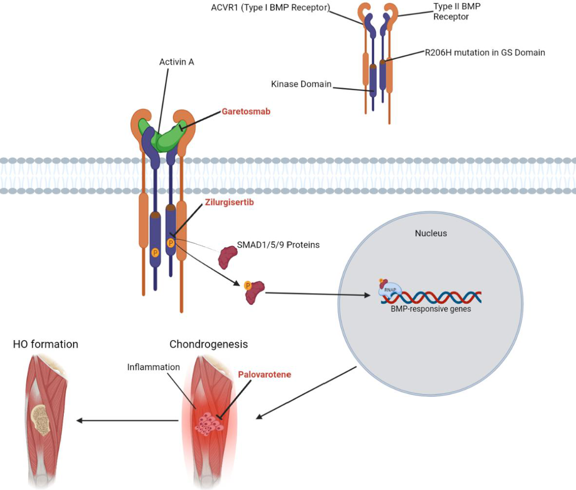
FOP starts with a heterozygous missense mutation in the ACVR1 gene. The ACVR1 receptor is a type I bone morphogenetic protein (BMP) receptor belonging to the transforming growth factor-β (TGFβ) superfamily [16]. Structurally, ACVR1 contains an extracellular ligand-binding region and intracellular Glycine-Serine (GS) and serine/threonine kinase domains. The most recurrent FOP-associated mutation is the c.617G>A substitution, resulting in an arginine-to-histidine substitution at residue 206 (p.R206H). This mutation lies within the GS domain, a regulatory region that controls activation of the receptor kinase and is therefore critical to normal ACVR1 signalling [17]. To understand the impact of this genetic defect, it is necessary to consider the normal function of ACVR1 in BMP signalling.
ACVR1 functions as part of a heterotetrameric BMP receptor complex at the cell surface. The receptor complex has two type I receptors and two type II receptors. The binding of BMP to type II receptors activates type I receptors, allowing ACVR1 to phosphorylate the SMAD1/5/9 proteins. These proteins then relay the signal toward the nucleus and regulate BMP-responsive gene transcription involved in processes such as skeletal development and differentiation [9].
Activin A is a multifunctional TGF-β superfamily cytokine involved in processes including hematopoiesis, embryonic development, stem cell maintenance, tissue repair, and fibrosis [18]. The p.R206H mutation alters the normal relationship between Activin A and ACVR1 by rendering the receptor aberrantly responsive to Activin A, which activates canonical BMP–SMAD1/5/9 signalling through the mutant receptor [8]. In contrast, Activin A acts as an antagonist of wild-type ACVR1-mediated BMP signalling by forming a non-signalling complex with wild-type ACVR1 and type II receptors [19]. Although Activin A has a central role in FOP pathogenesis, ACVR1R206H also exhibits enhanced signalling responses to several canonical BMP ligands, including BMP2, BMP4, BMP7, BMP9, and BMP10, suggesting that broader dysregulation of BMP-ligand signalling may contribute to disease pathogenesis [8]. Following experimentally-induced muscle injury in wild-type mice, Activin A expression increases as part of the local repair response, with infiltrating monocyte-derived macrophages serving as an important source within damaged skeletal muscle. In cultures of primary mouse muscle satellite cells, Activin A also promotes myoblast proliferation, linking the ligand to normal regenerative signalling [20]. Thus, in FOP, injury-associated Activin A is redirected from normal regenerative signalling toward a pathogenic osteogenic response.
The downstream consequences of mutant ACVR1–Activin A signalling are mediated by the cells that respond to this signal. Fibro/adipogenic progenitors (FAPs) are tissue-resident progenitor cells in skeletal muscle that are associated with the pathological accumulation of fibrotic and adipose tissue. In cultures of FAPs isolated from conditional ACVR1R206H knockin mice, FAPs expressing ACVR1R206H respond abnormally to activin ligands, activating osteogenic signalling that is not observed in wild-type FAPs. Activin A also induces phosphorylation of SMAD1/5/9 in ACVR1R206H-expressing FAPs, whereas wild-type FAPs remain unresponsive to the ligand, demonstrating that the mutation enables Activin A to act as an agonist of canonical BMP-like signalling in this disease-relevant progenitor population. In the same conditional knockin model, ACVR1R206H expression in FAPs was required for initiation of injury-induced heterotopic ossification, which proceeded through an endochondral pathway. Together, these findings identify mutant FAPs as a principal cellular intermediary linking aberrant Activin A–ACVR1 signalling to ectopic bone formation [21].
FOP lesions progress through a distinct sequence of cellular and tissue events during endochondral heterotopic ossification. Lesion formation begins with injury and degeneration of skeletal muscle and other connective tissues, accompanied by substantial infiltration of immune cells. A prominent inflammatory response develops within the damaged tissue, characterized by infiltration of CD45+ immune cells and abundant neutrophils surrounding injured myofibers. As the lesion progresses toward a fibroproliferative phase, expanding populations of fibroblastic cells contribute to the formation of fibroproliferative tissue, accompanied by the accumulation of activated macrophages and monocyte-lineage cells within degenerating muscle and newly formed fibroproliferative regions [21]. This phase is followed by chondrogenesis and the formation of ectopic cartilage. Chondrocytes subsequently undergo maturation and hypertrophy, and the hypertrophic cartilage is progressively replaced by bone through endochondral ossification [21,22].
Together, these findings establish a mechanistic pathway in which the ACVR1R206H mutation converts Activin A from a normal regenerative ligand into a pathological activator of BMP-SMAD signalling. Mutant FAPs then translate this abnormal signal into osteogenic differentiation and endochondral heterotopic bone formation. This molecular framework helps explain how a constitutive genetic lesion can remain clinically episodic and become activated in response to tissue injury and inflammation.
Figure 1.
Simplified molecular mechanism of FOP and targets of FDA-approved therapies. The R206H mutation in the glycine–serine (GS) domain of ACVR1 enables Activin A to activate BMP signalling through a heterotetrameric complex of type I and type II receptors. Subsequent phosphorylation of SMAD1/5/9 proteins promotes transcription of BMP-responsive genes, contributing to chondrogenesis and eventual heterotopic ossification (HO). Therapeutic targets shown include Activin A neutralization by garetosmab, inhibition of ACVR1 kinase activity by zilurgisertib, and suppression of chondrogenesis by palovarotene. “P” within the gold circles denotes phosphorylation. Created in BioRender. Chahal, K. (2026) https://BioRender.com/0o43myg.
4. Environmental Triggers of FOP.
FOP leads to progressive heterotopic ossification (HO), often involvingdiscrete, episodic flare-ups rather than continuous bone formation throughout life. However, new HO may form in the absence of recognized flare-ups, while flare-ups do not always lead to detectable new bone formation [6,23]. This episodic nature is key to understanding how environmental factors shape disease phenotype and influence lifestyle and clinical management. Flare-ups are commonly accompanied by symptoms such as swelling, pain, and stiffness and may ultimately result in the formation of HO. Importantly, HO can arise either spontaneously or following environmental insults, particularly soft-tissue trauma [6]. Thus, although the underlying basis of FOP is genetic, environmental factors play an important role in determining when and where disease activity occurs.
Mechanical soft-tissue trauma is among the most well-established environmental triggers of FOP flare-ups, with injuries and intramuscular injections triggering localized disease activity and subsequent HO [6]. Reported mechanical triggers include biopsies, dental procedures, falls, intramuscular injections, and muscular overuse [2,6,24]. Notably, even relatively minor soft-tissue trauma can precipitate flare-ups and subsequent HO, making routine physical impact clinically significant in patients with FOP [2,6]. As such, contact sports and activities that induce muscle fatigue are not recommended for patients [25]. This sensitivity to tissue injury substantially complicates clinical management because diagnostic and therapeutic procedures may themselves provoke disease activity.
Biopsies and surgical procedures are particularly problematic because tissue disruption intended to diagnose or remove existing lesions may instead induce further flare-ups and additional ossification of surrounding connective tissues [2]. Consequently, unnecessary invasive procedures are generally avoided whenever possible. Local injections are another important source of disease exacerbation. Intramuscular immunizations and anaesthetic injections used during dental procedures can damage local muscle and connective tissue, initiating inflammatory responses that may ultimately contribute to HO formation [6]. Accordingly, intramuscular immunizations should be avoided in individuals with FOP, with subcutaneous administration recommended for vaccines that can be administered by this route [25]. Collectively, the broad range of mechanical triggers demonstrates how ordinary physical stressors can acquire pathological significance in FOP, influencing daily activity, clinical decision-making, and disease management.
Although mechanical trauma is a well-recognized precipitant of FOP lesions and the associated inflammatory response, many episodes of FOP-related HO arise spontaneously, without an identifiable traumatic event. In addition, systemic illnesses and other non-traumatic exposures may precipitate flare-ups, further complicating efforts to determine the immediate cause of lesion formation. This variability in disease onset contributes to the unpredictability of FOP progression and makes both prevention and clinical management particularly challenging. Influenza-like illnesses have also been identified as potential precipitants of FOP flare-ups, with affected patients being substantially more likely to experience disease activity during such illnesses. In one worldwide survey, the risk of experiencing a flare-up during an influenza-like illness was increased by at least threefold [26]. Similar observations were reported in a later global survey, in which patients frequently associated flare-ups with viral illness, muscular overuse, or physical injury [6].
Importantly, approximately half of recent flare-ups reported in the global survey were considered spontaneous, highlighting the variability and unpredictability of lesion initiation [6]. Thus, an identifiable environmental insult is not a prerequisite for FOP progression. Together, these observations demonstrate that environmental factors can influence the timing and occurrence of disease activity but do not independently determine whether HO will develop. Instead, the variable response to trauma, systemic illness, and apparently spontaneous episodes suggests that disease expression ultimately depends on the interaction between environmental stimuli and the underlying genetic susceptibility.
5. Gene-Environment Interactions
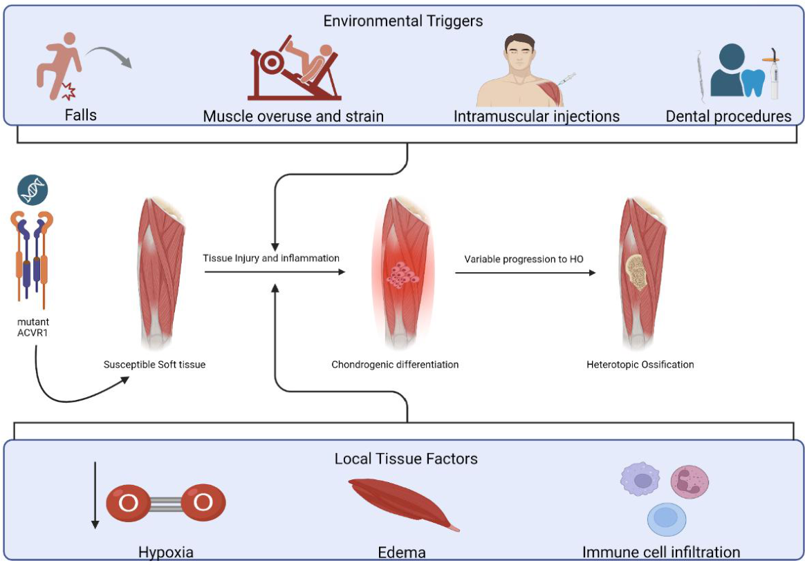
As mentioned previously, the ACVR1R206H mutation forms the genetic basis of FOP. In affected individuals, FAPs respond abnormally to Activin A, activating osteogenic signalling and promoting HO formation through an endochondral process. In contrast, following muscle injury, wild-type tissue progresses toward regeneration of muscle fibres, whereas ACVR1R206H-expressing tissue instead shows accumulation of chondrogenic and fibroblastic cells consistent with progression toward endochondral HO [21]. Thus, the mutation creates an underlying susceptibility to HO, whereas the local tissue environment influences whether this susceptibility is activated and progresses to lesion formation.
Inflammatory cell recruitment represents one component of this local environment. In ACVR1R206H models, macrophages and mast cells accumulate within developing lesions, and depletion of these populations reduces HO formation, indicating that immune-cell activity contributes to lesion development rather than acting as a passive consequence of injury [7]. However, the relationship between local disease activity and HO remains non-deterministic. In a previously conducted natural-history study, only 26.9% of imaged flare-ups developed new HO at the flare-up site by day 84, demonstrating that clinical flare-up activity does not invariably progress to additional ectopic bone formation [23].
Inflammation also influences HO formation through additional immune and tissue-level mechanisms. Toll-like receptors, which recognize damage-associated molecular patterns released following tissue injury, show increased activity during inflammation and can contribute to SMAD1/5/9 signalling, thereby reinforcing the same BMP signalling pathway involved in HO formation [27]. In addition, injured tissues in FOP can become locally hypoxic, which may prolong or intensify chondrogenic differentiation. Hypoxia has been associated with increased SMAD1/5/9 phosphorylation and enhanced BMP signalling in affected tissue. FOP cells exposed to hypoxic conditions also demonstrate greater chondrocyte differentiation and extracellular matrix formation than control cells, further illustrating how changes in the local inflammatory environment can amplify the effects of the underlying genetic mutation [28].
Disease expression also varies with age and anatomical location. Natural-history studies have reported a greater proportion of flare-ups in younger patients, particularly those aged under eight years, with flare-up frequency declining in older age groups [23]. The greater flare-up frequency observed during childhood may reflect heightened skeletal growth and bone turnover [29], greater exposure to falls and other injuries, and additional age-dependent factors; however, the mechanisms underlying this pattern remain unclear. The anatomical distribution of disease activity also changes across age groups, suggesting that age-dependent differences in tissue environment, mechanical exposure, or developmental state may modify how the underlying mutation is expressed clinically [6,23].
The presence of edema was another factor associated with progression from flare-up to HO formation. Flare-up sites presenting with edema were more likely to develop new HO than those without edema [23]. This association further supports the importance of the local tissue response in modifying disease expression, suggesting that the consequences of a flare-up depend not only on the underlying ACVR1 mutation but also on characteristics of the surrounding inflammatory and tissue environment.
Figure 2.
Simplified overview of gene–environment interactions contributing to HO in FOP. Mutant ACVR1 creates the genetic predisposition to HO in susceptible soft tissues. Environmental triggers, such as falls, muscle overuse and strain, intramuscular injections, and dental procedures, are possible initiators of tissue injury and inflammation. Local tissue factors, such as hypoxia, edema, and immune cell infiltration, contribute to a tissue environment that favours chondrogenic differentiation and subsequent HO through endochondral ossification. Progression to HO is variable and may occur without identifiable environmental triggers, referred to as spontaneous HO. Created in BioRender. Chahal, K. (2026) https://BioRender.com/hmy2xji.
6. Clinical Implications
Because trauma and inflammation can initiate HO in genetically susceptible tissue, the clinical management of FOP is exceptionally complicated. In this disease, prevention itself becomes an important component of treatment, as even relatively minor tissue injury may precipitate a flare-up and subsequent HO formation. Thus, patients are often advised to minimize activities associated with substantial risk of falls, blunt trauma, muscle strain, or other soft-tissue injury [30]. Although such precautions may reduce exposure to known environmental triggers, they can also impose significant restrictions on physical activity, recreation, independence, and participation in everyday life.
This challenge extends beyond accidental injury to necessary medical care. Iatrogenic trauma from intramuscular injections, biopsies, surgical procedures, and some dental interventions can provoke local inflammation and potentially initiate new HO formation [2]. As a result, procedures that are routine in the general population may require substantial modification or avoidance in individuals with FOP. Vaccination routes may need to be selected carefully, invasive diagnostic procedures should be minimized where alternatives exist, and dental care may require particular attention because trauma to the jaw and surrounding soft tissues can have severe functional consequences. Surgical excision of established heterotopic bone is also generally problematic, as the operative trauma itself may stimulate recurrent or even more extensive ossification [2,30]. This risk is supported by reports of substantial iatrogenic harm in FOP, with unnecessary invasive procedures following misdiagnosis associated with post-traumatic ossification and permanent loss of mobility in a considerable proportion of patients [13]. In the same cohort, 51% of patients reported complications following diagnostic or therapeutic interventions, while 49% experienced permanent complications, most commonly permanent loss of movement associated with exacerbation of heterotopic bone formation following traumatic interventions [13].
Disease progression further complicates medical management. Ankylosed joints, spinal deformity, and thoracic deformity can limit patient positioning, complicate imaging and venous access, and increase the difficulty of anaesthetic management [31]. Thoracic involvement is particularly important because restriction of the chest wall can impair pulmonary function and contribute to thoracic insufficiency syndrome, while ankylosis of the jaw and cervical spine may create additional challenges for airway management [32]. Given the multisystem complications of FOP, supportive care ideally requires a multidisciplinary care team comprising specialists from relevant fields to minimize treatment-associated complications [30]. Consequently, clinicians must carefully weigh the immediate benefits of an intervention against the possibility of provoking subsequent flare-ups, additional HO formation, or further functional decline.
This creates a clinical paradox in which necessary healthcare may itself act as an environmental trigger and contribute to disease progression. Accordingly, the management of FOP requires not only treatment of existing disease manifestations, but also careful modification of routine medical care to minimize avoidable tissue injury.
7. Therapeutic Strategies
Prior to the development of any FDA-approved therapies for FOP, therapeutic approaches were largely focused on the management of flare-ups and their accompanying symptoms. Glucocorticoids and other corticosteroids were commonly used to address appendicular flare-ups early in their development, usually within the first 24 hours of flare-up development, with the aim of reducing inflammation and edema and thereby alleviating associated symptoms [33]. Nonsteroidal agents, including NSAIDs and COX-2 inhibitors, were also commonly used in FOP but primarily served to manage flare-up-associated pain and inflammation. According to patient self-reports, corticosteroids were useful in reducing swelling, pain, and post-injury flare-ups. However, corticosteroids cannot reliably prevent HO formation following a flare-up [2]. Although they may alleviate the acute inflammatory symptoms and distress associated with flare-ups, they do not correct the underlying pathogenic signalling driven by mutant ACVR1. This limitation created a need for disease-modifying therapies capable of intervening more directly in the molecular and cellular mechanisms responsible for HO formation.
7.1. FDA-Approved Strategies
7.1.1. Palovarotene
Palovarotene, a nuclear retinoic acid receptor-γ (RAR-γ) agonist, was the first FDA-approved therapy for FOP. Its mechanism is based on the inhibition of chondrogenic differentiation, a critical step in the endochondral ossification process that ultimately leads to HO formation in FOP [34]. Under normal ligand-bound conditions, nuclear RARs participate in RAR-mediated transcriptional regulation. However, in their unliganded state, RARs recruit corepressors that suppress the expression of genes opposing chondrogenic differentiation. This repressive state promotes signalling through the PKA and p38 MAPK pathways and increases SOX9 activity, thereby facilitating chondrogenic differentiation. Concurrently, RAR-α expression is downregulated, while RAR-γ becomes concentrated within cartilaginous regions, with both receptors predominantly present in their unliganded states [35]. Later research found that activation of RAR-γ reduced HO more effectively than activation of RAR-α while producing little to no rebound effect following treatment withdrawal, providing the mechanistic basis for palovarotene [34].
Following these mechanistic findings, palovarotene was evaluated in preclinical models of FOP. In mice carrying the ACVR1R206H mutation, palovarotene reduced both injury-induced and spontaneous HO compared with untreated mice [36]. However, an important safety concern emerged during preclinical testing, as non-diseased mice administered palovarotene from the neonatal stage showed impaired skeletal growth [36]. A subsequent study further demonstrated skeletal toxicity associated with palovarotene treatment in wild-type mice [37].
Palovarotene was next evaluated in a phase 2 randomized, placebo-controlled, double-blind trial. Although the trial did not achieve statistical significance for its primary radiographic endpoint, a trend toward reduced HO in treated patients supported progression to a phase 3 clinical trial [38]. Palovarotene was subsequently evaluated in the phase 3 MOVE trial, an open-label study comparing 97 treated patients with 101 untreated participants from a natural-history study. The primary endpoint was annualized new HO volume measured by whole-body CT. Post-hoc analyses showed substantially lower new HO formation with palovarotene, corresponding to an approximately 54–60% reduction in annualized new HO volume compared with the natural-history cohort, although no significant improvement in functional outcomes was observed over the study period [39]. Long-term follow-up of MOVE showed that annualized new HO volume remained numerically lower with palovarotene over 48 months, with a 46.4% reduction relative to untreated natural-history controls, although the difference was not statistically significant (p = 0.0585). Exploratory within-patient analyses during treatment interruptions also suggested lower HO formation while patients were receiving palovarotene [40].
Skeletal toxicity represents a major limitation of palovarotene treatment, particularly in skeletally immature patients. At the 18-month MOVE analysis, premature physeal closure (PPC) or related epiphyseal abnormalities occurred in 21 of 57 patients (36.8%) younger than 14 years. With follow-up extended to 48 months, PPC was identified in 24 of 80 patients (30.0%) younger than 18 years, with all cases occurring in patients younger than 14 years. PPC was most commonly detected at the distal femur and tibia, highlighting the potential for palovarotene to disrupt normal growth-plate development in skeletally immature patients [39,40]. Consequently, palovarotene is restricted to females aged ≥8 years and males aged ≥10 years, reflecting the need to balance reductions in HO formation against potentially irreversible effects on skeletal growth [40].
7.1.2. Garetosmab
As mentioned earlier, the ACVR1R206H mutation causes the receptor to become aberrantly responsive to Activin A, resulting in BMP signalling through the SMAD1/5/9 pathway [7,8]. This mechanism provided another therapeutic target for FOP. In an inducible FOP mouse model, administration of an anti-Activin A antibody prevented HO formation for at least 6 weeks following induction and treatment, whereas HO was evident in control mice by 3 weeks [8]. These findings provided a preclinical rationale for directly targeting Activin A to prevent the aberrant BMP signalling that ultimately drives HO formation through the endochondral ossification process.
Garetosmab, the second FDA-approved therapy for FOP, is a monoclonal antibody that targets Activin A and builds on this therapeutic strategy. The recommended regimen for adults is a 10 mg/kg intravenous infusion administered over 60 minutes every four weeks, with a reduced dose of 3 mg/kg available when the standard dose is not tolerated [41]. LUMINA-1, the phase 2 clinical trial of garetosmab, was a randomized, double-blind, placebo-controlled study involving 44 patients, with 20 receiving garetosmab and 24 receiving placebo during the initial 28-week period, followed by a second 28-week open-label period. Although the primary endpoint of total HO activity did not reach statistical significance, formation of new HO lesions was strongly suppressed in the treatment group. Among patients who crossed over from placebo to garetosmab, the proportion developing new HO lesions decreased from 40.9% during the placebo period to 0% during subsequent garetosmab treatment. Treatment-emergent adverse events were frequent during LUMINA-1, with garetosmab associated with a greater overall adverse-event burden than placebo, particularly epistaxis, madarosis, and skin or soft-tissue infections [42]. During the open-label phases, five patients died. Investigators judged a connection between garetosmab treatment and the deaths to be unlikely, although the drug’s involvement could not be conclusively excluded [42]. Current FDA prescribing information warns of embryo-fetal toxicity, skin and soft tissue infections, and epistaxis, including serious cases requiring medical intervention. Garetosmab is consequently contraindicated during pregnancy [41].
The phase 3 OPTIMA trial further demonstrated substantial suppression of new HO at week 56, with reductions in new HO lesion rates of 94% with the 3 mg/kg dose and 90% with the 10 mg/kg dose. Clinician-assessed flare-ups were also significantly reduced with the 10 mg/kg dose, but not with the 3 mg/kg dose. Garetosmab was generally described as well tolerated, although a dose-dependent increase in skin and soft-tissue infections was observed [43].
Garetosmab’s neutralization of Activin A blocks an important component of the pathway linking environmental triggers, including injuries, to the formation of HO by preventing Activin A from binding mutant ACVR1. Its ability to reduce new HO lesions and clinically assessed flare-ups suggests that it may limit the progression of trigger-initiated inflammatory events into permanent HO. Nonetheless, because the treatment does not eliminate all disease activity and is currently approved only for adults, avoiding preventable trauma and other known triggers remains a key component of FOP management.
7.1.3. Zilurgisertib
ACVR1 kinase inhibitors are designed to suppress signalling from the mutant receptor and abnormal response to Activin A. The recurrent ACVR1R206H mutation occurs in the regulatory GS domain, which controls the activation of the kinase portion involved in SMAD1/5/9 phosphorylation and subsequent activation [44]. Zilurgisertib (Atebrioz) is an oral ACVR1 kinase inhibitor approved by the US Food and Drug Administration (FDA) on September 25, 2026, to reduce total new HO volume in patients 12 years and older. The recommended starting dose is 100 mg orally once daily [45]. In the double-blind, placebo-controlled phase 2 PROGRESS trial, the mean volume of new HO lesions at week 24 was 0.003 cm³ in the zilurgisertib group compared with 6.57 cm³ in the placebo group [46]. The trial’s primary endpoint was the proportion of patients with new HO lesions detected by whole-body CT at week 24. Fewer patients developed new lesions with zilurgisertib than with placebo (1/32 versus 5/30), although the difference did not reach statistical significance (p = 0.0986) [46]. In the open-label period between weeks 24 and 48, the study reported no additional HO lesions in either the group originally assigned zilurgisertib or the group that switched from placebo [46]. FDA prescribing information warns of embryo-fetal toxicity, and zilurgisertib is contraindicated during pregnancy. Clinically important drug interactions may require avoidance, dose adjustment, or monitoring when use together cannot be avoided [47]. By inhibiting mutant ACVR1 kinase activity, zilurgisertib may reduce the downstream signalling through which injury-associated Activin A contributes to HO formation.
7.2. Emerging and Future Therapeutic Strategies
7.2.1. Rapamycin
In addition to SMAD1/5/9 signalling, Activin A acting through mutant ACVR1 increases expression of ENPP2, which contributes to mTORC1 activation and promotes abnormal cartilage differentiation of mesenchymal stromal cells. Rapamycin inhibits mTORC1, targeting this downstream component of HO formation [48]. In FOP patient-derived mesenchymal stromal cells, rapamycin inhibited Activin A-induced chondrogenesis. It also suppressed Activin A-triggered HO in two mouse models [48]. Further in vivo mouse testing revealed rapamycin reduced not only naturally occurring HO, but also injury-induced HO [49]. Together, these preclinical findings suggest that mTORC1 inhibition could reduce HO arising both spontaneously and after injury. Rapamycin subsequently entered a randomized phase II/III trial in Japan to evaluate its efficacy and safety in patients with FOP [50].
7.2.2. AAV Gene Therapy
Recombinant adeno-associated virus (rAAV) vectors have been investigated for three approaches to FOP: increasing wild-type ACVR1 expression, selectively silencing the mutant ACVR1 transcript, and combining both strategies. In FOP-derived induced pluripotent stem cells and skeletal cells from FOP mice, the combined approach reduced Activin A-induced BMP signalling and osteogenic differentiation more than either strategy alone. It also reduced trauma-induced HO in treated mice [51]. A subsequent rAAV strategy targeting both mutant ACVR1 and Activin A expression also reduced aberrant BMP signalling and osteoblast differentiation [52]. By reducing mutant receptor expression and increasing wild-type receptor expression, the combined approach in [51] may weaken the abnormal response to Activin A following an injury or other inflammatory trigger. Increased wild-type ACVR1 could also favour the formation of non-signalling complexes with Activin A and type II receptors, although this contribution to the treatment effect has not been established. Despite these promising findings, the effects of these approaches in patients remain unknown, as they have only been evaluated preclinically.
7.2.3. siRNA
Another RNA-based therapeutic approach uses short interfering RNA (siRNA), which can be designed to selectively suppress mutant ACVR1 expression while preserving wild-type ACVR1. After delivery into cells, the siRNA guide strand directs the RNA-induced silencing complex (RISC) to a complementary mRNA, leading to its cleavage [53]. In cells with induced BMP signalling, treatment with siRNA reduced SMAD1/5/9 signalling to near-normal levels, whereas signalling in control cells was approximately doubled. A similar effect was observed for osteogenic differentiation, with treatment restoring osteogenic marker expression toward levels observed in normal cells [54]. A subsequent study further demonstrated the feasibility of allele-selective siRNA-mediated silencing of mutant ACVR1 while preserving wild-type allele expression [55]. By reducing mutant receptor expression, this approach could weaken the abnormal response to signals following injury or inflammation and potentially limit progression to HO. Whether it suppresses post-injury HO remains to be tested in vivo.
7.2.4. Antisense Oligonucleotides
Another emerging approach to treating FOP involves antisense oligonucleotide (ASO) therapy. ASOs are short, synthetic oligonucleotides that bind specific RNA sequences to alter downstream protein production. This can occur through two mechanisms relevant to FOP: (1) recruitment of RNase H to induce degradation of the target RNA or (2) exon skipping, in which an ASO binds to a selected region of pre-mRNA and alters splicing, causing the spliceosome to exclude the targeted exon from the mature transcript [56].
Shi et al. demonstrated the potential of ASOs for FOP using exon skipping to reduce ACVR1 mRNA expression, BMP signalling, osteogenic differentiation, and mineralization in mouse cells [57]. More recently, Maruyama et al. designed gapmer ASOs that promote RNase H-mediated degradation of mutant ACVR1 mRNA. In patient-derived fibroblasts, the gapmers significantly reduced mutant ACVR1R206H mRNA while largely preserving wild-type allele expression; total ACVR1 protein expression was also reduced. The gapmers also suppressed Activin A-induced osteogenic differentiation [58]. Subsequent incorporation of a single 2’-O-methyl (2’OMe) substitution at gap position 2 improved allele-specificity and increased gapmer levels in skeletal muscle and tendon, while reducing kidney and liver accumulation relative to unmodified gapmers in wild-type mice [59]. By reducing mutant receptor expression, this approach could weaken the response to inflammatory signals after injury. However, further in vivo evaluation in FOP models and eventual clinical testing are needed to establish its therapeutic efficacy and safety.
8. Conclusions
Fibrodysplasia ossificans progressiva is fundamentally a genetic disorder, but its clinical expression is strongly shaped by environmental and tissue factors. The recurrent ACVR1R206H mutation creates susceptibility to aberrant BMP signalling, yet the episodic nature of HO development reflects the additional influence of injury, inflammation, immune-cell recruitment, hypoxia, and changes in the local tissue environment. This interaction between the genetic basis of FOP and environmental factors helps explain why disease progression is highly variable and why minor environmental trauma can have major clinical consequences.
This interaction also has important implications for management and lifestyle. Patients and caregivers must take particular care to avoid preventable tissue injury, which remains central to clinical management, while therapies such as palovarotene, garetosmab, and zilurgisertib target distinct components of the pathogenic pathway. At the same time, emerging gene- and RNA-based strategies, including AAV-mediated gene therapy, siRNA, and antisense oligonucleotides, offer the possibility of targeting the underlying mutation more directly.
Future progress will depend on improving the selectivity, delivery, safety, and durability of these approaches while continuing to define the environmental and cellular factors that determine when and how genetic susceptibility progresses to active HO. Together, these advances may allow FOP treatment to move beyond suppression of disease activity toward increasingly precise therapeutic approaches.
Author Contributions
Conceptualization, K.C., H.M. and T.Y.; writing—original draft preparation, K.C. and H.M.; writing—review and editing, K.C., H.M. and T.Y. All authors have read and agreed to the published version of the manuscript.
Funding
This research received no external funding. T.Y. is supported by the Friends of Garrett Cumming Research Fund, Muscular Dystrophy Canada, the Canadian Institutes of Health Research (CIHR), the Women and Children’s Health Research Institute (WCHRI), the HM Toupin Neurological Science Research Fund, Alberta Innovates: Health Solutions (AIHS), Jesse’s Journey, the U.S. Department of Defense, and the Heart and Stroke Foundation Canada. H.M. is supported by a postdoctoral fellowship from WCHRI.
Institutional Review Board Statement
Not applicable.
Informed Consent Statement
Not applicable.
Data Availability Statement
No new data were created or analyzed in this study. Data sharing is not applicable to this article.
Acknowledgments
We thank the University of Alberta Faculty of Medicine and Dentistry, the University of Alberta Faculty of Graduate and Postdoctoral Studies, the Friends of Garrett Cumming Research Fund, Muscular Dystrophy Canada, the Canadian Institutes of Health Research (CIHR), the Women and Children’s Health Research Institute (WCHRI), the HM Toupin Neurological Science Research Fund, Alberta Innovates: Health Solutions (AIHS), Jesse’s Journey, and the Heart and Stroke Foundation Canada for their support.
Conflicts of Interest
T.Y. is a co-founder and shareholder of OligomicsTx Inc., which aims to commercialize antisense technology. The remaining authors declare that the research was conducted in the absence of any commercial or financial relationships that could be construed as a potential conflict of interest.
References
- De Brasi, D.; Orlando, F.; Gaeta, V.; De Liso, M.; Acquaviva, F.; Martemucci, L.; Mastrominico, A.; Di Rocco, M. Fibrodysplasia Ossificans Progressiva: A Challenging Diagnosis. Genes 2021, 12, 1187. [Google Scholar] [CrossRef]
- Connor, J.M.; Evans, D.A.P. Extra-Articular Ankylosis in Fibrodysplasia Ossificans Progressiva. Br. J. Oral Surg. 1982, 20, 117–121. [Google Scholar] [CrossRef]
- Kaplan, F.S.; Glaser, D.L. Thoracic Insufficiency Syndrome in Patients with Fibrodysplasia Ossificans Progressiva. Clin. Rev. Bone Min. Metab. 2005, 3, 213–216. [Google Scholar] [CrossRef]
- Hüning, I.; Gillessen-Kaesbach, G. Fibrodysplasia Ossificans Progressiva: Clinical Course, Genetic Mutations and Genotype-Phenotype Correlation. Mol. Syndromol. 2014, 5, 201–211. [Google Scholar] [CrossRef]
- Shore, E.M.; Xu, M.; Feldman, G.J.; Fenstermacher, D.A.; Cho, T.-J.; Choi, I.H.; Connor, J.M.; Delai, P.; Glaser, D.L.; LeMerrer, M.; et al. A Recurrent Mutation in the BMP Type I Receptor ACVR1 Causes Inherited and Sporadic Fibrodysplasia Ossificans Progressiva. Nat. Genet 2006, 38, 525–527. [Google Scholar] [CrossRef]
- Pignolo, R.J.; Bedford-Gay, C.; Liljesthröm, M.; Durbin-Johnson, B.P.; Shore, E.M.; Rocke, D.M.; Kaplan, F.S. The Natural History of Flare-Ups in Fibrodysplasia Ossificans Progressiva (FOP): A Comprehensive Global Assessment. J. Bone Min. Res. 2016, 31, 650–656. [Google Scholar] [CrossRef]
- Convente, M.R.; Chakkalakal, S.A.; Yang, E.; Caron, R.J.; Zhang, D.; Kambayashi, T.; Kaplan, F.S.; Shore, E.M. Depletion of Mast Cells and Macrophages Impairs Heterotopic Ossification in an Acvr1R206H Mouse Model of Fibrodysplasia Ossificans Progressiva. J. Bone Min. Res. 2018, 33, 269–282. [Google Scholar] [CrossRef]
- Hatsell, S.J.; Idone, V.; Wolken, D.M.A.; Huang, L.; Kim, H.J.; Wang, L.; Wen, X.; Nannuru, K.C.; Jimenez, J.; Xie, L.; et al. ACVR1R206H Receptor Mutation Causes Fibrodysplasia Ossificans Progressiva by Imparting Responsiveness to Activin A. Sci. Transl. Med. 2015, 7. [Google Scholar] [CrossRef]
- Omi, M.; Kaartinen, V.; Mishina, Y. Activin A Receptor Type 1–Mediated BMP Signaling Regulates RANKL-Induced Osteoclastogenesis via Canonical SMAD-Signaling Pathway. J. Biol. Chem. 2019, 294, 17818–17836. [Google Scholar] [CrossRef]
- Liljesthröm, M.; Pignolo, R.; Kaplan, F. Epidemiology of the Global Fibrodysplasia Ossificans Progressiva (FOP) Community. J. Rare Dis. Res. Treat. 2020, 5, 31–36. [Google Scholar] [CrossRef]
- Pignolo, R.J.; Hsiao, E.C.; Baujat, G.; Lapidus, D.; Sherman, A.; Kaplan, F.S. Prevalence of Fibrodysplasia Ossificans Progressiva (FOP) in the United States: Estimate from Three Treatment Centers and a Patient Organization. Orphanet J. Rare Dis. 2021, 16, 350. [Google Scholar] [CrossRef]
- Kaplan, F.S.; Xu, M.; Seemann, P.; Connor, J.M.; Glaser, D.L.; Carroll, L.; Delai, P.; Fastnacht-Urban, E.; Forman, S.J.; Gillessen-Kaesbach, G.; et al. Classic and Atypical Fibrodysplasia Ossificans Progressiva (FOP) Phenotypes Are Caused by Mutations in the Bone Morphogenetic Protein (BMP) Type I Receptor ACVR1. Hum. Mutat. 2009, 30, 379–390. [Google Scholar] [CrossRef]
- Kitterman, J.A.; Kantanie, S.; Rocke, D.M.; Kaplan, F.S. Iatrogenic Harm Caused by Diagnostic Errors in Fibrodysplasia Ossificans Progressiva. Pediatrics 2005, 116, e654–e661. [Google Scholar] [CrossRef]
- Breskin, A.; Popoola, V.O.; Davis, M.; Kile, S.; Souttou, A.; Rhee, S.; Knox, C. Measures of Mortality among People Living with Fibrodysplasia Ossificans Progressiva in the United States, 2017-2023. JBMR Plus 2026, 10. [Google Scholar] [CrossRef]
- Burdick, L.N.; DelVichio, A.H.; Hanson, L.R.; Griffith, B.B.; Bouchard, K.R.; Hunter, J.W.; Goldhamer, D.J. Sex as a Critical Variable in Basic and Pre-Clinical Studies of Fibrodysplasia Ossificans Progressiva. Biomolecules 2024, 14, 177. [Google Scholar] [CrossRef]
- Payne, T.L.; Postlethwait, J.H.; Yelick, P.C. Functional Characterization and Genetic Mapping of Alk8. Mech. Dev. 2001, 100, 275–289. [Google Scholar] [CrossRef]
- Haupt, J.; Xu, M.; Shore, E.M. Variable Signaling Activity by FOP ACVR1 Mutations. Bone 2018, 109, 232–240. [Google Scholar] [CrossRef]
- Morianos, I.; Papadopoulou, G.; Semitekolou, M.; Xanthou, G. Activin-A in the Regulation of Immunity in Health and Disease. J. Autoimmun. 2019, 104, 102314. [Google Scholar] [CrossRef]
- Aykul, S.; Corpina, R.A.; Goebel, E.J.; Cunanan, C.J.; Dimitriou, A.; Kim, H.J.; Zhang, Q.; Rafique, A.; Leidich, R.; Wang, X.; et al. Activin A Forms a Non-Signaling Complex with ACVR1 and Type II Activin/BMP Receptors via Its Finger 2 Tip Loop. eLife 2020, 9, e54582. [Google Scholar] [CrossRef]
- Yin, W.; Okamoto, K.; Terashima, A.; Pluemsakunthai, W.; Ono, T.; Ito-Kureha, T.; Akira, S.; Hashizume, Y.; Baron, R.; Ueha, S.; et al. Activin A Secretion by Muscle-Repairing Macrophages Induces Heterotopic Ossification in Mice. J. Clin. Investig. 2026, 136, e193797. [Google Scholar] [CrossRef]
- Lees-Shepard, J.B.; Yamamoto, M.; Biswas, A.A.; Stoessel, S.J.; Nicholas, S.-A.E.; Cogswell, C.A.; Devarakonda, P.M.; Schneider, M.J.; Cummins, S.M.; Legendre, N.P.; et al. Activin-Dependent Signaling in Fibro/Adipogenic Progenitors Causes Fibrodysplasia Ossificans Progressiva. Nat. Commun. 2018, 9, 471. [Google Scholar] [CrossRef]
- Kaplan, F.S.; Tabas, J.A.; Gannon, F.H.; Finkel, G.; Hahn, G.V.; Zasloff, M.A. The Histopathology of Fibrodysplasia Ossificans Progressiva. An Endochondral Process. J. Bone Jt. Surg. Am. 1993, 75, 220–230. [Google Scholar] [CrossRef]
- Pignolo, R.J.; Baujat, G.; Brown, M.A.; De Cunto, C.; Hsiao, E.C.; Keen, R.; Al Mukaddam, M.; Le Quan Sang, K.-H.; Wilson, A.; Marino, R.; et al. The Natural History of Fibrodysplasia Ossificans Progressiva: A Prospective, Global 36-Month Study. Genet. Med. 2022, 24, 2422–2433. [Google Scholar] [CrossRef]
- Pignolo, R.J.; Shore, E.M.; Kaplan, F.S. Fibrodysplasia Ossificans Progressiva: Diagnosis, Management, and Therapeutic Horizons. Pediatr. Endocrinol. Rev. 2013, 10, 437–448. [Google Scholar]
- Kaplan, F.S.; Al Mukaddam, M.; Baujat, G.; Hidalgo Bravo, A.; Brown, M.; Cali, A.; Cho, T.-J.; Crowe, C.; De Cunto, C.L.; Delai, P.L.R.; et al. Medical Guidelines for Fibrodysplasia Ossificans Progressiva. JBMR. Plus. 2025, 9, ziaf150. [Google Scholar] [CrossRef]
- Scarlett, R.F.; Rocke, D.; Kantanie, S.; Patel, J.; Shore, E.; Kaplan, F.S. Influenza-like Viral Illnesses and Flare-Ups of Fibrodysplasia Ossificans Progressiva. Clin. Orthop. Relat. Res. 2004, 275–279. [Google Scholar] [CrossRef]
- Wang, H.; Behrens, E.M.; Pignolo, R.J.; Kaplan, F.S. ECSIT Links TLR and BMP Signaling in FOP Connective Tissue Progenitor Cells. Bone 2018, 109, 201–209. [Google Scholar] [CrossRef]
- Wang, H.; Lindborg, C.; Lounev, V.; Kim, J.-H.; McCarrick-Walmsley, R.; Xu, M.; Mangiavini, L.; Groppe, J.C.; Shore, E.M.; Schipani, E.; et al. Cellular Hypoxia Promotes Heterotopic Ossification by Amplifying BMP Signaling. J. Bone Mineral. Res. 2016, 31, 1652–1665. [Google Scholar] [CrossRef]
- Rauchenzauner, M.; Schmid, A.; Heinz-Erian, P.; Kapelari, K.; Falkensammer, G.; Griesmacher, A.; Finkenstedt, G.; Högler, W. Sex- and Age-Specific Reference Curves for Serum Markers of Bone Turnover in Healthy Children from 2 Months to 18 Years. J. Clin. Endocrinol. Metab. 2007, 92, 443–449. [Google Scholar] [CrossRef]
- Akesson, L.S.; Savarirayan, R. Fibrodysplasia Ossificans Progressiva. In GeneReviews® [Internet]; Adam, M.P., Bick, S., Mirzaa, G.M., Eds.; University of Washington, Seattle: Seattle (WA), 2020. [Google Scholar]
- Kaplan, F.S.; Sponseller, P.D.; Pignolo, R.J. Thoracic Deformity in Fibrodysplasia Ossificans Progressiva. JBJS Rev. 2025, 13. [Google Scholar] [CrossRef]
- Butler, M.G.; Hayes, B.G.; Hathaway, M.M.; Begleiter, M.L. Specific Genetic Diseases at Risk for Sedation/Anesthesia Complications. Anesth. Analg. 2000, 91, 837–855. [Google Scholar] [CrossRef]
- Pignolo, R.J.; Shore, E.M.; Kaplan, F.S. Fibrodysplasia Ossificans Progressiva: Clinical and Genetic Aspects. Orphanet J. Rare Dis. 2011, 6, 80. [Google Scholar] [CrossRef]
- Shimono, K.; Tung, W.; Macolino, C.; Chi, A.H.-T.; Didizian, J.H.; Mundy, C.; Chandraratna, R.A.; Mishina, Y.; Enomoto-Iwamoto, M.; Pacifici, M.; et al. Potent Inhibition of Heterotopic Ossification by Nuclear Retinoic Acid Receptor-γ Agonists. Nat. Med. 2011, 17, 454–460. [Google Scholar] [CrossRef]
- Weston, A.D.; Chandraratna, R.A.S.; Torchia, J.; Underhill, T.M. Requirement for RAR-Mediated Gene Repression in Skeletal Progenitor Differentiation. J. Cell Biol. 2002, 158, 39–51. [Google Scholar] [CrossRef]
- Chakkalakal, S.A.; Uchibe, K.; Convente, M.R.; Zhang, D.; Economides, A.N.; Kaplan, F.S.; Pacifici, M.; Iwamoto, M.; Shore, E.M. Palovarotene Inhibits Heterotopic Ossification and Maintains Limb Mobility and Growth in Mice with the Human ACVR1R206H Fibrodysplasia Ossificans Progressiva (FOP) Mutation. J. Bone Min. Res. 2016, 31, 1666–1675. [Google Scholar] [CrossRef]
- Lees-Shepard, J.B.; Nicholas, S.-A.E.; Stoessel, S.J.; Devarakonda, P.M.; Schneider, M.J.; Yamamoto, M.; Goldhamer, D.J. Palovarotene Reduces Heterotopic Ossification in Juvenile FOP Mice but Exhibits Pronounced Skeletal Toxicity. eLife 2018, 7, e40814. [Google Scholar] [CrossRef]
- Pignolo, R.J.; Baujat, G.; Hsiao, E.C.; Keen, R.; Wilson, A.; Packman, J.; Strahs, A.L.; Grogan, D.R.; Kaplan, F.S. Palovarotene for Fibrodysplasia Ossificans Progressiva (FOP): Results of a Randomized, Placebo-Controlled, Double-Blind Phase 2 Trial. J. Bone Min. Res. 2022, 37, 1891–1902. [Google Scholar] [CrossRef]
- Pignolo, R.J.; Hsiao, E.C.; Al Mukaddam, M.; Baujat, G.; Berglund, S.K.; Brown, M.A.; Cheung, A.M.; De Cunto, C.; Delai, P.; Haga, N.; et al. Reduction of New Heterotopic Ossification (HO) in the Open-Label, Phase 3 MOVE Trial of Palovarotene for Fibrodysplasia Ossificans Progressiva (FOP). J. Bone Mineral. Res. 2023, 38, 381–394. [Google Scholar] [CrossRef]
- Pignolo, R.J.; Hsiao, E.C.; Al Mukaddam, M.; Baujat, G.; Berglund, S.K.; Cheung, A.M.; De Cunto, C.; Delai, P.; Haga, N.; Kannu, P.; et al. Palovarotene Impact on Fibrodysplasia Ossificans Progressiva (FOP): Data from Month 48 of the Phase 3 MOVE Trial. JBMR. Plus. 2026, ziag141. [Google Scholar] [CrossRef]
- U.S. Food and Drug Administration. PASATRU (Garetosmab-Grts) Injection, for Intravenous Use: Prescribing Information; U.S. Food and Drug Administration, 2026. [Google Scholar]
- Di Rocco, M.; Forleo-Neto, E.; Pignolo, R.J.; Keen, R.; Orcel, P.; Funck-Brentano, T.; Roux, C.; Kolta, S.; Madeo, A.; Bubbear, J.S.; et al. Garetosmab in Fibrodysplasia Ossificans Progressiva: A Randomized, Double-Blind, Placebo-Controlled Phase 2 Trial. Nat. Med. 2023, 29, 2615–2624. [Google Scholar] [CrossRef]
- Papa, R.; Rhee, S.; Delai, P.; Zhang, K.; Balcázar, I.B.; Rethanavelu, K.; Fujiwara, T.; Mishima, K.; Tabarkiewicz, J.; Funck-Brentano, T.; et al. Efficacy and Safety of Garetosmab, an Activin A-Blocking Antibody, in Fibrodysplasia Ossificans Progressiva (OPTIMA): A Randomised, Double-Blind, Placebo-Controlled, Phase 3 Trial. The Lancet 2026. [Google Scholar] [CrossRef]
- Allen, R.S.; Jones, W.D.; Hale, M.; Warder, B.N.; Shore, E.M.; Mullins, M.C. Reduced GS Domain Serine/Threonine Requirements of Fibrodysplasia Ossificans Progressiva Mutant Type I BMP Receptor ACVR1 in the Zebrafish. J. Bone Min. Res. 2023, 38, 1364–1385. [Google Scholar] [CrossRef]
- Center for Drug Evaluation and Research FDA Approves Third Treatment for Fibrodysplasia Ossificans Progressiva. Available online: https://www.fda.gov/drugs/news-events-human-drugs/fda-approves-third-treatment-fibrodysplasia-ossificans-progressiva (accessed on 25 September 2026).
- ENDO 2026. Available online: https://endo2026.eventscribe.net/fsPopup.asp?PresentationID=1846470 (accessed on 25 September 2026).
- U.S. Food and Drug Administration. ATEBRIOZ (Zilurgisertib) Tablets, for Oral Use: Prescribing Information; U.S. Food and Drug Administration, 2026. [Google Scholar]
- Hino, K.; Horigome, K.; Nishio, M.; Komura, S.; Nagata, S.; Zhao, C.; Jin, Y.; Kawakami, K.; Yamada, Y.; Ohta, A.; et al. Activin-A Enhances mTOR Signaling to Promote Aberrant Chondrogenesis in Fibrodysplasia Ossificans Progressiva. J. Clin. Invest 2017, 127, 3339–3352. [Google Scholar] [CrossRef]
- Maekawa, H.; Kawai, S.; Nishio, M.; Nagata, S.; Jin, Y.; Yoshitomi, H.; Matsuda, S.; Toguchida, J. Prophylactic Treatment of Rapamycin Ameliorates Naturally Developing and Episode -Induced Heterotopic Ossification in Mice Expressing Human Mutant ACVR1. Orphanet J. Rare Dis. 2020, 15, 122. [Google Scholar] [CrossRef]
- Multicenter Randomized Double-Blind Comparison Test Followed by Open-Label Continuous Administration Test of NPC-12T for Fibrodysplasia Ossificans Progressiva. Available online: https://center6.umin.ac.jp/cgi-open-bin/ctr_e/ctr_view.cgi?recptno=R000032495 (accessed on 27 September 2026).
- Yang, Y.-S.; Kim, J.-M.; Xie, J.; Chaugule, S.; Lin, C.; Ma, H.; Hsiao, E.; Hong, J.; Chun, H.; Shore, E.M.; et al. Suppression of Heterotopic Ossification in Fibrodysplasia Ossificans Progressiva Using AAV Gene Delivery. Nat. Commun. 2022, 13, 6175. [Google Scholar] [CrossRef]
- Yang, Y.-S.; Lin, C.; Ma, H.; Xie, J.; Kaplan, F.S.; Gao, G.; Shim, J.-H. AAV-Mediated Targeting of the Activin A-ACVR1R206H Signaling in Fibrodysplasia Ossificans Progressiva. Biomolecules 2023, 13, 1364. [Google Scholar] [CrossRef]
- Deerberg, A.; Willkomm, S.; Restle, T. Minimal Mechanistic Model of siRNA-Dependent Target RNA Slicing by Recombinant Human Argonaute 2 Protein. Proc. Natl. Acad. Sci. U S A 2013, 110, 17850–17855. [Google Scholar] [CrossRef]
- Kaplan, J.; Kaplan, F.S.; Shore, E.M. Restoration of Normal BMP Signaling Levels and Osteogenic Differentiation in FOP Mesenchymal Progenitor Cells by Mutant Allele-Specific Targeting. Gene Ther. 2012, 19, 786–790. [Google Scholar] [CrossRef]
- Takahashi, M.; Katagiri, T.; Furuya, H.; Hohjoh, H. Disease-Causing Allele-Specific Silencing against the ALK2 Mutants, R206H and G356D, in Fibrodysplasia Ossificans Progressiva. Gene Ther. 2012, 19, 781–785. [Google Scholar] [CrossRef]
- Lauffer, M.C.; van Roon-Mom, W.; Aartsma-Rus, A. Possibilities and Limitations of Antisense Oligonucleotide Therapies for the Treatment of Monogenic Disorders. Commun. Med. 2024, 4, 6. [Google Scholar] [CrossRef]
- Shi, S.; Cai, J.; de Gorter, D.J.J.; Sanchez-Duffhues, G.; Kemaladewi, D.U.; Hoogaars, W.M.H.; Aartsma-Rus, A.; Hoen, P.A.C. ’t; Dijke, P. ten Antisense-Oligonucleotide Mediated Exon Skipping in Activin-Receptor-Like Kinase 2: Inhibiting the Receptor That Is Overactive in Fibrodysplasia Ossificans Progressiva. PLoS ONE 2013, 8, e69096. [Google Scholar] [CrossRef]
- Maruyama, R.; Nguyen, Q.; Roshmi, R.R.; Touznik, A.; Yokota, T. Allele-Selective LNA Gapmers for the Treatment of Fibrodysplasia Ossificans Progressiva Knock Down the Pathogenic ACVR1R206H Transcript and Inhibit Osteogenic Differentiation. Nucleic Acid. Ther. 2022, 32, 185–193. [Google Scholar] [CrossRef]
- Anwar, S.; Hay, S.; Moriyama, H.; Mir, F.; Maruyama, R.; Yokota, T. Single-Base 2′OMe-Modified LNA and MOE Gapmers Selectively Silence ACVR1R206H in Fibrodysplasia Ossificans Progressiva. Mol. Ther. Nucleic Acids 2026, 37, 102937. [Google Scholar] [CrossRef]

Table 1.
Selected ACVR1 variants, receptor domains, and associated FOP phenotypes.
| ACVR1 Variant | Receptor Domain | Associated Phenotype |
| p.R206H | GS regulatory domain | Classic FOP: congenital great-toe malformation and early-onset progressive HO [12] |
| p.Q207E | GS regulatory domain | Classic great-toe malformation and early-onset HO, accompanied by severe growth restriction [12] |
| p.G328R | Protein kinase domain | Normal or minimal great-toe malformation and late-onset HO in patients [12] |
| p.G328E | Protein kinase domain | Childhood-onset HO with underdeveloped digits, missing toenails, scalp hair thinning, and mild cognitive impairment [12] |
| p.G328W | Protein kinase domain | Marked digit shortening or underdevelopment, absent toenails, scalp hair thinning, and mild cognitive impairments, along with childhood-onset HO [12] |
| p.G356D | Protein kinase domain | Variable HO onset and congenital great-toe and thumb abnormalities, such as absent or underdeveloped digits [12] |
| p.R375P | Protein kinase domain | Normal great toes, teenage-onset flare-ups, and slow, progressive HO, leading to limited movement by age 40 [12] |
Disclaimer/Publisher’s Note: The statements, opinions and data contained in all publications are solely those of the individual author(s) and contributor(s) and not of MDPI and/or the editor(s). MDPI and/or the editor(s) disclaim responsibility for any injury to people or property resulting from any ideas, methods, instructions or products referred to in the content. |
© 2026 by the authors. Licensee MDPI, Basel, Switzerland. This article is an open access article distributed under the terms and conditions of the Creative Commons Attribution (CC BY) license.
Copyright: This open access article is published under a Creative Commons CC BY 4.0 license, which permit the free download, distribution, and reuse, provided that the author and preprint are cited in any reuse.