

Submitted:
15 October 2026
Posted:
16 October 2026
You are already at the latest version
Abstract
Rotavirus A (RVA) is an important enteric pathogen of neonatal animals, whereas the genomic diversity of donkey-origin RVA remains incompletely defined. In 2025, an RVA strain was recovered from a diarrheic donkey foal in Shandong Province, China, and designated RVA/Donkey-wt/CHN/SD2503/2025/G3P[59] (SD2503). Viral RNA was subjected to Illumina NovaSeq paired-end sequencing, de novo assembly, genotype assignment, sequence comparison, and segment-specific maximum-likelihood phylogenetic analysis. All 11 genome segments were recovered, yielding the constellation G3-P[59]-I3-R3-C3-M3-A9-N3-T3-E3-H6. The VP4 gene was highly divergent: the closest BLASTn match was the P[24] rhesus strain TUCH, with 79.58% nucleotide identity, below the 80% threshold used to assign VP4 sequences to the same P genotype. The VP4 sequence was subsequently confirmed by the Rotavirus Classification Working Group as P[59]. Other segments showed discordant relationships. VP7 clustered with rabbit strain rab1404 with strong bootstrap support, whereas several internal genes grouped within genotype backgrounds represented by simian, rabbit, pangolin, equine, and other mammalian RVAs. The topological incongruence across segments, together with the unusual genotype constellation, is consistent with a complex reassortant evolutionary history involving cross-species-associated RVA lineages. SD2503 provides an 11-segment genomic framework for an independently identified donkey G3P[59] RVA and supports broader genomic surveillance of RVA in equids.
Keywords:
rotavirus A
; donkey
; G3P[59]
; P[59]
; whole-genome sequencing
; reassortment
; phylogeny
; cross-species evolution
1. Introduction
Rotavirus A (RVA) is a segmented double-stranded RNA virus and a major cause of acute gastroenteritis in the young of humans and many mammalian species [1,2,3,4]. Its 11 genome segments encode six structural proteins (VP1–VP4, VP6 and VP7) and five or six nonstructural proteins (NSP1–NSP5/6). The segmented genome provides repeated opportunities for reassortment when genetically distinct RVAs infect the same host, and this process is a major contributor to rotavirus diversity and host-associated evolution [2,3,4,5,6].
A whole-genome nomenclature is therefore used for RVA. Genotypes are assigned in the order Gx-P[x]-Ix-Rx-Cx-Mx-Ax-Nx-Tx-Ex-Hx, corresponding to VP7, VP4, VP6, VP1, VP2, VP3 and NSP1–NSP5, respectively [5,6]. This approach is particularly informative for unusual animal viruses because identical G/P combinations can occur on different internal-gene backgrounds. For VP4, an 80% nucleotide identity cutoff is used for assignment to the same P genotype [5,6].
Equine RVA is an important cause of diarrhea in foals, and G3P[12] and G14P[12] account for most well-characterized equine strains worldwide [7,8,9,10,11,12,13]. Nevertheless, atypical equine RVAs provide evidence that equids can acquire viruses or genome segments associated with other mammalian hosts. The Argentine strain E3198 possessed the constellation G3-P[3]-I3-R3-C3-M3-A9-N3-T3-E3-H6 and was related to simian and feline/canine-like RVAs [10]. Indian equine G3P[3] strains have also been shown to possess bat-associated genomic backgrounds [14].
Information on RVA infection in donkeys is limited. A severe enteritis outbreak in donkey foals in Shandong Province in 2019 was associated with RVA/Donkey-wt/CHN/Don01/2019/G3P[12]; its VP7 gene was atypical and related to bat-associated G3 viruses, whereas VP4 was equine-like P[12] [15]. A later Chinese donkey strain, HB01/China/2021/G3P[12], was also placed near atypical G3 lineages in VP7 analyses [16]. During preparation of this manuscript, another donkey RVA, HB2503, isolated in Hebei Province in 2025, was reported as G3P[59]. That study stated that P[59] had been confirmed by the Rotavirus Classification Working Group (RCWG), but detailed nucleotide data for HB2503 were not publicly available for direct whole-genome comparison [17].
Here, we characterize an independently identified G3P[59] RVA recovered from a diarrheic donkey foal in Shandong Province in 2025. We determined its complete 11-segment genotype constellation and compared the evolutionary placement of each segment to test whether SD2503 represents a conventional equine-like genome carrying an unusual VP4 gene or a more complex reassortant RVA assembled from cross-species-associated genomic lineages.
2. Materials and Methods
2.1. Sample Origin and Virus Isolation
A fecal swab was collected in 2025 from a 2-day-old donkey foal with diarrhea in Shandong Province, China. The swab material was suspended in sterile phosphate-buffered saline containing antibiotics and clarified by centrifugation. The supernatant was passed through a 0.22 μm membrane. The filtrate was mixed with an equal volume of Dulbecco’s modified Eagle medium (DMEM) containing 10 μg/mL crystalline trypsin and incubated at 37 °C for 60 min. Activated material was inoculated onto confluent Madin–Darby bovine kidney (MDBK) cell monolayers and allowed to adsorb for 1 h at 37 °C. Monolayers were then washed three times with serum-free DMEM and maintained in trypsin-supplemented DMEM. Cultures were examined daily for cytopathic changes. Blind passages were performed every 2–3 days after three freeze–thaw cycles, for up to three passages. Viral identity was confirmed by RT-PCR [18] and electron microscopy.
2.2. Electron Microscopy
Virus-containing material was examined by transmission electron microscopy (TEM) using a negative-staining procedure. Briefly, 20 μL of the virus suspension was applied to a 200-mesh carbon-coated copper grid and allowed to adsorb for 3–5 min. Excess liquid was removed with filter paper, after which the grid was negatively stained with 2% phosphotungstic acid for 1–2 min. Excess stain was removed with filter paper, and the grid was allowed to dry at room temperature. The prepared grids were examined using a Hitachi HT7800 transmission electron microscope, and representative viral particles were imaged.
2.3. RNA Library Preparation and Whole-Genome Sequencing
Viral RNA was processed for next-generation sequencing. According to the sequencing provider’s workflow, fragmented RNA was subjected to random reverse transcription and second-strand synthesis, followed by library construction. Libraries were sequenced on an Illumina NovaSeq 6000 platform using 150-bp paired-end reads. The run generated 12,805,186 reads before filtering and 12,680,940 reads after quality control, corresponding to 1,815,186,829 retained bases. The post-filter Q20 and Q30 rates were 97.20% and 93.51%, respectively, and the mean retained read length was 143 bp.
2.4. Read Processing, Assembly and Genome Annotation
Raw reads were quality filtered and adapter-trimmed using fastp [19]. Ribosomal RNA, host-derived and bacterial reads were removed by the provider’s BBMap-based filtering workflow. De novo assembly was performed using SPAdes and SOAPdenovo-based procedures [20]. Virus-associated contigs were identified by nucleotide database comparison. Coding regions were annotated using Prokka/Prodigal assisted procedures and manually checked against RVA references [21,22]. The overall metagenomic assembly contained non-RVA contigs and was therefore not interpreted as the length of a single viral genome. Eleven contigs corresponding to VP1–VP4, VP6, VP7 and NSP1–NSP5 were retained for SD2503 genome analysis.
2.5. Genotype Assignment and Sequence Comparison
Segment genotypes were assigned according to the RCWG whole-genome classification framework [5,6]. BLASTn searches were used to identify the closest available nucleotide matches. The VP4 sequence showed <80% nucleotide identity to the closest previously recognized P genotype in the available BLAST analysis and was submitted to the RCWG. It was confirmed as genotype P[59]. The nucleotide sequences of the 11 SD2503 genome segments were deposited in GenBank under accession numbers QB141057–QB141067.
2.6. Phylogenetic Analysis
Reference sequences were selected to represent the closest database matches, relevant equine and donkey strains, and informative RVA lineages from other mammalian hosts. Nucleotide sequences were aligned using MAFFT v7 [23]. Maximum-likelihood phylogenetic analyses were performed in MEGA12 [24]. For each segment, the best-fitting nucleotide substitution model was identified using the maximum-likelihood model-selection procedure and the Bayesian information criterion. Trees were re-constructed using the selected model with 1,000 standard bootstrap replicates. All codon positions and noncoding positions present in the alignments were retained, and gaps/missing data were handled using the 'use all sites' option. Bootstrap percentages ≥70 are displayed. SD2503 is indicated by a black diamond (◆). Because different reference sets were required for different segments, branch placement was interpreted segment by segment rather than as evidence of direct ancestry.
2.7. Use of Generative Artificial Intelligence
During manuscript preparation, ChatGPT (GPT-5.6 Sol, OpenAI) was used to assist with language editing, organization of author-provided information, and formatting. The tool was not used to generate experimental data or figures or to perform sequence or phylogenetic analyses. All scientific content and interpretations were reviewed and verified by the authors.
3. Results
3.1. Isolation and Morphological Identification of SD2503
Following inoculation of processed fecal material onto MDBK cells and serial passage, RVA was recovered and designated RVA/Donkey-wt/CHN/SD2503/2025/G3P[59] (SD2503). RT-PCR confirmed RVA in the culture material. Transmission electron microscopy revealed a rotavirus-like particle with the expected wheel-like morphology (Figure 1).
3.2. Sequencing Output and Genome Organization
The sequencing dataset retained 12.68 million reads after quality filtering, with Q20 and Q30 rates of 97.20% and 93.51%, respectively. Eleven RVA segments were recovered. Segment lengths ranged from 711 nt (NSP5) to 3,280 nt (VP1), and the predicted proteins ranged from 159 amino acids (NSP4) to 1,081 amino acids (VP1). The genome was assigned the constellation G3-P[59]-I3-R3-C3-M3-A9-N3-T3-E3-H6 (Table 1).
3.3. Divergent VP4 and Confirmation of P[59]
VP4 was the most divergent segment of SD2503. Its closest hit in the available BLASTn analysis was the P[24] rhesus RVA strain TUCH (FJ816611.1), with 79.582% nucleotide identity across a 2,346-nt alignment. This value was below the 80% nucleotide identity threshold used to assign VP4 sequences to the same P genotype [5,6]. The VP4 sequence was subsequently reviewed by the RCWG and confirmed as P[59]. In the broad VP4 maximum-likelihood tree containing representatives of recognized P genotypes, SD2503 formed a distinct branch rather than clustering within the conventional equine P[12] group (Figure 2).
3.4. VP7 Phylogeny Places SD2503 with an Atypical G3 Lineage
The VP7 segment was assigned to G3 but did not group with the principal conventional equine G3P[12] cluster. Instead, SD2503 formed a sister relationship with rabbit strain rab1404, supported by a bootstrap value of 96% (Figure 3). This topology agreed with the BLASTn result, in which rab1404 was the closest VP7 match at 95.45% nucleotide identity. Donkey strains Don01/2019 and HB01/2021 grouped elsewhere with bat-associated and atypical equine G3 viruses, emphasizing the diversity of G3 VP7 lineages detected in equids.
3.5. Internal-Gene Phylogenies Reveal Topological Incongruence
The nine internal-gene trees showed that SD2503 did not follow a single host-associated phylogenetic pattern (Figure 4). VP1 (R3) was placed near rabbit N5 and pangolin-associated R3 references; VP2 (C3) was positioned within a C3 lineage containing rabbit and simian representatives; and VP3 (M3) formed a strongly supported local grouping with rab1404 within a broader M3 assemblage. VP6 was assigned to I3 and remained distinct from the I1, I2, I6 and I17 reference groups represented in the selected dataset. NSP1 (A9) fell within an A9 assemblage containing rabbit and rhesus/simian viruses. NSP2 (N3) grouped with pangolin-associated N3 sequences, whereas NSP3 (T3) was embedded within a broad T3 lineage containing simian, rabbit, equine and pangolin-associated viruses. NSP4 was placed within E3, separate from the E2 cluster containing conventional equine strain 03V04954, and NSP5 (H6) grouped near rhesus/simian H6 viruses. These segment-specific placements are incongruent with one another and with the conventional equine G3P[12] genomic background.
3.6. Genotype Constellation Indicates a Distinct Equid RVA Background
The complete constellation of SD2503 was G3-P[59]-I3-R3-C3-M3-A9-N3-T3-E3-H6. This differs markedly from the common equine G3P[12] constellation, typically G3-P[12]-I6-R2-C2-M3-A10-N2-T3-E2-H7 [14,15]. At the genotype level, ten segments match the unusual equine E3198 background (G3-P[3]-I3-R3-C3-M3-A9-N3-T3-E3-H6), with VP4 representing the principal genotype difference [14]. However, the individual SD2503 trees do not consistently place every segment immediately with E3198 or with any single reference strain. The genome is therefore best interpreted as belonging to a broader cross-species-associated RVA gene pool rather than as a direct derivative of one previously sampled virus.
4. Discussion
This study provides whole-genome characterization of an independently identified donkey RVA carrying the newly recognized P[59] genotype. Three observations are central to the interpretation of SD2503. First, VP4 is sufficiently divergent from established P genotypes to cross the formal sequence boundary used in RVA classification, and its P[59] designation has been confirmed by the RCWG. Second, the remaining ten segments form an unusual I3-R3-C3-M3-A9-N3-T3-E3-H6 internal background rather than the genome constellation commonly associated with equine G3P[12] viruses. Third, the 11 segment trees are topologically incongruent, linking SD2503 to different cross-species-associated RVA lineages depending on the segment examined. Together, these features support a complex reassortant evolutionary history.
The VP4 finding is important because P-genotype diversity may be missed when surveillance is restricted to common equine G/P combinations. The 79.58% identity between SD2503 VP4 and P[24] TUCH lies below the 80% same-genotype cutoff established in the whole-genome classification framework [5,6]. In the broad VP4 tree, SD2503 remained distinct from P[12] equine viruses and from the P[24] reference cluster. The tree is not used here to redefine the genotype, because the formal P[59] assignment rests on RCWG confirmation; rather, it provides evolutionary context for the marked divergence of the sequence.
The VP7 tree adds a second unusual feature. SD2503 grouped with rabbit strain rab1404 with 96% bootstrap support. Rab1404 itself has a mosaic G3-P[22]-I2-R3-C3-M3-A9-N2-T3-E3-H3 genome and was interpreted as the product of multiple reassortment events involving animal RVA lineages [25]. This relationship should not be read as evidence that rabbits directly transmitted the virus to donkeys. Rather, it indicates that closely related G3 genes have circulated in a wider mammalian RVA network that also includes bat-, simian-, feline/canine- and equine-associated viruses [14,25,26,27,28].
The internal genes reinforce this interpretation. Several SD2503 segments occupy genotype groups represented by simian RRV-like, rabbit, pangolin-associated or atypical equine viruses, whereas VP6 is I3 and distinct from the I6 background of conventional equine G3P[12] strains. The overall genotype constellation is especially notable because it is identical to E3198 at ten genotype positions [14]. E3198 and Indian bat-associated equine G3P[3] strains illustrate that equids can harbor RVA genomes assembled from lineages that are not typical of the dominant equine G3P[12]/G14P[12] population [14,15]. Nevertheless, genotype identity does not establish direct ancestry. The absence of consistent nearest-neighbor relationships across the SD2503 trees argues against describing SD2503 simply as an E3198 derivative with a replaced VP4 segment.
Donkey RVA data remain sparse. Don01/2019 demonstrated that severe enteritis in donkey foals can be associated with a G3P[12] virus whose outer-capsid genes have discordant evolutionary affinities [15]. HB01/2021 further linked donkey G3 viruses with atypical equine/bat-related VP7 diversity [16]. The recent report of HB2503 as another G3P[59] donkey RVA is epidemiologically noteworthy [17], but direct genomic comparison is currently limited by the lack of publicly available nucleotide sequences in that report. Consequently, the present data support independent detection of P[59] in a Shandong donkey RVA but do not establish the geographic origin, direction of spread, or prevalence of this genotype.
Reassortment and interspecies transmission are well documented in RVA evolution [2,3,26,27,28,29,30,31]. Simian RRV and TUCH themselves have complex genome constellations [27], and feline/canine-like, bat, rabbit and equine RVAs provide examples in which individual segments trace to different host-associated lineages [14,15,25,26,27,28,29,31]. SD2503 fits this broader evolutionary pattern. The safest interpretation is therefore that its genome reflects historical reassortment among cross-species-associated RVA lineages. Demonstrating a specific transmission event or donor host would require denser contemporaneous sampling and complete genomes from candidate reservoir populations.
This study has limitations. Only one SD2503 isolate was characterized, so no inference can be made about the prevalence of P[59] in donkeys. Reference sequences are unevenly distributed across host species and geographic regions, and some relevant strains are represented only by partial sequences. Despite these constraints, the combination of RCWG-confirmed VP4 typing and 11-segment phylogenetic analysis provides a robust framework for recognizing SD2503 as a genetically unusual donkey RVA.
5. Conclusions
SD2503 is an independently identified donkey RVA with the genotype constellation G3-P[59]-I3-R3-C3-M3-A9-N3-T3-E3-H6. Its VP4 gene belongs to the newly recognized P[59] genotype, while its remaining genome segments show discordant relationships to several atypical mammalian RVA lineages. The 11-segment genome therefore supports a complex reassortant evolutionary history rather than a conventional equine G3P[12] background. Expanded whole-genome surveillance in donkeys, horses, wildlife and other sympatric mammals will be needed to determine the distribution of P[59] and clarify the evolutionary pathways that generated this constellation.
Author Contributions
Conceptualization, J.D. and T.H.; methodology, Q.L., K.Y., R.L., L.G., M.M., Y.X. and W.C.; software, Q.L., K.Y., M.W. and Z.H.; validation, M.W. and X.L.; formal analysis, Q.L. and J.M.; investigation, Q.L., K.Y. and X.L.; resources, X.L. and J.D.; data curation, J.D.; writing—original draft preparation, Q.L. and K.Y.; writing—review and editing, Q.L., L.G. and J.D.; visualization, Q.L. and X.L.; supervision, T.H. and J.D.; project administration, J.D.; funding acquisition, J.D. All authors have read and agreed to the published version of the manuscript.
Funding
This research was funded by the Key R&D Program of Shandong Province, China (Project No. 2025LZGC033), the National Key R&D Program of China (Project No. 2025YFD1800304), the Shandong Provincial Modern Agricultural Industry Technology System (Project No. SDAIT-27).
Institutional Review Board Statement
Not applicable.
Informed Consent Statement
Not applicable.
Data Availability Statement
The nucleotide sequences of the 11 genome segments of RVA/Donkey-wt/CHN/SD2503/2025/G3P[59] generated in this study have been deposited in GenBank under accession numbers QB141057–QB141067.
Acknowledgments
During manuscript preparation, a generative AI tool (ChatGPT-5.6 Sol ) was used by Q.L. to assist with language editing, organization of author-provided information, and formatting. All scientific interpretations, data, figures, references, and final wording were reviewed and verified by the authors, who take full responsibility for the content of the manuscript.
Conflicts of Interest
The authors declare no conflicts of interest.
References
- Estes, M.K.; Greenberg, H.B. Rotaviruses. In Fields Virology, 6th ed.; Knipe, D.M., Howley, P.M., Eds.; Lippincott Williams & Wilkins: Philadelphia, PA, USA, 2013; pp. 1347–1401. [Google Scholar]
- Martella, V.; Bányai, K.; Matthijnssens, J.; Buonavoglia, C.; Ciarlet, M. Zoonotic aspects of rotaviruses. Vet. Microbiol. 2010, 140, 246–255. [Google Scholar] [CrossRef] [PubMed]
- Patton, J.T. Rotavirus diversity and evolution in the post-vaccine world. Discov. Med. 2012, 13, 85–97. [Google Scholar] [PubMed]
- Pesavento, J.B.; Crawford, S.E.; Estes, M.K.; Prasad, B.V.V. Rotavirus proteins: structure and assembly. Curr. Top. Microbiol. Immunol. 2006, 309, 189–219. [Google Scholar] [CrossRef] [PubMed]
- Matthijnssens, J.; Ciarlet, M.; Heiman, E. Full genome-based classification of rotaviruses reveals a common origin between human Wa-like and porcine rotavirus strains and human DS-1-like and bovine rotavirus strains. J. Virol. 2008, 82, 3204–3219. [Google Scholar] [CrossRef] [PubMed]
- Matthijnssens, J.; Ciarlet, M.; Rahman, M. Recommendations for the classification of group A rotaviruses using all 11 genomic RNA segments. Arch. Virol. 2008, 153, 1621–1629. [Google Scholar] [CrossRef] [PubMed]
- Browning, G.F.; Begg, A.P. Prevalence of G and P serotypes among equine rotaviruses in the faeces of diarrhoeic foals. Arch. Virol. 1996, 141, 1077–1089. [Google Scholar] [CrossRef] [PubMed]
- Fukai, K.; Saito, T.; Fukuda, O.; Hagiwara, A.; Inoue, K.; Sato, M. Molecular characterisation of equine group A rotavirus, Nasuno, isolated in Tochigi Prefecture, Japan. Vet. J. 2006, 172, 369–373. [Google Scholar] [CrossRef] [PubMed]
- Garaicoechea, L.; Miño, S.; Ciarlet, M.; Fernández, F.; Barrandeguy, M.; Parreño, V. Molecular characterization of equine rotaviruses circulating in Argentinean foals during a 17-year surveillance period (1992–2008). Vet. Microbiol. 2011, 148, 150–160. [Google Scholar] [CrossRef] [PubMed]
- Miño, S.; Matthijnssens, J.; Badaracco, A. Equine G3P[3] rotavirus strain E3198 related to simian RRV and feline/canine-like rotaviruses based on complete genome analyses. Vet. Microbiol. 2013, 161, 239–246. [Google Scholar] [CrossRef] [PubMed]
- Nemoto, M.; Matsumura, T. Equine rotavirus infection. J. Equine Sci. 2021, 32, 1–9. [Google Scholar] [CrossRef]
- Nemoto, M.; Tsunemitsu, H.; Imagawa, H. Molecular characterization and analysis of equine rotavirus circulating in Japan from 2003 to 2008. Vet. Microbiol. 2011, 152, 67–73. [Google Scholar] [CrossRef] [PubMed]
- Papp, H.; Matthijnssens, J.; Martella, V.; Ciarlet, M.; Bányai, K. Global distribution of group A rotavirus strains in horses: a systematic review. Vaccine 2013, 31, 5627–5633. [Google Scholar] [CrossRef] [PubMed]
- Pathak, A.; Gulati, B.R.; Maan, S.; Mor, S.; Kumar, D.; Soman, R.; Punia, S.; Chaudhary, D.; Khurana, S.K. Complete genome sequencing reveals unusual equine rotavirus A of bat origin from India. J. Virol. 2022, 96, e01408-01422. [Google Scholar] [CrossRef] [PubMed]
- Dong, J.; Liu, G.; Gao, N. A reassortant G3P[12] rotavirus A strain associated with severe enteritis in donkeys (Equus asinus). Equine Vet. J. 2022, 54, 114–120. [Google Scholar] [CrossRef] [PubMed]
- Uprety, T.; Soni, S.; Sreenivasan, C.; Hause, B.M.; Naveed, A.; Ni, S.; Graves, A.J.; Morrow, J.K.; Meade, N.; Mellits, K.H.; et al. Genetic and antigenic characterization of two diarrhoeic-dominant rotavirus A genotypes G3P[12] and G14P[12] circulating in the global equine population. J. Gen. Virol. 2024, 105, 002016. [Google Scholar] [CrossRef] [PubMed]
- Zhang, Z.; Guo, X.; Guo, K.; Guo, W.; Du, C.; Yang, Y.; Dong, W.; Chu, X.; Lin, Y.; Wang, X. Development and validation of an antigen-capture ELISA for quantitative detection of donkey-origin rotavirus: application in vaccine manufacturing process monitoring. Microorganisms 2026, 14, 1874. [Google Scholar] [CrossRef] [PubMed]
- Zhu, W.; Dong, J.; Haga, T.; Goto, Y.; Sueyoshi, M. Rapid and sensitive detection of bovine coronavirus and group A bovine rotavirus from fecal samples by using one-step duplex RT-PCR assay. J. Vet. Med. Sci. 2011, 73, 531–534. [Google Scholar] [CrossRef] [PubMed]
- Chen, S.; Zhou, Y.; Chen, Y.; Gu, J. fastp: an ultra-fast all-in-one FASTQ preprocessor. Bioinformatics 2018, 34, i884–i890. [Google Scholar] [CrossRef] [PubMed]
- Bankevich, A.; Nurk, S.; Antipov, D. SPAdes: a new genome assembly algorithm and its applications to single-cell sequencing. J. Comput. Biol. 2012, 19, 455–477. [Google Scholar] [CrossRef] [PubMed]
- Hyatt, D.; Chen, G.L.; LoCascio, P.F.; Land, M.L.; Larimer, F.W.; Hauser, L.J. Prodigal: prokaryotic gene recognition and translation initiation site identification. BMC Bioinform. 2010, 11, 119. [Google Scholar] [CrossRef] [PubMed]
- Seemann, T. Prokka: rapid prokaryotic genome annotation. Bioinformatics 2014, 30, 2068–2069. [Google Scholar] [CrossRef] [PubMed]
- Katoh, K.; Standley, D.M. MAFFT multiple sequence alignment software version 7: improvements in performance and usability. Mol. Biol. Evol. 2013, 30, 772–780. [Google Scholar] [CrossRef] [PubMed]
- Kumar, S.; Stecher, G.; Suleski, M.; Sanderford, M.; Sharma, S.; Tamura, K. MEGA12: Molecular Evolutionary Genetic Analysis Version 12 for Adaptive and Green Computing. Mol. Biol. Evol. 2024, 41, msae263. [Google Scholar] [CrossRef] [PubMed]
- Matthijnssens, J.; Taraporewala, Z.F.; Yang, H. Simian rotaviruses possess divergent gene constellations that originated from interspecies transmission and reassortment. J. Virol. 2010, 84, 2013–2026. [Google Scholar] [CrossRef] [PubMed]
- Matthijnssens, J.; De Grazia, S.; Piessens, J. Multiple reassortment and interspecies transmission events contribute to the diversity of feline, canine and feline/canine-like human group A rotavirus strains. Infect. Genet. Evol. 2011, 11, 1396–1406. [Google Scholar] [CrossRef] [PubMed]
- He, B.; Yang, F.; Yang, W. Characterization of a novel G3P[3] rotavirus isolated from a lesser horseshoe bat: a distant relative of feline/canine rotaviruses. J. Virol. 2013, 87, 12357–12366. [Google Scholar] [CrossRef] [PubMed]
- Xia, L.; Fan, Q.; He, B. The complete genome sequence of a G3P[10] Chinese bat rotavirus suggests multiple bat rotavirus inter-host species transmission events. Infect. Genet. Evol. 2014, 28, 1–4. [Google Scholar] [CrossRef] [PubMed]
- Ghosh, S.; Shintani, T.; Kobayashi, N. Evidence for the porcine origin of equine rotavirus strain H-1. Vet. Microbiol. 2012, 158, 410–414. [Google Scholar] [CrossRef] [PubMed]
- Gillespie, J.; Kalica, A.; Conner, M.; Schiff, E.; Barr, M.; Holmes, D.; Frey, M. The isolation, propagation and characterization of tissue-cultured equine rotaviruses. Vet. Microbiol. 1984, 9, 1–14. [Google Scholar] [CrossRef] [PubMed]
- Nakagomi, O.; Nakagomi, T. Genetic diversity and similarity among mammalian rotaviruses in relation to interspecies transmission of rotavirus. Arch. Virol. 1991, 120, 43–55. [Google Scholar] [CrossRef] [PubMed]
Figure 1.
Representative transmission electron micrograph of the SD2503 virus preparation. A rotavirus-like particle is visible in the field. Scale bar, 100 nm.
Figure 1.
Representative transmission electron micrograph of the SD2503 virus preparation. A rotavirus-like particle is visible in the field. Scale bar, 100 nm.
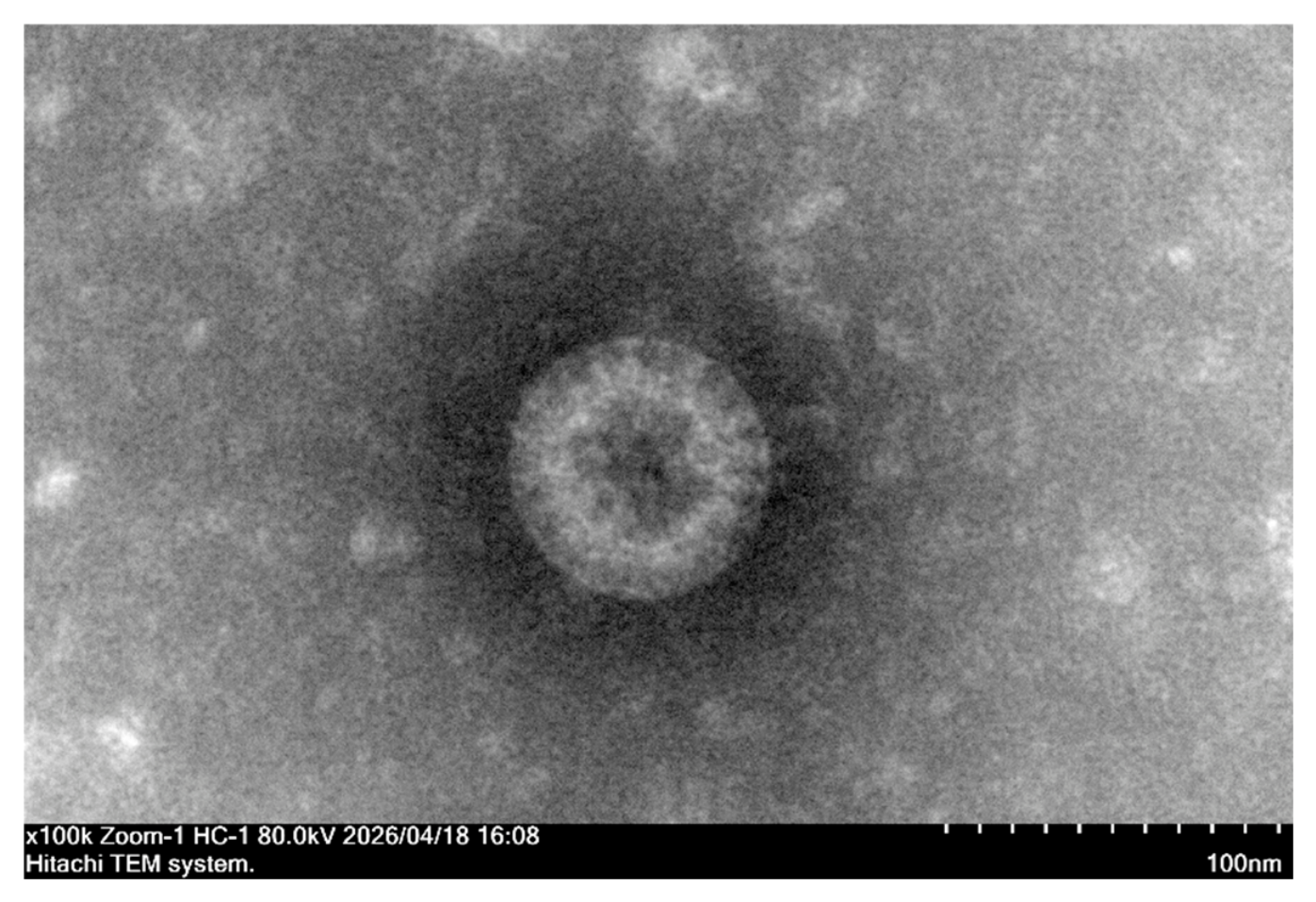
Figure 2.
Maximum-likelihood phylogeny of VP4. Representative RVA P genotypes were included to place the RCWG-confirmed P[59] sequence in a broad genotype context. SD2503 is marked by a black diamond (◆). Bootstrap values ≥70% are shown. The tree was reconstructed in MEGA12 using the BIC-selected model and 1,000 standard bootstrap replicates.
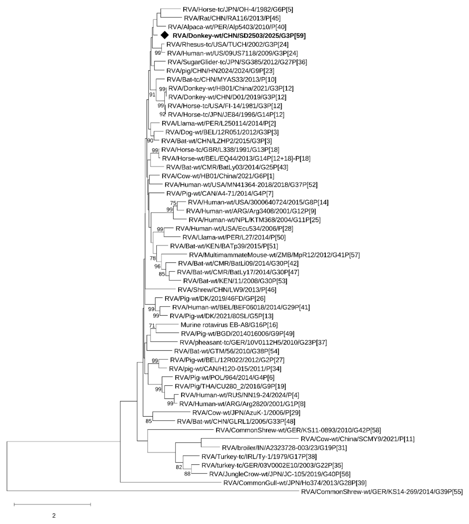
Figure 3.
Maximum-likelihood phylogeny of VP7. SD2503 (◆) clustered with rabbit strain rab1404 with 96% bootstrap support. Bootstrap values ≥70% are shown. The tree was reconstructed in MEGA12 under the BIC-selected model with 1,000 standard bootstrap replicates.
Figure 3.
Maximum-likelihood phylogeny of VP7. SD2503 (◆) clustered with rabbit strain rab1404 with 96% bootstrap support. Bootstrap values ≥70% are shown. The tree was reconstructed in MEGA12 under the BIC-selected model with 1,000 standard bootstrap replicates.
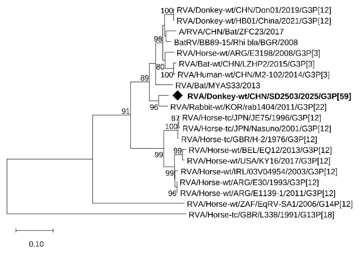
Figure 4.
Maximum-likelihood phylogenies of the nine internal genome segments of SD2503: (a) VP1; (b) VP2; (c) VP3; (d) VP6; (e) NSP1; (f) NSP2; (g) NSP3; (h) NSP4; and (i) NSP5. SD2503 is indicated by a black diamond (◆). Bootstrap values ≥70% are shown. Each segment was analyzed independently in MEGA12 using its BIC-selected nucleotide substitution model and 1,000 standard bootstrap replicates. Scale bars represent substitutions per site and differ among panels.
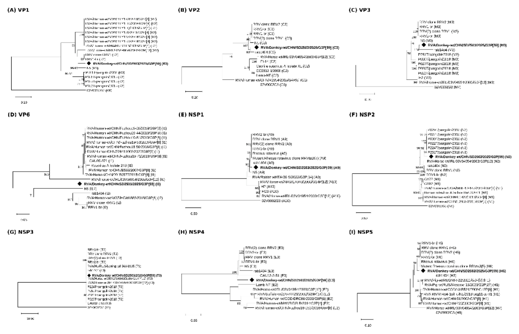

Table 1.
Genome segments of SD2503 and their closest BLASTn matches in the supplied whole-genome analysis.
Table 1.
Genome segments of SD2503 and their closest BLASTn matches in the supplied whole-genome analysis.
| Segment | Genotype | Length (nt) | Protein (aa) | Closest match | Accession | Identity (%) |
| VP1 | R3 | 3280 | 1081 | 2012841174/2012, human G8P[14] | KJ411432 | 93.415 |
| VP2 | C3 | 2703 | 887 | MS2015-1-0001, human G3P[10] | LC576609 | 96.707 |
| VP3 | M3 | 2583 | 844 | HB01/China/2021, donkey G3P[12] | ON377049 | 94.949 |
| VP4 | P[59] | 2355 | 776 | TUCH/2002, rhesus G3P[24] | FJ816611 | 79.582 |
| VP6 | I3 | 1413 | 391 | IAL-R566/2011, human G12P[9] | PP025572 | 89.788 |
| VP7 | G3 | 1055 | 297 | rab1404/2011, rabbit G3P[22] | MK751437 | 95.450 |
| NSP1 | A9 | 1624 | 492 | Rotavirus A type 1 NSP1 | OP963640 | 95.722 |
| NSP2 | N3 | 1105 | 288 | E3198/2008, horse G3P[3] | JX036372 | 93.732 |
| NSP3 | T3 | 1132 | 310 | RRV/Rotashield, simian | HQ846849 | 95.005 |
| NSP4 | E3 | 788 | 159 | K8/1977, human G1P[9] | JQ713651 | 95.393 |
| NSP5 | H6 | 711 | 198 | E3198/2008, horse G3P[3] | JX036375 | 98.628 |
Disclaimer/Publisher’s Note: The statements, opinions and data contained in all publications are solely those of the individual author(s) and contributor(s) and not of MDPI and/or the editor(s). MDPI and/or the editor(s) disclaim responsibility for any injury to people or property resulting from any ideas, methods, instructions or products referred to in the content. |
© 2026 by the authors. Licensee MDPI, Basel, Switzerland. This article is an open access article distributed under the terms and conditions of the Creative Commons Attribution (CC BY) license (http://creativecommons.org/licenses/by/4.0/).
Copyright: This open access article is published under a Creative Commons CC BY 4.0 license, which permit the free download, distribution, and reuse, provided that the author and preprint are cited in any reuse.