
Submitted:
14 October 2026
Posted:
16 October 2026
You are already at the latest version
Abstract
Background: Fascia has been overlooked throughout most of the history of anatomy, but many recent studies have begun to showcase its role. The ventral thigh has been extensively studied in various fields, including anatomy, biomechanics, physiotherapy, histology, and orthopedics. Therefore, the fascia lata is a good model for synthesizing the recent knowledge accumulated from studying this organ. Methods: A review was constructed to describe the characteristics of the deep fascia, with articles highlighting the main roles of the fascia lata selected for this purpose. Results: Forces travel through the fascia lata according to knee and hip positioning, optimizing energy sparing. When the knee is extended, the forces are transmitted to the iliotibial band whereas, when the knee is flexed, the forces are transmitted to the patella. Additionally, the fascia lata is a propri-oceptive organ. Its lengthening during a stretch is felt as pain while the mechanical tension is felt as a feeling of tension. The fascia lata can be lengthened and maybe reinforced by stretching training. Moreover, it is likely that its stiffness can be responsible for a few of the main running pathologies. Conclusions: The fascia lata may provide an insight into the major roles of the fascial tissue. This literature review put in relief a few gaps in fascia histology and bridging these lacunes would be profitable to better understand the physiology underlying its proprioceptive role. Finally, a better understanding of fascia physiology may improve clinical reasoning in various medical specialties.
Keywords:
fascia lata
; deep fascia
; stretching
; fascia proprioception
; iliotibial band
1. Introduction
Fascia used to be overlooked when studying anatomy [1,2], often considered as a packing organ but recent research, in many fields, has changed our vision of this organ. Fascia appears to play a major role in packing organs, in forces transmission [2,3] but, also, as a proprioceptive organ [4,5] with contractile properties [6,7]. Nevertheless, very few studies have examined the fascia as a proprioceptive organ and only 23 articles were found to describe fascia innervation [4]. Although fascia force transmission, and fascia proprioceptive role, are poorly known the front thigh constitutes an exception.
Indeed, the fascia lata and the rectus femoris have been widely studied in the last decade advancing our understanding of their physiology. Fascia lata histological structure is made of three layers and its biomechanical role is described accurately in many articles [8,9,10,11,12,13,14,15]. The fascia lata is widely used in surgical reconstructions [15]. Moreover, it was shown that the sensory regulation played a major role in limiting the stretch magnitude and intensity [16]. It was also proven that a bigger fascia lata taut surface equals a higher stretching intensity tolerance by one individual [12].
Therefore, the first goal of this review was to identify the force travel within the fascia lata and how this travel is influenced by the knee and the hip positioning. The second goal was to make hypotheses about the sensors at play and to examine the findings implications at the histologic level. A third goal was to examine how the fascia lata tensional network may adapt to the stretch and finally how it can be implicated in common pathologies such as iliotibial band syndrome or patellofemoral syndrome.
Due to the wide range of scientific fields involved, a systematic review was impossible to conduct. Thus, peer-reviewed articles were selected from various databases, such as Google Scholar, PubMed, and ScienceDirect, as well as other editors’ datasets. Various combinations of words such as “fascia,” “fascia lata,” “rectus femoris,” “quadriceps,” “fascia innervation,” “proprioceptors,” “Ruffini,” “Pacini,” “free nerve endings,” “stretching,” “sensory regulation,” “fascial sheet,” “fascia histology,” “patella travel,” and “iliofemoral band syndrome” were submitted to various database search engines to find the most relevant articles over a period of 6 months. These scientific studies explored the functional anatomy of the fascia lata, but readers must keep in mind that although there is abundant literature addressing the biomechanics of the fascia lata ex vivo, patellar tracking, and iliotibial band syndrome, the literature addressing the proprioception, innervation, and biomechanics of the fascia lata in vivo remains scarce. Nevertheless, fascia lata is one of the most studied fascia in the body due to its importance in medicine.
In this topical review, first, we show that despite its fineness, the fascia lata is a key structure in guiding the patella. To this regard, it is noteworthy that the fascia lata permits a shift of the passive forces travel according to the knee and the hip positionings authorizing to spare the locomotion energy. Then, we show how the fascia lata may be considered as a proprioceptive organ as its sensitive sensors, which are not yet identified, indicate precisely where the fascia is stretched. To this respect, the links between various sensors and the collagen network must be explored by histologists. Further, we show how the fascia can eventually be reinforced and lengthened by a mechanical tension applied on a regular basis. This adaptation to the tensile stress shows the dynamic nature of this tissue. In ending this review, we highlight the role of the fascia lata in daily clinical practice. Patello-femoral knee pain, iliotibial band syndrome or hip extension restriction illustrates how fascia lata excessive stiffness may impair human locomotion quality.
2. Fascia Lata Functional Anatomy and Force Transmission
One of the most remarkable functions of the fascia lata is to distribute the forces transmitted by its collagenic network to the tuberculum tractus iliotibialis or to the patella according to hip and knee positions (Figure 1). The tensor fascia lata is the only muscle which can tense the fascia lata and Schwartz et Al [17] used electromyograms to study its function. In their 1981 article, they wrote: “during walking the anteromedial fibers were electromyographically silent, whereas the posterolateral fibers were active near heel-strike. For all subjects, increased locomotor velocity (during jogging, running, and sprinting) was associated both with increased activity of the anteromedial fibers near toe-off apparently to assist deceleration of extension and acceleration of flexion of the thigh, and with increased activity of the posterolateral fibers at heel-strike”. The tensor fascia lata contraction pattern makes sense when the force travel within the fascia lata is considered. A recent review, by Hutchinson et Al. [18] explored the literature on the subject and most of the articles agreed on the primary role of the fascia lata. Its function was determined to be stabilizing the knee and maintaining the pelvis horizontal during the gait cycle (Figure 1 and Figure 0°). Additionally the literature suggested that the fascia lata plays a role in conserving energy during the gait cycle [13]. However, the role of the tensor fascia lata remained unclear when the knee was flexed. The fact that the anterior portion of the tensor fascia lata contracts during the knee flexion was subsequently elucidated when the coordinates of the stretched structure were calculated using the inverted tendon travel method and when the actual fascia lata force reaction was measured in vivo [11]. The present article demonstrated that the rotational torque at hip level could only be mediated by the fascia lata. Furthermore, a linear correlation between hip flexion and knee flexion was identified suggesting that an anatomical structure transmits constrains between the hip and the patella. The force travel calculated in [11] are represented Figure 1. The anatomical support for force transmission between the fascia lata and the patella was remarkably described by Terry et Al. [19]. The authors distinguee two functional components which are the ilio-patellar band and the iliotibial tract. Each of these two components transmit forces to the tibia or the patella according to knee and hip positioning. Another facet of force transmission was highlighted by the comparison between two mathematical models. The first one assumed that the force reaction to the stretch depended on the rectus femoris moment arm (In the field of biomechanics, the moment arm is defined as the shortest perpendicular distance between the axis of rotation of a joint and the line of action of a force) while the second one assumed that the force reaction to the stretch was dependent on the fascia taut surfaces. The experimental results were in accordance with the dependance on the taut fascia surface hypothesis [12] and a biomechanical assessment of local strains, measured directly from the thigh fascia showed similar results [20]. A recent experiment based on shear wave elastography demonstrated that stretching exerts its effects in the more lengthened area of the fascia, contingent to the knee and hip positioning [21]. This observation suggests the preeminent role of friction or viscosity in constraining diffusion through the fascial and muscular tissue. Furthermore, it underscores the complexity of the interactions between the different fascia and muscular layers. A comprehensive review of extant results suggests that the force travel and the surface involved presented Figure 1 are the more probable.
In addition to the information contained in traditional anatomy texts, regarding the fascia lata, a comprehensive understanding of its histological structure is necessary for an overview of its function [8,9,10,22]. Remarkably, the fascia lata is made of three layers of collagen fibers orientated mainly longitudinally (about 30%) but also obliquely and transversally [9] (Figure 2A). Another notable aspect of the fascia lata is its high elastic modulus (264.3 ± 90.3 MPa for men and 166.3 ± 94.7 MPa for women). These values may seem high considering its finesse but the total cross sectional area of the fascia lata is very close to the cross sectional area of the Achillean tendon (Figure 2C) [9]. From a physical perspective, the fascia lata can be represented as a wide flat elastic (Figure 2A) positioned with the best possible moment arm around the thigh (Figure 2B). Therefore, the fascia lata can store and release energy and this capacity is very close from the tendon’s elastic behavior (from 500 MPa). Szotek et Al [22] did manage to measure the average elastic modulus values which ranged between ca. 63–83 MPa for the longitudinal fascia lata specimens and between ca. 8–20 MPa for the transverse fascia lata specimens. They took pictures of collagen fibers associated with a few elastin fibers within a collagenic network organized with parallel fibers in each fascia lata layer. However, the most interesting fact reported by the authors was the pattern of collagen fiber ruptures observed after tensile stress tests. The ruptures were localized to specific areas of the fascial sheet, while the surrounding tissue remained intact (Figure 2D). This observation suggests heterogeneity in fiber tension within the tissue and the existence of “weak areas,” which were photographed by Szotek et al. [22]. Interestingly, this heterogeneity in the mechanical response of collagen fibers can likely be reduced chronically after 12 ten-minute stretching training sessions, as evidenced by a study conducted on high-level rugby players. [16]. Bonaldi et Al. [10] analyzed the viscoelastic behavior of fascia lata in vitro and demonstrated that the tissue could relax within a few seconds (from 6 to 10 s) for the short relaxation time and up to one hundred seconds for the long relaxation time. Observations of the fascia lata relaxation behavior were made in vivo during the process of accommodating an applied mechanical stress [23]. Studies have shown that repetitive stretching regimens can increase the rate at which passive fibers are recruited. [16]. Thus, the fascia lata is an efficient, adaptable, spring-like organ. Its peripheral positioning around the thigh provides the structure with the best possible moment arm to restitute the stretch energy (Figure 2B). The fascia’s relative fineness is compensated for by the large surface area that reacts to stretching.
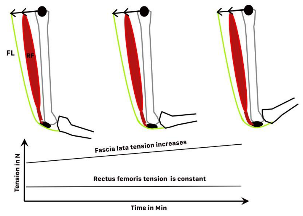
Historically, quadriceps stretching was considered to be a stretch of the rectus femoris [24]. However, recent research has revealed that the fascia lata plays a key role in this particular stretch associating hip extension to knee flexion. The preliminary finding was revealed through the use of ultrasonography which demonstrated that the fascia lata was stretched in a combination of knee flexion and hip extension [25]. Consequently, the tensor fascia lata was identified as a knee extensor when the knee is flexed, with forces traveling from the tensor fascia lata to the patella through the fascia lata (Figure 1). A secondary unexpected outcome emerged when two myo-elastography experiments demonstrated that the tension within the rectus femoris was not significantly modified by quadriceps stretching [26,27]. These results suggested that “the quadriceps stretching” has no visible effects on the rectus femoris. This component of quadriceps stretching has been corroborated by a mechanical analysis that entailed a reaction forces study and a calculation of the coordinates of the stretched structure [11]. The neutral role of the rectus femoris could be observed directly during a 10-minute stretch on one subject who had a complete tear of this left rectus femoris [11]. In this case, the tension developed by the rectus femoris remained constant despite the 25 mm fascia lata lengthening (Figure 3). Moreover, the stretch tolerance appeared to be more dependent on the fascia lata’s taut surfaces than on the rectus femoris moment arm [12]. This finding is consistent with another experiment where the participants were asked to point out the stretched area [11]. The experiment demonstrated that the centroids of the stretched areas, as identified by the participants, were located on a narrow anterior surface of the fascia lata. This finding was in accordance with the location predicted by the mathematical model (Figure 1). Taken together, these results reinforce the hypothesis that the fascia profundis plays a prominent role in the mechanism of stretching [3]. Finally, for the quadriceps stretching, the stretch tolerance seems to mainly depend on the fascia lata taut surfaces [12] and the fascia lata seems to be the only structure adapting to the stretching [11] as the rectus femoris’ viscoelasticity seems unaltered by the stretch (Figure 3) [11,26]. Interestingly, neither the tensor fascia lata nor the iliotibial band stiffness were modified by long quadriceps static stretching while hip extension increased following the intervention [28]. This experiment, by demonstrating that these two anatomical structures were not notably stretched by quadriceps stretching support the view that the most lateral fibers of the fascia lata get relaxed during quadriceps stretching as hypothesized by Germain and Perrin (Figure 1) [12].
Of course, when the knee and the hip are extended, the tensor fascia lata associated to the tractus iliotibial keeps the pelvis horizontal during the gait cycle (Figure 1) [13,17,18]. In fact, the fascia lata seems to be an organ specialized in sparing energy during bipodal locomotion whatever the degree of knee bending (Figure 1). This was demonstrated by Eng et Al [13] who could show that human fascia lata was far more efficient than chimpanzee fascia lata to spare energy while walking. From an evolutionary perspective, the possession of a low-volume muscle, such as the tensor fascia lata, which serves to regulate the tension of the fascia lata [15], can be regarded as a highly efficient form of adaptation. This low-volume muscle functions with low ATP expenditure to tense a super-efficient spring-like structure. Another study conducted by anatomists revealed that the force applied by the gluteus maximums could be transmitted directly to the fascia lata [29] and therefore to the patella when hip flexion is associated to knee flexion.
3. Evidence of Fascia Lata Proprioceptive and Sensory Role
Almost less is known when it comes to analyze the anatomical support for fascia proprioception. Consequently, a better comprehension of fascia physiology is hampered by the lack of knowledge about the anatomical support of the fascia proprioception. Nevertheless, a recent review listed fascia lata innervation at the macroscopic level [15]: the ilioingual nerve (L1), femoral branch of the genitofemoral nerve (L1–L2), lateral femoral cutaneous nerve (L2–L3) and cutaneous branch of the obturator nerve (L2–L4). It is also notable that a review [4] listed the nervous system involved in fascial tissue and described the main mechanoreceptors, or sensors. For the upper limbs, free nerves endings dominate followed by Pacini corpuscles and Ruffini corpuscles in about equal amounts [5]. Golgi tendon organs are also noticed close to the myofascial junctions [30]. The fascia lata innervation may be fairly similar to the other fascia but we did not find any anatomical studies targeting it.
However, the front thigh has the advantage of being a very simple biomechanical model that can be used to study the sensory afferences used by individuals to manage stretching magnitude and intensity. The fact that the fascia lata seems to be the only structure to adapt to the stretch during quadriceps stretching [11,12] facilitates the sensory afferences examination. Physiologists stressed the point that sensory end point should be used when studying stretching [31]. These sensory endpoints can be sheared in two categories: some of them represent the mechanical physical tension (resistance, stretch, tightness, stiffness) while others represent the sensation of pain (pain, discomfort). As far as fascia lata is concerned, an experiment was carried out to analyze the relationship between the sensations of tension felt by individuals and the level of mechanical tension using a principal component analysis [16]. The same analysis was conducted for the sensation of pain and the physical lengthening. The experimental results showed that the sensation of tension reflected the physical tension recorded while the sensation of pain was proportional to the measured lengthening. Considering these results, it can be hypothesized that the tension sensors are mechanically linked to the collagen fibers while the lengthening sensors are likely to be sensible of the fibers creep. The interesting point is that none of these sensors seem to be sensible to the phenomenon of accommodation as the values recorded before and after 12 10-minutes stretching sessions were the same [16]. In fact, from a physical perspective, the same number of fibers were recruited but the pattern of fibers recruitment was modified [16]. From an evolutionary perspective, this result makes sense, as training does not change the mechanical resistance of each collagen fiber. Anatomists and histologists will need to identify the relationship between each sensor (Ruffini, Pacini, and free nerve endings) and the collagen network to improve our understanding of stretching physiology at the tissue level. Nevertheless, a physiological study of the hand proprioceptive afferences suggest that the sensors responsible for the feeling of tension are Ruffini corpuscles [32] but this information is still hypothetical at the fascia lata level. It was also noted that long intense stretches could downregulate the underlying muscle “resting activity” [33] but the anatomical pathway between the fascia and the motoneuron is still unidentified. In summary, the tension and lengthening sensors would prevent fascia strain injuries.
Once we acknowledge that the fascia is a proprioceptive organ, the following question arises: Is the sensory feedback from the fascia lata precise? Only two recent studies can be used to answer this question. The first is a biomechanical assessment comparing the coordinates of the stretched front thigh, the reaction force of the front thigh, and the areas that participants felt were stretched. [11]. The authors concluded that the area perceived as stretched by the participant corresponded with the area calculated from the force reactions and force travel within the fascia lata. However, some unexpected results emerged from this experiment because the areas perceived as stretched were twice as big when the fascia’s taut surfaces were supposed to be smaller. In fact, the mathematical model describing the tension of the longitudinal fibers [12] suggested that the stretched area was smaller when the knee was more flexed (Figure 1). Additionally, it was observed that the participants’ designated fascia stretched areas were distributed in accordance with the model (Figure 1). This suggests that the stretched fascia surfaces are randomly distributed on the taut fascia lata surfaces and that there are variations between participants. The surfaces that are perceived as stretched are twice as large at 120° knee flexion as at 90° knee flexion, which may be due to more fibers overcoming the “alarm” threshold when a smaller surface bears the constraints. Finally, it was demonstrated that the taut area indicated by a participant could differ at 120° and 90°, suggesting that the fascial sheet was taut in different ways depending on the degree of knee flexion.
4. Dynamic Adaptation of the Fascia Lata and Histological Gaps
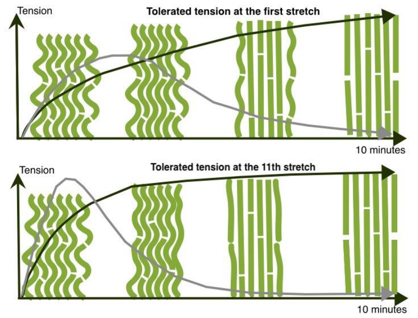
The fascia lata is specialized in energy storage and economy of bipodal locomotion [13]. Interestingly, it has been shown recently, that this dense collagenous tissue presents some adaptative capacity to repeated mechanical strain. In fact, prolonged intense stretching can extend and strengthened it [16,34] but the physical mechanisms underlying this reinforcement is not fully understood yet. It has been hypothesized that repeated stretching leads to tissue reorganization, improving its ability to resist tensile stress—similar to the structural adaptations observed in hydrogels [35] (Figure 4). Gels physics [35,36] authorizes some tissue adaptations to the main applied strain on short and long-time scales as observed in vivo [16,23]. The fascia lata dynamic adaptation to the stretch could be successfully modelized by a simple model involving two Maxwell bodies and an elastic recruitment element [37]. The elastic recruitment element mimics the fibers recruitment represented at two different timescales Figure 4 and Figure 6. The experimental results do show that the relaxation of the fascia lata in vivo (1-3 s) is faster than the relaxation reported by Bonaldi et Al in vitro (6 s) [10]. This suggests that the temperature of the fascia in vivo favorizes the mechanical stress absorption by the tissue. The fiber’s recruitment pattern looks the same at all time scales [16,23,37] suggesting that the changes observed after 12 stretches results from small changes in fibers positioning within each fascia layers. It is also noteworthy that the fascia lata seems to memorize the ideal fiber’s positioning to bear the applied constrains at all time scale considered from a few seconds to a few months [16,23,34,37]. From the physicist point of view the fascia lata appears as a hierarchical, hydrated composite whose macroscopic response emerges from the coupled effects of collagen fiber recruitment, fast collagen–glycosaminoglycan dissipation, and slow osmotic fluid redistribution but the comparison with hydrogels must be taken with caution. As local observations are not available yet for the fascia and as hydrated collagen composites are more complex than hydrogels this information must be considered as a plausible hypothesis at this stage.
Figure 4.
The regulation of a short stretch seems to be governed by two sensations: tension and pain. Straight lines represent taut collagen fibers and wavy lines represent slack fibers. The tension sensors (+ when activated and - when at rest) have not been identified, but they appear to be linked to collagen fibers and do not seem to show accommodation. Stretching regulation seems to be the result of an interplay between tension sensor activation (1), which stops the stretch after an elastic phase (1) and allows the tissue to relax (2) until the subject feels ready to stretch again (3). Then, the cycle can start again (4), recruiting more fibers (5, 6). It is likely that stretch tolerance increases with the recruitment of peripheral fibers, but the nature of this recruitment is not fully understood. Pain seems to increase regularly as the fibers creep. This may prevent tissue injury, but the nerves responsible for this sensation have not yet been identified. Sketch inspired by Germain and Regis. [23].
Figure 4.
The regulation of a short stretch seems to be governed by two sensations: tension and pain. Straight lines represent taut collagen fibers and wavy lines represent slack fibers. The tension sensors (+ when activated and - when at rest) have not been identified, but they appear to be linked to collagen fibers and do not seem to show accommodation. Stretching regulation seems to be the result of an interplay between tension sensor activation (1), which stops the stretch after an elastic phase (1) and allows the tissue to relax (2) until the subject feels ready to stretch again (3). Then, the cycle can start again (4), recruiting more fibers (5, 6). It is likely that stretch tolerance increases with the recruitment of peripheral fibers, but the nature of this recruitment is not fully understood. Pain seems to increase regularly as the fibers creep. This may prevent tissue injury, but the nerves responsible for this sensation have not yet been identified. Sketch inspired by Germain and Regis. [23].
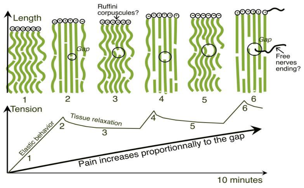
Figure 5.
The effects of repeated stretches on hydrogels. When disorganized fibers are stretched regularly, they tend to orient themselves in the main strain direction. These changes have two main effects. The first one is geometrical, and the alignment of the fibers lengthens the tissue. The second one is physical, and an increase of the stretch modulus results from the fibers alignment. Whereas the elastic modulus of a disorganized fibers network is low, as very few fibers are orientated to resist to the stretches, the elastic modulus of an aligned fibers network is high, as all the fibers react together. These combined two effects extend and strengthen the fibers network. Sketch inspired from Lin et al. [28].
Figure 5.
The effects of repeated stretches on hydrogels. When disorganized fibers are stretched regularly, they tend to orient themselves in the main strain direction. These changes have two main effects. The first one is geometrical, and the alignment of the fibers lengthens the tissue. The second one is physical, and an increase of the stretch modulus results from the fibers alignment. Whereas the elastic modulus of a disorganized fibers network is low, as very few fibers are orientated to resist to the stretches, the elastic modulus of an aligned fibers network is high, as all the fibers react together. These combined two effects extend and strengthen the fibers network. Sketch inspired from Lin et al. [28].
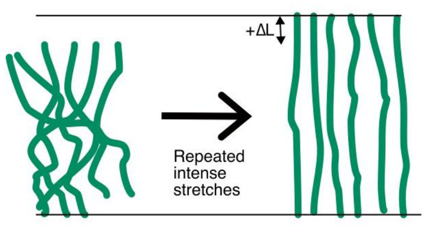
Figure 6.
Stretching training modifies the fascia’s mechanical reaction. The black curve shows the tension tolerated by a subject, and the gray curve shows the corresponding derivative representing fiber recruitment. Note that fibers are recruited twice as fast after the eleventh stretch. This demonstrates the tissue’s dynamic adaptation to stretching, causing the fibers to react collectively after long, intense training. Sketch inspired from Germain et Al [16].
Figure 6.
Stretching training modifies the fascia’s mechanical reaction. The black curve shows the tension tolerated by a subject, and the gray curve shows the corresponding derivative representing fiber recruitment. Note that fibers are recruited twice as fast after the eleventh stretch. This demonstrates the tissue’s dynamic adaptation to stretching, causing the fibers to react collectively after long, intense training. Sketch inspired from Germain et Al [16].
Research has shown that fascia can undergo dynamic reinforcing following intense stretching sessions [16]. The experiments on the front thigh demonstrate that passive fibers can be recruited within 10 minutes or over the course of a few months. Indeed, it was hypothesized that inter-fibers glycosaminoglycans might fixe the collagen fibers positioning for a few minutes or a few months to adapt to any tensile stress applied repeatedly. These hypotheses were formulated, following experiments exploring the fascia lata adaptation to the tensile stress on a 10-minutes time scale [23] and on a month timescale [16]. Regardless the intimate mechanisms at play, a deep change in fibers recruitment pattern was noticed following intense stretching training [16]. The observation that these mechanical adaptations are constrained by pain sensors and tension sensors suggests the key role of the mechanoreceptors in these adaptation mechanisms. In this regard, the absence of precise histological examinations of the links between these sensors and the collagen network is problematic to push further the stretching physiology understanding. Astonishingly, the collagen fibrils crawling within the tendon, is better understood since Screen and colleague managed to described it at the nanoscale level [38,39,40,41]. Alas, the intimate mechanisms at play within the fascia was not really examined and we lack direct observations of the fibers network following stretching or strengthening training. Considering the tissue similarities, the fibers rearrangement should be analogous in tendons, fascia or hydrogels but other experiments must be conducted to confirm this hypothesis. Three dimensional models, such as modified hydrogels [35] can mimic collagen fibers networks. These collagen-like fibers networks are promising to track more accurately fibers positioning and the physics of fibers reorganization following multiple stretches.
Whatever, the fascia, as a complex multiscale structure must be considered as a plastic organ and the fact that the fibroblasts have some contractile capacities [7] makes its physiology even more complex to apprehend. The main take away for researchers who want to study these phenomena of lengthening [16] and contraction [6,7] is that the experiments must last at least several days if not several months. Anatomists may contribute actively to fascia physiology understanding as it seems that fascia contractibility depends on the myofibroblasts density withing the tissue [6]. Consequently, knowing the statistical positioning of myofibroblasts in each fascia would be useful for building efficient physical models. Furthermore, the effects of myofibroblast contractility on the collagen network at the histologic level are still unknown and mapping the collagen network would be fundamental to understanding the interplay between myofibroblast contractions and the physical effects of stretching.
Finally, it is likely that fascial length is regulated by the interplay between external applied tensile stresses and myofibroblasts activities [6]. The physical stress is itself limited by a sensory regulation based on the sensation of tension and the sensation of pain [16]. These two phases can be observed during mammalian pregnancy where an impressive abdominal fascia lengthening is followed by a remarkable fascia retraction [42]. The fascia lata constitutes an outstanding structure to study the effects of the physical stress on a fascia as it is easy to assess physical changes within its structure [11,16,23,34].
5. Clinical Implications
The fascia lata is a critical component in surgical procedures, particularly in instances where the extraction of dense collagenic grafts is necessary [10,15,43,44]. A topical review has been published [15] that provides a comprehensive overview of the grafts extractions and subsequent utilization for various tissue replacements or reconstruction procedures. The fact that the fascia lata is highly accessible rationalizes its extraction with its vascular pedicel, a factor that can prove advantageous in the context of reconstruction surgeries [15].
The fascia lata fibrous network has been implicated in a few outlet syndromes. The entrapment of the lateral femoral cutaneous nerve is a causative factor for meralgia paresthetica, a prevalent neuropathy that can result from a wide range of etiologies [45]. This condition has been observed to result from a variety of factors, including pregnancy, trauma, excessive belt pressure, tumors, and surgical complications. These factors can affect the environment of the nerve when it traverses the fascia lata [46]. The ilioinguinal nerve (L1), femoral branch of the genitofemoral nerve (L1–L2), can also become entrapped within the fibrous network of the fascia lata on its proximal insertion on the ilioinguinal ligament. Tagliafico et al. [47] present a noteworthy analysis that exposes the intricacies of the anatomical structure of the nerves within the fascia lata. The treatment principles involving ultrasound-guided injections and surgery for the more invalidating cases remain consistent across all these conditions [45,46,47,48]. In the case of pudental neuralgia, physiotherapy and tissue mobilization are recognized as pivotal treatment modalities [49]. However, a paucity of literature has been identified concerning the manual treatment of nerves entrapment through the fascia lata.
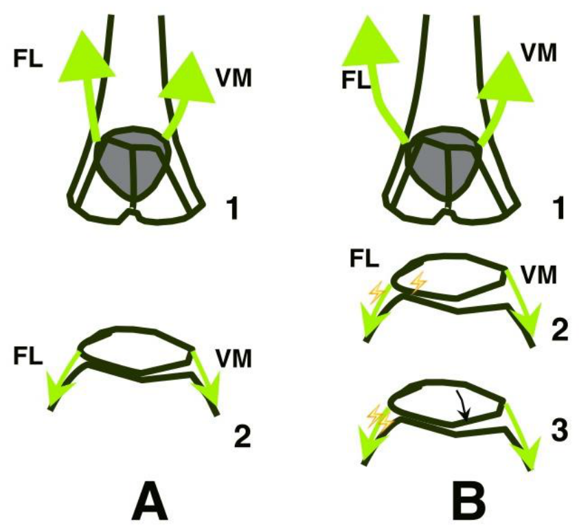
The fascia lata tensional network plays a pivotal role for two of the mains knee pathologies: the patellofemoral pain syndrome [50,51,52] and the iliotibial band syndrome [18,52,53,54,55]. Iliotibial band syndrome is a multifactorial condition. Its etiology involves training load, neuromuscular control, hip and pelvic mechanics, foot biomechanics, pain sensitivity, tissue capacity, and individual anatomical variation [54]. Patellofemoral pain syndrome is associated with patellar maltracking and dynamic valgus, and is linked to an imbalance in the vastus medialis and vastus lateralis muscles, as well as tightness in the hamstrings or iliotibial tract [56,57]. This review will focus solely on aspects directly related to the fascia lata tensional network. The recent literature on the subject suggests that both patellofemoral pain syndrome and iliotibial band syndrome are, in fact, overuse injuries resulting from excessive pressure on the lateral side of the patella [50,58] or on the deep layers of the iliotibial band [18]. Upon acknowledging the fascia lata as a wide dense collagenous sheet, a novel framework for comprehending these two major pathologies, which affect runners, emerges. As the recorded reaction forces during fascia lata stretching induce major compression of the iliotibial band or the lateral patella, the two pathologies may have the same biomechanical cause at the fascia lata level [11]. This may explain why the vastus medialis activation is delayed when patellar maltracking is registered in a subgroup of patellofemoral pain participants showing abnormal patellar tilt [59].
Figure 4.
Two of the most common knee pathologies may be related to the fascia lata (FL) tightness: the iliotibial band syndrome and the patellofemoral pain syndrome. Both pathologies may, in fact, result from the same mechanism. A: When the fascia lata is normally taut (1), the patella glides in the trochlea with a big contact surface, spreading the stress on all the cartilage surfaces, reducing the constraints per square centimeter. B: When the fascia lata is excessively taut on its lateral part (1), the patella is tilted on the lateral side of the trochlea, increasing dramatically the stress on the cartilage as the constraints are concentrated on a very small area (2). In this configuration, the lever arm of the vastus medialis (VM) contraction may increase the stress on the fascia lata fibers reaching the patella (3).
Figure 4.
Two of the most common knee pathologies may be related to the fascia lata (FL) tightness: the iliotibial band syndrome and the patellofemoral pain syndrome. Both pathologies may, in fact, result from the same mechanism. A: When the fascia lata is normally taut (1), the patella glides in the trochlea with a big contact surface, spreading the stress on all the cartilage surfaces, reducing the constraints per square centimeter. B: When the fascia lata is excessively taut on its lateral part (1), the patella is tilted on the lateral side of the trochlea, increasing dramatically the stress on the cartilage as the constraints are concentrated on a very small area (2). In this configuration, the lever arm of the vastus medialis (VM) contraction may increase the stress on the fascia lata fibers reaching the patella (3).
Another major concern in post-industrial societies is restricted hip extension. These may result from excessive sedentary behavior [60] or hip arthroplasties [61,62] but given the prevalence of both conditions, this is a significant issue in the field of rehabilitation. Hip extension appears to be primarily limited by soft tissues [63] and mainly by the fascia lata when the knee is flexed [11,12]. Therefore, introducing proper stretching sessions for the ageing population is likely to help, as the fascia lata can be lengthened [16,23].
For anatomists, the main point to remember is the dynamic nature of the fascia lata. It is important for them to emphasize both its contractile nature and its ability to be chronically lengthened in order to sensitize future physicians and orthopedists to fascia physiology. A comprehensive understanding of fascial function can facilitate clinical decision-making, particularly with regard to the efficacy of long, intense stretching as a treatment for patellofemoral pain syndrome, iliotibial band syndrome, and hip extension restriction. Viewing the fascia as a tensional network that influences patella travel may improve understanding of the consequences of post-surgical fibrosis. However, such clinical reasoning is only possible if the importance of the fascia is recognized during medical education and research.
6. Conclusions
The fascia lata is an amazingly complex organ, the role of which has been highlighted by many experiments. Surprisingly, it was found to be the only structure to adapt to stretching of the quadriceps. Considering all the data from the field of rehabilitation, it can be hypothesized that it is a significant proprioceptive organ, even though no histological research has looked for sensory organs within it. The fascia lata seems useful for locating the stretched area precisely during stretching. Its capacity to prevent tissue from excessive mechanical tension and to protect the fascial sheet from excessive lengthening collectively substantiate its designation as a proprioceptive organ. Interestingly, its anatomical length is an advantage in demonstrating how fascia can adapt to repeated stretching through chronic lengthening. Furthermore, it was demonstrated that tissue lengthening was accompanied by improved fiber recruitment. Finally, understanding all the mechanisms at play within the fascia has the potential to enhance clinical outcomes positively. Overall, the scientific evidence amassed by studying the fascia lata could be profitably integrated into anatomy courses to emphasize the importance of this organ, which was overlooked for most of the 20th century.
Author Contributions
The full article was written by Germain Franck and all the sketches are original.
Funding
This research received no external funding.
Institutional Review Board Statement
Not applicable.
Informed Consent Statement
Not applicable.
Data Availability Statement
Not applicable.
Acknowledgments
Marie Zaegel and Laure Lebon who helped with administration tasks when conducting many experiments cited in this review.
Conflicts of Interest
The author declares no conflicts of interest.
References
- Adstrum, S.; Nicholson, H. A history of fascia. Clin. Anat. 2019, 32(7), 862–70. [Google Scholar] [CrossRef]
- Stecco, C.; Pratt, R.; Nemetz, L.D.; Schleip, R.; Stecco, A.; Theise, N.D. Towards a comprehensive definition of the human fascial system. J. Anat. 2025, 246(6), 1084–98. [Google Scholar] [CrossRef]
- Stecco, C.; Pirri, C.; Fede, C.; Yucesoy, C.A.; De Caro, R.; Stecco, A. Fascial or Muscle Stretching? A Narrative Review. Appl. Sci. 2021, 11(1). [Google Scholar] [CrossRef]
- Suarez-Rodriguez, V.; Fede, C.; Pirri, C.; Petrelli, L.; Loro-Ferrer, J.F.; Rodriguez-Ruiz, D.; et al. Fascial Innervation: A Systematic Review of the Literature. Int. J. Mol. Sci. 2022, 23(10), 5674. [Google Scholar] [CrossRef]
- Stecco, C.; Gagey, O.; Belloni, A.; Pozzuoli, A.; Porzionato, A.; Macchi, V.; et al. Anatomy of the deep fascia of the upper limb. Second part: study of innervation. Morphologie 2007, 91(292), 38–43. [Google Scholar] [CrossRef]
- Schleip, R.; Gabbiani, G.; Wilke, J.; Naylor, I.; Hinz, B.; Zorn, A.; et al. Fascia Is Able to Actively Contract and May Thereby Influence Musculoskeletal Dynamics: A Histochemical and Mechanographic Investigation. Front Physiol. 2019, 10, 336. [Google Scholar] [CrossRef]
- Schleip, R.; Klingler, W. Active contractile properties of fascia. Clin. Anat. 2019, 32(7), 891–5. [Google Scholar] [CrossRef]
- Otsuka, S.; Shan, X.; Yoshida, K.; Yakura, T.; Naito, M.; Kawakami, Y. Site dependent elastic property of human iliotibial band and the effect of hip and knee joint angle configuration. J. Biomech. 2020, 109, 109919. [Google Scholar] [CrossRef]
- Otsuka, S.; Yakura, T.; Ohmichi, Y.; Ohmichi, M.; Naito, M.; Nakano, T.; et al. Site specificity of mechanical and structural properties of human fascia lata and their gender differences: A cadaveric study. J. Biomech. 2018, 77, 69–75. [Google Scholar] [CrossRef]
- Bonaldi, L.; Berardo, A.; Pirri, C.; Stecco, C.; Carniel, E.L.; Fontanella, C.G. Mechanical Characterization of Human Fascia Lata: Uniaxial Tensile Tests from Fresh-Frozen Cadaver Samples and Constitutive Modelling. Bioengineering 2023, 10(2), 226. [Google Scholar] [CrossRef]
- Germain, F.; Mayet, A.; Perrin, R. What mechanical and proprioceptive structures are involved in quadriceps stretching? Why is it important for patella travel? J. Bodyw. Mov. Ther. 2024, 40, 2001–10. [Google Scholar] [CrossRef]
- Germain, F.; Perrin, R. Stretch tolerance and elastic passive reaction of the quadriceps femoris seem to depend more on the fascia profundis taut surfaces than on the underlying stretched muscle. J. Anat. 2023, 243(6), 1059–65. [Google Scholar] [CrossRef]
- Eng, C.M.; Arnold, A.S.; Biewener, A.A.; Lieberman, D.E. The human iliotibial band is specialized for elastic energy storage compared with the chimp fascia lata. J. Exp. Biol. 2015, jeb, 117952. [Google Scholar] [CrossRef]
- Stecco, A.; Macchi, V.; Masiero, S.; Porzionato, A.; Tiengo, C.; Stecco, C.; et al. Pectoral and femoral fasciae: common aspects and regional specializations. Surg. Radiol. Anat. 2009, 31(1), 35–42. [Google Scholar] [CrossRef]
- Szczerba, N.; Waleszczyńska, J.; Jerka, D.; Bonowicz-Kozłowska, K.; Gagat, M. Fascia Lata and Tensor Fasciae Latae in Lower-Limb Reconstruction. J. Clin. Med. 2026, 15(14), 5547. [Google Scholar] [CrossRef]
- Germain, F.; Lemarchand, E.; Perrin, R. Sensory regulation and mechanical effects of sustained high intensity stretching of the anterior compartment of the thigh. J. Bodyw. Mov. Ther. 2020, 24(2), 18–25. [Google Scholar] [CrossRef]
- Schwartz, J.M.; Stern, J.T.; Paré, E.B. Functional differentiation within the tensor fasciae latae. A telemetered electromyographic analysis of its locomotor roles. J. Bone Jt. Surg. 1981, 63(9), 1457–71. [Google Scholar] [CrossRef]
- Hutchinson, L.A.; Lichtwark, G.A.; Willy, R.W.; Kelly, L.A. The Iliotibial Band: A Complex Structure with Versatile Functions. Sports Med. 2022, 52(5), 995–1008. [Google Scholar] [CrossRef]
- Terry, G.C.; Hughston, J.C.; Norwood, L.A. The anatomy of the iliopatellar band and iliotibial tract. Am. J. Sports Med. 1986, 14(1), 39–45. [Google Scholar] [CrossRef]
- Sednieva, Y.; Bruyère-Garnier, K.; Naaim, A.; Viste, A.; Gras, L.L. Assessment of the thigh skin and fascia strains during knee flexion-extension: an ex-vivo study. Comput Methods Biomech. BioMed Engin 2019, 22(sup1), S172–4. [Google Scholar] [CrossRef]
- Ley, C.D.; Valdes, E.M.; Murtagh, C.F.; Power, J.; Drust, B. Regional Differences in Muscle and Fascial Tissue Stiffness in the Rectus Femoris Are Dependent Upon Localised Stretching. Eur. J. Sport Sci. 2026, 26(3), e70109. [Google Scholar] [CrossRef]
- Szotek, S.; Dawidowicz, J.; Maksymowicz, K. Mechanical behaviour and structural characterization of human fascia lata. Acta Bioeng. Biomech. 2024, 26(4), 99–110. [Google Scholar] [CrossRef]
- Germain, F.; Regis, L. Mécanismes probables des étirements statiques en kinésithérapie: étude des aponévroses antérieures de la cuisse. Kinésithérapie Rev. 2016, 16(180), 3–12. [Google Scholar] [CrossRef]
- Björklund, M.; Hamberg, J.; Crenshaw, A.G. Sensory adaptation after a 2-week stretching regimen of the rectus femoris muscle. Arch. Phys. Med. Rehabil. 2001, 82(9), 1245–50. [Google Scholar] [CrossRef]
- Umehara, J.; Ikezoe, T.; Nishishita, S.; Nakamura, M.; Umegaki, H.; Kobayashi, T.; et al. Effect of hip and knee position on tensor fasciae latae elongation during stretching: An ultrasonic shear wave elastography study. Clin. Biomech. 2015, 30(10), 1056–9. [Google Scholar] [CrossRef]
- Nakamura, M.; Sato, S.; Murakami, Y.; Kiyono, R.; Yahata, K.; Sanuki, F.; et al. The Comparison of Different Stretching Intensities on the Range of Motion and Muscle Stiffness of the Quadriceps Muscles. Front Physiol. 2021, Volume 11-2020. [Google Scholar] [CrossRef]
- Kranjc, S.; Fink, M.; Nakamura, M.; Kozinc, Ž. Acute effects of proprioceptive neuromuscular facilitation stretching on rectus femoris muscle stiffness: a dose-response shear-wave elastography study. Front Physiol. 2025, Volume 15. [Google Scholar] [CrossRef]
- Bousie, J.; Myrrylainen, D.; Preston, P. Does static stretching of the TFL and ITB change muscle stiffness or hip range of motion in the short- or long-term? J. Sci. Med. Sport. 2022, 25, S77. [Google Scholar] [CrossRef]
- Antonio, S.; Wolfgang, G.; Robert, H.; Fullerton, B.; Carla, S. The anatomical and functional relation between gluteus maximus and fascia lata. J. Bodyw. Mov. Ther. 2013, 17(4), 512–7. [Google Scholar] [CrossRef]
- Schleip, R. Fascial plasticity – a new neurobiological explanation: Part 1. J. Bodyw. Mov. Ther. 2003, 7(1), 11–9. [Google Scholar] [CrossRef]
- Weppler, C.H.; Magnusson, S.P. Increasing Muscle Extensibility: A Matter of Increasing Length or Modifying Sensation? Phys. Ther. 2010, 90(3), 438–49. [Google Scholar] [CrossRef]
- Edin, B.B.; Abbs, J.H. Finger movement responses of cutaneous mechanoreceptors in the dorsal skin of the human hand. J. Neurophysiol. 1991, 65(3), 657–70. [Google Scholar] [CrossRef]
- Guissard, N.; Duchateau, J. Neural Aspects of Muscle Stretching. Exerc Sport Sci. Rev. 2006, 34(4), 154–8. [Google Scholar] [CrossRef]
- Germain, F.; Lemarchand, E. Un recrutement massif de fibres conjonctives semble accompagner les étirements longs et intenses de la loge antérieure de la cuisse. Sci. Amp Sports 2018, 5953(5), e181–262. [Google Scholar] [CrossRef]
- Lin, S.; Liu, J.; Liu, X.; Zhao, X. Muscle-like fatigue-resistant hydrogels by mechanical training. Proc. Natl. Acad. Sci. 2019, 116(21), 10244–9. [Google Scholar] [CrossRef]
- De Gennes, P.G. Reptation of stars. J. Phys. 1975, 36(12), 1199–203. [Google Scholar] [CrossRef]
- Germain, F.; Gibaud, T. In vivo measurements of fascia lata effective mechanics combined to a memory fiber–recruitment–viscoelastic modeling approach. Soft Matter 2026, 22(24), 4223–37. [Google Scholar] [CrossRef]
- Puxkandl, R.; Zizak, I.; Paris, O.; Keckes, J.; Tesch, W.; Bernstorff, S.; et al. Viscoelastic properties of collagen: synchrotron radiation investigations and structural model. In Philos Trans R Soc Lond B Biol Sci; Bailey, A.J., Macmillan, J., Shrewry, P.R., Tatham, A.S., Eds.; 28 Feb 2002; Volume 357, 1418, pp. 191–7. [Google Scholar] [CrossRef]
- Barreto, I.S.; Pierantoni, M.; Hammerman, M.; Törnquist, E.; Le Cann, S.; Diaz, A.; et al. Nanoscale characterization of collagen structural responses to in situ loading in rat Achilles tendons. Matrix Biol. 2023, 115, 32–47. [Google Scholar] [CrossRef]
- Gupta, H.S.; Seto, J.; Krauss, S.; Boesecke, P.; Screen, H.R.C. In situ multi-level analysis of viscoelastic deformation mechanisms in tendon collagen. J. Struct. Biol. 2010, 169(2), 183–91. [Google Scholar] [CrossRef]
- Screen, H.R.C.; Seto, J.; Krauss, S.; Boesecke, P.; Gupta, H.S. Extrafibrillar diffusion and intrafibrillar swelling at the nanoscale are associated with stress relaxation in the soft collagenous matrix tissue of tendons. Soft Matter 2011, 7(23), 11243. [Google Scholar] [CrossRef]
- Catanzarite, T.; Bremner, S.; Barlow, C.L.; Bou-Malham, L.; O’Connor, S.; Alperin, M. Pelvic muscles’ mechanical response to strains in the absence and presence of pregnancy-induced adaptations in a rat model. Am. J. Obstet. Gynecol. 2018, 218(5), 512.e1–512.e9. [Google Scholar] [CrossRef]
- Demontis, F.; Loi, G.; Mazzotti, E.; Rodriguez Reinoso, M.; Vecchio, F.; Faini, A.C.; et al. Uniaxial Tensile Tests and Digital Image Correlation Analysis for the Mechanical Characterization of Human Fascia Lata Under Different Decellularization Treatments. J. BioMed Mater. Res. A 2025, 113(11), e38012. [Google Scholar] [CrossRef]
- Derwin, K.A.; Baker, A.R.; Spragg, R.K.; Leigh, D.R.; Farhat, W.; Iannotti, J.P. Regional variability, processing methods, and biophysical properties of human fascia lata extracellular matrix. J. BioMed Mater. Res. A 2008, 84A(2), 500–7. [Google Scholar] [CrossRef]
- Gomez, Y.D.L.C.; Remotti, E.; Momah, D.U.; Zhang, E.; D. Swanson, D.; Kim, R.; et al. Meralgia Paresthetica Review: Update on Presentation, Pathophysiology, and Treatment. Health Psychol. Res. 2023, 11(1), 1. [Google Scholar] [CrossRef]
- Tagliafico, A.; Serafini, G.; Lacelli, F.; Perrone, N.; Valsania, V.; Martinoli, C. Ultrasound-Guided Treatment of Meralgia Paresthetica (Lateral Femoral Cutaneous Neuropathy): Technical Description and Results of Treatment in 20 Consecutive Patients. J. Ultrasound Med. 2011, 30(10), 1341–6. [Google Scholar] [CrossRef]
- Tagliafico, A.; Bignotti, B.; Cadoni, A.; Perez, M.M.; Martinoli, C. Anatomical study of the iliohypogastric, ilioinguinal, and genitofemoral nerves using high-resolution ultrasound. Muscle Nerve 2015, 51(1), 42–8. [Google Scholar] [CrossRef]
- Drakonaki, E.E.; Adriaensen, M.E.A.P.M.; Al-Bulushi, H.I.J.; Koliarakis, I.; Tsiaoussis, J.; Vanderdood, K. Sonoanatomy of the ilioinguinal, iliohypogastric, genitofemoral, obturator, and pudendal nerves: a practical guide for US-guided injections. J. Ultrason. 2022, 22(88), 44–50. [Google Scholar] [CrossRef]
- Hibner, M.; Desai, N.; Robertson, L.J.; Nour, M. Pudendal Neuralgia. J. Minim. Invasive Gynecol. 2010, 17(2), 148–53. [Google Scholar] [CrossRef]
- Esfandiarpour, F.; Lebrun, C.M.; Dhillon, S.; Boulanger, P. In-vivo patellar tracking in individuals with patellofemoral pain and healthy individuals. J. Orthop. Res. 2018, 36(8), 2193–201. [Google Scholar] [CrossRef]
- Nha, K.W.; Papannagari, R.; Gill, T.J.; Van De Velde, S.K.; Freiberg, A.A.; Rubash, H.E.; et al. In vivo patellar tracking: Clinical motions and patellofemoral indices. J. Orthop. Res. 2008, 26(8), 1067–74. [Google Scholar] [CrossRef]
- Merican, A.M.; Amis, A.A. Iliotibial band tension affects patellofemoral and tibiofemoral kinematics. J. Biomech. 2009, 42(10), 1539–46. [Google Scholar] [CrossRef]
- Falvey, E.C.; Clark, R.A.; Franklyn-Miller, A.; Bryant, A.L.; Briggs, C.; McCrory, P.R. Iliotibial band syndrome: an examination of the evidence behind a number of treatment options. Scand. J. Med. Sci. Sports 2010, 20(4), 580–7. [Google Scholar] [CrossRef]
- Louw, M.; Deary, C. The biomechanical variables involved in the aetiology of iliotibial band syndrome in distance runners – A systematic review of the literature. Phys. Ther. Sport 2014, 15(1), 64–75. [Google Scholar] [CrossRef]
- Puniello, M.S. Iliotibial Band Tightness and Medial Patellar Glide in Patients with Patellofemoral Dysfunction. J. Orthop. Sports Phys. Ther. 1993, 17(3), 144–8. [Google Scholar] [CrossRef]
- Petersen, W.; Ellermann, A.; Gösele-Koppenburg, A.; Best, R.; Rembitzki, I.V.; Brüggemann, G.; et al. Patellofemoral pain syndrome. Knee Surg. Sports Traumatol. Arthrosc. 2014, 22(10), 2264–74. [Google Scholar] [CrossRef]
- Xie, P.; István, B.; Liang, M. The Relationship between Patellofemoral Pain Syndrome and Hip Biomechanics: A Systematic Review with Meta-Analysis. Healthcare 2022, 11(1), 99. [Google Scholar] [CrossRef]
- Minghao, L.; Qiang, L.; Guoqing, C. Lateral patellar tilting can also result in cartilage lesions of tibial plateau - the characteristic features of excessive lateral pressure syndrome: a retrospective study of 141 cases. BMC Musculoskelet. Disord. 2025, 26(1), 443. [Google Scholar] [CrossRef]
- Pal, S.; Draper, C.E.; Fredericson, M.; Gold, G.E.; Delp, S.L.; Beaupre, G.S.; et al. Patellar Maltracking Correlates With Vastus Medialis Activation Delay in Patellofemoral Pain Patients. Am. J. Sports Med. 2011, 39(3), 590–8. [Google Scholar] [CrossRef]
- Boukabache, A.; Preece, S.J.; Brookes, N. Prolonged sitting and physical inactivity are associated with limited hip extension: A cross-sectional study. Musculoskelet. Sci. Pract. 2021, 51, 102282. [Google Scholar] [CrossRef]
- Tsukagoshi, R.; Tateuchi, H.; Fukumoto, Y.; Akiyama, H.; So, K.; Kuroda, Y.; et al. Factors Associated with Restricted Hip Extension during Gait in Women after Total Hip Arthroplasty. HIP Int. 2015, 25(6), 543–8. [Google Scholar] [CrossRef]
- Mazzoli, D.; Giannotti, E.; Longhi, M.; Prati, P.; Masiero, S.; Merlo, A. Age explains limited hip extension recovery at one year from total hip arthroplasty. Clin. Biomech. 2017, 48, 35–41. [Google Scholar] [CrossRef]
- Han, S.; Owens, V.L.; Patel, R.V.; Ismaily, S.K.; Harrington, M.A.; Incavo, S.J.; et al. The continuum of hip range of motion: From soft-tissue restriction to bony impingement. J. Orthop. Res. 2020, 38(8), 1779–86. [Google Scholar] [CrossRef]
Figure 1.
Fascia lata functional anatomy: When the hip is extended, the degree of knee flexion modifies deeply the force travel within the fascia lata. Strait green lines represent taut fibers while s lines represent slack fibers of the fascia. The black arrow shows how the main forces are distributed in the fascial sheet according to the degree of knee flexion. Note that at 0° the tensor fascia lata (TFL) transmits all the forces to the tuberculum tractus iliotibialis but that at 90 and 120° all the forces are transmitted to the patella. The oval represents the centroids of the stretched area location felt by the participants. It is also noteworthy that the moment arm of the peripheral fascia is always the most favorable to restitute the stretch energy.
Figure 1.
Fascia lata functional anatomy: When the hip is extended, the degree of knee flexion modifies deeply the force travel within the fascia lata. Strait green lines represent taut fibers while s lines represent slack fibers of the fascia. The black arrow shows how the main forces are distributed in the fascial sheet according to the degree of knee flexion. Note that at 0° the tensor fascia lata (TFL) transmits all the forces to the tuberculum tractus iliotibialis but that at 90 and 120° all the forces are transmitted to the patella. The oval represents the centroids of the stretched area location felt by the participants. It is also noteworthy that the moment arm of the peripheral fascia is always the most favorable to restitute the stretch energy.
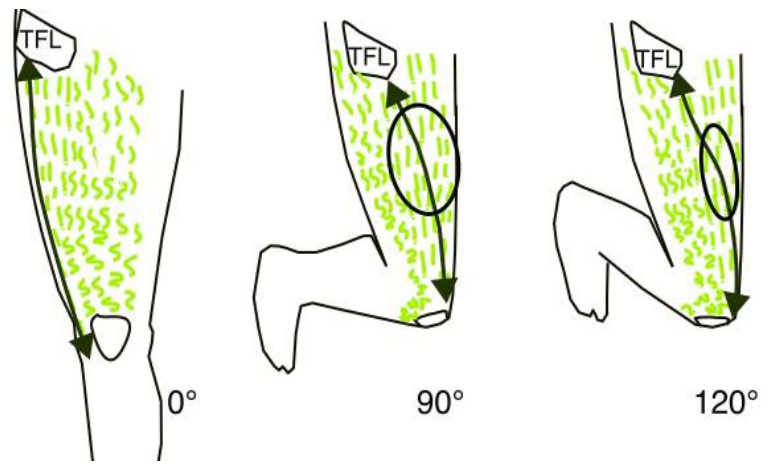
Figure 2.
A: The fascia lata is made of three layers of collagen fibers oriented longitudinally and transversally. B: Note that the moment arm of the fascia lata (MFL) is always more favorable than the moment arm of the rectus femoris (MRF) to restitute the stretch energy. C: Despite its finesse the total cross-sectional area of the fascia lata can reach 1 cm2 and can bear loads of 2000 N during stretching experiments. D: The fascial sheet is heterogeneous and some fibers bear most of the constrains (1). During a tensile test the tautest fibers can be ruptured and the constrains get concentrated at the peripheries of the rupture (2). Then, the chronical tolerable stretch of the tissue may homogenize the constrains between the preserved fibers (3). At the end of the healing process the fascia is a bit lengthened in the original taut area and new short collagen fibers can bridge the residual gap (4).
Figure 2.
A: The fascia lata is made of three layers of collagen fibers oriented longitudinally and transversally. B: Note that the moment arm of the fascia lata (MFL) is always more favorable than the moment arm of the rectus femoris (MRF) to restitute the stretch energy. C: Despite its finesse the total cross-sectional area of the fascia lata can reach 1 cm2 and can bear loads of 2000 N during stretching experiments. D: The fascial sheet is heterogeneous and some fibers bear most of the constrains (1). During a tensile test the tautest fibers can be ruptured and the constrains get concentrated at the peripheries of the rupture (2). Then, the chronical tolerable stretch of the tissue may homogenize the constrains between the preserved fibers (3). At the end of the healing process the fascia is a bit lengthened in the original taut area and new short collagen fibers can bridge the residual gap (4).
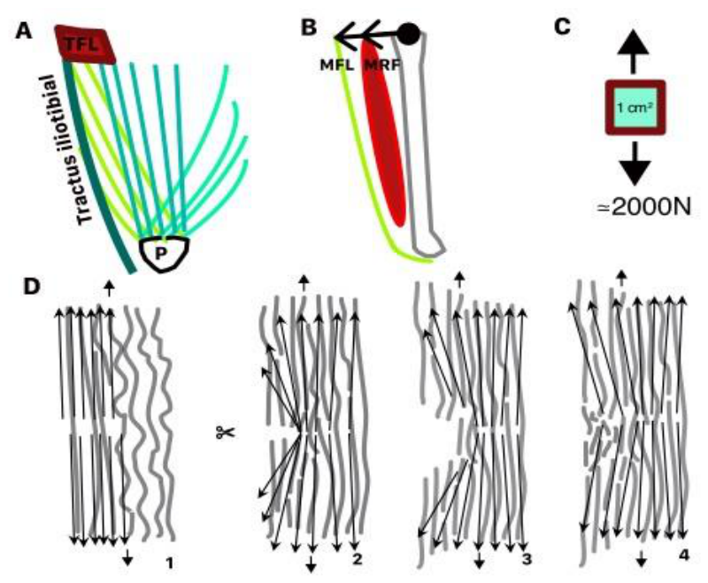
Figure 3.
During quadriceps stretching both the rectus femoris (RF) and the fascia lata (FL) lengthen but the only the tension of the fascia lata increases whereas the rectus femoris tension stay mostly constant. It was assumed that the rectus femoris tension was the result of the muscle resting activity.
Figure 3.
During quadriceps stretching both the rectus femoris (RF) and the fascia lata (FL) lengthen but the only the tension of the fascia lata increases whereas the rectus femoris tension stay mostly constant. It was assumed that the rectus femoris tension was the result of the muscle resting activity.
Disclaimer/Publisher’s Note: The statements, opinions and data contained in all publications are solely those of the individual author(s) and contributor(s) and not of MDPI and/or the editor(s). MDPI and/or the editor(s) disclaim responsibility for any injury to people or property resulting from any ideas, methods, instructions or products referred to in the content. |
© 2026 by the authors. Licensee MDPI, Basel, Switzerland. This article is an open access article distributed under the terms and conditions of the Creative Commons Attribution (CC BY) license (http://creativecommons.org/licenses/by/4.0/).
Copyright: This open access article is published under a Creative Commons CC BY 4.0 license, which permit the free download, distribution, and reuse, provided that the author and preprint are cited in any reuse.