

Submitted:
14 October 2026
Posted:
15 October 2026
You are already at the latest version
Abstract
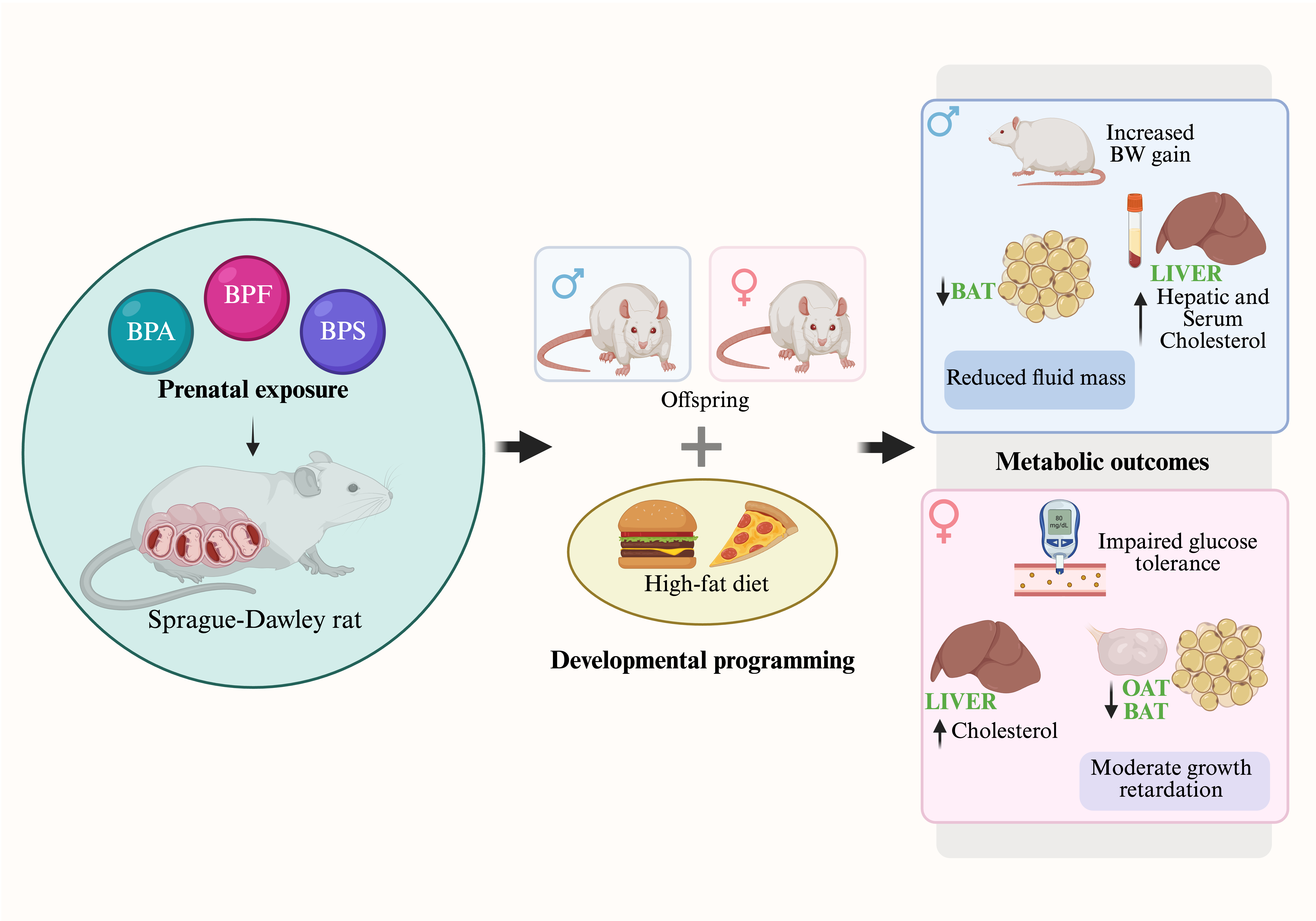
This study investigates the metabolic parameters of prenatal exposure to low doses of Bisphenol A (BPA) and its analogues, BPS and BPF, and the subsequent effects of high-fat diet (HFD) challenge in offspring during adulthood. Pregnant Sprague Dawley rats received saline (control), BPA or BPS (5 µg/kg BW), or BPF (1 µg/kg BW) via oral gavage from gestation days 6–21. Offspring growth, fat deposition, glucose tolerance, insulin, corticosterone, and cholesterol levels were measured in male and female offspring. Prenatal BPA exposure reduced food intake in males and induced pronounced glucose excursions in chow-fed females, which improved over time. In males, BPA caused a rapid 30-min glucose spike and increased body weight, fat, and lean mass following HFD. BPS exposure resulted in mild postnatal growth retardation and reduced food intake in chow-fed females, while males showed increased fluid mass. BPS females exhibited sharp glucose declines after 2 weeks, and HFD led to reduced brown adipose tissue. In males, BPS increased liver and serum cholesterol following HFD. BPF exposure increased water intake and led to greater fat accumulation after HFD, along with reduced fluid mass in males. HFD increased adiposity across groups, with sex-specific differences in body composition but no major effects on glucose tolerance. These findings demonstrate that low-dose prenatal exposure to BPA analogues alters metabolic parameters in offspring, with sex- and compound-specific effects, which can be modulated by dietary challenge.
Keywords:
bisphenol S (BPS)
; bisphenol F (BPF)
; prenatal exposure
; metabolic programming
; high-fat diet challenge
; sex-specific effects
; developmental origins of health and disease (DOHaD)
1. Introduction
There is accumulating evidence that humans are continuously exposed to a widespread plastic-derived chemical called Bisphenol A (BPA), an endocrine-disrupting chemical. BPA has been shown to have developmental, reproductive, immune, cardiovascular, and metabolic adverse outcomes [1]-[4]. The growing concern over BPA’s adverse health effects prompted a gradual industrial switch toward ‘BPA-free’ plastic products by using bisphenol analogs, such as Bisphenol S (BPS) or Bisphenol F (BPF) [5,6]. BPS and BPF are used in a variety of industrial applications and many consumer products [7], such as thermal receipts [8], personal care products [9,10], paper products [11], and fresh as well as packaged food (e.g., dairy and meat products, vegetables, canned foods) [12]. Like BPA, both BPS and BPF were detected in human serum [13] and urine [11] [14], human breast milk [10,15], maternal and cord blood serum [16,17] samples in concentrations and frequencies less than or comparable to BPA. These substitutes have very similar structural and chemical properties to BPA and possibly exhibit some endocrine-disrupting activities [18]-[20], but their potential obesogenic or diabetogenic role is still understudied.
BPS is reported to have estrogenic [21] [22], anti-androgenic [20] [23], and progestogenic properties [24]. BPS can also induce thyroid disruption via thyroid hormone receptor β [25,26]. BPS was shown to affect lipid homeostasis, human adipocyte differentiation, and expression of adipokines [27] [28]. While the ability of BPS to cross the placenta was ten times less than that of BPA, BPS was shown to accumulate in the fetal compartment following repeated maternal exposures [29], and to exhibit the longest fetal half-life when compared to BPA and BPF [30]. BPS was detected in amniotic fluid, fetal urine, and fetal plasma [30] and its exposure resulted in placental endocrine dysfunction [31,32], as well as a reduction in body length, body mass and gonad relative weights [33]. Dose-dependent changes in offspring body and organ weights were also observed after BPS perinatal exposure. Low-dose perinatal BPS treatment (100 ng/g BW) induced an increase in body, liver, and epididymal white adipose tissue weights of male mice offspring [34,35]. Perinatal exposure to very low doses of BPS (10 and 50 μg/kg BW) decreased food intake in male and female rats and reduced visceral adiposity in male offspring [36]. The current recommended no-observed-adverse-effect-level (NOAEL) for BPS is 10 mg/kg/day [37].
While BPS has been more extensively studied, emerging evidence suggests that BPF may exert comparable or even distinct endocrine-disrupting effects. BPF was shown to exhibit estrogenic [20], androgenic, anti-androgenic [20,23], and thyroidogenic modes of action [38] [33]. One study indicated that BPF has an anti-estrogenic property [39]. In vitro studies also revealed BPF cytotoxic and genotoxic effects [40]-[42]. In vivo studies have shown that direct exposure to BPF at high doses (20–750 mg/kg BW) reduces body weights and food consumption and increases organ weights of mature male and female rats [43,44]. At lower doses (0.044 or 4.4 mg/kg/day), male mice gained less weight than their control counterparts [45]. BPF was also shown to cross the placental barrier and reach the fetus. Residues of BPF were detected in the uterus, placenta, amniotic fluid, and fetuses at levels comparable to those in maternal blood [30,46,47]. Although a NOAEL for BPF has not been established yet, studies have classified it as a moderate reproductive [48] and a strong developmental hazard [49,50]. Nevertheless, comparative data directly evaluating BPA, BPS, and BPF under similar experimental conditions are limited, particularly at environmentally relevant exposure levels.
Despite accumulating evidence of endocrine and developmental effects, several critical gaps remain. There is a limited comparative assessment of BPA and its common substitutes, BPS and BPF, within the same experimental framework. Furthermore, data examining environmentally relevant low-dose exposures, long-term metabolic outcomes into adulthood, sex-specific differences, and interactions with postnatal dietary stressors are scarce. These limitations hinder a comprehensive understanding of the metabolic programming potential of BPA substitutes.
This study aimed to expose pregnant Sprague–Dawley rats to environmentally relevant low doses of BPA, BPS, and BPF and to evaluate offspring developmental trajectories and metabolic profiles in adulthood. Given evidence supporting a developmental “two-hit hypothesis,” whereby an early-life insult predisposes individuals to enhanced vulnerability following a later-life stressor [51,52], we further assessed whether postnatal exposure to a high-fat diet (HFD) would exacerbate metabolic alterations induced by prenatal bisphenol exposure. We hypothesized that prenatal exposure to BPS and BPF, like BPA, disrupts growth and glucose homeostasis in a sex-dependent manner and that these effects are exacerbated by an adult HFD challenge.
2. Materials and Methods
2.1. Experimental Design and Prenatal Exposure
Two independent animal experiments were conducted in a temperature-controlled (23 ± 2 °C), humidity-controlled (50 ± 20%), and light-controlled (12:12 h light–dark cycle) animal facility at the University of Georgia. All procedures were performed in accordance with the National Institutes of Health Guide for the Care and Use of Laboratory Animals and were approved by the University of Georgia Institutional Animal Care and Use Committee (IACUC).
The overall experimental design is shown in Figure 1. Detailed descriptions of animal husbandry, breeding procedures, and general maintenance have been reported previously [53]. Briefly, timed-pregnant Sprague–Dawley rats were orally administered phosphate-buffered saline (PBS; control; n = 12), bisphenol A (BPA; 5 μg/kg body weight [BW]/day; n = 12; Sigma-Aldrich, St. Louis, MO, USA), bisphenol F (BPF; 1 μg/kg BW/day; n = 10; Sigma-Aldrich), or bisphenol S (BPS; 5 μg/kg BW/day; n = 13; Sigma-Aldrich) from gestational day (GD) 6 through GD21.
Figure Dose selection was based on environmentally relevant exposure levels and regulatory benchmarks. The BPA and BPS doses used in this study were well below their established no-observed-adverse-effect levels (NOAELs) of 5 mg/kg/day and 10 mg/kg/day, respectively [54,55]. Although a NOAEL has not been established for BPF, the reported lowest-observed-adverse-effect level (LOAEL) is 20 mg/kg/day, and a proposed tolerable daily intake is 11 μg/kg/day [56]. The BPF dose administered in this study was therefore substantially lower than both reference values. Dams were considered the experimental units for all prenatal exposure analyses.
2.2. Postnatal Monitoring and HFD Challenge
Offspring were weaned at postnatal week (PNW) 3. Body weight (BW), food intake (FI), and water intake (WI) were recorded weekly from PNW3 to PNW12 to assess postnatal growth trajectories.
To evaluate susceptibility to a secondary metabolic stressor, a subset of male and female offspring was challenged with a high-fat diet (HFD) between PNW14 and PNW16 (45% kcal from fat, 20% protein, 35% carbohydrate; D12451, Research Diets Inc., New Brunswick, NJ). Remaining littermates were maintained on a standard chow diet (13% fat, 24% protein, 62% carbohydrate; PicoLab Rodent Diet 20, LabDiet; gross energy 4.11 kcal/g).
2.3. Postnatal Growth and Body Composition
Body weight, food intake, and water intake were monitored weekly from PNW3 to PNW12 for offspring maintained on chow. During the HFD challenge period (PNW14–PNW16), body weight and intake measurements were recorded daily for both chow- and HFD-fed groups.
Body composition, including fat mass, lean mass, and total body fluid mass, was assessed longitudinally before and after the HFD challenge (PNW14 and PNW16) using a quantitative magnetic resonance (QMR) analyzer (Bruker Minispec LF110 BCA Analyzer; Bruker Corp., The Woodlands, TX, USA). Measurements were performed in conscious animals to avoid the confounding effects of anesthesia on hydration status and tissue composition.
Prior to scanning, animals were gently placed into a cylindrical restraint tube sized appropriately for body weight to minimize movement while allowing unrestricted respiration. The restraint tube was then positioned horizontally within the analyzer bore. Each scan was completed within a short acquisition time, thereby limiting stress and motion artifacts.
The QMR system was calibrated daily using a manufacturer-supplied standard probe to ensure measurement accuracy and reproducibility. For each animal, a single scan was used for analysis, as the LF110 system provides high precision for repeated measures of fat, lean, and fluid mass. All measurements were conducted by the same trained operator to minimize inter-operator variability, and animals were returned immediately to their home cages following assessment.
2.4. Oral Glucose Tolerance Test
An oral glucose tolerance test (OGTT) was conducted before and during the HFD challenge at PNW14 (baseline), PNW15, and PNW16. Male and female offspring were fasted for approximately 15 h before testing and were orally gavaged with dextrose (2 g/kg BW). Blood samples were collected via tail nick at 0, 30, 60, 90, and 120 min following glucose administration. Blood glucose concentrations were measured using a OneTouch UltraMini glucometer (LifeScan Inc., USA) with GenUltimate test strips.
2.5. Tissue Collection And Estrous Cycle Monitoring
Estrous cyclicity in female offspring was monitored for 15 days prior to sacrifice using vaginal cytology. Vaginal smears were collected daily and stained with methylene blue solution (0.5% methylene blue and 1.6% potassium oxalate in water). Estrous stages were determined as previously described [57].
At PNW16, male rats and female rats in diestrus were euthanized by decapitation. Tissues were rapidly excised, weighed, flash-frozen on dry ice, and stored at −80 °C until analysis. Collected tissues included pituitary gland, adrenal glands, liver, pancreas, brown adipose tissue, and white adipose tissue depots (perirenal, abdominal, and gonadal). Relative organ weights were calculated by normalizing tissue weight to body weight and expressed as g/kg BW.
2.6. Serum Metabolic Analyses
At sacrifice, trunk blood was collected and allowed to clot at room temperature for 30 min, then centrifuged at 1500 × g for 10 min. Serum was isolated and stored at −80 °C until further analysis. Serum insulin concentrations were measured using a commercial radioimmunoassay (RIA) kit (Millipore, Cat# RI-13K) according to the manufacturer’s instructions. Serum corticosterone levels were measured using an RIA method utilizing tracer and standards from MP Biomedicals (DA I-125; Cat# 07120102) with primary and secondary antibodies generated in-house. All samples were analyzed in duplicate.
Serum total cholesterol and triglyceride levels were determined using CardioChek lipid panel test strips. Hepatic total cholesterol was quantified using a colorimetric assay kit (Cell Biolabs, Cat# STA-384) according to the manufacturer’s protocol.
2.7. Statistical Analysis
The dam was considered the experimental unit for prenatal exposure analyses. Two male offspring (one chow-fed and one HFD-fed) and two female offspring (one chow-fed and one HFD-fed) were randomly selected from each litter, resulting in 6–7 animals per sex per treatment group. Statistical analyses were performed using GraphPad Prism (version 11.0.0). Data are presented as mean ± SEM. Longitudinal measurements of body weight, food intake, water intake, and OGTT glucose values were analyzed using repeated-measures two-way ANOVA with time and treatment as independent variables, followed by Fisher’s LSD post hoc test.
One-way ANOVA followed by Bonferroni post hoc testing was used to analyze baseline parameters, including pre-HFD body weight, lean mass, fat mass, fluid mass, cumulative food intake, and incremental body weight gain. Two-way ANOVA followed by Tukey–Kramer post hoc testing was used to evaluate interactions between prenatal exposure and postnatal diet for metabolic parameters (serum corticosterone, insulin, triglycerides, and hepatic cholesterol) and percentage body weight change. Relative organ weights were analyzed using two-way ANOVA followed by Fisher’s LSD test. A p-value < 0.05 was considered statistically significant.
3. Results
3.1. Prenatal BPS and BPF Exposure Modestly Reduce Postnatal Growth in Female Offspring but Not Males
Body weight (BW) and food intake (FI) were monitored weekly from postnatal week 3 to week 12 to assess the programming effects of prenatal bisphenol exposure (Figure 2). BW increased significantly over time in both female and male offspring across all treatment groups (p<0.0001; Figure 2A,B). At weaning (week 3), female offspring prenatally exposed to BPF and BPS exhibited slightly lower BW compared with controls, although these differences did not reach statistical significance (p=0.0664; Figure 2A). However, a significant treatment effect on female postnatal growth trajectories was observed (p=0.0215), with BPS- and BPF-exposed females displaying modest growth retardation relative to controls, particularly at weeks 5, 7, and 8. Despite these differences in absolute BW, incremental BW gain did not differ significantly among female groups (Figure 2C).
Cumulative food intake was also reduced in female offspring exposed prenatally to bisphenols, with BPS-treated females consuming significantly less food compared with controls between weeks 3 and 12 (11486134 g, p=0.0353; Figure 2G). In contrast, male offspring showed no significant differences in absolute or incremental BW across treatment groups (Figure 2B,D). However, BPS- and BPF-exposed males had significantly reduced food intake compared with controls at multiple points between 3 and 12 weeks, and a significant treatment effect was detected for food intake (p=0.0178) (Figure 2F,G).
3.2. Prenatal BPF Exposure Selectively Alters Body Fluid Mass Without Affecting Adiposity or Lean Mass
Body composition was assessed at week 14 prior to dietary challenge (Figure 3). No significant treatment effects were observed in total body mass, fat mass, or lean mass in either sex (Figure 3A–D,G,H).
However, a significant treatment effect was detected for fluid mass in both females (p=0.0455) and males (p=0.0295) (Figure 3E,F). Male offspring exposed prenatally to BPF exhibited a significant reduction in fluid mass compared with controls (4.4660.453 g, p=0.0022), which may explain the increased water intake observed during the subsequent monitoring period (Figure 4T,V). Although male BPS offspring appeared to exhibit lower fat mass relative to controls, this difference did not reach statistical significance.
3.3. Prenatal Bisphenol Exposure Alters Feeding Behavior in Females and Enhances Diet-Dependent Body Weight Responses in Males
At week 14, a subset of offspring was challenged with a high-fat diet (HFD) for two weeks to evaluate susceptibility to a postnatal metabolic stressor (Figure 4). Among females maintained on chow, BPS-exposed offspring maintained lower BW throughout the monitoring period (240.4905.330 g, p=0.0054; Figure 4A,C) and exhibited reduced food and caloric intake compared with controls (Figure 4G,H). However, following HFD exposure, BW, food intake, and water intake in treated female offspring were comparable to those of control animals (Figure 4B–K), and body weight gain did not significantly differ between groups.
In males, prenatal bisphenol exposure did not significantly affect BW under chow conditions (Figure 4L,O). However, treatment effects became evident during HFD exposure, where differences in BW gain were observed between weeks 14 and 16 (Figure 4M,O). Significant interaction effects between prenatal treatment and diet were detected for BW, FI, and WI (Figure 4L–U), indicating increased diet-dependent susceptibility in male offspring.
3.4. Prenatal BPA and BPS Exposure Exacerbates HFD-Induced Body Weight and Lean Mass Gain in Male Offspring
Changes in total body mass and body composition during the HFD challenge are shown in Figure 5. In females, two-way ANOVA revealed a strong diet effect, with HFD-fed animals exhibiting greater increases in fat mass (p<0.0001), fluid mass (p=0.0053), lean mass (p<0.0001), and total BW (p<0.0001) compared with chow-fed controls (Figure 5A–D). No significant treatment effects were observed in females.
In contrast, male offspring exhibited both treatment and diet effects. HFD exposure induced significantly greater BW gain in BPA- (11.621.036%, p=0.0250) and BPS-exposed males (11.32%0.456%, p=0.0407; Figure 5E). BPA-exposed males also exhibited increased lean mass gain (8.8491.045%, p=0.0184; Figure 5H). Additionally, BPF-exposed males displayed a significant reduction in fluid mass during the HFD challenge (Figure 5G). Under chow conditions, treatment effects were minimal, except for increased fluid retention observed in BPS-exposed males.
3.5. Prenatal Bisphenol Exposure Transiently Impairs Glucose Tolerance prior to Dietary Challenge
Oral glucose tolerance tests (OGTT) were performed prior to HFD exposure (week 14) and during the HFD challenge (weeks 15 and 16) (Figure 6). Prior to dietary challenge, female offspring exposed to BPA exhibited significantly elevated glucose levels at 60 (191.4173.103 mg/dL, p=0.0191), 90 (169.9173.728 mg/dL, p=0.0211), and 120 min (151.253.703 mg/dL, p<0.0001) following glucose administration compared with controls (Figure 6A), indicating impaired glucose tolerance.
Similarly, male offspring showed treatment-dependent increases in glucose levels prior to HFD exposure. BPA-exposed males exhibited elevated fasting (0 min) (1162.928 mg/dL, p=0.0004) and early post-challenge (30 min) (189.4559.191 mg/dL, p=0.0010) glucose levels (Figure 6H,I), while BPS-exposed males showed transient hyperglycemia at 30 min (Figure 6H). These differences were no longer observed during the HFD challenge, and glucose tolerance was comparable among groups at weeks 15 and 16 (Figure 6C–F,J–M).
3.6. Prenatal Bisphenol Exposure Alters Brown Adipose Tissue Mass and Hepatic Cholesterol Without Major Endocrine Changes
At week 16, organs and tissues were collected for morphometric and metabolic analyses (Figure 7 and Figure 8). No significant treatment effects were observed in the relative weights of liver, pancreas, or adrenal glands in either sex (Figure 7A–C,G–I). Serum insulin and corticosterone concentrations were also comparable among groups (Figure 7D,F,J,L).
However, hepatic cholesterol levels were significantly elevated in the BPS-HFD males (1.818 0.816 M/g protein, p=0.0425). As expected, strong diet effects were observed in white and brown adipose tissues, particularly in males (p<0.0001; Figure 8B,D,F,H) and in female abdominal and ovarian adipose depots (p<0.0001; Figure 8C,E). Notably, major treatment-related differences were observed in brown adipose tissue (BAT). BPA-exposed males exhibited reduced BAT mass under chow conditions (0.3560.055 g/Kg BW, p=0.0317), whereas BPS-exposed females displayed a marked reduction in BAT mass, which was most pronounced following HFD exposure (0.5480.137 g/Kg BW, p=0.0319; Figure 8G,H).
3.7. BPS-Exposed Males Show Elevated Serum Cholesterol Levels Following HFD
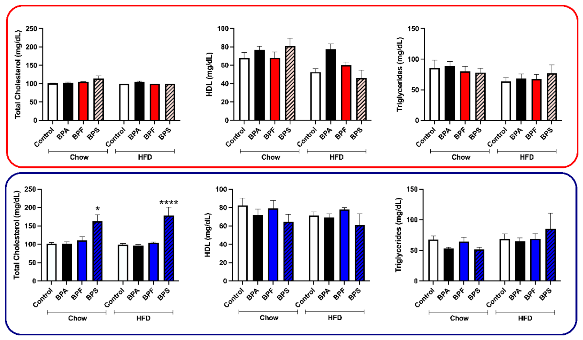
Serum lipid parameters were analyzed to determine whether prenatal bisphenol exposure altered metabolic responses to postnatal HFD challenge (Figure 9). In male offspring, a significant increase in total cholesterol was observed in the BPS-HFD group compared with controls (178.71421.712 mg/dL, p<0.0001; Figure 9D). Two-way ANOVA further revealed a significant main effect of treatment on total cholesterol levels (p<0.0001). However, no significant differences were detected in HDL cholesterol or triglyceride levels among male treatment groups.
In female offspring, individual treatment groups did not show significant differences in total cholesterol levels. Nevertheless, two-way ANOVA revealed strong diet (p<0.05) and diet × treatment interaction (p<0.05) on total cholesterol. For HDL cholesterol, significant main effects of diet (p<0.01), treatment (p<0.05), and diet × treatment interaction (p<0.05) were observed. Analysis of triglyceride levels revealed a significant main effect of diet (p<0.05) in female offspring. Overall, these findings suggest that prenatal bisphenol exposure, particularly BPS, may influence lipid metabolism in a sex-dependent manner, with male offspring showing greater susceptibility to HFD-induced hypercholesterolemia.
4. Discussion
Growing concerns regarding the endocrine-disrupting properties of bisphenol A (BPA) have led to its widespread replacement with structural analogs such as bisphenol S (BPS) and bisphenol F (BPF) in many consumer products [53,54]. However, the potential health consequences of these substitutes remain poorly understood. The present study investigated whether prenatal exposure to environmentally relevant low doses of BPS and BPF could program long-term metabolic alterations in offspring and whether these effects could be unmasked or amplified by a postnatal high-fat diet (HFD) challenge. Our findings demonstrate that prenatal exposure to BPS and BPF induces modest but detectable alterations in postnatal growth trajectories, feeding behavior, and metabolic responses in adulthood in a sex-specific manner. Male offspring exposed prenatally to these bisphenols exhibited increased susceptibility to diet-induced weight gain when challenged with HFD, suggesting that early-life exposure to BPA substitutes may predispose individuals to metabolic dysfunction later in life.
One of the notable findings of this study was the mild postnatal growth retardation observed in female offspring exposed prenatally to BPS and, to a lesser extent, BPF. This effect was accompanied by a reduction in cumulative food intake during early postnatal life. Similar reductions in food intake following perinatal exposure to low doses of BPS have been reported previously, suggesting that BPS may influence appetite regulation or energy balance during early development [11,55]. Endocrine-disrupting chemicals such as bisphenols are known to interfere with hormonal signaling pathways involved in metabolic regulation, including estrogen receptor signaling and hypothalamic neuroendocrine pathways controlling feeding behavior [21,56,57]. Disruption of these pathways during critical windows of development could alter the maturation of central circuits regulating appetite and energy homeostasis, potentially explaining the reduced food intake observed in early life.
Our findings also highlight the importance of developmental programming in determining long-term metabolic outcomes. Although female offspring displayed reduced body weight and caloric intake during the early postnatal period, these changes did not persist into adulthood when animals were challenged with HFD. In contrast, male offspring exposed prenatally to bisphenols exhibited greater body weight gain following HFD exposure despite minimal differences under chow-fed conditions. This pattern is consistent with the “developmental origins of health and disease” (DOHaD) hypothesis, which proposes that environmental exposures during early development can induce latent physiological changes that remain silent under normal conditions but become evident when individuals encounter metabolic stressors later in life [58]. In this context, prenatal exposure to bisphenols may create a metabolic vulnerability that is only revealed when animals are exposed to an obesogenic environment such as a high-fat diet.
Sex-specific responses to prenatal bisphenol exposure were also evident in this study. Female offspring were more sensitive to early growth alterations, whereas male offspring displayed greater susceptibility to HFD-induced weight gain. Sex differences in the effects of endocrine-disrupting chemicals have been widely documented and may reflect differences in hormonal regulation, adipose tissue distribution, and metabolic physiology between males and females [59]. Estrogen signaling plays a critical role in regulating energy balance and adipose tissue metabolism, and disruption of estrogen receptor pathways by bisphenols could lead to differential effects in males and females [60,61]. Additionally, sex-dependent differences in placental function and fetal hormone environments may influence the degree to which developing tissues are exposed to or respond to endocrine-disrupting chemicals during gestation [62].
Another notable observation in the present study was the alteration in body fluid mass observed particularly in BPF-exposed male offspring prior to dietary challenge. Although the physiological significance of this finding remains unclear, it may reflect subtle disruptions in fluid homeostasis or metabolic regulation. Bisphenols have been reported to affect kidney function, electrolyte balance, and hormonal systems involved in water regulation, including the renin–angiotensin and glucocorticoid signaling pathways [63]. The increased water intake observed during the later monitoring period may represent a compensatory response to these alterations.
Despite the modest early-life effects observed in female offspring, the HFD challenge revealed a stronger metabolic response in male animals exposed prenatally to bisphenols. Male offspring exposed to BPA and BPS showed significantly greater body weight gain during HFD exposure, suggesting that prenatal bisphenol exposure can enhance susceptibility to diet-induced obesity. Previous studies have reported similar findings, indicating that early-life exposure to endocrine-disrupting chemicals can exacerbate metabolic responses to high-calorie diets later in life [64]. Bisphenols have been shown to influence adipocyte differentiation and lipid metabolism through activation of peroxisome proliferator–activated receptor gamma (PPARγ) and other transcriptional regulators involved in adipogenesis [65,66]. Alterations in these pathways during fetal development could prime adipose tissue for enhanced lipid accumulation when exposed to an energy-rich diet [67].
In addition to changes in body weight, the present study identified alterations in brown adipose tissue (BAT) mass in both male and female offspring. BAT plays an important role in energy expenditure and thermogenesis, and reductions in BAT mass may impair the body’s ability to dissipate excess energy, thereby promoting weight gain [68]. In our study, BPA-exposed males displayed reduced BAT mass under chow conditions, whereas BPS-exposed females exhibited a marked reduction in BAT mass following HFD exposure. These findings suggest that bisphenol exposure may alter thermogenic capacity, which could contribute to metabolic vulnerability later in life.
The glucose tolerance tests performed in this study revealed transient impairments in glucose regulation prior to dietary challenge, particularly in BPA-exposed females and BPS-exposed males. However, these differences were not maintained during the HFD challenge period. These results suggest that prenatal bisphenol exposure may induce subtle alterations in glucose homeostasis that are not necessarily sustained or may be compensated for over time. It is also possible that the relatively short duration of the HFD challenge limited the ability to detect more pronounced metabolic disturbances. Longer dietary challenges may reveal stronger diabetogenic effects of prenatal bisphenol exposure.
Interestingly, prenatal bisphenol exposure did not produce major alterations in organ weights or circulating hormone levels, including insulin and corticosterone, at the time of sacrifice. This finding suggests that the metabolic effects observed in this study are relatively subtle and may involve changes at the molecular or cellular level rather than gross anatomical alterations. Future studies examining gene expression, hormonal signaling pathways, or epigenetic modifications in metabolic tissues may help clarify the mechanisms underlying these programming effects.
The present study has several strengths. First, it examined the developmental effects of two commonly used BPA substitutes at environmentally relevant low doses. Second, it incorporated both male and female offspring, allowing the identification of sex-specific effects. Third, the inclusion of a postnatal dietary challenge enabled the detection of latent metabolic vulnerabilities that may not be apparent under normal dietary conditions. However, several limitations should also be acknowledged. The study focused on a limited number of exposure doses and a single prenatal exposure window, which may not fully capture the range of possible developmental effects. Additionally, the duration of the HFD challenge was relatively short, which may have limited the detection of more pronounced metabolic abnormalities. Finally, although rodent models provide valuable insight into developmental toxicology, extrapolation of these findings to humans should be approached with caution.
In conclusion, the present study provides evidence that prenatal exposure to low doses of BPA substitutes, particularly BPS and BPF, can induce subtle but measurable alterations in postnatal growth and metabolic responses in a sex-dependent manner. Importantly, male offspring exposed prenatally to these chemicals displayed increased susceptibility to diet-induced weight gain during adulthood, supporting the concept that developmental exposure to endocrine-disrupting chemicals may predispose individuals to metabolic disorders later in life. Given the widespread human exposure to bisphenol analogs through food packaging, thermal receipts, and other consumer products, these findings raise important concerns regarding the metabolic safety of BPA substitutes. Further research is needed to elucidate the molecular mechanisms underlying these effects and to determine whether similar developmental programming effects occur in humans.
Author Contributions
JBD:Conceptualization, methodology, investigation, data curation, formal analysis, writing—original draft. AM: Formal analysis, visualization, writing—original draft and review & editing. AKP: Formal analysis, visualization, writing—original draft and review & editing. YJC: Methodology, investigation, formal analysis. MAM: Investigation, data curation, formal analysis. AK: Investigation, data curation, formal analysis. KR: Data curation (Bruker acquisition). AEA: Investigation, data curation. BKB: Investigation, data curation. DM: Investigation, data curation. EJC: Investigation, data curation. KC: Data curation (Bruker acquisition), formal analysis. PSM: Conceptualization, funding acquisition, methodology, supervision, formal analysis, writing—review & editing. SMJM: Conceptualization, funding acquisition, methodology, supervision, formal analysis, writing—review & editing.
Institutional Review Board Statement
The animal study protocol was reviewed and approved by the Institutional Animal Care and Use Committee (IACUC) of the University of Georgia (protocol number: [IACUC Protocol Number]; date of approval: [Approval Date]). All animal procedures were conducted in accordance with the approved protocol and applicable institutional guidelines for the care and use of laboratory animals.
Data Availability Statement
The data presented in this study are available from the corresponding author upon reasonable request.
Conflicts of Interest
The authors declare no conflicts of interest.
Abbreviations
The following abbreviations are used in this manuscript:
| BPA | Bisphenol A |
| BPS | Bisphenol S |
| BPF | Bisphenol F |
| DoHaD | Developmental origins of Health and Disease |
| NOAEL | No-Observed-Adverse-Effect-Level |
| LOAEL | Lowest-Observed-Adverse-Effect-Level |
| HFD | High Fat Diet |
| IACUC | Institutional Animal Care and Use Committee |
| GD | Gestation Day |
| PNW | Postnatal Week |
| QMR | Quantitative Magnetic Resonance |
| OGTT | Oral Glucose Tolerance Test |
| RIA | Radioimmuno Assay |
| BW | Body Weight |
| FI | Food intake |
| BAT | Brown Adipose Tissue |
| PPAR | Peroxisome Proliferator-Activated Receptor gamma |
References
- Rochester, J. R. Bisphenol a and human health: A review of the literature. Reprod. Toxicol. 2013, 42, 132–155. [Google Scholar] [CrossRef] [PubMed]
- Siracusa, J. S.; Yin, L.; Measel, E.; Liang, S.; Yu, X. Effects of bisphenol a and its analogs on reproductive health: A mini review. Reprod. Toxicol. 2018, 79, 96–123. [Google Scholar] [CrossRef] [PubMed]
- Xu, Z.; et al. Elucidating the mechanisms of environmental pollutant bpa inducing neurotoxicity through metabolomics and network toxicology strategy. Environ. Pollut. 2025, 381, 126591. [Google Scholar] [CrossRef] [PubMed]
- Guan, J.; et al. Multi-omics investigation of bisphenol a in gastrointestinal carcinogenesis: A network toxicology and molecular docking approach. Environ. Int. 2025, 203, 109785. [Google Scholar] [CrossRef] [PubMed]
- Xu, S.; et al. The role of endoplasmic reticulum stress in bps-induced disruption of endometrial decidualization. Ecotoxicol. Environ. Saf. 2025, 302, 118737. [Google Scholar] [CrossRef] [PubMed]
- Li, Z.; et al. Multi-omics analysis reveals bpf exposure causes hepatic glucose and lipid metabolism disorder in rats by disrupting energy homeostasis. Toxicology 2025, 515, 154130. [Google Scholar] [CrossRef] [PubMed]
- Wu, L. H.; et al. Occurrence of bisphenol s in the environment and implications for human exposure: A short review. Sci. Total Environ. 2018, 615, 87–98. [Google Scholar] [CrossRef] [PubMed]
- Liao, C.; Liu, F.; Kannan, K. Bisphenol s, a new bisphenol analogue, in paper products and currency bills and its association with bisphenol a residues. Environ. Sci. Technol. 2012, 46, 6515–6522. [Google Scholar] [CrossRef] [PubMed]
- Liao, C.; Kannan, K. A survey of alkylphenols, bisphenols, and triclosan in personal care products from china and the united states. Arch. Environ. Contam. Toxicol. 2014, 67, 50–59. [Google Scholar] [CrossRef] [PubMed]
- Cinkilli Aktağ, E.; et al. Associations of maternal behaviours and environmental exposures with melamine and bisphenol analogs in breast milk: A cross-sectional study. Int. J. Environ. Health Res. 2025, 35, 3923–3936. [Google Scholar] [CrossRef] [PubMed]
- Liao, C.; et al. Bisphenol s in urine from the united states and seven asian countries: Occurrence and human exposures. Environ. Sci. Technol. 2012, 46, 6860–6866. [Google Scholar] [CrossRef] [PubMed]
- Liao, C.; Kannan, K. Concentrations and profiles of bisphenol a and other bisphenol analogues in foodstuffs from the united states and their implications for human exposure. J. Agric. Food Chem. 2013, 61, 4655–4662. [Google Scholar] [CrossRef] [PubMed]
- Song, S.; et al. Serum concentrations of bisphenol a and its alternatives in elderly population living around e-waste recycling facilities in china: Associations with fasting blood glucose. Ecotoxicol. Environ. Saf. 2019, 169, 822–828. [Google Scholar] [CrossRef] [PubMed]
- Zhou, X.; Kramer, J. P.; Calafat, A. M.; Ye, X. Automated on-line column-switching high performance liquid chromatography isotope dilution tandem mass spectrometry method for the quantification of bisphenol a, bisphenol f, bisphenol s, and 11 other phenols in urine. J. Chromatogr. B Anal. Technol. BioMed Life Sci. 2014, 944, 152–156. [Google Scholar] [CrossRef] [PubMed]
- Niu, Y.; Wang, B.; Zhao, Y.; Zhang, J.; Shao, B. Highly sensitive and high-throughput method for the analysis of bisphenol analogues and their halogenated derivatives in breast milk. J. Agric. Food Chem. 2017, 65, 10452–10463. [Google Scholar] [CrossRef] [PubMed]
- Liu, B.; et al. Bisphenol a substitutes and obesity in us adults: Analysis of a population-based, cross-sectional study. Lancet Planet Health 2017, 1, e114–e122. [Google Scholar] [CrossRef] [PubMed]
- Liang, J.; et al. Effects of prenatal exposure to bisphenols on newborn leucocyte telomere length: A prospective birth cohort study in china. Environ. Sci. Pollut. Res. Int. 2023, 30, 25013–25023. [Google Scholar] [PubMed]
- Molina-Molina, J. M.; et al. In vitro study on the agonistic and antagonistic activities of bisphenol-s and other bisphenol-a congeners and derivatives via nuclear receptors. Toxicol. Appl. Pharmacol. 2013, 272, 127–136. [Google Scholar] [CrossRef] [PubMed]
- Jaskulak, M.; Zorena, K. Quantifying the metabolic health burden of increased bpa/bpf/bps endocrine-disrupting chemical exposure: A temporal and cross-country comparative analysis from 2000 to 2024. Sci. Total Environ. 2025, 1006, 180897. [Google Scholar] [CrossRef] [PubMed]
- Kitamura, S.; et al. Comparative study of the endocrine-disrupting activity of bisphenol a and 19 related compounds. Toxicol. Sci. 2005, 84, 249–259. [Google Scholar] [CrossRef] [PubMed]
- Grignard, E.; Lapenna, S.; Bremer, S. Weak estrogenic transcriptional activities of bisphenol a and bisphenol s. Toxicol. Vitr. 2012, 26, 727–731. [Google Scholar] [CrossRef] [PubMed]
- Kuruto-Niwa, R.; Nozawa, R.; Miyakoshi, T.; Shiozawa, T.; Terao, Y. Estrogenic activity of alkylphenols, bisphenol s, and their chlorinated derivatives using a gfp expression system. Environ. Toxicol. Pharmacol. 2005, 19, 121–130. [Google Scholar] [CrossRef] [PubMed]
- Satoh, K.; Ohyama, K.; Aoki, N.; Iida, M.; Nagai, F. Study on anti-androgenic effects of bisphenol a diglycidyl ether (badge), bisphenol f diglycidyl ether (bfdge) and their derivatives using cells stably transfected with human androgen receptor, ar-ecoscreen. Food Chem. Toxicol. 2004, 42, 983–993. [Google Scholar] [CrossRef] [PubMed]
- Rosenmai, A. K.; et al. Are structural analogues to bisphenol a safe alternatives? Toxicol. Sci. 2014, 139, 35–47. [Google Scholar] [CrossRef] [PubMed]
- Lu, L.; et al. Correction to “thyroid disruption by bisphenol s analogues via thyroid hormone receptor beta: In vitro, in vivo, and molecular dynamics simulation study”. Environ. Sci. Technol. 2019, 53, 9337. [Google Scholar] [CrossRef] [PubMed]
- Chouchene, L.; Rhouma, M. B.; Boughammoura, S.; Messaoudi, I.; Kessabi, K. Implications of early-life bps-induced thyroid disruption on adult female reproductive disorders. Chem. Biol. Interact. 2026, 425, 111891. [Google Scholar] [CrossRef] [PubMed]
- Boucher, J. G.; et al. Bisphenol a and bisphenol s induce distinct transcriptional profiles in differentiating human primary preadipocytes. PLoS ONE 2016, 11, e0163318. [Google Scholar] [CrossRef] [PubMed]
- Peshdary, V.; et al. Depot-specific analysis of human adipose cells and their responses to bisphenol s. Endocrinology 2020, 161. [Google Scholar] [CrossRef] [PubMed]
- Grandin, F. C.; et al. Bisphenol s instead of bisphenol a: Toxicokinetic investigations in the ovine materno-feto-placental unit. Environ. Int. 2018, 120, 584–592. [Google Scholar] [CrossRef] [PubMed]
- Gingrich, J.; et al. Toxicokinetics of bisphenol a, bisphenol s, and bisphenol f in a pregnancy sheep model. Chemosphere 2019, 220, 185–194. [Google Scholar] [CrossRef] [PubMed]
- Gingrich, J.; et al. Gestational bisphenol s impairs placental endocrine function and the fusogenic trophoblast signaling pathway. Arch. Toxicol. 2018, 92, 1861–1876. [Google Scholar] [CrossRef] [PubMed]
- Basak, S.; Varma, S.; Duttaroy, A. K. Modulation of fetoplacental growth, development and reproductive function by endocrine disrupters. Front Endocrinol. 2023, 14, 1215353. [Google Scholar] [CrossRef] [PubMed]
- Rochester, J. R.; Bolden, A. L. Bisphenol s and f: A systematic review and comparison of the hormonal activity of bisphenol a substitutes. Environ. Health Perspect. 2015, 123, 643–650. [Google Scholar] [CrossRef] [PubMed]
- Meng, Z.; et al. Perinatal exposure to bisphenol s (bps) promotes obesity development by interfering with lipid and glucose metabolism in male mouse offspring. Environ. Res. 2019, 173, 189–198. [Google Scholar] [CrossRef] [PubMed]
- Zhang, R.; et al. Prenatal bisphenol s exposure induces hepatic lipid deposition in male mice offspring through downregulation of adipose-derived exosomal mir-29a-3p. J. Hazard Mater. 2023, 453, 131410. [Google Scholar] [CrossRef] [PubMed]
- da Silva, B. S.; et al. Short and long-term effects of bisphenol s (bps) exposure during pregnancy and lactation on plasma lipids, hormones, and behavior in rats. Environ. Pollut. 2019, 250, 312–322. [Google Scholar] [CrossRef] [PubMed]
- Authority, E. F. S.; et al. 2020. Assessment of new information on bisphenol s (bps) submitted in response to the decision 1 under reach regulation (ec) no 1907/2006. Report No. 2397-8325. Wiley Online Library. [Google Scholar]
- Zhu, M.; et al. Bisphenol f disrupts thyroid hormone signaling and postembryonic development in xenopus laevis. Environ. Sci. Technol. 2018, 52, 1602–1611. [Google Scholar] [CrossRef] [PubMed]
- Stroheker, T.; Picard, K.; Lhuguenot, J. C.; Canivenc-Lavier, M. C.; Chagnon, M. C. Steroid activities comparison of natural and food wrap compounds in human breast cancer cell lines. Food Chem. Toxicol. 2004, 42, 887–897. [Google Scholar] [CrossRef] [PubMed]
- Cabaton, N.; et al. Genotoxic and endocrine activities of bis(hydroxyphenyl)methane (bisphenol f) and its derivatives in the hepg2 cell line. Toxicology 2009, 255, 15–24. [Google Scholar] [CrossRef] [PubMed]
- Kim, J. Y.; et al. Comparative study of cytotoxic signaling pathways in h1299 cells exposed to alternative bisphenols: Bpa, bpf, and bps. Toxicol. Res. (Camb) 2024, 13, tfae200. [Google Scholar] [CrossRef] [PubMed]
- Audebert, M.; Dolo, L.; Perdu, E.; Cravedi, J. P.; Zalko, D. Use of the gammah2ax assay for assessing the genotoxicity of bisphenol a and bisphenol f in human cell lines. Arch. Toxicol. 2011, 85, 1463–1473. [Google Scholar] [CrossRef] [PubMed]
- Higashihara, N.; et al. Subacute oral toxicity study of bisphenol f based on the draft protocol for the “enhanced oecd test guideline no. 407”. Arch. Toxicol. 2007, 81, 825–832. [Google Scholar] [CrossRef] [PubMed]
- Igarashi, T.; et al. Initial hazard assessment of 4-benzylphenol, a structural analog of bisphenol f: Genotoxicity tests in vitro and a 28-day repeated-dose toxicity study in rats. Regul. Toxicol. Pharmacol. 2018, 96, 64–75. [Google Scholar] [CrossRef] [PubMed]
- Drobna, Z.; et al. Bisphenol f has different effects on preadipocytes differentiation and weight gain in adult mice as compared with bisphenol a and s. Toxicology 2019, 420, 66–72. [Google Scholar] [CrossRef] [PubMed]
- Cabaton, N.; Chagnon, M. C.; Lhuguenot, J. C.; Cravedi, J. P.; Zalko, D. Disposition and metabolic profiling of bisphenol f in pregnant and nonpregnant rats. J. Agric. Food Chem. 2006, 54, 10307–10314. [Google Scholar] [CrossRef] [PubMed]
- Tuzimski, T.; et al. The association between the bisphenols residues in amniotic fluid and fetal abnormalities in polish pregnant women-its potential clinical application. Int. J. Mol. Sci. 2023, 24. [Google Scholar] [CrossRef] [PubMed]
- den Braver-Sewradj, S. P.; van Spronsen, R.; Hessel, E. V. S. Substitution of bisphenol a: A review of the carcinogenicity, reproductive toxicity, and endocrine disruption potential of alternative substances. Crit. Rev. Toxicol. 2020, 50, 128–147. [Google Scholar] [CrossRef] [PubMed]
- Catron, T. R.; et al. Host developmental toxicity of bpa and bpa alternatives is inversely related to microbiota disruption in zebrafish. Toxicol. Sci. 2019, 167, 468–483. [Google Scholar] [CrossRef] [PubMed]
- Bisphenol a alternatives in thermal paper. 2015.
- Prins, G. S.; Birch, L.; Tang, W. Y.; Ho, S. M. Developmental estrogen exposures predispose to prostate carcinogenesis with aging. Reprod. Toxicol. 2007, 23, 374–382. [Google Scholar] [CrossRef] [PubMed]
- Taylor, J. A.; et al. Interactive effects of perinatal bpa or des and adult testosterone and estradiol exposure on adult urethral obstruction and bladder, kidney, and prostate pathology in male mice. Int. J. Mol. Sci. 2020, 21. [Google Scholar] [CrossRef] [PubMed]
- Chen, D.; et al. Bisphenol analogues other than bpa: Environmental occurrence, human exposure, and toxicity-a review. Environ. Sci. Technol. 2016, 50, 5438–5453. [Google Scholar] [CrossRef] [PubMed]
- Zhu, M.; et al. Pathophysiologically relevant bisphenol s exposure accelerates aging by disrupting brown adipose tissue-regulated energy metabolism. Proc. Natl. Acad. Sci. U S A 2025, 122, e2420437122. [Google Scholar] [CrossRef] [PubMed]
- Clark, P. A.; Rogol, A. D. Growth hormones and sex steroid interactions at puberty. Endocrinol. Metab. Clin. North Am. 1996, 25, 665–681. [Google Scholar] [CrossRef] [PubMed]
- Gould, J. C.; et al. Bisphenol a interacts with the estrogen receptor alpha in a distinct manner from estradiol. Mol. Cell Endocrinol. 1998, 142, 203–214. [Google Scholar] [CrossRef] [PubMed]
- Li, Z.; Ren, Y.; Li, X.; Wang, W. Kdm2a interacts with estrogen receptor α to promote bisphenol a and s-induced breast cancer cell proliferation by repressing tet2 expression. Ecotoxicol. Environ. Saf. 2023, 262, 115132. [Google Scholar] [CrossRef] [PubMed]
- Lapehn, S.; Paquette, A. G. The placental epigenome as a molecular link between prenatal exposures and fetal health outcomes through the dohad hypothesis. Curr. Environ. Health Rep. 2022, 9, 490–501. [Google Scholar] [CrossRef] [PubMed]
- D’Archivio, M.; Coppola, L.; Masella, R.; Tammaro, A.; La Rocca, C. Sex and gender differences on the impact of metabolism-disrupting chemicals on obesity: A systematic review. Nutrients 2024, 16. [Google Scholar] [CrossRef] [PubMed]
- Maniradhan, M.; Calivarathan, L. Bisphenol a-induced endocrine dysfunction and its associated metabolic disorders. Endocr. Metab. Immune Disord. Drug Targets 2023, 23, 515–529. [Google Scholar] [CrossRef] [PubMed]
- Kaulgud, R. S.; Belur, S.; Anagharani, A.; Veeresh, S.; Shettar, A. K. Effects of bisphenol-a on human endocrine function and health outcomes: A review. Discov. Toxicol. 2025, 2, 24. [Google Scholar] [CrossRef]
- Marinello, W. P.; Patisaul, H. B. Endocrine disrupting chemicals (edcs) and placental function: Impact on fetal brain development. Adv. Pharmacol. 2021, 92, 347–400. [Google Scholar] [CrossRef] [PubMed]
- Hsu, C. N.; Tain, Y. L. Adverse impact of environmental chemicals on developmental origins of kidney disease and hypertension. Front Endocrinol. 2021, 12, 745716. [Google Scholar] [CrossRef] [PubMed]
- Varghese, S. V.; Hall, J. M. Bisphenol a substitutes and obesity: A review of the epidemiology and pathophysiology. Front Endocrinol. 2023, 14, 1155694. [Google Scholar] [CrossRef] [PubMed]
- Flores Gomez, D.; et al. Mechanistic insights of bpa alternatives on pparγ binding and the consequence on adipocyte differentiation. Environ. Sci. Technol. 2026, 60, 4526–4539. [Google Scholar] [CrossRef] [PubMed]
- Salehpour, A.; et al. Molecular mechanisms of vitamin d plus bisphenol a effects on adipogenesis in human adipose-derived mesenchymal stem cells. Diabetol. Metab. Syndr. 2021, 13, 41. [Google Scholar] [CrossRef] [PubMed]
- Longo, M.; et al. Low-dose bisphenol-a promotes epigenetic changes at pparγ promoter in adipose precursor cells. Nutrients 2020, 12. [Google Scholar] [CrossRef] [PubMed]
- Cheng, L.; et al. Brown and beige adipose tissue: A novel therapeutic strategy for obesity and type 2 diabetes mellitus. Adipocyte 2021, 10, 48–65. [Google Scholar] [CrossRef] [PubMed]
Figure 1.
Overview of the experimental design. Female Sprague–Dawley rats in proestrus were mated with males, and the day of mating was designated as gestational day (GD) 0. Pregnant dams received daily oral doses from GD6 to GD21 of saline (control; n = 7), BPA (5 μg/kg BW; n = 7), BPS (5 μg/kg BW; n = 7), or BPF (1 μg/kg BW; n = 7). To evaluate the effects of prenatal endocrine-disrupting chemical (EDC) exposure on postnatal growth and metabolic outcomes, a subset of male and female offspring was challenged with a high-fat diet (HFD) beginning at 14 weeks of age. Glucose tolerance and body composition were assessed before and after the HFD challenge. After two weeks of HFD exposure (16 weeks of age), male and female offspring (females in diestrus) were euthanized for tissue collection. Organs were dissected and weighed, and serum samples were collected for the assessment of insulin and corticosterone levels. Abbreviations: GD, gestational day; PND, postnatal day; HFD, high-fat diet; BPA, bisphenol A; BPS, bisphenol S; BPF, bisphenol F.
Figure 1.
Overview of the experimental design. Female Sprague–Dawley rats in proestrus were mated with males, and the day of mating was designated as gestational day (GD) 0. Pregnant dams received daily oral doses from GD6 to GD21 of saline (control; n = 7), BPA (5 μg/kg BW; n = 7), BPS (5 μg/kg BW; n = 7), or BPF (1 μg/kg BW; n = 7). To evaluate the effects of prenatal endocrine-disrupting chemical (EDC) exposure on postnatal growth and metabolic outcomes, a subset of male and female offspring was challenged with a high-fat diet (HFD) beginning at 14 weeks of age. Glucose tolerance and body composition were assessed before and after the HFD challenge. After two weeks of HFD exposure (16 weeks of age), male and female offspring (females in diestrus) were euthanized for tissue collection. Organs were dissected and weighed, and serum samples were collected for the assessment of insulin and corticosterone levels. Abbreviations: GD, gestational day; PND, postnatal day; HFD, high-fat diet; BPA, bisphenol A; BPS, bisphenol S; BPF, bisphenol F.
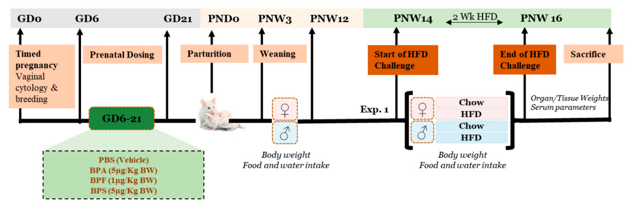
Figure 2.
Sex-dependent changes in body weight and food intake following BPA and its analogues in utero exposure. Changes in female offspring are shown in the top panels and male offspring in the bottom panels. Body weight (BW; g; A,B) and food intake (FI; g/day; E,F) were monitored weekly from postnatal weeks 3 to 12 in offspring maintained on chow diet. Incremental body weight (C,D) was calculated as the difference between body weight at week 12 and baseline body weight at week 3. Cumulative food intake over the 12 weeks is shown in panels G and H. Data are presented as mean ± SEM for 13–14 offspring per treatment group (7 dams per group). Statistical analyses were performed using repeated-measures ANOVA for longitudinal measurements (A,B,E,F) and one-way ANOVA followed by Bonferroni post hoc test for summary measures (C,D,G,H). *P < 0.05, **P < 0.01, ***P < 0.001 compared with control.
Figure 2.
Sex-dependent changes in body weight and food intake following BPA and its analogues in utero exposure. Changes in female offspring are shown in the top panels and male offspring in the bottom panels. Body weight (BW; g; A,B) and food intake (FI; g/day; E,F) were monitored weekly from postnatal weeks 3 to 12 in offspring maintained on chow diet. Incremental body weight (C,D) was calculated as the difference between body weight at week 12 and baseline body weight at week 3. Cumulative food intake over the 12 weeks is shown in panels G and H. Data are presented as mean ± SEM for 13–14 offspring per treatment group (7 dams per group). Statistical analyses were performed using repeated-measures ANOVA for longitudinal measurements (A,B,E,F) and one-way ANOVA followed by Bonferroni post hoc test for summary measures (C,D,G,H). *P < 0.05, **P < 0.01, ***P < 0.001 compared with control.
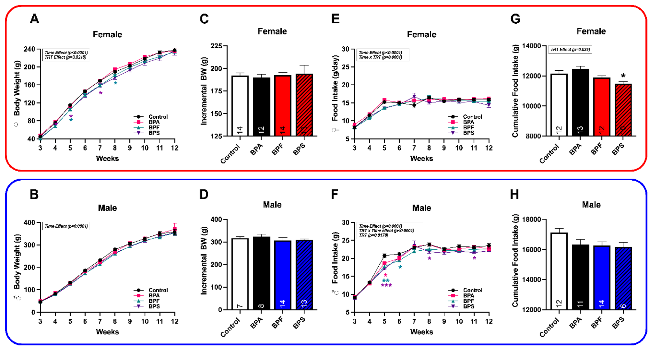
Figure 3.
Effect of in utero Bisphenol exposure on body composition at week 14. Body composition was assessed at 14 weeks of age before high-fat diet challenge. Top panels show female offspring and bottom panels show male offspring. Measurements include total body weight (A,B), fat mass (C,D), fluid mass (E,F), and lean mass (G,H) from offspring prenatally exposed to BPA, BPF, and BPS. Data are presented as mean ± SEM for 6–14 offspring per treatment group. Statistical analysis was performed using one-way ANOVA followed by Bonferroni post hoc testing. *P < 0.05, **P < 0.01 compared with control.
Figure 3.
Effect of in utero Bisphenol exposure on body composition at week 14. Body composition was assessed at 14 weeks of age before high-fat diet challenge. Top panels show female offspring and bottom panels show male offspring. Measurements include total body weight (A,B), fat mass (C,D), fluid mass (E,F), and lean mass (G,H) from offspring prenatally exposed to BPA, BPF, and BPS. Data are presented as mean ± SEM for 6–14 offspring per treatment group. Statistical analysis was performed using one-way ANOVA followed by Bonferroni post hoc testing. *P < 0.05, **P < 0.01 compared with control.
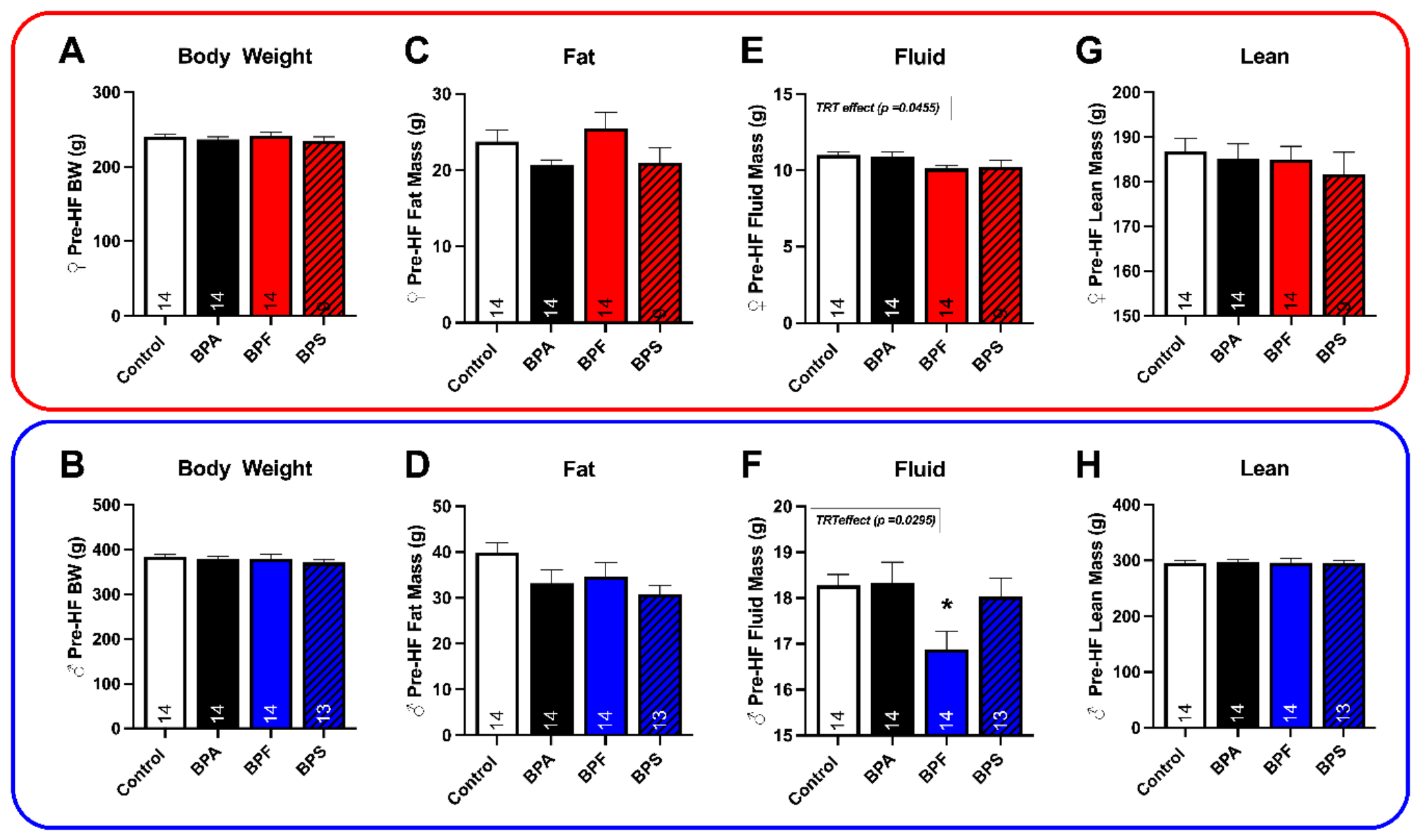
Figure 4.
Effects of prenatal bisphenol exposure and postnatal high-fat diet challenge on body weight and intake parameters. Female offspring are shown in the top panels and male offspring in the bottom panels. At 14 weeks of age, a subset of offspring was challenged with a high-fat diet (HFD) for two weeks (weeks 14–16). Daily body weight (BW; A,B,L,M), food intake (FI; E,F,P,Q), and water intake (WI; I,J,T,U) were recorded in animals maintained on chow or HFD. Average body weight (C,N), body weight gain (D,O), average food intake (G,R), caloric intake (H,S), and water intake (K,V) were calculated for the experimental period. Data are presented as mean ± SEM for 13–14 offspring per treatment group (7 dams per group). Statistical analysis was performed using repeated-measures ANOVA for longitudinal measurements and two-way ANOVA followed by Bonferroni post hoc tests for summary measures. *P < 0.05, **P < 0.01 vs control; +P < 0.05, ++P < 0.01 vs chow diet.
Figure 4.
Effects of prenatal bisphenol exposure and postnatal high-fat diet challenge on body weight and intake parameters. Female offspring are shown in the top panels and male offspring in the bottom panels. At 14 weeks of age, a subset of offspring was challenged with a high-fat diet (HFD) for two weeks (weeks 14–16). Daily body weight (BW; A,B,L,M), food intake (FI; E,F,P,Q), and water intake (WI; I,J,T,U) were recorded in animals maintained on chow or HFD. Average body weight (C,N), body weight gain (D,O), average food intake (G,R), caloric intake (H,S), and water intake (K,V) were calculated for the experimental period. Data are presented as mean ± SEM for 13–14 offspring per treatment group (7 dams per group). Statistical analysis was performed using repeated-measures ANOVA for longitudinal measurements and two-way ANOVA followed by Bonferroni post hoc tests for summary measures. *P < 0.05, **P < 0.01 vs control; +P < 0.05, ++P < 0.01 vs chow diet.
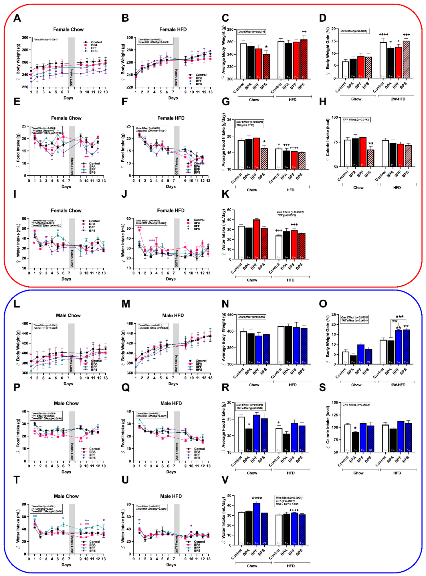
Figure 5.
Interaction effects of prenatal bisphenol exposure and postnatal high-fat diet challenge on body composition. Top panels represent female offspring, and bottom panels represent male offspring. Body composition was assessed before HFD exposure (week 14) and after two weeks of HFD challenge (week 16). Percent changes in total body mass (A,B), fat mass (C,D), fluid mass (E,F), and lean mass (G,H) were determined in control and prenatally exposed offspring maintained on chow or HFD. Data are presented as mean ± SEM for 6–14 offspring per treatment group and expressed as percent change. Sample sizes are indicated within the bars. Statistical analyses were performed using two-way ANOVA followed by Bonferroni post hoc tests. *P < 0.05, **P < 0.01, ***P < 0.001 vs control; +P < 0.05, ++P < 0.01, +++P < 0.001 vs chow diet.
Figure 5.
Interaction effects of prenatal bisphenol exposure and postnatal high-fat diet challenge on body composition. Top panels represent female offspring, and bottom panels represent male offspring. Body composition was assessed before HFD exposure (week 14) and after two weeks of HFD challenge (week 16). Percent changes in total body mass (A,B), fat mass (C,D), fluid mass (E,F), and lean mass (G,H) were determined in control and prenatally exposed offspring maintained on chow or HFD. Data are presented as mean ± SEM for 6–14 offspring per treatment group and expressed as percent change. Sample sizes are indicated within the bars. Statistical analyses were performed using two-way ANOVA followed by Bonferroni post hoc tests. *P < 0.05, **P < 0.01, ***P < 0.001 vs control; +P < 0.05, ++P < 0.01, +++P < 0.001 vs chow diet.
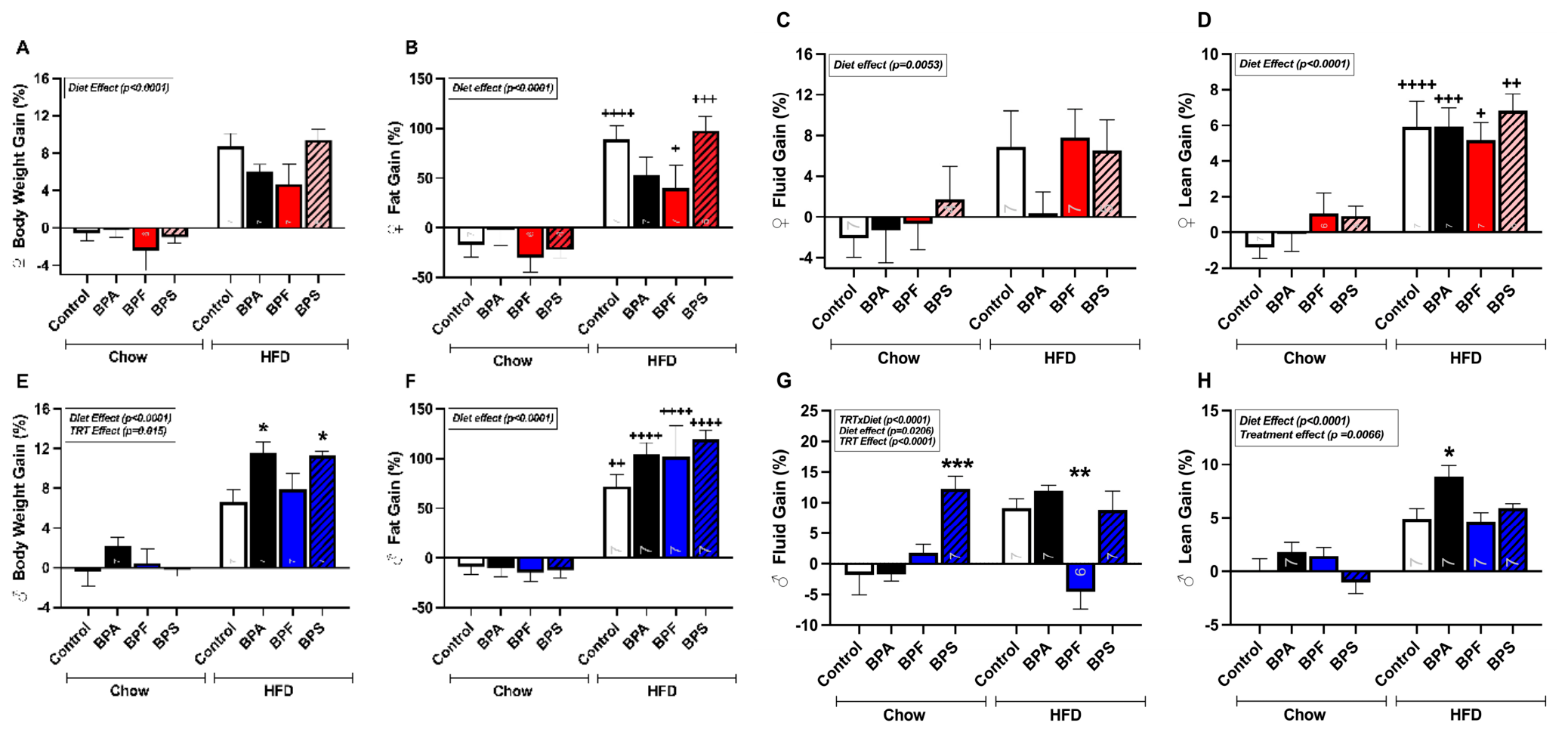
Figure 6.
Oral Glucose tolerance tests of control and Bisphenol treated offspring before and after HF diet exposure. Top panel reflect changes in female offspring and bottom panel depict changes in male offspring. One-way ANOVA was used to analyze fasting levels of glycemia (Fig. B,I) pre-HFD. Two-way ANOVA followed by Fisher’ LSD was used to analyze the differences in blood glucose levels over 120 minutes in control and treated offspring at week 14 pre-HFD exposure (Fig. A,H), after 2 weeks (Fig. C,J for Chow and Fig. D,K for HFD) and 4 weeks (Fig. E,L for Chow and Fig. F,M for HFD) of diet exposure. Data are presented as mean ± SEM for group averages of 4-14 offspring per treatment and diet group. Sample sizes are provided inside the bars. P values correspond to the main (Diet or treatment) or interaction effects. Asterisk (*) represents a significant difference from the control group (*p < 0.05, **p < 0.01, ***p < 0.001, ****p < 0.0001).
Figure 6.
Oral Glucose tolerance tests of control and Bisphenol treated offspring before and after HF diet exposure. Top panel reflect changes in female offspring and bottom panel depict changes in male offspring. One-way ANOVA was used to analyze fasting levels of glycemia (Fig. B,I) pre-HFD. Two-way ANOVA followed by Fisher’ LSD was used to analyze the differences in blood glucose levels over 120 minutes in control and treated offspring at week 14 pre-HFD exposure (Fig. A,H), after 2 weeks (Fig. C,J for Chow and Fig. D,K for HFD) and 4 weeks (Fig. E,L for Chow and Fig. F,M for HFD) of diet exposure. Data are presented as mean ± SEM for group averages of 4-14 offspring per treatment and diet group. Sample sizes are provided inside the bars. P values correspond to the main (Diet or treatment) or interaction effects. Asterisk (*) represents a significant difference from the control group (*p < 0.05, **p < 0.01, ***p < 0.001, ****p < 0.0001).
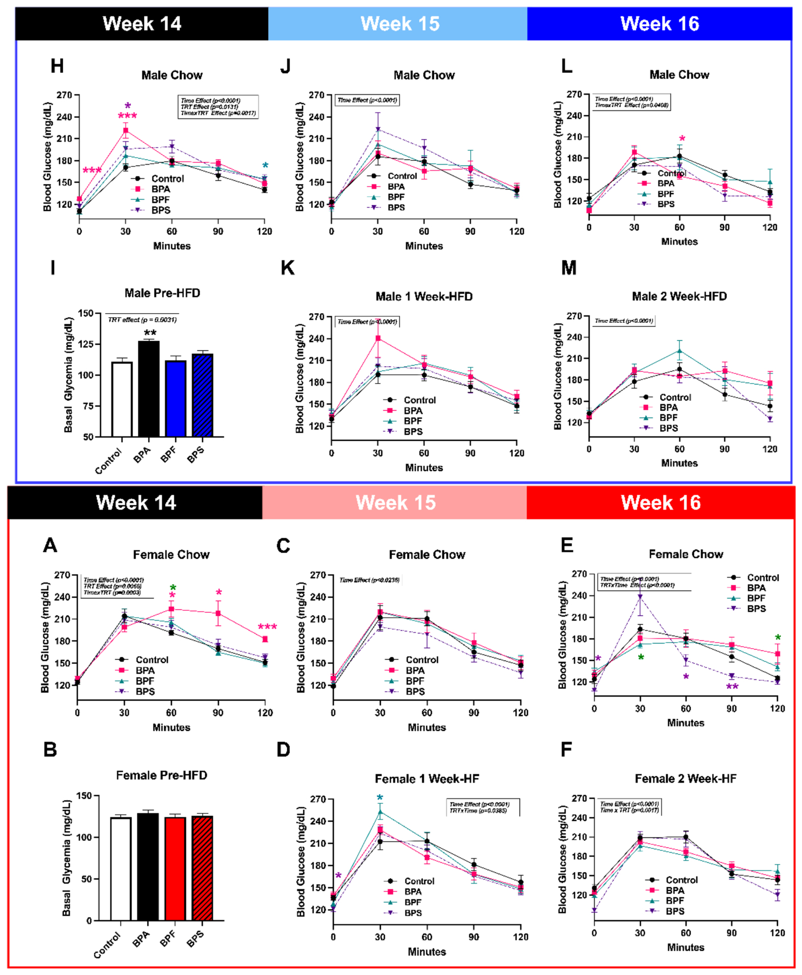
Figure 7.
Interaction effect of postnatal high-fat challenge and in utero treatment of Bisphenols on organ relative weights and metabolic parameters. Two left panels reflect changes in female offspring, and two right panels depict changes in male offspring. In experiment 1, serum and organs were isolated after sacrifice at week 16. Liver (Fig. A,G), pancreas (Fig. C,I), and adrenal (Fig. E,K) relative weights were measured. Liver cholesterol was extracted and quantified (μM/μg; Fig. B,D). Serum and was further processed for insulin (pM; Fig. D,J) , corticosterone (ng/mL; Fig. F,L). Data are analyzed using Two-way ANOVA followed by Bonferroni post hoc test and presented as mean ± SEM for group averages of 6-7 offspring per treatment group. Sample sizes are provided inside the bars. P-values correspond to the main effects (Diet or treatment). Comparisons within treatment: *p < 0.05.
Figure 7.
Interaction effect of postnatal high-fat challenge and in utero treatment of Bisphenols on organ relative weights and metabolic parameters. Two left panels reflect changes in female offspring, and two right panels depict changes in male offspring. In experiment 1, serum and organs were isolated after sacrifice at week 16. Liver (Fig. A,G), pancreas (Fig. C,I), and adrenal (Fig. E,K) relative weights were measured. Liver cholesterol was extracted and quantified (μM/μg; Fig. B,D). Serum and was further processed for insulin (pM; Fig. D,J) , corticosterone (ng/mL; Fig. F,L). Data are analyzed using Two-way ANOVA followed by Bonferroni post hoc test and presented as mean ± SEM for group averages of 6-7 offspring per treatment group. Sample sizes are provided inside the bars. P-values correspond to the main effects (Diet or treatment). Comparisons within treatment: *p < 0.05.
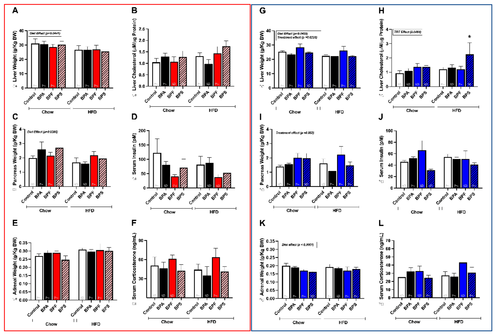
Figure 8.
Interaction effect of postnatal high-fat challenge and in utero treatment of Bisphenols on adipose tissue relative weights. Top panels reflect changes in female offspring and bottom panels depict changes in male offspring. In experiment 1, organs were dissected and weighed after sacrifice at week 16. Relative weights of perirenal (Fig. A,B), abdominal (Fig. C,D), gonadal (Fig. E,F), and brown (Fig. G,H) adipose tissues were measured. Data are analyzed using Two-way ANOVA followed by Bonferroni post hoc test and presented as mean ± SEM for group averages of 6-14 offspring per treatment group. Sample sizes are provided inside the bars. P-values correspond to the main effects (Diet or treatment). Comparisons within treatment: *p < 0.05. Abbreviations: PAT=Perirenal adipose tissue, AAT=Abdominal adipose tissue; OAT=Ovarian adipose tissue; BAT = Brown adipose tissue.
Figure 8.
Interaction effect of postnatal high-fat challenge and in utero treatment of Bisphenols on adipose tissue relative weights. Top panels reflect changes in female offspring and bottom panels depict changes in male offspring. In experiment 1, organs were dissected and weighed after sacrifice at week 16. Relative weights of perirenal (Fig. A,B), abdominal (Fig. C,D), gonadal (Fig. E,F), and brown (Fig. G,H) adipose tissues were measured. Data are analyzed using Two-way ANOVA followed by Bonferroni post hoc test and presented as mean ± SEM for group averages of 6-14 offspring per treatment group. Sample sizes are provided inside the bars. P-values correspond to the main effects (Diet or treatment). Comparisons within treatment: *p < 0.05. Abbreviations: PAT=Perirenal adipose tissue, AAT=Abdominal adipose tissue; OAT=Ovarian adipose tissue; BAT = Brown adipose tissue.
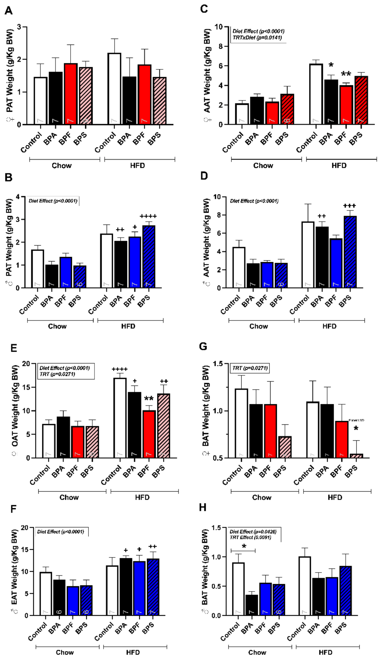
Figure 9.
Effects of prenatal bisphenol exposure and postnatal high-fat diet challenge on serum lipid profile. Top panels represent female offspring, and bottom panels represent male offspring. Serum lipid parameters were measured at 16 weeks of age following two weeks of high-fat diet (HFD) challenge or maintenance on chow diet. Measurements include total cholesterol (A,B), HDL cholesterol (C,D), and triglycerides (E,F). Data are presented as mean ± SEM for group averages of 6–14 offspring per treatment group. Statistical analyses were performed using two-way ANOVA followed by Bonferroni post hoc tests. *P < 0.05, **P < 0.01, ***P < 0.001 vs control; +P < 0.05, ++P < 0.01 vs chow diet.
Figure 9.
Effects of prenatal bisphenol exposure and postnatal high-fat diet challenge on serum lipid profile. Top panels represent female offspring, and bottom panels represent male offspring. Serum lipid parameters were measured at 16 weeks of age following two weeks of high-fat diet (HFD) challenge or maintenance on chow diet. Measurements include total cholesterol (A,B), HDL cholesterol (C,D), and triglycerides (E,F). Data are presented as mean ± SEM for group averages of 6–14 offspring per treatment group. Statistical analyses were performed using two-way ANOVA followed by Bonferroni post hoc tests. *P < 0.05, **P < 0.01, ***P < 0.001 vs control; +P < 0.05, ++P < 0.01 vs chow diet.
Disclaimer/Publisher’s Note: The statements, opinions and data contained in all publications are solely those of the individual author(s) and contributor(s) and not of MDPI and/or the editor(s). MDPI and/or the editor(s) disclaim responsibility for any injury to people or property resulting from any ideas, methods, instructions or products referred to in the content. |
© 2026 by the authors. Licensee MDPI, Basel, Switzerland. This article is an open access article distributed under the terms and conditions of the Creative Commons Attribution (CC BY) license (http://creativecommons.org/licenses/by/4.0/).
Copyright: This open access article is published under a Creative Commons CC BY 4.0 license, which permit the free download, distribution, and reuse, provided that the author and preprint are cited in any reuse.