

Submitted:
15 October 2026
Posted:
15 October 2026
You are already at the latest version
Abstract
The adult mammalian heart does not regenerate. After myocardial infarction, cardiomyocytes are lost permanently because the surviving cells are terminally differentiated and post-mitotic. Regenerative vertebrates such as zebrafish and newts retain proliferative, centriole-bearing cardiomyocytes and repair the heart completely, whereas mammals lose this capacity within the first postnatal week. I propose that the centriole functions as a differentiation ratchet: a heritable structural input that couples proliferative potential to centriole integrity through the mitotic surveillance pathway (MSP). The strongest natural evidence comes from the heart. Shortly after birth, mammalian cardiomyocytes undergo programmed centrosome disassembly, with pericentriolar material relocating to the nuclear envelope. Pharmacological depletion of centrioles is sufficient to drive the post-mitotic transition in human cardiomyocytes, and failure of this step is pathogenic: a mutation in the centrosomal gene RTTN causes infantile dilated cardiomyopathy. Because the adult cardiomyocyte is one of the few naturally and permanently acentriolar somatic cells, it offers a ready-made testbed for asking whether the ratchet can be reversed. A single-nucleus transcriptomic analysis of the developing human heart (fetal, young and adult) presented here shows that this shutdown is actively installed during maturation rather than a passive decay: adult cardiomyocytes reduce their centriolar program—PLK4, CEP192, SASS6, STIL and NEK2 are all down-regulated—while the p53 effector p21 (CDKN1A) is elevated, the signature of a p53-maintained post-mitotic lock. The roadmap proceeds in three steps, all conducted in cell culture: (i) release the G0 lock and assess TRIM37 and PLK1 status; (ii) restore a licensed centriole from an autologous low-entropy adult stem cell, with PLK1 and Ect2 co-delivery; and (iii) enforce a strict safety gate that includes TRIM37, p53 pathway, and contractile function assessment. Eight pre-registered, falsifiable hypotheses are stated below, and the principal risks are analyzed.
Keywords:
centriole
; centrosome
; differentiation
; cardiomyocyte
; cardiac regeneration
; acentriolar state
; cell-cycle re-entry
; TRIM37
; mitotic surveillance
; PLK1
1. Introduction: The Regeneration Gap of the Adult Mammalian Heart
Heart failure after myocardial infarction is a leading cause of death in the aging population, and the fundamental obstacle to repair is cellular. The adult mammalian heart loses a large fraction of its cardiomyocytes to injury, and the survivors are effectively post-mitotic: their proliferative capacity is negligible, so damaged tissue is replaced by non-contractile scar rather than by new muscle (Tzahor & Poss, 2017). The adult human heart does retain a low but measurable rate of cardiomyocyte renewal, with a transient wave of proliferation in early life (Bergmann et al., 2009; Mollova et al., 2013), but the magnitude of this turnover is far too small to restore an infarcted myocardium (Alkass et al., 2015; Leone et al., 2015).
The failure to regenerate is not a universal feature of vertebrates. Zebrafish regenerate a resected or cryoinjured heart completely, through dedifferentiation and proliferation of pre-existing cardiomyocytes (Poss et al., 2002; Jopling et al., 2010; Kikuchi et al., 2010), and adult newts rebuild the ventricular wall by recapitulating developmental cardiogenesis (Witman et al., 2011). A key cellular correlate of this difference is that regenerative species retain centriole-bearing, division-competent cardiomyocytes, whereas mammals lose centrosome integrity within the first postnatal week (Zebrowski et al., 2015). Understanding what locks the mammalian cardiomyocyte out of the division cycle—and whether that lock can be released safely—is therefore a central problem of cardiac biology and regenerative medicine.
The developmental timing of the lock is well characterized. In mice and rats, cardiomyocytes exit the cell cycle within the first postnatal week, in a wave of terminal maturation marked by binucleation, centrosome disassembly, and engagement of p53 signaling (Li et al., 1996; Soonpaa et al., 1997; Zebrowski et al., 2015; Liu et al., 2007). In human cardiomyocytes, centrosome reduction occurs during maturation, with pericentriolar material relocalizing from the centriole to the nuclear envelope (Ng et al., 2020). The neonatal mouse heart can regenerate after apex resection at postnatal day 1 but loses this capacity by day 7 (Porrello et al., 2011). Once the window closes, injury elicits a fibrotic rather than a proliferative response, indicating that the post-mitotic state is actively maintained by developmental programs rather than passively decayed (Porrello et al., 2011; Tzahor & Poss, 2017).
The concept that the centriole might couple differentiation state to proliferative capacity has a long history. More than two decades ago, Tkemaladze and Chichinadze proposed that the centriole (diplosome) could act as a self-reproducing determinant of morphogenetic status, carrying differentiation information through asymmetric inheritance (Tkemaladze & Chichinadze, 2005a, 2005b, 2010; Tkemaladze, 2023). The present framework retains that structural intuition but specifies a molecular execution mechanism—the mitotic surveillance pathway (MSP)—that makes the hypothesis experimentally tractable in a defined cell type.
2. The Centriole as a Differentiation Ratchet
2.1. The Hypothesis
I propose that the centriole functions as a differentiation ratchet: a heritable structural input that couples proliferative potential to centriole integrity through the mitotic surveillance pathway (MSP). In the conventional view, the centriole and the centrosome are division organelles required to organize the mitotic spindle. The ratchet view differs in one essential respect: it assigns the centriole a state-fixing role through the MSP. A cell that retains functional centrioles remains division-competent; a cell that has lost or dismantled its centrioles experiences delayed mitotic spindle assembly, which activates the MSP and locks the cell into a differentiated, non-proliferative state.
The MSP is a timing mechanism that monitors mitotic duration, treating abnormally prolonged mitosis as a sign of cellular stress (Sparr & Meitinger, 2025). When a defined mitotic duration is exceeded, the 53BP1–USP28 module stabilizes p53, which activates p21 and induces durable G1 arrest (Fong et al., 2016; Lambrus et al., 2016; Meitinger et al., 2016). The centriole thus serves as one structural input, among several, that determines whether mitosis completes within the surveillance window.
The MSP is not unique to centriole loss: other distinct defects that prolong mitosis—kinetochore–microtubule attachment errors, spindle assembly checkpoint dysfunction, or delayed chromosome congression—also activate the 53BP1–USP28–p53 axis and induce durable G1 arrest (Fong et al., 2016). What distinguishes the centriole from these transient triggers is its role as a heritable structural marker. The mother–daughter asymmetry of the centriole pair is not reset at each division; it carries forward information about replicative history through the asymmetric inheritance of the older mother centriole, which is distinguished by maturation-associated post-translational modifications (Yamashita et al., 2007; Wang et al., 2009; Nigg & Holland, 2018). A spindle assembly error is corrected or the cell dies; it does not accumulate across divisions. Centriole age does. The ratchet metaphor is therefore precise in one respect: the centriole converts a transient mitotic event (delayed spindle assembly) into a heritable state (differentiation) through the directional inheritance of a structurally aged organelle.
This ratchet is not absolute, however. GSK-3β inhibition reverses centrosome reduction in postnatal rat cardiomyocytes and coincides with reinitiated proliferation (Ng et al., 2020). The differentiated-to-proliferative (D→P) transition, while strongly disfavored under physiological conditions, is not mechanistically forbidden. The ratchet is better described as a highly asymmetric switch than as an irreversible lock.
The state that is fixed is not a simple binary of dividing versus not dividing. The centriole is a carrier of differentiation information through its asymmetric inheritance. In the Drosophila male germline, the mother centrosome remains anchored at the hub–germline-stem-cell interface and is inherited by the self-renewing stem cell, whereas the daughter centrosome migrates away with the differentiating daughter (Yamashita et al., 2007). In the developing mouse neocortex, the centrosome retaining the older mother centriole stays in the ventricular zone and is preferentially inherited by renewing radial glial progenitors; removal of ninein disrupts this asymmetric segregation and causes premature depletion of the progenitor pool (Wang et al., 2009; Imai et al., 2010). A transplanted centriole could therefore anchor a specific differentiation state through mechanisms that remain to be defined.
Three implications follow. Centriole loss can promote differentiation through MSP activation. Restoring proliferative potential requires overcoming the MSP, either by restoring rapid mitosis or by modulating the p53 pathway. The system is resettable in specific windows, as demonstrated by the natural acentriolar windows of reproduction.
2.2. Evidence that centriole loss locks differentiation through the MSP
The clearest somatic example is the heart. In mice and rats, cardiomyocytes lose centrosome integrity shortly after birth: centrosomal proteins relocalize from the centriole to the nuclear envelope, ciliogenesis is lost, and the nuclear envelope takes over as the microtubule-organizing center (Zebrowski et al., 2015; Ng et al., 2020; Vergarajauregui et al., 2020). Zebrowski and colleagues concluded that loss of centrosome integrity “is associated with, and can promote,” cell-cycle exit (Zebrowski et al., 2015). Adult zebrafish and newt cardiomyocytes, which retain proliferative capacity, maintain centrosome integrity (Zebrowski et al., 2015; Jopling et al., 2010; Witman et al., 2011). In the rat, binucleated postnatal cardiomyocytes can re-enter the cell cycle but divide poorly, with a ceiling near 6% (Leone & Engel, 2019).
The causal direction was tested directly in human induced pluripotent stem cell–derived cardiomyocytes. Pharmacological depletion of centrioles with centrinone, a PLK4 inhibitor, was sufficient to drive the post-mitotic transition and aspects of maturation (Ng et al., 2020). The same study identified an upstream trigger: activation of the Hippo kinases MST1/2 caused centrosome disassembly and relocalization of pericentriolar material to perinuclear membranes, an effect mediated by the kinases themselves rather than by YAP–TEAD, because the TEAD inhibitor verteporfin had no effect on centrosome organization (Ng et al., 2020). This connects the Hippo pathway, long known to restrain cardiomyocyte proliferation and heart size (Heallen et al., 2011), directly to the centrosome. Mechanistically, MST1/2 cooperate with the centrosomal kinase NEK2 to phosphorylate C-Nap1 and rootletin, the linker proteins required for centrosome cohesion, triggering centrosome disjunction—the detachment of mother and daughter centrioles—at the onset of mitosis (Mardin et al., 2010). This cell-cycle-regulated event is distinct from the developmental centrosome reduction program described above, which operates over a timescale of days during postnatal cardiomyocyte maturation.
Reversibility of centrosome reduction. Ng et al. (2020) noted that in postnatal rat cardiomyocytes, GSK-3β inhibition reversed centrosome reduction in parallel with reinitiated proliferation. This observation suggests that remnant centrosomes in postnatal myocytes may retain functionality and that the capacity to reassemble centrosomes may decline with age. This finding directly supports the core hypothesis that the ratchet is reversible: if centrosome reduction can be pharmacologically reversed, then the logical premise of centriole transplantation—that the acentriolar state is not an irreversible terminal state—is independently validated.
Centrosome loss in hPSC-derived cardiomyocytes appeared irreversible: washout after five days of centrinone did not restore centrosome numbers (Ng et al., 2020). This contrasts with cancer cell lines, which return to an intrinsic centrosome set point after centrinone washout (Wong et al., 2015), and with untransformed cells, which can progress through G1 without centrioles and assemble them de novo after centrosome removal (Uetake et al., 2007).
The mechanistic link to MSP is well established. Genome-wide CRISPR screens identified 53BP1 and USP28 as essential components acting upstream of p53 in response to centrosome loss (Fong et al., 2016; Lambrus et al., 2016; Meitinger et al., 2016). USP28 directly deubiquitinates p53, and 53BP1 bridges the interaction, stabilizing p53 and evoking p21-dependent arrest. This pathway also responds to prolonged mitosis independently of centrosome loss, indicating that the centriole is not the sole determinant of arrest (Fong et al., 2016). The MSP threshold is approximately 90 minutes in the cell types examined: 53BP1 and USP28 stabilize p53 after centrosome loss or extended mitotic duration (>90 minutes) (Fong et al., 2016).
TRIM37 as an enforcer of singularity. A second determinant is TRIM37, a centrosomal ubiquitin ligase. Meitinger et al. (2020) demonstrated that acentrosomal spindle assembly following PLK4 inhibition depends on TRIM37 levels. Low TRIM37 accelerates acentrosomal spindle assembly and improves proliferation; high TRIM37 inhibits acentrosomal spindle assembly through degradation of the centrosomal component CEP192, leading to mitotic failure (Meitinger et al., 2020). TRIM37 prevents PLK4 from self-assembling into centrosome-independent condensates that serve as ectopic microtubule-organizing centers. This establishes TRIM37 as a critical modifier of the cellular response to centriole loss (see also Yeow et al., 2020; Balestra et al., 2021).
Functional precision of TRIM37. The same CRISPR screen showed that TRIM37 knockout cells form ectopic centrosomal-component foci that suppress the mitotic defects associated with centrosome loss (Meitinger et al., 2016). For the present hypothesis this matters in two ways. A low-TRIM37 state may be permissive for exogenous centriole integration, because TRIM37 restricts the formation of ectopic microtubule-organizing centers; conversely, high TRIM37 degrades CEP192 and inhibits acentrosomal spindle assembly, which could prevent proliferation even of cells that carry an imported centriole (Meitinger et al., 2020).
Failure of the ratchet is disease. A patient with infantile dilated cardiomyopathy carried compound heterozygous mutations in RTTN (rotatin), a centrosomal protein; whole-exome sequencing, together with CRISPR/Cas9 knockout and correction, identified RTTN as the causal gene (Chun et al., 2023). In the patient’s cardiomyocytes, the centrosome persisted at the centriole instead of undergoing the programmed relocalization to the nuclear envelope, producing global microtubule-network defects and impaired maturation (Chun et al., 2023). The case links a defect in the programmed relocation step to a non-syndromic human cardiomyopathy. This is an n = 1 observation and is treated as plausibility evidence, not as a confirmed mechanism.
Independent validation: centrosome loss triggers differentiation. Renzova et al. (2018) provided independent validation of the causal link between centrosome loss and differentiation. Using human embryonic and induced pluripotent stem cells, they demonstrated that inactivation of the PLK4–STIL module leads to centrosome depletion, which in turn triggers prolonged mitosis, p53 stabilization, loss of pluripotency markers, and induction of differentiation. Acentrosomal mitosis was markedly prolonged relative to controls, and this prolongation correlated with p53 activation and the loss of pluripotency markers. This work establishes that centrosome loss is not merely associated with differentiation but can actively promote it through mechanisms that parallel the mitotic surveillance pathway.
2.3. One-Way Operation and Resettable Windows
The germline and the early embryo show that the ratchet can be reset. In oocytes, centrioles are eliminated and the meiotic spindle assembles without canonical centrosomes; after fertilization, the embryo re-establishes its centrioles from the paternally delivered centriole (Manandhar et al., 2005; Avidor-Reiss et al., 2015; Fishman et al., 2018). Pharmacological centriole depletion is reversible in cancer cell lines, which recover to their centriole set point after washout (Wong et al., 2015). In contrast, untransformed cells that lose centrosomes arrest in a p53-, p38-, and p21-dependent G1 state that is not reversed by washout (Mikule et al., 2007; Wong et al., 2015). Normal cells can pass through G1 without centrioles and rebuild them de novo after removal, indicating that the centrosome is not an obligatory component of the G1-to-S transition machinery (Uetake et al., 2007).
Centriole duplication is governed by a licensing mechanism tightly coupled to the cell cycle. The PLK4–STIL complex assembles in G1, separated in time from centriole assembly in S phase; this can be read as a licensing event that ensures duplication occurs once and only once per cycle (Arquint & Nigg, 2016; Nigg & Holland, 2018). CDK1–cyclin B binds STIL and competes with PLK4 binding, preventing untimely licensing in S and G2 (Zitouni et al., 2016). Within a single cycle, a G1 cell carries a mother and a daughter centriole joined by a linker; at the G1-to-S transition a procentriole assembles orthogonally at the proximal end of each pre-existing centriole, and toward the end of G2 the linker is severed to allow centrosome separation and bipolar spindle assembly (Fırat-Karalar & Stearns, 2014; Mardin et al., 2010).
Recent work has clarified the centriole elimination program during oogenesis across model organisms. In C. elegans, 88% of somatic cells lose their centrioles during embryogenesis, following a rigid developmental schedule (Kalbfuss & Gönczy, 2023). In planarians, which have lost centrosomes, proliferating neoblasts divide without centrioles, and centrioles are assembled de novo only in terminally differentiating ciliated cells (Azimzadeh et al., 2012). In Drosophila, Polo kinase activity maintains pericentriolar material integrity, and its decline triggers elimination (Pimenta-Marques et al., 2016).
Mechanism of centriole elimination. Pimenta-Marques et al. (2016) demonstrated that centriole stability is not an intrinsic property but requires a Polo kinase– and PCM-dependent maintenance program. PCM downregulation during oogenesis leads to centriole loss; ectopic expression of Polo kinase tethered to centrioles prevented PCM loss and maintained functional centrioles during meiosis, when they are normally eliminated entirely. This finding redefines the “lifespan” of the centriole as a tissue-specific, regulated program rather than a fixed structural feature. Pimenta-Marques et al. (2024) further showed that Polo kinase requires ANA1/CEP295 to maintain centriole stability and that ANA1 expression prevents centriole loss upon PCM downregulation.
Unpublished hypothesis. A related hypothesis, not yet peer-reviewed, proposes that centriole elimination may serve as a gateway to reprogramming (Tqemaladze, unpublished observations). This concept aligns with our framework but is presented here as an unverified proposition rather than established evidence.
2.4. Transcriptomic Evidence: The Centriole and Proliferation Programs are Developmentally Shut Down in Human Cardiomyocytes
If the ratchet view is correct, the transcriptome of human cardiomyocytes should show a coordinated developmental shutdown of the centriolar machinery as the cells become post-mitotic, together with the engagement of the p53–p21 effector arm that maintains the arrest. I tested this prediction directly in a published single-nucleus RNA-sequencing dataset of healthy human left-ventricular myocardium spanning three developmental stages—fetal (n = 3 donors), young (n = 3) and adult (n = 3) (Sim et al., 2021; GEO accession GSE156703). After quality control and restriction to cardiomyocyte nuclei (TNNT2/MYH6/MYH7/NPPA/TTN expression), 44,938 of 50,511 nuclei were retained (Fetal 25,306; Young 12,374; Adult 7,258). Three pre-specified gene modules were scored per nucleus: a centriole/centrosome module (33 structural and assembly genes), an MSP/p53 module (14 genes), and a proliferation module (28 genes).
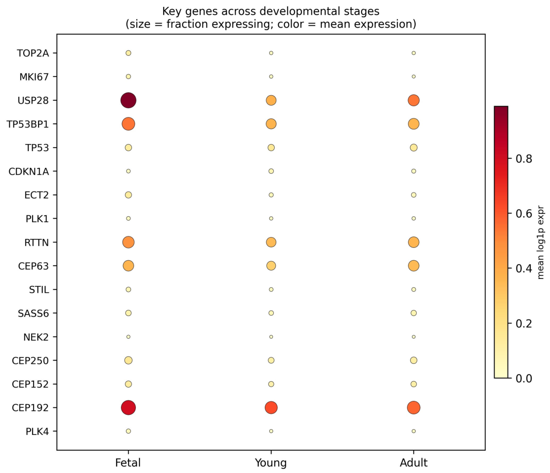
Centriolar program shutdown. The centriole module score declined from fetal to adult cardiomyocytes (mean ± SEM: Fetal −0.013 ± 0.001 vs Adult −0.041 ± 0.001; two-sided Mann–Whitney p = 1.4 × 10⁻¹¹⁸; rank-biserial r = −0.18), with the same direction in every donor (Figure 1a; Figure 2). The reduction was broad rather than confined to one component: the centriole-licensing kinase PLK4 (log2 fold change, Adult vs Fetal = −2.14), the cartwheel/assembly factors SASS6 (−0.27) and STIL (−1.26), the pericentriolar scaffold CEP192 (−0.48), the centrosome linker component CEP250 (−0.44), the centrosome-cohesion kinase NEK2 (−3.48), and the cytokinesis/cytoskeletal factor ECT2 (−1.34) were all reduced, as was RTTN (−0.39), the gene whose mutation causes infantile dilated cardiomyopathy. TRIM37, the enforcer of centriole singularity whose status is assessed in Step 1 of the roadmap, was essentially unchanged (log2 FC ≈ 0.01; p = 0.55), consistent with TRIM37 acting as a constitutive modifier rather than a developmentally regulated switch.
Exit from the cell cycle. The proliferation module collapsed in adult cardiomyocytes (Fetal 0.021 ± 0.001 vs Adult −0.029 ± 0.001; p < 10⁻³⁰⁰; r = −0.49), driven by markers such as MKI67 (log2 FC = −2.96) and TOP2A. Adult cardiomyocytes have therefore transcriptionally exited the division cycle, as expected for a post-mitotic, naturally acentriolar cell.
p53–p21 effector engagement. The p53 pathway effector CDKN1A (p21) and TP53 itself were elevated in adult relative to fetal cardiomyocytes (CDKN1A log2 FC +0.35, p = 4 × 10⁻¹⁰; TP53 +0.23, p = 1 × 10⁻⁷), consistent with a p53–p21-maintained G0 lock. The module-level MSP/p53 score was lower in adult nuclei (Fetal 0.067 ± 0.001 vs Adult 0.050 ± 0.001; p = 4 × 10⁻²²), and the mitotic-duration sensors TP53BP1 and USP28 were transcriptionally reduced (USP28 log2 FC = −1.13). This is the expected pattern for a cell that has already left the cycle: the sensors that monitor prolonged mitosis are no longer transcribed once mitosis has stopped, whereas the effector that maintains the durable arrest—p21—remains elevated. The combination of centriolar-program shutdown, proliferation collapse, and elevated p21 in the same cell population is the signature the ratchet framework predicts for the adult, naturally acentriolar cardiomyocyte.
3. The Adult Cardiomyocyte as a Natural Acentriolar Testbed
The adult mammalian cardiomyocyte is one of the few terminally differentiated somatic cells that are naturally and permanently acentriolar. This makes it an unusually clean experimental system: rather than inventing safe centriole elimination, we need to ask whether the lock can be opened and a fresh, licensed centriole installed. The transcriptomic shutdown of the centriolar program across human cardiac development (Section 2.4) confirms that this lock is actively installed during maturation rather than passively decayed.
Three features make the system attractive. First, the acentriolar state is structurally well characterized: the nuclear-envelope microtubule-organizing center is organized by AKAP6, which tethers centrosomal proteins and links the Golgi and nucleus via AKAP9, generating two pools of microtubules (Vergarajauregui et al., 2020; Zebrowski et al., 2015). Second, releasing the lock is experimentally accessible by published means: constitutively active YAP stabilizes microtubules and drives sarcomere disassembly and cell-cycle re-entry in adult cardiomyocytes (Zhang et al., 2026); clonal analysis of YAP5SA-expressing cardiomyocytes shows efficient division, with >20% of clones containing ≥2 cells and an S phase of ≈48 hours, whose completion is promoted by P21 inhibition (Morikawa et al., 2025); the GSK-3β inhibitor CHIR99021 combined with the p300/CBP inhibitor A-485 induces regenerative cardiac cells and improves post-infarction heart function in mice (Zhou et al., 2024); and transient OSK (Oct4, Sox2, Klf4) reprogramming primes dedifferentiation and overcomes the cytokinesis barrier (Abad et al., 2013; Yan et al., 2026). Third, the contrast case is built in: zebrafish and newt adult cardiomyocytes retain centrosome integrity and divide (Zebrowski et al., 2015; Jopling et al., 2010; Witman et al., 2011).
Primary cilia, which are nucleated from the mother centriole, reinforce the view of the centriole as a signaling hub in cardiac development. Cilia coordinate early cardiogenesis and hedgehog signaling in cardiomyocyte differentiation: the pluripotent P19.CL6 mouse line, which can differentiate into beating cardiomyocytes, forms primary cilia that contain essential hedgehog pathway components, and disrupting cilia blocks hedgehog signaling and the appearance of beating cardiomyocytes; Ift88-null mouse embryos, which form no cilia, show ventricular dilation and abnormal outflow tract development (Clement et al., 2009).
4. Experimental Roadmap
The roadmap comprises three steps, all conducted in cell culture; no manipulation is performed in the intact heart.
4.1. Step 1—Release the G0 Lock and Assess TRIM37 and PLK1 Status
Dedifferentiate human or mammalian cardiomyocytes in culture using verified levers: YAP5SA-mediated microtubule stabilization (Zhang et al., 2026; Morikawa et al., 2025), the CHIR99021 + A-485 two-compound protocol (Zhou et al., 2024), and transient OSK reprogramming (Yan et al., 2026). Readouts follow established methodology (Leone et al., 2015): sarcomere disassembly (α-actinin displacement), cell-cycle re-entry (Ki67, phospho-H3, EdU incorporation), and G0-exit transcriptomes. A compound advances to the next phase only if it produces both sarcomere disassembly and S-phase entry; a lever that yields one without the other is scored as partial and is not carried forward. Each lever is run with matched non-cardiomyocyte controls, and with at least three biological replicates.
A. Release the G0 lock. The levers described above.
B. Suppress the elimination program and assess TRIM37 and PLK1 status. Centriole stability is not intrinsic but requires a Polo kinase– and PCM-dependent maintenance program. TRIM37, as the enforcer of centrosome assembly singularity, sets the direction of the cellular response to centriole loss: high TRIM37 inhibits acentrosomal spindle assembly through CEP192 degradation, leading to mitotic failure; low TRIM37 accelerates acentrosomal spindle assembly, improving proliferation. Step 1 must therefore include: (i) quantification of TRIM37 levels by qPCR and Western blot; (ii) determination of the “acceptable zone”—a TRIM37 level that permits rapid mitosis when a functional centriole is present, but not when it is absent; (iii) if TRIM37 is too high, testing whether temporary TRIM37 suppression (siRNA or small molecule) creates a licensing window that allows the transplanted centriole to functionally integrate rather than be eliminated. PLK1 expression and activity must also be measured, because PLK1 is required for centriole maturation and functional MTOC formation.
In parallel, quantify TRIM37 expression and protein levels in the cardiomyocyte population. If TRIM37 is high, test whether TRIM37 knockdown or inhibition facilitates acentrosomal proliferation (Meitinger et al., 2020). If TRIM37 is low, determine why centriole loss still triggers arrest—likely through MSP activation rather than acentrosomal spindle failure.
4.2. Step 2—Centriole import and PLK1/Ect2 co-delivery in cell culture
Centrioles are isolated from autologous low-entropy adult stem cells using established centrosome-isolation protocols (Mitchison & Kirschner, 1984; Bornens et al., 1987), prepared and licensed in vitro (≤2 centrioles, correct cell-cycle phase, intact ultrastructure verified by electron microscopy), and transferred into the G0-released cell by microinjection or membrane fusion. The dual purpose of the transplant is explicit: (i) to anchor the desired differentiation through the state-fixing role of the imported centriole (Yamashita et al., 2007; Wang et al., 2009)—and (ii) to restore rapid mitotic spindle assembly, thereby preventing MSP activation.
The feasibility of transferring a functional centrosome into a recipient mammalian cell has been demonstrated in principle (Heidemann & Kirschner, 1975; Khodjakov & Rieder, 2001), but not, to my knowledge, with the efficiency, purity, or post-transfer functional persistence required for the cardiomyocyte application. Centriole import is therefore treated as a technical hypothesis to be validated in Step 2, not as an established capability. Pre-registered feasibility milestones: (i) recovery of structurally intact centrioles from autologous stem cells at ≥70% purity as assessed by electron microscopy and pericentrin immunoblot; (ii) microtubule nucleation from imported centrioles in ≥50% of recipient cells within 2 hours of delivery, quantified by cold-recovery assays; (iii) persistence of imported centrioles through at least one cell cycle in ≥30% of recipients. Failure to meet these milestones terminates the import arm and redirects the roadmap to pharmacological or genetic reconstitution of centriole biogenesis in situ.
Overcoming PLK1 deficiency. A critical obstacle to centriole functional integration is the postnatal loss of PLK1 expression. Coxon et al. (2009) demonstrated that PLK1 mRNA and protein decline progressively during postnatal cardiomyocyte development, becoming undetectable in adult rat hearts. Overexpression of wild-type or constitutively active Plk1(T210D) alone was insufficient to induce cell cycle progression in isolated neonatal or adult rat cardiomyocytes, because PLK1 failed to increase Cdc2/cyclin B1 activity. However, Lam et al. (2025) showed that combined overexpression of Plk1(T210D) and Ect2—a RhoA guanine nucleotide exchange factor—induces cytokinesis in postnatal cardiomyocytes; in the prevailing model, Plk1 phosphorylates RACGAP1 (HsCyk-4), creating a docking site for the Ect2 BRCT domain that activates RhoA at the cleavage furrow (Lam et al., 2025). The dual-transgenic approach prevented binucleation and induced widespread cardiomyocyte mitosis, but constitutive expression caused cardiac enlargement, contractile failure, and death before 2 weeks of age. Inducible, transient activation improved post-myocardial infarction systolic function, at the cost of a temporary decline in contractile performance. Therefore, Step 2 must include: (i) co-delivery of Plk1(T210D) and Ect2 using adenoviral vectors or mRNA-lipid nanoparticles; (ii) strict temporal control of induction (24–48 hours); and (iii) assessment of transplanted centriole stability under PLK1 overexpression conditions, because PLK1 is a central regulator of centriole duplication and centrosome maturation.
Alternative path: de novo biogenesis. If centriole import efficiency falls below the pre-registered milestones, the roadmap will pivot to testing whether forced expression of PLK4, STIL, and SAS-6 can induce de novo centriole assembly in cardiomyocytes. Uetake et al. (2007) demonstrated that untransformed human cells can assemble centrioles de novo after centrosome removal, and the PLK4-STIL-SAS-6 module is the core duplication machinery (Arquint & Nigg, 2016). The critical question is whether cardiomyocytes retain the molecular competence for de novo biogenesis.
Control injections—sham, inactivated centrosomes, and deliberately unlicensed (over-numbered) preparations—are included so that each hypothesis is tested against a defined negative.
Critical variable: Whether the imported centriole is sufficient to prevent MSP activation depends on whether it restores mitotic duration below the surveillance threshold (90 minutes). This must be measured directly by time-lapse imaging of mitotic duration in recipient cells.
4.3. Step 3—Safety Gate
Reactivated cardiomyocytes are known to amplify centrioles (more than four, paired and unpaired), form multipolar spindles that resolve into pseudo-bipolar spindles, distribute centrioles unevenly, and divide with a low (~6%) ceiling (Leone & Engel, 2019); centriole amplification is a recognized route to aneuploidy in proliferating cells (Nigg & Holland, 2018). The safety gate is therefore the scientific core of the proposal. It enforces: imported centriole number (≤2); licensing (correct phase, intact structure); ploidy and karyotype stability; absence of multipolar spindles and of chromosome instability; TRIM37 expression status, interpreted bidirectionally: high TRIM37 inhibits acentrosomal spindle assembly through CEP192 degradation (Meitinger et al., 2020), potentially preventing the proliferation of any cells that fail to receive a licensed centriole; low TRIM37 accelerates acentrosomal mitosis, potentially allowing uncontrolled division of acentriolar cells. Both extremes are flagged for exclusion. The acceptable zone is a TRIM37 level that permits rapid mitosis when a functional centriole is present, but not when it is absent. p53 pathway activation status (p21, phospho-p53, 53BP1 recruitment) is also assessed. Contractile function is monitored as a safety endpoint, given the documented contractile cost of forced cytokinesis (Lam et al., 2025). Single-cell entropy, computed from scRNA-seq transcriptomes using the approach of Teschendorff and Enver (2017), is used as a qualitative consistency check rather than a quantitative gate. The prediction is directional: cells that have successfully received a licensed centriole and resumed safe division should exhibit entropy trajectories that converge toward the cardiomyocyte lineage, not toward a pluripotent or transformed state. Genomic integrity is assessed by whole-genome sequencing, with tumorigenicity assays—anchorage-independent growth and teratoma formation where transplantation is considered—as the final gate.
Primary outcomes are the fraction of cells passing each gate and the frequency and geometry of anaphase spindles; secondary outcomes are ploidy distributions and single-cell entropy trajectories. Sample sizes are set so that a doubling of the division rate between a licensed and an unlicensed arm is detected with 80% power at α = 0.05. Assuming a baseline division rate of 5% and a doubling to 10% (absolute increase of 5%), a χ² test requires n ≈ 350 cells per arm; accounting for 20% attrition, n ≈ 440 per arm. Batch effects are addressed by stratified randomization and mixed-effects modeling with batch as a random intercept. All image analysis is performed by observers blinded to arm. Protocols, analysis pipelines, and the gate criteria are registered before data collection.
5. Falsifiable Hypotheses
The following hypotheses are pre-registered; each has an explicit falsification criterion.
- H-CR0a (Technical feasibility). Structurally intact centrioles can be isolated from autologous adult stem cells and transferred with sufficient purity to persist for ≥1 cell cycle in recipient cardiomyocytes. Falsified if imported centrioles fail to meet the pre-registered purity, nucleation, or persistence milestones.
- H-CR0b (Functional restoration). Centriole import into G0-released acentriolar cardiomyocytes restores the capacity for safe division in culture. Falsified if no safe division is obtained in any human or mammalian model.
- H-CR1 (Preparation is a prerequisite). The G0 lock must be released before import. Falsified if division occurs without prior preparation.
- H-CR2 (Licensing). Imported centrioles must be licensed (≤2, correct phase); unlicensed import produces multipolar spindles and chromosome instability. Falsified if ≤2 licensed centrioles still yield multipolar spindles at the same rate as unlicensed import.
- H-CR3 (Youth of the source). Centrioles from low-entropy autologous adult stem cells confer safer division than centrioles from high-entropy (aged) cells. Source selection criteria: low-entropy stem cells are defined by (i) passage number ≤ 3, (ii) centriolar polyglutamylation below the 25th percentile of the donor population as measured by GT335 immunofluorescence intensity normalized to centriole number, and (iii) no detectable Δ2-tubulin signal. Falsified if no measurable difference in division fidelity or single-cell entropy is seen between high- and low-entropy sources.
- H-CR4 (Safety). Prepared and imported cells maintain genomic integrity and ploidy and are not tumorigenic. Falsified if the genome-integrity or tumorigenicity gate fails.
- H-CR5 (Differentiation anchoring). The imported centriole anchors the specific differentiation state chosen before transplant rather than merely re-enabling generic division. Operationalization: cardiomyocyte identity retention is defined as cTnT⁺/α-actinin⁺ ≥90%, vimentin⁺ ≤5%, and no emergence of Ki67⁺/Nkx2.5⁻ proliferative progenitors at day 7 post-import. Falsified if these thresholds are not met.
- H-CR6 (MSP dependence). The imported centriole restores division by reducing mitotic duration below the MSP surveillance threshold, and this effect is modulated by TRIM37 status. Falsified if mitotic duration remains prolonged despite import, or if TRIM37 status does not predict outcomes. The MSP threshold is defined as approximately 90 minutes based on the findings of Fong et al. (2016) and Meitinger et al. (2016): 53BP1 and USP28 stabilize p53 after centrosome loss or extended mitotic duration (>90 minutes), triggering p21-dependent G1 arrest.
6. Weak Points and Mitigations
- Multipolarity and chromosome instability on reactivation. A known risk (Leone & Engel, 2019; Nigg & Holland, 2018). Mitigation: the licensing gate (H-CR2) is the core design feature; every reactivation protocol is paired with centriole-number and spindle-geometry quality control.
- Import is necessary but not sufficient. The G0 lock—Rb/E2F silencing and cyclin-dependent kinase inhibitors (Ahuja et al., 2007; Sdek et al., 2011), Meis1 (Mahmoud et al., 2013), Hippo (Heallen et al., 2011), and the sarcomere—must be released first; otherwise imported centrioles are not licensed.
- TRIM37 status. High TRIM37 can block acentrosomal spindle assembly even after import, whereas low TRIM37 may allow acentriolar division and thereby make import unnecessary (Meitinger et al., 2020; Balestra et al., 2021). This variable is assessed in Step 1, and both extremes are defined as exclusion criteria: the acceptable zone is a TRIM37 level that permits rapid mitosis with a licensed centriole but not without one.
- PLK1 deficiency. PLK1 is undetectable in adult cardiomyocytes, and PLK1 alone is insufficient to drive division (Coxon et al., 2009). Co-delivery of Plk1(T210D) and Ect2 is required, but the contractile cost of forced cytokinesis must be managed (Lam et al., 2025).
- Source youth. Centrioles from adult stem cells carry accumulated aging-related changes (Goodell & Rando, 2015; López-Otín et al., 2023); H-CR3 tests whether low-entropy selection of the source improves outcomes.
- In vitro versus in vivo. All centriole transfer is performed in culture; any therapeutic path uses prepared cells (cell transplantation), never in-vivo centriole injection.
- Model distance. The RTTN/iDCM case is n = 1 and is treated as plausibility.
- Cell-type specificity. Some YAP/CHIR/OSK effects may reflect a general dedifferentiation response rather than a cardiomyocyte-specific one; matched non-cardiomyocyte controls are included.
- MSP competence in cardiomyocytes is unverified. The MSP has been characterized largely in transformed and immortalized cell lines such as U2OS and hTERT-RPE1 (Fong et al., 2016; Meitinger et al., 2016), but its activity in cardiomyocytes has not been directly measured. However, USP28 is expressed in cardiomyocytes (Han et al., 2024), and the prevalent shorter isoform of USP28—the functionally active form—is critical for p53 stabilization (Belal et al., 2025). The transcriptomic analysis of the developing human heart presented in Section 2.4 now provides direct evidence in the target cell type: TP53BP1, USP28, CDKN1A and TP53 are all expressed in human cardiomyocytes, and the MSP effector arm CDKN1A (p21) and TP53 are elevated in adult relative to fetal cardiomyocytes. The transcriptional reduction of the mitotic-duration sensors themselves in adult cells is the expected consequence of cell-cycle exit (Section 2.4).
7. Relation to Alternative Explanations
Three alternatives deserve explicit treatment. (i) Centriole loss could be a bystander of cell-cycle exit rather than its cause. The centrinone experiment in hPSC-derived cardiomyocytes argues against this: centriole depletion alone sufficed to promote the post-mitotic transition and aspects of maturation (Ng et al., 2020). (ii) The MSP could be the true master switch, with the centriole merely one upstream trigger. This is fully compatible with the ratchet view as redefined here: the centriole is a structural input that determines mitotic duration, and the MSP converts prolonged mitosis into durable arrest (Fong et al., 2016; Lambrus et al., 2016). (iii) Ploidy and multinucleation, rather than centrioles, could be the actual barrier. Binucleated rat cardiomyocytes can re-enter the cell cycle and assemble aberrant spindles, which shows that binucleation per se does not prevent division but also that the dividing cells are abnormal (Leone & Engel, 2019); the safety gate addresses this risk directly. The roadmap is designed so that these alternatives are discriminated by H-CR2 (licensing), H-CR5 (anchoring), and H-CR6 (MSP dependence).
8. Conclusion
The differentiation ratchet framework, in which the centriole serves as a heritable structural input to the mitotic surveillance pathway, offers a unified account of why the adult mammalian heart cannot regenerate: cardiomyocytes lose their centrioles shortly after birth, mitotic spindle assembly becomes inefficient, and the MSP locks them out of the division cycle through p53–p21 signaling. Because nature has already produced a permanently acentriolar somatic cell—the cardiomyocyte—the hypothesis can be tested directly, entirely in cell culture, with a falsifiable, staged roadmap. If the technical and safety milestones are met, this approach would provide a testbed for cardiac regenerative cells and, more broadly, for restoring proliferative potential in post-mitotic tissue.
9. Methods: Public Transcriptomic Data Analysis
Data source. Single-nucleus RNA-sequencing count matrices for 54,140 nuclei from healthy human left-ventricular myocardium at three developmental stages (fetal, young, adult; three donors each) were obtained from GEO accession GSE156703 (Sim et al., 2021). Count matrices (10x Genomics format) were loaded per sample and merged into a single AnnData object, with features mapped by gene symbol.
Quality control. Nuclei with fewer than 300 detected genes, fewer than 1,000 total counts, or more than 200,000 total counts were removed, as were nuclei with more than 15% mitochondrial transcripts, retaining 50,511 nuclei. Counts were normalized to 10,000 transcripts per nucleus and log1p-transformed.
Cardiomyocyte identification. Nuclei were assigned to the cardiomyocyte compartment if the mean normalized expression of TNNT2, MYH6, MYH7, NPPA and TTN exceeded 0.1; 44,938 nuclei were retained (Fetal 25,306; Young 12,374; Adult 7,258).
Module scoring. Three pre-specified gene modules were scored per nucleus with scanpy score_genes using default control-gene sampling: (i) centriole/centrosome, 33 genes (TRIM37, PLK4, CEP192, CEP152, CEP250, CEP128, CEP63, CEP44, CEP131, CEP78, CEP97, PCNT, NIN, NINL, OFD1, SASS6, STIL, CETN2, CETN3, CNTROB, ODF2, TUBG1, TUBGCP2–6, MZT1, MZT2A/B, CDK5RAP2, CCDC61, NEK2); (ii) MSP/p53, 14 genes (TP53BP1, USP28, CDKN1A, TP53, MDM2, MDM4, GADD45A/B, BTG2, SESN1/2, BBC3, PMAIP1, RPS27L); (iii) proliferation, 28 genes (MKI67, TOP2A, PCNA, PLK1, AURKA/B, CCNB1/2, CDC20, BIRC5, CDK1, CCNE1/2, MCM2/3/5/6/7, UBE2C, CENPF, NUSAP1, TPX2, KIF11/23, RRM2, TK1, TYMS, PCLAF).
Statistics. Per-cell module scores and per-gene normalized expression were compared between fetal and adult cardiomyocytes with two-sided Mann–Whitney U tests; effect sizes are reported as rank-biserial correlations (r = 1—2U/(n₁n₂)); gene-level log2 fold changes were computed on mean normalized expression with a small pseudocount. Every module-level result was confirmed in each of the three donors per stage. Analyses were performed in Python 3.10 (scanpy 0.11.4, scipy, pandas); scripts are available from the author on request.
Data availability
All data are public (GEO GSE156703; Sim et al., 2021). Analysis scripts and figures are deposited in the project repository and available on request.
Competing interests
The author declares no competing interests.
References
- Abad, M., Mosteiro, L., Pantoja, C., Cañamero, M., Rayon, T., Ors, I., Graña, O., Megías, D., Domínguez, O., Martínez, D., Manzanares, M., Ortega, S., & Serrano, M. (2013). Reprogramming in vivo produces teratomas and iPS cells with totipotency features. Nature, 502(7471), 340–345. [CrossRef]
- Ahuja, P., Sdek, P., & MacLellan, W. R. (2007). Cardiac myocyte cell cycle control in development, disease, and regeneration. Physiological Reviews, 87(2), 521–544. [CrossRef]
- Alkass, K., Panula, J., Westman, M., Wu, T.-D., Guerquin-Kern, J.-L., & Bergmann, O. (2015). No evidence for cardiomyocyte number expansion in preadolescent mice. Cell, 163(4), 1026–1036. [CrossRef]
- Arquint, C., & Nigg, E. A. (2016). The PLK4–STIL–SAS-6 module at the core of centriole duplication. Biochemical Society Transactions, 44(5), 1253–1263. [CrossRef]
- Avidor-Reiss, T., Khire, A., Fishman, E. L., & Jo, K. H. (2015). Atypical centrioles during sexual reproduction. Frontiers in Cell and Developmental Biology, 3, Article 21. [CrossRef]
- Azimzadeh, J., Wong, M. L., Downhour, D. M., Sánchez Alvarado, A., & Marshall, W. F. (2012). Centrosome loss in the evolution of planarians. Science, 335(6067), 461–463. [CrossRef]
- Balestra, F. R., Domínguez-Calvo, A., Wolf, B., Busso, C., Buff, A., Averink, T., Lipsanen-Nyman, M., Huertas, P., Ríos, R. M., & Gönczy, P. (2021). TRIM37 prevents formation of centriolar protein assemblies by regulating Centrobin. eLife, 10, Article e62640. [CrossRef]
- Belal, H., Ng, E. F. Y., Ohta, M., & Meitinger, F. (2025). Cancer-associated USP28 missense mutations disrupt 53BP1 interaction and p53 stabilization. Nature Communications, 16(1), Article 10310. [CrossRef]
- Bergmann, O., Bhardwaj, R. D., Bernard, S., Zdunek, S., Barnabé-Heider, F., Walsh, S., Zupicich, J., Alkass, K., Buchholz, B. A., Druid, H., Jovinge, S., & Frisén, J. (2009). Evidence for cardiomyocyte renewal in humans. Science, 324(5923), 98–102. [CrossRef]
- Bornens, M., Paintrand, M., Berges, J., Marty, M. C., & Karsenti, E. (1987). Structural and chemical characterization of isolated centrosomes. Cell Motility and the Cytoskeleton, 8(3), 238–249. [CrossRef]
- Chun, Y. W., Miyamoto, M., Williams, C. H., Neitzel, L. R., Silver-Isenstadt, M., Cadar, A. G., Fuller, D. T., Fong, D. C., Liu, H., Lease, R., Kim, S., Katagiri, M., Durbin, M. D., Wang, K.-C., Feaster, T. K., Sheng, C. C., Neely, M. D., Sreenivasan, U., Cortes-Gutierrez, M., … Hong, C. C. (2023). Impaired reorganization of centrosome structure underlies human infantile dilated cardiomyopathy. Circulation, 147(17), 1291–1303. [CrossRef]
- Clement, C. A., Kristensen, S. G., Møllgård, K., Pazour, G. J., Yoder, B. K., Larsen, L. A., & Christensen, S. T. (2009). The primary cilium coordinates early cardiogenesis and hedgehog signaling in cardiomyocyte differentiation. Journal of Cell Science, 122(17), 3070–3082. [CrossRef]
- Coxon, C. H., Bicknell, K. A., Moseley, P. L., & Brooks, G. (2009). Overexpression of Plk1 does not induce cell division in rat cardiac myocytes in vitro. PLoS ONE, 4(8), e6752. [CrossRef]
- Fırat-Karalar, E. N., & Stearns, T. (2014). The centriole duplication cycle. Philosophical Transactions of the Royal Society B: Biological Sciences, 369(1650), Article 20130460. [CrossRef]
- Fishman, E. L., Jo, K., Nguyen, Q. P. H., Kong, D., Royfman, R., Cekic, A. R., Khanal, S., Miller, A. L., Simerly, C., Schatten, G., Loncarek, J., Mennella, V., & Avidor-Reiss, T. (2018). A novel atypical sperm centriole is functional during human fertilization. Nature Communications, 9(1), Article 2210. [CrossRef]
- Fong, C. S., Mazo, G., Das, T., Goodman, J., Kim, M., O’Rourke, B. P., Izquierdo, D., & Tsou, M.-F. B. (2016). 53BP1 and USP28 mediate p53-dependent cell cycle arrest in response to centrosome loss and prolonged mitosis. eLife, 5, Article e16270. [CrossRef]
- Goodell, M. A., & Rando, T. A. (2015). Stem cells and healthy aging. Science, 350(6265), 1199–1204. [CrossRef]
- Han, J., Lin, L., Fang, Z., Ye, B., Han, X., Xu, J., Han, B., Min, J., Qian, J., Wu, G., Wang, Y., & Liang, G. (2024). Cardiomyocyte-derived USP28 negatively regulates antioxidant response and promotes cardiac hypertrophy via deubiquitinating TRIM21. Theranostics, 14(16), 6236–6248. [CrossRef]
- Heallen, T., Zhang, M., Wang, J., Bonilla-Claudio, M., Klysik, E., Johnson, R. L., & Martin, J. F. (2011). Hippo pathway inhibits Wnt signaling to restrain cardiomyocyte proliferation and heart size. Science, 332(6028), 458–461. [CrossRef]
- Heidemann, S. R., & Kirschner, M. W. (1975). Aster formation in eggs of Xenopus laevis: Induction by isolated basal bodies. Journal of Cell Biology, 67(1), 105–117. [CrossRef]
- Imai, J. H., Wang, X., & Shi, S.-H. (2010). Kaede-centrin1 labeling of mother and daughter centrosomes in mammalian neocortical neural progenitors. Current Protocols in Stem Cell Biology, Chapter 5, Unit 5A.5. [CrossRef]
- Jopling, C., Sleep, E., Raya, M., Martí, M., Raya, A., & Izpisúa Belmonte, J. C. (2010). Zebrafish heart regeneration occurs by cardiomyocyte dedifferentiation and proliferation. Nature, 464(7288), 606–609. [CrossRef]
- Kalbfuss, N., & Gönczy, P. (2023). Extensive programmed centriole elimination unveiled in C. elegans embryos. Science Advances, 9(22), eadg8682. [CrossRef]
- Khodjakov, A., & Rieder, C. L. (2001). Centrosomes enhance the fidelity of cytokinesis in vertebrates and are required for cell cycle progression. Journal of Cell Biology, 153(1), 237–242. [CrossRef]
- Kikuchi, K., Holdway, J. E., Werdich, A. A., Anderson, R. M., Fang, Y., Egnaczyk, G. F., Evans, T., Macrae, C. A., Stainier, D. Y. R., & Poss, K. D. (2010). Primary contribution to zebrafish heart regeneration by gata4+ cardiomyocytes. Nature, 464(7288), 601–605. [CrossRef]
- Lam, N. T., Nguyen, N. U. N., Elhelaly, W. M., Hsu, C.-C., Menendez-Montes, I., Xiao, F., Ali, S. R., Vo, N., Briard, N., El-Feky, L., Omari, Q. M., Cardoso, A. C., Liu, Y., Ahmed, M. S., Li, S., Thet, S., Xing, C., Zangi, L., & Sadek, H. A. (2025). Induced cytokinesis generates highly proliferative mononuclear cardiomyocytes at the expense of contractility. Circulation, 151(14), 1009–1023. [CrossRef]
- Lambrus, B. G., Daggubati, V., Uetake, Y., Scott, P. M., Clutario, K. M., Sluder, G., & Holland, A. J. (2016). A USP28–53BP1–p53–p21 signaling axis arrests growth after centrosome loss or prolonged mitosis. Journal of Cell Biology, 214(2), 143–153. [CrossRef]
- Leone, M., & Engel, F. B. (2019). Pseudo-bipolar spindle formation and cell division in postnatal binucleated cardiomyocytes. Journal of Molecular and Cellular Cardiology, 134, 69–73. [CrossRef]
- Leone, M., Magadum, A., & Engel, F. B. (2015). Cardiomyocyte proliferation in cardiac development and regeneration: A guide to methodologies and interpretations. American Journal of Physiology: Heart and Circulatory Physiology, 309(8), H1237–H1250. [CrossRef]
- Li, F., Wang, X., Capasso, J. M., & Gerdes, A. M. (1996). Rapid transition of cardiac myocytes from hyperplasia to hypertrophy during postnatal development. Journal of Molecular and Cellular Cardiology, 28(8), 1737–1746. [CrossRef]
- Liu, Y., Tang, M. K., Cai, D. Q., Li, M., Wong, W. M., Chow, P. H., & Lee, K. K. (2007). Cyclin I and p53 are differentially expressed during the terminal differentiation of the postnatal mouse heart. Proteomics, 7(1), 23–32. [CrossRef]
- López-Otín, C., Blasco, M. A., Partridge, L., Serrano, M., & Kroemer, G. (2023). Hallmarks of aging: An expanding universe. Cell, 186(2), 243–278. [CrossRef]
- Mahmoud, A. I., Kocabas, F., Muralidhar, S. A., Kimura, W., Koura, A. S., Thet, S., Porrello, E. R., & Sadek, H. A. (2013). Meis1 regulates postnatal cardiomyocyte cell cycle arrest. Nature, 497(7448), 249–253. [CrossRef]
- Manandhar, G., Schatten, H., & Sutovsky, P. (2005). Centrosome reduction during gametogenesis and its significance. Biology of Reproduction, 72(1), 2–13. [CrossRef]
- Mardin, B. R., Lange, C., Baxter, J. E., Hardy, T., Scholz, S. R., Fry, A. M., & Schiebel, E. (2010). Components of the Hippo pathway cooperate with Nek2 kinase to regulate centrosome disjunction. Nature Cell Biology, 12(12), 1166–1176. [CrossRef]
- Meitinger, F., Anzola, J. V., Kaulich, M., Richardson, A., Stender, J. D., Benner, C., Glass, C. K., Dowdy, S. F., Desai, A., Shiau, A. K., & Oegema, K. (2016). 53BP1 and USP28 mediate p53 activation and G1 arrest after centrosome loss or extended mitotic duration. Journal of Cell Biology, 214(2), 155–166. [CrossRef]
- Meitinger, F., Ohta, M., Lee, K.-Y., Watanabe, S., Davis, R. L., Anzola, J. V., Kabeche, R., Jenkins, D. A., Shiau, A. K., Desai, A., & Oegema, K. (2020). TRIM37 controls cancer-specific vulnerability to PLK4 inhibition. Nature, 585(7825), 440–446. [CrossRef]
- Mikule, K., Delaval, B., Kaldis, P., Jurczyk, A., Hergert, P., & Doxsey, S. (2007). Loss of centrosome integrity induces p38-p53-p21-dependent G1-S arrest. Nature Cell Biology, 9(2), 160–170. [CrossRef]
- Mitchison, T., & Kirschner, M. (1984). Microtubule assembly nucleated by isolated centrosomes. Nature, 312(5991), 232–237. [CrossRef]
- Mollova, M., Bersell, K., Walsh, S., Savla, J., Das, L. T., Park, S.-Y., Silberstein, L. E., Dos Remedios, C. G., Graham, D., Colan, S., & Kühn, B. (2013). Cardiomyocyte proliferation contributes to heart growth in young humans. Proceedings of the National Academy of Sciences of the United States of America, 110(4), 1446–1451. [CrossRef]
- Morikawa, Y., Kim, J. H., Li, R. G., Liu, L., Liu, S., Deshmukh, V., Hill, M. C., & Martin, J. F. (2025). YAP overcomes mechanical barriers to induce mitotic rounding and adult cardiomyocyte division. Circulation, 151(1), 76–93. [CrossRef]
- Ng, D. C. H., Richards, D. K., Mills, R. J., Ho, U. Y., Perks, H. L., Tucker, C. R., Voges, H. K., Pagan, J. K., & Hudson, J. E. (2020). Centrosome reduction promotes terminal differentiation of human cardiomyocytes. Stem Cell Reports, 15(4), 817–826. [CrossRef]
- Nigg, E. A., & Holland, A. J. (2018). Once and only once: Mechanisms of centriole duplication and their deregulation in disease. Nature Reviews Molecular Cell Biology, 19(5), 297–312. [CrossRef]
- Pimenta-Marques, A., Bento, I., Lopes, C. A. M., Duarte, P., Jana, S. C., & Bettencourt-Dias, M. (2016). A mechanism for the elimination of the female gamete centrosome in Drosophila melanogaster. Science, 353(6294), aaf4866. [CrossRef]
- Pimenta-Marques, A., Perestrelo, T., Reis-Rodrigues, P., Duarte, P., Ferreira-Silva, A., Lince-Faria, M., & Bettencourt-Dias, M. (2024). Ana1/CEP295 is an essential player in the centrosome maintenance program regulated by Polo kinase and the PCM. EMBO Reports, 25(1), 102–127. [CrossRef]
- Porrello, E. R., Mahmoud, A. I., Simpson, E., Hill, J. A., Richardson, J. A., Olson, E. N., & Sadek, H. A. (2011). Transient regenerative potential of the neonatal mouse heart. Science, 331(6020), 1078–1080. [CrossRef]
- Poss, K. D., Wilson, L. G., & Keating, M. T. (2002). Heart regeneration in zebrafish. Science, 298(5601), 2188–2190. [CrossRef]
- Renzova, T., Bohaciakova, D., Esner, M., Pospisilova, V., Barta, T., Hampl, A., & Cajanek, L. (2018). Inactivation of PLK4-STIL module prevents self-renewal and triggers p53-dependent differentiation in human pluripotent stem cells. Stem Cell Reports, 11(4), 959–972. [CrossRef]
- Sdek, P., Zhao, P., Wang, Y., Huang, C.-J., Ko, C. Y., Butler, P. C., Weiss, J. N., & MacLellan, W. R. (2011). Rb and p130 control cell cycle gene silencing to maintain the postmitotic phenotype in cardiac myocytes. Journal of Cell Biology, 194(3), 407–423. [CrossRef]
- Sim, C. B., Phipson, B., Ziemann, M., Rafehi, H., Mills, R. J., Watt, K. I., Abu-Bonsrah, K. D., Kalathur, R. K. R., Voges, H. K., Dinh, D. T., Ter Huurne, M., Vivien, C. J., Kaspi, A., Kaipananickal, H., Hidalgo, A., Delbridge, L. M. D., Robker, R. L., Gregorevic, P., Dos Remedios, C. G., … Porrello, E. R. (2021). Sex-specific control of human heart maturation by the progesterone receptor. Circulation, 143(16), 1614–1628. [CrossRef]
- Soonpaa, M. H., Koh, G. Y., Pajak, L., Jing, S., Wang, H., Franklin, M. T., Kim, K. K., & Field, L. J. (1997). Cyclin D1 overexpression promotes cardiomyocyte DNA synthesis and multinucleation in transgenic mice. Journal of Clinical Investigation, 99(11), 2644–2654. [CrossRef]
- Sparr, C., & Meitinger, F. (2025). Prolonged mitosis: A key indicator for detecting stressed and damaged cells. Current Opinion in Cell Biology, 92, Article 102449. [CrossRef]
- Teschendorff, A. E., & Enver, T. (2017). Single-cell entropy for accurate estimation of differentiation potency from a cell’s transcriptome. Nature Communications, 8, Article 15599. [CrossRef]
- Tkemaladze, J. (2023). Reduction, proliferation, and differentiation defects of stem cells over time: A consequence of selective accumulation of old centrioles in the stem cells? Molecular Biology Reports, 50(3), 2751–2761. [CrossRef]
- Tkemaladze, J., & Chichinadze, K. (2005a). Potential role of centrioles in determining the morphogenetic status of animal somatic cells. Cell Biology International, 29(5), 370–374. [CrossRef]
- Tkemaladze, J. V., & Chichinadze, K. N. (2005b). Centriolar mechanisms of differentiation and replicative aging of higher animal cells. Biochemistry (Moscow), 70(11), 1288–1303. [CrossRef]
- Tkemaladze, J., & Chichinadze, K. (2010). Centriole, differentiation, and senescence. Rejuvenation Research, 13(2–3), 339–342. [CrossRef]
- Tzahor, E., & Poss, K. D. (2017). Cardiac regeneration strategies: Staying young at heart. Science, 356(6342), 1035–1039. [CrossRef]
- Uetake, Y., Loncarek, J., Nordberg, J. J., English, C. N., La Terra, S., Khodjakov, A., & Sluder, G. (2007). Cell cycle progression and de novo centriole assembly after centrosomal removal in untransformed human cells. Journal of Cell Biology, 176(2), 173–182. [CrossRef]
- Vergarajauregui, S., Becker, R., Steffen, U., Sharkova, M., Esser, T., Petzold, J., Billing, F., Kapiloff, M. S., Schett, G., Thievessen, I., & Engel, F. B. (2020). AKAP6 orchestrates the nuclear envelope microtubule-organizing center by linking Golgi and nucleus via AKAP9. eLife, 9, Article e61669. [CrossRef]
- Wang, X., Tsai, J.-W., Imai, J. H., Lian, W.-N., Vallee, R. B., & Shi, S.-H. (2009). Asymmetric centrosome inheritance maintains neural progenitors in the neocortex. Nature, 461(7266), 947–955. [CrossRef]
- Witman, N., Murtuza, B., Davis, B., Arner, A., & Morrison, J. I. (2011). Recapitulation of developmental cardiogenesis governs the morphological and functional regeneration of adult newt hearts following injury. Developmental Biology, 354(1), 67–76. [CrossRef]
- Wong, Y. L., Anzola, J. V., Davis, R. L., Yoon, M., Motamedi, A., Kroll, A., Seo, C. P., Hsia, J. E., Kim, S. K., Mitchell, J. W., Mitchell, B. J., Desai, A., Gahman, T. C., Shiau, A. K., & Oegema, K. (2015). Reversible centriole depletion with an inhibitor of Polo-like kinase 4. Science, 348(6239), 1155–1160. [CrossRef]
- Yamashita, Y. M., Mahowald, A. P., Perlin, J. R., & Fuller, M. T. (2007). Asymmetric inheritance of mother versus daughter centrosome in stem cell division. Science, 315(5811), 518–521. [CrossRef]
- Yan, Y., Huang, Y., Cao, C., Li, D., Che, Y., Wang, Q., Liu, Y., Zhang, Q., Hu, Y., Zhang, X., & Wang, L. (2026). OSK-mediated partial reprogramming induces cardiomyocyte dedifferentiation, overcomes cytokinesis barriers, and promotes post-MI endogenous cardiac regeneration. Journal of Molecular and Cellular Cardiology, 215, 36–50. [CrossRef]
- Yeow, Z. Y., Lambrus, B. G., Marlow, R., Zhan, K. H., Durin, M.-A., Evans, L. T., Scott, P. M., Phan, T., Park, E., Ruiz, L. A., Moralli, D., Knight, E. G., Badder, L. M., Novo, D., Haider, S., Green, C. M., Tutt, A. N. J., Lord, C. J., Chapman, J. R., & Holland, A. J. (2020). Exploiting TRIM37-driven centrosome dysfunction to eliminate 17q23-amplified breast cancer cells. Nature, 585(7825), 447–452. [CrossRef]
- Zebrowski, D. C., Vergarajauregui, S., Wu, C.-C., Piatkowski, T., Becker, R., Leone, M., Hirth, S., Ricciardi, F., Falk, N., Giessl, A., Just, S., Braun, T., Weidinger, G., & Engel, F. B. (2015). Developmental alterations in centrosome integrity contribute to the post-mitotic state of mammalian cardiomyocytes. eLife, 4, Article e05563. [CrossRef]
- Zhang, F., Cusick, J. K., Liang, J., Wang, F., Fernandes, A. T., Gafranek, J. T., Waxman, J. S., Chen, Z., Yutzey, K. E., & Liu, S. (2026). YAP promotes microtubule growth to facilitate sarcomere disassembly in adult cardiomyocytes. Circulation, 154(10), 934–955. [CrossRef]
- Zhou, W., He, K., Wang, C., Wang, P., Wang, D., Wang, B., Geng, H., Lian, H., Ma, T., Nie, Y., & Ding, S. (2024). Pharmacologically inducing regenerative cardiac cells by small molecule drugs. eLife, 13, Article e93405. [CrossRef]
- Zitouni, S., Francia, M. E., Leal, F., Montenegro Gouveia, S., Nabais, C., Duarte, P., Gilberto, S., Brito, D., Moyer, T., Kandels-Lewis, S., Ohta, M., Kitagawa, D., Holland, A. J., Karsenti, E., Lorca, T., Lince-Faria, M., & Bettencourt-Dias, M. (2016). CDK1 prevents unscheduled PLK4–STIL complex assembly in centriole biogenesis. Current Biology, 26(9), 1127–1137. [CrossRef]
Figure 1.
Differentiation ratchet in the developing human heart (GSE156703; single-nucleus RNA-sequencing of cardiomyocytes). (a) Centriole/centrosome, (b) MSP/p53 and (c) proliferation module scores across fetal, young and adult stages. Boxes: interquartile range with median; points: per-nucleus values (subsampled); p values are two-sided Mann–Whitney U tests comparing fetal and adult nuclei.
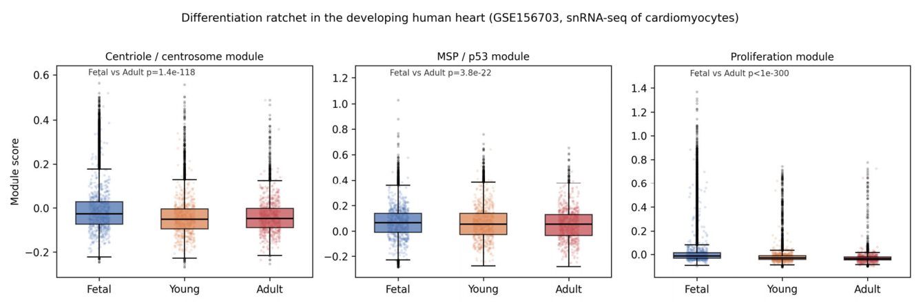
Figure 2.
Expression of key centriolar, p53-pathway and proliferation genes across fetal, young and adult human cardiomyocytes. Dot size: fraction of nuclei expressing the gene; dot color: mean normalized (log1p) expression.
Figure 2.
Expression of key centriolar, p53-pathway and proliferation genes across fetal, young and adult human cardiomyocytes. Dot size: fraction of nuclei expressing the gene; dot color: mean normalized (log1p) expression.
Disclaimer/Publisher’s Note: The statements, opinions and data contained in all publications are solely those of the individual author(s) and contributor(s) and not of MDPI and/or the editor(s). MDPI and/or the editor(s) disclaim responsibility for any injury to people or property resulting from any ideas, methods, instructions or products referred to in the content. |
© 2026 by the authors. Licensee MDPI, Basel, Switzerland. This article is an open access article distributed under the terms and conditions of the Creative Commons Attribution (CC BY) license (http://creativecommons.org/licenses/by/4.0/).
Copyright: This open access article is published under a Creative Commons CC BY 4.0 license, which permit the free download, distribution, and reuse, provided that the author and preprint are cited in any reuse.