

Submitted:
15 October 2026
Posted:
30 October 2026
You are already at the latest version
Abstract
This article reviews reported coupling properties of astrocytes in neural circuits, functional structures, and molecular mechanisms linked to astrocytic specialized domains and spatiotemporal specificity of Ca2+ signaling. We propose a model in which astrocytes act as a bridging mechanism from microcircuit specialization to functional connectivity of brain regions and nuclei, addressing which signals are involved, how integration is achieved, and how these processes contribute to long-term memory and brain–body dimension of consciousness. The model posits that during consciousness and cognition astrocytes and their functional structures bridge neuronal encoding of multimodal embodied perception, integrate and transmit this information, and generate lagged, low-frequency Ca2+ waves that correspond to current state of consciousness or cognition. Through a relatively global signal, astrocytes shape the dynamics of cortical connectome and act at the level of brain dynamics as a downstream bridging router for memory, thereby shaping inertial circuits of consciousness over extended timescales. At the microenvironmental level, region-specific astrocytes and their subtypes shape neural circuits through metabolic coupling based on layer-specific spatial transcriptomes, thereby reversibly shaping cellular and synaptic basis of long-term memory. Through their broad receptor repertoire, astrocytes also serve as both terminal integrators of perceptual information and a neurochemical basis for information integration. At the circuit level, syncytium-based global Ca2+ waves form a global signal that locally and specifically influences cortical activity while globally regulating it, thereby coordinating cortical connectivity and spatial transcriptional profiles associated with consciousness. In embodied consciousness, astrocytes bridge bidirectional communication between consciousness as brain–body synchrony and bodily rhythms such as blood flow and respiration via neurovascular coupling and global signals.
Keywords:
astrocytes
; calcium waves
; global signal
; brain-body synchrony
; information integration
1. Introduction
1.1. Current Dilemmas in Consciousness Research
The levels and contents of consciousness are an important dimension for understanding the nature of consciousness. Consciousness can be divided into different levels, from perception to cognition and metacognition (Leisman et al., 2024). Consciousness is generally understood to comprise two principal components: wakefulness and the content of consciousness. Wakefulness refers to the degree of alertness or arousal of an organism and is mainly regulated by the ascending reticular activating system (ARAS) of the brainstem.(Whyte et al., 2024, Llinás and Paré, 1991) The content of consciousness refers to the specific quality of subjective experience—what we perceive and experience—and involves activity of thalamocortical networks.(Whyte et al., 2024) Cognition, by contrast, is defined at the interface between the neural substrates and mechanisms of consciousness (Gammel et al., 2023, Wang et al., 2026).
Contemporary consciousness and cognitive neuroscience face challenges in multivariate connectome analysis and empirical validation of cognitive mechanisms. Causal intervention techniques such as resting-state functional magnetic resonance imaging (rs-fMRI), transcranial magnetic stimulation–electroencephalography (TMS–EEG), and spectral dynamic causal modelling (spDCM) (Lee et al., 2019) —together with strategies for decoding neurochemical signals, including fluorescent dyes, genetically encoded optical sensors, and two-photon imaging (Kalugin et al., 2026), are being used to resolve the spatiotemporal dynamics of neural circuits with increasing precision. Nevertheless, the core questions remain: how cognition and consciousness emerge from the mapping between neural circuits and the cortex; what constitutes the neural architecture of embodied consciousness; and how these phenomena correspond to empirically testable physiological mechanisms (Thompson and Varela, 2001).
Current research focuses on precisely defining the neural correlates of consciousness (NCC) (Lee et al., 2022, Friedman et al., 2023) and exploring the relationship between consciousness and information integration, including the neural mechanisms of disorders of consciousness and the influence of the body on consciousness (and "brain-body synchrony"). Dominant theoretical frameworks mainly revolve around the Global Neuronal Workspace Theory (GNWT), Integrated Information Theory (IIT), and Recurrent Processing Theory (RPT). These theories explain the mechanisms of con- sciousness along different dimensions: global broadcasting of information, the intrinsic integrative capacity of the system, and recurrent activity in local sensory cortices. In recent years, with the development of no-report paradigms and causal interventions such as TMS-EEG, researchers are attempting to gradually dissociate the prerequisites and consequences of consciousness and to localize the minimal set of neural mechanisms sufficient for generating specific contents of consciousness. In parallel, growing research interest has recently emerged regarding the role of the astrocytic syncytium in higher-order functions such as cognition and even consciousness—particularly its non-trophic, background contributions. With accumulating cellular- and circuit-level evidence, this issue is becoming an important topic in neuroscience.
Neurons serve as interactive conduits and as primary encoders and routers of perceived information. Because of their presumed pre-established harmony and salience, they were among the first cellular substrates studied as memory carriers. However, their highly functionally specialized neural architecture makes them limited functional entities as information-circuit carriers and recipients of synaptic structural remodeling, rather than an absolute, unified hub.
1.2. Recent Paradigm Shift Regarding Astrocytes
In recent years, research on astrocytes has shifted from the traditional view of them as neuronal support cells to a paradigm in which they are recognized as key computational units actively participating in information processing and cognitive function in the central nervous system. Beyond basal metabolic support and homeostatic protection, neuroglial integration and feedback of molecular signals at the levels of gene transcriptomes and neuromodulators reflect their role as important hubs of neural information in neuronal interactions and as bridges in higher-order functions such as memory and cognition. Over the past decade or so, as the pathological value of neuroglia, especially astrocytes, has been increasingly explored and recognized, their metabolic coupling in higher cognitive functions such as memory and cognition within brain circuits has gradually been appreciated for its dynamic significance. Meanwhile, the neurochemical substrates by which astrocytes modulate memory have been reported by various studies in recent years. This has extended to possible roles of astrocytic cellular and molecular metabolic mechanisms, including neurovascular coupling, in memory and cognition. Therefore, the possible involvement of astrocytes in consciousness has gradually become a critical topic in the field of consciousness neuroscience.
2. Functional Structures and Properties of Astrocytes
Astrocytes are primarily located in the central nervous system, and their prominent functional structures, such as the astrocytic syncytium, exhibit laminar hierarchy and stage-dependent dynamic properties that evolve spatiotemporally. Learning-related astrocytic engrams are mainly distributed in core brain regions closely associated with learning, memory, and emotional processing, with the most conclusive evidence coming from studies of the hippocampus, amygdala, and nucleus accumbens. In these recognized key regions, astrocytic ensembles exist in a sparse but highly functionally coordinated manner, and their spatial localization shows marked anatomical overlap and functional coupling with neuronal engrams (Williamson et al., 2025, ichi Dewa et al., 2025). Moreover, in the hippocampus, astrocytic engrams have been shown to be essential for the encoding and retrieval of episodic memory, particularly in subregions such as CA1 and the dentate gyrus, where astrocytes directly participate in the formation of memory traces by sensing neuronal activity and regulating the synaptic microenvironment (Sánchez Romero and Navarrete, 2026).
Going further, as causal integrative circuit elements proposed as necessary by both the Global Neuronal Workspace Theory (GNWT) and Integrated World Modeling Theory (IWMT), neuroglia situated downstream of multimodal sensory relay neurons and receiving encoded information function as integrative circuit hubs that exhibit the broad receptor expression is necessary for neural information storage. This highly complex and environmentally heterogeneous receptor system not only expresses G protein-coupled receptors (GPCRs), ionotropic receptors, and multiple transporters but also displays marked spatiotemporal dynamics in subcellular localization, receptor expression patterns, and signal transduction mechanisms across different brain regions, developmental stages, and pathological states. Interestingly, transplantation of glia-enriched human cortical organoids into the retrosplenial cortex of immunodeficient (NSG) mice generated the interlaminar, protoplasmic, fibrous, and varicose projection astrocytes whose the major anatomical subclasses of human cortical astrocytes first defined in adult human cortex (Oberheim et al., 2009). And, spatial transcriptomics revealed layer-specific expression patterns among these subclasses. Consistent with the transplantation, the human glial chimeric mice exhibit enhanced long-term potentiation and improved learning and memory (Han et al., 2013, Zhang and Barres, 2013, Franklin and Bussey, 2013), suggesting a unique shaping capacity of human astrocytes in synaptic plasticity and learning (Goldman et al., 2015).
2.1. Astrocytic Calcium Waves and Oscillations
Astrocytic calcium waves constitute a complex biochemical signaling language in the central nervous system that is distinct from neuronal action potentials (Veiga et al., 2025, Semyanov et al., 2020). Through spatiotemporally dynamic changes in calcium ion concentration, they regulate synaptic transmission, neurovascular coupling, and global brain homeostasis (Haydon and Carmignoto, 2006a). This signaling mechanism involves not only release from intracellular endoplasmic reticulum calcium stores via inositol (1,4,5)-trisphosphate (IP3) receptors but also intercellular propagation through gap junction proteins such as Connexin 43 (Cx43), thereby forming synchronized calcium oscillation networks spanning hundreds of cells (Panatier et al., 2011, Wang et al., 2006). Studies have also demonstrated that astrocytic calcium signaling exhibits marked kinetic alterations in physiological aging, neurodegenerative diseases such as Alzheimer’s disease, and ischemic injury, directly affecting neural circuit synchrony and cognitive function (Beckers et al., 2024, Emrich et al., 2022, Tabi et al., 2024).
Notably, within neural circuits, and especially at the metabolically coupled neurochemical dynamic termini of PAPs—perisynaptic astrocytic processes formed at the tripartite synapse between astrocytic domains and neurons—that mediate bidirectional astrocyte-neuron communication, astrocytes often link changes in intracellular calcium concentra- tion to differential release of calcium waves. These responses can range from localized microdomain calcium events to cell-wide calcium waves, thereby profoundly influencing neural circuit synchrony, synaptic plasticity, and cerebral hemodynamics. In the hypothesis proposed in this article, this is regarded as the foundation of a bridging mechanism through which coherent astrocytic neurochemical coupling at the cellular level connects to global consciousness integration and participates in the bidirectional “brain-body” dimension of consciousness.
In current molecular and dynamical models of calcium waves, the core driver of astrocytic calcium signaling lies in the release and refilling cycle of intracellular calcium stores. IP3 receptors serve as key channels whose sensitivity is finely regulated by G protein-coupled receptor (GPCR) pathways (Lines et al., 2025). When neurons release glutamate or ATP to activate metabotropic glutamate receptors (mGluRs) or P2Y receptors on astrocytes, phospholipase C is activated and produces IP3, thereby triggering endoplasmic reticulum calcium release (Eroglu, 2025, Wang et al., 2023). This process is not linearly additive but exhibits highly nonlinear dynamical features. Moreover, because glutamate and ATP derived from neurons serve as substrates, this calcium wave-inducing pathway also implies an intriguing metabolically coupled transduction, a capacity that has often been regarded as protection against excitotoxicity and homeostatic regulation in the past, rather than as a potential encoder of neural information, which should be considered. Computational modeling studies indicate that the feedback loop between calcium-induced calcium release (CICR) and IP3 degradation determines the frequency and amplitude of calcium oscillations (Wang et al., 2023, Lines et al., 2025). For example, the Li-Rinzel model and its derivatives successfully simulate the bifurcation behavior of astrocytes under different stimulus intensities, from resting state to periodic oscillations and then to chaos. This phase transition of calcium waves, which differs mechanistically from linear dynamics, provides a similar premise for fitting phase transitions of conscious states and dynamically stable states.
In this article, this is proposed as a possible molecular mechanism for a non-neuronal global signal with background-signal characteristics, as well as a molecular-level dynamical basis for coordinating and participating in multimodal sensory encoding and thereby shaping the denominator of neural entropy.
2.2. Astrocytic Syncytium
The astrocytic syncytium is a highly functionally integrated glial network structure in the central nervous system mediated by gap junctions. Unlike a true syncytium, the astrocytic syncytium is an intercellular network composed entirely of discrete cellular components. While preserving certain astrocyte-specific morphological and transcriptional properties of spatial differentiation, this structure transcends the limitations of traditional single-cell function. It enables astrocytes to act as a unified physiological unit and, through subtype-differentiated syncytial coupling strength, connexin composition, network topology, and functional effects, to exert complex systemic roles in ion homeostasis, metabolite distribution, neural signal modulation, and pathological defense.
The formation of the astrocytic syncytium mainly depends on two key gap junction proteins: Connexin 43 (Cx43) and Connexin 30 (Cx30) (5,6). Cx43 is the predominant connexin in adult brain tissue and is responsible for establishing extensive intercellular coupling channels, whereas Cx30 is gradually upregulated in specific brain regions and later developmental stages, jointly regulating network permeability and selectivity. These hemichannels dock on adjacent cell membranes to form complete gap junction channels, allowing free diffusion of small molecules with a molecular weight of less than 1 kDa, such as potassium ions, calcium ions, glucose metabolites, lactate, ATP, and second messengers such as IP3 and cAMP. This mechanism propagates intercellular calcium waves and exchanges cell death signals. Recent studies confirm that this syncytial property is not static but dynamically evolves with developmental stage and exhibits complex dynamic roles in various neurological diseases.
2.3. Learning-Associated Astrocytes
Learning-associated astrocytic engrams (LAAs), as a cellular material basis for memory recall, are thought to be coupled with the activation of neuronal ensembles termed engrams during learning and are sufficient to trigger memory retrieval. They also exhibit strong association with anatomical regions closely related to memory tasks and are mainly identified in the hippocampus and its associated cortical regions. During learning, specific astrocytic subpopulations are activated and form engrams, whose spatial distribution highly overlaps anatomically with synchronously activated neuronal engrams. Moreover, LAA processes finely ensheathe neuronal synapses; this positional proximity enables real-time monitoring and regulation of synaptic transmission while providing favorable spatial coupling for integrating encoded information with calcium wave propagation.
2.4. Cortex-Specific Morphology of Astrocytes
Astrocytes exhibit rich morphological and molecular subtype diversity across transcriptomes, brain regions, and species. They also display pre-established harmony-like cross-species conservation in brain connectome and circuit architecture and in close metabolic coupling with neurons, which is considered a shared necessary prerequisite for cognitive capacity in organisms. Interestingly, astrocytic morphology and function are highly related to their local microenvironment and neural circuits, reflecting regional transcriptomic specificity similar to that of neurons. Such anatomical and transcriptomic specificity generally signifies high functional complexity, enabling astrocytes to shape specialized microcircuit properties for different cognitive modalities across brain regions and neural circuits.
2.5. Metabolic Coupling Regulation of Memory by Astrocytes
Reports on the mechanisms and evidence of memory within the paradigm shift in astrocytes have become a major focus in neuroscience. The major contributions of astrocytes to synaptic plasticity and memory are mediated through gliotrans- mitters such as D-serine at the tripartite synapse. Key mediators of astrocyte-neuron communication participate in LTP and object recognition memory, as represented by cannabinoid type 1 receptor (CB1R)-related reversible modulation of long-term memory by exogenous D-serine and by adenosine receptor-mediated modulation of synaptic plasticity in the amygdala downstream of astrocyte activation, as reported by (Martin-Fernandez et al., 2017) (Sánchez Romero and Navarrete, 2026, Robin et al., 2018). Currently, reports on astrocytic engrams and their related molecular mechanisms in memory formation are accumulating at a considerable rate, and increasingly valuable evidence regarding metabolic dynamics continues to emerge with the development of astrocyte-specific activity control.
Cognition undoubtedly requires memory and experience. In visual perception, which constitutes a substantial proportion of cognition, astrocytes, in multiple forms such as Müller cells, show functional coupling with neurons and marked proportional differences, demonstrating their active rather than merely passive supporting role in visual perception. Similarly, in brain regions closely related to spatial memory, such as the hippocampus, astrocytes not only dynamically regulate synaptic plasticity through the tripartite synapse but also participate in encoding spatial location and reward information. They form "glial place cells" corresponding to place cells that encode position in hippocampal place fields via firing rates and temporal sequences, secondarily integrating inputs from boundary cells and periodic grid cells to construct sequence-dependent cognitive maps (Yu, 2022). The complexity of this neuron-glia interaction involves not only the complex metabolic dynamics of the astrocyte-neuron lactate shuttle but also astrocytic calcium ion dynamics and calcium wave release (Li et al., 2025). The latter has recently been shown to have a common basis and heterogeneous effects across different brain regions. In spatial memory paradigms, astrocytes exhibiting persistent calcium activity in hippocampal CA1 show encoding specificity for location information in mice in virtual environments (Adi et al., 2022), which is generally regarded as a neural process underlying spatial cognition.

Table 1.
The Constitution of Consciousness.
| Level | Functional substrate | Level of action | Pathological causal studies | Refs |
|---|---|---|---|---|
| Structural connectivity (SC) |
White matter fiber tracts; intracortical axonal projections |
Structural foundations of physical connections, including wiring transmission, ephaptic coupling, volume transmission, and neurovascular coupling |
Impaired structural connectivity of thalamus-basal ganglia-frontal circuits in patients with disorders of consciousness |
(Weng et al., 2017) |
| Network topology |
Modularity, hubs, rich-club organization | Balance between segregation and integration | Collapse of global integration and enhanced local segregation in anesthesia/coma |
(Luppi et al., 2019) |
| Intrinsic neural timescale (INT) |
Local recurrent connection strength; synaptic time constants |
Temporal integration window: fast and slow sensory and association cortices |
Disrupted dynamic transitions of temporal-scale states in DoC patients |
(Buccellato et al., 2025) |
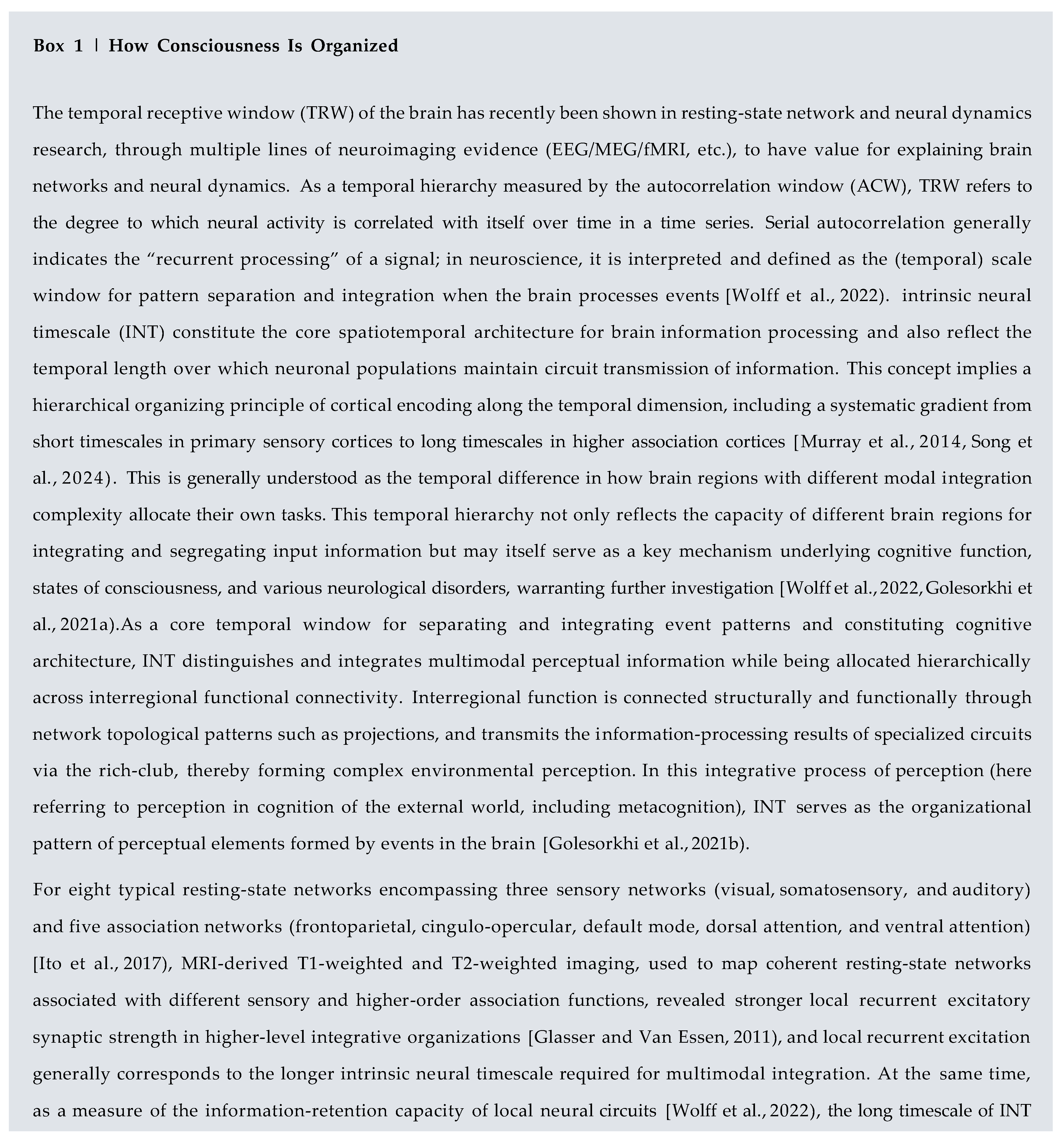
Interestingly, during retrieval of auditory fear memory, in lateral septum (LS) brain slices—LS being among multiple brain regions activated by c-Fos expression and implicated in motivated behavior, anxiety, and contextual fear conditioning—immunostaining for glutamic acid decarboxylase 65 and 67 kDa isoforms (GAD65 and GAD67, markers of GABAergic neurons) and tyrosine hydroxylase (TH, a marker of dopaminergic neurons) was detected, whereas vesicular glutamate transporter 1 (vGluT1) was not detected (Chen et al., 2024). The differential detection of vGluT1 in neurotransmitter systems containing glutamatergic excitatory inputs may arise from possible changes in glial activation states within the LS under post-shock stress in mice, which may in turn affect the detection of vesicular glutamate transporters during the experiment through metabolic coupling in bidirectional dialogue with neurons. The possibility of such bidirectional dialogue has been demonstrated in glia-neuron metabolic coupling, as represented by molecular mechanisms such as ZDHHC enzyme-mediated protein palmitoylation (Fan et al., 2024). However, direct molecular profiling studies of LS astrocytes remain relatively scarce, and direct evidence for this specific mechanism in the LS still requires further elucidation (Seo et al., 2024, Xie et al., 2025).
2.6. Neurovascular Coupling
As shown by the perivascular astrocytic endfeet spontaneously formed by astrocytes transplanted by astrocytes transplanted into the retrosplenial cortex of immunodeficient (NSG) mice by. human astrocytes in vivo connect to microvessels through spontaneously formed perivascular astrocytic endfeet, regulating the nutrient exchange required by the brain and sensing neuronal activity through perisynaptic processes. As key relay elements in neurovascular coupling (NVC), astrocytes link local neuronal activity to local cerebral blood flow. Moreover, astrocytes serve as critical mediators of “Brain-Body Synchrony,” enabling neural drive that is not unidirectional from affective states and levels of consciousness to the circulation and its neurovascular coupling. This will be elaborated in the subsequent module on the "brain-body" dimension of consciousness. Multiple studies have demonstrated that altered states of consciousness, such as sleep, anesthesia, or disorders of consciousness, directly lead to decoupling or remodeling of neurovascular coupling properties, whereas emotional processing elicits local increases in blood flow and systemic cardiovascular responses through activation of specific brain regions.
3. Astrocytic Dynamical Mechanisms of Signal Wiring, Mapping, and Integration
3.1. Network Topology, Structural Connectivity, and Temporal Scale Shape Cognition and Consciousness
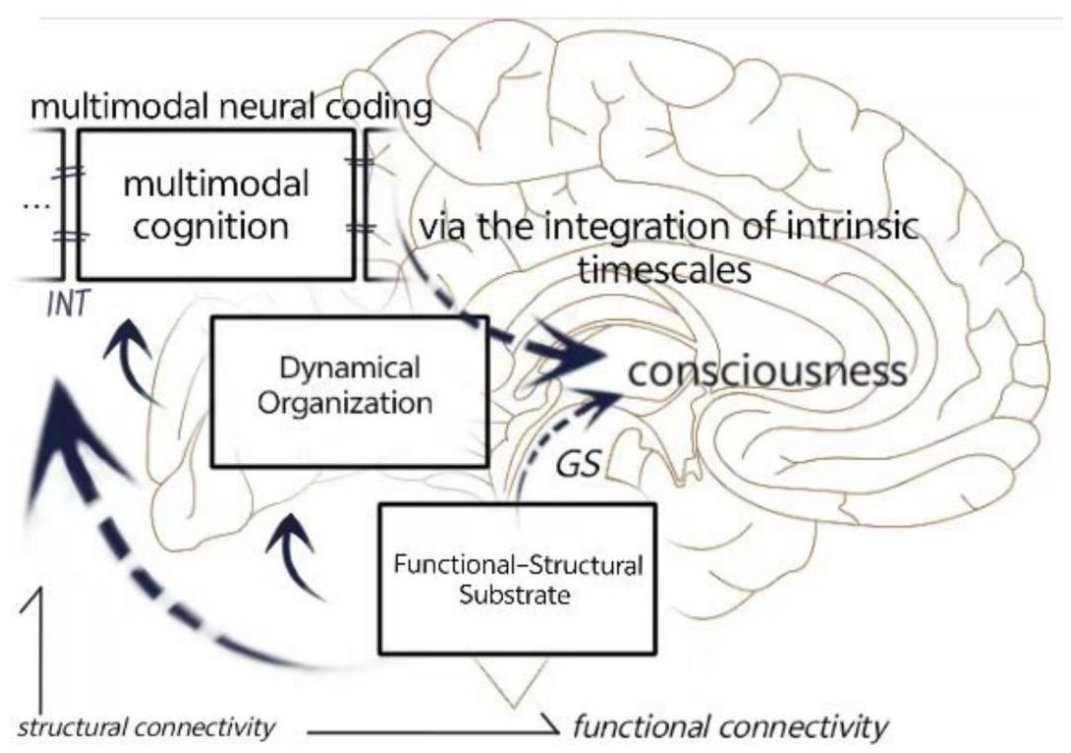
Structural connectivity delineates the intrinsic dynamical space. Network topology, as the basis of functional connectiv- ity, determines the architecture of information segregation and integration. Meanwhile, temporal scale determines how much history each node can integrate; cognition is dynamic information processing running on this three-layer architec- ture, whereas consciousness corresponds to the capacity of this architecture to maintain a “rich, rapidly reconfigurable integrated state.” Neural entropy, then, is an combinatorial explosion generated on the basis of the intrinsic topological structure, modular nodes, and dynamic change mechanisms of the structure.
The gradients of structural connectivity (connection strength, myelination) determine the hierarchy of temporal scales, which in turn defines the “core-periphery” organization in topology (Golesorkhi et al., 2021a, Wang, 2022).
At the level of the brain connectome, consciousness uses the rich-club—a set of highly interconnected hubs that constitute traffic hubs for global integration—as a structural domain for functional connectivity and information integration, in which cortical areas process perceptual, cognitive, or metacognitive inputs and generate neural signal encoding for projection(Senden et al., 2014, Baggio et al., 2015). At the cellular and circuit levels, astrocytes act as microscopic units of information integration through neurovascular coupling and local calcium waves evoked in microcircuits in response to the primary encoding transmitted by neurons.
3.2. Evidence on How Information Is Mapped and Integrated into Consciousness in the Structural and Signaling Mechanisms of the Cortical Connectome
In functional magnetic resonance imaging (fMRI) studies, the global signal is usually defined as the average of whole-brain voxel time series. Showed that the global signal is not merely noise but a mixed signal containing neural contributions. Further reviewed evidence that the global signal has a neural basis and is tightly coupled with infra-slow-frequency neural activity and low-frequency physiological signals such as respiration and heartbeat. They also reported possible mechanisms by which subcortical nuclei, such as the nucleus basalis of Meynert (NBM) in the basal forebrain, raphe nuclei, and substantia nigra, influence cortical activity through multi-regional modulation and thereby participate in global signal generation and arousal regulation. These bodily physiological signals are rhythmic; however, although well recognized in the literature, what has not yet received sufficient attention is that they also undergo bidirectional communication and changes in tandem with brain-derived states such as consciousness and emotion. The neural substrate unit in the brain for this process is the astrocyte, which continuously emits dynamic calcium waves within circuits in neurovascular coupling (NVC) (Haydon, 2006b, Pereira, 2011).
The global signal (GS), as a potential coordinating signal of global brain activity studied in fMRI, has been demonstrated in recent studies to be tightly coupled with bodily signals such as respiration, heartbeat, and vascular dynamics and to be capable of regulating arousal levels. At the same time, as a dynamic topographical map influencing perception and cognition, the GS coordinates different forms of cognition by modulating activity across cortical regions and networks (Zhang and Northoff, 2022). As an infra-slow-frequency global activity, it differs from the rapid electrical conduction of neurons and the rapid calcium waves of astrocytes, and it may be a widespread neural signal potentially originating from nuclei such as the substantia nigra. Moreover, the broad distribution of astrocytes and their infra-slow-frequency calcium waves, as spatiotemporal fluctuating signals, together with the structural connectivity through which endfeet directly couple to neurovascular units, serve as a bridging mechanism. The relatively broad spatial extent of infra-slow-frequency calcium waves in the astrocytic syncytium spanning the cortex provides a highly promising neural substrate framework for the GS.
Studies of intrinsic neural timescale (INT) hierarchical gradients in cognitive impairment have shown stable and significant INT alterations in the inferior temporal cortex (Zhang et al., 2023), caudate nucleus, and globus pallidus in Alzheimer’s disease (AD) converters, indicating that longer dwell times of neural information in lower-level regions impair cognitive ability. This degenerative trend is typically accompanied by marked INT prolongation in lower-level regions, revealing that INT does not function as an infinitely deployable allocation scale, nor as a fixed scale distributed across cortical levels during cognition. Rather, it may serve as a dynamic window mediated by specific cellular or domain substrates.
Notably, in this theory of consciousness, astrocytes coupled to neurovasculature via endfeet also mediate the modulation of the GS by bodily rhythmic signals such as blood flow through this coupling unit. This is also consistent with corresponding principles on the basis of existing pathological theories. Astrocytes in neurovascular coupling, as structural units that bidirectionally bridge "brain-body" states, correspond in this bidirectional action to the global signal that similarly maintains and regulates arousal levels, thereby constituting a three-dimensional jigsaw puzzle of the "brain-body" dimension of consciousness. In the functional and structural connectivity accounted for by this hypothesis, a promising mutual correspondence among nodes emerges: during graded propofol-induced loss of consciousness (LOC)(Chen et al., 2024), low-frequency power of electrophysiological signals increases sharply (Chen et al., 2009). At the systems level, the corresponding loss of functional coupling shown by astrocytes in the hippocampus-thalamus- mPFC pathway during cascading deactivation revealed by fMRI has explanatory potential. In particular, before LOC onset, fMRI signal synchrony within this pathway temporarily increases, which may point to a component of another consciousness pathway mechanism whose pathological causality has not yet been fully elucidated.
In addition, animal studies by (Seo et al., 2024) show that multiple regions participate in inducing and mediating a specific behavior, which supports a global response to behavior in the brain’s embodied states of consciousness (Yeates et al., 2022). For example, the lateral septum (LS) forms close bidirectional loop connections with the hippocampus. The LS receives glutamatergic projections from hippocampal CA3 (subiculum) and CA1; after integrating emotional and stress-related information, it projects back to the hippocampus via GABAergic or cholinergic fibers, regulating hippocampal rhythmic oscillations and synaptic plasticity. Locally in the hippocampus, this regulation depends on pyramidal cell axons sending Schaffer collaterals to CA1 and, at the same time, emitting recurrent collaterals in CA3 that form predominantly glutamatergic excitatory synapses with other pyramidal cells (including inhibitory interneurons), acting on AMPA/NMDA receptors. This forms autoexcitatory/recurrent loops that amplify signals and participate in pattern completion and working memory; however, hypersynchronization can lead to the pathological features of epilepsy.
3.3. Distinct Signaling Mechanisms and Integrative Systems of Astrocytes Across the Cortex
As a hierarchical level of circuit construction, astrocytic calcium waves can be divided into local calcium signal excitation and global calcium waves. In normal neural circuit activity, the most classic trigger for astrocytic calcium signaling originates from glutamate, the excitatory neurotransmitter released from presynaptic terminals. When neurons fire action potentials and release glutamate, some glutamate molecules diffuse to adjacent astrocytic processes (PAPs), activating metabotropic glutamate receptors on their membranes, particularly the mGluR5 subtype (Panatier et al., 2011, Wang et al., 2006). mGluR5 belongs to the G protein-coupled receptor (GPCR) family; upon activation, it couples to Gq/11 proteins and subsequently activates phospholipase C-β (PLC-β) (Araque et al., 1999). PLC-β catalyzes the hydrolysis of the membrane phospholipid phosphatidylinositol-4,5-bisphosphate (PIP2), generating the second messengers inositol-1,4,5-trisphosphate (IP3) and diacylglycerol (DAG) (Berridge and Irvine, 1984). The generated IP3 diffuses into the cytoplasm and binds to IP3 receptors (IP3Rs) located on the endoplasmic reticulum (ER) membrane. IP3R is a ligand-gated calcium channel whose open probability is dually regulated by IP3 concentration and cytosolic calcium concentration, exhibiting a characteristic "bell-shaped dependence": low cytosolic calcium concentrations synergize with IP3 to promote channel opening (calcium-induced calcium release, CICR), whereas high calcium concentrations inhibit channel activity (7,6). This mechanism enables astrocytes to generate local and transient calcium elevations, i.e., "microdomain" calcium signals, upon receiving high-frequency synaptic stimulation. A single astrocyte can contact approximately 100,000 synapses (Bushong et al., 2002), yet its calcium signals appear to be autonomously confined to independent subcellular domains within certain chemical kinetics. This spatial confinement ensures that astrocytes can finely regulate specific synapses rather than indiscriminately affecting the entire cell. This local calcium signaling mechanism typically serves as an integration mechanism downstream of neuronal signal transmission (its role at the memory level is not the focus of discussion here). As a form of cellular-level integration, its function is determined by tight chemical coupling rather than by global breadth at the connectome level, making this integration almost inevitably lagging and retentive in large-scale neural dynamics. The extended inferences from these dynamical properties are also consistent with current global signal research and the consolidation of long-term memory.
Beyond local synaptic inputs, astrocytes also broadly express neuromodulator receptors that trigger global calcium waves. Among these, one particularly striking feature is that they have been reported in relevant studies to be associated with long-term emotional states of consciousness, such as acetylcholine, norepinephrine, dopamine, and ATP receptors. These neuromodulators are typically released from long-range nuclei, have slower time courses and broader spatial ranges, and can regulate the overall excitability state of astrocytes (Lines et al., 2025, Eroglu, 2025). When these neuromodulators (e.g.,norepinephrine via α 1-adrenergic receptors, or ATP via P2Y receptors) activate astrocytes, they likewise elevate intracellular IP3 levels through the Gq-PLC-IP3 pathway. As effectors bridging previously received neural encoding, the subsequent calcium wave signals thereby exert subtle state modulation over a relatively broad spatial domain.
When stimulus intensity is sufficiently large or IP3 concentration reaches a specific threshold, local calcium release can self-regenerate through the CICR mechanism and propagate within the cell in the form of waves, generating a "calcium surge" or global calcium wave (Wang et al., 2023). The propagation speed of such global calcium waves is approximately 10–30 µm/s, sufficient to cover the entire astrocyte and its complex process network (Bootman and Lipp, 1999). In barrel cortex experiments in mice under awake or aroused states, large-scale, short-lived astrocytic calcium spikes have been observed. These calcium waves depend on IP3-mediated ER calcium release and are closely associated with enhanced sensory transmission. In addition, the nitric oxide (NO) signaling pathway also participates in regulating this process. NO produced by eNOS and nNOS expressed in astrocytes can promote calcium influx through S-nitrosylation modification of calcium homeostasis modulator 1 (CALHM1) channels or release ATP through Connexin 43 (Cx43) hemichannels, further amplifying calcium signals. This chemically coupled local differentiation, as a downstream bridging route for memory, shapes the inertial circuits of consciousness over extended timescales, suggesting a potential dynamical mechanism by which astrocytes help bridge immediate neural information across functional levels.
Figure 1.
Multilevel signaling bridging mechanisms of astrocytes: From synaptic microenvironment to network calcium waves. (a) The broad spectrum of receptors expressed by astrocytes at the tripartite synapse and their coupled neurochemical dynamics reflect Ca2+ dynamics within active intracellular or syncytial astrocytic domains, and, under the influence of regional heterogeneity such as spatiotemporal specificity and circuit specialization, astrocytes release Ca2+ responses with different spatial ranges and levels of influence according to the degree of integration; (b) Astrocytes support the neurochemical shaping of neuronal long-term memory in circuit specialization through glutamate shuttling, inositol trisphosphate release, lactate, and other mechanisms; (c) When “brain–body” phenomena such as emotion-induced alterations in bodily rhythmic signals occur, astrocytes in the immediate neural circuits act as a bridging mechanism; on the basis of astrocytic endfeet serving as neurovascular coupling units, due to task demands during cognitive load, they may also lead to endfeet decoupling and form domains with other astrocytes to support the cognitive task operations of neural circuits; (d) In the hypothesis of this paper, after information circuit processing and neurochemical cognitive integration, the relatively global Ca2+ waves released downstream of the coupled events in the astrocytic syncytium serve as the neural basis for the infra-slow-frequency global signal of the human brain, forming a dynamic topographic map of the connectome. Figure credits: Illustrations of CA1 pyramidal neuron, blue astrocyte, and human brain sagittal outline from SciDraw.io by Stoyo Karamihalev and Andrew Hardaway (2026), used under CC-BY 4.0. Modifications: colors and arrangement adapted.
Figure 1.
Multilevel signaling bridging mechanisms of astrocytes: From synaptic microenvironment to network calcium waves. (a) The broad spectrum of receptors expressed by astrocytes at the tripartite synapse and their coupled neurochemical dynamics reflect Ca2+ dynamics within active intracellular or syncytial astrocytic domains, and, under the influence of regional heterogeneity such as spatiotemporal specificity and circuit specialization, astrocytes release Ca2+ responses with different spatial ranges and levels of influence according to the degree of integration; (b) Astrocytes support the neurochemical shaping of neuronal long-term memory in circuit specialization through glutamate shuttling, inositol trisphosphate release, lactate, and other mechanisms; (c) When “brain–body” phenomena such as emotion-induced alterations in bodily rhythmic signals occur, astrocytes in the immediate neural circuits act as a bridging mechanism; on the basis of astrocytic endfeet serving as neurovascular coupling units, due to task demands during cognitive load, they may also lead to endfeet decoupling and form domains with other astrocytes to support the cognitive task operations of neural circuits; (d) In the hypothesis of this paper, after information circuit processing and neurochemical cognitive integration, the relatively global Ca2+ waves released downstream of the coupled events in the astrocytic syncytium serve as the neural basis for the infra-slow-frequency global signal of the human brain, forming a dynamic topographic map of the connectome. Figure credits: Illustrations of CA1 pyramidal neuron, blue astrocyte, and human brain sagittal outline from SciDraw.io by Stoyo Karamihalev and Andrew Hardaway (2026), used under CC-BY 4.0. Modifications: colors and arrangement adapted.
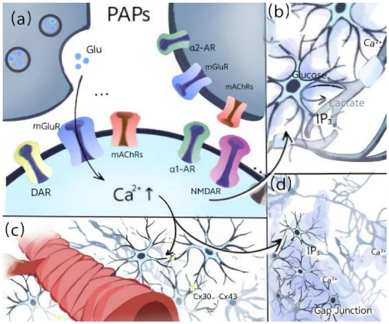
From a systematic perspective, downstream of neuron circuits with different brain region characteristics, when neurons release neurotransmitters or neuromodulators based on cortical transcriptomes or activation pathways, neurotransmitters and neuromodulators respectively bridge calcium wave-coupled release across different time scales and intensities through the broad receptor expression and response repertoire of astrocytes, spatiotemporal transcriptome expression, and functional structures such as engrams or the syncytium. The latter, through different spatial ranges of action—for example, when a specific brain region or nucleus is activated—expresses fast or slow, local or relatively global calcium wave patterns based on a certain combinatorial pattern of the aforementioned astrocytic transcriptional features and functional structures. This in turn induces, promotes, or shapes a specific neural dynamic. This ranges from the small scale (circuits) to the large scale—due to integration and subsequent lagging—of infra-slow-frequency power and the global signal (GS) that connects with and relates to bodily rhythmic signals.
Figure 2.
Hierarchical Connectivity Underlying Consciousness.
4. Astrocytic Bridging of the “Brain-Body” Dimension of Consciousness
Consciousness is not an isolated product of the brain but arises through complex "brain-body synchrony" and structured periodic correspondence (Young et al., 2026), which together shape the embodied nature of consciousness. Different states of consciousness, including wakefulness, sleep, anesthesia, and disorders of consciousness, exhibit markedly different neurovascular dynamical features. During sleep inertia (a brief period of low alertness after awakening from sleep), neurovascular coupling changes dynamically with the recovery of alertness. that the lagged coupling between electroencephalographic features and functional MRI signals changes significantly during awakening, indicating the functional dynamics of NVC mechanisms across transitions in states of consciousness.
In more extreme altered states of consciousness, such as general anesthesia or disorders of consciousness caused by severe brain injury, differential involvement of thalamic nuclei coordinates transitions in states of consciousness. Through regulation of astrocytic activity, this mediates coupling and decoupling at perivascular astrocytic endfeet, thereby influencing whole-brain blood flow patterns.
In contrast, in wakefulness, rapid eye movement (REM) sleep, and psychedelic states, neural entropy is higher, which is usually accompanied by higher levels of astrocytic metabolic activity to maintain normal neurovascular coupling. Conversely, in deep non-rapid eye movement (NREM) sleep, anesthesia, and disorders of consciousness, neural entropy is lower, often accompanied by reduced efficiency or decoupling of neurovascular coupling, and decoupling of endfeet also implies a decline in astrocytic function.
As a bridging mechanism through which astrocytes exert "brain-body" influences in the thinking aspect of consciousness, the demand of cognitive tasks on cerebral blood flow is reflected not only in local activation but also in the regulation of overall network efficiency. Studies of individuals performing dual tasks have reported a negative correlation between neurovascular coupling status and cognitive performance, namely neurovascular decoupling under high cognitive load. Based on astrocytic properties, one possible explanation is that perivascular astrocytic endfeet decouple to meet the metabolic dynamic demands under cognitive load, thereby providing the frequent calcium wave feedback required by task circuits for cognitive integration.
5. Conclusions
In summary, by reviewing the specialized functional structures and molecular mechanisms of astrocytes, this article proposes a theoretical hypothesis in which astrocytes act as bridges in a global and hierarchically progressive mechanism of cognitive shaping, integration, and "brain-body synchrony" consciousness. It addresses how astrocytic mechanisms couple from cellular-level structures through circuit mechanisms up to regional functional properties and connectivity architecture at the cortical and connectome levels. It also addresses how astrocytes, as a multilevel neural dynamical mechanism, bridge the modal integration of cognition and the "brain-body" dimension of consciousness capable of bidirectional communication with bodily signals.
However, it should be noted that the bridging mechanism of astrocytes in the complex constitution of cognition and consciousness does not exclude the neurodynamical roles of neurons and their functional structures, such as engrams, as essential flow channels of consciousness. On the contrary, their roles remain crucial as inertial carriers of microcircuit specialization and long-term memory, as myelin structures associated with oligodendrocytes, as key structural units of interareal cortical projections, and especially as functional neurons in the functional projections of brain regions or nuclei.
- The blue astrocyte illustration was created by Andrew Hardaway (2026), available from SciDraw.io (DOI: 10.5281/zenodo.5348394), licensed under CC-BY 4.0. The author modified and color-edited the original illustration.
- The human brain sagittal outline was created by Stoyo Karamihalev (2026), available from SciDraw.io (DOI: 10.5281/zenodo.4312476), licensed under CC-BY 4.0. The author modified and color-edited the original illustration.
Funding
This research received no external funding.
Acknowledgments
The author would like to thank the following open-access resources for providing the illustration materials used in the figures:.
Additionally, the schematic drawing depicting the smooth muscle coverage on the neurovasculature was adapted based on the graphical representation presented in the study by ), DOI: 10.3389/fnetp.2023.1162757.
Competing interests
The author declares no competing interests.
References
- Leisman, Gerry; Alfasi, Rahela; D’Angiulli, Amedeo. From sensory motor and perceptual development to primary consciousness in the fetus: converging neural, behavioral, and imaging correlates of cognition-mediated emergent transitions. Curr. Opin. Behav. Sci. 2024, 60, 101455. [Google Scholar] [CrossRef]
- Whyte, Christopher J.; Redinbaugh, Michelle J.; Shine, James M.; Saalmann, Yuri B. Thalamic contributions to the state and contents of consciousness. Neuron 2024, 112(10), 1611–1625. [Google Scholar] [CrossRef]
- Llinás, Rodolfo; Paré, Denis. Of dreaming and wakefulness. Neuroscience 1991, 44(3), 521–535. [Google Scholar] [CrossRef]
- Gammel, Theresa C.; Leor, Alkadaa N.; Saadon, John Servider; Saluja, Sabir; Servider, Jordan; Cleri, Nathaniel A.; Egnor, Michael; Davis, Raphael P.; Huang, Chuan; Saalmann, Yuri B.; Mofakham, Sima; Mikell, Charles B. Brain circuitry of consciousness: A review of current models and a novel synergistic model with clinical application. Neurosurg. Pract. 2023, 4(2), e00031. [Google Scholar] [CrossRef]
- Wang, Fangting; Xue, Yufei; Di, Haibo. From detection to decision: a conceptual framework of uncertainty migration in disorders of consciousness. Front. Neurol. 2026, 17, 1940448. [Google Scholar] [CrossRef]
- Lee, Heonsoo; Golkowski, Daniel; Jordan, Denis; Berger, Sebastian; Ilg, Rüdiger; Lee, Joseph; Mashour, George A.; Lee, UnCheol; Avidan, Michael S.; Blain-Moraes, Stefanie; et al. Relationship of critical dynamics, functional connectivity, and states of consciousness in large-scale human brain networks. NeuroImage 2019, 188, 228–238. [Google Scholar] [CrossRef]
- Kalugin, Peter N.; Soden, Paul A.; Massengill, Crystian I.; Amsalem, Oren; Porniece, Marta; Stolberg, Zachary B.; Guarino, Diana C.; Tingley, David; Zhang, Stephen X.; Benson, Jordan C.; Hammell, Madalon F.; Tong, David M.; Ausfahl, Charlotte D.; Lacey, Tiara E.; Courtney, Ya’el; Hochstetler, Alexandra; Lutas, Andrew; Wang, Huan; Geng, Lan. [CrossRef]
- Li; Li, Bohan; Li, Yulong; Lehtinen, Maria K.; Andermann, Mark L. Simultaneous, real-time tracking of many neuromodulatory signals with multiplexed optical recording of sensors on a micro-endoscope. Neuron 2026. [Google Scholar] [CrossRef]
- Thompson, Evan; Varela, Francisco J. Radical embodiment: neural dynamics and consciousness. Trends Cogn. Sci. 2001, 5(10), 418–425. [Google Scholar] [CrossRef]
- Lee, Minji; Sanz, Leandro R. D.; Barra, Alice; Wolff, Audrey; Nieminen, Jaakko O.; Boly, Melanie; Rosanova, Mario; Casarotto, Silvia; Bodart, Olivier; Annen, Jitka; Thibaut, Aurore; Panda, Rajanikant; Bonhomme, Vincent; Massimini, Marcello; Tononi, Giulio; Laureys, Steven; Gosseries, Olivia; Lee, Seong-Whan. Quantifying arousal and awareness in altered states of consciousness using interpretable deep learning. Nat. Commun. 2022, 13(1), 1064. [Google Scholar] [CrossRef]
- Friedman, Garrett; Turk, Katherine W.; Budson, Andrew E. The current of consciousness: Neural correlates and clinical aspects. Curr. Neurol. Neurosci. Rep. 2023, 23(7), 345–352. [Google Scholar] [CrossRef]
- Williamson, Michael R.; Kwon, Wookbong; Woo, Junsung; Ko, Yeunjung; Maleki, Ehson; Yu, Kwanha; Murali, Sanjana; Sardar, Debosmita; Deneen, Benjamin. Learning-associated astrocyte ensembles regulate memory recall. Nature 2025, 637, 478–486. [Google Scholar] [CrossRef]
- Dewa, Kenichi; Kaseda, Kodai; Kuwahara, Aoi; Kubotera, Hideaki; Yamasaki, Ayato; Awata, Natsumi; Komori, Atsuko; Holtz, Mika A.; Kasai, Atsushi; Skibbe, Henrik; Takata, Norio; Yokoyama, Tatsushi; Tsuda, Makoto; Numata, Genri; Nakamura, Shun; Takimoto, Eiki; Sakamoto, Masayuki; Ito, Minako; Masuda, Takahiro; Nagai, Jun. The astrocytic ensemble acts as a multiday trace to stabilize memory. Nature 2025, 648(8092), 146–156. [Google Scholar] [CrossRef]
- Sánchez Romero, Javier; Navarrete, Marta. Astroengrams: rethinking the cellular substrate for memory. In Nature Reviews Neuroscience; ISSN 1471-0048, April 2026; Volume 27, 4, pp. 289–300. [Google Scholar] [CrossRef]
- Oberheim, Nancy Ann; Takano, Takahiro; Han, Xiaoning; He, Wei; Lin, Jane H. C.; Wang, Fushun; Xu, Qiwu; Wyatt, Jeffrey D.; Pilcher, Webster; Ojemann, Jeffrey G.; Ransom, Bruce R.; Goldman, Steven A.; Nedergaard, Maiken. Uniquely hominid features of adult human astrocytes. J. Neurosci. 2009, 29(10), 3276–3287. [Google Scholar] [CrossRef]
- Han, Xiaoning; Chen, Michael; Wang, Fushun; Windrem, Martha; Wang, Su; Shanz, Steven; Xu, Qiwu; Oberheim, Nancy Ann; Bekar, Lane; Betstadt, Sarah; Silva, Alcino J.; Takano, Takahiro; Goldman, Steven A.; Nedergaard, Maiken. Forebrain engraftment by human glial progenitor cells enhances synaptic plasticity and learning in adult mice. Cell Stem Cell 2013, 12(3), 342–353. [Google Scholar] [CrossRef]
- Zhang, Ye; Barres, Ben A. A smarter mouse with human astrocytes. BioEssays 2013, 35(10), 876–80. [Google Scholar] [CrossRef]
- Franklin, Robin J. M.; Bussey, Timothy J. Do your glial cells make you clever? Cell Stem Cell 2013, 12(3), 265–266. [Google Scholar] [CrossRef]
- Goldman, Steven A.; Nedergaard, Maiken; Windrem, Martha S. Modeling cognition and disease using human glial chimeric mice. Glia 2015, 63(8), 1483–1493. [Google Scholar] [CrossRef]
- Veiga, Alexandra; Abreu, Daniela Sofia; et al. Calcium-dependent signaling in astrocytes: downstream mechanisms and implications for cognition. J. Neurochem. 2025, 169(2), e70019. [Google Scholar] [CrossRef]
- Semyanov, Alexey; Henneberger, Christian; Agarwal, Amit. Making sense of astrocytic calcium signals — from acquisition to interpretation. Nat. Rev. Neurosci. 2020, 21(10), 551–564. [Google Scholar] [CrossRef]
- Haydon, Philip G.; Carmignoto, Giorgio. Astrocyte control of synaptic transmission and neurovascular coupling. Physiol. Rev. 2006a, 86(3), 1009–1031. [Google Scholar] [CrossRef]
- Panatier, Aude; Vallée, Joanne; Haber, Michael; Murai, Keith K.; Lacaille, Jean-Claude; Robitaille, Richard. Astrocytes are endogenous regulators of basal transmission at central synapses. Cell 2011, 146(5), 785–798. [Google Scholar] [CrossRef]
- Wang, Xiaohai; Lou, Nanhong; Xu, Qiwu; Tian, Gui-Fang; Peng, Wei-Gang; Han, Xu; Kang, Jian; Takano, Takahiro; Nedergaard, Maiken. Astrocytic Ca2+ signaling evoked by sensory stimulation in vivo. Nat. Neurosci. 2006, 9(6), 816–823. [Google Scholar] [CrossRef]
- Beckers, Pauline; Doyen, Pierre J.; Hermans, Emmanuel. Modulation of type 5 metabotropic glutamate receptor- mediated intracellular calcium mobilization by regulator of g protein signaling 4 (rgs4) in cultured astrocytes. Cells 2024, 13(4), 291. [Google Scholar] [CrossRef]
- Emrich, Scott M.; Yoast, Ryan E.; Trebak, Mohamed. Physiological functions of crac channels. Annu. Rev. Physiol. 2022, 84(1), 355–379. [Google Scholar] [CrossRef]
- Tabi, C. B.; Tiayo, T. K.; Etémé, A. S.; Fouda, H. P. E.; Kofané, T. C. Bifurcation of synchronized nonlinear intercellular Ca2+ oscillations induced by bi-directional paracrine signaling and inositol 1,4,5-triphosphate-cytosolic- Ca2+ interaction. Phys. Lett. A 2024, 498, 129332. [Google Scholar] [CrossRef]
- Lines, Justin; Corkrum, Michelle; Aguilar, J.; Araque, Alfonso. The duality of astrocyte neuromodulation: As- trocytes sense neuromodulators and are neuromodulators. J. Neurochem. 2025, 169(4), e70054. [Google Scholar] [CrossRef]
- Eroglu, Cagla. Astrocytes, hidden puppet masters of the brain. Science 2025, 388(6748), 705–706. [Google Scholar] [CrossRef]
- Wang, Fushun; Wang, Wei; Gu, Simeng; Qi, Dan; Smith, Nathan A.; Peng, Weiguo; Dong, Wei; Yuan, Jiajin; Zhao, Binbin; Mao, Ying; Cao, Peng; Lu, Qing Richard; Shapiro, Lee A.; Yi, S. Stephen; Wu, Erxi; Huang, Jason H. Distinct astrocytic modulatory roles in sensory transmission during sleep, wakefulness, and arousal states in freely moving mice. Nat. Commun. 2023, 14(1), 2186. [Google Scholar] [CrossRef]
- Martin-Fernandez, Mario; Jamison, Stephanie; Robin, Laurie M.; Zhao, Zhe; Martin, Eduardo D.; Aguilar, Juan; Benneyworth, Michael A.; Marsicano, Giovanni; Araque, Alfonso. Synapse-specific astrocyte gating of amygdala-related behavior. Nat. Neurosci. 2017, 20(11), 1540–1548. [Google Scholar] [CrossRef]
- Robin, Laurie M; Oliveira da Cruz, José F; Langlais, Valentin C; Martin-Fernandez, Mario; Metna-Laurent, Mathilde; Busquets-Garcia, Arnau; Bellocchio, Luigi; Soria-Gomez, Edgar; Papouin, Thomas; Varilh, Marjorie; Sher- wood, Mark W; Belluomo, Ilaria; Balcells, Georgina; Matias, Isabelle; Bosier, Barbara; Van Eeckhaut, Filippo Drago Ann; Georges, Ilse Smolders Francois; Araque, Alfonso; Panatier, Aude; Oliet, Stéphane H R; Marsicano, Giovanni. Astroglial cb1 receptors determine synaptic d-serine availability to enable recognition memory. Neuron 2018, 98(5), 935–944.e5. [Google Scholar] [CrossRef]
- Yu, Xinzhu. Hippocampal astrocytes represent navigation space. PLoS Biol. 2022, 20(3), e3001568. [Google Scholar] [CrossRef]
- Li, Xinyue; Ding, Lu; Nie, Hong; Deng, David Y. B. Calcium signaling in astrocytes and its role in the central nervous system injury. Mol. Neurobiol. 2025, 62(11), 14947–14965. [Google Scholar] [CrossRef]
- Adi, Doron; Alon, Rubin; Aviya, Benmelech-Chovav; Netai, Benaim; Tom, Carmi; Ron, Refaeli; Nechama, Novick; Tirzah, Kreisel; Yaniv, Ziv; Inbal, Goshen. Hippocampal astrocytes encode reward location. Nature 2022, 609(7928), 772–778. [Google Scholar] [CrossRef]
- Chen, Xiaoai; Cramer, Samuel R.; Chan, Dennis C. Y.; Han, Xu; Zhang, Nanyin. Sequential deactivation across the thalamus-hippocampus-mpfc pathway during loss of consciousness. bioRxiv 2024. [Google Scholar] [CrossRef]
- Fan, Xiaoqing; Sun, Suling; Zhang, Siyu; Li, Shuyang; Bi, Wenxu; Chen, Xueran; Wang, Wei; Fang, Zhiyou. GFAP palmitoylation mediated by ZDHHC23 in spinal astrocytes contributes to the development of neuropathic pain. Reg. Anesth. Pain Med. 2024, 49(11), 821–830. [Google Scholar] [CrossRef]
- Seo, Kain; Won, Sanghyun; Lee, Hee-Yoon; Sin, Yeonju; Lee, Sangho; Park, Hyejin; Kim, Yong Geon; Yang, Seo Young; Kim, Dong-Jae; Suk, Kyoungho; Koo, Ja Wook; Baek, Myungin; Choi, Se-Young; Lee, Hyosang. Astrocytic inhibition of lateral septal neurons promotes diverse stress responses. Nat. Commun. 2024, 15(1), 10091. [Google Scholar] [CrossRef]
- Xie, Yajun; Reid, Christopher M.; Garcia, Miguel Turrero; Dale-Huang, Fiona; Granados, Alejandro A.; Lu, Yi; Li, Jiwen; Hanson, Sarah M.; Leon, Walter R. Mancia; Liu, Jonathan; Adam, Manal; Mosto, Olivia; Pisco, Angela O.; Zhang, Ye; Alvarez-Buylla, Arturo; Harwell, Corey C. Astrocyte specification in the mouse septum is shaped by both developmental origin and local signals. Nat. Neurosci. 28, 1676–1687, 2025. [CrossRef]
- Wang, Meiyan; Zhang, Lei; Novak, Sammy Weiser; Yu, Jingting; Gallina, Iryna S.; Xu, Lynne L.; Lim, Christina K.; Fernandes, Sarah; Shokhirev, Maxim N.; Williams, April E.; Saxena, Monisha D.; Coorapati, Shashank; Parylak, Sarah L.; Quintero, Cristian; Molina, Elsa; Andrade, Leonardo R.; Manor, Uri; Gage, Fred H. Morphological diversification and functional maturation of human astrocytes in glia-enriched cortical organoids transplanted in the mouse brain. Nat. Biotechnol. Online ahead of print. 2025, 43(1), 52–62. [Google Scholar] [CrossRef]
- Weng, Ling; Xie, Qiuyou; Zhao, Ling; Zhang, Ruibin; Ma, Qing; Wang, Junjing; Jiang, Wenjie; He, Yanbin; Chen, Yan; Li, Changhong; Ni, Xiaoxiao; Xu, Qin; Yu, Ronghao; Huang, Ruiwang. Abnormal structural connectivity between the basal ganglia, thalamus, and frontal cortex in patients with disorders of consciousness. Cortex 2017, 90, 71–87. [Google Scholar] [CrossRef]
- Luppi, Andrea I.; Craig, Michael M.; Pappas, Ioannis; Finoia, Paola; Williams, Guy B.; Allanson, Judith; Pickard, John D.; Owen, Adrian M.; Naci, Lorina; Menon, David K.; Stamatakis, Emmanuel A. Consciousness-specific dynamic interactions of brain integration and functional diversity. Nat. Commun. 2019, 10(1), 4616. [Google Scholar] [CrossRef]
- Buccellato, Andrea; Zang, Di; Çatal, Yasir; Ventura, Bianca; Facca, Massimiliano; Qi, Zengxin; Bisiacchi, Patrizia; Del Felice, Alessan- dra; Wu, Xuehai; Northoff, Georg. Dynamic topographies of intrinsic neural timescales: a key role for consciousness. Comput. Biol. Med. 197, 111102, 2025. [CrossRef]
- Golesorkhi, Mehrshad; Gomez-Pilar, Javier; Zilio, Federico; Berberian, Nareg; Wolff, Annemarie; Yagoub, Mustapha C. E.; Northoff, Georg. The brain and its time: intrinsic neural timescales are key for input processing. Commun. Biol. 2021a, 4(1), 970. [Google Scholar] [CrossRef]
- Wang, Xiao-Jing. Theory of the multiregional neocortex: Large-scale neural dynamics and distributed cognition. Annu. Rev. Neurosci. 2022, 45(1), 533–560. [Google Scholar] [CrossRef]
- Senden, Mario; Deco, Gustavo; de Reus, Marcel A.; Goebel, Rainer; van den Heuvel, Martijn P. Rich club organization supports a diverse set of functional network configurations. NeuroImage 2014, 96, 174–182. [Google Scholar] [CrossRef]
- Baggio, Hugo C.; Segura, Barbara; Junque, Carme; de Reus, Marcel A.; Sala-Llonch, Roser; Van den Heuvel, Martijn P. Rich club organization and cognitive performance in healthy older participants. J. Cogn. Neurosci. 2015, 27(9), 1801–1810. [Google Scholar] [CrossRef]
- Anumba, Nmachi; Maltbie, Eric; Pan, Wen-Ju; LaGrow, Theodore J.; Xu, Nan; Keilholz, Shella. Spatial and spectral components of the bold global signal in rat resting-state functional mri. Magn. Reson. Med. 2023, 90(6), 2486–2499. [Google Scholar] [CrossRef]
- Zhang, Jianfeng; Northoff, Georg. Beyond noise to function: reframing the global brain activity and its dynamic topography. Commun. Biol. 2022, 5(1), 1350. [Google Scholar] [CrossRef]
- Haydon, Philip G.; Carmignoto, Giorgio. Astrocyte control of synaptic transmission and neurovascular coupling. Physiol. Rev. 2006b, 86(3), 1009–1031. [Google Scholar] [CrossRef]
- Pereira, Alfredo. Biophysical mechanisms supporting conscious perception: Prospects for an artificial astrocyte. Nat. Preced. 2011. [Google Scholar] [CrossRef]
- Aiying Zhang, Kenneth Wengler, Xi Zhu, Guillermo Horga, Terry E. Goldberg, Seonjoo Lee, and Alzheimer’s Disease Neuroimaging Initiative. Altered hierarchical gradients of intrinsic neural timescales in mild cognitive impairment and alzheimer’s disease. bioRxiv, sep 2023. Preprint. Posted September 27, 2023; not certified by peer review. [CrossRef]
- Chen, A.C.; Yin, Y.L.; Wang, P.P.; Wang, Y.; Wu, A.S.; Yue, Y. Eeg default mode network in the anesthetized human brain: Spectral field powers markedly enhanced by i.v. propofol at ventromedial prefrontal brain. NeuroImage 2009, 47, S155. [Google Scholar] [CrossRef]
- Yeates, Dylan C. M.; et al. Parallel ventral hippocampus-lateral septum pathways differentially regulate approach- avoidance conflict. Nat. Commun. 2022, 13(1), 3349. [Google Scholar] [CrossRef]
- Araque, A.; Sanzgiri, R. P.; Parpura, V.; Haydon, P. G. Astrocyte-induced modulation of synaptic transmission. Can. J. Physiol. Pharmacol. 1999, 77(9), 699–706. [Google Scholar] [CrossRef]
- Berridge, Michael J.; Irvine, Robin F. Inositol trisphosphate, a novel second messenger in cellular signal transduction. Nature 1984, 312(5992), 315–321. [Google Scholar] [CrossRef]
- Bushong, Eric A.; Martone, Maryann E.; Jones, Ying Z.; Ellisman, Mark H. Protoplasmic astrocytes in ca1 stratum radiatum occupy separate anatomical domains. J. Neurosci. 2002, 22(1), 183–192. [Google Scholar] [CrossRef]
- Bootman, Martin D.; Lipp, Peter. Calcium signalling: ringing changes to the ‘bell-shaped curve’. Curr. Biol. 1999, 9, R685–R687. [Google Scholar] [CrossRef]
- Wolff, Annemarie; Berberian, Nareg; Golesorkhi, Mehrshad; Gomez-Pilar, Javier; Zilio, Federico; Northoff, Georg. Intrinsic neural timescales: temporal integration and segregation. Trends Cogn. Sci. 2022, 26(2), 159–173. [Google Scholar] [CrossRef]
- Murray, John D.; Bernacchia, Alberto; Freedman, David J.; Romo, Ranulfo; Wallis, Jonathan D.; Cai, Xinying; Padoa-Schioppa, Camillo; Pasternak, Tatiana; Seo, Hyojung; Lee, Daeyeol; Wang, Xiao-Jing. A hierarchy of intrinsic timescales across primate cortex. Nat. Neurosci. 2014, 17(12), 1661–1663. [Google Scholar] [CrossRef]
- Song, Min; Shin, Eun Ju; Seo, Hyojung; Soltani, Alireza; Steinmetz, Nicholas A.; Lee, Daeyeol; Jung, Min Whan; Paik, Se-Bum. Hierarchical gradients of multiple timescales in the mammalian forebrain. Proc. Natl. Acad. Sci. 2024, 121(51), e2415695121. [Google Scholar] [CrossRef]
- Golesorkhi, Mehrshad; Gomez-Pilar, Javier; Tumati, Shankar; Fraser, Maia; Northoff, Georg. Temporal hierarchy of intrinsic neural timescales converges with spatial core-periphery organization. Commun. Biol. 2021b, 4(1), 277. [Google Scholar] [CrossRef]
- Ito, Takuya; Kulkarni, Kaustubh R.; Schultz, Douglas H.; Mill, Ravi D.; Chen, Richard H.; Solomyak, Levi I.; Cole, Michael W. Cognitive task information is transferred between brain regions via resting-state network topology. Nat. Commun. 2017, 8, 1027. [Google Scholar] [CrossRef]
- Glasser, Matthew F.; Van Essen, David C. Mapping human cortical areas in vivo based on myelin con- tent as revealed by t1- and t2-weighted mri. J. Neurosci. 2011, 31(32), 11597–11616. [Google Scholar] [CrossRef]
- Wang, X. J. Synaptic basis of cortical persistent activity: The importance of NMDA receptors to working memory. J. Neurosci. 1999, 19(21), 9587–9603. [Google Scholar] [CrossRef]
- Demirtas¸, Murat; Burt, Joshua B.; Helmer, Markus; Ji, Jie Lisa; Adkinson, Brendan D.; Glasser, Matthew F.; Van Essen, David C.; Sotiropoulos, Stamatios N.; Anticevic, Alan; Murray, John D. Hierarchical heterogeneity across human cortex shapes large-scale neural dynamics. Neuron 2019, 101(6), 1181–1194.e13. [Google Scholar] [CrossRef]
- Northoff, Georg; Huang, Zirui. How do the brain’s time and space mediate consciousness and its different dimen- sions? temporo-spatial theory of consciousness (TTC). Neurosci. Biobehav. Rev. 2017, 80, 630–645. [Google Scholar] [CrossRef]
- Giurfa, Martin. Cognition with few neurons: Higher-order learning in insects. Trends Neurosci. 2013, 36(5), 285–294. [Google Scholar] [CrossRef]
- Solvi, Cwyn; Gutierrez Al-Khudhairy, Selene; Chittka, Lars. Bumble bees display cross-modal object recognition between visual and tactile senses. Science 2020, 367(6480), 910–912. [Google Scholar] [CrossRef]
- Stacho, Martin; Herold, Christina; Rook, Noemi; Wagner, Hermann; Axer, Markus; Amunts, Katrin; Güntürkün, Onur. A cortex-like canonical circuit in the avian forebrain. Science 2020, 369(6511), eabc5534. [Google Scholar] [CrossRef]
- Young, Asa; Ericson, Marissa; Schooler, Jonathan W. I sync, therefore i am: brain–body synchrony in typical and disordered consciousness. Neurosci. Conscious. 2026, 1, niag028, 2026. [Google Scholar] [CrossRef]
- Wang, Zhitong John; Lee, Hsin-Chien; Chuang, Chun-Hsiang; Hsiao, Fan-Chi; Lee, Shwu-Hua; Hsu, Ai-Ling; Wu, Changwei W. Traces of EEG-fMRI coupling reveals neurovascular dynamics on sleep inertia. Sci. Rep. 2024, 14(1), 1537. [Google Scholar] [CrossRef]
- Lia, Annamaria; Di Spiezio, Alessandro; Speggiorin, Michele; Zonta, Micaela. Two decades of astro- cytes in neurovascular coupling. Front. Netw. Physiol. 2023, 3, 1162757. [Google Scholar] [CrossRef] [PubMed] [PubMed Central]
Disclaimer/Publisher’s Note: The statements, opinions and data contained in all publications are solely those of the individual author(s) and contributor(s) and not of MDPI and/or the editor(s). MDPI and/or the editor(s) disclaim responsibility for any injury to people or property resulting from any ideas, methods, instructions or products referred to in the content. |
© 2026 by the author. Licensee MDPI, Basel, Switzerland. This article is an open access article distributed under the terms and conditions of the Creative Commons Attribution (CC BY) license.
Copyright: This open access article is published under a Creative Commons CC BY 4.0 license, which permit the free download, distribution, and reuse, provided that the author and preprint are cited in any reuse.