

Submitted:
12 October 2026
Posted:
14 October 2026
You are already at the latest version
Abstract
Cell migration contributes to tissue homeostasis, repair, and regeneration by controlling the spatial organization of cells within tissues. Although extensively studied in development and wound healing, migration has received comparatively less attention than proliferation and differentiation in regenerative biology. Migrating cells dynamically regulate polarity, cytoskeletal organization, adhesion, extracellular-matrix interactions, guidance sensing, and metabolism and can move individually or collectively. Importantly, migration can be mechanistically separable from proliferation and, in some regenerative contexts, can precede proliferative expansion. In the heart, cardiomyocytes undergo structural remodeling associated with protrusive and migratory behavior, while vascular, immune, and extracellular-matrix responses shape the regenerative environment. In liver and kidney injury, collective cell migration contributes to early wound closure before substantial proliferation, whereas zebrafish fin and axolotl limb regeneration illustrate how cell movement is coordinated with blastema formation and positional information. In this review, we synthesize the molecular and cellular mechanisms regulating migration during tissue homeostasis, repair, and regeneration and compare individual and collective migration across tissues and species, with particular emphasis on cardiac repair and regeneration. We further discuss experimental approaches, therapeutic opportunities, and outstanding questions. We propose that successful regeneration should be viewed not only as a problem of cell proliferation and differentiation but also as a spatial process requiring regenerative cells to reach appropriate locations and integrate into functional tissue architecture.
Keywords:
cell migration
; tissue regeneration
; cardiac regeneration
; cardiomyocyte
; cytoskeleton
; collective migration
; extracellular matrix
; mitochondria
; wound healing
; mechanotransduction
1. Introduction
Tissue homeostasis, repair, and regeneration are related but distinct biological processes. Homeostasis maintains tissue structure and function under steady-state conditions, whereas repair and regeneration occur in response to injury. Repair restores tissue integrity or function, often without fully replacing lost structures, whereas regeneration involves replacement of lost cells and reconstruction of tissue architecture and function. Regenerative biology has traditionally emphasized cell proliferation, stem and progenitor cell activation, lineage specification, and differentiation, yet generating new cells is only one component of successful regeneration. Newly generated or surviving cells must also occupy appropriate spatial positions, interact with neighboring cells and the extracellular matrix (ECM), and integrate into functional tissue architecture. Cell migration can contribute to homeostasis, repair, and regeneration, although its biological role and relationship to proliferation vary among tissues and biological contexts [1].
Cell migration is a highly coordinated multistep process involving establishment of front–rear polarity, formation of membrane protrusions, remodeling of the actin cytoskeleton, generation of traction forces, modulation of cell–matrix and cell–cell adhesion, and retraction of the trailing edge [1]. In intact tissues, these processes are further influenced by three-dimensional matrix architecture, neighboring cell populations, tissue-scale mechanical forces, and chemical or electrical guidance cues. Cells may migrate individually or collectively, with collective migration allowing groups of cells to preserve intercellular contacts while coordinating polarity, force generation, and directional movement [2,3].
These principles are particularly relevant to regeneration because reconstruction of damaged tissue can present a spatial problem: regenerative cells must not only be generated or activated but, in many contexts, must also reach and appropriately populate regions of tissue loss. Evidence from multiple regenerative systems suggests that migration is not simply a secondary consequence of proliferation. In several settings, migration can precede proliferation or migration-related processes can be disrupted despite preserved proliferative capacity [4,5,6,7,8]. Thus, understanding regeneration requires consideration of both the generation of regenerative cells and their spatial deployment within the tissue.
This review examines cell migration as a distinct but integrated component of tissue regeneration, while drawing comparisons with migration during tissue repair, homeostasis, and development. We first discuss the molecular machinery that enables cells to polarize, generate force, interact with the ECM, and respond to guidance cues. We then compare individual and collective migration across tissues and species, with particular emphasis on cardiac repair and regeneration. Finally, we discuss experimental approaches, therapeutic opportunities, and outstanding questions raised by the emerging view that successful regeneration requires coordinated regulation of cell generation, spatial deployment, and functional integration.
2. Cell Migration as a Distinct Component of Regeneration
A useful conceptual distinction in regeneration can be made between cell generation and cell positioning. Proliferation determines whether sufficient cells are available to replace those lost or damaged, whereas migration determines whether regenerative cells can reach and appropriately populate regions requiring reconstruction. Differentiation, patterning, and structural integration subsequently determine whether these cells contribute to restoration of tissue architecture and function. These processes are closely coordinated but are not necessarily synchronous or governed by identical mechanisms. Thus, successful regeneration requires not only the generation of appropriate cell populations but also their spatial deployment and functional integration.
Cardiac regeneration provides evidence that cytoskeletal and protrusive remodeling can be experimentally separable from proliferation. In Hippo-deficient mouse hearts, cardiomyocytes near injured regions develop protrusions associated with actin cytoskeletal remodeling. Disruption of dystrophin impairs this cytoskeletal remodeling and regenerative outcome despite preserved cardiomyocyte proliferation, indicating that increased cell production alone is insufficient when the structural remodeling required for tissue reconstruction is compromised [4].
Kidney repair provides a clearer temporal example. In a zebrafish model of acute tubular injury, collective epithelial migration is an early reparative response, whereas proliferation occurs later. Mechanical stretch generated during migration was proposed to link early tissue closure to subsequent proliferative expansion [9]. Similarly, multimodal analysis of human and mouse liver regeneration identified migratory hepatocytes at necrotic borders and showed that collective wound closure can precede hepatocyte proliferation [6]. Together with the cardiac studies described above, these findings support a broader model in which migration and migration-associated structural remodeling can be regulated separately from proliferation and, in some tissues, can precede substantial proliferative expansion.
This distinction may also have therapeutic implications. In tissues where cell migration is required for regeneration, stimulating proliferation alone may be insufficient if regenerative cells cannot reach and appropriately populate damaged regions. Conversely, increasing cell motility indiscriminately could promote maladaptive responses, including fibrosis, inflammatory-cell infiltration, or pathological invasion. Therapeutic strategies should therefore aim not simply to increase migration, but to promote appropriately timed, directed, and cell-type-specific movement while preserving cell identity and tissue organization. This conceptual framework is summarized in Figure 1.
3. Molecular Mechanisms of Regenerative Cell Migration
3.1. Cell Polarity and Directional Sensing
Migrating cells must establish spatial asymmetry. Signaling pathways concentrate protrusive activity at the leading edge while coordinating adhesion turnover, actomyosin contraction, and rear retraction [1]. Rho-family GTPases are central regulators of these processes, linking extracellular cues to cytoskeletal remodeling. Integrins and associated adhesion complexes couple cytoskeletal forces to the extracellular environment, while cadherin-based junctions support coordinated movement during collective migration [1,2,3]. Developmental systems provide direct examples of how transcriptional programs activate this machinery. In cardiopharyngeal precursor cells of Ciona intestinalis, fibroblast growth factor signaling and the forkhead transcription factor FoxF induce RhoDF, which cooperates with Cdc42 to promote membrane protrusion and migration [10].
Regenerative cells may reuse components of developmental migration programs after injury. A key unresolved question is whether injury-induced migratory states represent partial reactivation of embryonic migration modules or distinct adult repair programs adapted to inflammatory and mechanically altered environments.
3.2. Actin Cytoskeleton, Protrusion, and Contractile Remodeling
Actin polymerization drives lamellipodial and filopodial protrusions, whereas myosin-dependent contractility and adhesion turnover generate cell-body translocation [11,12,13]. During regeneration, these processes may require differentiated cells to transiently relinquish highly specialized architecture. Cardiomyocytes are a striking example because mature sarcomeres are optimized for contraction rather than migration. In zebrafish, AP-1-dependent chromatin remodeling promotes sarcomere disassembly and cardiomyocyte protrusion during regeneration [14]. In Hippo-deficient hearts, YAP regulates genes involved in F-actin polymerization and cytoskeleton–ECM coupling, and border-zone cardiomyocytes develop protrusive morphologies associated with regenerative repair [4].
The requirement for cytoskeletal remodeling suggests that regenerative competence includes structural plasticity in addition to proliferative potential. Recent work further shows that YAP promotes microtubule growth and sarcomere disassembly in adult cardiomyocytes, linking microtubule remodeling to cardiomyocyte dedifferentiation and regenerative repair [15]. For cardiomyocytes, any transition toward a migratory and reparative state must eventually be reversed so that cells can reassemble contractile architecture and electrically integrate with surviving myocardium.
3.3. Integrins, Cell Adhesion, and Traction
Migration requires force transmission between the cytoskeleton and the extracellular environment. Integrins and associated adhesion complexes couple actin-based forces to ECM ligands and function as mechanosensitive signaling platforms [1]. In regenerative tissues, the composition, density, and stiffness of the matrix can therefore determine whether cells can move productively. Dense connective tissues provide an instructive contrast: high matrix density and stiffness can restrict endogenous cell mobility and limit repair, while appropriately designed biomaterials can promote reparative cell recruitment [16].
Cell–cell adhesion is equally important during collective migration. Cadherin-based junctions allow neighboring cells to remain mechanically coupled while establishing supracellular polarity and coordinating force generation [2]. This balance between cohesion and motility is essential for epithelial repair, where tissue continuity must be restored without sacrificing barrier organization.
3.4. Extracellular-Matrix Remodeling and Mechanotransduction
The ECM is not merely a passive substrate. Injury changes collagen organization, matrix stiffness, protease activity, and the availability of adhesion ligands, thereby creating either barriers or permissive paths for regenerative movement. In zebrafish cardiac regeneration, mutation of mmp14b reduces macrophage recruitment, collagen degradation, and cardiomyocyte protrusion into injured tissue, whereas cardiomyocyte-specific mmp14b overexpression enhances invasion into the wound [5]. Conversely, pathological matrix remodeling can direct maladaptive migration: spatial stiffness gradients in fibrotic tissues can promote fibroblast durotaxis in vivo, linking ECM mechanics to fibroblast recruitment and activation through a FAK-paxillin-YAP mechanosensory axis [17]. More broadly, regenerative cardiac mechanobiology integrates matrix mechanics, cellular forces, and mechanotransduction to regulate cell behavior [18]. Together, these findings indicate that cardiomyocyte protrusive and wound-invasion behavior can be influenced by both intrinsic cytoskeletal programs and remodeling of the injury environment. Where direct displacement or trajectory measurements have been demonstrated, these behaviors can additionally be interpreted as components of regenerative migration [5].
Mechanical feedback may also couple migration to later regenerative responses. In injured kidney epithelium, collective migration generates cell stretch that is associated with subsequent proliferation [9]. Such findings suggest that migration can change the mechanical state of a tissue and thereby alter signaling programs controlling growth and repair.
3.5. Metabolic and Mitochondrial Support of Migration
Migration is energetically demanding, and studies across diverse cell types support important roles for mitochondrial positioning, dynamics, calcium handling, and redox signaling in cell motility. However, direct evidence linking these mitochondrial mechanisms specifically to regenerative migration remains comparatively limited. We therefore first summarize general principles established in migratory cells and then consider their potential relevance to tissue regeneration. Mitochondrial organization can influence migratory competence not only through total ATP production but also through spatially restricted bioenergetic and signaling functions.
Mitochondrial dynamics can also influence migratory persistence and immune-cell polarity. Anterior mitochondrial localization in migrating epithelial cancer cells correlates with faster movement and greater directional persistence, whereas perturbation of mitochondrial fission, fusion, or Miro1-dependent trafficking reduces these properties [19]. In lymphocytes, Miro1 links mitochondria to microtubule motor machinery during chemokine-induced polarization, and disrupting this linkage impairs migration [20]. These findings indicate that mitochondrial positioning is integrated with the spatial organization of cytoskeletal and signaling networks rather than functioning solely as a source of bulk ATP.
Mitochondria can additionally regulate migration through calcium and redox signaling. In Caenorhabditis elegans epidermal wound repair, injury-triggered mitochondrial Ca2+ uptake through the mitochondrial calcium uniporter generates a local mitochondrial ROS burst that promotes actin-dependent wound closure through redox-sensitive regulation of Rho GTPase activity [21]. Similarly, Drp1-mediated mitochondrial fission contributes to calcium responses, ROS production, F-actin organization, and wound closure in Drosophila epithelium [22]. These studies suggest that mitochondria can function as local signaling hubs that translate injury-associated calcium changes into cytoskeletal remodeling.
The relevance of mitochondrial dynamics to tissue repair is also evident in endothelial cells. In human endothelial cells, AMPK-dependent Drp1 activation promotes mitochondrial fission and redistribution toward lamellipodia, increases local mitochondrial energetics, and supports endothelial migration and angiogenesis; inhibition of Drp1 suppresses lamellipodia formation and migration [23]. This provides a mechanistic link between mitochondrial remodeling and vascular repair, a particularly relevant process for regeneration of ischemic tissues.
Recent work further suggests that mitochondrial subpopulations can be specialized for particular migratory structures. A distinct population termed METEORs was identified within a subset of filopodia and was enriched in the MICOS complex, the mitochondrial calcium uniporter, and the mitochondrial motor-associated protein MYO19. Preventing mitochondrial localization to these filopodia reduced filopodial length and cellular motility [24]. Despite these advances, direct evidence for mitochondrial polarization during regeneration remains limited. Whether mitochondrial redistribution, local ATP production, calcium handling, or redox signaling directly regulates cardiomyocyte, hepatocyte, or epithelial migration in regenerating tissues remains largely unresolved.
3.6. Guidance Cues: Chemical, Mechanical, and Electrical Signals
Regenerative cells must not only become motile but also move in the correct direction. Guidance information can arise from soluble chemical gradients (chemotaxis), gradients or organization of matrix-bound ligands (haptotaxis), mechanical properties and force transmission (mechanotaxis, including durotaxis), and endogenous electrical fields (electrotaxis). These cues are often superimposed rather than acting independently. At tissue scale, propagating mechanical signals can also encode wound geometry [25]. Chemotactic ligands can activate polarity pathways [1], while matrix stiffness, fiber alignment, tension, and confinement can bias the same cytoskeletal machinery toward particular trajectories [26,27].
Mechanical information can also propagate through a collectively migrating tissue and encode the location of injury. In the amputated adult zebrafish tailfin, De Leon et al. identified a cell-density wave that propagated away from the amputation edge during collective migration of basal epithelial cells. The maximum propagation distance scaled with amputation level, while experimental and theoretical analyses linked wave velocity to tissue tension [25]. The findings support a model in which collective migration is not only a response to a wound but can transmit mechanical information about wound geometry and thereby contribute to positional sensing during regeneration.
The relative importance of chemotaxis, mechanotaxis, haptotaxis, and electrotaxis is likely to vary among tissues. In vivo, these signals may interact with one another and with tissue geometry, cell–cell adhesion, and matrix remodeling. Integrating multiple guidance systems into regenerative models will therefore be necessary to explain how cells identify wound borders, encode positional information, and choose productive migration paths. The zebrafish tailfin mechanical-wave study provides an especially striking example in which collective movement itself can generate spatial information about the injury [25]. These mechanisms are integrated schematically in Figure 2.
4. Individual and Collective Migration
4.1. Individual Migration
4.1.1. Individual Migration as a Plastic Behavior
Individual migration refers to movement in which a cell navigates without continuous physical coupling to a migrating cohort. Although often introduced as a single mode, individual migration is highly plastic. Mesenchymal, amoeboid, and lobopodial behaviors represent alternative strategies for generating displacement through environments that differ in dimensionality, matrix architecture, confinement, and adhesion. Friedl and Wolf proposed that migration mode emerges from the integration of cell-intrinsic properties with environmental variables rather than from rigidly predetermined programs [28]. This plasticity is particularly relevant to regeneration because cells moving through an injured tissue encounter rapidly changing matrix composition, stiffness, confinement, inflammatory signals, and cell–cell interactions. A migration strategy that is effective on a two-dimensional culture surface may therefore not predict movement within a three-dimensional tissue [28,29,30].
4.1.2. Mesenchymal Migration: Protrusion, Adhesion, and Persistence
Mesenchymal migration is characterized by polarized protrusions, integrin-dependent adhesion, actomyosin contractility, and coordinated rear retraction. Actin polymerization at the leading edge generates lamellipodia and filopodia, while focal adhesions connect the cytoskeleton to the ECM and transmit traction forces. The Arp2/3 complex illustrates how these processes can be uncoupled: disrupting Arp2/3-dependent lamellipodia in fibroblasts can preserve aspects of cell movement while markedly reducing directional migration, indicating that protrusion architecture contributes to directional persistence rather than simply determining migration speed [31]. Thus, regenerative migration should be evaluated using parameters such as directionality, persistence, turning behavior, and protrusion dynamics rather than displacement alone [30,31].
4.1.3. Amoeboid and Lobopodial Migration
Amoeboid migration provides an alternative strategy in which cells rely more heavily on cortical actomyosin contractility and deformation of the cell body and less on stable matrix adhesions. Lobopodial migration represents another mechanically distinct behavior in which highly contractile cells generate blunt protrusions through three-dimensional matrices. Cells can transition between these modes as the physical environment changes, highlighting that migration is an adaptive process rather than a fixed phenotype [28,30]. In regeneration, such plasticity may permit immune cells, fibroblasts, and progenitor populations to move through matrix environments that differ substantially from those used in conventional two-dimensional assays. The biological objective is therefore not maximal motility but selection of a migration mode that permits productive, directional movement without inappropriate invasion.
4.1.4. Front–Rear Polarity and Directional Control
Individual migration requires spatial separation of signaling activities that promote protrusion at the front from those that drive contraction and adhesion turnover at the rear. Rho-family GTPases form a central regulatory network in this process: Rac1 promotes leading-edge actin assembly, Cdc42 contributes to polarity and directional sensing, and RhoA–ROCK signaling promotes actomyosin contractility and rear retraction [32,33,34,35]. These pathways interact through reciprocal inhibition and feedback, allowing cells to stabilize a leading edge while suppressing competing protrusions elsewhere. This spatial organization is especially important during regeneration, where cells must move toward a defined injury rather than simply increase random motility. Experimental studies showing that defects in Cdc42 or Arp2/3 can preferentially impair directional persistence reinforce the distinction between how fast a cell moves and how accurately it reaches its destination [31,34].
4.1.5. Focal Adhesions as Mechanochemical Sensors
Cell–matrix adhesion must be continuously assembled, strengthened, and released during migration. Focal adhesions therefore function not only as mechanical attachments but also as mechanosensitive signaling platforms. Force measurements have shown that individual focal adhesions generate dynamic traction forces that permit cells to sense ECM rigidity, with FAK, paxillin, and vinculin contributing to rigidity-dependent directional migration [36]. More broadly, integrins and focal-adhesion complexes integrate matrix ligand availability, force, and cytoskeletal tension [37,38,39]. FAK can couple mechanical inputs to YAP activation, linking local adhesion forces with transcriptional programs that regulate migration, growth, and tissue remodeling [40]. These observations provide a mechanistic framework for understanding why changes in ECM stiffness during regeneration can alter migratory behavior.
4.1.6. Three-Dimensional Confinement and Nuclear Mechanics
Migration through native tissue introduces physical constraints that are poorly captured by two-dimensional assays. Cells may need to squeeze through matrix pores smaller than the nucleus, making nuclear deformability a rate-limiting component of movement. Microfabricated confinement studies demonstrated that nuclear mechanics could determine whether cells successfully traverse narrow three-dimensional spaces [41]. Quantitative imaging studies further emphasize that individual migration in three-dimensional matrices encompasses multiple modes that cannot always be inferred from two-dimensional cell morphology [29,30]. This distinction is particularly relevant to regeneration because scar tissue and remodeled ECM can create dense, anisotropic, or heterogeneous environments. Future studies of regenerative migration should therefore combine live three-dimensional imaging with measurements of cell shape, nuclear deformation, matrix architecture, and traction.
4.1.7. Mitochondrial Positioning and Local Energy Supply
Migration requires continuous ATP consumption for cytoskeletal remodeling, adhesion turnover, membrane trafficking, and ion homeostasis. Mitochondria are increasingly recognized as spatially regulated components of this process. AMPK activity promotes mitochondrial trafficking toward leading-edge protrusions, increasing local mitochondrial mass and ATP availability during migration and matrix invasion [42]. Miro1-dependent mitochondrial positioning can likewise shape intracellular energy gradients required for protrusive activity, focal-adhesion dynamics, and cell migration [43]. These findings suggest that energetic support is spatially organized rather than uniformly distributed throughout the cell. Evidence from regenerative neural progenitors further links mitochondrial dynamics to migration, but whether comparable mitochondrial polarization operates in regenerating cardiomyocytes, hepatocytes, or epithelial cells remains an important open question.
4.1.8. Individual Migration in Regeneration: What Should Be Measured?
Because individual migration is multifactorial, experimental interpretation should distinguish at least four properties: motility, directionality, persistence, and environmental adaptation. Standard scratch-wound or transwell assays can quantify displacement but provide limited information about three-dimensional confinement, matrix remodeling, or in vivo guidance. Advanced live imaging and quantitative trajectory analysis can resolve migration speed, persistence, turning frequency, polarity changes, and the transition between migration modes [29,30]. For regenerative biology, direct demonstration of cell movement should ideally be complemented by perturbations that alter migration while preserving cell survival and proliferation. This approach is especially important for testing whether migration is causally required for tissue regeneration rather than simply associated with a regenerative cell state.
4.2. Collective Migration
4.2.1. Collective Migration as an Emergent Tissue Behavior
Collective migration is the coordinated movement of multiple cells that remain coupled through cell–cell contacts while interacting with the surrounding ECM. Migrating groups can form sheets, strands, clusters, tubes, or other higher-order structures. Collective migration is not simply the sum of individual cell movements: intercellular adhesion, force transmission, biochemical communication, and shared polarity generate properties that may not emerge in isolated cells [2,3,44]. This organization is particularly advantageous for regenerative tissues because cells can move while maintaining tissue continuity and coordinating the restoration of tissue architecture.
4.2.2. Leader–Follower Organization and Dynamic Leadership
Many collective migratory systems exhibit spatial organization between cells at the front and cells behind them. Leader cells frequently display enhanced protrusive activity and sensitivity to environmental cues, whereas follower cells transmit forces and maintain cohesion. However, leadership is not necessarily a permanent cell identity. Mechanical interactions among neighboring cells can determine which cells occupy the front, and leader-like behavior can emerge dynamically as environmental conditions change [44]. Developmental systems further demonstrate that collective movement can integrate cell–cell adhesion with extracellular guidance cues rather than relying on a single specialized leader population [45]. These findings suggest that regenerative collectives should be viewed as dynamic mechanical networks rather than fixed populations with predefined roles.
4.2.3. Cell–Cell Adhesion and Force Transmission
Collective migration requires a balance between cohesion and flexibility. Cadherin-based junctions allow cells to transmit actomyosin-generated forces across the tissue while maintaining physical continuity. Excessive adhesion can limit rearrangement, whereas insufficient adhesion can cause fragmentation and loss of coordinated movement. Collective migration therefore depends on regulated remodeling of cell–cell junctions rather than complete junctional dissolution [2,3,44,45]. This principle is directly relevant to wound repair, where the migrating population must close the defect while preserving barrier or tissue integrity. It also provides a mechanistic basis for why cells that migrate efficiently as isolated cells may behave differently when embedded within an intact tissue.
4.2.4. Collective Guidance by Chemical and Mechanical Cues
Collectives integrate chemotactic, haptotactic, and mechanical information across multiple spatial scales. Soluble gradients can bias the direction of movement, while matrix alignment and stiffness can provide additional directional information [27]. Because forces can be transmitted across cell–cell junctions, a local mechanical asymmetry can influence movement of cells that are not themselves directly exposed to the original cue. These findings suggest how collective durotaxis can emerge from local cell–matrix sensing combined with intercellular force propagation [36,44]. In regenerating tissues, such coupling may allow groups of cells to respond coherently to a wound boundary or remodeled matrix rather than following independent and potentially conflicting trajectories. Mechanical waves provide a further example of tissue-scale mechanical guidance. In the amputated adult zebrafish tailfin, a propagating basal epithelial cell-density wave was linked to amputation level and tissue tension, suggesting that collective migration can transmit positional information about wound geometry during regeneration [25].
4.2.5. Coordination of Migration with Tissue Remodeling
Collective migration can actively reshape its environment. Leader cells may reorganize or modify the ECM, while movement of followers generates stresses that alter cell shape, adhesion, and signaling. Thus, tissue migration is bidirectional: the environment guides movement, and the moving collective simultaneously changes the environment [44,45]. This reciprocal interaction is evident in regenerative settings in which migrating hepatocytes, epithelial cells, or cardiac cells encounter a matrix that is itself undergoing rapid remodeling. The consequence is that migration should be considered part of a feedback loop connecting cellular movement, mechanical state, and tissue repair.
4.2.6. Initiation, Termination, and Migration-Mode Switching
Successful collective migration requires not only initiation but also timely termination. A wound-directed collective must become motile when a defect is detected and stop or reorganize once tissue continuity has been restored. Recent work on collective migration emphasizes that initiation and termination are distinct regulatory problems and that collective migration cannot be reduced to a simple epithelial–mesenchymal transition/mesenchymal–epithelial transition sequence [3,44]. In regenerative tissues, failure to terminate migration could disrupt tissue architecture, whereas premature termination could leave defects incompletely repaired. This concept also reinforces the broader principle that migratory competence must be regulated in space and time.
4.2.7. Individual and Collective Migration Are Interconvertible
Individual and collective migration are best considered points along a continuum rather than mutually exclusive states. Cells can detach from groups, become incorporated into collectives, or switch migration mode in response to changes in adhesion, matrix architecture, confinement, or signaling [28,44,45]. This plasticity is particularly relevant during regeneration, where a cell population may initially disperse toward a wound, organize into a cohesive sheet, and subsequently differentiate or become quiescent after reaching its destination. Understanding these transitions may be essential for designing therapies that enhance regenerative migration without promoting pathological invasion.
4.2.8. Implications for Regenerative Biology
Collective migration provides a mechanism for coupling cell movement to preservation and reconstruction of tissue architecture. Liver regeneration, intestinal epithelial turnover, and skin wound closure provide prominent examples, while cardiac repair increasingly appears to involve coordinated movement of cardiomyocytes with vascular, epicardial, immune, and stromal populations. Across these systems, the key question is not simply whether cells migrate, but how collective behavior determines where cells move, how forces are distributed, and when movement is terminated. The tissue-specific examples in Section 5 examine how these general principles are deployed during regeneration.
5. Cell Migration in Tissue Repair and Regeneration
Representative roles of cell migration across tissue repair, regeneration, homeostasis, and development are summarized in Table 1.
5.1. Heart: A Multicellular Migration Problem
The heart is a central model for regenerative migration because restoration of injured myocardium requires coordinated movement and remodeling of multiple cell populations. Recent reviews emphasize the contributions of cardiomyocytes, fibroblasts, immune cells, endothelial cells, and other non-cardiomyocyte populations across zebrafish and mammalian models [46,47]. In zebrafish, cardiomyocyte migration into the injury is directly required for regeneration: CXCL12a–CXCR4b signaling and photoconversion experiments demonstrated movement of cardiomyocytes into the wound, and disruption of this pathway impaired regeneration without eliminating cardiomyocyte proliferation [48]. Cardiomyocytes immediately adjacent to the injury are preferentially recruited to the regenerated myocardium, and lineage tracing indicates that their contribution is strongly influenced by their original myocardial layer [49]. These findings support a model in which regenerative cardiomyocytes are spatially recruited rather than uniformly drawn from the entire myocardium [48,49]. The multicellular organization of this regenerative response is summarized in Figure 3.
Cardiac regeneration is closely linked to structural remodeling. AP-1 promotes sarcomere disassembly and cardiomyocyte protrusion during zebrafish regeneration [14], while YAP and ERBB2-dependent signaling promotes cytoskeletal remodeling, junctional dissolution, migration-related behavior, and ECM turnover in regenerative cardiomyocytes [4,50]. In a neonatal mouse apical-resection model, angiogenesis was reported to precede cardiomyocyte ingrowth, and migrating cardiomyocytes were frequently observed in association with newly formed vessels [51]. These findings suggest that vascular remodeling may influence the spatial organization of cardiomyocyte repopulation in this specific regenerative model. In zebrafish, newly regenerated coronary vessels form a scaffold that supports subsequent cardiomyocyte repopulation and are guided by coordinated epicardial Cxcl12–Cxcr4 and endocardial Vegfa signaling [52]. Thus, the timing and geometry of vascular regeneration may help determine where and how cardiomyocytes can repopulate injured myocardium.
The cardiac migratory niche extends beyond cardiomyocytes. Injury activates and expands the epicardium, and a subset of epicardial-derived cells undergoes EMT, invades the wound, and contributes to vascular regeneration [53]. Hyaluronic acid and its receptor HMMR are required for epicardial EMT during zebrafish heart regeneration, linking ECM composition to migratory behavior [54]. Macrophages also regulate the regenerative environment: a Wt1b-positive macrophage population exhibits distinct migratory behavior, and Wt1b regulates macrophage migration and retention during heart and fin regeneration [55]. At the infarct border, macrophage recruitment and Mmp14b-dependent collagen degradation promote cardiomyocyte protrusion and invasion [5]. Cardiac fibroblasts provide an important counterexample because they accumulate in the infarct zone during repair and subsequently contribute to myofibroblast differentiation and scar formation [56]. These observations emphasize that migration is not intrinsically regenerative; its effect depends on cell type, timing, spatial context, and the state of the extracellular environment.
Importantly, regenerative models are not uniformly equivalent. The neonatal mouse heart has been reported to regenerate after injury [57], but large-cohort and model-comparison studies have emphasized sensitivity to injury type, resection size, surgical technique, and assessment method [58,59,60]. The extent to which neonatal mammalian regeneration reproduces the spatially coordinated migratory programs observed in zebrafish therefore remains an important translational question.
Cardiac regeneration is also shaped by vascular, neural, lymphatic, and immune cues [61]. Coronary revascularization can precede or scaffold cardiomyocyte repopulation [51,52,62], while endocardial retinoic-acid and Notch responses contribute to injury signaling and inflammation control [63,64]. Neuregulin-1, Jak1/STAT3 signaling, and acute inflammatory signals regulate cardiomyocyte regenerative responses [65,66,67]. Nerves likewise influence post-injury cardiomyocyte proliferation and regeneration [68,69]. These findings emphasize that cell migration is embedded within a temporally coordinated tissue-wide response rather than operating as an isolated cardiomyocyte program.
The extracellular environment also provides active spatial information. Injury-activated epicardial cells deposit fibronectin, and loss of fibronectin disrupts regeneration without directly suppressing cardiomyocyte proliferation, implicating ECM–integrin signaling in the regenerative response [70]. In parallel, hyaluronic acid–HMMR signaling promotes epicardial EMT and cell motility [54]. Together with the requirement for MMP14b-dependent collagen remodeling at the wound border [5], these studies support a model in which matrix composition and matrix turnover help determine where regenerative cells can move.
5.1.1. Cardiomyocyte Migration and Spatial Recruitment
Direct evidence for cardiomyocyte migration is strongest in zebrafish. Following injury, cardiomyocytes near the wound undergo partial dedifferentiation and are recruited toward the damaged region. CXCL12a–CXCR4b signaling contributes to this spatial redistribution, and perturbation of this pathway reduces cardiomyocyte migration and impairs regeneration without proportionately eliminating proliferative responses [48]. These findings provide direct evidence that cardiomyocyte migration can be mechanistically separable from proliferation in zebrafish heart regeneration. Fate-mapping studies further indicate that cardiomyocytes adjacent to the lesion make disproportionate contributions to regenerated myocardium and that their original myocardial layer is largely preserved [49,71]. Together, these findings support a model of local recruitment and short-range redistribution rather than unrestricted mixing of cardiomyocytes across the organ [48,49].
5.1.2. Epicardial, Vascular, Endocardial, and Lymphatic Remodeling
Cardiomyocyte migration occurs within a broader vascular and epicardial remodeling program. After neonatal mouse apical resection, endothelial ingrowth is detectable before cardiomyocyte migration, and most migrating cardiomyocytes align with newly formed vessels [51]. In zebrafish, coronary revascularization begins rapidly after injury and produces a vascular scaffold for cardiomyocyte repopulation; superficial sprouting is guided by epicardial Cxcl12–Cxcr4 signaling, whereas intraventricular sprouting is regulated by endocardial Vegfa [52]. The epicardium itself becomes migratory after injury. Lepilina et al. showed that epicardial tissue expands over the injured heart and that a subset of epicardial-derived cells undergoes EMT and invades the wound to support neovascularization [53]. Hyaluronic acid and its receptor HMMR are required for epicardial EMT, further linking matrix composition to migration [54]. Lymphatic remodeling adds another layer of spatial organization: lymphatic endothelial cells produce signals that support cardiomyocyte survival and proliferation, and lymphatic signaling contributes to neonatal cardiac regeneration and post-infarction repair [72]. Although these effects are not direct evidence that lymphatic migration drives cardiomyocyte movement, they indicate that regenerative cell behavior occurs within a dynamically remodeled vascular and lymphatic niche [72,73,74,75].
5.1.3. Immune Cells, Fibroblasts, and the Migratory Niche
Immune and stromal cells actively shape the cardiac migratory niche. Wt1b-positive macrophages represent a pro-regenerative macrophage state with distinct migratory and retention behavior during zebrafish organ regeneration; loss of Wt1b alters macrophage recruitment and impairs heart and fin regeneration [55]. At the cardiac wound border, macrophage recruitment and Mmp14b-mediated collagen remodeling directly regulate cardiomyocyte protrusion [5]. Fibroblast migration provides an important contrast: cardiac fibroblasts accumulate in the infarct zone during the granulation phase and contribute to extracellular-matrix remodeling, but subsequently differentiate into myofibroblasts and contribute to scar formation [56]. Thus, directed cellular migration can contribute to tissue repair in some contexts, whereas related migratory responses can accompany pathological remodeling in others [5,56].
5.1.4. Guidance Signals and Therapeutic Cell Homing
Guidance systems provide potential entry points for manipulating cardiac cell migration. The CXCL12–CXCR4 axis directs cardiomyocyte recruitment during zebrafish regeneration [48] and contributes to vascular sprouting [52]. The SDF-1/CXCR4 axis also promotes stem-cell homing to ischemic myocardium; experimental blockade of CXCR4 reduces myocardial recruitment of circulating stem cells and attenuates the reparative effects of combined stem-cell mobilization and DPP-IV inhibition [76]. Thymosin β4 likewise promotes cardiac and endothelial cell migration and was associated with improved repair through an integrin-linked kinase-dependent mechanism [77]. These studies demonstrate that migration can be manipulated experimentally, but they also highlight the need to control cell identity, timing, dose, and anatomical targeting to avoid maladaptive remodeling.
5.2. Liver: Collective Wound Closure
The liver provides one of the clearest examples that restoration of tissue continuity can begin through migration before substantial proliferative expansion. Using multimodal single-nucleus and spatial transcriptomic analyses of human acute liver injury together with mouse injury models and four-dimensional intravital imaging, Matchett et al. identified an ANXA2-positive hepatocyte population with a migratory phenotype at the edge of necrotic regions [6]. These cells developed polarized morphology and moved collectively into the damaged area, contributing directly to wound closure. Importantly, closure of the necrotic region preceded the major proliferative response, and hepatocyte-specific depletion of ANXA2 impaired migration and wound closure. This finding provides direct evidence that hepatocyte migration is a distinct and temporally separable component of liver repair.
The liver also illustrates how growth-factor signaling can couple tissue injury to the cytoskeletal machinery required for movement. Hepatocyte growth factor (HGF) and its receptor c-Met are established regulators of liver regeneration, and their activities encompass cell survival, proliferation, morphogenesis, and motility [78,79]. In hepatic progenitor-cell models, HGF/c-Met signaling promotes migration and invasion through phosphatidylinositol 3-kinase-dependent mechanisms without requiring a classical epithelial-to-mesenchymal transition, supporting an organized migratory phenotype in which cells retain aspects of epithelial cohesion while moving as groups [78]. These findings broaden the conventional view of HGF as a mitogenic factor and suggest that motogenic signaling is an integral component of regenerative growth-factor responses.
Cell–matrix interactions provide an additional layer of regulation. Integrin-linked kinase and related adhesion pathways influence hepatic repair and the response of hepatocytes to their extracellular environment [80]. Thus, the liver wound can be viewed as a dynamic migratory landscape in which hepatocytes or progenitor cells respond to chemotactic and growth-factor signals while simultaneously sensing matrix composition and mechanical constraints. Together, the human and experimental data support a general principle: successful tissue restoration requires both spatial continuity and restoration of appropriate cell populations, and migration can precede substantial proliferative expansion during this process.
5.3. Kidney: Migration Before Proliferation
Kidney repair provides another strong example in which cell migration represents an early response to tissue injury. In a laser-ablation model of zebrafish pronephros injury, tubular epithelial cells rapidly moved into the damaged region and collectively resealed the lesion before a later increase in proliferation [9]. Time-lapse imaging demonstrated coordinated epithelial movement rather than simple passive displacement, indicating that active migration is an intrinsic component of renal repair.
The relationship between migration and proliferation in kidney repair is particularly informative. Collective epithelial movement changes cell geometry and produces tissue-scale mechanical strain, suggesting that migration itself can generate signals that influence subsequent proliferation [9]. Growth-factor signaling contributes to this process: HGF promotes renal epithelial migration through cytohesin-dependent mechanisms involving the small GTPases Arf6 and Rac1, and disruption of this pathway impairs HGF-mediated recovery after acute kidney injury [81]. These findings connect a classical regenerative growth factor to the polarity and actin-remodeling pathways required for cell movement.
Cell–matrix interactions are similarly important. Injury induces alpha5-integrin expression in tubular epithelium, and alpha5-integrin-dependent interactions with fibronectin support epithelial migration during repair [82]. Conversely, stabilization of HIF can inhibit renal epithelial migration and alter organization of actin-associated structures, focal adhesions, and paxillin, illustrating how oxygen-sensing pathways can modify the physical competence of epithelial cells to move [83]. In zebrafish pronephros repair, histone deacetylases and NF-κB also contribute to the immediate migratory response, with HDAC6-dependent regulation of cytoskeletal protrusions providing a mechanistic link between injury signaling and cell motility [84]. Thus, kidney repair integrates growth-factor signaling, adhesion, cytoskeletal remodeling, oxygen sensing, and injury-induced transcriptional responses to coordinate an early migratory phase with subsequent proliferative restoration.
Together with the liver, the kidney supports a model in which migration can precede proliferation and may help establish the physical conditions that permit later tissue expansion. This temporal organization is important when interpreting regenerative phenotypes: a reduction in cell number does not necessarily imply that proliferation is the primary defect, because impaired migration can prevent surviving or newly generated cells from reaching and resealing the damaged region.
5.4. Skin Wound Repair
Skin wound healing represents a canonical example of collective epithelial migration and provides a mechanistically rich system for understanding how individual motility is coordinated at the tissue scale. Following injury, keratinocytes at the wound edge polarize, form protrusions, reorganize actin, and migrate to restore epithelial coverage. This response occurs in parallel with inflammatory-cell recruitment, fibroblast activation, angiogenesis, and extracellular-matrix remodeling, making skin a useful model for studying how multiple cellular programs are coordinated during tissue repair [8].
Within the epithelial sheet, leader cells establish directional information that is transmitted to follower cells. TGF-β-stimulated epithelial migration is accompanied by spatially patterned ERK activity, and leader-cell positioning is linked to sustained wound-directed movement [85]. EGFR localization and clustering can activate PLCγ1 in leader cells, further connecting receptor organization to directional migration [86]. These observations illustrate that collective migration does not represent a simple average of individual cell motility; specialized cells at the wound edge can organize directional behavior across a multicellular sheet.
Mechanical signals also directly regulate this process. PIEZO1, a mechanically activated ion channel, exhibits spatially restricted enrichment at the wound edge and can promote local cellular retraction, slowing collective keratinocyte migration when activated excessively [87]. Metabolic stress pathways can likewise affect migration. GCN2 coordinates amino-acid availability and reactive oxygen species to regulate RAC1-dependent lamellipodia and focal-adhesion dynamics during keratinocyte collective migration [88]. Computational and experimental modeling further indicates that cell–cell adhesion is not simply an obstacle to movement; coordinated adhesion can sustain collective migration and maintain tissue coherence [89]. Skin repair therefore demonstrates how biochemical signaling, mechanical sensing, metabolism, and intercellular coupling converge to generate directional movement.
Rapid wound closure illustrates an important distinction between repair and regeneration. Migration can restore tissue continuity without replacing all lost cell types or recreating the original three-dimensional architecture. Accordingly, migration should be interpreted as a mechanistic component of tissue restoration whose outcome depends on the degree to which subsequent proliferation, differentiation, patterning, and integration reconstruct the original tissue. Skin nevertheless provides a powerful mechanistic framework because many of the same modules—actin remodeling, integrin signaling, mechanosensation, polarity, and collective force transmission—are relevant to regeneration in internal organs. The challenge is to determine which components of this well-characterized wound-healing machinery are reused during regenerative replacement of complex three-dimensional tissues such as the heart.
5.5. Intestinal Injury and Regeneration
In acute injury, intestinal restitution likewise relies on rapid epithelial migration that can precede extensive proliferation. The classic concept of restitution emphasizes the ability of surviving epithelial cells bordering a wound to flatten, polarize, extend protrusions, and migrate across the denuded surface [90]. Small GTPases provide important molecular control: Rho-family signaling regulates epithelial migration during the early phase of mucosal restitution, linking cell injury to actin remodeling and motility [91]. Store-operated calcium signaling provides another layer of regulation. TRPC1 contributes to early restitution after wounding, and RhoA can enhance this pathway to promote epithelial movement [92,93].
More recent work places these migratory responses within an ECM- and mechanotransduction-dependent program. YAP/TAZ activity links extracellular-matrix remodeling to epithelial reprogramming during intestinal regeneration, providing a mechanism through which changes in the wound environment can alter epithelial state [94]. Thus, intestinal repair integrates chemical, calcium-dependent, cytoskeletal, and mechanical signals. The intestine also provides an important conceptual contrast with tissues that only regenerate after severe injury: because epithelial migration is a normal component of homeostasis, the machinery for rapid movement is continuously maintained and can be rapidly deployed after damage.
Intestinal injury therefore reinforces a principle also evident in liver and kidney repair: rapid migration can restore physical continuity before cell populations are numerically replenished. In this setting, surviving epithelial cells deploy cytoskeletal, calcium-dependent, and mechanotransduction programs to reseal the damaged surface, after which proliferation and differentiation restore epithelial capacity. This temporal separation makes intestinal restitution a useful model for distinguishing early migratory repair from later regenerative growth.
6. Migration in Epimorphic and Highly Regenerative Vertebrate Models
6.1. Zebrafish Fin Regeneration
Zebrafish fin regeneration provides an experimentally accessible system in which cell movement can be directly visualized and experimentally dissociated from proliferation and differentiation. During early fin regeneration, epidermal and connective-tissue cells rapidly redistribute toward the amputation plane, establishing the wound epidermis and the cellular environment in which the blastema develops [95,96]. Long-term live imaging of larval fin-fold regeneration further revealed an accumulation of migratory, shape-changing fibroblasts near the wound, suggesting that cell movement contributes to formation of a signaling-competent regenerative niche [96].
The fin also demonstrates that migration can be experimentally dissociated from dedifferentiation. Mature osteoblasts elongate and align along the proximodistal axis before migrating toward injury sites; these morphological changes require actomyosin dynamics. Importantly, osteoblast dedifferentiation and migration can be experimentally separated: NF-κB and retinoic-acid signaling regulate dedifferentiation without necessarily controlling migration, whereas actomyosin dynamics and complement signaling influence migration without equivalently controlling dedifferentiation [97]. These findings argue against treating dedifferentiation, migration, and proliferation as a single regenerative state.
Additional studies show that the regenerative epidermis is itself dynamically heterogeneous. Epidermis-derived populations display different fates and rearrangements over the course of regeneration, indicating that early wound closure and later blastema organization involve distinct spatial behaviors [98]. Together, these observations support a model in which injury first generates broad responses that alter morphology and cell position, after which additional positional and tissue-specific signals determine which cells accumulate within a functional blastema. The fin therefore provides an unusually clear experimental system for testing whether migration is permissive, instructive, or merely associated with subsequent regenerative growth.
For comparison with the heart, the key lesson is that cell movement can be experimentally dissociated from proliferative or differentiation responses. This raises the possibility that regenerative competence depends on the ability of cells to execute several partially separable programs—injury sensing, migration, dedifferentiation, proliferation, and differentiation—in the correct spatial and temporal sequence. Importantly, migration alone does not establish regeneration; successful appendage reconstruction requires subsequent proliferation, patterning, and structural integration.
6.2. Axolotl Limb Regeneration
Axolotl limb regeneration extends the study of regenerative migration to a complex tetrapod appendage in which cells from multiple mature tissues contribute to a blastema. Following amputation, the wound surface is rapidly covered by a wound epidermis, and cells derived from the surrounding skin and underlying tissues participate in formation and organization of the blastema [99,100,101]. Early lineage and cell-tracing studies demonstrated migration of dermal cells toward and beneath the wound epidermis, emphasizing that blastema formation depends on spatial redistribution of cells as well as changes in their proliferative and differentiation states [100,102].
Migration in the axolotl is closely linked to extracellular-matrix remodeling and signaling between tissue layers. Studies of early limb regeneration have implicated coordinated changes in wound signaling and extracellular-matrix interactions in activation and organization of the blastema [103,104]. These observations reinforce the broader principle that migratory behavior is controlled by a combination of intracellular cytoskeletal machinery and extracellular signals rather than by a single “regeneration factor”.
The blastema also illustrates the importance of positional information. Regenerating cells must migrate into the appropriate spatial compartment while retaining or re-establishing information about the structure that must be regenerated. Single-cell transcriptomic profiling of the axolotl blastema has revealed substantial heterogeneity among epithelial, mesenchymal, and hematopoietic populations, providing a molecular framework for connecting cell identity with spatial behavior [105]. Historically, experiments showing contributions from multiple mature tissues also established that cells entering the blastema need not simply return to an embryonic state; lineage relationships and positional interactions constrain their eventual fates [101].
Thus, axolotl regeneration adds an important dimension to the migration problem: motility must be coordinated with positional identity and tissue patterning. Conceptually, efficient migration without appropriate positional information could lead cells to populate the wrong region and impair regeneration. This principle is relevant to cardiac repair, where regenerating cells must not only reach an infarcted or resected region but also integrate with the highly ordered architecture of the myocardium.
For regenerative medicine, the axolotl also provides a cautionary lesson. The high regenerative capacity of this species does not imply that every migratory response is intrinsically regenerative; wound epidermis formation, progenitor recruitment, ECM remodeling, and positional patterning are coupled but distinguishable processes. Comparative studies should therefore ask which migratory behaviors are necessary for functional regeneration and which are generic responses to injury.
7. Cell Migration in Tissue Homeostasis
Tissue homeostasis requires continuous replacement, repositioning, and removal of cells while preserving organ architecture and function. The intestinal epithelium provides a particularly informative model because cell production, migration, differentiation, and extrusion occur continuously along a stereotyped crypt–villus axis. Unlike injury-induced regeneration, homeostatic migration operates without overt tissue loss; nevertheless, it uses many of the same cytoskeletal, adhesive, and mechanical mechanisms that are redeployed during repair. Examining this constitutive tissue flow therefore provides a useful framework for asking whether regenerative migration represents the activation of entirely new programs or the redirection of pre-existing homeostatic motility.
7.1. Intestinal Epithelial Turnover
The mammalian intestinal epithelium is among the most rapidly renewing adult tissues. LGR5-positive crypt-base columnar stem cells continuously generate progeny that populate all major epithelial lineages, establishing a spatially organized system in which cell production at the crypt base is coupled to progressive movement toward the villus tip [106]. Classical models interpreted this upward movement largely as passive displacement generated by proliferative pressure. Consistent with this view, Parker et al. found that villus migration velocity was tightly coupled to crypt proliferation: reducing or transiently arresting proliferation proportionally reduced epithelial movement, and migration resumed when proliferation recovered [107].
Direct imaging and biophysical studies subsequently refined this model by demonstrating that epithelial cells also generate active migratory forces. Krndija et al. showed that steady-state villus movement requires Arp2/3-dependent actin dynamics and is characterized by actin-rich basal protrusions oriented in the direction of migration. Cells moved collectively with limited neighbor exchange while maintaining both apicobasal and front–rear polarity [7]. This work established that intestinal turnover cannot be explained solely by passive mitotic pressure. Rather, proliferation supplies cellular flux while active migration and tissue mechanics help transmit that flux through the epithelium [108].
Mechanical mapping of intestinal organoids further revealed that crypt–villus organization is partitioned into distinct force-generating compartments. The stem-cell compartment pushes and folds the extracellular matrix through apical constriction, whereas the transit-amplifying region exerts basal traction; cells are subsequently pulled out of the crypt along a gradient of increasing tissue tension [109]. These observations reconcile apparently competing proliferation-driven and active-migration models by suggesting that epithelial flow emerges from coupled cell production, actomyosin-generated forces, cell–matrix traction, and collective movement rather than from any single mechanism.
Importantly, intestinal epithelial cells must migrate while preserving a continuous barrier. Actin, microtubules, and intermediate filaments coordinate cell polarity, junctional integrity, intracellular trafficking, and force transmission during epithelial renewal [110]. Thus, homeostatic migration differs fundamentally from isolated-cell locomotion: displacement occurs within a mechanically coupled sheet in which individual cell movement must remain compatible with epithelial cohesion and function.
7.2. Homeostatic Cell Extrusion and Mechanical Balance
Continuous cell addition must be balanced by cell elimination. At the villus tip, epithelial cells are removed through extrusion, an active process in which neighboring cells assemble actomyosin structures that expel the selected cell while simultaneously closing the resulting gap [111]. Migration and extrusion therefore represent complementary and mechanically coupled components of continuous epithelial turnover: cells are generated in crypts, redistributed along the villus, and ultimately removed without disrupting barrier integrity.
This coupling emphasizes that tissue homeostasis is not simply a linear sequence of proliferation followed by migration and cell loss. Changes in proliferation alter cell flux; migration redistributes cells and tissue tension; differentiation changes adhesive and mechanical properties; and extrusion feeds back on local density and force balance. The intestinal epithelium is therefore best viewed as a mechanically regulated tissue-flow system in which cell number, position, and elimination are coordinated continuously.
7.3. Stem-Cell Niches, Tissue Flow, and Spatial Organization
The crypt is both a biochemical niche and a mechanical compartment. As stem-cell progeny move away from the crypt base, they encounter changing WNT, NOTCH, EGF, BMP, and other niche signals that accompany progressive lineage specification [112]. Migration along the crypt–villus axis is therefore simultaneously a physical trajectory and a transition through distinct signaling environments. Spatial position therefore contributes to cell state rather than representing merely a consequence of cell movement.
The relationship between position and identity is also more plastic than a strictly hierarchical model would predict. Intestinal epithelial populations can reacquire stem-like properties after injury when the normal stem-cell compartment is compromised, indicating that differentiation state and niche occupancy can be reconfigured during regeneration [113,114]. This plasticity suggests a general principle for regenerative biology: cells that migrate into a damaged region must not only arrive at the correct location but also acquire or retain the functional state appropriate to that location.
7.4. What Homeostasis Reveals About Regeneration
The intestine provides a direct bridge between homeostatic and reparative migration. During steady-state turnover, epithelial cells move along a stereotyped crypt–villus trajectory while maintaining tissue cohesion. Following superficial injury, viable epithelial cells rapidly redirect their motility toward the wound in a process termed restitution. Early restitution can occur before substantial proliferation and depends on actin remodeling and Rho-family GTPases; inhibition of Rho signaling impairs both basal and growth-factor-stimulated epithelial migration [91]. Rac1 further promotes restitution through PLC-gamma1-dependent Ca2+ signaling, linking small-GTPase activity to local calcium control of migration [115].
These observations suggest that injury does not necessarily create a migratory program de novo. Instead, repair may redirect or amplify motility mechanisms already used during normal epithelial turnover. The same broad machinery—actin remodeling, cell–cell adhesion, cell–matrix interaction, mechanical force transmission, and Rho-family signaling—operates in both settings, but the spatial objective changes from maintaining continuous epithelial flux to closing a newly created tissue defect.
This comparison also highlights an important contrast with the heart. Intestinal epithelial cells are constitutively motile as part of normal tissue maintenance, whereas adult cardiomyocytes are highly specialized, structurally constrained, and largely immobile. Cardiac regeneration therefore appears to require a more profound transition in cellular architecture, including sarcomere disassembly and acquisition of protrusive behavior, before migration can occur. Comparing constitutively migratory epithelia with normally immobile cardiomyocytes may help distinguish universal migration machinery from tissue-specific barriers that limit regenerative competence.
8. Developmental Cell Migration as a Blueprint for Regeneration
8.1. Cardiopharyngeal and Cardiac Precursor Migration
Development and regeneration share a common requirement: cells must be generated, positioned, and organized into functional structures. Developmental systems have therefore provided many of the principles used to understand regenerative migration. In Ciona intestinalis, FGF signaling and FoxF activate RhoDF, which cooperates with Cdc42 to promote protrusive behavior in cardiopharyngeal precursor cells [10].
8.2. Developmental Cardiomyocyte and Epicardial Migration
Developmental cardiac models extend this principle beyond cardiopharyngeal precursors. During zebrafish heart development, ErbB2 signaling contributes to cardiomyocyte organization during trabeculation [116]. Developmental cardiac morphogenesis also involves coordinated movements of multiple cardiac cell populations, providing a mechanistic reference for interpreting migration-related programs in regeneration. These developmental observations provide a mechanistic reference for understanding how migration-related programs may be deployed during cardiac regeneration.
8.3. Development Versus Regeneration
These developmental observations raise a central question for regenerative biology: to what extent does regeneration reactivate developmental migration programs? Injury-induced cells may partially reacquire developmental signaling modules involving FGF, Rho-family GTPases, ERBB2/YAP, adhesion remodeling, and extracellular-matrix guidance, yet adult wounds also contain inflammatory, fibrotic, and mechanical features that are absent during normal morphogenesis. Regeneration should therefore not be assumed to simply recapitulate development. Comparative analysis can instead distinguish conserved migration modules from injury-specific adaptations and identify developmental programs that might be safely reactivated for repair.
9. Experimental Approaches for Studying Regenerative Migration
9.1. Comparative Model Systems
No single experimental model captures all aspects of regenerative migration. Zebrafish provide optical accessibility, genetic tractability, and robust regeneration of heart, fin, and kidney, enabling direct analysis of cell movement during successful repair [9,46,95]. Axolotl limbs provide a complementary tetrapod model for blastema formation and positional patterning [99]. Neonatal mouse models are useful for testing mammalian regenerative mechanisms, but the extent of regeneration after ventricular apical resection has been debated. Although Porrello et al. reported substantial regenerative potential in neonatal mouse hearts [57], subsequent work failed to reproduce complete regeneration in large cohorts and reported persistent fibrosis after apical resection [117]. These differences highlight the importance of injury model, surgical technique, strain, and outcome measures when interpreting mammalian regeneration studies. Recent reviews also emphasize the roles of fibroblasts, immune cells, and endothelial cells in shaping regenerative outcomes across zebrafish and mammalian hearts [47,118].
9.2. Direct Versus Indirect Evidence of Cell Migration
Experimental evidence for migration should distinguish true displacement from changes in morphology, proliferation, or tissue deformation. Live-cell and intravital imaging provide the strongest direct evidence of movement. Lineage tracing can define the origin and destination of migrating populations, while photoconversion and pulse-labeling approaches can resolve local cell displacement. Fixed-tissue imaging can identify protrusions, polarity, and spatial redistribution but should ideally be combined with functional perturbation.
9.3. Molecular, Spatial, and Mechanical Approaches
Single-cell RNA sequencing and spatial transcriptomics can identify transient migratory states and map them relative to wound borders, but transcriptional signatures alone do not establish movement. Mechanical measurements, matrix imaging, traction-force approaches, and perturbation of cytoskeletal, adhesion, or guidance pathways are needed to establish causality. Organoids and engineered three-dimensional matrices provide experimentally controlled intermediate systems between two-dimensional culture and intact organisms.
10. Therapeutic Opportunities and Risks
10.1. Manipulating Migratory Competence and Guidance
Where migration has been demonstrated to be rate-limiting for tissue restoration, migratory competence and guidance pathways represent potential therapeutic targets. Experimental strategies may therefore seek to enhance directed migration, modify the wound environment, or improve cellular access to injured tissue while preserving cell identity and appropriate tissue architecture [76,77,119].
Potential intervention points include cytoskeletal regulators that promote a transient migratory phenotype, controlled ECM remodeling that opens permissive paths, immune modulation that creates regenerative niches, metabolic support of energetically demanding migration, and biomaterials that provide directional or mechanical guidance. Experimental studies of SDF-1/CXCR4 signaling and thymosin β4 illustrate that manipulating cardiac cell homing or motility can influence repair in preclinical settings [76,77]. Electrical cues may also represent a potential intervention point in epithelial wound-healing contexts where endogenous electric fields contribute to wound-directed migration [26].
10.2. Engineering the Wound Environment: Lessons from Dense Connective Tissues
Dense connective tissues provide an important counterexample to highly regenerative organs because the extracellular matrix is both a structural scaffold and a physical barrier to endogenous cell movement. Cartilage, meniscus, tendon, and ligament contain relatively sparse populations of cells embedded within dense collagen-rich matrices. As a result, successful repair depends not only on the availability of reparative cells but also on whether those cells can physically enter the damaged region [16].
The joint-repair literature demonstrates that the wound interface itself can determine migratory competence. Biomaterial-mediated matrix reprogramming of the meniscal wound environment showed that partial remodeling of dense collagenous matrix could increase access for endogenous cells and improve repair-related responses [120]. This concept has led to the broader view that biomaterials should not simply provide a passive scaffold but can be designed to modify matrix architecture, biochemical cues, and cellular accessibility [16,121]. Migration of endogenous ligament cells into collagen–glycosaminoglycan scaffolds provides direct evidence that extracellular matrices can serve as substrates for recruitment and tissue repopulation [122,123].
Chemotactic and growth-factor strategies can further increase recruitment. G-CSF-based recruitment in nanoporous gels, for example, has been used to increase endogenous cell participation and improve repair of critical-size osteochondral defects [124]. These studies illustrate a general regenerative strategy: rather than relying exclusively on exogenous cell transplantation, the wound can be engineered to attract endogenous cells and provide a permissive migratory environment.
Dense connective tissues also demonstrate why migration must be distinguished from fibrosis and inflammation. Increased cellular recruitment is not necessarily beneficial if the infiltrating cells produce persistent inflammation or maladaptive matrix deposition. Thus, the objective is not to maximize cell entry but to regulate which cells migrate, where they migrate, when they migrate, and what state they adopt after reaching the injury. This principle closely parallels cardiac regeneration, where macrophage- and fibroblast-mediated ECM remodeling can either support regenerative migration or contribute to scar formation.
Dense connective tissues illustrate a general therapeutic principle that also applies to the heart: regenerative migration depends on both intrinsic motility and the accessibility of the wound environment. Matrix density, pore structure, stiffness, chemotactic cues, and inflammatory composition can determine whether reparative cells reach an injury. Rather than globally increasing motility, regenerative strategies may therefore need to engineer a permissive but spatially controlled niche that recruits the appropriate endogenous cells and supports their subsequent differentiation and integration.
10.3. Risks of Uncontrolled Migration
However, enhancing migration is not inherently beneficial. Many of the same pathways that enable regenerative movement also participate in fibrosis, inflammatory infiltration, and cancer invasion [2,17,56]. The therapeutic objective should not be maximal motility, but appropriately timed and spatially controlled migration that supports tissue restoration without promoting fibrosis, inflammation, or pathological invasion. Therapeutic strategies must therefore control cell type, timing, magnitude, and direction of migration, followed by stable integration and restoration of tissue-specific function.
11. Outstanding Questions and Future Directions
Migration versus proliferation. When does migration precede, follow, or occur simultaneously with proliferation? Kidney and liver studies indicate that migration can precede proliferation, whereas cardiac studies show that migration-related remodeling can be disrupted despite preserved proliferation [4,6,9]. Defining this temporal relationship across tissues may identify distinct therapeutic windows.
Developmental reactivation. To what extent do regenerating cells reuse developmental migration programs? Comparative analysis of transcription factors, Rho-family GTPases, adhesion systems, and guidance cues could distinguish conserved modules from injury-specific mechanisms [10,116].
Migration-to-integration transition. How do cells terminate migration and re-establish mature architecture? This is especially important for cardiomyocytes, which must transition from a structurally plastic state back to aligned sarcomeres, intercellular junctions, and electrical coupling.
Mitochondrial and metabolic control. Whether polarized mitochondrial trafficking, local ATP generation, calcium handling, and redox signaling regulate migration in regenerating cardiomyocytes or other parenchymal cells remains largely unknown.
Multicellular coordination. How do macrophages, fibroblasts, endothelial cells, and ECM-producing cells collectively determine whether an injury environment is permissive for regenerative movement? Spatial and multicellular analyses will be essential [5,55,118,119,125,126,127,128].
Species differences. Why do zebrafish and axolotl support extensive regenerative migration while adult mammalian tissues often progress toward fibrosis? Comparative studies should distinguish differences in intrinsic cell plasticity from differences in ECM, immune responses, vascularization, and tissue mechanics [99,117,129].
12. Conclusions
Tissue regeneration is not solely a problem of generating new cells; it is also a problem of positioning them. Across mammalian organ repair, highly regenerative vertebrate appendages, tissue homeostasis, and development, cells use related but context-dependent programs of polarity, cytoskeletal remodeling, adhesion, extracellular-matrix interaction, guidance sensing, and metabolic support to move through tissues. Separating these biological contexts clarifies that migration can help maintain tissue architecture during homeostasis, contribute to tissue construction during development, restore continuity after injury, or contribute directly to regeneration of lost structures.
The cross-organ literature supports a more nuanced model in which proliferation contributes to the generation of regenerative cell populations, while migration can control their spatial redistribution in tissues where cells must reach regions of injury. Importantly, migration is not sufficient to produce regeneration on its own; successful tissue reconstruction requires coordinated regulation of cell movement, proliferation, differentiation, positional information, and structural integration.
Understanding how migration is initiated, guided, coordinated, and terminated may help explain differences between effective regeneration and predominantly reparative or fibrotic responses. Therapeutic regeneration may therefore require not only generating appropriate cell populations but also enabling their controlled movement into damaged regions and their subsequent integration into the tissue architecture.
Author Contributions
Emma Faye Murry and Xiaoxin Chen: led the conceptualization of the manuscript, conducted the literature search, and led the preparation of the original draft. Marco P. De Leon: provided significant input in conceiving the review, was actively involved in writing. Xiaoxin Chen: led the editing process.
Funding
.
Data Availability Statement
No new data were created or analyzed in this study. Data sharing is not applicable to this article.
Acknowledgments
We thank the Huang Laboratory at the Cardiovascular Research Institute, University of California San Francisco, for its support. This work was supported by an American Heart Association Postdoctoral Fellowship and a UCSF Program for Breakthrough Biomedical Research Postdoctoral Independent Research Grant. The authors declare that generative AI was not used for literature searching, conceptualization, scientific interpretation, or preparation of the original manuscript draft. ChatGPT-5.6 Sol was used solely during manuscript revision to improve grammar, clarity, and writing style. ChatGPT-5.6 Sol was used to generate graphical icons and illustrations, which were subsequently edited, organized, and assembled by the authors in BioRender to produce the final figures.
Conflicts of Interest
The authors declared that this work was conducted in the absence of any commercial or financial relationships that could be construed as a potential conflict of interest.:.
References
- Ridley, A.J.; Schwartz, M.A.; Burridge, K.; Firtel, R.A.; Ginsberg, M.H.; Borisy, G.; Parsons, J.T.; Horwitz, A.R. Cell migration: Integrating signals from front to back. Science 2003, 302, 1704–1709. [Google Scholar] [CrossRef] [PubMed]
- Friedl, P.; Gilmour, D. Collective cell migration in morphogenesis, regeneration and cancer. Nat. Rev. Mol. Cell Biol. 2009, 10, 445–457. [Google Scholar] [CrossRef] [PubMed]
- Cheung, K.J.; Horne-Badovinac, S. Collective cell migration modes in development, tissue repair and cancer. Nat. Rev. Mol. Cell Biol. 2025, 26, 741–758. [Google Scholar] [CrossRef] [PubMed]
- Morikawa, Y.; Zhang, M.; Heallen, T.; Leach, J.; Tao, G.; Xiao, Y.; Bai, Y.; Li, W.; Willerson, J.T.; Martin, J.F. Actin cytoskeletal remodeling with protrusion formation is essential for heart regeneration in Hippo-deficient mice. Sci. Signal. 2015, 8, ra41. [Google Scholar] [CrossRef] [PubMed]
- Constanty, F.; Wu, B.; Wei, K.-H.; Lin, I.-T.; Dallmann, J.; Guenther, S.; Lautenschlaeger, T.; Priya, R.; Lai, S.-L.; Stainier, D.Y.R.; et al. Border-zone cardiomyocytes and macrophages regulate extracellular matrix remodeling to promote cardiomyocyte protrusion during cardiac regeneration. Nat. Commun. 2025, 16, 3823. [Google Scholar] [CrossRef] [PubMed]
- Matchett, K.P.; Wilson-Kanamori, J.R.; Portman, J.R.; Kapourani, C.A.; Fercoq, F.; May, S.; Zajdel, E.; Beltran, M.; Sutherland, E.F.; Mackey, J.B.G.; et al. Multimodal decoding of human liver regeneration. Nature 2024, 630, 158–165. [Google Scholar] [CrossRef] [PubMed]
- Krndija, D.; El Marjou, F.; Guirao, B.; Richon, S.; Leroy, O.; Bellaiche, Y.; Hannezo, E.; Matic Vignjevic, D. Active cell migration is critical for steady-state epithelial turnover in the gut. Science 2019, 365, 705–710. [Google Scholar] [CrossRef] [PubMed]
- Peña, O.A.; Martin, P. Cellular and molecular mechanisms of skin wound healing. Nat. Rev. Mol. Cell Biol. 2024, 25, 599–616. [Google Scholar] [CrossRef] [PubMed]
- Palmyre, A.; Lee, J.; Ryklin, G.; Camarata, T.; Selig, M.K.; Duchemin, A.-L.; Nowak, P.; Arnaout, M.A.; Drummond, I.A.; Vasilyev, A. Collective epithelial migration drives kidney repair after acute injury. PLoS ONE 2014, 9, e101304. [Google Scholar] [CrossRef] [PubMed]
- Christiaen, L.; Davidson, B.; Kawashima, T.; Powell, W.; Nolla, H.; Vranizan, K.; Levine, M. The transcription/migration interface in heart precursors of Ciona intestinalis. Science 2008, 320, 1349–1352. [Google Scholar] [CrossRef] [PubMed]
- Pollard, T.D.; Borisy, G.G. Cellular motility driven by assembly and disassembly of actin filaments. Cell 2003, 112, 453–465. [Google Scholar] [CrossRef] [PubMed]
- Vicente-Manzanares, M.; Ma, X.; Adelstein, R.S.; Horwitz, A.R. Non-muscle myosin II takes centre stage in cell adhesion and migration. Nat. Rev. Mol. Cell Biol. 2009, 10, 778–790. [Google Scholar] [CrossRef] [PubMed]
- Le Clainche, C.; Carlier, M.-F. Regulation of actin assembly associated with protrusion and adhesion in cell migration. Physiol. Rev. 2008, 88, 489–513. [Google Scholar] [CrossRef] [PubMed]
- Beisaw, A.; Kuenne, C.; Guenther, S.; Dallmann, J.; Wu, C.-C.; Bentsen, M.; Looso, M.; Stainier, D.Y.R. AP-1 contributes to chromatin accessibility to promote sarcomere disassembly and cardiomyocyte protrusion during zebrafish heart regeneration. Circ. Res. 2020, 126, 1760–1778. [Google Scholar] [CrossRef] [PubMed]
- Zhang, F.; Cusick, J.K.; Liang, J.; Wang, F.; Fernandes, A.T.; Gafranek, J.T.; Waxman, J.S.; Chen, Z.; Yutzey, K.E.; Liu, S. YAP promotes microtubule growth to facilitate sarcomere disassembly in adult cardiomyocytes. Circulation 2026. [Google Scholar] [CrossRef] [PubMed]
- Qu, F.; Guilak, F.; Mauck, R.L. Cell migration: Implications for repair and regeneration in joint disease. Nat. Rev. Rheumatol. 2019, 15, 167–179. [Google Scholar] [CrossRef] [PubMed]
- Al-Hilal, T.A.; Chrysovergi, M.-A.; Grasberger, P.E.; Liu, F.; Auernheimer, V.; Zhou, Y.; Xiao, Z.; Leon-Duque, M.A.; Santos, A.; Islam, T.; et al. Durotaxis is a driver and potential therapeutic target in lung fibrosis and metastatic pancreatic cancer. Nat. Cell Biol. 2025, 27, 1543–1554. [Google Scholar] [CrossRef] [PubMed]
- Majid, T.; Eroglu, E. Mechanobiology of cardiac regeneration. Curr. Opin. Genet. Dev. 2026, 99, 102474. [Google Scholar] [CrossRef] [PubMed]
- Desai, S.P.; Bhatia, S.N.; Toner, M.; Irimia, D. Mitochondrial localization and the persistent migration of epithelial cancer cells. Biophys. J. 2013, 104, 2077–2088. [Google Scholar] [CrossRef] [PubMed]
- Morlino, G.; Barreiro, O.; Baixauli, F.; Robles-Valero, J.; González-Granado, J.M.; Villa-Bellosta, R.; Cuenca, J.; Sánchez-Sorzano, C.O.; Veiga, E.; Martín-Cófreces, N.B.; Sánchez-Madrid, F. Miro-1 links mitochondria and microtubule Dynein motors to control lymphocyte migration and polarity. Mol. Cell Biol. 2014, 34, 1412–1426. [Google Scholar] [CrossRef] [PubMed]
- Xu, S.; Chisholm, A.D. C. elegans epidermal wounding induces a mitochondrial ROS burst that promotes wound repair. Dev. Cell 2014, 31, 48–60. [Google Scholar] [CrossRef] [PubMed]
- Ponte, S.; Carvalho, L.; Gagliardi, M.; Campos, I.; Oliveira, P.J.; Jacinto, A. Drp1-mediated mitochondrial fission regulates calcium and F-actin dynamics during wound healing. Biol. Open 2020, 9, bio048629. [Google Scholar] [CrossRef] [PubMed]
- Wang, L.-T.; He, P.-C.; Li, A.-Q.; Cao, K.-X.; Yan, J.-W.; Guo, S.; Jiang, L.; Yao, L.; Dai, X.-Y.; Feng, D.; Xu, Y.-M.; Tan, N. Caffeine promotes angiogenesis through modulating endothelial mitochondrial dynamics. Acta Pharmacol. Sin. 2021, 42, 2033–2045. [Google Scholar] [CrossRef] [PubMed]
- Marlar-Pavey, M.; Tapias-Gomez, D.; Mettlen, M.; Friedman, J.R. Compositionally unique mitochondria in filopodia support cellular migration. Curr. Biol. 2025, 35, 1227–1241.e6. [Google Scholar] [CrossRef] [PubMed]
- De Leon, M.P.; Wen, F.-L.; Paylaga, G.J.; Wang, Y.-T.; Roan, H.-Y.; Wang, C.-H.; Hsiao, C.-D.; Lin, K.-H.; Chen, C.-H. Mechanical waves identify the amputation position during wound healing in the amputated zebrafish tailfin. Nat. Phys. 2023, 19, 1362–1370. [Google Scholar] [CrossRef]
- Zhao, M.; Song, B.; Pu, J.; Wada, T.; Reid, B.; Tai, G.; Wang, F.; Guo, A.; Walczysko, P.; Gu, Y.; et al. Electrical signals control wound healing through phosphatidylinositol-3-OH kinase-γ and PTEN. Nature 2006, 442, 457–460. [Google Scholar] [CrossRef] [PubMed]
- Roca-Cusachs, P.; Sunyer, R.; Trepat, X. Mechanical guidance of cell migration: lessons from chemotaxis. Curr. Opin. Cell Biol. 2013, 25, 543–549. [Google Scholar] [CrossRef] [PubMed]
- Friedl, P.; Wolf, K. Plasticity of cell migration: A multiscale tuning model. J. Cell Biol. 2010, 188, 11–19. [Google Scholar] [CrossRef] [PubMed]
- Driscoll, M.K.; Danuser, G. Quantifying modes of 3D cell migration. Trends Cell Biol. 2015, 25, 749–759. [Google Scholar] [CrossRef] [PubMed]
- Te Boekhorst, V.; Preziosi, L.; Friedl, P. Plasticity of cell migration in vivo and in silico. Annu. Rev. Cell Dev. Biol. 2016, 32, 491–526. [Google Scholar] [CrossRef] [PubMed]
- Suraneni, P.; Rubinstein, B.; Unruh, J.R.; Durnin, M.; Hanein, D.; Li, R. The Arp2/3 complex is required for lamellipodia extension and directional fibroblast cell migration. J. Cell Biol. 2012, 197, 239–251. [Google Scholar] [CrossRef] [PubMed]
- Raftopoulou, M.; Hall, A. Cell migration: Rho GTPases lead the way. Dev. Biol. 2004, 265, 23–32. [Google Scholar] [CrossRef] [PubMed]
- Ridley, A.J. Rho GTPase signalling in cell migration. Curr. Opin. Cell Biol. 2015, 36, 103–112. [Google Scholar] [CrossRef] [PubMed]
- Lawson, C.D.; Ridley, A.J. Rho GTPase signaling complexes in cell migration and invasion. J. Cell Biol. 2018, 217, 447–457. [Google Scholar] [CrossRef] [PubMed]
- Krause, M.; Gautreau, A. Steering cell migration: lamellipodium dynamics and the regulation of directional persistence. Nat. Rev. Mol. Cell Biol. 2014, 15, 577–590. [Google Scholar] [CrossRef] [PubMed]
- Plotnikov, S.V.; Pasapera, A.M.; Sabass, B.; Waterman, C.M. Force fluctuations within focal adhesions mediate ECM-rigidity sensing to guide directed cell migration. Cell 2012, 151, 1513–1527. [Google Scholar] [CrossRef] [PubMed]
- Kechagia, J.Z.; Ivaska, J.; Roca-Cusachs, P. Integrins as biomechanical sensors of the microenvironment. Nat. Rev. Mol. Cell Biol. 2019, 20, 457–473. [Google Scholar] [CrossRef] [PubMed]
- Kanchanawong, P.; Calderwood, D.A. Organization, dynamics and mechanoregulation of integrin-mediated cell–ECM adhesions. Nat. Rev. Mol. Cell Biol. 2023, 24, 142–161. [Google Scholar] [CrossRef] [PubMed]
- Chastney, M.R.; Kaivola, J.; Leppänen, V.-M.; et al. The role and regulation of integrins in cell migration and invasion. Nat. Rev. Mol. Cell Biol. 2025, 26, 147–167. [Google Scholar] [CrossRef] [PubMed]
- Lachowski, D.; Cortes, E.; Robinson, B.; Rice, A.; Rombouts, K.; Del Río Hernández, A.E. FAK controls the mechanical activation of YAP, a transcriptional regulator required for durotaxis. FASEB J. 2018, 32, 1099–1107. [Google Scholar] [CrossRef] [PubMed]
- Davidson, P.M.; Denais, C.; Bakshi, M.C.; Lammerding, J. Nuclear deformability constitutes a rate-limiting step during cell migration in 3-D environments. Cell. Mol. Bioeng. 2014, 7, 293–306. [Google Scholar] [CrossRef] [PubMed]
- Cunniff, B.; McKenzie, A.J.; Heintz, N.H.; Howe, A.K. AMPK activity regulates trafficking of mitochondria to the leading edge during cell migration and matrix invasion. Mol. Biol. Cell 2016, 27, 2662–2674. [Google Scholar] [CrossRef] [PubMed]
- Schuler, M.-H.; Lewandowska, A.; Di Caprio, G.; Skillern, W.; Upadhyayula, S.; Kirchhausen, T.; Shaw, J.M.; Cunniff, B. Miro1-mediated mitochondrial positioning shapes intracellular energy gradients required for cell migration. Mol. Biol. Cell 2017, 28, 2159–2169. [Google Scholar] [CrossRef] [PubMed]
- Mayor, R.; Etienne-Manneville, S. The front and rear of collective cell migration. Nat. Rev. Mol. Cell Biol. 2016, 17, 97–109. [Google Scholar] [CrossRef] [PubMed]
- Scarpa, E.; Mayor, R. Collective cell migration in development. J. Cell Biol. 2016, 212, 143–155. [Google Scholar] [CrossRef] [PubMed]
- Tahara, N.; Brush, M.; Kawakami, Y. Cell migration during heart regeneration in zebrafish. Dev. Dyn. 2016, 245, 774–787. [Google Scholar] [CrossRef] [PubMed]
- Travisano, S.I.; Lien, C.-L. Cardiac regeneration and repair in zebrafish and mammalian models. Curr. Cardiol. Rep. 2025, 27, 95. [Google Scholar] [CrossRef] [PubMed]
- Itou, J.; Oishi, I.; Kawakami, H.; Glass, T.J.; Richter, J.; Johnson, A.; Lund, T.C.; Kawakami, Y. Migration of cardiomyocytes is essential for heart regeneration in zebrafish. Development 2012, 139, 4133–4142. [Google Scholar] [CrossRef] [PubMed]
- Tekeli, I.; Garcia-Puig, A.; Notari, M.; García-Pastor, C.; Aujard, I.; Jullien, L.; Raya, A. Fate predetermination of cardiac myocytes during zebrafish heart regeneration. Open Biol. 2017, 7, 170116. [Google Scholar] [CrossRef] [PubMed]
- Aharonov, A.; Shakked, A.; Baruch Umansky, K.; Savidor, A.; Genzelinakh, A.; Kain, D.; Lendengolts, D.; Revach, O.-Y.; Morikawa, Y.; Dong, J.; et al. ERBB2 drives YAP activation and EMT-like processes during cardiac regeneration. Nat. Cell Biol. 2020, 22, 1346–1356. [Google Scholar] [CrossRef] [PubMed]
- Ingason, A.B.; Goldstone, A.B.; Paulsen, M.J.; Thakore, A.D.; Truong, V.N.; Edwards, B.B.; Eskandari, A.; Bollig, T.; Steele, A.N.; Woo, Y.J. Angiogenesis precedes cardiomyocyte migration in regenerating mammalian hearts. J. Thorac. Cardiovasc. Surg. 2018, 155, 1118–1127.e1. [Google Scholar] [CrossRef] [PubMed]
- Marín-Juez, R.; El-Sammak, H.; Helker, C.S.M.; Kamezaki, A.; Mullapuli, S.T.; Bibli, S.-I.; Foglia, M.J.; Fleming, I.; Poss, K.D.; Stainier, D.Y.R. Coronary revascularization during heart regeneration is regulated by epicardial and endocardial cues and forms a scaffold for cardiomyocyte repopulation. Dev. Cell 2019, 51, 503–515.e4. [Google Scholar] [CrossRef] [PubMed]
- Lepilina, A.; Coon, A.N.; Kikuchi, K.; Holdway, J.E.; Roberts, R.W.; Burns, C.G.; Poss, K.D. A dynamic epicardial injury response supports progenitor cell activity during zebrafish heart regeneration. Cell 2006, 127, 607–619. [Google Scholar] [CrossRef] [PubMed]
- Missinato, M.A.; Tobita, K.; Romano, N.; Carroll, J.A.; Tsang, M. Extracellular component hyaluronic acid and its receptor Hmmr are required for epicardial EMT during heart regeneration. Cardiovasc. Res. 2015, 107, 487–498. [Google Scholar] [CrossRef] [PubMed]
- Sanz-Morejón, A.; García-Redondo, A.B.; Reuter, H.; Marques, I.J.; Bates, T.; Galardi-Castilla, M.; Große, A.; Manig, S.; Langa, X.; Ernst, A.; et al. Wilms tumor 1b expression defines a pro-regenerative macrophage subtype and is required for organ regeneration in the zebrafish. Cell Rep. 2019, 28, 1296–1306.e6. [Google Scholar] [CrossRef] [PubMed]
- van Nieuwenhoven, F.A.; Turner, N.A. The role of cardiac fibroblasts in the transition from inflammation to fibrosis following myocardial infarction. Vasc. Pharmacol. 2013, 58, 182–188. [Google Scholar] [CrossRef] [PubMed]
- Porrello, E.R.; Mahmoud, A.I.; Simpson, E.; Hill, J.A.; Richardson, J.A.; Olson, E.N.; Sadek, H.A. Transient regenerative potential of the neonatal mouse heart. Science 2011, 331, 1078–1080. [Google Scholar] [CrossRef] [PubMed]
- Xin, M.; Kim, Y.; Sutherland, L.B.; Murakami, M.; Qi, X.; McAnally, J.; Porrello, E.R.; Mahmoud, A.I.; Tan, W.; Shelton, J.M.; et al. Hippo pathway effector Yap promotes cardiac regeneration. Proc. Natl. Acad. Sci. USA 2013, 110, 13839–13844. [Google Scholar] [CrossRef] [PubMed]
- Bryant, D.M.; O’Meara, C.C.; Ho, N.N.; Gannon, J.; Cai, L.; Lee, R.T. A systematic analysis of neonatal mouse heart regeneration after apical resection. J. Mol. Cell. Cardiol. 2015, 79, 315–318. [Google Scholar] [CrossRef] [PubMed]
- Konfino, T.; Landa, N.; Ben-Mordechai, T.; Leor, J. The type of injury dictates the mode of repair in neonatal and adult heart. J. Am. Heart Assoc. 2015, 4, e001320. [Google Scholar] [CrossRef] [PubMed]
- Chen, X.; Zhong, X.; Huang, G.N. Heart regeneration from the whole-organism perspective to single-cell resolution. npj Regen. Med. 2024, 9, 34. [Google Scholar] [CrossRef] [PubMed]
- Kim, J.; Wu, Q.; Zhang, Y.; Wiens, K.M.; Huang, Y.; Rubin, N.; Shimada, H.; Handin, R.I.; Chao, M.Y.; Tuan, T.-L.; et al. PDGF signaling is required for epicardial function and blood vessel formation in regenerating zebrafish hearts. Proc. Natl. Acad. Sci. USA 2010, 107, 17206–17210. [Google Scholar] [CrossRef] [PubMed]
- Kikuchi, K.; Holdway, J.E.; Major, R.J.; Blum, N.; Dahn, R.D.; Begemann, G.; Poss, K.D. Retinoic acid production by endocardium and epicardium is an injury response essential for zebrafish heart regeneration. Dev. Cell 2011, 20, 397–404. [Google Scholar] [CrossRef] [PubMed]
- Münch, J.; Grivas, D.; González-Rajal, Á.; Torregrosa-Carrión, J.L.; de la Pompa, J.L. Notch signalling restricts inflammation and serpine1 expression in the dynamic endocardium of the regenerating zebrafish heart. Development 2017, 144, 1425–1440. [Google Scholar] [CrossRef] [PubMed]
- Han, C.; Nie, Y.; Lian, H.; Liu, R.; He, F.; Huang, H.; Hu, S. Acute inflammation stimulates a regenerative response in the neonatal mouse heart. Cell Res. 2015, 25, 1137–1151. [Google Scholar] [CrossRef] [PubMed]
- Gemberling, M.; Bailey, T.J.; Hyde, D.R.; Poss, K.D. Nrg1 is an injury-induced cardiomyocyte mitogen for the endogenous heart regeneration program in zebrafish. eLife 2015, 4, e05871. [Google Scholar] [CrossRef] [PubMed]
- Fang, Y.; Gupta, V.; Karra, R.; Holdway, J.E.; Kikuchi, K.; Poss, K.D. Translational profiling of cardiomyocytes identifies an early Jak1/Stat3 injury response required for zebrafish heart regeneration. Proc. Natl. Acad. Sci. USA 2013, 110, 13416–13421. [Google Scholar] [CrossRef] [PubMed]
- Mahmoud, A.I.; O’Meara, C.C.; Gemberling, M.; Zhao, L.; Bryant, D.M.; Zheng, R.; Gannon, J.B.; Cai, L.; Choi, W.-Y.; Egnaczyk, G.F.; et al. Nerves regulate cardiomyocyte proliferation and heart regeneration. Dev. Cell 2015, 34, 387–399. [Google Scholar] [CrossRef] [PubMed]
- Payumo, A.Y.; Chen, X.; Hirose, K.; Chen, X.; Hoang, A.; Khyeam, S.; Yu, H.; Wang, J.; Chen, Q.; Powers, N.; Chen, L.; Bigley, R.B.; Lovas, J.; Hu, G.; Huang, G.N. Adrenergic-thyroid hormone interactions drive postnatal thermogenesis and loss of mammalian heart regenerative capacity. Circulation 2021, 144, 1000–1003. [Google Scholar] [CrossRef] [PubMed]
- Wang, J.; Karra, R.; Dickson, A.L.; Poss, K.D. Fibronectin is deposited by injury-activated epicardial cells and is necessary for zebrafish heart regeneration. Dev. Biol. 2013, 382, 427–435. [Google Scholar] [CrossRef] [PubMed]
- Kikuchi, K.; Holdway, J.E.; Werdich, A.A.; Anderson, R.M.; Fang, Y.; Egnaczyk, G.F.; Evans, T.; MacRae, C.A.; Stainier, D.Y.R.; Poss, K.D. Primary contribution to zebrafish heart regeneration by gata4+ cardiomyocytes. Nature 2010, 464, 601–605. [Google Scholar] [CrossRef] [PubMed]
- Liu, X.; De la Cruz, E.; Gu, X.; Balint, L.; Oxendine-Burns, M.; Terrones, T.; Ma, W.; Kuo, H.-H.; Lantz, C.; Bansal, T.; et al. Lymphoangiocrine signals promote cardiac growth and repair. Nature 2020, 588, 705–711. [Google Scholar] [CrossRef] [PubMed]
- Gancz, D.; Raftrey, B.C.; Perlmoter, G.; Marín-Juez, R.; Semo, J.; Matsuoka, R.L.; Karra, R.; Raviv, H.; Moshe, N.; Addadi, Y.; et al. Distinct origins and molecular mechanisms contribute to lymphatic formation during cardiac growth and regeneration. eLife 2019, 8, e44153. [Google Scholar] [CrossRef] [PubMed]
- Gancz, D.; Perlmoter, G.; Yaniv, K. Formation and growth of cardiac lymphatics during embryonic development, heart regeneration, and disease. Cold Spring Harb. Perspect. Biol. 2020, 12, a037176. [Google Scholar] [CrossRef] [PubMed]
- Klaourakis, K.; Vieira, J.M.; Riley, P.R. The evolving cardiac lymphatic vasculature in development, repair and regeneration. Nat. Rev. Cardiol. 2021, 18, 368–379. [Google Scholar] [CrossRef] [PubMed]
- Theiss, H.D.; Vallaster, M.; Rischpler, C.; Krieg, L.; Zaruba, M.-M.; Brunner, S.; Vanchev, Y.; Fischer, R.; Gröbner, M.; Huber, B.; et al. Dual stem cell therapy after myocardial infarction acts specifically by enhanced homing via the SDF-1/CXCR4 axis. Stem Cell Res. 2011, 7, 244–255. [Google Scholar] [CrossRef] [PubMed]
- Bock-Marquette, I.; Saxena, A.; White, M.D.; DiMaio, J.M.; Srivastava, D. Thymosin beta 4 activates integrin-linked kinase and promotes cardiac cell migration, survival and cardiac repair. Nature 2004, 432, 466–472. [Google Scholar] [CrossRef] [PubMed]
- Suárez-Causado, A.; Caballero-Díaz, D.; Bertrán, E.; Roncero, C.; Addante, A.; García-Álvaro, M.; Fernández, M.; Herrera, B.; Porras, A.; Fabregat, I.; Sánchez, A. HGF/c-Met signaling promotes liver progenitor cell migration and invasion by an epithelial-mesenchymal transition-independent, phosphatidyl inositol 3-kinase-dependent pathway in an in vitro model. Biochim. Biophys. Acta Mol. Cell Res. 2015, 1853, 2453–2463. [Google Scholar] [CrossRef] [PubMed]
- Zhao, Y.; Ye, W.; Wang, Y.-D.; Chen, W.-D. HGF/c-Met: A key promoter in liver regeneration. Front. Pharmacol. 2022, 13, 808855. [Google Scholar] [CrossRef] [PubMed]
- Apte, U.; Gkretsi, V.; Bowen, W.C.; Mars, W.M.; Luo, J.-H.; Donthamsetty, S.; Orr, A.; Monga, S.P.S.; Wu, C.; Michalopoulos, G.K. Enhanced liver regeneration following changes induced by hepatocyte-specific genetic ablation of integrin-linked kinase. Hepatology 2009, 50, 844–851. [Google Scholar] [CrossRef] [PubMed]
- Reviriego-Mendoza, M.M.; Santy, L.C. The cytohesin guanosine exchange factors (GEFs) are required to promote HGF-mediated renal recovery after acute kidney injury in mice. Physiol. Rep. 2015, 3, e12442. [Google Scholar] [CrossRef] [PubMed]
- White, L.R.; Blanchette, J.B.; Ren, L.; Awn, A.; Trpkov, K.; Muruve, D.A. The characterization of alpha5-integrin expression on tubular epithelium during renal injury. Am. J. Physiol. Ren. Physiol. 2007, 292, F567–F576. [Google Scholar] [CrossRef] [PubMed]
- Müller, S.; Djudjaj, S.; Lange, J.; Iacovescu, M.; Goppelt-Struebe, M.; et al. HIF stabilization inhibits renal epithelial cell migration and is associated with cytoskeletal alterations. Sci. Rep. 2018, 8, 9497. [Google Scholar] [CrossRef] [PubMed]
- Zhuang, M.; Scholz, A.; Walz, G.; Yakulov, T.A. Histone deacetylases cooperate with NF-κB to support the immediate migratory response after zebrafish pronephros injury. Int. J. Mol. Sci. 2022, 23, 9582. [Google Scholar] [CrossRef] [PubMed]
- Chapnick, D.A.; Liu, X. Leader cell positioning drives wound-directed collective migration in TGFβ-stimulated epithelial sheets. Mol. Biol. Cell 2014, 25, 1586–1593. [Google Scholar] [CrossRef] [PubMed]
- Kim, C.S.; Yang, X.; Jacobsen, S.; Masters, K.S.; Kreeger, P.K. Leader cell PLCγ1 activation during keratinocyte collective migration is induced by EGFR localization and clustering. Bioeng. Transl. Med. 2019, 4, e10138. [Google Scholar] [CrossRef] [PubMed]
- Holt, J.R.; et al. Spatiotemporal dynamics of PIEZO1 localization controls keratinocyte migration during wound healing. eLife 2021, 10, e65415. [Google Scholar] [CrossRef] [PubMed]
- Miles, R.R.; Amin, P.H.; Barriera Diaz, M.; Misra, J.; Aukerman, E.; Das, A.; Ghosh, N.; Guith, T.; Knierman, M.D.; Roy, S.; Spandau, D.F.; Wek, R.C. The eIF2 kinase GCN2 directs keratinocyte collective cell migration during wound healing via coordination of reactive oxygen species and amino acids. J. Biol. Chem. 2021, 297, 101257. [Google Scholar] [CrossRef] [PubMed]
- Nardini, J.T.; Chapnick, D.A.; Liu, X.; Bortz, D.M. Modeling keratinocyte wound healing dynamics: Cell–cell adhesion promotes sustained collective migration. J. Theor. Biol. 2016, 400, 103–117. [Google Scholar] [CrossRef] [PubMed]
- Lacy, E.R. Epithelial restitution in the gastrointestinal tract. J. Clin. Gastroenterol. 1988, 10, S72–S77. [Google Scholar] [CrossRef] [PubMed]
- Santos, M.F.; McCormack, S.A.; Guo, Z.; Okolicany, J.; Zheng, Y.; Johnson, L.R.; Tigyi, G. Rho proteins play a critical role in cell migration during the early phase of mucosal restitution. J. Clin. Invest. 1997, 100, 216–225. [Google Scholar] [CrossRef] [PubMed]
- Rao, J.N.; Platoshyn, O.; Golovina, V.A.; Liu, L.; Zou, T.; Marasa, B.S.; Turner, D.J.; Yuan, J.X.-J.; Wang, J.-Y. TRPC1 functions as a store-operated Ca2+ channel in intestinal epithelial cells and regulates early mucosal restitution after wounding. Am. J. Physiol. Gastrointest. Liver Physiol. 2006, 290, G782–G792. [Google Scholar] [CrossRef] [PubMed]
- Chung, H.K.; Rathor, N.; Wang, S.R.; Wang, J.-Y.; Rao, J.N. RhoA enhances store-operated Ca2+ entry and intestinal epithelial restitution by interacting with TRPC1 after wounding. Am. J. Physiol. Gastrointest. Liver Physiol. 2015, 309, G759–G767. [Google Scholar] [CrossRef] [PubMed]
- Yui, S.; Azzolin, L.; Maimets, M.; Pedersen, M.T.; Fordham, R.P.; Hansen, S.L.; Larsen, H.L.; Guiu, J.; Alves, M.R.P.; Rundsten, C.F.; et al. YAP/TAZ-dependent reprogramming of colonic epithelium links ECM remodeling to tissue regeneration. Cell Stem Cell 2018, 22, 35–49.e7. [Google Scholar] [CrossRef] [PubMed]
- Poleo, G.; Brown, C.W.; Laforest, L.; Akimenko, M.A. Cell proliferation and movement during early fin regeneration in zebrafish. Dev. Dyn. 2001, 221, 380–390. [Google Scholar] [CrossRef] [PubMed]
- Mateus, R.; Pereira, T.; Sousa, S.; de Lima, J.E.; Pascoal, S.; Saúde, L.; Jacinto, A. In vivo cell and tissue dynamics underlying zebrafish fin fold regeneration. PLoS ONE 2012, 7, e51766. [Google Scholar] [CrossRef] [PubMed]
- Sehring, I.M.; Mohammadi, H.F.; Haffner-Luntzer, M.; Ignatius, A.; Huber-Lang, M.; Weidinger, G. Zebrafish fin regeneration involves generic and regeneration-specific osteoblast injury responses. eLife 2022, 11, e77614. [Google Scholar] [CrossRef] [PubMed]
- Shibata, E.; Ando, K.; Murase, E.; Kawakami, A. Heterogeneous fates and dynamic rearrangement of regenerative epidermis-derived cells during zebrafish fin regeneration. Development 2018, 145, dev162016. [Google Scholar] [CrossRef] [PubMed]
- McCusker, C.; Bryant, S.V.; Gardiner, D.M. The axolotl limb blastema: Cellular and molecular mechanisms driving blastema formation and limb regeneration in tetrapods. Regeneration 2015, 2, 54–71. [Google Scholar] [CrossRef] [PubMed]
- Gardiner, D.M.; Muneoka, K.; Bryant, S.V. The migration of dermal cells during blastema formation in axolotls. Dev. Biol. 1986, 118, 488–493. [Google Scholar] [CrossRef] [PubMed]
- Roy, S.; Lévesque, M. Limb regeneration in axolotl: Is it superhealing? Sci. World J. 2006, 6, 12–25. [Google Scholar] [CrossRef] [PubMed]
- Muneoka, K.; Fox, W.F.; Bryant, S.V. Cellular contribution from dermis and cartilage to the regenerating limb blastema in axolotls. Dev. Biol. 1986, 116, 256–260. [Google Scholar] [CrossRef] [PubMed]
- Makanae, A.; Satoh, A. Early regulation of axolotl limb regeneration. Anat. Rec. 2012, 295, 1566–1574. [Google Scholar] [CrossRef] [PubMed]
- Lévesque, M.; Gatien, S.; Finnson, K.; Desmeules, S.; Villiard, E.; Pilote, M.; Philip, A.; Roy, S. Transforming growth factor β signaling is essential for limb regeneration in axolotls. PLoS ONE 2007, 2, e1227. [Google Scholar] [CrossRef] [PubMed]
- Leigh, N.D.; Dunlap, G.S.; Johnson, K.; Mariano, R.; Oshiro, R.; Wong, A.Y.; Bryant, D.M.; Miller, B.M.; Ratner, A.; Chen, A.; et al. Transcriptomic landscape of the blastema niche in regenerating adult axolotl limbs at single-cell resolution. Nat. Commun. 2018, 9, 5153. [Google Scholar] [CrossRef] [PubMed]
- Barker, N.; van Es, J.H.; Kuipers, J.; Kujala, P.; van den Born, M.; Cozijnsen, M.; Haegebarth, A.; Korving, J.; Begthel, H.; Peters, P.J.; Clevers, H. Identification of stem cells in small intestine and colon by marker. LgrNature 2007, 449, 1003–1007. [Google Scholar] [CrossRef] [PubMed]
- Parker, A.; Maclaren, O.J.; Fletcher, A.G.; Muraro, D.; Kreuzaler, P.A.; Byrne, H.M.; Maini, P.K.; Watson, A.J.M.; Pin, C. Cell proliferation within small intestinal crypts is the principal driving force for cell migration on villi. FASEB J. 2017, 31, 636–649. [Google Scholar] [CrossRef] [PubMed]
- Kopf, A.; Sixt, M. Gut homeostasis: Active migration of intestinal epithelial cells in tissue renewal. Curr. Biol. 2019, 29, R1091–R1093. [Google Scholar] [CrossRef] [PubMed]
- Pérez-González, C.; Ceada, G.; Greco, F.; Matejčić, M.; Gómez-González, M.; Castro, N.; Menendez, A.; Kale, S.; Krndija, D.; Clark, A.G.; et al. Mechanical compartmentalization of the intestinal organoid enables crypt folding and collective cell migration. Nat. Cell Biol. 2021, 23, 745–757. [Google Scholar] [CrossRef] [PubMed]
- Perrin, L.; Matic Vignjevic, D. The emerging roles of the cytoskeleton in intestinal epithelium homeostasis. Semin. Cell Dev. Biol. 2023, 150–151, 23–27. [Google Scholar] [CrossRef] [PubMed]
- Gu, Y.; Rosenblatt, J. New emerging roles for epithelial cell extrusion. Curr. Opin. Cell Biol. 2012, 24, 865–870. [Google Scholar] [CrossRef] [PubMed]
- Gehart, H.; Clevers, H. Tales from the crypt: new insights into intestinal stem cells. Nat. Rev. Gastroenterol. Hepatol. 2019, 16, 19–34. [Google Scholar] [CrossRef] [PubMed]
- Barker, N. Adult intestinal stem cells: critical drivers of epithelial homeostasis and regeneration. Nat. Rev. Mol. Cell Biol. 2014, 15, 19–33. [Google Scholar] [CrossRef] [PubMed]
- Clevers, H. Shifting paradigms in tissue stem cell biology: Insights from the intestine. Cell 2026, 189, 706–724. [Google Scholar] [CrossRef] [PubMed]
- Rao, J.N.; Liu, S.V.; Zou, T.; Liu, L.; Xiao, L.; Zhang, X.; Bellavance, E.; Yuan, J.X.-J.; Wang, J.-Y. Rac1 promotes intestinal epithelial restitution by increasing Ca2+ influx through interaction with phospholipase C-gamma1 after wounding. Am. J. Physiol. Cell Physiol. 2008, 295, C1499–C1509. [Google Scholar] [CrossRef] [PubMed]
- Liu, J.; Bressan, M.; Hassel, D.; Huisken, J.; Staudt, D.; Kikuchi, K.; Poss, K.D.; Mikawa, T.; Stainier, D.Y.R. A dual role for ErbB2 signaling in cardiac trabeculation. Development 2010, 137, 3867–3875. [Google Scholar] [CrossRef] [PubMed]
- Andersen, D.C.; Ganesalingam, S.; Jensen, C.H.; Sheikh, S.P. Do neonatal mouse hearts regenerate following heart apex resection? Stem Cell Rep. 2014, 2, 406–413. [Google Scholar] [CrossRef] [PubMed]
- Ryan, R.; Moyse, B.R.; Richardson, R.J. Zebrafish cardiac regeneration—Looking beyond cardiomyocytes to a complex microenvironment. Histochem. Cell Biol. 2020, 154, 533–548. [Google Scholar] [CrossRef] [PubMed]
- Poch, C.M.; Foo, K.S.; De Angelis, M.T.; Jennbacken, K.; Santamaria, G.; Bähr, A.; Wang, Q.-D.; Reiter, F.; et al. Migratory and anti-fibrotic programmes define the regenerative potential of human cardiac progenitors. Nat. Cell Biol. 2022, 24, 659–671. [Google Scholar] [CrossRef] [PubMed]
- Qu, F.; Pintauro, M.P.; Haughan, J.E.; Henning, E.A.; Esterhai, J.L.; Schaer, T.P.; Mauck, R.L.; Fisher, M.B. Repair of dense connective tissues via biomaterial-mediated matrix reprogramming of the wound interface. Biomaterials 2015, 39, 85–94. [Google Scholar] [CrossRef] [PubMed]
- Mauck, R.L.; Burdick, J.A. From repair to regeneration: biomaterials to reprogram the meniscus wound microenvironment. Ann. Biomed. Eng. 2015, 43, 529–542. [Google Scholar] [CrossRef] [PubMed]
- Murray, M.M.; Martin, S.D.; Spector, M. Migration of cells from human anterior cruciate ligament explants into collagen–glycosaminoglycan scaffolds. J. Orthop. Res. 2000, 18, 557–564. [Google Scholar] [CrossRef] [PubMed]
- Murray, M.M.; Spector, M. The migration of cells from the ruptured human anterior cruciate ligament into collagen–glycosaminoglycan regeneration templates in vitro. Biomaterials 2001, 22, 2393–2402. [Google Scholar] [CrossRef] [PubMed]
- Tang, S.; Zhang, R.; Bai, H.; Shu, R.; Chen, D.; He, L.; Zhou, L.; Liao, Z.; Chen, M.; Pei, F.; Mao, J.J.; Shi, X. Endogenous chondrocytes immobilized by G-CSF in nanoporous gels enable repair of critical-size osteochondral defects. Mater. Today Bio 2023, 24, 100933. [Google Scholar] [CrossRef] [PubMed]
- Simões, F.C.; Cahill, T.J.; Kenyon, A.; Gavriouchkina, D.; Vieira, J.M.; Sun, X.; Pezzolla, D.; Ravaud, C.; Masmanian, E.; Weinberger, M.; et al. Macrophages directly contribute collagen to scar formation during zebrafish heart regeneration and mouse heart repair. Nat. Commun. 2020, 11, 600. [Google Scholar] [CrossRef] [PubMed]
- Godwin, J.W.; Debuque, R.; Salimova, E.; Rosenthal, N.A. Heart regeneration in the salamander relies on macrophage-mediated control of fibroblast activation and the extracellular landscape. npj Regen. Med. 2017, 2, 22. [Google Scholar] [CrossRef] [PubMed]
- Hu, B.; Lelek, S.; Spanjaard, B.; El-Sammak, H.; Guedes Simões, M.; Mintcheva, J.; Aliee, H.; Schäfer, R.; Meyer, A.M.; Theis, F.J.; et al. Origin and function of activated fibroblast states during zebrafish heart regeneration. Nat. Genet. 2022, 54, 1227–1237. [Google Scholar] [CrossRef] [PubMed]
- Sun, J.; Peterson, E.A.; Chen, X.; Wang, J. ptx3a+ fibroblast/epicardial cells provide a transient macrophage niche to promote heart regeneration. Cell Rep. 2024, 43, 114092. [Google Scholar] [CrossRef] [PubMed]
- Poss, K.D.; Wilson, L.G.; Keating, M.T. Heart regeneration in zebrafish. Science 2002, 298, 2188–2190. [Google Scholar] [CrossRef] [PubMed]
- Kim, H.J.; Shaker, M.R.; Cho, B.; Cho, H.M.; Kim, H.; Kim, J.Y.; Sun, W. Dynamin-related protein 1 controls the migration and neuronal differentiation of subventricular zone-derived neural progenitor cells. Sci. Rep. 2015, 5, 15962. [Google Scholar] [CrossRef] [PubMed]
Figure 1.
Cell migration is a distinct but coordinated component of tissue regeneration. Schematic illustrating the temporal and functional relationships among injury sensing, migratory activation, cell migration, proliferation, differentiation, and tissue integration during regeneration. Tissue injury generates biochemical, mechanical, and structural signals that can activate migratory programs and direct cells toward damaged regions. Migration and migration-associated remodeling can be regulated separately from proliferation and, in some tissues, can precede substantial proliferative expansion. These processes are subsequently coordinated with differentiation, patterning, and structural integration to restore tissue architecture and function. The lower schematic illustrates conceptual, tissue-dependent temporal relationships among these processes.
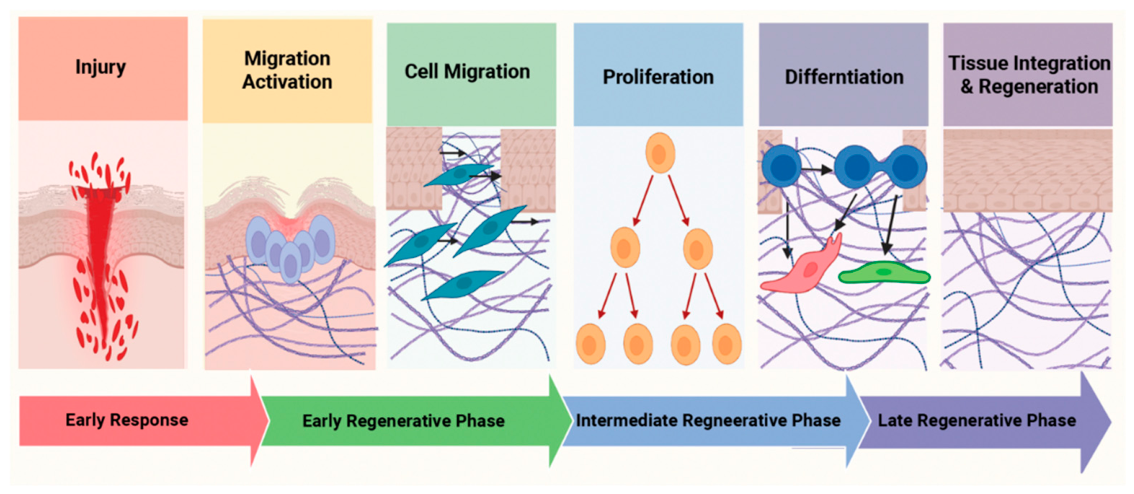
Figure 2.
Molecular and environmental mechanisms controlling regenerative cell migration. Regenerative migration is coordinated by cell-intrinsic machinery and extracellular guidance signals. Establishment of cell polarity, cytoskeletal remodeling, cell–ECM adhesion and traction, actomyosin contractility, and metabolic and mitochondrial support collectively generate and sustain cell movement. Migrating cells simultaneously integrate chemical gradients, ECM-associated adhesive cues, mechanical properties of the tissue environment, and endogenous electrical signals to determine migration direction and persistence. These mechanisms are dynamically interconnected and their relative contributions vary among cell types, tissues, and regenerative contexts.
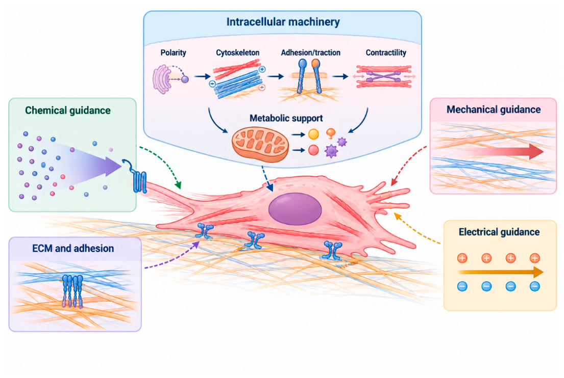
Figure 3.
Cardiac regeneration as a coordinated multicellular migration and tissue-remodeling process. Schematic illustrating the coordinated cellular movements and tissue remodeling that accompany cardiac repair and regeneration. Following myocardial injury, cardiomyocytes, epicardial cells, endothelial cells, immune cells, fibroblasts, and lymphatic endothelial cells undergo spatial redistribution, migration, or remodeling around the injured region. These responses contribute to cardiomyocyte repopulation, revascularization and lymphatic remodeling, inflammatory regulation, extracellular-matrix turnover, and restoration of tissue architecture. The enlarged injury region emphasizes that cardiac regeneration emerges from coordinated interactions among multiple cell populations and the dynamically remodeled extracellular environment rather than from cardiomyocyte proliferation alone.
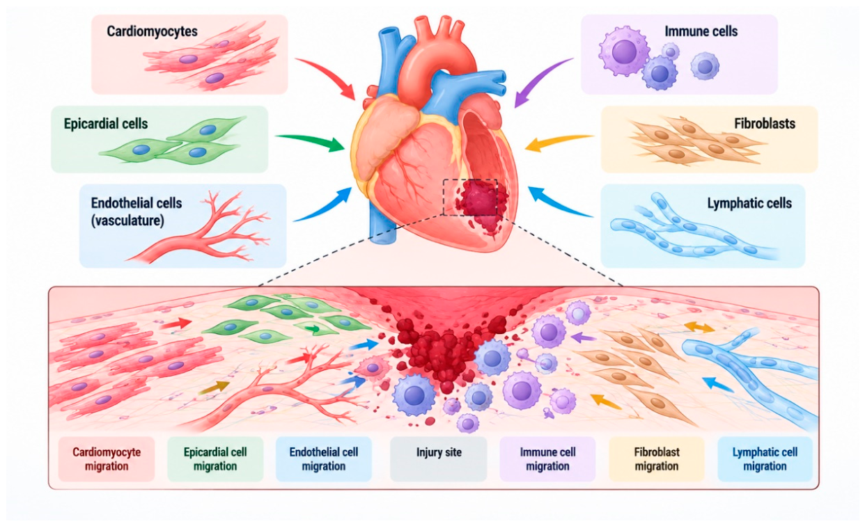

Table 1.
Representative roles of cell migration across tissue repair, regeneration, homeostasis, and development.
Table 1.
Representative roles of cell migration across tissue repair, regeneration, homeostasis, and development.
| Tissue/context | Cell type | Migration mode | Key mechanism | Regenerative significance | Refs. |
|---|---|---|---|---|---|
| Heart | Cardiomyocytes and cardiac niche cells | Protrusive/local and multicellular | YAP-actin remodeling; dystrophin; Mmp14b; macrophage/ECM remodeling | Migration-related remodeling can be experimentally dissociated from proliferation | [4,5,14,15] |
| Heart–vascular niche | Endothelial/coronary cells | Sprouting/collective | Epicardial Cxcl12–Cxcr4; endocardial Vegfa | Vascular ingrowth can precede cardiomyocyte migration and provide a scaffold | [51,52] |
| Heart–epicardium | Epicardial-derived cells | Collective/EMT-like invasion | FGF signaling; hyaluronic acid–HMMR | Epicardial migration supports neovascularization and repair | [53,54] |
| Heart–immune niche | Macrophages | Directed recruitment/retention | Wt1b-dependent migration; Mmp14b-dependent ECM remodeling | Immune-cell positioning shapes the permissiveness of the cardiac wound | [5,55,125] |
| Heart–lymphatic niche | Lymphatic endothelial cells | Vascular remodeling | Lymphoangiocrine signaling/Reelin | Lymphatic remodeling supports cardiac growth and repair | [72,73,74,75] |
| Liver | Hepatocytes | Collective | ANXA2-associated migratory state | Necrotic wound closure can precede proliferation | [6] |
| Kidney | Tubular epithelial cells | Collective | Migration-induced cell stretch/mechanical feedback | Early tubule resealing precedes later proliferation | [9,81,82,83,84] |
| Intestine | Epithelial cells | Collective | Arp2/3-dependent basal protrusions | Steady-state crypt-villus turnover | [7,106,107,108,109,110] |
| Skin | Keratinocytes and stromal/immune cells | Individual + collective | Multicellular wound signaling and ECM remodeling | Re-epithelialization and repair | [8,85,86,87,88,89] |
| Zebrafish fin | Epithelial and connective-tissue cells | Local/collective | Rapid wound-directed movement | Wound epidermis and blastema formation | [25,95,96,97,98] |
| Axolotl limb | Blastema-contributing cells | Multicellular | Wound epithelium, neural signals, positional information | Blastema formation and patterned limb regeneration | [99,100,101,102,103,104,105] |
| Neural progenitors | Adult neural progenitor cells | Individual | Drp1-dependent mitochondrial dynamics | Migration linked to neuronal differentiation | [130] |
| Joint/connective tissue | Mesenchymal and immune cells | Interstitial | ECM density/stiffness; biochemical cues | Can support repair or pathological inflammation | [16,120,121,122,123,124] |
Disclaimer/Publisher’s Note: The statements, opinions and data contained in all publications are solely those of the individual author(s) and contributor(s) and not of MDPI and/or the editor(s). MDPI and/or the editor(s) disclaim responsibility for any injury to people or property resulting from any ideas, methods, instructions or products referred to in the content. |
© 2026 by the authors. Licensee MDPI, Basel, Switzerland. This article is an open access article distributed under the terms and conditions of the Creative Commons Attribution (CC BY) license (http://creativecommons.org/licenses/by/4.0/).
Copyright: This open access article is published under a Creative Commons CC BY 4.0 license, which permit the free download, distribution, and reuse, provided that the author and preprint are cited in any reuse.