
Preprint
Article

This version is not peer-reviewed.

Topical Oxygen Treatment Restores Vaginal Microbiota Balance in Women with Vaginitis and Bacterial Vaginosis: An Exploratory Study

A peer-reviewed version of this preprint was published in:
Microorganisms 2026, 14(11), 2458. https://doi.org/10.3390/microorganisms14112458

Submitted:

12 October 2026

Posted:

13 October 2026

You are already at the latest version

Abstract

(1) Background: Vaginal dysbiosis is strongly implicated in the pathophysiology of vaginitis and bacterial vaginosis (BV). Standard antimicrobial therapy may temporarily reduce pathogen burden but often disrupts beneficial Lactobacillus species, contributing to recurrence. Topical oxygen treatment may promote selective suppression of anaerobic pathogens and restore Lactobacillus-dominated communities. This preliminary study aimed to evaluate the effect of topical oxygen treatment—alone or adjunctive to systemic antimicrobial treatment—on vaginal microbiota composition. (2) Methods: Twenty-two symptomatic women affected by either BV or vulvovaginitis were enrolled and assigned to either topical oxygen or standard antimicrobial treatment plus topical oxygen. Oxygen delivery (3 L/min for 20 minutes, twice weekly for 2 weeks) was performed by using the Caress Flow device. Vaginal microbiota composition, pH and Lactobacillus presence were evaluated at baseline and 10–14 days post-treatment using the 16S Metagenomic Sequencing Library Preparation, sequencing and analysis. (3) Results: Topical oxygen alone increased the negative swabs from 4 (36.4%) to 9 (81.8%), p=0.06 and maintained stably high the total Lactobacillus spp. 92.60% [67.88–96.16%] at baseline and 91.50% [87.4–95.0%] pre-treatment. Oxygen plus antimicrobial treatment markedly reduced strict anaerobes (pre-treatment: 24.8% [15.3-36.5] to 9.0% [1.3-17.4] after treatment, p=0.05) and decreased BV-associated genera including Atopobium and Sneathia. Both oxygen-based regimens showed trends toward restoration or maintenance of Lactobacillus dominance. (4) Conclusions: Topical oxygen treatment favourably modulates the vaginal microbiota and may accelerate recovery from dysbiosis especially when used combined with standard antimicrobial treatment. These findings support larger trials to assess clinical efficacy and recurrence prevention.

Keywords: 
;  ;  ;  

1. Introduction

The vaginal microbiota plays a critical role in maintaining mucosal homeostasis and protecting against infections, largely through the activity of Lactobacillus species, which produce lactic acid and antimicrobial factors [1,2]. The healthy vaginal microenvironment is a dynamic and finely balanced ecosystem dominated by Lactobacillus species, most notably L. crispatus, L. gasseri, L. jensenii, and L. iners. These beneficial bacteria maintain vaginal health primarily through the fermentation of glycogen—released by estrogen-stimulated vaginal epithelial cells—into lactic acid. This process maintains a low physiological vaginal pH (typically between 3.5 and 4.5), which serves as a primary chemical barrier against the colonization and overgrowth of opportunistic pathogens. In addition to lactic acid production, lactobacilli exert competitive exclusion by secreting hydrogen peroxide (H2O2), bacteriocins, and biosurfactants, while physically blocking pathogen adherence to the epithelial lining. Importantly, the various vaginal Lactobacillus species possess distinct functional genomes that influence their resilience during environmental transitions. For instance, L. crispatus is widely recognized as the hallmark of a highly stable and protective microbiome, owing to its robust production of D-lactic acid and hydrogen peroxide, which strongly inhibit pathogen growth. Conversely, L. iners possesses a significantly smaller genome, lacks the ability to produce D-lactic acid, and encodes inersolysin, a pore-forming toxin upregulated under inflammatory or dysbiotic conditions. Despite these differences, both species are highly aerotolerant due to their cellular mechanisms for managing oxidative stress. While strict anaerobes succumb to oxygen exposure, these facultative species leverage specialized antioxidant defenses, allowing them to survive and rapidly repopulate the vaginal niche once the competing anaerobic load is cleared by oxygen therapy.
Disruption of this ecosystem allows a vaginal dysbiosis underlying bacterial vaginosis (BV) and mixed forms of vulvovaginitis [3]. Bacterial vaginosis is clinically characterized by a depletion of protective lactobacilli, an elevated vaginal pH (> 4.5), the presence of 'clue cells' on microscopic examination, and the absence of a significant local inflammatory reaction. This dysbiosis is driven by a polymicrobial proliferation of anaerobic and facultative pathogens, including Gardnerella vaginalis, Mobiluncus spp., Bacteroides spp., Peptostreptococcus spp., Fusobacterium spp., Mycoplasma hominis, and Ureaplasma urealyticum. Standard antimicrobial treatments, although effective in clearing acute infection, may further disturb the microbiota by reducing beneficial Lactobacillus species, potentially contributing to increase recurrence rates [4,5]. Consequently, attention has focused on therapeutic strategies capable not only of eradicating pathogens but also of restoring a Lactobacillus-dominant environment.
On the other hand, few attentions have been focused on the fact that most of the bacterial species sustaining BV (Gardnerella vaginalis, Atopobium vaginae, Megasphera, Prevotella and Sneathia), classified as anaerobia bacteria since their proliferation could take place only in the absence of oxygen. Indeed, the presence of oxygen in the environment induced a block of bacterial growth [6]. Although Lactobacillus spp. are included among anaerobia, they are still aero-tolerant and insensitive to oxygen exposure [7].
Topical oxygen treatment may represent one such innovative approach. The physical microenvironment of the vagina, particularly its oxygen tension and oxidation-reduction (redox) potential, plays a pivotal role in regulating microbial dynamics. In a healthy state, the vagina maintains a low but definitive oxygen tension (typically ranging between 15 and 30 mmHg). However, during BV-associated dysbiosis, the massive proliferation of anaerobic microorganisms rapidly depletes this localized oxygen, driving the redox potential down and creating an ideal niche for obligate anaerobes to thrive. Obligate anaerobes lack critical antioxidant enzymes, such as superoxide dismutase (SOD), catalase, and peroxidase. Exposure to molecular oxygen is toxic to these organisms as it leads to the intracellular accumulation of highly reactive oxygen species (ROS), including singlet oxygen and hydroxyl radicals, which induce lethal oxidative damage to bacterial proteins, lipids, and nucleic acids. Conversely, while Lactobacillus species are classified as facultative anaerobes or microaerophiles, they are aerotolerant. They possess alternative metabolic enzymes, such as NADH oxidases and manganese-dependent pseudocatalases, enabling them to survive and metabolize efficiently in oxygenated environments [9,10]. This metabolic divergence suggests that raising local oxygen tension could selectively suppress strict pathogens while preserving or actively promoting the re-growth of protective lactobacilli.
Preliminary clinical observation from our group [10] suggest that topical oxygen insufflation may accelerate symptom improvement, possibly through microbial rebalancing [11]. Thus, this exploratory study investigates the impact of topical oxygen treatment, used alone or as an adjunct to antimicrobial treatment, on vaginal microbiota composition.

2. Materials and Methods

2.1. Study Population

An exploratory, randomized, open-label interventional pilot study was conducted on women presenting with symptoms of vaginitis or BV. The recruitment and clinical procedures were conducted at the Obstetrics Unit of the Mother-Infant Department, Azienda Ospedaliero-Universitaria (AOU) of Modena, Italy (a tertiary referral hospital outpatient clinic). Diagnoses were established at screening using standard clinical criteria during the outpatient visit. Patients were diagnosed based on the presence of characteristic vulvovaginal symptoms (including abnormal vaginal discharge, pruritus, burning, or dyspareunia), bed-side vaginal pH measurement (with pH > 4.5 indicative of BV), and qualitative microscopic evaluation of standard vaginal wet mount smears (to assess the presence of leukocytes, yeast cells, or clue cells). Inclusion criteria comprised symptomatic BV or non-specific vaginitis and collection of Informed consent. This was an ancillary study of a pilot, open label, monocentric trial in which women of fertile age with a diagnosis of vaginal infections were enrolled, after informed consent was obtained. Inclusion criteria were aged 18-45 years, diagnosis of vaginal infections of different etiology, ability to fill-in the symptom’s sheet and the comprehension of the informed consent sheet. Exclusion criteria were: vulvo-vaginal atrophy, amenorrhea since at least 3 months and exposure to antibiotics in the previous 30 days.
Patients were instructed to avoid sexual intercourse, vaginal douching, and the use of any topical intravaginal products, lubricants, or spermicides for at least 48 hours prior to both sampling visits. Furthermore, clinical visits and samplings were scheduled to avoid active menses.
Due to the explorative nature of the study, the sample size was determined a priori as being composed by 22 women, 11 each group, deemed sufficient to assess the variation in microbiota composition with focus on Lactobacillus dominance and the facultative vs. strict anaerobe distribution in the two study groups.

2.2. Study Design

At baseline (Visit 1), clinicians collected vaginal swabs for microbiota analysis. Participants were 1:1 allocated to topical oxygen alone or antibiotic treatment plus topical oxygen. Randomization was managed by a midwife who used sealed envelopes containing treatment allocation that was performed through a random series of numbers. Treatment was disclosed after the patient’s consent was signed.
A group received vaginal high concentration oxygen supply (O2 concentration >95%, 3 L/min for 20 minutes), twice/week for 2 weeks; the other group received vaginal oxygen added to a targeted antibiotic or antimycotic treatment for 5-7 days. All topical oxygen administrations were performed using the Caress Flow device (Caress Flow - Bologna, Italy), a specialized medical apparatus designed for gynecological applications. The system utilizes molecular sieves to concentrate oxygen from ambient room air, delivering high-purity medical gas (oxygen concentration >95%) at low, non-traumatic pressures. The gas was administered intravaginally via a sterile, single-use, medical-grade plastic cannula. The cannula features a rounded, trailblazing, atraumatic tip and multiple lateral micro-apertures strategically positioned to ensure a uniform, 360-degree radial distribution of oxygen across the vaginal mucosa, vaginal walls, and posterior fornix. This specific design avoids localized barotrauma or the risk of gas embolism, as the vaginal canal remains open to the atmosphere, allowing the continuous escape of excess gas during the 20-minute procedure. The flow rate was calibrated and maintained at 3 L/min. Each session lasted exactly 20 minutes, and the procedure was performed twice weekly for a total of two consecutive weeks (4 sessions in total).2.3. Microbiome assay
Vaginal samples were collected using sterile vaginal swabs and immediately placed in sterile collection tubes (eNAT, Copan, Italy). To ensure consistency and avoid contamination, vaginal swabs for microbiota analysis were collected using a standardized clinical technique. Specifically, the clinician carefully rotated sterile swabs against the lateral vaginal wall and the posterior vaginal fornix, ensuring that contact with the cervical mucus plug was strictly avoided.
Samples were stored at −80 °C until DNA extraction. All samples were processed using the same collection, storage, and handling protocol to minimize technical variability. Pre- and post-treatment vaginal microbiome analyses were performed by NEXT Genomics S.R.L. (Sesto Fiorentino, Florence, Italy) using a 16S rRNA gene amplicon sequencing workflow, including DNA extraction, library preparation, sequencing, and primary bioinformatics analysis. To prevent bias in microbial representation, DNA extraction was optimized to ensure the efficient lysis of Gram-positive bacteria, which possess thick peptidoglycan cell walls. Total metagenomic DNA was extracted from 200 µL of vaginal swab suspension in eNAT buffer using the QIAamp® Fast DNA Stool Mini Kit (Qiagen, Hilden, Germany) with critical modifications to the manufacturer's standard protocol. Briefly, samples were transferred to sterile 2 mL screw-cap tubes containing 0.3 g of sterile, acid-washed zirconia/silica beads (0.1 mm diameter; BioSpec Products, Bartlesville, OK, USA). Mechanical disruption (bead-beating) was performed using a high-speed cell homogenizer (FastPrep-24, MP Biomedicals, Solon, OH, USA) at a speed of 6.0 m/s for 40 seconds. This physical disruption was immediately followed by chemical lysis via incubation with 1 mL of InhibitEX buffer at 70°C for 10 minutes, and subsequent enzymatic digestion with lysozyme (20 mg/mL) and Proteinase K (20 mg/mL) at 56°C for 30 minutes. The remaining steps, including inhibitor adsorption, silica-membrane binding, washing, and elution in 50 µL of sterile elution buffer, were performed in accordance with the manufacturer's instructions. The same optimized extraction procedure was applied to all baseline and post-treatment samples to ensure consistency.
16S amplification was performed with primers: Forward: 5’-CCTACGGGNGGCWGCAG-3’ and Reverse: 5’-GACTACHVGGGTATCTAATCC-3’ (Klindworth et al., 2013), which target the hypervariable V3 and V4 region of the 16S rRNA gene. Each PCR reaction was assembled according to the Illumina 16S Metagenomic Sequencing Library Preparation protocol (Illumina, San Diego, CA). A negative control, consisting of all reagents used during sample processing (16S amplification and library preparation) but without biological material, was included in the workflow to monitor potential contamination. Libraries were quantified using a Qubit fluorometer (Invitrogen Co., Carlsbad, CA) and pooled to an equimolar amount of each index-tagged sample to a final concentration of 4nM, including the Phix Control Library. Pooled samples were subject to cluster generation and sequenced on the MiSeq platform (Illumina, San Diego, CA) in a 2x250 paired-end format. The raw sequence files generated (FASTQ files) underwent quality control analysis with FastQC. Demultiplexed paired-end reads were processed using QIIME2. Quality filtering, denoising, paired-end merging, and chimera removal were performed using the DADA2 plugin with default parameters, generating amplicon sequence variants (ASVs). To ensure the highest standard of data integrity, stringent quality control criteria were enforced during sequence processing. Demultiplexed reads were first evaluated for quality; bases with a Phred score below Q30 were trimmed from the 3’ ends, and reads shorter than 200 base pairs after trimming were discarded. The DADA2 pipeline executed denoising, which models sequencing errors to distinguish true biological variants from sequencing artifacts. Forward and reverse reads were merged with a minimum overlap of 20 base pairs. Chimeric sequences, which can arise during PCR amplification, were identified and removed de novo using the consensus method in DADA2, resulting in a high-purity dataset of non-chimeric ASVs. Samples with low sequencing depth (fewer than 10,000 high-quality reads) were excluded from downstream taxonomic profiling to prevent undersampling bias. The rarefaction curves were plotted to confirm that the sequencing depth successfully reached a plateau, capturing the complete microbial diversity of the vaginal microenvironment. Taxonomic classification was performed using a Naive Bayes classifier trained on the SILVA SSURef NR99 database (release 138.2), trimmed to the amplified V3–V4 region. The output of this workflow consisted of taxonomic classifications across multiple taxonomic ranks. Given the known limitations of 16S rRNA gene amplicon sequencing, species-level assignments were considered putative and interpreted as relative changes in sequence assignment rather than absolute bacterial quantification.

2.4. Statistical Analysis

Data were analyzed using the statistical package Stata 16.1 (StataCorp. 2019. College Station, TX, USA). Due to the compositional nature of vaginal microbiota data (expressed as relative abundances from 0 to 100%) and their non-normal distribution, non-parametric statistics were utilized. Comparisons of continuous variables (including bacterial relative abundances and pH) between pre- and post-treatment paired samples were performed using the non-parametric Wilcoxon signed-rank test. Categorical variables were compared using the McNemar test. Continuous variables are presented in the text as Median (Interquartile Range, 25th-75th percentiles) [IQR:Q1-Q3], and in figures the Boxes represent the Median and IQR: Q1–Q3. Categorical variables (vaginal swab outcomes) are presented as N (%). Relative abundances were calculated as the percentage of high-quality reads assigned to each taxon relative to the total number of reads retained for each sample, following quality filtering and rarefaction to a standard sequencing depth in QIIME2. The P value ≤ 0.05 was considered as the threshold for defining statistical significance. Bacteria were grouped according to their oxygen requirements into strict or facultative anaerobes, with the first class of anaerobes being incapable of aerobic metabolism and the second one being able to grow in both the presence and absence of oxygen.

3. Results

This section may be divided by subheadings. It should provide a concise and precise desc

3.1. Topical Oxygen

Eleven women belonged to the group treated solely with topical oxygen treatment. The main results of the vaginal swab showed a stable pH and Lactobacilli’s presence after treatment, but an increase of negative swabs was found after the treatment completion (negative swabs: pre- 4 (36.4%) vs post- 9 (81.8%), p=0.06), Table 1. The overall structure of the vaginal microbiota demonstrated notable signs of recovery after treatment, particularly with respect to Lactobacillus prevalence (Figure 1A).
More substantial changes emerged within the Lactobacillus community. Overall, Lactobacillus spp. median of relative abundance was 92.60% [IQR: 67.88–96.16%] at baseline becoming 91.50% [IQR: 87.4–95.0%] after treatment, suggesting a stability of protective lactobacilli following oxygen exposure. Increases were observed in several key Lactobacillus taxa, including sequences assigned to L. crispatus, L. iners, and L. helveticus. While these individual changes did not achieve statistical significance, their collective upward trend supports the overall maintenance of Lactobacillus dominance in the post-treatment microbiome (Figure 1A).
At the broader ecological level, the distribution of facultative and strict anaerobes remained generally stable. Facultative anaerobes showed only a minimal increase from 80.09% [IQR: 72.4-83.9] to 81.85% [IQR: 75.30-85.43], a change that was not statistically significant. Similarly, strict anaerobes exhibited a slight reduction from 6.9% [IQR: 4.5-9.6] to 5.6 [IQR: 3.7-7.7]. Parallelly, a consistent, multi-taxa reduction in key dysbiosis-associated anaerobic pathogens was observed, including Bacteroides spp. (median: 0.07% vs 0.06%, p = 0.1270), Peptoniphilus spp. (median: 0.01% vs 0.01%, p = 0.1484), and opportunistic Streptococcus spp. (median: 0.14% vs 0.07%, p = 0.3711). Taken together, these data demonstrate that topical oxygen exerts a targeted suppressive pressure on anaerobic pathobionts while preserving the protective Lactobacillus barrier.
A comprehensive taxonomic breakdown of the vaginal microbiota before and after topical oxygen monotherapy is presented in Table 1.
Taken together, the findings from the oxygen-only group indicate that topical oxygen treatment can favourably modulate the vaginal microbiota by promoting the re-expansion of endogenous Lactobacillus species while simultaneously reducing select anaerobic pathogens, even in the absence of concurrent antimicrobial treatment.

3.2. Antibiotic Treatment Plus Topical Oxygen

In the cohort receiving combined treatment with topical oxygen and standard antimicrobial treatment, the main results of the vaginal swab showed a stable pH (median 5.00 (IQR: 4.88–5.13) pre vs. 4.75 (IQR: 4.50–5.00) post, p = 0.1875), and Lactobacilli’s presence, while a trend toward reduction of positive and increase of negative swabs can be highlighted after treatment, Table 2.
Lactobacillus taxa also benefited from the combined regimen. Overall, Lactobacillus spp. increased from a median of 49.08% (IQR: 1.14–96.81%) pre-treatment to 85.19% (IQR: 44.63–97.49%) post treatment, a substantial improvement, though not statistically significant (p=0.13). Among individual species, L. iners demonstrated a notable and statistically significant rise, increasing from 18.9% (14.3-23.4) to 47.9% (20.7-56.4) (p=0.05). This increase may reflect the early colonizing behavior of L. iners, which often expands before more stable Lactobacillus species re-establish dominance (Figure 1B).
Interestingly, the microbiological response was more pronounced in this study group, particularly regarding anaerobic taxa associated with bacterial vaginosis. The most striking change in this group was the significant reduction in strict anaerobes, which decreased from 24.8% (15.3-36.5) at baseline to 9.0% (1.3-17.4) after treatment (p=0.05). This marked decline aligns with the known susceptibility of anaerobic organisms to both antimicrobial therapy and the elevated oxygen tension achieved through insufflation. At the same time, facultative anaerobes showed an upward trend, although the change did not reach statistical significance (p = 0.12). This shift suggests a transition toward a more oxygen-tolerant and potentially more stable community structure (Table 2).
The combined treatment also resulted in meaningful reductions in several pathogens strongly associated with dysbiosis. Clostridium spp. decreased significantly (from 0.04% (IQR: 0.02–3.43%) pre vs. 0.02% (IQR: 0.01–0.16%) post, p=0.06), while Sneathia spp., a hallmark organism of severe BV-associated dysbiosis and adverse reproductive outcomes, showed a dramatic reduction (from 0.00% [IQR: 0.00–26.41%] pre vs. 0.00% [IQR: 0.00–0.00%], p=0.04), mirroring the pattern observed in the oxygen-only group and reinforcing the hypothesis that oxygen exposure selectively inhibits anaerobic pathobionts (Table 2). Conversely, a statistically significant increase was observed in the relative abundance of Staphylococcus spp. (from 0.00% [IQR: 0.00–0.01%] pre vs. 0.02% [IQR: 0.01–0.09%] post, p = 0.03; Table 2); however, its absolute representation remained extremely low (well below 0.1%), rendering this shift clinically negligible.
In this combined treatment group, other BV-associated anaerobes also demonstrated sharp, clinically favourable declines (Table 2). Atopobium spp. demonstrated a highly statistically significant reduction, decreasing from a baseline relative abundance 0.00% (IQR: 0.00–2.37%) pre vs. 0.00% (IQR: 0.00–0.00%) post-treatment (p = 0.01). Gardnerella spp. relative abundance was reduced from 0.01% (IQR: 0.00–3.86%) to 0.00% (IQR: 0.00–3.18%), (p = 0.84). Conversely a very strong significant increase in Streptococcus spp. (from 0.02% [IQR: 0.00–0.03%] to 0.71% [IQR: 0.14–2.83%], p = 0.002). While these Gram-positive, facultative taxa expanded, their presence did not clinically translate into symptomatic vulvovaginitis.
Overall, the data suggests that the combination of topical oxygen treatment and standard antimicrobial treatment exerts a synergistic effect on microbiota restoration. The regimen not only suppresses anaerobic pathogens more effectively than antimicrobials alone but also facilitates the re-emergence of Lactobacillus species, supporting a transition toward a more physiologically balanced vaginal ecosystem.

4. Discussion

This preliminary study indicates that topical oxygen treatment may beneficially modulate the vaginal microbiota in women affected by vaginitis and BV. The observed stability maintenance of highly distributed Lactobacillus spp. in the oxygen-only group supports the concept that oxygen enrichment favours the recovery of protective species, consistent with previous mechanistic studies demonstrating Lactobacillus resilience in oxygenated microenvironments [8,9].
We acknowledge that the relatively small sample size of our exploratory cohorts (N = 11 per group) represents a critical limitation regarding statistical power, which likely accounts for the lack of statistically significant changes in overall Lactobacillus relative abundance (p > 0.05) despite a clear, clinically positive upward trend. In small-scale pilot studies of vaginal dysbiosis, high inter-individual baseline variability often masks statistical significance when using conservative non-parametric tests. Rather than a lack of therapeutic efficacy, the observed transition—characterized by a shift from a highly heterogeneous, dysbiotic state to a more uniform, Lactobacillus-enriched profile—supports the clinical value of topical oxygen. This is particularly evident in the combined therapy cohort, where Lactobacillus spp. relative abundance expanded from 49.08% (1.14–96.81%) to 85.19% (44.63–97.49%), signaling an active, early-stage recovery of the vaginal microenvironment.
A highly compelling finding of our updated, non-parametric analysis is the highly significant and dramatic reduction of Atopobium spp. (p = 0.01) in the combined treatment group. Atopobium vaginae is a critical, Gram-positive anaerobic pathogen that plays a pivotal role in the initiation and structural maintenance of the highly resilient polymicrobial biofilms characteristic of bacterial vaginosis. Crucially, A. vaginae is notoriously associated with high rates of metronidazole resistance and recurrent dysbiosis, representing a major hurdle in standard gynecological care. The fact that the addition of topical oxygen to standard antimicrobial therapy successfully eradicated or minimized Atopobium spp. relative abundance represents a major therapeutic milestone. By altering the localized redox potential, oxygen therapy selectively targets the metabolic vulnerability of strict anaerobes like Atopobium and Sneathia (p = 0.04), effectively disrupting the physical scaffold of the pathogenic biofilm and paving the way for long-term clinical recovery.
The combined oxygen + antimicrobial regimen showed significant reductions in strict anaerobes, including BV-associated organisms such as Sneathia and Clostridium, which aligns with evidence that anaerobic taxa are particularly sensitive to increased oxygen tension [13]. Restoration of Lactobacillus dominance is crucial, as persistent dysbiosis is a recognized driver of BV recurrence [4,14]. Although these shifts were modest, they indicate that oxygen treatment did not disrupt the overall balance of major microbial categories. The significant expansion of Lactobacillus iners observed post-treatment in the combined therapy group warrants deeper ecological discussion. In vaginal microbial ecology, L. iners is frequently characterized as a transitional or 'pioneer' species. Due to its unique genomic plasticity, L. iners is capable of surviving in both dysbiotic (high pH, inflamed) and eubiotic (low pH) environments. During the acute phase of recovery following topical oxygen and antimicrobial treatment, the rapid suppression of strict anaerobes leaves an open ecological niche. L. iners, with its rapid replication rate and high tolerance to environmental fluctuations, is often the first to expand, acting as an intermediate colonizer. This transitional phase is critical, as the metabolic activity of L. iners helps to re-acidify the vaginal microenvironment, thereby preparing the physiological ground for the subsequent, long-term re-establishment of more stable, protective species such as L. crispatus. Therefore, the post-treatment increase in L. iners should not be viewed as a failure of therapy, but rather as an active, early phase of the vaginal ecosystem's natural transition toward complete eubiosis.
The results suggest that oxygen treatment may serve as a microbiome-supportive intervention capable of accelerating ecosystem recovery following antimicrobial treatment. This is consistent with previous exploratory studies where oxygen application reduced pathogen burden and improved vaginal microenvironment parameters [10]. Furthermore, also other in vitro studies confirmed antimicrobial oxygen activity on P. gingivalis in patients affected by periodontitis where the treatment fostered eubiosis [15].
Conversely, the statistically significant expansion of Streptococcus spp. (p = 0.002) and Staphylococcus spp. (p = 0.03) in the combined therapy group should be interpreted through an ecological lens. In a healthy vaginal niche, the clearance of dominant anaerobic pathobionts creates an open biophysical space. In the immediate post-treatment window, aerotolerant, facultative Gram-positive taxa such as Streptococcus and Staphylococcus can rapidly expand to occupy this niche. It is vital to emphasize that the relative abundance of Staphylococcus remained exceedingly low (well below 0.1%), rendering this shift clinically negligible. Furthermore, none of the patients exhibited clinical symptoms of aerobic vaginitis or local irritation during the follow-up, demonstrating that this early taxonomic rearrangement represents a transient, safe transitional phase of ecological restructuring rather than a secondary infection.
Lastly, it is worth emphasizing that the reporting of non-parametric medians and IQRs clearly highlights the extreme sparsity and inter-individual heterogeneity of the vaginal microbiome in bacterial vaginosis. Rather than representing ubiquitous low-level colonizers, pathogenic anaerobes such as Atopobium vaginae and Sneathia spp. manifest as acute, patient-specific blooms. The fact that topical oxygen combined therapy achieved a statistically significant clearance of these organisms (p < 0.05) demonstrates that oxygen insufflation effectively eradicates these high-abundance pathogenic clusters in affected patients, driving the entire cohort toward a uniform, non-detectable state.
Our study relied on 16S rRNA gene amplicon sequencing targeting the V3–V4 region, which provides robust characterization of overall community structure but has intrinsic limitations in species-level resolution for some bacterial taxa. Accordingly, species-level findings should be interpreted as relative changes in sequence assignment rather than absolute quantification. Nevertheless, the observed trends in dominant vaginal Lactobacillus species—such as L. iners—are consistent with previous vaginal microbiome studies using similar approaches and support the biological relevance of the reported shifts. Future studies integrating targeted qPCR or shotgun metagenomics will be valuable to further refine species-level resolution.
Regarding clinical tolerability and patient acceptability, our study demonstrated that topical oxygen treatment delivered via the Caress Flow system was exceptionally well tolerated. None of the twenty-two participants reported pain, burning, local irritation, or any other adverse events during or after the 20-minute vaginal insufflation sessions. This is a clinically relevant finding, as standard intravaginal antimicrobial therapies (such as creams, gels, or ovules) are frequently associated with local discomfort, messy discharge, or compliance issues. The subjective evaluations and symptom sheets collected during the follow-up visit (10–14 days post-treatment) indicated a high level of patient satisfaction, with participants describing the non-invasive procedure as comfortable and highly acceptable. This high compliance and positive patient perception suggests that topical oxygen therapy represents a patient-friendly, non-invasive alternative or adjunctive option for managing vaginal dysbiosis.However, the small sample size and absence of a long-term follow-up limit the generalizability of these findings. Additionally, the absence of a standard-of-care control arm (i.e., an antimicrobial-alone group) represents a limitation inherent to the preliminary and exploratory nature of this pilot study. However, the promising findings emerging from this work justify and support larger, sham-controlled, and multi-arm randomized trials to fully characterize the synergistic effect of topical oxygen and standard antimicrobial treatments. Furthermore, we acknowledge that enrolling patients with different etiologies, including both bacterial vaginosis and non-specific or fungal vaginitis, introduced a degree of population heterogeneity that may have confounded baseline microbiome profiles. To address this variation, our exploratory analysis was designed to focus on broad, high-level ecological shifts—specifically the ratio of protective Lactobacillus species to strict and facultative anaerobes—which are common denominators of vaginal dysbiosis across different etiologies. Nevertheless, future clinical trials utilizing larger and more homogeneous patient cohorts, restricted to specific clinical diagnoses (e.g., pure BV vs. specific vaginitis subtypes), are highly warranted to fully dissect etiology-specific microbiome dynamics.

5. Conclusions

In conclusion, topical oxygen treatment delivered via the Caress Flow system represents a highly promising, patient-friendly, and non-invasive therapeutic approach for restoring vaginal microbiota balance in women affected by vaginitis and bacterial vaginosis. Whether administered as a standalone therapy or even more as an adjunct to standard antimicrobial regimens, localized oxygen insufflation selectively suppresses pathogenic, obligate anaerobes while preserving and fostering the recovery of protective, aerotolerant Lactobacillus species.
In an era of escalating antimicrobial resistance and alarmingly high recurrence rates associated with standard antibiotic treatments, this technology offers an innovative, antibiotic-sparing alternative for gynecological care. By shifting the local redox potential, topical oxygen therapy targets the biophysical environment that supports dysbiosis, rather than relying solely on chemical eradication. Future large-scale, sham-controlled, multi-center clinical trials with extended follow-up periods are highly warranted to fully validate the clinical efficacy of this approach, characterize its impact on polymicrobial biofilms, and establish standardized treatment protocols tailored to specific vaginal dysbiosis profiles.

Supplementary Materials

The following supporting information can be downloaded at the website of this paper posted on Preprints.org, Figure S1: title; Table S1: title; Video S1: title.

Author Contributions

Conceptualization, F.F.; methodology, M.C.; software, M.C. and S.C.; validation, D.M. and M.C..; formal analysis, D.M.. M.C. and S.C.; investigation, F.F.. E.B. and F.G.; data curation, D.M. and M.C..; writing—original draft preparation, D.M. and M.C.; writing—review and editing, F.F. I.N. M.C, F.G; supervision, F.F. and I.N. All authors have read and agreed to the published version of the manuscript.

Funding

This research received no external funding.

Institutional Review Board Statement

Ethical review and approval were waived for this study by the local Ethic Committee “Comitato etico territoriale di Area Vasta Emilia Nord (CET AVEN)” as it was conducted as a service evaluation of existing practices within this setting. Despite the exemption, the study adhered to the ethical principles of the Declaration of Helsinki.

Data Availability Statement

The data that support the findings of this study are available from the corresponding author, upon reasonable request.

Conflicts of Interest

The authors declare no conflicts of interest.

References

  1. Ravel, J.; Brotman, R.M. Translating the vaginal microbiome: gaps and opportunities. Microbiome 2016, 4, 23–31. [Google Scholar]
  2. Boris, S.; Barbés, C. Role played by lactobacilli in controlling the population of vaginal pathogens. Microbes Infect. 2000, 2, 543–6. [Google Scholar] [CrossRef] [PubMed]
  3. Onderdonk, A.B.; Delaney, M.L.; Fichorova, R.N. The human microbiome during bacterial vaginosis. Clin. Microbiol. Rev. 2016, 29, 223–38. [Google Scholar] [CrossRef] [PubMed]
  4. Bradshaw, C.S.; Sobel, J.D. Current treatment of bacterial vaginosis—limitations and need for innovation. J. Infect. Dis. 2016, 214 (Suppl 1), S14–20. [Google Scholar] [CrossRef] [PubMed]
  5. Swidsinski, A.; Verstraelen, H.; Loening-Baucke, V.; et al. Presence of a polymicrobial biofilm in bacterial vaginosis. PLoS ONE 2013, 8, e53997. [Google Scholar] [CrossRef] [PubMed]
  6. Goldman, L.; Schafer, A.I. (Eds.) Goldman’s Cecil Medicine. (Twenty-Fourth Edition); Elsevier Press, 2012. [Google Scholar]
  7. André, A.C.; Debande, L.; Marteyn, B.S. The selective advantage of facultative anaerobes relies on their unique ability to cope with changing oxygen levels during infection. Cell Microbiol. 2021, 23(8), e13338. [Google Scholar] [CrossRef] [PubMed]
  8. Zheng, H.; Brennan, T.; Shetty, A. Oxygen and redox regulation in the vaginal microbiome: implications for dysbiosis. J. Reprod. Immunol. 2021, 148, 103–12. [Google Scholar]
  9. Eschenbach, D.A. Oxygen tension and redox potential of the vaginal environment: effect on microbial populations. Am. J. Obstet. Gynecol. 1989, 160, 1130–5. [Google Scholar]
  10. Facchinetti, F.; Longo, M.; Danieli, A.; Melis, B.; Garoia, F.; Bergamini, E. Vaginal supplementation of high concentration Oxygen for the treatment of vaginal infections: A pilot, controlled trial. Eur. Gynecol. Obstet. 2024, 7(1), 23–26. [Google Scholar]
  11. Rossi, M.; D’Andrea, F.; Bianchi, P. Topical oxygen therapy for vaginal dysbiosis: a pilot clinical experience. Gynecol. Obstet. Invest. 2020, 85, 340–6. [Google Scholar]
  12. Bush, L.M.; Vazquez-Pertejo, M.T. Anaerobic bacteria: overview. MSD Manual Professional Edition, 2025. [CrossRef] [PubMed]
  13. Fredricks, D.N.; Fiedler, T.L.; Marrazzo, J.M. Molecular identification of bacteria associated with BV. N Engl. J. Med. 2005, 353, 1899–911. [Google Scholar] [CrossRef] [PubMed]
  14. Muzny, C.A.; Kardas, P.; Griswold, M.E. Role of recurrent dysbiosis in BV pathophysiology. Clin. Infect. Dis. 2020, 71, 1109–16. [Google Scholar]
  15. Müller-Heupt, L.K.; Wiesmann-Imilowski, N.; Schröder, S.; et al. Oxygen-Releasing Hyaluronic Acid-Based Dispersion with Controlled Oxygen Delivery for Enhanced Periodontal Tissue Engineering. Int. J. Mol. Sci. 2023, 24(6), 5936. [Google Scholar] [CrossRef] [PubMed]
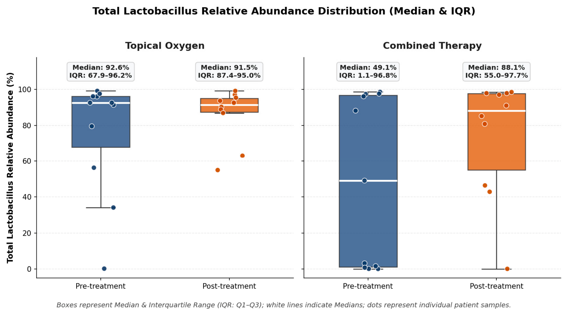
Figure 1. Lactobacilli distribution (%) pre- and post-treatment in the two study groups. Boxes represent the Median and Interquartile Range (IQR: Q1–Q3); white horizontal lines indicate Medians, and overlaid points represent individual patient values across species (L. crispatus, L. gasseri, L. iners, L. jensenii, L. helveticus).
Preprints 237896 g001
Table 1. Main vaginal swab results and Bacteria spp. distribution pre- and post-treatment with topical oxygen.
Pre-treatment
(N=11)
Post-treatment
(N=11)
P value
pH 4.50 (4.50–4.75) 4.50 (4.50–4.75) 1.00
Lactobacilli presence 6 (54.5%) 1 (14.3%) 0.50
Vaginal swab result
Negative
Leucocytosis
Positive

4 (36.4%)
3 (27.2%)
4 (36.4%)

9 (81.8%)
1 (9.1%)
1 (9.1%)
0.06
Bacteria spp,
Anaerococcus spp 0.00 (0.00–0.07) 0.01 (0.00–0.03) 0.14
Aerococcus spp 0.00 (0.00–0.00) 0.00 (0.00–0.00) 0.37
Atopobium spp 0.00 (0.00–0.01) 0.00 (0.00–0.00) 0.12
Bacteroides spp 0.07 (0.02–0.12) 0.06 (0.02–0.10) 0.13
Bifidobacterium spp 0.00 (0.00–0.02) 0.00 (0.00–0.01) 0.81
Clostridium spp 0.01 (0.00–0.10) 0.01 (0.00–0.03) 0.29
Dialister spp 0.00 (0.00–0.02) 0.00 (0.00–0.07) 0.46
Gardnerella spp 0.00 (0.00–0.04) 0.00 (0.00–0.04) 1.00
Megasphera spp 0.00 (0.00–0.00) 0.00 (0.00–0.00) 0.31
Peptoniphulus spp 0.01 (0.00–0.12) 0.01 (0.00–0.09) 0.14
Prevotella spp 0.01 (0.00–0.30) 0.13 (0.01–0.27) 0.44
Sneathia spp 0.00 (0.00–0.00) 0.00 (0.00–0.00) 0.25
Staphylococcus spp 0.01 (0.00–0.16) 0.02 (0.00–0.10) 0.44
Streptococcus spp 0.14 (0.02–0.50) 0.07 (0.04–0.13) 0.37
Ureaplasma spp 0.00 (0.00–0.01) 0.00 (0.00–0.00) 1.00
Note: Continuous variables (pH and bacterial relative abundances) are presented as Median (IQR: Q1–Q3, 25th–75th percentiles). Categorical clinical swab variables are presented as N (%). P-values in bold red are statistically significant (p ≤ 0.05) calculated using the non-parametric Wilcoxon signed-rank test for paired continuous data or McNemar test for binarized categorical data. The relative abundances were calculated as the percentage of high-quality reads assigned to each taxon relative to the total reads per sample, after quality filtering and rarefaction to a standard depth in QIIME2.
Table 2. Main vaginal swab results and Bacteria spp. distribution pre- and post-treatment with antibiotic therapy plus topical oxygen.
Pre-treatment
(N=11)
Post-treatment
(N=11)
P value
pH 5.00 (4.88–5.13) 4.75 (4.50–5.00) 0.18
Lactobacilli presence 5 (45.4%) 6 (54.5%) 0.37
Vaginal swab result
Negative
Leucocytosis
Positive

4 (36.4%)
4 (36.4%)
3 (27.2%)

8 (72.3%)
3 (27.2%)0
0.12
Bacteria spp,
Actinomyces spp 0.00 (0.00–0.00) 0.00 (0.00–0.07) 0.07
Aerococcus spp 0.00 (0.00–0.12) 0.00 (0.00–0.00) 0.56
Atopobium spp 0.00 (0.00–2.37) 0.00 (0.00–0.00) 0.01
Bacteroides spp 0.11 (0.06–0.13) 0.10 (0.03–0.14) 0.46
Bifidobacterium spp 0.00 (0.00–0.30) 0.00 (0.00–0.02) 0.29
Clostridium spp 0.04 (0.02–3.43) 0.02 (0.01–0.16) 0.06
Dialister spp 0.01 (0.00–0.78) 0.00 (0.00–0.01) 0.12
Gardnerella spp 0.01 (0.00–3.86) 0.00 (0.00–3.18) 0.84
Megasphera spp 0.00 (0.00–0.92) 0.00 (0.00–0.01) 0.13
Peptoniphulus spp 0.01 (0.00–0.23) 0.01 (0.00–0.02) 0.46
Prevotella spp 0.45 (0.05–8.75) 0.06 (0.02–0.35) 0.83
Sneathia spp 0.00 (0.00–26.41) 0.00 (0.00–0.00) 0.04
Staphylococcus spp 0.00 (0.00–0.01) 0.02 (0.01–0.09) 0.03
Streptococcus spp 0.02 (0.00–0.03) 0.71 (0.14–2.83) 0.002
Ureaplasma spp 0.00 (0.00–0.01) 0.00 (0.00–0.14) 1.00
Note: Continuous variables (pH and bacterial relative abundances) are presented as Median (IQR: Q1–Q3, 25th–75th percentiles). Categorical clinical swab variables are presented as N (%). P-values in bold red are statistically significant (p ≤ 0.05) calculated using the non-parametric Wilcoxon signed-rank test for paired continuous data or McNemar test for binarized categorical data. The relative abundances were calculated as the percentage of high-quality reads assigned to each taxon relative to the total reads per sample, after quality filtering and rarefaction to a standard depth in QIIME2.
Disclaimer/Publisher’s Note: The statements, opinions and data contained in all publications are solely those of the individual author(s) and contributor(s) and not of MDPI and/or the editor(s). MDPI and/or the editor(s) disclaim responsibility for any injury to people or property resulting from any ideas, methods, instructions or products referred to in the content.
Copyright: This open access article is published under a Creative Commons CC BY 4.0 license, which permit the free download, distribution, and reuse, provided that the author and preprint are cited in any reuse.
