

Submitted:
09 October 2026
Posted:
10 October 2026
You are already at the latest version
Abstract
The metabolism of drugs takes place for the most part in the liver. Next to the liver, however, microbiota in the gut also play an active role in the metabolism of drugs, either in generating active compounds or deactivation of the drug. In vivo, the gastrointestinal tract is populated by a complex, and highly individual microbiota. Gut organoids are powerful ex vivo tools for studying drug metabolism. Organoids also allow for studying the role of microbiota in drug metabolism. The technical challenges in co-culturing eukaryotic organoids with prokaryotic bacteria can be overcome. Next steps will be to mimic the complex composition of the gut microbiome. This manuscript provides a state of the art overview of current concepts and of most promising avenues for future research and applications in drug development.
Keywords:
gut microbiome
; gut organoids
; oral drugs
1. Introduction
It is well known and accepted that a given medicine is not equally effective for everyone. A drug that has great benefits for one person may cause adverse effects for another because of an interplay of various factors. Some of these include but are not limited to genetic makeup of the patient, lifestyle, age, sex, and environmental factors. Understanding, accounting, and prediction of these differential effects is one of the main reasons why drug discovery and development is such a long and expensive process, usually taking over a decade and around US$2-3 billion before a drug appears in pharmacies [1,2].
Many pharmaceuticals are taken by the oral route, which is generally considered the most convenient and acceptable route of administration. Before an oral drug reaches the systemic circulation though, it passes the gastrointestinal tract and is therefore exposed to variable pH, digestive enzymes, and an extensive population of gut microorganisms [3,4,5]. This means that the absorption of oral drugs is shaped by more than just human digestive enzymes, as the gut microbiome also plays a role, functioning as a metabolic organ and directly affecting metabolism of xenobiotics [6]. Interindividual differences in gut microbial composition affects its ability to activate, inactivate, or toxify drugs [7].
These drug-microbiome interactions are clinically important, and a well-known example is the drug levodopa (L-DOPA), which is used to treat Parkinson’s disease [8]. Both host and gut microbial enzymes influence the amount of L-DOPA that reaches the brain. To prevent peripheral methylation and increase the amount of L-DOPA reaching the brain, the drug is often prescribed with catechol-O-methyltransferase inhibitors (COMT-Is) such as tolcapone and entacapone. A study performed by Verdegaal et al. in 2026 showed that COMT-Is have an unintended side effect on the gut microbiome. Specifically, the inhibitors have antibacterial properties, suppressing certain gut bacteria such as Bacteroides and Firmicutes. With less competition, the tyrosine decarboxylase (tyrDC) gene-carrying Enterococcus species thrive. These enterococci then decarboxylate L-DOPA to dopamine in the intestinal lumen, thereby reducing the amount of the drug available for absorption [9]. Another clinical example of drug-microbiome interaction is the conversion of two drugs used to treat varicella zoster infections, brivudine and sorivudine. Gut bacteria can generate toxic metabolites from these drugs: bromovinyluracil (BVU). BVU inhibits dihydropyrimidine dehydrogenase and increases the toxicity of 5-fluorouracil. In a study done in 1991, the two drugs lead to life-threatening toxicity in conventional, but not gnotobiotic mice, showing a clear toxic influence by the gut microbiome [10]. Together, L-DOPA, brivudine, and sorivudine show that the gut microbiome is able to alter both the efficacy and safety of oral drugs and that drug-microbiome interactions need to be taken into account in preclinical models to more accurately predict the gut bacterial influence [11].
Currently, a perfect model of the human gut containing all necessary enzymes, cell types, physiological conditions, and microbes does not exist. Therefore, results from multiple models are necessary to create a more complete picture. For example, Caco-2 monolayers and Transwell systems are high-throughput models useful for basic toxicity and permeability testing, but they lack a critical drug metabolizing enzyme CYP3A4, as its expression is negligible in this model [3,12,13,14]. Dynamic systems such as the TNO gastrointestinal models (TIM), TIM-1 and TIM-2 mimic luminal pH, peristalsis, enzymatic activity, and microbial activity, but they do not include host tissue [15]. Gut organoids, organoids-on-a-chip, and organoid co-cultures address some of the aforementioned limitations because they more closely mimic the human intestinal lining, including its cell types, gene-expression, and barrier function [16]. However, standard organoid cultures generally do not contain bacteria, and organoid co-cultures are technically challenging due to oxygen environment mismatch between the two, nutrient competition, bacterial overgrowth, and mutually inhibitory effects. This results in limited and short experimental windows [17,18,19,20].
With this background in mind, the central research question of this literature review is: “Is the inclusion of a gut microbiome in preclinical oral drug testing necessary?”. To answer this question, relevant literature was retrieved through multiple PubMed searches conducted between January and May of 2026. Search terms included combinations of keywords and MeSH terms such as “preclinical drug testing models” including “gut organoids”, “intestinal organoids drug testing”, as well as other models, “drug microbiome interactions”, “co-cultures”, “pharmacomicrobiomics”, “pharmacoecology”, and other relevant terms. Backward and forward snowballing was used on key articles to find additional literature. Papers were included if they discussed preclinical oral drug models, drug metabolism, and drug-microbiome interactions in the context of oral drug development. Reviews, white papers, grey literature, and original research articles written in English were included.
This literature review is structured around three main topics to answer the research question. First, there is an overview of human host intestinal drug metabolism by both cytochrome P450 (CYP) and non-CYP enzymes, as well as transporters. Then, an overview of microbial metabolism is given, outlining how gut microbes can (de)activate and toxify drugs. Furthermore, non-steroidal anti-inflammatory drugs are included as an example of the bidirectionality of drug-microbiome interactions, as they have an effect on the bacterial population in the gut. Next, major preclinical gut models are compared and contrasted, highlighting each model’s benefits and limitations.
A couple of conclusions can be reached from this analysis: firstly, there is currently not a single model that is able to fully capture the complicated interplay between peristalsis, enzymatic conditions, epithelial structure, and the gut microbiome. Instead, each model has its own strengths and weaknesses when it comes to in vivo accuracy. Secondly, standard organoids are high-throughput and best used for studies into epithelial toxicity, patient-specific responses, and barrier integrity [16,21]. Organoid co-cultures and TIM-2 models however, are still low-throughput, but best used when both direct epithelial and microbial effects on a given drug must be observed [17,22]. Therefore, the inclusion of the gut microbiome in preclinical drug testing is not always necessary. Standard intestinal organoids are sufficient for many preclinical drug testing questions about toxicity, barrier integrity, and expression and regulation of host enzymes. However, microbiome components can be added if pharmacomicrobiomical and/or pharmacoecological effects must be looked into, e.g. L-DOPA.
With that in mind, the way forward would be to move towards an integrated approach, rather than using the current preclinical drug testing models in isolation. Instead, results from each model should be carefully assessed and combined to provide a clear picture [23]. Expansion of organoid biobanks and microbiome libraries would allow for reproducible studies of how host genetics and microbiome composition shape drug interactions [21]. Furthermore, development of indirect approaches, such as placing organoids downstream of TIM-1 and TIM-2 to expose them to metabolites from the upper GI tract and gut microbiota, should be explored. Finally, consistent terminology for specific drug-microbiome interactions needs to be adopted by the academic community to better reflect the bidirectional nature of these interactions [24].
2. Drug Metabolism in the Gut
2.1. Intestinal Metabolism of Drugs
Though oral administration is the most convenient route of administration for drugs, it comes with several challenges. Before a drug reaches systemic circulation, it must deal with the gastrointestinal (GI) tract’s digestive systems. These include varying pH values, enzymes, poor solubility, and low permeability. Oral bioavailability (F), the fraction of an orally administered drug that reaches the systemic circulation in its unaltered form. Therefore, not only does oral bioavailability reflect how well the drug is absorbed from the GI tract, but also how much of it is lost to first-pass metabolism, which is the breakdown and removal of drugs, or elimination. F is affected by all of the aforementioned factors and it is determined by three parameters: fraction absorbed (Fa), fraction escaping gut metabolism and secretion (Fg), and fraction escaping hepatic elimination (Fh)[3,4,5]. Although Fh has a greater impact on F than Fg due to the liver’s superior capacity for drug metabolism, Fg has been proven to be a significant contributor to F, with around 30% of 309 analyzed drugs showing Fg <0.8. Put simply, this means that over 20% of the absorbed dose is lost in the intestinal wall before even reaching the liver via the portal vein, which can largely reduce systemic exposure [25]. Therefore, Fg has an important role in determining oral drug exposure and interactions between drugs and the gut microbiome.
The clinical relevance of Fg can be mostly attributed to the presence of intestinal cytochrome P450 enzymes (CYP). Though mainly expressed in the liver, CYP enzymes are also present in the kidneys, placenta, adrenal glands, skin, and GI tract [26,27,28]. These hemoproteins are crucial players in phase I drug metabolism, the purpose of which is to make the drug more water soluble, which is done by either adding or exposing polar functional groups on the drug molecule. This is done through oxidation, reduction, and hydrolysis reactions which prepare the drug for phase II conjugation reactions [29]. In phase II reactions, molecules such as glucuronate or sulfate are attached to the exposed/added polar group from phase I and the result is a soluble molecule that can be easily eliminated through urine or bile [30].
The human genome contains at least 57 genes that encode functional CYP enzymes, which are divided into 18 families and 43 subfamilies. However, despite this variety, CYP1, CYP2, and CYP3 families are accountable for the metabolism of 70-80% of drugs [28,31]. These families consist of CYP1A2, CYP2A6, CYP2B6, CYP2C8, CYP2C19, CYP2D6,
CYP2J2, CYP2E1, CYP3A4, and CYP3A5. Together, CYP3A4 and CYP2D6 account for ~50% CYP-associated drug metabolism (Figure 1).
Each family also has a subfamily with different characteristics. For example, most drug metabolizing CYP enzymes are mainly expressed in the liver, but CYP1A1 is mainly located in extrahepatic tissues, the lungs in particular. Contrastingly, its isoform, CYP1A2, is only expressed in the cytoplasm of hepatocytes [32,33,34]. As for the largest CYP family, CYP2, most of its pharmacologically important genes, or pharmacogenes, are highly polymorphic. Its key isoforms are CYP2D6 and CYP2C9, which have different active site properties as CYP2D6 has a preference for basic compounds and CYP2C9 favors weakly acidic substances [28,35,36]. Despite its involvement in the metabolism of ~20% of commonly used drugs in psychiatry, oncology, cardiology, and pain management, CYP2D6 only accounts for 2-4% of total hepatic content. Its genetic variants cause up to 60-fold differences in enzyme activity between individuals that can result in poor (PM), intermediate (IM), normal (NM) or ultra-rapid (UM) metabolizer phenotypes that have a substantial effect on drug response [35,36].
Drawing on these different phenotypes, a comprehensive review of global CYP2D6 allele and phenotype frequencies by Kane (2021, updated in 2025), showed that NMs comprise the largest group worldwide and serve as the reference standard, whereas IMs, PMs, and UMs make up a minority with important clinical consequences. PMs, who lack functional CYP2D6 activity, are at a higher risk of drug-induced toxicity when administered standard doses of active CYP2D6 substrates because the lack of enzyme activity leads to active drug accumulation. Furthermore, they might not experience therapeutic benefits from prodrugs such as codeine or tramadol which need CYP2D6-mediated bioactivation to produce active opioid metabolites [36,37]. On the other hand, UMs carry gene duplications that carry CYP2D6 duplication(s), which results in significantly higher amounts of the enzyme being produced. UMs can also fall victim to drug toxicity, because codeine and other prodrugs get converted into their active forms very quickly, potentially leading to overdose. However, drugs that are already active get broken down too fast, so the UM patient may not respond to treatment at all [36,38]. The bottom line here is that one standard drug dose is not a one-size-fits all as it can result in toxicity in one patient and total therapeutic failure in another.
Continuing the trend of high polymorphism in the CYP2 family, common reduced-function alleles, CYP2C9*2 and *3 are associated with reduced enzyme activity, leading to an increased risk of developing hypoglycemia and (GI) bleeding [35]. An outlier of this family and the CYPs in general is CYP2J2, which is primarily a cardiovascular CYP, unlike its predominantly hepatic siblings. Rather than completely dedicating itself to drug metabolism, CYP2J2’s main role is to produce signaling molecules that maintain cardiovascular homeostasis [28,39].
Two key members of the CYP3 family are CYP3A4/5 polymorphs that metabolize 30-50% of known drugs [28,35,40]. Due to genetic polymorphism, the two enzymes have differences in activity and pharmacokinetics (PK), both affecting a drug’s toxicity and efficacy. Still, they are both localized in the endoplasmic reticulum (ER) in the liver and small intestine, specifically in the intestinal epithelial cells, enterocytes [5,40]. These cells are the first metabolic site for orally administered drugs.
Not only is CYP3A4 one of the most intensively studied enzymes due to its significance in phase I drug metabolism, but it is also one of the most abundant drug-metabolizing enzyme/transporter proteins (DMET). However, a global proteomics approach by Singh et al. in 2024, performed on microsomes, which are subcellular derived from the endoplasmic reticulum. Microsomes enable membrane-bound DMETs to be both isolated and concentrated from tissue cells, making them easier to detect and quantify with mass spectrometry. The study showed that the intestinal landscape of enzymes is broader than just CYP3A4. Though still part of the top three DMETs in the human intestine, it is preceded by carboxylesterase 2 (CES2) and uridine diphosphate-glucuronosyltransferase 2B17 (UGT2B17). Laizure and Parker and Wang et al. describe carboxylesterases as promiscuous enzymes because of their large active sites that can hydrolyze a variety of compounds, such as esters, thioesters, amides, and carbamates [41,42]. Likewise, UGT enzymes are characterized as promiscuous as well due to their flexible binding sites that glucuronidate substrates such as aglycones including steroids, NSAIDs, and other xenobiotics [43]. Singh et al.’s results show that unlike the liver that is heavily dominated by CYP3A4, the intestinal DMET profile is less CYP-centered. The study also showed a 5.4 times higher amount of microsomal glutathione S-transferase 1 (MGST1) compared to the liver [44]. One of GSTs’ roles is to protect the cell against oxidative toxic stress and a number of toxic molecules. This finding suggests that the intestine is not just useful for absorption, but it also has a detoxification system of its own [45].
In addition to drug-metabolizing enzymes, transporters such as P-glycoprotein (P-gp/ABCB1), breast cancer resistance protein (BRCP)/ABCG2), and multidrug resistance-associated protein 2 (MRP2/ABCC2) are expressed in the intestinal epithelium. Singh et al. (2024) detected P-gp in both intestinal and kidney microsomes, which are membrane-bound vesicles derived from the endoplasmic reticulum. BCRP only in the intestine. MCRP was ubiquitously expressed in all three tissues [44].
Specific contributions of liver and intestinal CYP3A4/5 enzymes vary by drug and individual, but liver and intestinal metabolism do not perfectly mirror each other [25,46], as shown by global proteomics studies. Additionally, there is high variability between DMET proteins in the intestines of different individuals, especially UGTB17, CES2, and MGST2 [44]. Thus, understanding liver- and intestine-specific contributions is crucial for predicting drug bioavailability.
2.2. Personalized Medicine and Effects of Drugs on the Gut Microbiome and Vice-Versa
Every individual is different and differences in one’s reactions to drugs depend on many factors, not just different expressions of CYP and non-CYP proteins. Pharmacogenomics, a term introduced in 1959 by Friedrich Vogel, is the study on how an individual’s genome affects the way the body responds to drugs. However, Vogel was not the first person to realize that one’s genetic makeup affects drug metabolism. Over two and a half millennia have passed since Pythagoras concluded that different people can respond to drugs in different ways (Figure 2). Known for discouraging his students from eating fava beans, the ancient philosopher came to the earliest-known pharmacogenetic observation: he noticed that the ingestion of fava beans was fatal for some and caused no ill effects to others [47,48].
Pharmacogenomics is a form of personalized medicine, but it does not account for the valuable community of microorganisms residing in the gut and their effects on drugs and vice-versa. Rizkallah et al. addressed this gap and gave this new field a name in 2010: pharmacomicrobiomics, which looks at the many ways that the host microbiota can alter drugs. Aziz et al. then created the Pharmacomicrobiomics web portal in 2011 [49]. Another term, pharmacoecology, first appeared in the Flexner’s 2008 review [50]. Heirali et al. define pharmacoecology as a way to describe the effects that drugs and probiotics have on the composition and function of the host’s microbiome. The authors also credit Rizkallah et al. for the definition of pharmacomicrobiomics and note that the two terms for drug-microbiome interactions are used inconsistently across literature. Considering the bidirectionality of interactions between drugs and the gut microbiota, Heirali et al. argue that standardized terminology is needed to distinguish between the two. [24].
When searched on PubMed, “pharmacoecology” only generates 6 papers, all of which were published over 3 years ago at the time of writing this review. On the other hand, “pharmacomicrobiomics” yields 170 results and the field has been gaining traction steadily since 2012, with 41 papers being published in 2025. Similarly, though on a larger scale, the search query “+drug +microbiome +interaction” lead to almost 2000 results in 2025 and over 7800 overall. Though the exact terms have yet to be adopted at a larger scale, there is certainly an increase of interest in how the microbiome affects drug metabolism and toxicity.
Gut microbiota can have beneficial effects for the host, such as protection against the invasion of pathogenic bacteria and synthesis of important metabolites. The human GI tract contains trillions of microorganisms, ~10 times the amount of the host body cells and ~100 times as many genes [51]. Within a given person, there are about 300 to 1000 different species, though 99% of them belong to 30 or 40 species [52]. Some of their metabolic roles include metabolizing indigestible carbohydrates (e.g. cellulose) into short chain fatty acids, synthesizing vitamins B and K as well as bile acids, cholesterol, conjugated fatty acids, and some neurochemicals [53,54]. Apart from these roles, the various microorganisms in the gut and their metabolic effects should be considered in drug testing because of their ability to activate or inactivate xenobiotics [7].
Gut microbes interact with orally administered drugs during bile recycling, or enteropathic circulation, and via deconjugation of metabolites processed by the liver from the bile. These interactions can take place in synergy or competition with epithelial cells that express CYP enzymes and conjugases involved in phase II reactions [7,55]. The microbiota-host co-metabolism transcends the human host’s hepatic pathways and contributes to differences in PK and PD between individuals. The human hosts’ enzymatic systems mainly rely on conjugation and oxidation reactions to facilitate the excretion of xenobiotics, whereas bacteria mainly use hydrolytic and reductive reactions to keep said xenobiotics inside and extract energy from them. In the 2024 review by Shayeganpour, the author argues that the reason for this is because microbes keep toxic chemicals and xenobiotics inside to extract energy from them, whereas the human body encourages excretion of said compounds. Additionally, human CYPs need high levels of oxygen, whereas bacterial reductases thrive in the poorly oxygenated environment of the gut. Not only that, but gut bacteria have significantly more types of CYP enzymes than humans, as they are predicted to express over 3000 types [56]. Though bacterial CYPs (bCYPs) are structurally homologous to human CYPs (hCYPs), meaning that they share the haemothiolate core necessary for C-H bond oxidation, they differ in functional properties. While hCYPs are membrane-bound enzymes anchored to the ER that rely on NADPH cytochrome P450 reductase (also in the ER) as their electron donor, bCYPs are soluble cytosolic proteins that use ferredoxins, iron-sulfur shuttle proteins, to transfer electrons [57,58,59]. Because hCYPs and their reductase are located next to each other in the ER, their electron transfer is quick and efficient. BCYPs on the other hand, rely on collision by chance because they, along with their redox partner proteins, float freely in the cytoplasm, making the process slower [59,60,61]. Furthermore, instead of being optimized for pharmaceutical compounds, bCYPs’ active sites are optimized for molecules naturally produced by bacteria or used inside of their own cells, such as fatty acids, terpenes, and sterols, resulting in slow processing of pharmaceutics by bacteria. This effect is further exacerbated by the anaerobic conditions in the colon, which is where most gut microbe population is the densest. As all CYPs are monooxygenases that need O2 as a co-substrate, the hypoxic environment in the gut lumen restricts BCYP-mediated drug oxidation regardless of enzyme availability. Although the information from available literature suggests that bCYPs in the human gut do not have a significant impact on drug metabolism compared to bCYPs, there are currently no studies that explicitly state or quantify this [59,62,63].
Though there are other examples of DMETs that exist in both humans and their gut bacteria, such as β-glucuronidases, hydrolases, and reductases, there are bound to be enzyme variants that human lack or ones that do not exist at all due to the vast number of bacterial species. For example, the descending colon and rectum are dominated by the species of Bacteroides and Clostridium that produce azoreductases to facilitate cleavage of azo-bonds, -N=N-, of azo-bonded prodrugs which are used to treat ulcerative colitis and inflammatory bowel disease (IBD) [55,64,65,66]. In humans, azo reduction by microbiota increases the activity of olsalazine, a drug used to treat various inflammatory diseases. In some cases, the azo reduction to 5-aminosalicylicacid was responsible for side effects such as anorexia, nausea, headache, and more, even though the drug itself is not acutely toxic depending on the dose [7,67].
Still, there are some instances where microbial metabolic contribution is negligible compared with the host’s clearance and bioactivation processes. Furthermore, the host’s and bacteria’s drug-metabolism can generate the same metabolites, making it difficult to verify their origin from plasma measurements alone [10]. Most literature on drug-microbiome interactions focuses on identifying drugs that have significant interactions, so negligible effects are rarely explicitly stated or looked into, besides the “obvious” pharmaceutics that are highly absorbed in the upper GI tract. One example is oral omeprazole, which does not reach the colon because of its rapid absorption in the small intestine [68]. Nonsteroidal anti-inflammatory drugs (NSAIDs) are also generally considered negligible when it comes to metabolic breakdown by gut microbes, but they have been shown to affect pharmacoecology because they drive dysbiosis. NSAIDs such as diclofenac, ibuprofen, and aspirin have been shown to exhibit antimicrobial activity against both Gram-positive and negative bacteria by inadvertently targeting crucial bacterial processes (DNA replication, biofilm formation) [69]. However, Gram-positive bacteria are more prone to cell membrane disruption because of their peptidoglycan cell wall, which is highly permeable and porous to small molecules, allowing the NSAID to interact with the membrane underneath and disrupt the bacterium’s homeostasis [70,71,72,73]. Gram-negative bacteria on the other hand, have an extra layer of armor: an outer lipid membrane that prevents NSAIDs from ever reaching their cell wall. This causes opportunistic Gram-negative populations to grow while Gram-positive populations decline [74,75]. Overall, these findings show that even in cases where the bacterial metabolism is not significant, certain drugs can still influence the bacterial composition of the gut.
Gut microbial diversity has an influence the drug tamoxifen, the most commonly prescribed oral drug used to prevent the recurrence of estrogen receptor-positive breast cancer in both women and men [76]. Its therapeutic efficacy is mainly linked to its role as an estrogen receptor antagonist, though the drug’s therapeutic effects are subjected to interindividual variability, resulting in one in three people experiencing breast cancer recurrence [76]. It is theorized that the variable drug response is due to interindividual differences in the expression of DMETs and efflux proteins, though this is only a small piece of the puzzle [76,77]. Therefore, Alam et al. introduced the gut microbiota as an additional factor responsible for differences in therapeutic efficacy between patients due to potential differences in microbial composition. Tamoxifen is mainly metabolized by CYP enzymes in the liver, which convert it to the primary active metabolite endoxifen [78]. Then, endoxifen is glucuronidated by UDP-glucuronosyltransferases before being directed the intestine through biliary excretion. Although up to 69% of metabolites in the bile are reabsorbed by enteropathic recirculation, returning the active metabolite endoxifen depends on the hydrolysis of endoxifen-glucuronide [76]. Alam et al. tested and proved the idea that this hydrolysis could be performed by gut bacterial beta-glucuronidases. In their experiments, some mice were kept germ-free, while others and the control group were given a mixture of fecal samples from five healthy human donors. All mice were administered tamoxifen daily for a period of 10 days and it was evident that the gnotobiotic mice had lower circulating tamoxifen than microbiota-colonized mice. Not only is the presence of gut bacteria important for increased therapeutic efficacy of tamoxifen, but so is the functional capacity of said bacteria. Beta-glucuronidase activity and abundance varies between bacterial species, the presence of which depends on the individual and therefore affects the amount of endoxifen that is regenerated [76].
To isolate host and bacterial metabolism, Zimmermann-Kogadeeva et al. also used a gnotobiotic mouse model. However, this model consisted of three sections of the small intestine (duodenum, jejunum, ileum), three sections of the large intestine (cecum, proximal and distal colon), gallbladder, and a central compartment that represented processes that happened in the serum, liver, and other tissues. The germ-free mice allowed the researchers to establish a baseline for host-only metabolism and then isolate the microbial contribution by using conventional mouse models. Zimmermann-Kogadeeva et al. used this model on three drugs: brivudine, sorivudine, and clonazepam [10,11]. As for clonazepam, the gut microbiome dominated the production of its metabolites, 7-aminoclonazepam and aminohydroxyclonazepam, accounting for 78% and 66% of their circulating serum levels, respectively. These metabolites are not considered toxic, but they are pharmacologically active. However, bromovinyluracil, the result of both intestinal and microbial metabolism of sorivudine and brivudine, is toxic. Conventional mouse models had ~5 times higher serum bromovinyluracil than gnotobiotic mice [11,79,80]. These results show that microbiome contribution can increase systemic exposure to harmful drug metabolites.
Another example of the gut microbiota’s role in producing toxic metabolites are nitroreductases that can reduce nitro groups to amines. This is important for the metabolism of benzodiazepines that contain said groups. In a study done by Takeno and Sakai in 1991, they orally administered the benzodiazepine nitrazepam to gestating rats. The bacteria in the rat cecum reduced the drug to its teratogenic metabolites, 7-aminonitrazepam and 7-acetylaminonitrazepam. These metabolites accounted for 30% of the original oral dose in urine and feces, but this percentage dropped to 2% in pregnant rats that were given antibiotics prior to nitrazepam. Takeno and Sakai concluded that antibiotic treatment inhibited microbiota-driven metabolism and consequently the nitrazepam-induced fetal malformations [65,81]. Therefore, drug metabolism by microbiome is an important factor that should be included in preclinical research. The key examples of microbial drug metabolism and population changes to the gut bacteria discussed in this section are summarized in Figure 3.
3. In Vitro Cellular and Non-Cellular Models for Drug Testing
3.1. Caco-2 Cells
Caco-2 (Cancer coli-2) cells, a human adenocarcinoma cell monolayer established in the 1970’s, are often referred to as the “gold standard” in vitro model for drug testing, particularly useful for Fa of small molecule drugs [3,12,13]. Under normal culture conditions and after 21 days, this cell line can spontaneously differentiate in enterocytes which display microvilli, tight junctions, carrier-mediated transport systems, and they possess a brush border [82,83]. The brush border in human small intestinal enterocytes consists of up to 3000 densely packed microvilli per cell. Microvilli are cylindrical projections of the cell membrane and their role is to increase the absorptive surface area [84]. They are found in fully differentiated Caco-2 cells as well, but they are less uniform than those of in vivo enterocytes [85]. This was shown in the 2018 atomic force microscopy study conducted by Franz et al. Caco-2 microvilli organized themselves into repeating (hexagonal or rhombic) patterns, but they were less tightly packed than native enterocytes [86]. As a result of fewer microvilli, there is less apical surface area and therefore fewer transporter proteins being exposed which influences carrier-mediated drugs. More specifically, fewer transporter proteins means that Caco-2 cells tend to underestimate the transcellular absorption of these drugs when compared to the situation in vivo. However, lipophilic drugs that are absorbed passively are mostly unaffected because they diffuse across the membrane [87].
Another difference between Caco-2 cells and in vivo human small enterocytes is that Caco-2 cells have tighter tight junctions, which is hypothesized to be due to their colonic origin which maintained its tightness [12,13,88]. Compared to the small intestine, the colon has tighter tight junctions because it is supposed to reabsorb water and electrolytes and retain waste before excretion, unlike the small intestine which is meant to reabsorb nutrients. Consequently, this makes it more challenging for drugs to pass through the Caco-2 cell tight junctions than it is in the human small intestine [12,89]. Overall, Caco-2 cells both under- and overestimate drug absorption.
Furthermore, DMETs are involved in drug absorption as well and Caco-2 cells express enzymes such as UGTs and CESs. However, their CYP expression is negligible, lacking CYP3A4 in particular. CYP3A4 mRNA has been reported to be up to 871-fold. As previously mentioned, CYP3A4 is important for determining intestinal availability because drugs are more vulnerable to high gut extraction than others. Therefore, drugs subject to extensive CYP3A4 metabolism will have overestimated bioavailability when measured in Caco-2 cells [3]. Other disadvantages include inter-laboratory variations and lack of the mucus layer that surrounds the intestinal epithelium of in vivo human cells.
To alleviate some of these disadvantages and provide a more accurate representation of the cellular complexity of the intestinal epithelium, Caco-2 cells can be co-cultured with other cell lines [90]. For example, to address the lack of a true mucus layer, Caco-2 cells can be cultured with mucus-producing HT29 cells [91].
3.2. Transwell Systems
Another commercially available model is transwell inserts, or cell culture inserts with porous membranes that divide a well into two compartments, specifically an apical (upper) and a basolateral (lower) one. Transwells allow epithelial cells to grow as a polarized layer with separate access to the “lumen” and “blood” sides of the barrier, which is essential for studying the gut because the intestinal epithelium has distinct upper and lower compartments that have different responses to signals/drugs. Therefore, transwells are useful for studying intestinal transport and permeability [14].
When it comes to drug testing, transwells are mainly used in combination with intestinal cell lines, such as the Caco-2 line mentioned above, to assess passive permeability, active transport, efflux, and lastly, to derive the apparent permeability coefficient, Papp. Papp is a number that shows how quickly it takes for a drug to cross the “gut barrier” in transwell/Caco-2 tests. It takes around 2-3 weeks of culture for Caco-2 cells to differentiate into enterocyte-like monolayers with tight junctions, microvilli, as well as express brush-border enzymes and transporters [13]. Overall, this makes it possible to assess and group compounds into high, moderate, or low permeability categories for oral absorption. Generally, barrier integrity in these assays is measured with transepithelial electrical resistance (TEER), an electrical test. TEER is a number that shows if the artificial intestinal barrier is tight and healthy (high TEER), or leaky (low TEER) [92].
3.3. Acellular Gut Models for Drug Testing
3.3.1. The TIM Model
Beyond static drug testing models such as Caco-2 cells and transwells, also dynamic systems, such as TNO’s GI models (TIM), TIM-1 and TIM-2 have been developed. TIM systems recreate the luminal environment, mimicking the body’s temperature and peristaltic movements of the human gut. Moreover, pH and electrolyte concentrations are well-regulated, which offers insight into the release, solubility and availability for the absorption of orally ingested drugs, as well microbiome-mediated metabolism [15,93]. TIM-1 is a computer-controlled simulator (Figure 4, [94]) of the GI tract from the stomach to the small intestine and TIM-2 (Figure 5, [95]) is a simulator of the large intestine. Though both models are used to evaluate various drugs, TIM-1 is generally operated without a complex microbiota because the main purpose is to analyze the effects of pH, temperature, gastric emptying, and release of digestive enzymes and fluids on a given drug. Still, microorganisms can be added to the model to see whether they are able to survive passage through the upper GI tract, but TIM-1 is not intended to support a stable microbial community [15,96]. Contrastingly, TIM-2 was made to simulate the large intestine, so it contains fecal microbiota. The model recreates standardized physiological conditions such as low O2 levels, mixing, removal of fermentation products, and more, making it a useful tool for studying how gut microbes interact with xenobiotics.
Even so, TIM systems have certain limitations, which are mentioned in a 2026 White Paper written by López Mármol et al., a group of experienced TIM users. While they acknowledge that TIM systems are some of the most (bio)relevant commercially available oral in vitro models, they are unable to simulate drug absorption and cannot accurately capture drug permeability, active uptake, or efflux transporter activity. This is because drug removal from the intestinal compartment happens through forced filtration through a polysulfone filtration unit rather than passive diffusion. As a result, the system measures bioaccessibility instead of true absorption. One of the primary limitations, however, is filter binding. Lipophilic drugs adsorb onto the polysulfone material, an effect that is enhanced by the high surface areas of the filters. Filter binding can result in an underestimation of the bioaccessible fraction of the drug because it is bound to the filter rather than the filtrate. This becomes a data reliability issue when 10% or more of the administered dose is recovered from filter rinses. Outside of technical limitations, TIM experiments are also time- and resource-consuming, making it difficult to perform the same experiment more than a couple of times. However, regulatory guidelines generally require data from at least three to six replicates to achieve statistical significance. Therefore, TIM data is used as supporting rather than primary evidence in regulatory filings [22].
3.3.2. SHIME®
Another example of a gut model that recreates the gastrointestinal tract is the Simulator of the Human Intestinal Microbial Ecosystem (SHIME®). ProDigest and Ghent University jointly registered this name in 2010, though SHIME® is an advanced model of a simulator created by Macfarlane et al. in 1989. The original three-stage colon system evolved to five sequential and computer-controlled reactors that mimic the stomach, small intestine, and the ascending, transverse, and descending colon, all of which are maintained at 37 °C [97,98]. Moreover, there is region-specific control of pH, retention time, mixing, and continuous flushing of the headspace with nitrogen or nitrogen/carbon dioxide gas to maintain anaerobiosis in the colonic compartments. SHIME® is generally inoculated with fecal microbiota from a single donor and run for several weeks. This gives the microbial community an adequate amount of time to adapt to local environmental conditions and to establish stable and region-specific fermentation profiles. Therefore, SHIME is suitable for drug testing in cases where the primary interest is how oral drugs are biotransformed by colon microbiota over a period of several weeks [97,99].
Still, similarly to the TIM models, SHIME® also has limitations that complicate its use as a stand-alone pharmacokinetic model. The typical setup of the model lacks an absorption compartment, which means that easily digestible carbohydrates and proteins that would normally be taken up by the small intestine must be removed so as not to overload the colon microbiota. One way to remove these nutrients is to formulate the feed medium in such a way that most of the compounds are hydrolyzed or removed, or to place a dialysis unit after the small intestine compartment to strip out monomers before the digest reaches the colon. This means that SHIME is fed with a semi-pre-digested medium rather than a physiologically accurate one. Therefore, SHIME is better suited for studying microbial metabolism of compounds that escape absorption by the small intestine, such as the aforementioned azo-bonded prodrugs that reach the colon largely intact and rely on cleavage by colonic bacteria.
3.4. Gut Organoids
Gut organoids are promising alternatives to acellular gut models. Gut organoids that make up for the lack of complexity in traditional cell lines and limitations of other models. Dating back to 2009, Sato et al. made human intestinal organoids originating from a single adult stem cell [100]. These 3D epithelial structures mimic both the function and structure of the human intestine, containing goblet, Paneth, enteroendocrine, and tuft cells. Gut organoids reflect in vivo gene expression, making them more physiologically relevant for drug testing and disease modeling than the models mentioned above [16,21]. Youhanna and Lauschke point out that the apical surface of 3D organoids is hidden inside the lumen, making it difficult to expose organoids to oral drugs in a realistic way [90]. The authors also acknowledge the lack of systematic absorption studies and benchmarking data. The promise that organoids would revolutionize oral bioavailability studies remains to be fulfilled. Therefore, most current work on intestinal permeability uses organoid-derived epithelial cells in 2D monolayers on transwells or microfluidic chips. Under these conditions epithelial cells form tight, polarized layers, causing them to essentially behave as advanced forms of Caco-2 cells or primary crypt models [90].
In vitro drug permeability studies can provide valuable information about how drugs interact with epithelial barriers. However, assessing the potential toxicity of a drug is important as well. GI toxicity is a common side effect of oral drugs, causing diarrhea, constipation, nausea, abdominal pain, and vomiting. Belair et al. note that unlike cardiac and hepatic toxicity, GI toxicity is not treated with the same level of importance as it accounts for less than 10% of discontinuation in the clinic [101] . They stress that even though a drug is still effective from a regulatory perspective, its benefit might be reduced in cases of severe GI symptoms, potentially causing the patient to skip doses or even stop treatment completely. Current preclinical models, such as rodents, do not match human GI toxicity too accurately and Belair et al. argue that there is a need for new approaches. Building on this, they built a 3D human ileal enteroid model from dissociated primary human ileal tissue, specifically designed for assessing drug-induced GI toxicity. Here, cells self-assemble into crypt-containing structures in Matrigel, and after sufficient culture time, differentiate into various intestinal cells. Then, when the differentiated enteroids were exposed to a reference set of 31 drugs and IC50 (half maximal inhibitory concentration) values were normalized to clinical Cmax, expressing toxicity relative to real-life patient exposure. Belair et al. report that their assay can predict clinical drug-induced diarrhea with ~90% accuracy, showing that organoids are a useful tool for the assessment of GI toxicity. Still, they acknowledge the importance of using existing animal models rather than fully replacing them with organoids [101].
Across reviews, there is agreement that organoids are suitable tools in cases where patient-specific genetics factors are of importance. Examples of this include modelling the variability in toxicity between individual patients such as in IBD, and the possible impact of environmental factors. Organoids also can be powerful tools for the creation of organoid biobanks through cryopreservation, which is best performed when organoids are close to reaching their maximal size [21]. Despite their numerous potential applications, a number of practical limitations will have to be overcome, such as expensive and variable matrices, differences between batches, the absence of vascular, stromal, and immune systems, lack of peristalsis, and the aforementioned permeability testing issues [21,90]. These practical limitations make it challenging to compare different studies, even when the same protocols are used. Thus, current drug testing methods need to be integrated to predict bioavailability as accurately as possible [102]. Caco-2 cells, transwell systems, acellular gut models as well as organoid data could be combined for the best experimental results. Although this would be the ideal course of action, it is not an absolute necessity for all drugs as it may add an unnecessary layer of complexity.
3.5. Organoids on a Chip
Standard organoid cultures are static in the sense that they do not experience ongoing waves of contraction and relaxation. To address this, organoids on a chip (OOCs) were first developed in the early and mid-2010s [103,104]. In an optimistic view they have been described as biomimetic systems with the key functional units of living organs [105]. Originally OOCs were developed as microfabricated cell culture devices that reproduce the most basic functional units required for the physiological function of a certain organ [106]. The word “chip” in OOC stems from the original production of computer microchips because similar techniques are applied to OOCs to create microchannels and structures of similar size as human cells [107]. In the context of using these chips for in vitro modelling of the human gut, intestinal OOCs generally include either primary epithelial cells or cells derived from organoids. The cells are subsequently cultured on porous membranes within microfluidic channels where they get exposed to controlled luminal flow, shear stress, and induced peristalsis. This dynamic environment allows for a more physiologically accurate drug exposure conditions and intestinal epithelial cell response than static organoid cultures [108,109].
OOCs have limitations as well. Firstly, once a chip is designed and produced, its structure is locked in place, limiting how well they can mimic the changes which can occur in vivo. The cells must adapt to the chip, rather than the other way around [106]. Secondly, many gut organoids depend on Matrigel, mixtures made from the basement membranes of animal tumors that contain various proteins found in the extracellular membrane and growth factors [110]. However, the exact composition of these mixtures can vary between batches, making it difficult to reproduce. This variability can pose a problem for OOCs because different (concentrations of) growth factors and levels of matrix stiffness can impede organoid growth, differentiation, and drug responses in ways that are not directly related to the drug being tested [111,112,113]. Furthermore, certain chip materials (such as poly(dimethylsiloxane)) can absorb small hydrophobic compounds and thus affect the true pharmacological readouts [106,111,114,115]. Finally, similarly to other configurations of organoids, OOCs lack a fully functional vascular network and complex microbiota, meaning that they cannot capture the whole spectrum of host-microbiome interactions that affect drug metabolism and toxicity [106,116].
3.6. Organoid Co-Cultures with Microbiota
Because the gut microbiome has a significant impact on the metabolism of xenobiotics, it is attractive to consider co-culturing microbiota with gut organoids. Although gut organoids already contain major epithelial cells, an important question to ask is whether that would be sufficient to assess the physiological effects of pharmaceutics on their own. It thus has been argued that the inclusion of immune, neural, vascular, and mesenchymal cells, as well as microbes and/or their metabolites transforms organoids would lead to more physiologically relevant “mini intestines” [19,117,118,119]. In the Department of Medical Microbiology of the UMC Utrecht, Drs. Schaafsma and de Zoete have been successful in setting up gut organoids in co-culture with 3 different species of anaerobic bacteria (Figure 4). For periods up to 72 hours, both the organoids as well as the bacteria are staying alive and functional. These systems can be developed further to include larger consortia of bacteria, and in principle also the full microbiota of individual patients.
Co-culturing gut organoids with microbiota has the potential to add an extra layer of complexity that would be missed in sterile organoids. However, there are a number of major challenges that comes with integrating these two, mainly related to viability constraints and bacterial overgrowth [17,18,19]. There is an obvious oxygen mismatch between the organoids, which require a normoxic environment (~21% O2), and most anaerobic gut bacteria that die within hours of being in an environment containing more than 1-5% of O2 [20,120]. Additionally, bacteria and host cells deplete glucose and amino acids and acidify media within 24 hours, leading to apoptosis of the organoids [121,122]. Bacteria and organoid cells can have a mutual inhibitory effect, as organoids, especially Paneth cells, secrete defensins which act as natural antibiotics [123,124]. Conversely, bacterial proteases and lipopolysaccharides can disrupt tight junctions and cause lysis [117,125]. Overall, these co-culture challenges result could be overcome when using short experimental windows, but that would make them less suitable for chronic pharmacomicrobiomics [120].
The central question therefore is how to prevent bacteria from overgrowing the organoid while remaining representative of the in vivo human gut microbiota? First, the culture setup and oxygen gradient can be made in such a way that epithelial and bacterial cell requirements are separated. In so-called hemi-anaerobic transwell systems, the luminal side is kept hypoxic or anaerobic for the microbes, while the basolateral side remains normoxic for the cells of the organoid. This system allows co-cultures with strict anaerobes without subjecting organoids to the hypoxic environment [126]. Secondly, techniques such as microinjecting a bacterial inoculum into the organoid lumen or using apical-out organoids, give precise control over the starting bacterial load and restrict bacteria primarily to the luminal surface. This way, barrier loss and overgrowth are delayed, but these techniques need to be explored further because they are low-throughput and technically demanding [127]. Moreover, most current organoid and gut microbe co-cultures use single bacterial species or low-complexity microbial communities. Therefore, these models are useful for studies with the purpose of investigating how a specific microbe and the gut epithelium respond to a drug, rather than researching in vivo pharmacoecology [127,128,129].
A middle-ground option would be to expose gut organoids to known microbial metabolites. This indirect exposure to the microbiota has been used in organoid and organoid-derived monolayers to investigate postbiotic effects of the compound under study on barrier integrity, immune signaling, and nutrient transport [130]. For example, Freire et al. used intestinal organoids from patients with gluten-intolerant celiac disease and healthy individuals. These organoids were generated from duodenal biopsies, subjected to RNA sequencing, and used as a model to study gluten response and effects of molecules produced by the gut microbiota in celiac disease. The specific bacterial products of interest included butyrate, lactate, and polysaccharide A from Bacteroides fragilis. Freire et al. showed that organoids from patients with celiac disease exhibit distinct genetic changes in gut barrier integrity, innate immune signaling, and stem cell function. When they exposed these organoids to gliadin, the specific protein in gluten that triggers an immune reaction in the disease, the organoids displayed increased intestinal permeability and elevated secretion of proinflammatory cytokines compared to non-celiac organoid controls. However, pre-treatment with the aforementioned bacterial products resulted in preservation of barrier function and reduced secretion of cytokines induced by gliadin. These data show how a complex microbial metabolism can be functionally separated and analyzed in vitro, without direct exposure of the organoid to bacteria [131].
Apart from being directly aimed at microbial metabolites, several systems have been developed to address the oxygen and nutrient constraint problems of co-cultures. The intestinal Hemi-Anaerobic Co-culture System (iHACS) is the first system to make co-cultures of anaerobic bacteria with the human colonic epithelial monolayer, possible. iHACS consists of a hypoxic apical chamber, sealed with a plug made of butyl rubber, and a normoxic basal chamber. The system allowed for coculture of the organoid monolayer with obligate anaerobes such as Clostridium butyricum, Akkermansia muciniphila, and the aforementioned Bacteroides fragilis, for up to 5 days [132].
4. Discussion
Gut organoids are powerful ex vivo tools for studying drug metabolism. In vivo, the GI tract is populated by a complex and highly individual microbiota. Gut microbiota play an active role in the metabolism of drugs, either by activating or deactivating them. Because of these interindividual differences in gut microbiota between patients, personalized medicine should include the personal microbiome.
4.1. Organoids as Tools for Personalized Medicine
When developing organoids as tools for personalized medicine, one of the main strengths of patient-derived epithelial cell cultures is their ability to retain donor-specific characteristics. This principle is demonstrated in the context of the research approach for cystic fibrosis by Aalbers et al. (2026). Cystic fibrosis is a disease caused by pathogenic variants in the cystic fibrosis transmembrane conductance regulator (CFTR) gene. Despite existing therapies that target the underlying molecular defect of the disease, individual responses vary between patients, even in cases with the same CFTR mutations. To address this variation in response, Aalbers et al. cryopreserved nasal epithelial cells from 23 patients with cystic fibrosis. Those epithelial cells retain donor-specific responses to CFTR targeted-therapies and Aalbers et al. found that these in vitro responses correlate with clinical outcomes at the cohort level. This approach allows clinicians to accurately assess which drug a patient is likely to respond to before actually administer it and preventing serious side effects. Though this study used nasal epithelial cultures rather than gut organoid ones, it illustrates the broader concept of this review: patient-derived epithelial models capture the interindividual (patho)physiological variability that other preclinical models cannot [133].
4.2. The Ideal Experimental Approach for Drug Testing
Animal models are widely used in the preclinical stages of drug testing, but they have (sometimes major) limitations such as poor prediction of human physiology, species-specific differences, as well as ethical concerns. Yet, animal models remain being used due to years of established protocols, regulatory acceptance, ADME profiling, immune interactions, and long-term efficacy readouts that are unachievable in other models. These benefits slow down efforts to develop alternative models. Also, completely excluding animal models from the equation is not possible because they provide the essential integration of all physiological systems. Although Gewirtz et al. acknowledge this, they also recognize that organoids, the ones co-cultured with gut bacteria especially, have the potential to reduce the number of or even replace animal models altogether. This is why a stepwise preclinical testing framework is proposed, starting with a 2D cell culture model at appropriate concentrations, progressing to 3D organoids and co-cultures with immune cells, and finally, as the last step relevant animal models [23].
While organoids and organoid co-culture offer important insights into intestinal epithelial responses, they also come with practical obstacles, which were discussed in depth above. Therefore, rather than immediately starting with patient-derived organoids, an indirect approach may be the best option. As the TIM-1 system is a simulator of the GI tract from the stomach to the small intestine with relevant physiological conditions, it is useful for the evaluation of the bioaccessible fraction of an oral drug after digestion. A proposed workflow (Figure 5) would include the gut organoid being downstream of TIM-1 and receiving its products, which is more physiologically relevant than exposing the organoid with the starting compound.
Whether organoid co-cultures or “simple” organoids are necessary in the indirect approach depends entirely on the research question. If the question is “Which drug metabolites are produced by gut bacteria and how do they affect the epithelium”, then a co-culture experiment may not be necessary. Instead, an indirect approach might be more useful. The drug of choice can be incubated with gut bacteria first and the resulting metabolite(s) can be collected in a cell-free fraction which can be applied to the organoid for its potential effects on the epithelium. The indirect approach isolates the microbial metabolism from that of the host, making it less complicated to determine whether the bacteria are producing biologically active metabolites of the pharmaceutic. On the other hand, if the research question is not about metabolite production alone but about how in vivo bacteria interact with the intestinal epithelium, then a co-culture system is more fitting. In this case, live bacteria are needed to observe live host-microbe interactions such as bacterial attachment, immune activation, or barrier disruption.
Building on the preclinical testing framework proposed by Gewirtz et al. in 2026 for oncology drug testing, a similar plan can be constructed for the preclinical assessment of orally administered drugs. However, Gewirtz et al. focus on cancer drugs, so the framework, shown in Figure 6, has been adapted by removing oncology-specific steps. Because the main focus was on gut-relevant steps, the main question shifted from “does this drug kill tumor cells” to “how does this drug behave in the gut and how does it affect the microbiome and vice-versa?”. The core idea, however, remains the same: experimental models should progress from simple to more complex systems. In this framework, 2D models such as Caco-2 cells remain useful for initial screening, while more advanced models such as organoids, TIM systems, and co-cultures are only introduced when a higher complexity level of physiological relevance is necessary.
4.3. Pharmacomicrobiomics and Pharmacology: The Rising Relevance of Personalized Medicine and the Need for Appropriate Nomenclature
As illustrated in Figure 7, interest in personalized medicine has grown substantially over the past two decades. PubMed publications retrieved with the search term “personalized medicine” rose from fewer than 250 per year at the beginning of the millennium to over 55 000 in 2025. The rising number of publications reflects an increase of interest in research that seeks to abandon a uniform approach to medical treatment.
An appropriate, let alone an accepted, term for the field of research dealing with the interaction of (gut) bacteria with drug metabolism has not been defined. Pharmacomicrobiomics and pharmacoecology can both be considered because they take into the account differences in the composition and function of the gut microbiome. Despite the increasing interest in the relationship between the gut microbiome and pharmaceutics however, the terms “pharmacomicrobiomics” and “pharmacoecology” have yet to be adopted by a wider audience.
The PubMed search in Figure 8 shows how fragmented and inconsistent the terminology used to describe drug and microbiome interactions is. Heirali et al. highlight the bidirectionality of drug-microbiome interactions and therefore emphasize the need for agreed-upon terminology in this multidisciplinary field [24]. As indicated above, there are cases in which the microbial metabolism of drugs is negligible compared to that of the human host. The drug itself, however, can have an effect on the composition of microbiota. Adopting appropriate terms for this field of research would make it easier to find relevant literature and it would make academic communication between pharmacologists, microbiologists, immunologists, and clinicians more efficient and precise [24].
4.4. Drug Testing and Administration in Children
Special attention should be paid to testing drugs for efficacy and side effects in children. Children have been described as “therapeutic orphans” in 1968, because many drugs lack pediatric-specific safety, dosing, and efficacy data, and unfortunately the situation has not improved greatly in almost 60 years. Instead, drug testing and administration and children often rely on the extrapolation of data from studies done on adults [134]. This approach neglects differences in PK and (patho)physiology that reflect growth and maturational stages of pediatric patients, as well as differences in the abundance of DMETs [135,136,137,138]. Moreover, the microbiome should be considered as well. Although the gut microbiome is shaped around the age of 3 years, the full maturation process can last for 6 years from birth [139]. The role of gut microbes is not routinely considered when it comes to drug administration in adults, let alone children. Instead, drug dosing prioritizes weight-based adjustments extrapolated from adults, which is not (or not always) directly applicable to children [140]. While the importance of personalized medicine in adults is well recognized, the same should be considered for children. Gut organoids could be an important step in tailoring the optimal drug dosage for pediatric patients. Though generating organoids requires biopsies, which is an invasive procedure. Depending on the underlying disease, the necessary cells could be obtained during diagnostic biopsies without additional procedures. Organoids from these biopsies can be cryopreserved (and expanded) in biobanks to support further research while preserving patient genetics and pathophysiology [21]. Several studies have already reported setting up patient-derived intestinal organoids from infants and children with IBD and cystic fibrosis (CF). As an example, intestinal epithelial organoids from 94 pediatric patients with IBD representing various clinical and genetic characteristics. The organoids preserved the patient-specific IBD pathophysiology [141]. Similarly, Chan et al. established a biobank of intestinal organoids and matched gut bacteria from 27 recently diagnosed pediatric IBD patients, allowing the study of personalized host-microbe interactions [142]. As for CF, patient-derived intestinal organoids from children successfully predicted individual drug responses to CFTR modulators[143,144,145]. Furthermore, de Poel et al. demonstrated that CFTR function measured in organoids derived from rectal biopsies correlated with clinical disease severity [146]. Overall, these proof-of-concept studies demonstrate that organoids are reliable tools for personalized drug testing [147].
Still, until now most patient-derived organoid studies neglect the microbiome, despite evidence of gut microbes impacting drug metabolism [19]. This is especially important in young children who may not have a fully developed gut microbiome, making its contribution to drug metabolism much less predictable than adults [139]. As indicated, pediatric patients are an underrepresented group in preclinical drug testing, so there first needs to be more of an interest in generating pediatrics-specific pharmacological data before organoid co-cultures can be implemented [148]. Fortunately, a number of regulatory initiatives offer a basis for inclusion of children in drug development studies, such as the Pediatric Research Equity Act in the United States [149] and the Pediatric Investigation Plan in Europe [150]. However, these initiatives do not address preclinical model requirements, as they primarily focus on clinical inclusion of children. Still, initiatives like these are needed to promote inclusion of preclinical models derived from pediatric patients and not merely adults. It is important to invest in preclinical models that accurately reflect pediatric physiology. Patient-derived organoids, both co-cultures and standalone models, have the potential to move beyond a one-size-fits-all approach in drug development [129].
5. Conclusion and Future Directions
This review aimed to integrate current knowledge on the metabolism of oral drugs in human hosts with that of drug metabolism by the gut microbiota. The capabilities and limitations of major preclinical drug testing models were assessed and combined with insights from pharmacomicrobiomics and pharmacoecology to address the central questions of this paper.
It has to be concluded that there is no single model that can accurately reproduce the complex physiology of the human body, as well as the bidirectional interactions between the gut microbiome and ingested pharmaceutics. The current “gold-standard” models in drug testing (Caco-2 cell monolayers and transwells), are in fact “gilded standards”: they are valuable because of their simplicity, but the limited expression of important drug metabolizing enzymes is just one of the major limitation. Non-cellular systems such as TIM-1 and TIM-2 provide a more accurate representation of pH changes and transit in the gut, both of which are not incorporated in intestinal epithelial cell models. A downside is that these non-cellular systems are low-throughput, difficult to reproduce, and expensive. Gut organoids, on the other hand, recapitulate intestinal cell diversity and they maintain a donor-specific genetic background. They provide patient-specific responses that cannot be reproduced by using cell lines or animal models alone. Still, “standard” organoid cultures do not include the complex community of microorganisms that is present in the human gut. This could be overcome by culturing intestinal organoids with gut microbes. At this moment however, this technology is still at its infancy, due to the fact that patient-derived cells and microbes are competing for nutrients, oxygen mismatches, and mutual inhibition. Currently, co-culture experiments tend to only last for limited amount of time, which is not optimal for physiologically relevant pharmacoecological results. Therefore, standard organoids seem to be the best option for high-throughput, physiologically relevant results, though they must be combined with other available models for maximal accuracy.
Looking ahead, intestinal organoids could be improved in several ways, both with and without added microbiota. First of all, since gut organoids can be cryopreserved, it might prove useful to develop biobanks with gut region-specific organoids of the duodenum, jejunum, ileum, and colon. Furthermore, microbiome libraries should continue to expand with bacteria from a variety of donors, including children. The next step would be to extend the duration of co-culture, as well as including more complex populations of microorganisms. As additional models are required to properly test drugs, indirect experimental methods should be explored more. For example, cell-free models such as TIM-1 could provide the metabolite(s) that are normally produced by the GI tract in vivo, which could then be further processed in a gut organoid (refer back to figure 6 for more details).
It is safe to say that the study of drug-microbiome interactions is an interdisciplinary field, and the all inclusive research team may include pharmacologists, tissue culture specialists, immunologists, microbiologists, and clinicians. The most appropriate name for this novel field of pharmaceutical research will surface, hopefully in the near future.
This literature review brought together current knowledge on oral drug metabolism in human hosts, as well as drug metabolism by the gut microbiota. Furthermore, the capabilities and limitations of major preclinical drug testing models were assessed and combined with insights from pharmacomicrobiomics and pharmacoecology to address the central question of this paper.
Author Contributions
K.H; conceptualization; writing—original draft preparation, G. T. R.; supervision, conceptualization; writing—review and editing. All authors have read and agreed to the published version of the manuscript.
Acknowledgments
We thank Dr. Rump for valuable feedback on the manuscript. We also thank Drs. Schaafsma and de Zoete for sharing their data and providing us with the illustrations for figure 4 .
Conflicts of Interest
The authors declare no conflicts of interest.
Abbreviations
The following abbreviations are used in this manuscript:
| LDPA | Levodopa |
| COMT-Is | Catechol-O-methyltransferase inhibitors |
| BVU | Bromovinyluracil |
| TIM | TNO gastrointestinal models |
| CYP | Cytochrome P450 |
| GI | Gastrointestinal |
| F | Oral bioavailability |
| Fa | Fraction absorbed |
| Fg | Fraction escaping gut metabolism |
| Fh | Fraction escaping heptatic metabolism |
| PM | Poor metabolized |
| IM | Intermediate metabolizer |
| NM | Normal metabolizer |
| UM | Ultra-rapid metabolizer |
| PK | Pharmacokinetics |
| ER | Endoplasmir reticulum |
| DMET | Drug-metabolizing enzyme/transporter proteins |
| CES2 | Carboxylesterase 2 |
| UGT2B17 | Uridine diphosphate-glucuronosyltransferase 2B17 |
| MGST1 | Microsomal glutathione S-transferase 1 |
| BRCP | Breast-cancer resistance protein |
| bCYP | Bacterial CYP |
| hCYP | Human CYP |
| IBD | Inflammatory bowel disease |
| NSAID | Non-steroidal anti-inflammatory drug |
| Caco-2 | Cancer coli-2 |
| TEER | Transepithelial electrical resistance |
| SHIME® | Simulator of the Human Intestinal Microbial Ecosystem |
| OOC | Organoid on a chip |
| iHACS | Intestinal Hemi-Anaerobic Co-culture System |
| CFTR | Cystic fibrosis transmembrane conductance regulator |
| CF | Cystic fibrosis |
References
- Berdigaliyev, N.; Aljofan, M. An Overview of Drug Discovery and Development. Future Med. Chem. 2020, 12(10), 939–47. [Google Scholar] [CrossRef] [PubMed]
- Antolović, R.; Jelić, D.; Marković, D. Preclinical Research in Drug Discovery. In Drug Interactions in Gastroenterology: A Clinical Guide; Banić, M., Erceg, D., Eds.; Springer Nature Switzerland: Cham, 2024; pp. 13–28. [Google Scholar] [CrossRef]
- Chothe, P.P.; Whitcher-Johnstone, A.; Karve, A.; et al. Advancing Predictions of Oral Drug Absorption, CYP3A4 Induction, and Transporter-Mediated Interactions Using a Human Primary Intestinal 3D Model (EpiIntestinalTM). Clin. Pharmacol. Ther. 2025, 118(6), 1499–511. [Google Scholar] [CrossRef] [PubMed]
- Sietsema, W.K. The absolute oral bioavailability of selected drugs. Int. J. Clin. Pharmacol. 1989, 27(4), 179–211. [Google Scholar]
- Alqahtani, S.; Bukhari, I.; Albassam, A.; et al. An update on the potential role of intestinal first-pass metabolism for the prediction of drug–drug interactions: the role of PBPK modeling. Expert Opin. Drug Metab. Toxicol. 2018, 14(6), 625–34. [Google Scholar] [CrossRef] [PubMed]
- Valencia, S.; Zuluaga, M.; Florian Pérez, M.C.; et al. Human Gut Microbiome: A Connecting Organ Between Nutrition, Metabolism, and Health. Int. J. Mol. Sci. 2025, 26(9), 4112. [Google Scholar] [CrossRef] [PubMed]
- Li, P.H.; He, J.; Jia, P.W. The influence of gut microbiota on drug metabolism and toxicity. Expert Opin. Drug Metab. Toxicol. 2015, 12(1), 31. [Google Scholar] [CrossRef] [PubMed]
- Discovery and inhibition of an interspecies gut bacterial pathway for Levodopa metabolism | Science. n.d. https://www.science.org/doi/10.1126/science.aau6323 (24 Aug. 2026, date last accessed). [CrossRef]
- Verdegaal, A.A.; Oh, J.; Javdan, B.; et al. A drug–microbiome–drug interaction impacts co-prescribed medications for Parkinson’s disease. Nat. Microbiol. 2026, 11(5), 1387–409. [Google Scholar] [CrossRef] [PubMed]
- Zimmermann-Kogadeeva, M.; Zimmermann, M.; Goodman, A.L. Insights from pharmacokinetic models of host-microbiome drug metabolism. Gut Microbes n.d., 11(3), 587–96. [Google Scholar] [CrossRef] [PubMed]
- Zimmermann, M.; Zimmermann-Kogadeeva, M.; Wegmann, R.; et al. Separating host and microbiome contributions to drug pharmacokinetics and toxicity. Science 2019, 363(6427), eaat9931. [Google Scholar] [CrossRef] [PubMed]
- Panse, N.; Gerk, P.M. The Caco-2 Model: Modifications and enhancements to improve efficiency and predictive performance. Int. J. Pharm. 2022, 624, 122004. [Google Scholar] [CrossRef] [PubMed]
- Postal, B.G.; Aguanno, D.; Thenet, S.; et al. Rapid Evaluation of Intestinal Paracellular Permeability Using the Human Enterocytic-Like Caco-2/TC7 Cell Line. In Permeability Barrier, Methods in Molecular Biology; Turksen, K., Ed.; Springer US: New York, NY, 2021; pp. 2367.13–26. [Google Scholar] [CrossRef] [PubMed]
- Wright, C.W.; Li, N.; Shaffer, L.; et al. Establishment of a 96-well transwell system using primary human gut organoids to capture multiple quantitative pathway readouts. Sci. Rep. 2023, 13(1), 16357. [Google Scholar] [CrossRef] [PubMed]
- Erpelinck, S.; Meijers, T.; Ariga, K. TIM Gastrointest. Syst. n.d. [CrossRef] [PubMed]
- Xu, X.; Zhang, Y.; Huang, G.; et al. Advances and applications of gut organoids: modeling intestinal diseases and therapeutic development. Life Med. 2025, 4(2), lnaf012. [Google Scholar] [CrossRef] [PubMed]
- Puschhof, J.; Pleguezuelos-Manzano, C.; Martinez-Silgado, A.; et al. Intestinal organoid cocultures with microbes. Nat. Protoc. 2021, 16(10), 4633–49. [Google Scholar] [CrossRef] [PubMed]
- Kim, M.B.; Hwangbo, S.; Jang, S.; et al. Bioengineered Co-culture of organoids to recapitulate host-microbe interactions. Mater. Today Bio 2022, 16, 100345. [Google Scholar] [CrossRef] [PubMed]
- Zheng, L.; Zhan, Y.; Wang, C.; et al. Technological advances and challenges in constructing complex gut organoid systems. Front Cell Dev. Biol. 2024, 12. [Google Scholar] [CrossRef] [PubMed]
- Kakni, P.; Jutten, B.; Teixeira Oliveira Carvalho, D.; et al. Hypoxia-tolerant apical-out intestinal organoids to model host-microbiome interactions. J. Tissue Eng. 2023, 14, 20417314221149208. [Google Scholar] [CrossRef] [PubMed]
- Leach, S.T.; Wheeler, T.; Bowcock, N.; et al. The emerging roles of intestinal organoid models in inflammatory bowel disease research. Expert Rev. Gastroenterol. Hepatol. 2026, 20(3), 247–56. [Google Scholar] [CrossRef] [PubMed]
- López Mármol, Á.; Barker, R.; Koziolek, M.; et al. An expert’s view on the application of TIM technology in the development of oral drug products. Mol. Pharm. 2026, 23(4), 2219–45. [Google Scholar] [CrossRef] [PubMed]
- Gewirtz DA, Teicher B, Greenberg E. Why preclinical models for cancer drug development fail. Clin Cancer Res published online 4 Mar. 2026. [CrossRef] [PubMed]
- Heirali, A.; Moossavi, S.; Arrieta, M.C.; et al. Principles and Terminology for Host–Microbiome–Drug Interactions. Open Forum Infect. Dis. 2023, 10(5), ofad195. [Google Scholar] [CrossRef] [PubMed]
- Varma, M.V.S.; Obach, R.S.; Rotter, C.; et al. Physicochemical Space for Optimum Oral Bioavailability: Contribution of Human Intestinal Absorption and First-Pass Elimination. J. Med. Chem. 2010, 53(3), 1098–108. [Google Scholar] [CrossRef] [PubMed]
- Almazroo, O.A.; Miah, M.K.; Venkataramanan, R. Drug Metabolism in the Liver. Clin. Liver Dis. 2017, 21(1), 1–20. [Google Scholar] [CrossRef] [PubMed]
- Danielson, P.B. The Cytochrome P450 Superfamily: Biochemistry, Evolution and Drug Metabolism in Humans. Curr. Drug Metab. 2002, 3(6), 561–97. [Google Scholar] [CrossRef] [PubMed]
- Cytochrome P450 Enzymes and Drug Metabolism in Humans. n.d. https://www.mdpi.com/1422-0067/22/23/12808 (20 Mar. 2026, date last accessed).
- Mohsin N ul, A.; Farrukh, M.; Shahzadi, S.; et al. Drug Metabolism: Phase I and Phase II Metabolic Pathways. In Drug Metabolism and Pharmacokinetics; IntechOpen, 2024. [Google Scholar] [CrossRef]
- Jancova, P.; Anzenbacher, P.; Anzenbacherova, E. Phase II drug metabolizing enzymes. BioMed Pap. Med. Fac. Univ. Palacky. Olomouc Czechoslov 2010, 154(2), 103–16. [Google Scholar] [CrossRef] [PubMed]
- Ingelman-Sundberg, M. Human drug metabolising cytochrome P450 enzymes: properties and polymorphisms. Naunyn Schmiedebergs Arch. Pharmacol. 2004, 369(1), 89–104. [Google Scholar] [CrossRef] [PubMed]
- Wei, C.; Cacavale, R.J.; Kehoe, J.J.; et al. CYP1A2 is expressed along with CYP1A1 in the human lung. Cancer Lett. 2001, 164(1), 25–32. [Google Scholar] [CrossRef] [PubMed]
- Tissue expression of CYP1A2 - Summary - The Human Protein Atlas. n.d. https://www.proteinatlas.org/ENSG00000140505-CYP1A2/tissue (25 Apr. 2026, date last accessed).
- Ma, Q.; Lu, A.Y.H. CYP1A Induction and Human Risk Assessment: An Evolving Tale of in Vitro and in Vivo Studies. Drug Metab. Dispos. 2007, 35(7), 1009–16. [Google Scholar] [CrossRef] [PubMed]
- Zanger, U.M.; Schwab, M. Cytochrome P450 enzymes in drug metabolism: Regulation of gene expression, enzyme activities, and impact of genetic variation. Pharmacol. Ther. 2013, 138(1), 103–41. [Google Scholar] [CrossRef] [PubMed]
- Taylor, C.; Crosby, I.; Yip, V.; et al. A Review of the Important Role of CYP2D6 in Pharmacogenomics. Genes 2020, 11(11), 1295. [Google Scholar] [CrossRef] [PubMed]
- Dean L, Kane M. Codeine Therapy and CYP2D6 Genotype. In: Pratt VM, Scott SA, Pirmohamed M et al. (eds), Medical Genetics Summaries. Bethesda (MD): National Center for Biotechnology Information (US), 2012. http://www.ncbi.nlm.nih.gov/books/NBK100662/ (13 May 2026, date last accessed).
- Kane M. CYP2D6 Overview: Allele and Phenotype Frequencies. In: Medical Genetics Summaries [Internet]. n.p.: National Center for Biotechnology Information (US), 2025. https://www.ncbi.nlm.nih.gov/books/NBK574601/ (13 May 2026, date last accessed).
- Aliwarga, T.; Evangelista, E.A.; Sotoodehnia, N.; et al. Regulation of CYP2J2 and EET Levels in Cardiac Disease and Diabetes. Int. J. Mol. Sci. 2018, 19(7), 1916. [Google Scholar] [CrossRef] [PubMed]
- Zhang, Y.; Wang, Z.; Wang, Y.; et al. CYP3A4 and CYP3A5: the crucial roles in clinical drug metabolism and the significant implications of genetic polymorphisms. PeerJ 2024, 12, e18636. [Google Scholar] [CrossRef] [PubMed]
- Laizure, S.C.; Parker, R.B. Is genetic variability in carboxylesterase-1 and carboxylesterase-2 drug metabolism an important component of personalized medicine? Xenobiotica 2020, 50(1), 92–100. [Google Scholar] [CrossRef] [PubMed]
- Wang, D.; Zou, L.; Jin, Q.; et al. Human carboxylesterases: a comprehensive review. Acta Pharm. Sin. B 2018, 8(5), 699–712. [Google Scholar] [CrossRef] [PubMed]
- Ahire, D.; Mariasoosai, C.; Naji-Talakar, S.; et al. Promiscuity and Quantitative Contribution of UGT2B17 in Drug and Steroid Metabolism Determined by Experimental and Computational Approaches. J. Chem. Inf. Model 2024, 64(2), 483–98. [Google Scholar] [CrossRef] [PubMed]
- Singh, D.K.; Ahire, D.; Davydov, D.R.; et al. Differential Tissue Abundance of Membrane-Bound Drug Metabolizing Enzymes and Transporter Proteins by Global Proteomics. Drug Metab. Dispos. 2024, 52(11), 1152–60. [Google Scholar] [CrossRef] [PubMed]
- Glutathione transferases: substrates, inihibitors and pro-drugs in cancer and neurodegenerative diseases | Oncogenesis. n.d. https://www-nature-com.utrechtuniversity.idm.oclc.org/articles/s41389-017-0025-3 (9 Apr. 2026, date last accessed).
- Zhu, Y.; D’Agostino, J.; Zhang, Q.Y. Role of Intestinal Cytochrome P450 (P450) in Modulating the Bioavailability of Oral Lovastatin: Insights from Studies on the Intestinal Epithelium-Specific P450 Reductase Knockout Mouse. Drug Metab. Dispos. 2011, 39(6), 939–43. [Google Scholar] [CrossRef] [PubMed]
- Meletis, J. Favism - From the ‘avoid fava beans’ of Pythagoras to the present. Haema 2004, 7 1 17-21 7, 17. [Google Scholar]
- Somogy, A. Evolution of pharmacogenomics. Proc. West Pharmacol. Soc. 2008, 51, 1–4. [Google Scholar] [PubMed]
- Elrakaiby, M.; Dutilh, B.; Rizkallah, M.; et al. Pharmacomicrobiomics: The Impact of Human Microbiome Variations on Systems Pharmacology and Personalized Therapeutics. Omics J. Integr. Biol. 2014, 18. [Google Scholar] [CrossRef] [PubMed]
- Flexner, C. Pharmacoecology: a new name for an old science. Clin. Pharmacol. Ther. 2008, 83(3), 375–9. [Google Scholar] [CrossRef] [PubMed]
- Gill, S.R.; Pop, M.; Deboy, R.T.; et al. Metagenomic analysis of the human distal gut microbiome. Sci. 2006, 312(5778), 1355–9. [Google Scholar] [CrossRef] [PubMed]
- Shapira, M. Gut Microbiotas and Host Evolution: Scaling Up Symbiosis. Trends Ecol. Evol. 2016, 31(7), 539–49. [Google Scholar] [CrossRef] [PubMed]
- LeBlanc, J.G.; Milani, C.; Giori, G.S.; de; et al. Bacteria as vitamin suppliers to their host: a gut microbiota perspective. Curr. Opin. Biotechnol. Food Biotechnol. • Plant Biotechnol. 2013, 24(2), 160–8. [Google Scholar] [CrossRef] [PubMed]
- Forsythe, P.; Sudo, N.; Dinan, T.; et al. Mood and gut feelings. Brain Behav. Immun. 2010, 24(1), 9–16. [Google Scholar] [CrossRef] [PubMed]
- Enright, E.F.; Joyce, S.A.; Gahan, C.G.M.; et al. Impact of Gut Microbiota-Mediated Bile Acid Metabolism on the Solubilization Capacity of Bile Salt Micelles and Drug Solubility. Mol. Pharm. 2017, 14(4), 1251–63. [Google Scholar] [CrossRef] [PubMed]
- Shayeganpour, A. Capacity of the Human Gut Microbiome in Metabolism of Drugs and Its Interaction with CYP450 Enzymes. In Metabolism Annual Volume 2024; IntechOpen, 2024; Volume n.p. [Google Scholar] [CrossRef]
- What makes a P450 tick? - ScienceDirect. n.d. https://www-sciencedirect-com.utrechtuniversity.idm.oclc.org/science/article/pii/S0968000412001892?via%3Dihub (18 May 2026, date last accessed).
- Winkler, M.; Hemschemeier, A.; Jacobs, J.; et al. Multiple ferredoxin isoforms in Chlamydomonas reinhardtii – Their role under stress conditions and biotechnological implications. Eur J Cell Biol Molecular Biology of Complex Functions of Botanical Systems 2010, 89(12), 998–1004. [Google Scholar] [CrossRef] [PubMed]
- Kelly, S.L.; Kelly, D.E. Microbial cytochromes P450: biodiversity and biotechnology. Where do cytochromes P450 come from, what do they do and what can they do for us? Philos. Trans. R Soc. B Biol. Sci. 2013, 368(1612), 20120476. [Google Scholar] [CrossRef] [PubMed]
- Bakkes, P.J.; Riehm, J.L.; Sagadin, T.; et al. Engineering of versatile redox partner fusions that support monooxygenase activity of functionally diverse cytochrome P450s. Sci. Rep. 2017, 7(1), 9570. [Google Scholar] [CrossRef] [PubMed]
- Brignac-Huber, L.; Reed, J.R.; Backes, W.L. Organization of NADPH-Cytochrome P450 Reductase and CYP1A2 in the Endoplasmic Reticulum—Microdomain Localization Affects Monooxygenase Function. Mol. Pharmacol. 2011, 79(3), 549–57. [Google Scholar] [CrossRef] [PubMed]
- Mclean, K.; Hans, M.; Munro, A. Cholesterol, an essential molecule: Diverse roles involving cytochrome P450 enzymes. Biochem Soc. Trans. 2012, 40, 587–93. [Google Scholar] [CrossRef] [PubMed]
- Singhal, R.; Shah, Y.M. Oxygen battle in the gut: Hypoxia and hypoxia-inducible factors in metabolic and inflammatory responses in the intestine. J. Biol. Chem. 2020, 295(30), 10493–505. [Google Scholar] [CrossRef] [PubMed]
- Sousa, T.; Yadav, V.; Zann, V.; et al. On the Colonic Bacterial Metabolism of Azo-Bonded Prodrugsof 5-Aminosalicylic Acid. J. Pharm. Sci. 2014, 103(10), 3171–5. [Google Scholar] [CrossRef] [PubMed]
- Xie, Y.; Hu, F.; Xiang, D.; et al. The metabolic effect of gut microbiota on drugs. Drug Metab. Rev. 2020, 52(1), 139–56. [Google Scholar] [CrossRef] [PubMed]
- Azo Compound - an overview | ScienceDirect Topics. n.d. https://www-sciencedirect-com.utrechtuniversity.idm.oclc.org/topics/chemistry/azo-compound (19 Apr. 2026, date last accessed).
- Wadworth, A.N.; Fitton, A. Olsalazine. Drugs 1991, 41(4), 647–64. [Google Scholar] [CrossRef] [PubMed]
- Sun, C.; Chen, L.; Shen, Z. Mechanisms of gastrointestinal microflora on drug metabolism in clinical practice. Saudi Pharm. J. SPJ 2019, 27(8), 1146–56. [Google Scholar] [CrossRef] [PubMed]
- Okpala, O.E.; Rondevaldova, J.; Kokoska, L. Anti-inflammatory drugs as potential antimicrobial agents: a review. Front Pharmacol. 2025, 16, 1557333. [Google Scholar] [CrossRef] [PubMed]
- Jurtshuk P. Bacterial Metabolism. In: Baron S (ed.), Medical Microbiology, 4th edn. Galveston (TX): University of Texas Medical Branch at Galveston, 1996. http://www.ncbi.nlm.nih.gov/books/NBK7919/ (5 May 2026, date last accessed).
- Majeski M, Erickson N, Schmidt R. NSAIDs and Their Interactions with Bacterial Growth. Celebr Scholarsh Creat Day 2018- 6 May 2022. https://digitalcommons.csbsju.edu/ur_cscday/179.
- Zádori, Z.S.; Király, K.; Al-Khrasani, M.; et al. Interactions between NSAIDs, opioids and the gut microbiota - Future perspectives in the management of inflammation and pain. Pharmacol. Ther. 2023, 241, 108327. [Google Scholar] [CrossRef] [PubMed]
- Frontiers | Antimicrobial Activity of Non-steroidal Anti-inflammatory Drugs on Biofilm: Current Evidence and Potential for Drug Repurposing. n.d. https://www.frontiersin.org/journals/microbiology/articles/10.3389/fmicb.2021.707629/full (5 May 2026, date last accessed).
- Zádori, Z.S.; Király, K.; Al-Khrasani, M.; et al. Interactions between NSAIDs, opioids and the gut microbiota - Future perspectives in the management of inflammation and pain. Pharmacol. Ther. 2023, 241, 108327. [Google Scholar] [CrossRef] [PubMed]
- Frontiers | Gut Microbiota in NSAID Enteropathy: New Insights From Inside. n.d. https://www.frontiersin.org/journals/cellular-and-infection-microbiology/articles/10.3389/fcimb.2021.679396/full (5 May 2026, date last accessed).
- Alam, Y.; Hakopian, S.; Ortiz de Ora, L.; et al. Variation in human gut microbiota impacts tamoxifen pharmacokinetics. mBio 2025, 16(1), e0167924. [Google Scholar] [CrossRef] [PubMed]
- Helland, T.; Alsomairy, S.; Lin, C.; et al. Generating a Precision Endoxifen Prediction Algorithm to Advance Personalized Tamoxifen Treatment in Patients with Breast Cancer. J. Pers. Med. 2021, 11(3), 201. [Google Scholar] [CrossRef] [PubMed]
- Patel P, Jacobs TF. Tamoxifen. In: StatPearls. Treasure Island (FL): StatPearls Publishing, 2026. http://www.ncbi.nlm.nih.gov/books/NBK532905/ (25 Aug. 2026, date last accessed).
- Basit H, Kahwaji CI. Clonazepam. In: StatPearls. Treasure Island (FL): StatPearls Publishing, 2026. http://www.ncbi.nlm.nih.gov/books/NBK556010/ (5 May 2026, date last accessed).
- Nakayama, H.; Kinouchi, T.; Kataoka, K.; et al. Intestinal anaerobic bacteria hydrolyse sorivudine, producing the high blood concentration of 5-(E)-(2-bromovinyl)uracil that increases the level and toxicity of 5-fluorouracil. Pharmacogenetics 1997, 7(1), 35–43. [Google Scholar] [CrossRef] [PubMed]
- Takeno, S.; Sakai, T. Involvement of the intestinal microflora in nitrazepam-induced teratogenicity in rats and its relationship to nitroreduction. Teratology 1991, 44(2), 209–14. [Google Scholar] [CrossRef] [PubMed]
- Lea, T. Caco-2 Cell Line. In The Impact of Food Bioactives on Health: In Vitro and Ex Vivo Models; Verhoeckx, K., Cotter, P., López-Expósito, I., et al., Eds.; Springer International Publishing: Cham, 2015; pp. 103–11. [Google Scholar] [CrossRef] [PubMed]
- Ritarossi, C.; Prota, V.; De Battistis, F.; et al. Nanoparticle Uptake and Crossing by Human In Vitro Models of Intestinal Barriers: A Scoping Review. Nanomaterials 2025, 15(15), 1195. [Google Scholar] [CrossRef] [PubMed]
- Phototransduction in Limulus Photoreceptors; 1 Jan 2010; pp. 381–8. [CrossRef]
- Danielsen, E.M.; Hansen, G.H. Lipid rafts in epithelial brush borders: atypical membrane microdomains with specialized functions. Biochim Biophys. Acta BBA-Biomembr. 2003, 1617(1), 1–9. [Google Scholar] [CrossRef] [PubMed]
- Franz, J.; Grünebaum, J.; Schäfer, M.; et al. Rhombic organization of microvilli domains found in a cell model of the human intestine. PLoS ONE 2018, 13(1), e0189970. [Google Scholar] [CrossRef] [PubMed]
- Ferreira, F.J.N.; Machado, T.M.; Guilhon-Simplicio, F. Toward a Mechanistic Framework for Intestinal Drug Permeability: Integrating In Silico Modeling with Biorelevant Assays. ACS Omega n.d., 11(9), 14158–67. [Google Scholar] [CrossRef] [PubMed]
- Lennernäs, H.; Palm, K.; Fagerholm, U.; et al. Comparison between active and passive drug transport in human intestinal epithelial (caco-2) cells in vitro and human jejunum in vivo. Int. J. Pharm. 1996, 127(1), 103–7. [Google Scholar] [CrossRef]
- Sun, H.; Chow, E.C.; Liu, S.; et al. The Caco-2 cell monolayer: usefulness and limitations. Expert Opin. Drug Metab. Toxicol. 2008, 4(4), 395–411. [Google Scholar] [CrossRef] [PubMed]
- Youhanna, S.; Lauschke, V.M. The Past, Present and Future of Intestinal In Vitro Cell Systems for Drug Absorption Studies. J. Pharm. Sci. 2021, 110(1), 50–65. [Google Scholar] [CrossRef] [PubMed]
- Verhoeckx, K.; Cotter, P.; López-Expósito, I.; et al. (Eds.) The Impact of Food Bioactives on Health; Springer International Publishing: Cham, 2015. [Google Scholar] [CrossRef] [PubMed]
- Corning® HTS Transwell®-96 Permeable Support Protocols for Drug Transport Guidelines for Use. n.d. https://www.corning.com/catalog/cls/documents/protocols/t_HTS_Transwell_96_Protocols_Drug_Transport_CLS-AN-058.pdf (3 May 2026, date last accessed).
- Venema K. The TNO In Vitro Model of the Colon (TIM-2). In: Verhoeckx K, Cotter P, López-Expósito I et al. (eds), The Impact of Food Bioactives on Health: In Vitro and Ex Vivo Models. Cham (CH): Springer, 2015. http://www.ncbi.nlm.nih.gov/books/NBK500161/ (2 May 2026, date last accessed).
- Dickinson, P.A.; Abu Rmaileh, R.; Ashworth, L.; et al. An investigation into the utility of a multi-compartmental, dynamic, system of the upper gastrointestinal tract to support formulation development and establish bioequivalence of poorly soluble drugs. AAPS J. 2012, 14(2), 196–205. [Google Scholar] [CrossRef] [PubMed]
- Maas, E.; Penders, J.; Venema, K. Investigating the survival and activity of a bacteriophage in the complex colon environment with the use of a dynamic model of the colon (TIM-2). Microb. Pathog. 2023, 178, 106061. [Google Scholar] [CrossRef] [PubMed]
- Venema, K.; Verhoeven, J.; Verbruggen, S.; et al. Probiotic survival during a multi-layered tablet development as tested in a dynamic, computer-controlled in vitro model of the stomach and small intestine (TIM-1). Lett. Appl. Microbiol. 2019, 69(5), 325–32. [Google Scholar] [CrossRef] [PubMed]
- Van de Wiele T, Van den Abbeele P, Ossieur W et al. The Simulator of the Human Intestinal Microbial Ecosystem (SHIME®). In: Verhoeckx K, Cotter P, López-Expósito I et al. (eds), The Impact of Food Bioactives on Health: In Vitro and Ex Vivo Models. Cham (CH): Springer, 2015. http://www.ncbi.nlm.nih.gov/books/NBK500150/ (7 Aug. 2026, date last accessed).
- Englyst, H.N.; Hay, S.; Macfarlane, G.T. Polysaccharide breakdown by mixed populations of human faecal bacteria. FEMS Microbiol. Ecol. 1987, 3(3), 163–71. [Google Scholar] [CrossRef]
- Decroos, K.; Eeckhaut, E.; Possemiers, S.; et al. Administration of equol-producing bacteria alters the equol production status in the Simulator of the Gastrointestinal Microbial Ecosystem (SHIME). J. Nutr. 2006, 136(4), 946–52. [Google Scholar] [CrossRef] [PubMed]
- Sato, T.; Vries, R.G.; Snippert, H.J.; et al. Single Lgr5 stem cells build crypt-villus structures in vitro without a mesenchymal niche. Nature 2009, 459(7244), 262–5. [Google Scholar] [CrossRef] [PubMed]
- Belair, D.G.; Visconti, R.J.; Hong, M.; et al. Human ileal organoid model recapitulates clinical incidence of diarrhea associated with small molecule drugs. Toxicol. Vitr. Int. J. Publ. Assoc. BIBRA 2020, 68, 104928. [Google Scholar] [CrossRef] [PubMed]
- Chen, J.; Yuan, Z.; Tu, Y.; et al. Experimental and computational models to investigate intestinal drug permeability and metabolism. Xenobiotica Fate Foreign Compd. Biol. Syst. 2023, 53(1), 25–45. [Google Scholar] [CrossRef] [PubMed]
- Esch, E.W.; Bahinski, A.; Huh, D. Organs-on-chips at the frontiers of drug discovery. Nat. Rev. Drug Discov. 2015, 14(4), 248–60. [Google Scholar] [CrossRef] [PubMed]
- Huh, D.; Hamilton, G.A.; Ingber, D.E. From Three-Dimensional Cell Culture to Organs-on-Chips. Trends Cell Biol. 2011, 21(12), 745–54. [Google Scholar] [CrossRef] [PubMed]
- Esch, E.W.; Bahinski, A.; Huh, D. Organs-on-chips at the frontiers of drug discovery. Nat. Rev. Drug Discov. 2015, 14(4), 248–60. [Google Scholar] [CrossRef] [PubMed]
- Park, S.E.; Georgescu, A.; Huh, D. Organoids-on-a-chip. Science 2019, 364(6444), 960–5. [Google Scholar] [CrossRef] [PubMed]
- Bhatia, S.N.; Ingber, D.E. Microfluidic organs-on-chips. Nat. Biotechnol. 2014, 32(8), 760–72. [Google Scholar] [CrossRef] [PubMed]
- Bein, A.; Shin, W.; Jalili-Firoozinezhad, S.; et al. Microfluidic Organ-on-a-Chip Models of Human Intestine. Cell Mol. Gastroenterol. Hepatol. 2018, 5(4), 659–68. [Google Scholar] [CrossRef] [PubMed]
- Yokoi, F.; Deguchi, S.; Takayama, K. Advancing intestinal disease research using gut-on-a-chip. Regen. Ther. 2025, 29, 541–50. [Google Scholar] [CrossRef] [PubMed]
- Kleinman, H.K.; Martin, G.R. Matrigel: Basement membrane matrix with biological activity. Semin Cancer Biol. 3D Cult. Cancer Biol. 2005, 15(5), 378–86. [Google Scholar] [CrossRef] [PubMed]
- Liu, K.; Chen, X.; Fan, Z.; et al. From organoids to organoids-on-a-chip: Current applications and challenges in biomedical research. Chin. Med. J. (Engl) 2025, 138(7), 792–807. [Google Scholar] [CrossRef] [PubMed]
- Baker, L.A.; Tiriac, H.; Clevers, H.; et al. Modeling Pancreatic Cancer with Organoids. Trends Cancer 2016, 2(4), 176–90. [Google Scholar] [CrossRef] [PubMed]
- Lai, W.; Geliang, H.; Bin, X.; et al. Effects of hydrogel stiffness and viscoelasticity on organoid culture: a comprehensive review. Mol. Med. 2025, 31, 83. [Google Scholar] [CrossRef] [PubMed]
- van der Meer, A.D.; van den Berg, A. Organs-on-chips: breaking the in vitro impasse. Integr. Biol. 2012, 4(5), 461–70. [Google Scholar] [CrossRef] [PubMed]
- Berthier, E.; Young, E.W.K.; Beebe, D. Engineers are from PDMS-land, Biologists are from Polystyrenia. Lab Chip 2012, 12(7), 1224–37. [Google Scholar] [CrossRef] [PubMed]
- Bossink, E.G.B.M.; Segerink, L.I.; Odijk, M. Organ-on-Chip Technology for Aerobic Intestinal Host – Anaerobic Microbiota Research. Organs---Chip 2022, 4, 100013. [Google Scholar] [CrossRef]
- Guzauskiene, J.; Valentelyte, D.; Butaite, G.; et al. Modeling gut inflammation using intestinal organoids: Advances, challenges, and future perspectives. Best Pract Res Clin Gastroenterol Cutting Edge Developments and Novel Targets in IBD 2025, 78, 102048. [Google Scholar] [CrossRef] [PubMed]
- Bogoslowski, A.; An, M.; Penninger, J.M. Incorporating Immune Cells into Organoid Models: Essential for Studying Human Disease. Organoids 2023, 2(3), 140–55. [Google Scholar] [CrossRef]
- Min, S.; Kim, S.; Cho, S.W. Gastrointestinal tract modeling using organoids engineered with cellular and microbiota niches. Exp. Mol. Med. 2020, 52(2), 227–37. [Google Scholar] [CrossRef] [PubMed]
- Fofanova, T.Y.; Karandikar, U.C.; Auchtung, J.M.; et al. A novel system to culture human intestinal organoids under physiological oxygen content to study microbial-host interaction. PLoS ONE 2024, 19(7), e0300666. [Google Scholar] [CrossRef] [PubMed]
- Yokota, J.; Yamashita, T.; Inui, T.; et al. Comparison of culture media for human intestinal organoids from the viewpoint of pharmacokinetic studies. Biochem Biophys. Res. Commun. 2021, 566, 115–22. [Google Scholar] [CrossRef] [PubMed]
- A novel system to culture human intestinal organoids under physiological oxygen content to study microbial-host interaction | PLOS One. n.d. https://journals.plos.org/plosone/article?id=10.1371%2Fjournal.pone.0300666 (19 Apr. 2026, date last accessed).
- Erkert, L.; Haag, L.M.; Becker, C. Paneth cells as orchestrators of epithelial barrier defense and emerging therapeutic targets in inflammatory bowel disease. Semin Immunopathol. 2026, 48(1), 1. [Google Scholar] [CrossRef] [PubMed]
- Kakni, P.; Jutten, B.; Teixeira Oliveira Carvalho, D.; et al. Hypoxia-tolerant apical-out intestinal organoids to model host-microbiome interactions. J. Tissue Eng. 2023, 14, 20417314221149208. [Google Scholar] [CrossRef] [PubMed]
- Lakemeyer, M.; Latorre, R.; Blazkova, K.; et al. A Bacteroides fragilis protease activates host PAR2 to induce intestinal pain and inflammation. Cell Host Microbe 2025, 33(10), 1686–1702.e11. [Google Scholar] [CrossRef] [PubMed]
- Sasaki, N.; Miyamoto, K.; Maslowski, K.M.; et al. Development of a Scalable Coculture System for Gut Anaerobes and Human Colon Epithelium. Gastroenterology 2020, 159(1), 388–390.e5. [Google Scholar] [CrossRef] [PubMed]
- Puschhof, J.; Pleguezuelos-Manzano, C.; Martinez-Silgado, A.; et al. Intestinal organoid cocultures with microbes. Nat. Protoc. 2021, 16(10), 4633–49. [Google Scholar] [CrossRef] [PubMed]
- Poletti, M.; Arnauts, K.; Ferrante, M.; et al. Organoid-based Models to Study the Role of Host-microbiota Interactions in IBD. J. Crohns Colitis 2020, 15(7), 1222–35. [Google Scholar] [CrossRef] [PubMed]
- Ahn, J.S.; Kang, M.J.; Kim, Y.S.; HS. Intestinal organoids as advanced modeling platforms to study the role of host-microbiome interaction in homeostasis and disease. BMB Rep. 2023, 56(1), 15–23. [Google Scholar] [CrossRef] [PubMed]
- Bozzetti, V.; Senger, S. Organoids Technologies for the Study of Intestinal Microbiota-Host Interactions. Trends Mol. Med. 2022, 28(4), 290–303. [Google Scholar] [CrossRef] [PubMed]
- Freire, R.; Ingano, L.; Serena, G.; et al. Human gut derived-organoids provide model to study gluten response and effects of microbiota-derived molecules in celiac disease. Sci. Rep. 2019, 9(1), 7029. [Google Scholar] [CrossRef] [PubMed]
- Sasaki, N.; Miyamoto, K.; Maslowski, K.M.; et al. Development of a Scalable Coculture System for Gut Anaerobes and Human Colon Epithelium. Gastroenterology 2020, 159(1), 388–390.e5. [Google Scholar] [CrossRef] [PubMed]
- Aalbers, B.L.; Amatngalim, G.D.; Aarts, E.M.; et al. Functional, Cohort-Level Assessment of CFTR Modulator Responses Using Biobanked Nasal Epithelial Cells from Individuals with Cystic Fibrosis. J. Pers. Med. 2026, 16(1), 51. [Google Scholar] [CrossRef] [PubMed]
- Germovsek, E.; Barker, C.I.S.; Sharland, M.; et al. Pharmacokinetic–Pharmacodynamic Modeling in Pediatric Drug Development, and the Importance of Standardized Scaling of Clearance. Clin. Pharmacokinet. 2019, 58(1), 39–52. [Google Scholar] [CrossRef] [PubMed]
- Shirkey, H. Therapeutic orphans. J. Pediatr. 1968, 72(1), 119–20. [Google Scholar] [CrossRef] [PubMed]
- Rahurkar, S.; Ouyang, J.; Jonnalagadda, P.; et al. Identifying Pediatric Drug Safety Knowledge Gaps: An Integrated Approach Leveraging Real-World Data, a Biomedical Knowledge Base, and Postmarketing Surveillance Data. Pharmacotherapy 2025, 45(10), 667–76. [Google Scholar] [CrossRef] [PubMed]
- Rodriguez, W.; Selen, A.; Avant, D.; et al. Improving Pediatric Dosing Through Pediatric Initiatives: What We Have Learned. Pediatrics 2008, 121(3), 530–9. [Google Scholar] [CrossRef] [PubMed]
- Thakur, A.; Subash, S.; Ahire, D.; et al. Developmental expression of drug transporters and conjugating enzymes involved in enterohepatic recycling: Implication for pediatric drug dosing. Clin. Pharmacol. Ther. 2024, 116(6), 1615–26. [Google Scholar] [CrossRef] [PubMed]
- Bankole, T.; Li, Y. The early-life gut microbiome in common pediatric diseases: roles and therapeutic implications. Front Nutr. 2025, 12. [Google Scholar] [CrossRef] [PubMed]
- Pai, M.P. Drug dosing based on weight and body surface area: mathematical assumptions and limitations in obese adults. Pharmacotherapy 2012, 32(9), 856–68. [Google Scholar] [CrossRef] [PubMed]
- Shojaei Jeshvaghani, Z.; Argmann, C.; Vries, M.H.; de; et al. A Collection of Patient-Derived Intestinal Organoid Lines Reveals Epithelial Phenotypes Associated with Genetic Drivers of Pediatric Inflammatory Bowel Disease. Inflamm. Bowel Dis. 2026, 32(1), 9–24. [Google Scholar] [CrossRef] [PubMed]
- Chan, E.; Chan, W.H.; Kerr, G.; et al. Patient-derived intestinal organoids as a model for site-specific mucosal bacterial interactions in paediatric inflammatory bowel disease. Sci. Rep. 2026, 16(1), 15359. [Google Scholar] [CrossRef] [PubMed]
- Rodriguez Mier, N.; Cuyx, S.; De Boeck, K.; et al. Correlation between patient-derived intestinal organoids and clinical responses to CFTR modulators in people with cystic fibrosis homozygous for F508del. J. Cyst. Fibros 2025, 24(5), 939–45. [Google Scholar] [CrossRef] [PubMed]
- CFTR modulators: transformative therapies for cystic fibrosis | Journal of Managed Care & Specialty Pharmacy. n.d. https://www.jmcp.org/doi/10.18553/jmcp.2021.27.2.281 (25 May 2026, date last accessed). [CrossRef]
- Berkers, G.; van Mourik, P.; Vonk, A.M.; et al. Rectal Organoids Enable Personalized Treatment of Cystic Fibrosis. Cell Rep. 2019, 26(7), 1701–1708.e3. [Google Scholar] [CrossRef] [PubMed]
- de Poel, E.; Lefferts, J.W.; Beekman, J.M. Intestinal organoids for Cystic Fibrosis research. J. Cyst. Fibros ECFS Cyst. Fibros. Res. 2020, 19, S60–4. [Google Scholar] [CrossRef] [PubMed]
- Lefferts, J.W.; Bierlaagh, M.C.; Kroes, S.; et al. CFTR Function Restoration upon Elexacaftor/Tezacaftor/Ivacaftor Treatment in Patient-Derived Intestinal Organoids with Rare CFTR Genotypes. Int. J. Mol. Sci. 2023, 24(19), 14539. [Google Scholar] [CrossRef] [PubMed]
- Public Workshop: EVALUATING INCLUSION AND EXCLUSION CRITERIA IN CLINICAL TRIALS. n.d.
- Research C for DE and. Pediatric Research Equity Act | PREA. FDA 18 Aug. 2026. https://www.fda.gov/drugs/development-resources/pediatric-research-equity-act-prea (30 Sept. 2026, date last accessed).
- Paediatric investigation plans | European Medicines Agency (EMA). 10 Aug. 2015. https://www.ema.europa.eu/en/human-regulatory-overview/research-development/paediatric-medicines-research-development/paediatric-investigation-plans (30 Sept. 2026, date last accessed).
Figure 1.
Contributions of major CYP isoforms to drug metabolism. CYP3A4/5 contribute the largest proportion, followed by CYP2D6 and CYP2C9. The figure was adapted from data reported by Zhang et al. (2024).
Figure 1.
Contributions of major CYP isoforms to drug metabolism. CYP3A4/5 contribute the largest proportion, followed by CYP2D6 and CYP2C9. The figure was adapted from data reported by Zhang et al. (2024).
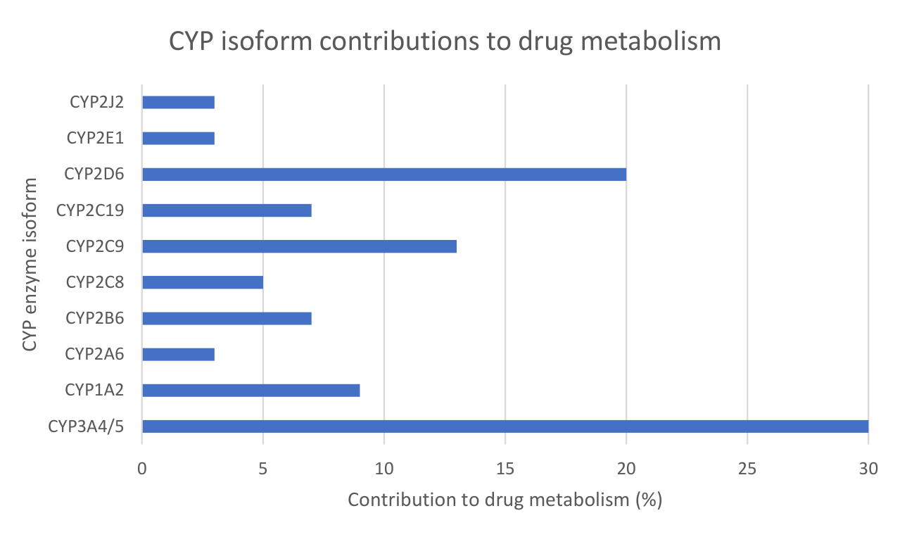
Figure 2.
Founding fathers of pharmacogenomics, Pythagoras and Friedrich Vogel. Line art created with Perplexity AI (Claude Sonnet 4.6, 2026).
Figure 2.
Founding fathers of pharmacogenomics, Pythagoras and Friedrich Vogel. Line art created with Perplexity AI (Claude Sonnet 4.6, 2026).
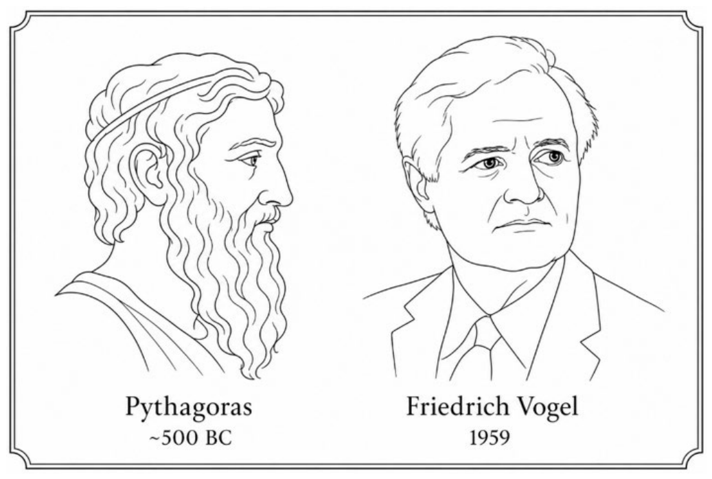
Figure 3.
Bidirectional interactions between drugs and the gut microbiome. The left panel (pharmacomicrobiomics) shows examples of microbial drug metabolites, including the generation of the toxic metabolite bromovinyluracil, pharmacologically active metabolites.
Figure 3.
Bidirectional interactions between drugs and the gut microbiome. The left panel (pharmacomicrobiomics) shows examples of microbial drug metabolites, including the generation of the toxic metabolite bromovinyluracil, pharmacologically active metabolites.
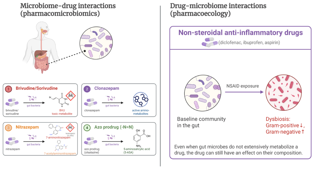
Figure 4.
Gut organoids cultured with bacteria under hypoxic conditions. (a) Shows a low power image of gut organoids, cultured for 2 days at 2% O2. Actin expression in red, Muc1 in blue. (b) Is an electron micrograph of a similar culture with the bacteria seen in close contact with the organoid cells. Illustrations courtesy of Dr. Freke Schaafsma and Dr. Marcel de Zoete, University Medical Center Utrecht.
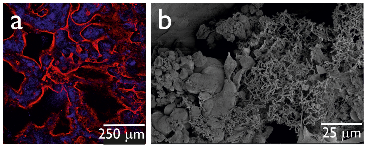
Figure 5.
Proposed indirect model for evaluating oral drug effects on intestinal epithelium. Adapted from Dickinson et al. (87) [2012]. The expanded illustration of the TIM-1 model can be found in figure. The illustration of the TIM-1 system has been taken from Dickinson et al. [94].
Figure 5.
Proposed indirect model for evaluating oral drug effects on intestinal epithelium. Adapted from Dickinson et al. (87) [2012]. The expanded illustration of the TIM-1 model can be found in figure. The illustration of the TIM-1 system has been taken from Dickinson et al. [94].
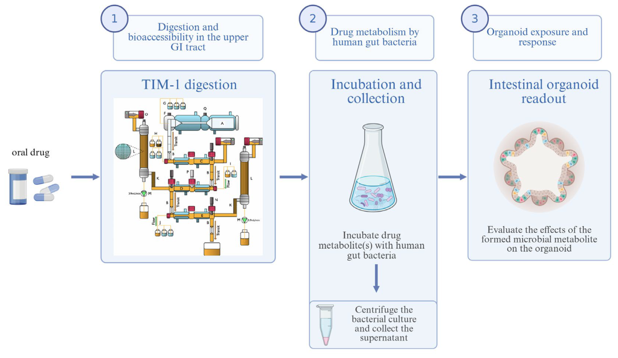
Figure 6.
Proposed experimental framework for oral drug testing models, adapted from Gewirtz et al. [2026]. The framework progresses from simple screening models to increasingly complex ones, as the research purpose turns from basic permeability tests.
Figure 6.
Proposed experimental framework for oral drug testing models, adapted from Gewirtz et al. [2026]. The framework progresses from simple screening models to increasingly complex ones, as the research purpose turns from basic permeability tests.
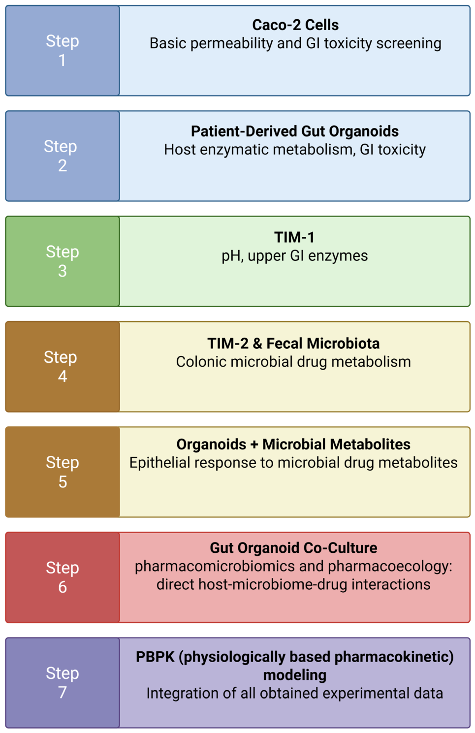
Figure 7.
Growth in the number of PubMed publications related to personalized medicine by publication year. Data retrieved from PubMed in May 2026.
Figure 7.
Growth in the number of PubMed publications related to personalized medicine by publication year. Data retrieved from PubMed in May 2026.
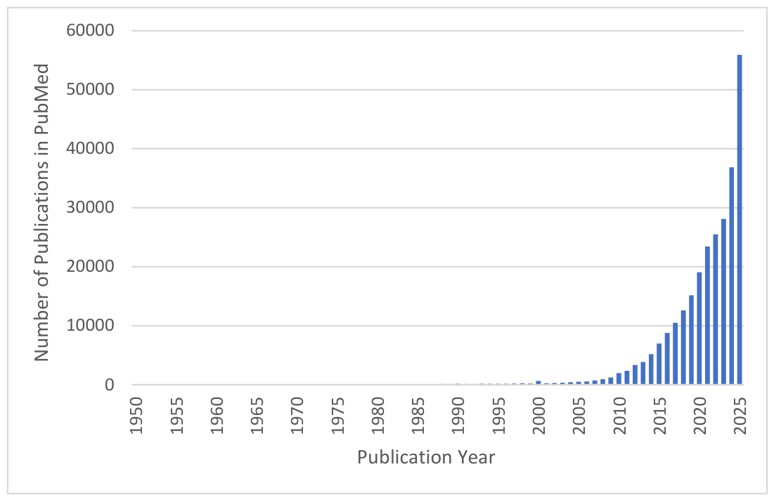
Figure 8.
Development of pharmacomicrobiomics research over time. Shown are the annual numbers of PubMed publications retrieved using the keywords “drug + microbiome + interaction” (blue columns, left Y-axis) and “pharmacomicrobiomics” (orange line right Y-axis).
Figure 8.
Development of pharmacomicrobiomics research over time. Shown are the annual numbers of PubMed publications retrieved using the keywords “drug + microbiome + interaction” (blue columns, left Y-axis) and “pharmacomicrobiomics” (orange line right Y-axis).
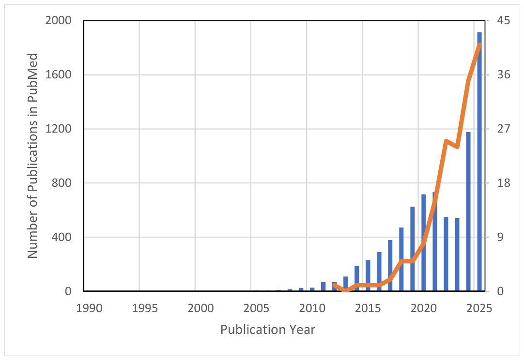
Disclaimer/Publisher’s Note: The statements, opinions and data contained in all publications are solely those of the individual author(s) and contributor(s) and not of MDPI and/or the editor(s). MDPI and/or the editor(s) disclaim responsibility for any injury to people or property resulting from any ideas, methods, instructions or products referred to in the content. |
© 2026 by the authors. Licensee MDPI, Basel, Switzerland. This article is an open access article distributed under the terms and conditions of the Creative Commons Attribution (CC BY) license.
Copyright: This open access article is published under a Creative Commons CC BY 4.0 license, which permit the free download, distribution, and reuse, provided that the author and preprint are cited in any reuse.