

Submitted:
09 October 2026
Posted:
10 October 2026
You are already at the latest version
Abstract
Plants are prone to injury in nature, yet how woody stems repair damaged protective and vascular tissues remains poorly understood. We established an in vitro bridge-culture system in hybrid poplar to track regeneration after longitudinal stem cutting. Lignin deposition began in diverse cells beneath the wound by 2 days after cutting (DAC), marking an early wound response. Repair then followed distinct tissue-specific trajectories. In the cortex and secondary phloem, cell division generated callus-like tissue that covered the wound by 2–4 DAC. New phellogen subsequently formed beneath the callus and produced phelloderm and ligno-suberized phellem, forming a wound periderm resembling native periderm by 12 DAC. In vascular cambium and young phloem, cells expanded and proliferated to reconnect the wound by 4 DAC, while restoration of cambium- and phloem-associated gene expression by 8–12 DAC indicated vascular recovery. Auxin responses also differed between tissues. PtaDR5::GUS activity was transiently induced during periderm regeneration but persisted in vascular regions until vascular development resumed. Exogenous auxin promoted periderm regeneration but had little effect on vascular cell divisions, whereas inhibiting polar auxin transport impaired vascular wound closure. Thus, periderm and vascular tissues undergo coordinated yet distinct repair processes regulated by tissue-specific auxin responses and transport.
Keywords:
wound repair
; in vitro system
; periderm regeneration
; vascular regeneration
; cellular dynamics
; auxin
; hybrid poplar
1. Introduction
Plants are frequently injured by abiotic and biotic stresses from the environment as well as human activities such as grafting and harvesting. When the bark of a stem is cut or wounded mechanically, both outer protective tissues and inner vascular tissues could be disrupted. Damage to the barrier tissues increases water loss and the risk of pathogen entry, whereas injury to internal tissues can impair long-distance transport and secondary growth. Repair must therefore restore a protective boundary and reconnect damaged tissues. Such processes are not only important for plant survival and post-injury growth, but also for vegetative propagation, crop storage and biomass production of bark-derived materials [1].
Two lateral meristems, phellogen and vascular cambium, contribute to the radial growth of stems in woody plants. The phellogen, also called cork cambium, produces phellem (cork) outward and phelloderm inward to form periderm. Mature phellem cells deposit suberin and lignin to execute their protective functions [2]. The vascular cambium lies between the secondary phloem and xylem and produces both vascular tissues during radial growth. Studies in Arabidopsis and trees have identified key regulators of periderm development as well as vascular development [1,3]. Several transcription factors such as AINTEGUMENTA (ANT), WUSCHEL-RELATED HOMEOBOX 4 (WOX4) and BREVIPEDICELLUS/KNOTTED1-LIKE HOMEOBOX 1 (BP/KNAT1) are associated with both phellogen and vascular cambium activity [3]. The receptor-like kinase PHLOEM INTERCALATED WITH XYLEM (PXY) is the vascular cambium specific regulator [4]. A number of MYB family genes including MYB67, MYB68 and MYB93 as well as NAC family gene NAC038 are involved in phellem maturation and suberin deposition [1,5,6,7,8]. In addition, LONG-CHAIN ACYL-COA SYNTHETASE 2 (LACS2), CYTOCHROME P450 86B1 (CYP86B1), and GLYCEROL-3-PHOSPHATE ACYLTRANSFERASE 5 (GPAT5) participate in suberin formation [9]. Therefore, re-establishing these two meristems alongside resumption of gene expression of these cell type maker genes provides a basis for rebuilding an outer barrier and repairing internal vascular tissues after stem injury.
The morphological dynamics of wound repair in stems and roots depend on the regeneration system used. In Arabidopsis, when the inflorescence stem is incised transversely, parenchyma cells of the pith and protoxylem proliferate and acquire vascular cambium identity around the incision during tissue reunion and vascular reconnection [10,11]. How the barrier tissue is restored in the incised stem, however, remains poorly understood. In a second system, a longitudinal cut in the mature region of Arabidopsis roots growing on agar plates rapidly activates periderm regeneration from vascular cells. Reporters for periderm marker genes have shown that phellem identity is induced at the wound site by 1 day after injury (1 dai), followed by periclinal cell divisions and expression of phellogen genes at 2–3 dai. A functional barrier is recovered once ligno-suberized phellem-like cells appear by 4 dai [12]. This system shows no pronounced vascular repair [12]. In poplar, removing the outer bark, which consists mainly of periderm and part of the phloem, induces wound periderm formation from the phloem, and the dynamics of wound-induced suberization have been well characterized [13]. When the entire bark is removed by girdling, by contrast, the remaining immature xylem cells regenerate phloem and vascular cambium through trans-differentiation and dedifferentiation, thereby resuming the secondary vascular system. Xylem ray cells in this system expand and divide to form a callus that covers the girdled trunk, and periderm tissues later arise from the outermost callus [14,15]. Secondary vascular tissue regeneration after bark girdling has been studied in both in planta [14] and in vitro [15,16] systems, but the dynamics and regulation of periderm regeneration have not been resolved in detail. Mechanical wounds in nature often damage both barrier tissues and inner vascular tissues. A regeneration system that mimics the repair of all disrupted tissues is therefore needed to understand the full wound healing process in plants.
Hormonal regulation of tissue regeneration has been investigated widely in various plant systems. Among many phytohormones, auxin plays important roles during vascular and periderm development, and is crucial for maintaining the meristematic activities of both vascular and cork cambia [3,17]. Auxin response and polar auxin transport are also involved in vascular regeneration and tissue reunion in both herbivorous and woody plants. After the Arabidopsis inflorescence stem is incised, auxin accumulates mainly in the upper region of incised site 1–5 days after cutting and diminishes when the reunion is completed around 7 days, as revealed by the auxin-response promoter reporter DR5::GUS [10]. During Arabidopsis graft formation, DR5 is peaked when vascular reconnection occurs in the graft junction at 5 days and diminishes by 10 days [18]. A number of auxin-responsive downstream genes are required for tissue reunion or grafting formation. For example, around an Arabidopsis stem incision, auxin induces the expression of NAC transcription factor ANAC071, RELATED TO AP2.6L (RAP2.6L), AUXIN RESPONSE FACTOR 6 (ARF6) and ARF8[10,19]. Mutations of ANACs and ARFs delay tissue reunion and graft formation [10,19,20]. Using an inducible cell ablation system, we recently demonstrate that polar auxin transport is required for the regeneration and re-positioning of stem cells in the vascular cambium after ablation [21]. In poplar trees, auxin is essential for re-establishment of auxin maximum and vascular cambium regeneration after bark girdling in the in vitro stem culture system [15]. Thus, it would be very interesting to investigate how auxin response and transport contribute to periderm regeneration during wound repair in the stems of trees.
Here, we established a bridge-culture system to track the wound repair after a longitudinal cut in poplar stems. These cuts typically traversed the whole bark and reached the vascular cambium, exposing the cortex, phloem and vascular cambium at the wound surface. This system therefore allows periderm and vascular regeneration to be monitored simultaneously under cultured conditions. Using time-course histological and transcriptional analyses, we uncovered distinct spatiotemporal dynamics of periderm and vascular regeneration during this wound healing process. Combining poplar DR5::GUS transgenic trees with exogenous chemical treatments, we further examined the roles of auxin and polar auxin transport in this process. Together, our results established a new in vitro platform and provided a tissue-resolved framework for studying how protective and conductive tissues are rebuilt after wounding in trees.
2. Results
2.1. Establishment of an In Vitro Bridge-Culture System for Monitoring Wound Repair in Poplar Stems
To recapitulate the natural wound repair process that occurs after mechanical injury to tree stems, we developed an in vitro culture system using the stem segments harvested from greenhouse-grown hybrid poplar (Figure 1A). Stem segments were collected from 15th–35th internodes of 2–3-month-old Populus alba × P. glandulosa (clone 84K, hereafter referred to as 84K), and surface sterilized before being placed on culture medium. Each internode was then longitudinally incised on one side, and positioned across a split plate medium setup that we designated as the bridge-culture system (Figure 1A). In this system, a central strip of solid medium was removed to create a gap, so that each stem segment was laid across the gap. Both ends of the internode remained in contact with the medium, whereas the wounded region was suspended above the empty space. The wound surface faced upward and remained exposed to the air without contacting the medium. This setup maintained a stable wound orientation and allowed continuous observation and sampling of the exposed and regenerated tissues (Figure 1A).
The intact poplar stem contains native periderm, cortex and secondary vascular tissues (phloem, vascular cambium and xylem) (Figure 1B, mock). The longitudinal cuts typically reached the depth of vascular cambium or secondary xylem of the stem, thereby exposing native periderm, cortex, phloem, and vascular cambium along the wound surface (Figure 1B, cut). This mechanical injury created a spatially organized damage gradient, enabling simultaneous monitoring of the spatiotemporal repair dynamics of all tissue types disrupted by wounding. At the whole-mount level, the open gap caused by the longitudinal incision remained clearly distinguishable at 2 days after cutting (DAC). By 12 DAC, newly regenerated tissues had covered most of the exposed region (Figure 1C). These observations showed that the bridge-culture system provides a controlled and accessible experimental platform to dissect the cellular and molecular changes during wound repair of poplar stems.
2.2. Cellular and Molecular Dynamics of Periderm Regeneration in Wounded Poplar Stems
To characterize wound repair at the anatomical and molecular levels, we combined histological analysis with time-course RNA sequencing of all regenerated tissues (Figure 2, Figure 3, Figure S1 and Figure S2). We found that outer barrier tissue and inner vascular tissue regeneration occurred in distinct domains around the wound sites. We therefore dissected the regeneration of the periderm and vascular tissues separately (Figure 2).
At 2 DAC, lignin deposition was detected with Basic Fuchsin (BF) staining in cells beneath the exposed wound surface across cortex, phloem, and vascular cambial regions (Figure 2A), indicating that a shared early wound response was activated across all damaged tissues. Consistent with this histochemical observation, genes responsible for lignin biosynthesis and polymerization [22] showed elevated expression at 2 DAC relative to 0 DAC (Figure 3A, Table S1). These included the monolignol biosynthesis genes 4-COUMARATE: COENZYME A LIGASE 1 (4CL1), CAFFEOYL-COA O-METHYLTRANSFERASE 1 (CCoAOMT1), CCoAOMT2 and CINNAMYL ALCOHOL DEHYDROGENASE 1 (CAD1), as well as a putative lignin polymerization-related gene PEROXIDASE 52 (PRX52) [23]. Expression of two lignin-related regulators, MYB63 [24] and WILLY LIM 1 (WLIM1) [25], was induced in a similar manner (Figure 3A, Table S1). The upregulation of 4CL1 and MYB63 was further confirmed by Reverse Transcription Quantitative PCR (RT-qPCR; Figure 3B). This lignified boundary remained distinguishable throughout regeneration and was therefore used to mark the original cut surface in subsequent analyses (Figure 2B,D, dashed lines).
Periderm regeneration was observed mainly in cortex and phloem domains adjacent to the cut surface (Figure 2A,B). At 4 DAC, loosely arranged callus-like cells appeared on the wound-facing side of the lignified boundary in the cortex and phloem regions (Figure 2A,B). This anatomical change was accompanied by increased expression of genes associated with cell expansion, cell division and callus formation (Figure S2A,B, Table S1). In particular, transcription factors regulating callus formation such as WRKY75, LATERAL ORGAN BOUNDARIES DOMAIN 18 (LBD18) and LBD36 [26,27] showed higher expression at 2–4 DAC (Figure S2B, Table S1).
On the inner side of the lignified boundary, periclinal divisions became prominent from 4 DAC (Figure 2A,B). The activation of cell proliferation at this stage was supported by increased expression of cyclins, cyclin-dependent kinases, and other cell-cycle-related genes (Figure S2C, Table S1). These divisions generated cell files oriented parallel to the wound surface, giving rise to regenerating phellogen (rPg) cells. Periclinal cell division continued by 9 DAC, resulting in a progressive increase in the number of rPg cells (Figure 2B). Consistent with this anatomical progression, poplar homologues of ANT, WOX4 and KNAT1, transcription factors known to regulate meristematic activity in the vascular cambium and phellogen of Arabidopsis [3,28], were upregulated at 4 DAC and remained highly expressed by 9 DAC (Figure 3C, Table S1). Our RT-qPCR analysis confirmed the induction of ANT and WOX4 expression (Figure 3D). Together, the periclinal cell divisions beneath the callus and along the wound surface, accompanied by elevated phellogen-related gene expression, indicated the recovery of phellogen activity along the exposed wound surface during periderm regeneration.
Using the suberin-specific dye Fluorol Yellow 088 (FY) and the lignin-specific dye BF, ligno-suberized phellem (Phe) cells were readily visualized in the native periderm (Figure 2A,C). At 8–9 DAC, FY- and BF-positive regenerated phellem (rPhe) cells were detected external to the regenerated phellogen (rPg) in both the cortex and phloem regions, indicating the formation of new phellem cells from rPg cells (Figure 2A–C). The regenerated periderm exhibited an organization closely resembling that of the native periderm (Figure 2C). To further confirm the rPhe identity, we examined the expression of genes associated with phellem differentiation and suberin biosynthesis. MYB67 has been reported to promote phellem maturation [5], whereas MYB68, MYB93, and NAC038 are associated with suberization [1]. LACS2, CYP86B1 and GPAT5 encode enzymes involved in suberin biosynthesis [2,9]. The poplar homologues of these genes exhibited higher transcript levels at 9 DAC than at 2 or 4 DAC (Figure 3E, Table S1). Similar expression dynamics were observed for FATTY ACYL-ACP THIOESTERASE B (FATB), HYDROXYACYL-ACP DEHYDRATASE (HCD), LACCASE 3 (LAC3), GPAT6, GPAT7, and GPAT8 [2,9] (Figure 3E, Table S1). RT-qPCR experiments verified the expression changes of MYB67, CYP86B1, and GPAT8 during regeneration (Figure 3F).
Notably, callus cells at the wound surface also underwent lignification during the later stages of periderm regeneration (Figure 2C,D). Collectively, the histochemical detection of phellem-associated cell-wall polymers and the coordinated induction of genes involved in phellem differentiation and suberin biosynthesis demonstrated the successful re-establishment of the periderm in the cortex and phloem of wounded poplar stems.
2.3. Repair of Vascular Cambium and Secondary Phloem After Wounding
In parallel with periderm re-establishment at the wound surface, vascular regeneration proceeded within the inner phloem and vascular cambium domains (Figure 2A,B,E), revealing distinct tissue-specific dynamics of wound repair. During periderm regeneration, callus formation and subsequent lignification occurred at the wound surface, providing a transient protective barrier before the new periderm was fully established (Figure 2A–D). In contrast, vascular regeneration was initiated by the early proliferation of callus-like cells, which were detectable as early as 2 DAC (Figure 2A, 2DAC). These cells subsequently expanded and proliferated extensively, filling the wound gap and reconnecting the severed vascular tissues by 4 DAC (Figure 2E).
Consistent with this early cell expansion and proliferative response, genes associated with primary cell-wall remodeling were upregulated during the initial stages of vascular regeneration (Figure S2A, Table S1). For example, EXPANSIN A8 (EXPA8), XYLOGLUCAN ENDOTRANSGLUCOSYLASE /HYDROLASE 41 (XTH41), PECTIN METHYLESTERASE 20 (PME20) and PME1 [29] showed increased gene expression at 2 DAC (Figure S2A, Table S1). CELLULASE 1 (CEL1) displayed higher expression at 4 DAC, whereas POLYGALACTURONASE INVOLVED IN EXPANSION 3 (PGX3), XTH2 and CELLULOSE SYNTHASE A6 (CESA6) homologues [30,31,32] remained highly expressed until 9 DAC (Figure S2A, Table S1). These transcript profiles indicated that cell-wall modification was activated early during vascular regeneration and continued as the newly formed tissues expanded, reorganized, and re-established continuity across the wound.
By 9 DAC, newly formed cells within the vascular region became more regularly organized on both sides of the lignified boundary, with both periclinal and anticlinal divisions evident (Figure 2E). In addition to the shared regulators of phellogen and vascular cambium identity, including ANT, KNAT1 and WOX4 (Figure 3C,D), PXY, a receptor-like kinase associated with vascular cambium activity [4], was strongly induced at 4 DAC and reached its expression peak at 9 DAC (Figure 3C, Table S1). Several genes associated with phloem development also showed higher expression at 9 DAC (Figure S2D, Table S1). These included ALTERED PHLOEM DEVELOPMENT (APL), PHLOEM EARLY DOF 2 (PEAR2), CALLOSE SYNTHASE 7 (CALS7), and SIEVE-ELEMENT-OCCLUSION-RELATED 1 (SEOR1) [33,34]. Taken together, these anatomical changes and transcriptional profiling suggested the repair of vascular cambium and phloem in the wounded regions.
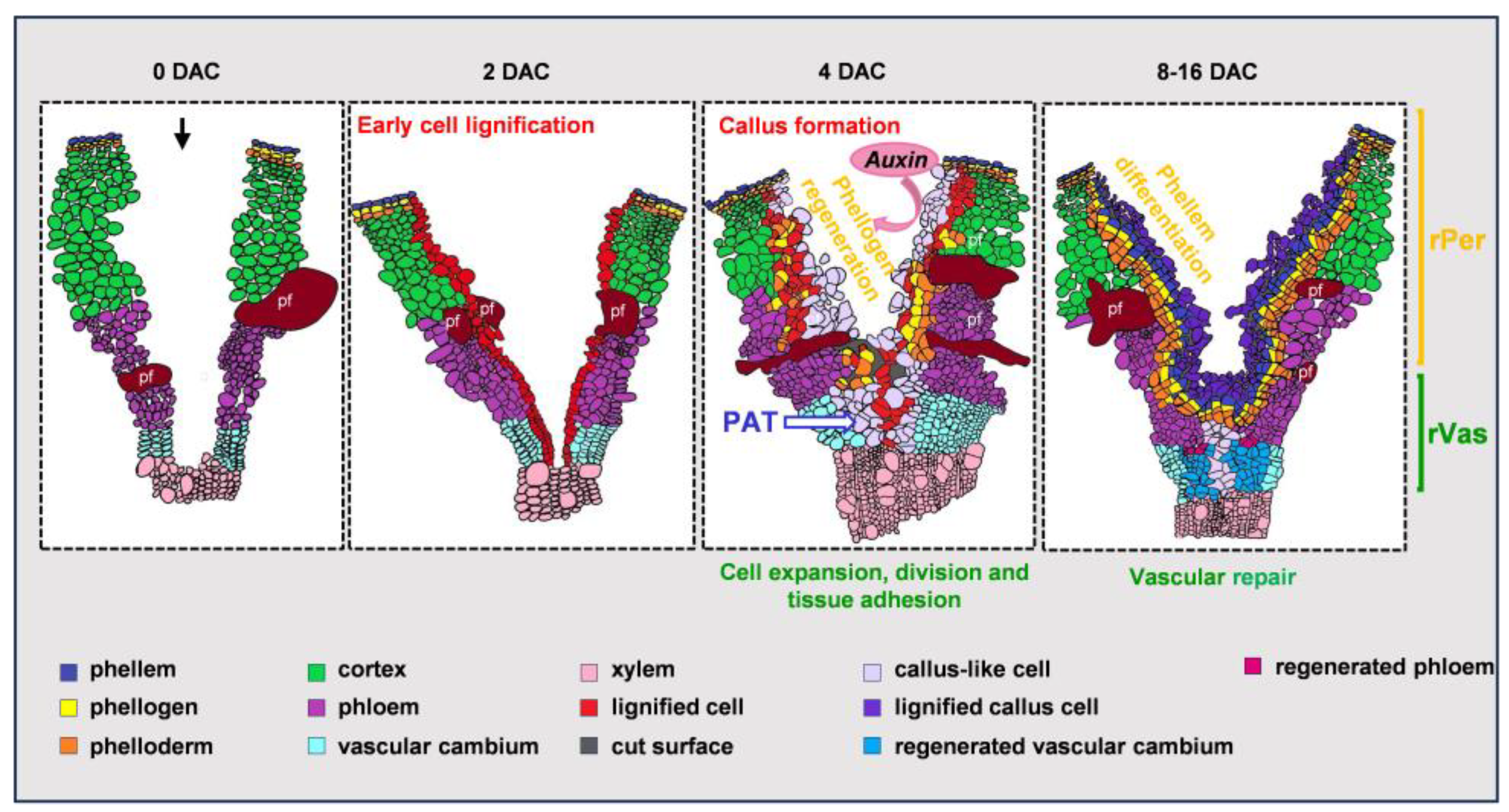
Thus, by monitoring the regeneration of both periderm and vascular tissues in the same wounded poplar stem, our system enabled direct comparison of tissue-specific wound responses simultaneously. We found that these two domains revealed distinct spatial and temporal repair dynamics: periderm tissues prioritized barrier formation and lignification, whereas internal vascular tissues initiated early cell growth and proliferation to restore tissue adhesion and connectivity. Collectively, these findings establish a framework for studying coordinated wound responses across stem tissues, while also providing a useful platform for investigating the molecular mechanisms that regulate these processes.
2.4. Tissue Specific Auxin Responses During Periderm and Vascular Regeneration
Auxin regulates periderm formation [3], vascular development [17] and vascular regeneration in several plant systems [10,15,21]. We therefore examined genes involved in auxin metabolism, transport, signaling and responses in our wound repair system in poplar stems (Figure 4A, Table S1).
Genes implicated in auxin homeostasis and metabolism exhibited distinct temporal profiles during regeneration. IAA-ALANINE RESISTANT 1 (IAR1), REVERSAL OF SAV3 PHENOTYPE 1 (VAS1), ACYL-ACTIVATING ENZYME 18 (AAE18), IAA CARBOXYLMETHYLTRANSFERASE 1 (IAMT1), and METHYL ESTERASE 17 (MES17) [35] showed higher expression after cutting, with several genes reaching their highest transcript abundance at 4 DAC (Figure 4A, Table S1). Auxin transport-related genes also changed with time. Homologues of auxin efflux carrier PIN-FORMED 1b (PIN1b) showed higher relative expression at 2 DAC, whereas auxin influx carrier gene AUXIN RESISTANT 1 (AUX1) and several other PINs [36] reached their expression maximum at 9 DAC (Figure 4A, Table S1). Auxin signaling associated genes, including AUXIN RESISTANT 6 (AXR6), AUXIN SIGNALING F-BOX 2 (AFB2), RELATED TO UBIQUITIN 1 (RUB1), and RUB1-CONJUGATING ENZYME 1 (RCE1) [37,38] increased rapidly following cutting (Figure 4A, Table S1). SMALL AUXIN UP RNA (SAUR) genes were induced as early as 2 DAC, whereas most AUXIN RESPONSE FACTOR (ARF) genes [39] showed greater expression at later stages and peaked at 9 DAC (Figure 4A, Table S1). Overall, these gene expression patterns indicated that auxin homeostasis, transport and signaling were dynamically regulated throughout wound repair.
Next, to monitor the spatiotemporal dynamics of auxin responses during tissue regeneration after cutting, we employed the auxin-responsive promoter reporter PtaDR5::GUS in hybrid clone INRA 7171–B4 (P. tremula × P. alba) [15,40]. In intact poplar stems, DR5 activity was primarily detected in the vascular cambium [15] and was absent from the cortex and phloem (Figure 4B, 0 DAC). We therefore first examined PtaDR5::GUS activity in the cortex and phloem of poplar, where the periderm regenerated. GUS staining became detectable near the wound site at 2 DAC and became strongest at 4 DAC in regions undergoing prominent periclinal division during phellogen regeneration (Figure 4B, Figure 2, 0 DAC). By 9 DAC, only weak signals remained in the cell files along the cut surface. In contrast, DR5 activity in the vascular region maintained high at 9 DAC (Figure S4A), coinciding with the recovery of vascular cambium and phloem gene expression (Figure 3C and Figure S2D).
These observations revealed tissue-specific dynamics in auxin responses during regeneration. During periderm regeneration, PtaDR5::GUS activity was transiently induced at 2–4 DAC in the cortex and phloem, reached its maximum during phellogen regeneration, and subsequently declined as the periderm regeneration completed. In the damaged vascular region, however, DR5 activity persisted until the resumption of vascular development. Thus, auxin responses were activated earlier and transiently during periderm regeneration but were sustained during vascular regeneration.
2.5. Distinct Roles of Auxin and Polar Auxin Transport in Periderm and Vascular Regeneration
To further investigate the roles of auxin and/or polar auxin transport (PAT) in periderm and vascular regeneration after wounding, we treated cut 84K poplar stem segments with the exogenous auxin 1-naphthaleneacetic acid (NAA) or the PAT inhibitor N-(1-naphthyl) phthalamic acid (NPA). Stem segments were cut and cultured on medium supplemented with DMSO control, 5 μM NAA, or 100 μM NPA, and the effects on periderm regeneration (Figure 4C–E and S3) and vascular regeneration (Figure S4B–D) were subsequently assessed.
To quantify regenerative progression, we measured the percentage of cells along the cut surface that participated in regeneration relative to the total number of cells analyzed (Figure 4D and Figure S4C). We also quantified division frequency, defined as the number of division events per dividing cell (Figure 4E and Figure S4D). We termed these parameters the percentage of regeneration and frequency of cell division, respectively.
First, to determine how NAA and NPA affect barrier re-establishment, we quantified the regenerative cell divisions that separately contributing to callus formation and periderm regeneration (Figure 4C,D and S3). Our data revealed that NAA treatment increased the proportion of dividing cells at 2 and 4 DAC significantly, but the increase was not statistically significant at 9 DAC (Figure 4C,D). It also promoted cell proliferation in each cell file during periderm regeneration over time (Figure 4C,E). However, NAA had no obvious effect on callus formation (Figure 4C–E). NPA treatment did not significantly change either the percentage of dividing cells or the frequency of periclinal cell divisions in periderm regeneration relative to DMSO control. It only inhibited callus proliferation at 9 DAC (Figure 4C–E). Altogether, these results demonstrated that exogenous auxin enhanced periclinal cell division and phellogen regeneration in the cortex and phloem regions, while polar auxin transport was important for callus formation during barrier repair.
Previous study has shown that vascular cambium regeneration after girdling in cultured poplar stems requires exogenous auxin when endogenous phytohormone sources are removed [15]. We then examined vascular regeneration in our in vitro system, in which wound repair proceeded without exogenous hormones (Figure 1 and Figure 2). Quantification of regenerative cell divisions in the vascular region (Figure S4) showed that exogenous NAA did not significantly affect either the percentage or frequency of dividing cells at all time points when compared to DMSO control (Figure S4C,D), suggesting that endogenous auxin was sufficient to support vascular regeneration in this system. However, inhibition of PAT with NPA disrupted vascular wound repair. At 4 DAC, the wound gap remained open in 7 out of 9 NPA-treated segments, whereas no open gaps were observed in DMSO- or NAA-treated stems (Figure S4B). In line with this observation, cell proliferation in the damaged vascular domain was reduced at 4 DAC upon NPA treatment (Figure S4C). Thus, although additional auxin was not required, PAT was essential for the timely re-establishment of tissue adhesion and wound closure in the vascular region.
Collectively, our in vitro regeneration system enabled us to resolve the cellular dynamics and hormonal regulation of wound repair following cutting of the woody poplar stem. Our findings highlight that barrier and vascular tissues undergo distinct regenerative trajectories, consistent with their different structural and physiological roles. Barrier repair was associated with the coordinated production of callus-like cells and periderm tissues, whereas vascular repair involved the expansion of cells surrounding the wound and the timely restoration of tissue continuity. These processes were also differentially regulated by auxin: exogenous NAA promoted periderm regeneration but had little effect on vascular regenerative divisions, while PAT mainly affected callus formation in the periderm region and was required for efficient wound closure in vascular region.
3. Discussion
Wound repair is crucial for plant survival under both natural and cultivated conditions. In trees, the ability to regenerate rapidly in response to stress also affects the quality of wood and bark biomass. Several regeneration systems have been used to investigate the dynamics and regulatory mechanisms of vascular regeneration and tissue reunion [1,4,10,11,15,18,19], as well as wound periderm formation in stems and roots [1,12,13]. However, no system has comprehensively examined the coordination of regeneration across different tissues within the same platform. Our newly established in vitro bridge-culture system enables us to monitor the dynamics and investigate the regulatory mechanisms of periderm and vascular regeneration simultaneously.
3.1. Longitudinal Cutting Induces Ectopic Lignification and Wound Periderm Restoration
The periderm is a secondary protective tissue that replaces the epidermis during normal development, forming the native periderm, and replaces damaged barrier tissues following injury, forming the wound periderm [1]. The phellem provides an effective barrier against water loss and pathogen invasion, largely because its cell walls contain suberin and lignin [1]. Accordingly, wound-induced suberization represents an important step in restoring a functional protective barrier over injured regions [12,13]. In addition, lignin accumulation at the wound site may further contribute to the establishment and reinforcement of this barrier [1,13,22].
When mature Arabidopsis roots are longitudinally cut to the depth of phloem, cells beneath the wound acquire phellem identity within 1 day. This is followed by cell proliferation and the expression of phellogen marker genes, while suberin deposition in the phellem cells marks the completion of periderm regeneration 4 days after injury [12]. In this system, the periderm directly reconstitutes from adjacent vascular cells in a short time frame.
In poplar stems, removal of a piece of outer bark, including the native periderm and part of the phloem, results in periderm regeneration through two successive stages: the formation of a temporary boundary zone followed by the development of a wound periderm [13]. This is similar to wound healing in potato tubers [41,42]. During wound-induced suberization in poplar, chemical and molecular profiling reveal sequential accumulation of lignin and suberin monomers in the boundary zone and developing phellem before suberin lamellae become histologically detectable [13]. However, the polyphenolic domain of the wound phellem is chemically distinct from the lignin deposited in the boundary zone [13]. Thus, both the boundary zone and the wound periderm originate from phloem cells in this system and may contribute to barrier formation.
In our system, early cell lignification beneath the wound, followed by the formation and lignification of callus-like tissue, appear to reinforce the wound boundary before a functional wound phellem is replenished (Figure 2, Figure 5 and Figure S2B). The emergence of callus in our system might be associated with the high moisture in the Petri dishes and the greater depth of cut. Successive wound phellogen initiates from cortex and phloem cells located on the inner side of the lignified boundary, leading to the reconstitution of wound periderm. These observations suggest that the dynamics and cellular changes of periderm regeneration depend on both wound depth and environmental conditions. Our RNA–seq results also showed increased expression of genes enriched in the native periderm during wound-periderm formation, consistent with previous findings in poplar [13]. However, the functions of these candidate genes have not yet been demonstrated in periderm regeneration. Generating and analyzing stable transgenic poplar lines carrying periderm marker genes could help validate these findings at the genetic and molecular levels, as has been done in Arabidopsis [12].
3.2. Vascular Repair After Cutting Involves Tissue Reunion and Reestablishment
Vascular regeneration has been studied in different systems of poplar and Arabidopsis [10,11,14,15,16,18,21]. When nearly all vascular cambial cells around the circumference are eliminated by girdling or ablation, differentiating xylem cells serve as the source for regeneration [14,15,16,21]. By contrast, when vascular continuity is disrupted transversely by incision or grafting, parenchyma cells in the pith and protoxylem contribute to vascular reconnection [10,11,18]. In our system, the phloem and vascular cambium are locally disconnected in the longitudinal direction. Our results showed that phloem and vascular cambium cells on both sides of the cut expand and proliferate, ultimately bridging the gap with newly formed vascular cells and closing the wound in this region (Figure 2A,E and 5).
Unlike periderm regeneration in the outer region of the gap, cell proliferation in the vascular region occurs in both periclinal and anticlinal directions, resulting in a disorganized cellular arrangement. Consequently, cell identities in this region are difficult to distinguish based solely on anatomical observations. Gene expression analysis revealed the upregulation of marker genes associated with the vascular cambium and phloem, suggesting the restoration of vascular tissues (Figure 3 and Figure S2). Further characterization using genetic marker lines for distinct vascular cell types will improve our understanding of the mechanisms underlying vascular repair.
Overall, through detailed anatomical analyses and gene expression profiling, we demonstrated that the periderm and vascular tissues regenerate through distinct, tissue-specific processes. However, the coordinated regeneration of these tissues is essential for the effective repair and restoration of the wound. Our study establishes a framework for understanding wound repair in secondary tissues, including the two lateral meristems, the phellogen and vascular cambium. The sequential and coordinated activation of these meristems enables the re-establishment of both protective outer tissues and internal vascular continuity. These findings provide a foundation for further investigating the cellular signals and molecular regulation that coordinate regeneration across tissue boundaries during secondary growth.
3.3. Differential Roles of Auxin and Polar Auxin Transport (PAT) in Periderm and Vascular Regeneration
Auxin and PAT have been implicated in wound-induced regeneration across several systems; however, little is known about their role in periderm regeneration. Following bark girdling in poplar, the phloem and vascular cambium regenerate without exogenous auxin in field grown trees, whereas regeneration fails in cultured girdled stem segments grown without auxin supplementation [14,15,16]. In poplar, auxin response is rapidly induced at the wound site and later becomes restricted to the newly formed vascular cambium [15]. Similarly, in incised Arabidopsis stems, auxin response peaks above the incision and declines as the tissues reconnect [10,11]. These observations suggest that auxin signaling is dynamically regulated during wound repair and the requirement for exogenous auxin depends on the experimental system and its physiological context.
In intact poplar stem, auxin response is strongest in vascular cambium and relatively weak in periderm, cortex and phloem (Figure 4A,B), indicating stronger basal auxin signaling in the vascular region. Because PtaDR5::GUS reporter reflects signaling output rather than auxin concentration directly, this pattern alone does not show the relative auxin levels in these tissues. Nevertheless, the greater sensitivity of periderm regeneration to exogenous auxin supplementation than that of vascular regeneration (Figure 4 and Figure S4) may reflect differences in their pre-existing auxin signaling or auxin requirements. Intriguingly, PAT is required for timely tissue reunion during vascular repair (Figure S4). PAT may also contribute by establishing auxin distributions during repair, although its role in our system remains to be tested. Genetic perturbation of auxin biosynthesis, signaling or transport, together with cell-type-specific markers, could help determine how auxin and PAT coordinate the regeneration of periderm and vascular tissues in woody plants.
Other hormonal and molecular signals may also contribute to wound repair, either independently of auxin or through interactions with it. Auxin and cytokinin have been reported to act differently during secondary vascular tissue regeneration in poplar [15]. Jasmonate contributes to the spatial regulation of wound responses in incised Arabidopsis stems [10], while abscisic acid promotes wound-induced suberization in potato [47]. Moreover, gas diffusion as a new regulatory mechanism for periderm repair [12] may also work in our system. Exploring these signals hence should clarify whether they regulate periderm regeneration, vascular repair or both. More broadly, the distinct responses of periderm and vascular tissues to auxin and NPA support the view that wound regeneration in woody stems is governed by tissue-specific regulatory mechanisms rather than by a single, uniform wound response.
4. Materials and Methods
4.1. Plant Materials and Growth Conditions
Two hybrid poplar genotypes were used in this study. 84K poplar (Populus alba × P. glandulosa) was used for system establishment, histology, RNA sequencing, RT-qPCR, and pharmacological treatments. INRA 7171–B4 poplar (P. tremula × P. alba) carrying DR5::GUS (PtaDR5::GUS) was used to monitor auxin-responses.
Plants were propagated in vitro on woody plant medium at 24 °C under a 16 h light/8 h dark photoperiod. Approximately one-month-old plantlets were transferred to a 1:3 (v/v) mixture of potting soil and vermiculite. Plants were grown in a greenhouse at 24 °C under the same photoperiod. Stem segments were used from 2–3-month-old soil-grown plants.
4.2. Stem Wounding and Bridge Culture
Stem segments spanning internodes 15th–35th were harvested and rinsed under running tap water. Surface trichomes and axillary buds were gently removed. The segments were transferred into 50 mL tubes with diluted laboratory detergent and agitated for 10 min. After three times with tap water for (10 min each) per rinse, all subsequent operations were performed in a laminar-flow hood.
The stems were immersed in 70% (v/v) ethanol for 1 min and then in 10% (v/v) commercial bleach for 10 min. They were rinsed five times with sterile water( 2 min per rinse) and blotted dry on sterile filter paper. A sterile scalpel was used to make a single longitudinal cut through the native periderm, cortex, phloem, and vascular cambium to the xylem. The wounded surface was oriented upward during culture.
The bridge-culture system was modified from a previously reported in vitro stem-segment culture system for studying secondary vascular tissue formation in poplar [15]. Medium was poured into sterile Petri dishes and allowed to solidify. A strip approximately 1 cm wide was then removed from the center of each dish, creating two supporting blocks separated by an air gap. The open dishes were dried under sterile airflow for 10 min. Each segment was positioned across the gap with both ends resting on the medium and the wounded region suspended above the gap. Cultures were maintained at 24 °C under a 16 h light/8 h dark photoperiod.
The basal regeneration medium contained 2.2 g L⁻¹ Murashige and Skoog salts, 0.5 g L⁻¹ MES, 30 g L⁻¹ sucrose, and 10 g L⁻¹ agar. The pH was adjusted to 5.85 before autoclaving.
4.3. Histology and Histochemical Staining
Wound regions were cut into approximately 5 mm segments. Tissue samples were fixed in formalin-acetic acid-alcohol containing 70% ethanol, glacial acetic acid, and commercial formaldehyde solution at 90:5:5 (v/v/v). Vacuum infiltration was performed for 2 h, and samples were incubated in fixative overnight at 4 °C prior to three rinses with phosphate-buffered saline (PBS).
For hand sectioning, transverse sections were cut with a razor blade and collected in PBS. For resin sections, fixed samples were dehydrated in a graded ethanol series (30%, 50%, 70%, 85%, 95%, and 100%), embedded in Technovit 7100 resin following the manufacturer’s instructions and sectioned transversely at 5 μm.
Toluidine Blue O and SCRI Renaissance 2200 (SR) were applied to visualize cell walls. Basic Fuchsin (BF) was used to visualize lignified cell walls, whereas Fluorol Yellow 088 (FY) was used to detect suberized cell walls. Images were acquired with a Leica DM2500 microscope or a Zeiss Axio Imager Z1 microscope.
4.4. RNA Isolation, Library Preparation, and Sequencing
RNA–seq samples were harvested at 0, 2, 4, and 9 days after cutting (DAC). Wound-region samples at 0 DAC were collected immediately after wounding. In parallel, 0 DAC whole-bark samples (0 DAC–W) were collected as a reference. For wound-region sampling, the native periderm was stripped off, while newly formed tissues and their adjacent underlying cells were retained. The approximate sampling thickness was approximately 0.25–0.35 mm at 2 DAC, 0.30–0.40 mm at 4 DAC, and 0.35–0.45 mm at 9 DAC (Figure S1). Three independent biological replicates were included for each time point. All samples were snap-frozen in liquid nitrogen and stored at −80 °C prior to RNA extraction.
Total RNA was extracted with the Polysaccharide Polyphenol Plant Total RNA Extraction Kit (Tiangen, Beijing, China; DP441). RNA concentration, purity, and integrity were assessed before library preparation. Libraries were prepared and sequenced on an Illumina NextSeq 500 platform by Beijing Biomarker Technologies Co., Ltd. (Beijing, China).
Raw read quality was checked with FastQC. Adapter sequences and low-quality bases were trimmed with Trim Galore. Clean reads were aligned to the Populus trichocarpa v4.0 reference genome using HISAT2. Gene expression was quantified as fragments per kilobase of transcript per million mapped reads (FPKM), which were used for expression visualization. The FPKM values used to generate the heatmaps are provided in Table S1. Heatmaps were generated with TBtools [48]. Values were log2-transformed after addition of a small pseudocount and normalized per gene.
4.5. Reverse Transcription and Quantitative PCR
Total RNA for RT-qPCR was extracted with the Polysaccharides and Polyphenols-rich StarSpin Plant RNA Kit (GenStar, Beijing, China; P134). One microgram of total RNA was reverse transcribed with the Evo M–MLV RT Mix Kit (Accurate Biology, Changsha, China; AG11728). Diluted cDNA was amplified using SYBR Green Premix Pro Taq HS qPCR Kit (Accurate Biology; AG11701) on a CFX96 Real-Time PCR Detection System (Bio-Rad, Hercules, CA, USA). ACTIN served as reference gene. Relative transcript abundance was calculated via the 2−ΔΔCt method. Three technical reactions were performed for each biological sample. Data were analyzed by one-way ANOVA followed by Tukey’s multiple-comparisons test. Different lowercase letters indicate significant differences at P < 0.05. Primer sequences are listed in Table S2.
4.6. Histochemical Detection of DR5::GUS Activity
Following cutting treatment, stem segments of PtaDR5::GUS were collected at 0, 2, 4, and 9 DAC and sectioned into approximately 5-mm-long pieces. Samples were fixed in ice-cold 90% (v/v) acetone for 1 h. After removal of the fixative, samples were immersed in commercial GUS staining solution (Coolaber, Beijing, China; SL7160), vacuum infiltrated, and incubated at 37 °C. Stained samples were washed twice in 70% ethanol for 30 min, hand sectioned, and examined with a Leica DM2500 microscope.
4.7. Auxin Treatments and Quantification of Cell Division
Following cutting, 84K stem segments were transferred to regeneration medium supplemented with 5 μM NAA, 100 μM NPA, or an equivalent volume of DMSO as the control. Stem segments were collected at 2, 4, and 9 DAC, and transverse sections were stained with SR and BF. In the cortex and phloem, a newly formed wall parallel to the wound surface was scored as evidence of recent periclinal division. We recorded the percentage of cell files containing a recent division, the number of callus-like cells, and the number of newly formed cells undergoing periclinal division. In the vascular cambium, newly formed cells were counted. The counted cell populations are illustrated in Figure S3. Cells were quantified in the regions shown in Figure 4C and Figure S4B. Data were analyzed by one-way ANOVA followed by Tukey’s multiple-comparisons test. Differences among treatments at each time point were considered significant at P < 0.05.
5. Conclusions
In this study, we established the bridge-culture system as a stable and accessible platform for analyzing regeneration in woody stems. Using this system, we show that distinct tissues employ different repair strategies (Figure 5), consistent with their biological functions. Poplar reconstructs the wound barrier through successive steps: early lignification provides temporary sealing, callus-like tissue covers the cut surface to protects inner tissues, and it becomes lignified before the wound periderm is fully established. In contrast, vascular repair occurs mainly through tissue reunion and reconnection driven by cell expansion and proliferation. Although functional phloem recovery needs further demonstration, this process shares common features with vascular regeneration in other systems. In our current system, endogenous phytohormone levels are sufficient to support wound repair in the absence of exogenous hormones. We show that applying exogenous auxin at the same concentration promotes regenerative cell division in periderm tissues (Figure 5) but not in vascular tissues, possibly because the vascular cambium maintains higher endogenous auxin levels. On the other hand, the reduced cell proliferation and delayed tissue attachment caused by NPA suggest that PAT is important for vascular repair, as observed in many other regeneration systems (Figure 5). Thus, auxin and PAT contribute to wound repair through tissue-specific mechanisms. Future studies using transgenic or genome-edited poplar lines should further elucidate the molecular genetic roles of auxin and other regulators.
Supplementary Materials
The following supporting information can be downloaded at the website of this paper posted on Preprints.org. Figure S1. Sampling of wound-region tissues for RNA sequencing from longitudinally cut poplar stems. Figure S2. Expression of genes associated with the cell expansion, callus formation, cell cycle and phloem development during wound repair in poplar stems. Figure S3. Identification of cell populations used for quantitative analysis of wound repair. Figure S4. Auxin promotes cell division in the vascular cambium region during wound repair in poplar stems. Table S1. FPKM values of all genes displayed in the heatmaps. Table S2. Primers used in this study.
Author Contributions
J.Z. conceived the project. J.Z. and X.W. designed the experiments. X.W. and J.H. conducted the experiments. X.W., M.Z. and J.H. performed the data analysis. X.W. and J.Z. wrote the manuscript. All authors have read and agreed to the published version of the manuscript.
Funding
This work was supported by the National Natural Science Foundation of China (Grant No. 32171741 to J.Z.) and Fundamental Research Funds for Central Universities (Grant Nos. 63233049, 63243123 and 63263181 to J.Z.).
Data Availability Statement
The raw RNA-sequencing data are available in the NCBI Sequence Read Archive under BioProject accession number PRJNA1532084. The FPKM values used to generate the heatmaps are provided in Table S1. Other data supporting the findings of this study are available from the corresponding author upon reasonable request.
Acknowledgments
During the preparation of this manuscript, the authors used ChatGPT (OpenAI, GPT-6o) for the purposes of improving the wording of selected sentences. The authors have reviewed and edited the output and take full responsibility for the content of this publication.
Conflicts of Interest
The authors declare no conflict of interest.
Abbreviations
The following abbreviations are used in this manuscript:
| DAC | day after cutting |
| DAC–W | day after cutting-whole bark |
| dai | day after injury |
| PAT | polar auxin transport |
| pf | phloem fiber |
| VC | vascular cambium |
| Xy | xylem |
| Phe | phellem |
| rPhe | regenerated phellem |
| rPg | regenerated phellogen |
| rPer | regenerated periderm |
| Cal | callus-like cell |
| rVas | regenerated vascular cambium |
| SR | SCRI Renaissance 2200 |
| BF | Basic Fuchsin |
| TBO | Toluidine Blue O |
| FY | Fluorol Yellow 088 |
| NAA | 1-naphthaleneacetic acid |
| NPA | N-(1-naphthyl) phthalamic acid |
| RT-qPCR | Reverse Transcription Quantitative PCR |
References
- Serra, O.; Mähönen, A.P.; Hetherington, A.J.; Ragni, L. The making of plant armor: The periderm. Annu. Rev. Plant Biol. 2022, 73, 405–432. [Google Scholar] [CrossRef] [PubMed]
- Woolfson, K.N.; Esfandiari, M.; Bernards, M.A. Suberin biosynthesis, assembly, and regulation. Plants 2022, 11, 555. [Google Scholar] [CrossRef] [PubMed]
- Xiao, W.; Molina, D.; Wunderling, A.; Ripper, D.; Vermeer, J.E.M.; Ragni, L. Pluripotent pericycle cells trigger different growth outputs by integrating developmental cues into distinct regulatory modules. Curr. Biol. 2020, 30, 4384–4398.e5. [Google Scholar] [CrossRef] [PubMed]
- Etchells, J.P.; Turner, S.R. The PXY-CLE41 receptor ligand pair defines a multifunctional pathway that controls the rate and orientation of vascular cell division. Development 2010, 137, 767–774. [Google Scholar] [CrossRef] [PubMed]
- Miller, C.N.; Jarrell-Hurtado, S.; Haag, M.V.; Ye, Y.S.; Simenc, M.; Alvarez-Maldonado, P.; Behnami, S.; Zhang, L.; Swift, J.; Papikian, A.; et al. A single-nuclei transcriptome census of the Arabidopsis maturing root identifies that MYB67 controls phellem cell maturation. Dev. Cell 2025, 60, 1377–1391. [Google Scholar] [CrossRef] [PubMed]
- Molina, D.; Horvath, S.; Zhang, X.; Xiao, W.; Ragab, N.; Ripper, D.; Kilian, J.; Andersen, T.G.; Ragni, L. Members of the MYB68 subclade orchestrate cork differentiation by regulating stem cell proliferation and suberin deposition. Cell Rep. 2025, 44, 116048. [Google Scholar] [CrossRef] [PubMed]
- Legay, S.; Guerriero, G.; André, C.; Guignard, C.; Cocco, E.; Charton, S.; Boutry, M.; Rowland, O.; Hausman, J.-F. MdMyb93 is a regulator of suberin deposition in russeted apple fruit skins. New Phytol. 2016, 212, 977–991. [Google Scholar] [CrossRef] [PubMed]
- Chen, Y.-H.; Straube, J.; Khanal, B.P.; Zeisler-Diehl, V.; Suresh, K.; Schreiber, L.; Debener, T.; Knoche, M. Apple fruit periderms (russeting) induced by wounding or by moisture have the same histologies, chemistries and gene expressions. PLoS ONE 2022, 17, e0274733. [Google Scholar] [CrossRef] [PubMed] [PubMed Central]
- Vishwanath, S.J.; Delude, C.; Domergue, F.; Rowland, O. Suberin: Biosynthesis, regulation, and polymer assembly of a protective extracellular barrier. Plant Cell Rep. 2015, 34, 573–586. [Google Scholar] [CrossRef] [PubMed]
- Asahina, M.; Azuma, K.; Pitaksaringkarn, W.; Yamazaki, T.; Mitsuda, N.; Ohme-Takagi, M.; Yamaguchi, S.; Kamiya, Y.; Okada, K.; Nishimura, T.; et al. Spatially selective hormonal control of RAP2.6L and ANAC071 transcription factors involved in tissue reunion in Arabidopsis. Proc. Natl. Acad. Sci. USA 2011, 108, 16128–16132. [Google Scholar]
- Matsuoka, K.; Sato, R.; Matsukura, Y.; Kawajiri, Y.; Iino, H.; Nozawa, N.; Shibata, K.; Kondo, Y.; Satoh, S.; Asahina, M. Wound-inducible ANAC071 and ANAC096 transcription factors promote cambial cell formation in incised Arabidopsis flowering stems. Commun. Biol. 2021, 4, 369. [Google Scholar] [CrossRef] [PubMed] [PubMed Central]
- Iida, H.; Abreu, I.; López Ortiz, J.; Peralta Ogorek, L.L.; Shukla, V.; Mäkelä, M.; Lyu, M.; Shapiguzov, A.; Licausi, F.; Mähönen, A.P. Plants monitor the integrity of their barrier by sensing gas diffusion. Nature 2025, 644, 130–138. [Google Scholar] [CrossRef] [PubMed]
- Rains, M.K.; Caron, L.; Canam, T.; Bernards, M.A. Chemical and molecular characterization of wound-induced suberization in poplar bark. Plants 2022, 11, 1183. [Google Scholar] [CrossRef] [PubMed]
- Zhang, J.; Gao, G.; Chen, J.-J.; Taylor, G.; Cui, K.-M.; He, X.-Q. Molecular features of secondary vascular tissue regeneration after bark girdling in Populus. New Phytol. 2011, 192, 869–884. [Google Scholar] [CrossRef] [PubMed]
- Chen, J.-J.; Wang, L.-Y.; Immanen, J.; Nieminen, K.; Spicer, R.; Helariutta, Y.; Zhang, J.; He, X.-Q. Differential regulation of auxin and cytokinin during the secondary vascular tissue regeneration in Populus trees. New Phytol. 2019, 224, 188–201. [Google Scholar] [CrossRef] [PubMed]
- Zhang, Y.; Wang, L.; Wu, Y.; Wang, D.; He, X.-Q. Gibberellin promotes cambium reestablishment during secondary vascular tissue regeneration after girdling in an auxin-dependent manner in Populus. J. Integr. Plant Biol. 2024, 66, 86–102. [Google Scholar] [CrossRef] [PubMed]
- Smetana, O.; Mäkilä, R.; Lyu, M.; Amiryousefi, A.; Sánchez Rodríguez, F.; Wu, M.-F.; Solé-Gil, A.; Leal Gavarrón, M.; Siligato, R.; Miyashima, S.; et al. High levels of auxin signalling define the stem-cell organizer of the vascular cambium. Nature 2019, 565, 485–489. [Google Scholar] [CrossRef] [PubMed]
- Melnyk, C.W.; Schuster, C.; Leyser, O.; Meyerowitz, E.M. A developmental framework for graft formation and vascular reconnection in Arabidopsis thaliana. Curr. Biol. 2015, 25, 1306–1318. [Google Scholar] [CrossRef] [PubMed] [PubMed Central]
- Pitaksaringkarn, W.; Matsuoka, K.; Asahina, M.; Miura, K.; Sage-Ono, K.; Ono, M.; Yokoyama, R.; Nishitani, K.; Ishii, T.; Iwai, H.; et al. ARF6 and ARF8 contribute to tissue reunion in incised Arabidopsis inflorescence stems. Plant Biotechnol. 2014, 31, 49–53. [Google Scholar] [CrossRef]
- Matsuoka, K.; Sugawara, E.; Aoki, R.; Takuma, K.; Terao-Morita, M.; Satoh, S.; Asahina, M. Differential cellular control by cotyledon-derived phytohormones involved in graft reunion of Arabidopsis hypocotyls. Plant Cell Physiol. 2016, 57, 2620–2631. [Google Scholar] [CrossRef] [PubMed]
- Zhang, X.; Wang, X.; Smetana, O.; Mäkelä, M.; Mäkilä, R.; Scarpella, E.; Zhang, J.; Mähönen, A.P. Auxin transport positions stem cells in the vascular cambium during normal development and regeneration. Proc. Natl. Acad. Sci. USA 2025, 122, e2511087122. [Google Scholar] [CrossRef] [PubMed]
- Han, X.; Zhao, Y.; Chen, Y.; Xu, J.; Jiang, C.; Wang, X.; Zhuo, R.; Lu, M.-Z.; Zhang, J. Lignin biosynthesis and accumulation in response to abiotic stresses in woody plants. For. Res. 2022, 2, 9. [Google Scholar] [CrossRef] [PubMed] [PubMed Central]
- Vanholme, R.; Demedts, B.; Morreel, K.; Ralph, J.; Boerjan, W. Lignin biosynthesis and structure. Plant Physiol. 2010, 153, 895–905. [Google Scholar] [CrossRef] [PubMed] [PubMed Central]
- Zhou, J.; Lee, C.; Zhong, R.; Ye, Z.-H. MYB58 and MYB63 are transcriptional activators of the lignin biosynthetic pathway during secondary cell wall formation in Arabidopsis. Plant Cell 2009, 21, 248–266. [Google Scholar] [CrossRef] [PubMed] [PubMed Central]
- Kawaoka, A.; Kaothien, P.; Yoshida, K.; Endo, S.; Yamada, K.; Ebinuma, H. Functional analysis of tobacco LIM protein Ntlim1 involved in lignin biosynthesis. Plant J. 2000, 22, 289–301. [Google Scholar] [CrossRef] [PubMed]
- Zhang, Y.; Cai, G.; Zhang, K.; Sun, H.; Huang, L.; Ren, W.; Ding, Y.; Wang, N. PdeERF114 recruits PdeWRKY75 to regulate callus formation in poplar by modulating the accumulation of H₂O₂ and the relaxation of cell walls. New Phytol. 2024, 241, 732–746. [Google Scholar] [CrossRef] [PubMed]
- Fan, M.; Xu, C.; Xu, K.; Hu, Y. LATERAL ORGAN BOUNDARIES DOMAIN transcription factors direct callus formation in Arabidopsis regeneration. Cell Res. 2012, 22, 1169–1180. [Google Scholar] [CrossRef] [PubMed] [PubMed Central]
- Xiao, W.; Yang, L.; Ji, H.; Molina, D.; Chen, H.; Yu, S.; Miao, Y.; Ripper, D.; Deng, S.; Bayer, M.; et al. An AINTEGUMENTA phosphoswitch controls bilateral stem cell activity during secondary growth. Proc. Natl. Acad. Sci. USA 2025, 122, e2510538122. [Google Scholar] [CrossRef] [PubMed]
- Cosgrove, D.J. Catalysts of plant cell wall loosening. F1000Research 2016, 5, 119. [Google Scholar] [CrossRef] [PubMed] [PubMed Central]
- Anderson, C.T.; Kieber, J.J. Dynamic construction, perception, and remodeling of plant cell walls. Annu. Rev. Plant Biol. 2020, 71, 39–69. [Google Scholar] [CrossRef] [PubMed]
- Rui, Y.; Xiao, C.; Yi, H.; Kandemir, B.; Wang, J.Z.; Puri, V.M.; Anderson, C.T. POLYGALACTURONASE INVOLVED IN EXPANSION3 functions in seedling development, rosette growth, and stomatal dynamics in Arabidopsis thaliana. Plant Cell 2017, 29, 2413–2432. [Google Scholar] [CrossRef] [PubMed] [PubMed Central]
- Desprez, T.; Juraniec, M.; Crowell, E.F.; Jouy, H.; Pochylova, Z.; Parcy, F.; Höfte, H.; Gonneau, M.; Vernhettes, S. Organization of cellulose synthase complexes involved in primary cell wall synthesis in Arabidopsis thaliana. Proc. Natl. Acad. Sci. USA 2007, 104, 15572–15577. [Google Scholar] [CrossRef] [PubMed] [PubMed Central]
- Bonke, M.; Thitamadee, S.; Mähönen, A.P.; Hauser, M.-T.; Helariutta, Y. APL regulates vascular tissue identity in Arabidopsis. Nature 2003, 426, 181–186. [Google Scholar] [CrossRef] [PubMed]
- Miyashima, S.; Roszak, P.; Sevilem, I.; Toyokura, K.; Blob, B.; Heo, J.-O.; Mellor, N.; Help-Rinta-Rahko, H.; Otero, S.; Smet, W.; et al. Mobile PEAR transcription factors integrate positional cues to prime cambial growth. Nature 2019, 565, 490–494. [Google Scholar] [CrossRef] [PubMed] [PubMed Central]
- Casanova-Sáez, R.; Mateo-Bonmatí, E.; Ljung, K. Auxin metabolism in plants. Cold Spring Harb. Perspect. Biol. 2021, 13, a039867. [Google Scholar] [CrossRef] [PubMed] [PubMed Central]
- Hammes, U.Z.; Pedersen, B.P. Structure and function of auxin transporters. Annu. Rev. Plant Biol. 2024, 75, 185–209. [Google Scholar] [CrossRef] [PubMed]
- de Roij, M.; Borst, J.W.; Weijers, D. Protein degradation in auxin response. Plant Cell 2024, 36, 3025–3035. [Google Scholar] [CrossRef] [PubMed] [PubMed Central]
- Mergner, J.; Schwechheimer, C. The NEDD8 modification pathway in plants. Front. Plant Sci. 2014, 5, 103. [Google Scholar] [CrossRef] [PubMed] [PubMed Central]
- Weijers, D.; Wagner, D. Transcriptional responses to the auxin hormone. Annu. Rev. Plant Biol. 2016, 67, 539–574. [Google Scholar] [CrossRef] [PubMed]
- Spicer, R.; Tisdale-Orr, T.; Talavera, C. Auxin-responsive DR5 promoter coupled with transport assays suggest separate but linked routes of auxin transport during woody stem development in Populus. PLoS ONE 2013, 8, e72499. [Google Scholar] [CrossRef] [PubMed] [PubMed Central]
- Lulai, E.C.; Corsini, D.L. Differential deposition of suberin phenolic and aliphatic domains and their roles in resistance to infection during potato tuber wound healing. Physiol. Mol. Plant Pathol. 1998, 53, 209–222. [Google Scholar] [CrossRef]
- Lulai, E.C.; Neubauer, J.D. Wound-induced suberization genes are differentially expressed, spatially and temporally, during closing layer and wound periderm formation. Postharvest Biol. Technol. 2014, 90, 24–33. [Google Scholar] [CrossRef]
- Chen, J.-J.; Zhang, J.; He, X.-Q. Tissue regeneration after bark girdling: An ideal research tool to investigate plant vascular development and regeneration. Physiol. Plant. 2014, 151, 147–155. [Google Scholar] [CrossRef] [PubMed]
- Zhang, Y.; Wang, X.; Zhang, J.; He, X.-Q. Plant in situ tissue regeneration: Dynamics, mechanisms and implications for forestry research. For. Res. 2023, 3, 8. [Google Scholar] [CrossRef] [PubMed] [PubMed Central]
- Stobbe, H.; Schmitt, U.; Eckstein, D.; Dujesiefken, D. Developmental stages and fine structure of surface callus formed after debarking of living lime trees (Tilia sp.). Ann. Bot. 2002, 89, 773–782. [Google Scholar] [CrossRef] [PubMed] [PubMed Central]
- Chano, V.; López, R.; Pita, P.; Collada, C.; Soto, Á. Proliferation of axial parenchymatic xylem cells is a key step in wound closure of girdled stems in Pinus canariensis. BMC Plant Biol. 2015, 15, 64. [Google Scholar] [CrossRef] [PubMed] [PubMed Central]
- Soliday, C.L.; Dean, B.B.; Kolattukudy, P.E. Suberization: Inhibition by washing and stimulation by abscisic acid in potato disks and tissue culture. Plant Physiol. 1978, 61, 170–174. [Google Scholar] [CrossRef] [PubMed] [PubMed Central]
- Chen, C.; Chen, H.; Zhang, Y.; Thomas, H.R.; Frank, M.H.; He, Y.; Xia, R. TBtools: An integrative toolkit developed for interactive analyses of big biological data. Mol. Plant 2020, 13, 1194–1202. [Google Scholar] [CrossRef] [PubMed]
Figure 1.
Establishment of an in vitro bridge-culture system for monitoring wound repair in poplar stems. (A) Schematic representation of stem segment preparation and bridge culture of 84K poplar (Populus alba × P. glandulosa, clone 84K). The yellow dashed box indicates the sampled stem region. Scale bars, 10 cm for the plant and 2 cm for the stem segments. (B) SCRI Renaissance 2200 (SR, white) and Basic Fuchsin (BF, red) staining of mock and longitudinally cut stems in 84K poplar at 0 day after cutting (DAC). SR was used to visualize cell walls, and BF was used to visualize lignified cell walls. Scale bar, 50 μm. (C) Representative wounded stem segments at 2 and 12 DAC in 84K poplar. Scale bars, 1 cm. pf, phloem fiber. VC, vascular cambium. Xy, xylem. White arrows indicate the cut site.
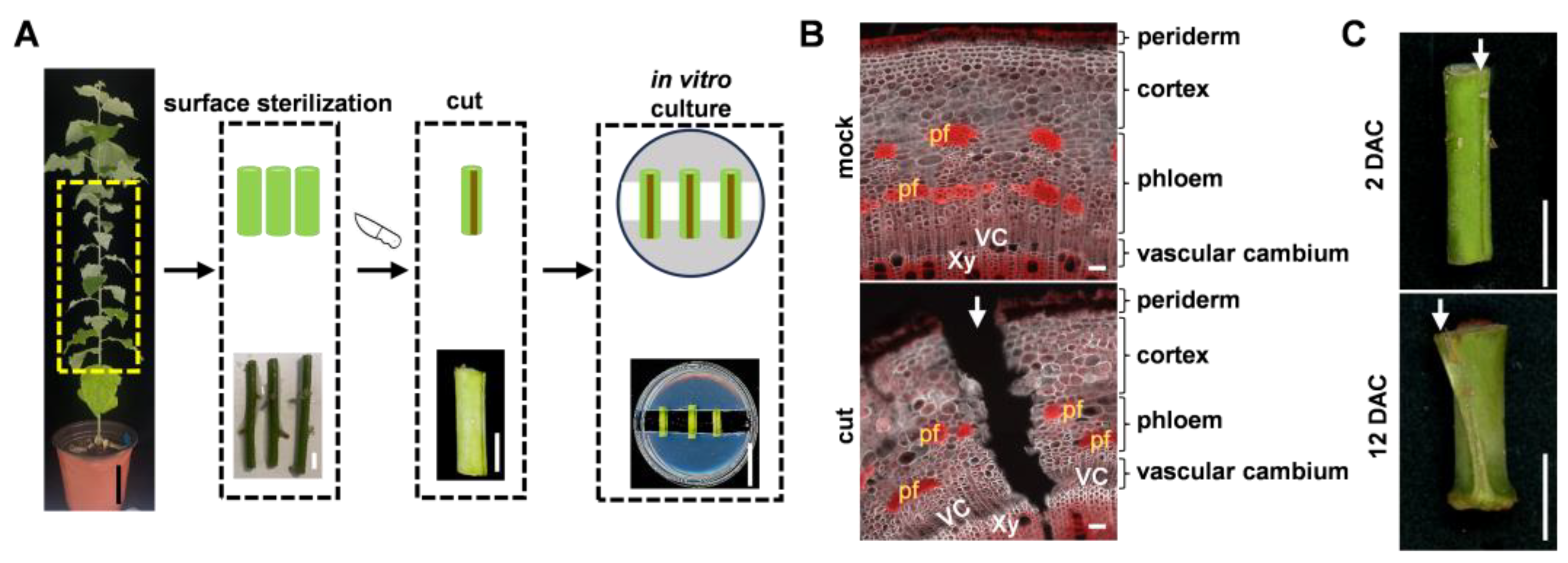
Figure 2.
Morphological dynamics of tissue-specific wound repair after longitudinal cutting of poplar stems. (A, B, E) Representative transverse sections showing cellular dynamics at the cutting site in 84K poplar (Populus alba × P. glandulosa). (A) Low-magnification images of the wound region at 0, 2, 4, 8, and 12 days after cutting (DAC). SCRI Renaissance 2200 (SR, white) was used to visualize cell walls, and Basic Fuchsin (BF, red) was used to visualize lignified cell walls. Scale bars, 100 μm. (B, E) Higher-magnification images of the cortex (B), phloem (B), and vascular cambium (E) regions at 0, 4, and 9 DAC. Toluidine Blue O (TBO) was used to visualize cell walls. Scale bars, 100 μm. (C) SR, BF, and Fluorol Yellow 088 (FY, yellow) staining of the cortex and phloem regions in 84K poplar at 2 and 8 DAC. FY was used to visualize suberized cell walls. Scale bars, 100 μm. (D) BF staining of the cortex, phloem and vascular cambium regions in 84K poplar at 16 DAC. Scale bars, 50 μm. pf, phloem fiber. VC, vascular cambium. Xy, xylem. Phe, phellem (magenta arrows). rPhe, regenerated phellem (yellow arrows). rPg, regenerated phellogen (red asterisks). Cal, callus-like cell. rVas, regenerated vascular cambium (red asterisks). White and black arrows indicate the cut site. Half orange bracket indicates the domains of regenerated periderm (rPer) and half green bracket indicates the domains of regenerated vascular tissues (rVas). Green dots indicate regenerated parenchyma cells. Yellow and green dashed lines indicate outline collapsed cells.
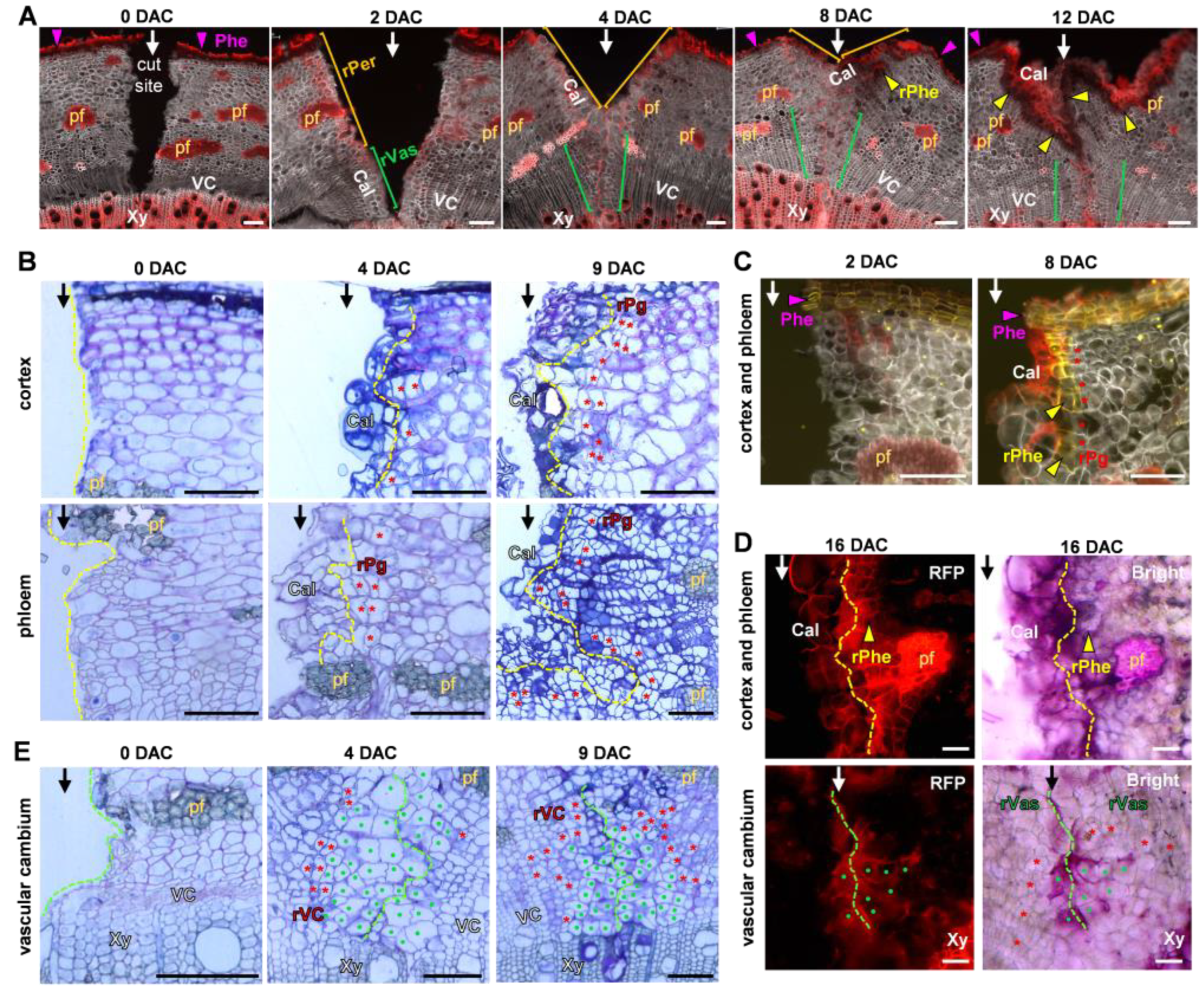
Figure 3.
Temporal expression dynamics of marker genes during wound repair in poplar stems. (A, C, E) Heatmaps showing relative expression of lignification (A), phellogen and vascular cambium activity (C), and phellem differentiation and suberin formation (E) related genes in 84K poplar (Populus alba × P. glandulosa) at 0 DAC–W (day after cutting-whole bark) and 0, 2, 4, and 9 DAC. FPKM values were log2-transformed and standardized within each gene. Red and blue indicate relatively high and low expression, respectively. Gene identifiers refer to Populus trichocarpa loci. (B, D, F) Reverse transcription quantitative PCR (RT-qPCR) analysis of the relative expression levels of 4CL1 and MYB63 (B), ANT and WOX4a (D), and MYB67, CYP86B1, and GPAT8 (F) in 84K poplar at 0 DAC–W and 0, 2, 4, and 9 DAC. Values are means ± SD. Different lowercase letters indicate significant differences among time points according to one-way ANOVA followed by Tukey’s multiple-comparisons test (P < 0.05).
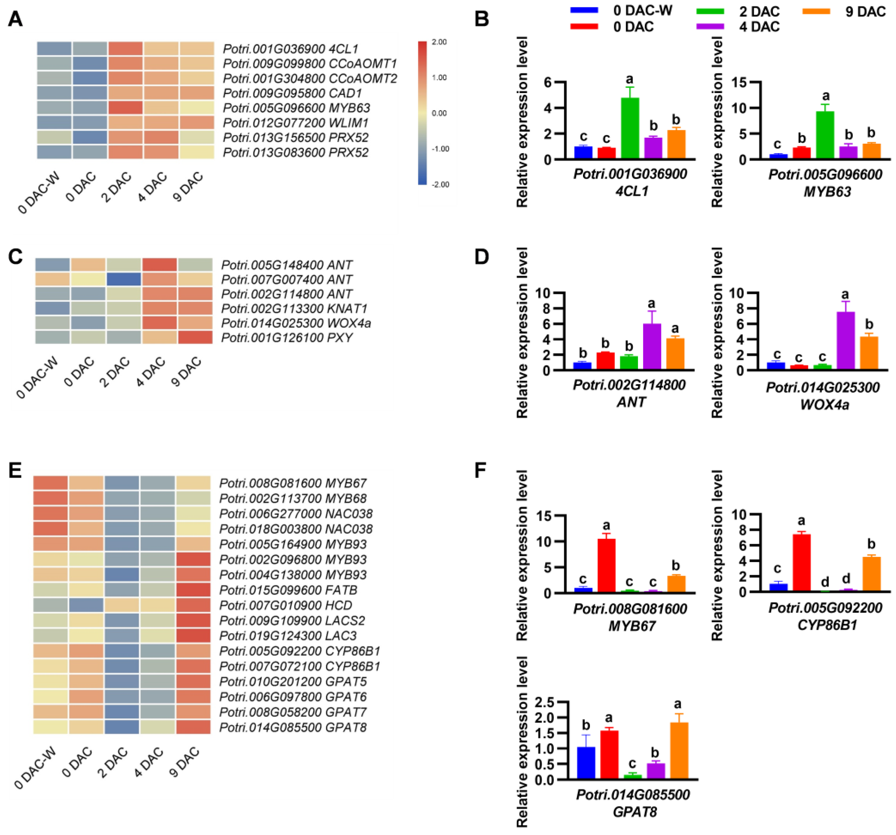
Figure 4.
Auxin promotes periclinal division in the cortex and phloem regions during wound repair in poplar stems. (A) Heatmap showing the relative expression of genes associated with auxin metabolism, transport, signaling, and transcriptional responses in 84K poplar (Populus alba × P. glandulosa) at 0 DAC–W (day after cutting-whole bark) and 0, 2, 4, and 9 DAC. FPKM values were log2-transformed and standardized within each gene. Red and blue indicate relatively high and low expression, respectively. Gene identifiers refer to Populus trichocarpa loci. (B) GUS staining showing PtaDR5::GUS reporter activity in the cortex and phloem of INRA 7171–B4 poplar (Populus tremula × P. alba) at 0, 2, 4, and 9 DAC. Scale bars, 100 μm. (C) SCRI Renaissance 2200 (SR, white) and Basic Fuchsin (BF, red) staining of the cortex and phloem regions of 84K stem segments cultured on medium containing DMSO, 5 μM 1-naphthaleneacetic acid (NAA), or 100 μM N-(1-naphthyl) phthalamic acid (NPA). Samples were collected at 2, 4, and 9 DAC. SR was used to visualize cell walls, and BF was used to visualize lignified cell walls. Scale bars, 100 μm. (D) The percentage of callus formation (left) and periderm regeneration (right) in the cortex and phloem regenerated regions, as shown in (C). (E) The frequency of cell division in callus (left) and cell division in regenerated periderm (right) in the cortex and phloem regenerated regions, as shown in (C). Values are means ± SEM. Different lowercase letters indicate significant differences among treatments at each time point according to one-way ANOVA followed by Tukey’s multiple-comparisons test (P < 0.05). pf, phloem fiber. White arrows indicate the cut site. Red asterisks indicate newly formed phellogen-like cells, based on their anatomical position. Red arrows mark the recent periclinal divisions. Yellow dashed lines indicate outline collapsed cells.
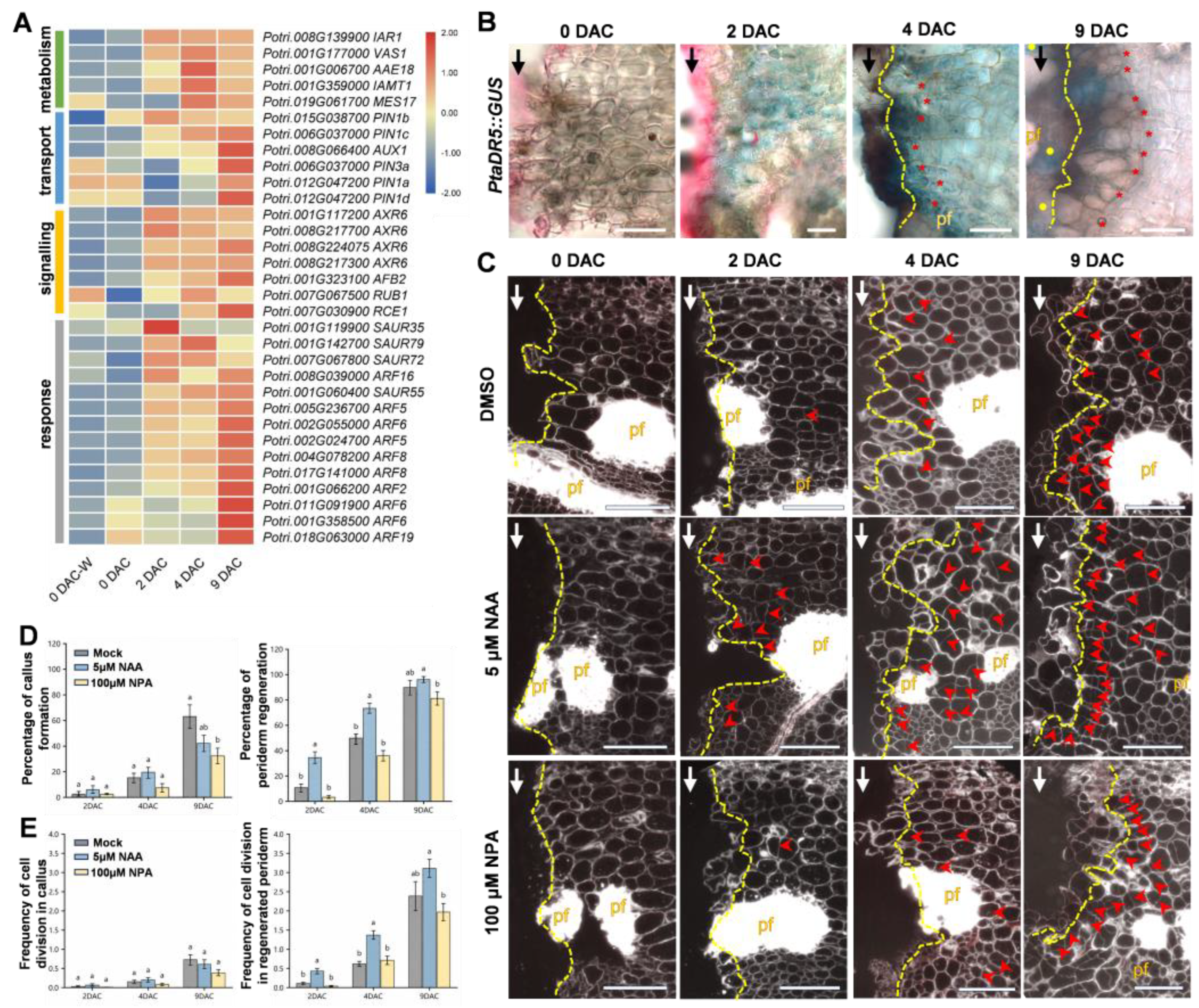
Figure 5.
Working model of coordinated tissue regeneration during wound repair following longitudinal cutting of poplar stems. The schematics illustrate the proposed progression of tissue regeneration and the roles of auxin and polar auxin transport (PAT). At 0 day after cutting (DAC), the longitudinal cut (black arrow) reaches xylem, exposing the cortex, phloem, and vascular cambium. At 2 DAC, cells beneath the wound surface become lignified as an early wound response. At 4 DAC, callus-like cells emerge from cells adjacent to the wound surface and subsequently undergo lignification. On the inner side of the lignified boundary, periclinal divisions of cortex and phloem cells initiate phellogen regeneration from 4 DAC. From 8 to 16 DAC, the newly formed phellogen further proliferates and differentiates into phelloderm and ligno-suberized phellem, thereby establishing a wound-induced periderm in the outer wound regions. Exogenous auxin promotes regenerative cell divisions during this process. In the damaged vascular region, cells along the cut surface expand and divide actively, filling the gap and closing the wound by 4 DAC. Such process requires PAT. Vascular repair continues until the vascular cambium and phloem are restored at 8–14 DAC. Cell types are distinguished by different colours. pf, phloem fiber. Half orange bracket indicates the domains of regenerated periderm (rPer) and half green bracket indicates the domains of regenerated vascular tissues (rVas).
Disclaimer/Publisher’s Note: The statements, opinions and data contained in all publications are solely those of the individual author(s) and contributor(s) and not of MDPI and/or the editor(s). MDPI and/or the editor(s) disclaim responsibility for any injury to people or property resulting from any ideas, methods, instructions or products referred to in the content. |
© 2026 by the authors. Licensee MDPI, Basel, Switzerland. This article is an open access article distributed under the terms and conditions of the Creative Commons Attribution (CC BY) license (http://creativecommons.org/licenses/by/4.0/).
Copyright: This open access article is published under a Creative Commons CC BY 4.0 license, which permit the free download, distribution, and reuse, provided that the author and preprint are cited in any reuse.