

Submitted:
09 October 2026
Posted:
10 October 2026
You are already at the latest version
Abstract
Transdermal drug delivery is an effective and, for some substances, the only feasible method. The most effective and advanced method for its implementation is the use of microneedles made of bi-odegradable polymeric materials. This study proposes a method for producing transdermal delivery patches containing microneedles on their surface. The method involves developing a 3D model of the patch master mold, 3D printing it, filling the intermediate mold with polydimethylsiloxane, and then filling the patch with a biopolymer. The biopolymers studied were PLGA (poly(lactic-co-glycolic acid)), PLA (polylactic acid), and PDLLA (poly-d,l-lactic acid). PLGA showed the best results. Two microneedle array topology variants were used. In the first case, the needles were arranged in a 34 × 34 array with a tip-to-tip spacing of 500 µm and a height of 800 µm. The base thickness was 5 mm, and the side dimensions were 20 mm. A second design variant featuring larger needles was pro-posed; this array consisted of 21 × 21 needles with a height of 1.6 mm.
Keywords:
transdermal delivery
; microneedles
; biopolymers
; poly(lactic-co-glycolic acid)
; polylactic acid
; poly-d
; l-lactic acid
1. Introduction
When developing a new treatment for any disease, the choice of drug delivery method is crucial. Traditional methods, such as oral administration or subcutaneous injections, are often ineffective and cause numerous side effects. Therefore, transdermal drug delivery methods, which have attracted the attention of researchers in recent decades due to a number of significant advantages, may be a promising option [1].
A transdermal therapeutic system (TTS) is a dosed, soft, well-tolerated dosage form for external use that can be manufactured as patches or films that release a drug over a specific, controlled period of time [2].
The advantages of this method compared with alternatives [3] include: higher rate of drug delivery, resulting in a more rapid onset of therapeutic effect; elimination of complications for patients associated with oral administration (e.g. difficulty swallowing and chewing); maintenance of a constant concentration of the active substance of the drug in the blood; ease and painlessness of administration (many transdermal delivery methods can be used by patients themselves); elimination of negative effects on the gastrointestinal mucosa (since the drug enters the systemic circulation directly, often through the skin). The method also helps to prevent first-pass metabolism through the liver; the absence of hydrolysis and degradation of administered drugs in an acidic environment or under the influence of enzymes. This increases the bioavailability of various drugs, the possibility of immediate discontinuation of treatment if side effects occur, a decrease in the frequency of administration and prescription of the drug due to a longer delivery time of small doses of the active substance [4]. However, transdermal drug delivery also has limitations associated with the mechanism of substance penetration into the body due to passive diffusion through the skin barrier, in particular, the stratum corneum [5].
Transdermal delivery using patches involves exposure directly to the skin – largest external organ of the body, attractive for use as a drug delivery route. Transdermal patch technology is currently used in anti-smoking, anti-inflammatory, osteoporosis, contraceptive, motion sickness, and cardiovascular patches. Researchers are striving to improve the technology by employing various chemical and physical approaches to deliver more complex drug formulations [6]. Transdermal patches can be single-layer or multilayer, and differ in the method of drug application and release. Due to this technological diversity and the use of various materials, a large number of drugs are currently effectively delivered using this technology [7]. Transdermal delivery methods allow the use of drugs based on polymer nanoparticles, the delivery of which is difficult by traditional routes due to low solubility and bioavailability [8,9].
One method of transdermal drug delivery is the use of microneedles—minimally invasive, micron-sized devices that penetrate the stratum corneum of the skin and release therapeutic agents through various mechanisms [10]. Typically, such devices are made of polymers, ceramics, steel, or silicon [11,12]. Microneedle arrays are used to enhance drug penetration, which helps overcome the disadvantages of subcutaneous administration using conventional needles.
Based on the method of active substance release, microneedles can be divided into five main types: solid (designed to create microscopic holes in the skin layer through which the drug will subsequently penetrate), hollow (similar to solid microneedles, but additionally contain reservoirs that deliver the drug directly to the injection site), coated microneedles (solid microneedles, the drug is applied directly to their outer surface), soluble (the drug is encapsulated in a non-toxic polymer that dissolves upon contact with the skin), and hydrogel-forming (the drug is also placed in a polymer, but upon dissolution, a hydrogel is formed) [13]. When choosing the type of microneedle, one considers the drugs used, available technology, and the point of contact of the patches with the skin. The use of dissolvable microneedles requires careful consideration of the polymeric materials used. They must be biocompatible and safe for humans, and dissolve without leaving sharp waste after insertion under the influence of body heat. Ideally, the microneedles should be durable enough to be inserted into any skin type, and their dissolution should be controlled to ensure consistent release.
The aim of the study was to develop and test in practice a patch with microneedles made of a biodegradable polymers (PLA, PLGA). We determined that PDLLA is best suited for the fabrication of microneedles and proposed two patch topologies.
2. Materials and Methods
Materials
Anycubic Water Wash Resin (Anycubic, China) was used to produce master molds using 3D printing.
Sylgard 184 Silicone Elastomer Kit (Dow Europe GMBH C/O Dow Silicones, Germany) was used to produce molds using soft lithography.
The following biopolymers were used: Medical Grade PLGA (Novokhim, Russia) with a lactide/glycolide ratio of 48-52%/48-52%, a molecular weight of 1,000–80,000 Da, a polydispersity coefficient of <2, and a residual monomer content of <0.5%, PLA for 3D printing (MAKO, China), and Medical Grade PDLLA (Shanghai Epoch Material Co., Ltd., China). To prepare the microneedle patch, several biopolymer solutions in acetone of varying concentrations were prepared.
Analytical grade acetone (Vekton, Russia) was used as the solvent for the biopolymers.
Medicinal substances
The following drugs were selected as model active substances: metformin, sibutramine, and tesofensine.
Metformin is a widely used drug for the treatment of type 2 diabetes mellitus, with a molecular weight of 165.63 g/mol. It functions by lowering blood glucose levels and increasing insulin sensitivity. Metformin reduces blood glucose by inhibiting gluconeogenesis and enhancing tissue sensitivity to insulin, which promotes weight loss and lowers the risk of cardiovascular disease [14,15]. Although the drug is actively used to treat obesity, it is associated with a number of serious side effects when administered orally. Structurally, metformin is a small, highly polar molecule containing multiple amino groups; this confers high water solubility and renders it virtually non-lipophilic. Due to these properties, metformin permeates lipid membranes poorly but is ideally suited for delivery in a soluble form.
Sibutramine is a drug used in conjunction with diet and exercise for weight loss. It acts as a serotonin, norepinephrine, and dopamine reuptake inhibitor, reducing appetite and increasing the feeling of satiety. This drug has demonstrated efficacy in the treatment of obesity, polycystic ovary syndrome, sleep apnea syndrome, etc. [16]. The molecular weight of sibutramine is 279.85 g/mol. Unlike metformin, it is a larger and more lipophilic molecule featuring an aromatic ring and chlorine substitution, which enables it to effectively penetrate biological membranes.
Tesofensine is a drug currently in the final stages of research for weight loss. It was originally developed to treat neurodegenerative disorders, such as Alzheimer’s and Parkinson’s diseases [17]. Its administration reduces meal frequency and increases energy expenditure by blocking the reuptake of serotonin, norepinephrine, and dopamine. Tesofensine has demonstrated efficacy in treating obesity, including the type 2 diabetes that often accompanies it [18]. It is a structurally complex molecule with a molecular weight of approximately 371 g/mol and high lipophilicity; its higher molecular weight compared to the other drugs discussed influences its pharmacokinetics and bioavailability across various routes of administration.
The selection of these drugs as model substances for encapsulation within a polymer matrix is based on differences in their chemical structure, lipophilicity, and mechanisms of action, enabling an assessment of the versatility of transdermal delivery using dissolving microneedles.
3D Printing of Master Molds
Preparation for printing the casting molds consisted of creating a 3D model of the desired structure. KOMPAS-3D (v21) was selected as the modeling software environment. Two patch topologies were used. In the first, the needles were arranged in a 34 × 34 array with a tip spacing of 500 µm, and the pyramid height was 800 µm. The base thickness was 5 mm for ease of mold handling, and the sides were 20 mm. An alternative design was proposed with larger needles (to facilitate printing and subsequent mold handling). With the substrate parameters maintained at 20 x 20 mm and 5 mm thick, the resulting array of 21 x 21 needles now has a height of 1.6 mm. The resulting models were loaded as .STL files and printed on an Anycubic Photon Mono x6Ks printer (Anycubic, China).
Making PDMS Casting Molds
The resulting photopolymer resin master mold was used to create a PDMS mold with holes. The PDMS mold was cast using the master mold, which was glued into a Petri dish. To do this, the PDMS base was first mixed with the hardener in a 10:1 ratio. A vacuum desiccator with a pump was used to remove air bubbles. The degassed PDMS was then placed in an oven for curing at 60°C for 4 hours. The cured PDMS replica was separated from the photopolymer master mold using a sharp scalpel blade.
Filling the molds with biopolymer solutions
For 25 ml of a 30% PLGA, PLA, and PDLLA solution, 7.5 ml of biopolymer was mixed with 17.5 ml of solvent and stirred on an orbital shaker for 2 hours at 110 min-1. The biopolymer solution was then poured into the PDMS mold using a syringe until the entire volume was filled (approximately 5 ml per patch). To ensure better solution flow into the microneedle cavities, the filled molds were placed in a vacuum desiccators and then left in a fume hood to harden and remove evaporating solvent. Finally, the mold was placed in an oven at 60 ºC for 1 hour and cooled at room temperature overnight. The finished patch was removed from the mold manually. For 25 ml of a 40% PDLLA solution, 10 ml of the biopolymer was mixed with 15 ml of solvent and stirred on an orbital shaker for 4 hours at 150 min-1. The biopolymer solution was then poured into a PDMS mold using a syringe until the entire volume was filled (approximately 5 ml per patch). To ensure better solution flow into the microneedle cavities, the filled molds were placed in a fume hood to remove evaporating solvent. They were then placed in an oven at 60 ºC for 15-30 minutes, stirred, and cooled at room temperature overnight. The finished mold was manually removed from the mold.
Images of the patches and PDMS mold topology were obtained using a Biomed-2 microscope (Micromed, Russia) with an ELP camera model FHD01M-MFV for image transfer to a computer.
Determination of drug release kinetics
The release kinetics of drugs from biopolymer-based microneedles were studied *in vitro* using freshly prepared phosphate-buffered saline (PBS). Fabricated samples—based on PLGA solutions encapsulating sibutramine, metformin, tesofensine, and a mixture of metformin and sibutramine at various concentrations (8 samples in total)—were placed in 100 mL of PBS at 25°C under constant stirring with a magnetic stirrer to maintain medium homogeneity. To evaluate drug release efficiency, 1 mL aliquots were withdrawn from the PBS, and the removed volume was replenished with fresh buffer. Samples were collected at 15, 30, 45, and 60 minutes, and at 2, 3, 24, and 48 hours from the start of the experiment for metformin and sibutramine. For the two remaining samples, additional aliquots were taken at 72 and 96 hours. The collected samples were frozen at -80°C and sent for analysis of their drug content.
The concentration of the active substance in the samples was determined using chromatography-mass spectrometry (CMS). Working solutions of the drugs (metformin, sibutramine, and tesofensine) were prepared for analysis by diluting a stock solution (5 mg/mL concentration), which had been prepared from a 2.5 mg drug sample. Prior to analysis, the samples were diluted tenfold with deionized water. Chromatographic separation was performed using an Agilent 1290 Infinity liquid chromatograph equipped with an Intensity Solo 1.8 C18-2 column. The components were separated based on their retention time in the chromatographic column; the mobile phase consisted of ammonium acetate in water and acetonitrile, flowing at a rate of 0.3 mL/min]. Detection was performed using a Bruker Q-TOF Maxis Impact mass spectrometer equipped with an electrospray ionization source. The operating conditions for the mass spectrometric detector were as follows:
– Drying gas (nitrogen) flow rate: 8 L/min
– Nebulizer gas pressure: 29 psi
– Transfer capillary temperature: 220°C
– Capillary voltage: 4500 V
– Ion detection range: m/z 50–1300 amu (in positive ionization mode).
Quantification was performed using the external calibration method within a concentration range of 5–100 ng/mL.
Studies on laboratory animals
The study utilized 132 sexually mature male Wistar SPF rats weighing 263 ± 45 g (aged 2–3 months). The source of the breeding stock was the "Laboratory Animal Breeding Facility" (NPP) of the Shemyakin-Ovchinnikov Institute of Bioorganic Chemistry (RAS), Moscow. The animals were housed in a barrier-type facility under standard conditions with controlled humidity and a 12-hour light/dark cycle, with free access to food and water.
Experimental groups consisted of animals treated with drugs delivered via a transdermal delivery system (microneedle patch). Control groups included intact animals, as well as obese animals treated with a drug-free microneedle patch. Comparison groups consisted of animals treated with weight-loss drugs administered intragastrically in a standard pharmacological form, mirroring their clinical use.
3. Results
The process of fabricating microneedle patches using soft lithography (casting into a PDMS mold) requires a master mold—one that is durable, capable of withstanding high temperatures, and accurately conveys the relief of the structure being fabricated. For this purpose, 3D printing was chosen. Preparation for printing involves creating a 3D model of the desired structure. KOMPAS-3D (v21) was selected as the software environment for modeling.
Creating a 3D model in KOMPAS-3D begins with analyzing the information model and mentally dividing it into simple base bodies. The structure from [19], consisting of a substrate (parallelepiped) and an array of needles (regular quadrangular pyramids), was chosen as a reference. The needles were arranged in a 34 × 34 array with a tip spacing of 500 µm, and the pyramid height was 800 µm. For ease of manipulation, the base thickness was chosen to be 5 mm, and the sides were 20 mm. The resulting model is shown in Figure 1.a, and its printed version is shown in Figure 1.b.
As an alternative, a structure with larger needles was proposed (to facilitate printing and subsequent mold handling). While maintaining the substrate parameters—20 x 20 mm, 5 mm thick—the resulting array of 21 x 21 needles now has a height of 1.6 mm. The model is shown in Figure 2.a, and the printed version is shown in Figure 2.b.
These models were used to subsequently produce master molds.
There are two main types of 3D printers, differing in the layer formation technology: filament and photopolymer. The former are characterized by affordability and high printing speed, can use a wide range of plastics with different characteristics, and direct the material onto the work surface layer by layer. The latter type of 3D printing, often used in medicine and jewelry, allows for the creation of detailed and complex models of structures using a special photopolymer that hardens under ultraviolet radiation. Since the structure size of the transdermal patches being developed is hundreds of microns, the photopolymer method, characterized by greater accuracy, was chosen for printing the master molds. Files from the KOMPAS-3D software package were converted to a suitable format, and the master molds were printed at ETU "LETI."
Molds for casting PDMS-based microneedle patches were fabricated using soft lithography. The process flow for this manufacturing method is shown in Figure 3.
The finished PDMS molds are shown in Figure 4.
The quality of the mold for casting was assessed visually using a microscope. The resulting image (Figure 4.b) demonstrated that the microneedle array (the mold consists of pyramidal holes) was transferred satisfactorily, maintaining the dimensions and spacing between the pyramids.
The microneedle patch for transdermal delivery was made of polylactic acid and its copolymers, which are biocompatible and readily available polymeric materials. PLA, PLGA, and PDLLA microneedle array options were considered. After curing the three molds, PLGA and PDLLA solutions were selected as they harden quickly after filling the holes. The PDLLA solution retains its microneedle geometry after removal from the mold, while the PLGA solution slowly flows after apparent stabilization, destroying the resulting relief (Figure 5).
However, increasing the PLGA content to 35% resolved this issue. Over a 48-hour period, the PDLLA samples retained their shape, whereas the PLGA sample exhibited deformation and partial dissolution of the needles. By the 168-hour (7 days) mark, the PDLLA patches showed slight deformation while maintaining their structural relief, whereas the PLGA sample exhibited complete dissolution of the needles. Ultimately, given the intended duration of patch use and the dissolution rate of the PLGA microneedles, this polymer proved effective for the objectives at hand.
Dissolving PLA in acetone at the same concentration as other biopolymers, as well as at twice the concentration, yielded no results. The polymer swelled and expanded, but did not dissolve completely, preventing its use in further experiments (Figure 6).
Therefore, it was decided to use a PLGA solution in acetone to fill the molds.
The most important step in filling the molds with the biopolymer solution is to completely fill the microneedles. Due to the high viscosity of the solution, accurate relief transfer cannot be achieved without additional stimulation of the sample, as this creates microcavities near the filled holes. Figure 7.a shows the resulting microcavity, which has destroyed some of the microneedles. Figure 7.b show images of the transferred microneedle array using a biopolymer solution with a higher concentration. This significantly reduces the number of air bubbles near the surface and within the patch (dark areas in the photo), improving the quality of the resulting structure. After several experiments, a higher concentration of the biopolymer solution in acetone was selected (40% solution instead of 30% for PDLLA and 35% solution instead of 30% for PLGA), and the option of heating the molds in an oven at 60°C followed by stirring to remove any air bubbles that had floated to the surface was chosen. A comparison of the recovered castings of solutions of different concentrations and heating times is shown in Figure 7.
Typically, such devices are fabricated from polymers, ceramics, steel, or silicon [11,12]. Soluble microneedles produced via 3D printing are known, but they lack sufficient strength [13]. We have proposed a method for fabricating microneedle patches that possess adequate strength, as the rigid biodegradable polymer PDLLA is used.
The scheme of the step-by-step formation of the microneedle patch is shown in Figure 8.
During the first 48 hours, approximately 7% of the total encapsulated metformin is released into the buffer solution (Figure 9.a). A comparison of data from the first and second days revealed that the highest release rate occurs during the initial period (the first two hours). Subsequently, the release rate slows down and reaches a "plateau," where the amount of substance released over 24 hours becomes comparable to the amount released over 48 hours. These results confirm earlier findings regarding the effective duration of patch use, which is approximately two days. Further research into optimizing the composition of the polymeric microneedles could be conducted to achieve a more uniform drug release over an extended period.
Analysis of the obtained data reveals a significantly slower release of sibutramine compared to metformin, which was previously studied under similar conditions (Figure 9.,b). Specifically, over a period of two days (48 hours), only about 1% of the total encapsulated substance was released into the buffer solution. While the release rate remains relatively high and constant during the initial period (the first 2 hours), a gradual slowdown follows—though the release of the drug substance into the solution does not cease entirely. The amount of released substance continues to increase with the duration of the sample's exposure to the solution. Given the drug's low and relatively uniform release rate, the key factor determining the effective duration of patch use becomes the dissolution rate of the needles themselves, rather than the rate at which the drug substance is released. This may necessitate the development of formulations with an accelerated polymer matrix degradation rate to achieve the required therapeutic dose within a specific timeframe.
To assess the feasibility of tesofensine release, its concentration within the polymer matrix was increased, based on the assumption that sufficient release would occur if the fraction of the drug transferred to the buffer solution reached 20%. Analysis of data from samples with drug concentrations five times higher than those initially tested (increasing from 7.31 µg/mL to 36.54 µg/mL) revealed a relationship between tesofensine release efficiency and its initial content in the polymer matrix. The graphs clearly demonstrate the non-linear dynamics of this process: an initial phase lasting approximately 20–40 hours is characterized by a relatively slow release rate, followed by a significant acceleration in the rate of release. By the 96-hour mark, the tesofensine concentration reaches 525 µg/mL, and the fraction of released substance rises to 1.437%.
In experiments involving laboratory animals, no deterioration in clinical condition or mortality was observed throughout the study.
No deterioration in clinical condition or animal mortality was observed throughout the experiment.
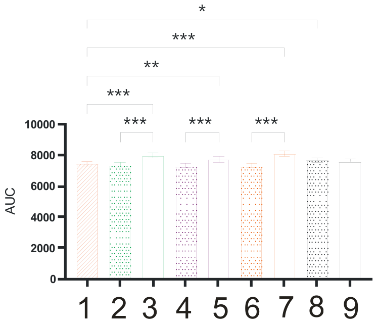
No statistically significant differences in body weight were found among the animals in the experimental groups during the course of the study. However, the area under the curve (AUC) values were higher in the "Obesity Control" group compared to rats treated with metformin, sibutramine, or tesofensine via the intragastric route, or with a combination of metformin and sibutramine via microneedle patches. Furthermore, the AUC in animals administered metformin, sibutramine, and tesofensine intragastrically was significantly higher than in rats receiving the same drugs transdermally (Figure 10). This may indicate a slower release of the drugs.
Before and after the 5-week treatment period, a statistically significant increase in blood glucose levels (postprandial hyperglycemia) was observed 60 minutes after the administration of 40% glucose in the intact control, obesity control, and empty patch control groups.
In rats treated with metformin and tesofensine via the transdermal route, postprandial hyperglycemia persisted both before and after the treatment course (at 60 min). In animals treated orally with sibutramine, no increase in blood glucose levels was observed 60 minutes after the administration of 40% glucose following the 5-week therapy. Furthermore, rats treated with tesofensine transdermally showed a reduction in glycemia levels relative to baseline values (pre-treatment) at the 120-minute mark.
Before and after the 5-week therapy course, a significant reduction in postprandial glycemia was recorded in all rats treated transdermally with the drugs (metformin, sibutramine, tesofensine), occurring in both the peak absorption phase (60 min) and the glucose utilization phase (120 min). In the groups of rats treated with sibutramine and tesofensine via microneedle patches, a complete flattening of the glycemic curve was also observed after the 5-week treatment period. In animals treated with the combination of pharmacological agents (metformin + sibutramine) via either a microneedle patch or intragastric administration, a statistically significant reduction in postprandial glycemia was observed during the phase of peak glucose absorption (60 min), whereas no significant changes were noted during the late phase of the test.
4. Discussion
Transdermal drug delivery systems have been developed intensively in recent years, but the approach proposed in the present work, the use of microneedles made of biodegradable polymers, has so far been reported in only a small number of experimental studies. The reason for adopting it is that, when drugs are applied to the skin surface, their access to the deeper skin layers and to the subcutaneous adipose tissue is limited by the stratum corneum, which restricts the permeability of most hydrophobic and hydrophilic drugs [20]. Several strategies have been proposed to overcome this limitation and to increase drug penetration through the skin: iontophoresis, sonophoresis, electroporation, microemulsions, solid lipid nanoparticles and a number of others [20]. Of these, patches carrying microneedles on their surface have attracted the greatest attention. In their properties they occupy an intermediate position between injectable dosage forms, being less invasive, and oral dosage forms, over which they offer better bioavailability and the absence of hepatic metabolism. The method is therefore not only minimally invasive but also permits the use of lower doses, which is both economically advantageous and safer. A further important benefit is the prolonged action and the stable effect of the drugs delivered in this way, provided that the composition of the biodegradable polymer has been selected correctly.
The size and structure of microneedles depend on the purpose for which they are intended: action on the deep skin layers in anti-ageing therapy, entry of the drug into the bloodstream for a systemic effect, or action on the subcutaneous adipose tissue. The patches with biodegradable microneedles developed in this work are intended for the delivery of drugs into the subcutaneous adipose tissue in order to act on its morphology and function in a targeted manner. A number of drugs are known to activate the transformation of white adipocytes into beige ones and to activate brown adipocytes. The presence and high activity of beige and brown adipocytes improve metabolic parameters (glycaemia, lipid levels and others) and produce a moderate weight loss or restrain weight gain. Under systemic exposure (oral or injectable administration) these effects are weak, and over the past decade the targeted delivery of browning agents into adipose tissue for the prevention and treatment of obesity and the associated metabolic disorders has therefore become increasingly popular [21,22]. This is precisely the field of application planned for the system of targeted drug delivery into adipose tissue developed here.
The choice of the polymer used as a carrier of the drug substance is governed by the required release rate, which depends on the physical properties of the polymer itself. To obtain the necessary combination of properties, copolymers are often used that combine the characteristics of units of different nature. Such materials have to maintain a constant drug release rate over a long period. In the studies described in the literature that pursue the same aim as ours, hyaluronic acid (HA) and its blends with other polymers are the biodegradable polymers most often used. HA has traditionally been used as the main component for the manufacture of dissolving microneedles applied in cosmetology to deliver active substances into the deep skin layers. On drying, hyaluronic acid becomes hard enough for microneedles of various sizes and shapes to be formed; on contact with the skin they dissolve and release the active substances they carry, such as peptides, vitamins and ceramides. The use of this material also provides deep hydration and stimulates regeneration. For the delivery of drugs into the subcutaneous adipose tissue, a blend of hyaluronic acid and poly(lactic-co-glycolic acid) (PLGA) has also been used [21,22,23], a blend of HA with poly(vinyl alcohol) (PVA) [24] and a blend of PVA with poly(vinylpyrrolidone) (PVP) [25].
In our opinion, the copolymer of lactic and glycolic acids (PLGA) is one of the most promising materials for dissolving microneedles. Synthetic polyesters such as PLGA have earned recognition owing to their adjustable degradation rate and good biocompatibility [14]. This biocompatible and biodegradable polymer is widely used in medical devices, including transdermal drug delivery systems, since it causes no inflammatory reactions on contact with living organisms. PLGA is a thermoplastic polyester approved for medical use. Its degradation in vivo proceeds faster than in vitro under the influence of biological substances and of the immune response [11]. Being a linear copolymer, PLGA can be obtained from lactic and glycolic acid monomers in various ratios, which makes it possible to vary the molecular weight and the degradation rate. This ratio determines the degradation rate of PLGA, which may range from several days to several months. The main monomers for the synthesis of PLGA are lactic and glycolic acids. At a higher lactic acid content PLGA becomes less hydrophilic and degrades more slowly. Racemic lactic acid, a mixture of the D- and L-isomers, is often used [13].
The PLGA copolymer composition was chosen in our study because it allowed the required characteristics of the targeted delivery system to be obtained, in particular the necessary rate of biodegradation. Biodegradation of the microneedles within 48-72 h was needed to reach the required level of drug release. On the basis of the experiments carried out, the optimal composition and processing regime for the microneedle patch prototype were determined: a 35% solution of PLGA in acetone with subsequent curing at 140 °C. This variant gave the most balanced characteristics with respect to a number of key parameters. First, a high reproducibility of the microneedles was achieved, which is an important factor for the stability of the performance properties of the patch. Second, the rate of biodegradation of the polymer matrix after removal from the mould proved to be controllable, which makes it possible to predict the kinetics of drug release. In addition, the thickness of the specimen obtained was optimal for convenient removal from the mould and for further use. The chosen regime favoured the formation of a homogeneous structure with a minimum number of defects, which had a positive effect on the mechanical strength and thus ensured the reliability and efficiency of the patch in subsequent studies. This copolymer is therefore a biocompatible and readily available polymeric material, and it showed the properties required, namely sufficient strength, stability and plasticity. Among the notable features of this biodegradable polymer is the presence of lactate in its composition, a substance that itself activates browning, which may enhance the efficiency of the targeted delivery system.
In our earlier publications we showed that the system developed is able to provide prolonged release, as the chosen copolymer biodegrades, of drugs that increase the activity of beige (metformin) or brown (sibutramine, tesofensine) adipose tissue. The PLGA system was found to be suitable for the gradual release of the drug into the tissue at the required concentration, and differences in the release rate were revealed depending on the type of drug and on whether it had been mixed with another drug [26]. In another publication we described the changes that occur in experimental animals (Wistar rats) with metabolic dysfunction (impaired lipid and carbohydrate metabolism) after the application of microneedle patches loaded with the drugs studied, in comparison with intragastric administration of the same drugs. Transdermal delivery was accompanied by considerably lower values of the body weight AUC than intragastric administration. Transdermal administration provided a marked improvement in carbohydrate metabolism, with a significant reduction in postprandial glycaemia and a pronounced flattening of the glycaemic curve in the groups given sibutramine and tesofensine. Biochemical analysis confirmed a significant improvement in the lipid profile (cholesterol and triglyceride levels) after transdermal therapy in comparison with the groups given the drugs intragastrically [27]. In the same model of metabolic dysfunction we also demonstrated a considerable increase in the expression of UCP1 and PGC1a in brown adipose tissue after the application of microneedle patches loaded with the drugs studied. Without drug treatment, this metabolic model showed whitening of the brown adipose tissue, that is, a pronounced decrease in the expression of UCP1 and PGC1a, and administration of the same drugs by the oral route did not normalise the expression of UCP1 and PGC1a, in contrast to targeted delivery by microneedles. The whole body of data obtained thus confirmed that the system developed is suitable for the delivery of the drugs selected for the study.
Summing up, it may be noted that, in spite of the large number of studies carried out in this field, the TTS we have developed is unique. By a system we mean the whole complex, that is, a biopolymer of a definite composition and concentration, the drug dissolved in it and the structure that has been formed. Our TTS comprises microneedles made of a biodegradable polymeric carrier, which contains the copolymer of lactide and glycolide (PLGA) and the drugs dissolved in it: metformin, sibutramine and tesofensine. An analysis of the literature showed that only the work of Abbasi et al. [19] is sufficiently close to ours in methodological terms. The system these authors developed was likewise based on a copolymer of lactic and glycolic acids, but it differed in a number of respects. PLGA (50:50) was used to incorporate the drug, but with a different solvent, dimethylformamide (DMF), at 200 mg/mL (20%). The copolymer dissolved in dimethylformamide (DMF) at 200 mg/mL, with the metformin loaded into it, was placed into the mould with a pipette, approximately 30-40 µL per patch. PLGA 50:50 was also used in our work, but acetone served as the solvent, the final concentration of the solution was 35%, and the filling was performed not only with metformin: four variants of microneedles were developed, containing metformin, sibutramine, a mixture of metformin and sibutramine, and tesofensine. The concentrations and the amounts of the drugs loaded into the system also differed considerably. In addition, the possibility of using sibutramine and tesofensine in biodegradable needles to induce the activity of brown adipose tissue was assessed for the first time in our work. Judging by the results, these features of the composition had a substantial effect on the rate of drug release. In the development of Abbasi et al. the system was used over a considerably shorter period of time (24 h), and within this period the drug was released almost completely [19]. Our TTS was applied for 48 h, and even over this period the release of the drugs was not complete. According to the data we obtained [26], the rate of drug release depended not only on the ratio of lactide to glycolide, as noted by Abbasi et al., but also on the concentration of the drugs in the copolymer, on the molecular weight of the drug and on the mixing of two drugs. Nevertheless, an amount of drug sufficient to obtain the required effect entered the adipose tissue [27. Since in the work of Abbasi et al. the system was applied to the inguinal region (inducible beige adipose tissue) in mice, a direct comparison of efficiency with our development is not possible. As a result, the unique features of the TTS we have designed are a considerably longer period of application (48 h) and the possibility of using different drugs and their mixture, which provides a prolongation of the effect on the activity of brown adipose tissue and gives an advantage for translation into the clinic.
5. Conclusions
A manufacturing process for patches with biodegradable microneedles for transdermal drug delivery was selected, and a laboratory technology for their production was developed based on this process. When considering the materials used to create the molds for future casting, it was decided to use PDMS polymer, the working features of which had been previously developed by the authors. To fabricate the patches themselves, two models with different topologies were created in the KOMPAS-3D software package to select the best option for subsequent work. Master molds were printed using 3D models on a photopolymer printer. These molds were used to create PDMS molds for subsequent casting of biopolymer solutions. As a result, PDMS molds were produced and cast with biopolymer solutions in acetone. PLGA, PLA, and PDLLA were studied as biopolymers. PDLLA demonstrated the best performance. The resulting patches with biodegradable microneedles can be used for transdermal drug delivery.
Author Contributions
Conceptualization A.Y.B.and D.V.K.; Methodology D.V.K.; Validation A.I.N.; Formal analysis A.I.N.; Investigation A.I.N.; Data curation A.I.N.;Writing—original draft preparation D.V.K.; Writing—review and editing A.Y.B.; Supervision A.Y.B.and D.V.K.; Project administration A.Y.B. All authors have read and agreed to the published version of the manuscript.
Funding
This research and APC were funded by the Russian Science Foundation (Agreement № 24-15-20006 of 12 April 2024) and Saint-Petersburg Science Foundation (Agreement № 24-15-20006 of 24 May 2024).
Institutional Review Board Statement
“Not applicable” for this etap of studies (not involving humans or animals.).
Informed Consent Statement
Not applicable.
Data Availability Statement
The original contributions presented in this study are included in the article. Further inquiries can be directed to the corresponding author.
Acknowledgments
This work has been performed using the resources of Research Equipment Sharing Center “Preclinical Translational Research Centre” at Almazov National Medical Research Center.
Conflicts of Interest
The authors of this work declare that they have no conflicts of interest.
Abbreviations
The following abbreviations are used in this manuscript:
| PLGA | Poly lactic-co-glycolic acid |
| PLA | Poly lactic acid |
| PDLLA | Poly-d,l-lactic Acid |
| TTS | Transdermal therapeutic system |
| PDMS | Polydimethylsiloxane |
| 3D | Three-dimensional |
References
- Balakrishnan, P.; Gopi, S. Revolutionizing transdermal drug delivery: unveiling the potential of cubosomes and ethosomes. J. Mater. Chem. B 2024, 12(18), 4335–4360. Available online: https://pubs.rsc.org/tb/article-abstract/12/18/4335/833802/Revolutionizing-transdermal-drug-delivery?redirectedFrom=fulltext. [CrossRef]
- Shestakov, N.; Pyatigorskaya, N. Current aspects of the use of excipients as exemplified by transdermal therapeutic system technology. Pharmacy 2019, 68, 10–14. Available online: https://pharmaciyajournal.ru/ru/25419218-2019-08-02. [CrossRef]
- Hupfeld, S.; Gravem, H. Depotplastre som administrasjonsprinsipp for legemidler [Transdermal therapeutic systems for drug administration]. Tidsskr. Nor. Laegeforen 2009, 129(6), 532–3. Available online: https://tidsskriftet.no/2009/03/legemidler-i-praksis/depotplastre-som-administrasjonsprinsipp-legemidler.
- Farlow, M.; Somogyi, M. Transdermal patches for the treatment of neurologic conditions in elderly patients: a review. Prim. Care Companion CNS Disord. 2011, 13(6), PCC.11r01149. Available online: https://www.psychiatrist.com/pcc/transdermal-patches-treatment-neurologic-conditions/. [CrossRef]
- Beregovyh, V.; Pjatigorskaja, N.; Prudkevich, Ju.; Kedik, S. Transdermal therapeutic systems of drug delivery. Fine Chem. Technol. 2012, 7(5), 17–22. Available online: https://www.finechem-mirea.ru/jour/article/view/689/735?locale=ru_RU.
- Hanbali, O.; Khan, H.; Sarfraz, M.; Arafat, M.; Ijaz, S.; Hameed, A. Transdermal patches: Design and current approaches to painless drug delivery. Acta Pharm. 2019, 69(2), 197–215. Available online: https://reference-global.com/article/10.2478/acph-2019-0016. [CrossRef]
- Sabbagh, F.; Kim, B. Recent advances in polymeric transdermal drug delivery systems. J. Control Release 2022, 341, 132–146. Available online: https://www.sciencedirect.com/science/article/abs/pii/S0168365921006209?via%3Dihub. [CrossRef]
- Murthy, N.; Paul, S.; Chauhan, H.; Singh, S. Polymeric Nanoparticles for Transdermal Delivery of Polyphenols. Curr. Drug Deliv. 2022, 19(2), 182–191. Available online: https://www.eurekaselect.com/article/116830. [CrossRef]
- Kalave, S.; Chatterjee, B.; Shah, P.; Misra, A. Transdermal Delivery of Macromolecules Using Nano Lipid Carriers. Curr. Pharm. Des. 2021, 27(42), 4330–4340. Available online: https://www.eurekaselect.com/article/117403. [CrossRef]
- Singh, P.; Carrier, A.; Chen, Y.; Lin, S.; Wang, J.; Cui, S.; Zhang, X. Polymeric microneedles for controlled transdermal drug delivery. J. Control Release 2019, 315, 97–113. Available online: https://www.sciencedirect.com/science/article/abs/pii/S0168365919305863?via%3Dihub. [CrossRef]
- Ita, K. Transdermal Delivery of Drugs with Microneedles-Potential and Challenges. Pharmaceutics 2015, 7(3), 90–105. Available online: https://www.mdpi.com/1999-4923/7/3/90. [CrossRef]
- Larrañeta, E.; Lutton, R.; Woolfson, A.; Donnelly, R. Microneedle arrays as transdermal and intradermal drug delivery systems: Materials science, manufacture and commercial development. Mater. Sci. Eng. R Rep. 2016, 104, 1–32. Available online: https://www.sciencedirect.com/science/article/pii/S0927796X16300213?via%3Dihub. [CrossRef]
- Dharadhar, S.; Majumdar, A.; Dhoble, S.; Patravale, V. Microneedles for transdermal drug delivery: a systematic review. Drug Dev. Ind. Pharm. 2019, 45(2), 188–201. Available online: https://www.tandfonline.com/doi/10.1080/03639045.2018.1539497?url_ver=Z39.88-2003&rfr_id=ori:rid:crossref.org&rfr_dat=cr_pub%20%200pubmed. [CrossRef]
- Kuznetsov, K. O.; et al. Metformin and malignant neoplasms: a possible mechanism of antitumor action and prospects for use in practice. Probl. Endocrinol. 2022, 68(5), 45–55. Available online: https://pmc.ncbi.nlm.nih.gov/articles/PMC9762452/. [CrossRef]
- Lv, Z.; Guo, Y. Metformin and its benefits for various diseases. Frontiers in endocrinology. 2020. Available online: https://pmc.ncbi.nlm.nih.gov/articles/PMC7212476/.
- Tziomalos, K.; Krassas, G. E.; Tzotzas, T. The use of sibutramine in the management of obesity and related disorders: an update. In Vascular health and risk management; 2009; pp. 441–452. Available online: https://pmc.ncbi.nlm.nih.gov/articles/PMC7212476/.
- Bara-Jimenez, W.; et al. Effect of monoamine reuptake inhibitor NS 2330 in advanced Parkinson's disease. Mov. Disord. Off. J. Mov. Disord. Soc. 2004, 19(10), 1183–1186. Available online: https://pubmed.ncbi.nlm.nih.gov/15390018/. [CrossRef]
- Singh, R.; et al. Tesofensine. Triple monoamine reuptake inhibitor of dopamine, norepinephrine and serotonin; Treatment of obesity. Drugs of the Future 2018, 43(11). Available online: https://access.portico.org/Portico/auView?auId=ark:%2F27927%2Fpjbf78xj5qr. [CrossRef]
- Abbasi, M.; Fan, Z.; Dawson, J.; Wang, S. Transdermal Delivery of Metformin Using Dissolving Microneedles and Iontophoresis Patches for Browning Subcutaneous Adipose Tissue. Pharmaceutics 2022, 14(4), 879. Available online: https://pmc.ncbi.nlm.nih.gov/articles/PMC9029293/. [CrossRef]
- Phatale, V.; Vaiphei, K.; Jha, S.; Patil, D.; Agrawal, M.; Alexander, A. Overcoming skin barriers through advanced transdermal drug delivery approaches. J. Control. Release 2022, 351, 361–380. Available online: https://pubmed.ncbi.nlm.nih.gov/36169040/. [CrossRef]
- Zhang, Y.; Liu, Q.; Yu, J.; Yu, S.; Wang, J.; Qiang, L.; Gu, Z. Locally Induced Adipose Tissue Browning by Microneedle Patch for Obesity Treatment. ACS Nano 2017, 11, 9223–9230. Available online: https://pmc.ncbi.nlm.nih.gov/articles/PMC6812556/. [CrossRef]
- Than, A.; Liang, K.; Xu, S.; Sun, L.; Duan, H.; Xi, F.; Xu, C.; Chen, P. Transdermal delivery of anti-obesity compounds to subcutaneous adipose tissue with polymeric microneedle patches. Small Methods 2017, 1, 1700269. [Google Scholar] [CrossRef]
- Peng, H.; Zhang, C.; Wang, M.; Zhang, W.; Xu, B.; Yan, X.; Xin, H.; Wang, X. Black phosphorus modified soluble microneedle patch for painless, effective and accurate body slimming. Appl. Mater. Today 2020, 19, 100577. [Google Scholar] [CrossRef]
- Bao, C.; Li, Z.; Liang, S.; Hu, Y.; Wang, X.; Fang, B.; Wang, P.; Chen, S.; Li, Y. Microneedle patch delivery of capsaicin-containing α-lactalbumin nanomicelles to adipocytes achieves potent anti-obesity effects. Adv. Funct. Mater. 2021, 31, 2011130. [Google Scholar] [CrossRef]
- Feng, M.; Jiang, G.; Sun, Y.; Aharodnikau, U.E.; Yunusov, K.; Liu, T.; Zeng, Z.; Solomevich, S. Integration of metformin-loaded mesoporous bioactive glass nanoparticles and free metformin into polymer microneedles for transdermal delivery on diabetic rats. Inorg. Chem. Commun. 2022, 144, 109896. [Google Scholar] [CrossRef]
- Babenko, A.; Edemskaya, E.; Murashko, E. Development of a novel technology for targeted drugs delivery into adipose tissue. Complex Issues of Cardiovascular Diseases. 2026.
- Zasypkin, G.; Podyacheva, E.; Antonyuk, I.; Vasyutina, M.; Egorova, N.; Snezhkova, J.; Korolev, D.; Babenko, A.; Toropova, Ya. Metabolic Effects of Transdermally Delivered Pharmacological Agents in a Rat Model of Diet-Induced Metabolic Dysfunction. J. Evol. Biochem. Physiol. 2026, 62, 974–985. [Google Scholar] [CrossRef]
Figure 1.
Visualization of the patch model: a — Model developed in KOMPAS-3D; b — visualization of the resulting topology using a light microscope.
Figure 1.
Visualization of the patch model: a — Model developed in KOMPAS-3D; b — visualization of the resulting topology using a light microscope.
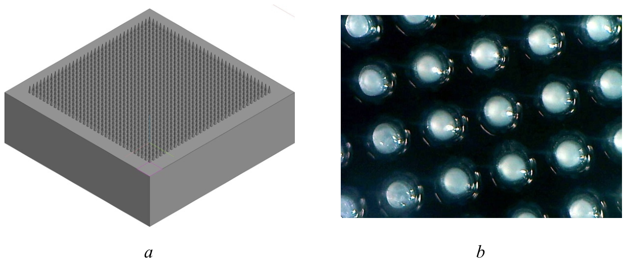
Figure 2.
Visualization of a patch model with large needles: a — Model developed in KOMPAS-3D; b — visualization of the resulting topology using a light microscope.
Figure 2.
Visualization of a patch model with large needles: a — Model developed in KOMPAS-3D; b — visualization of the resulting topology using a light microscope.
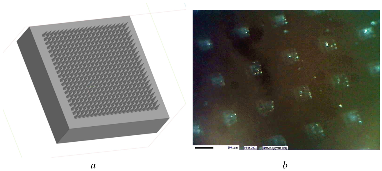
Figure 3.
Sequence of steps for pouring PDMS molds.
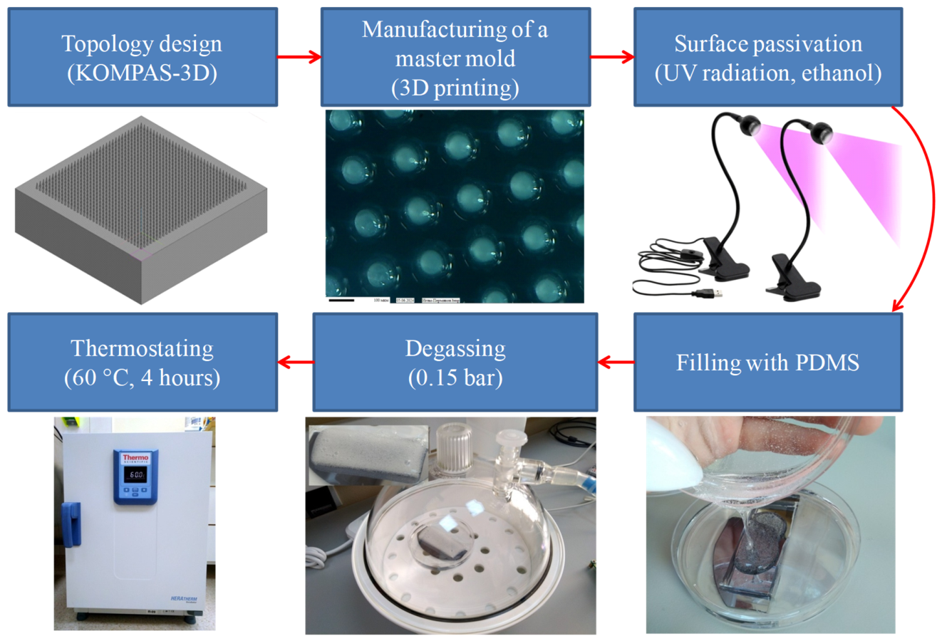
Figure 4.
Finished PDMS mold: a – macro image; b – microscopic image of the relief.
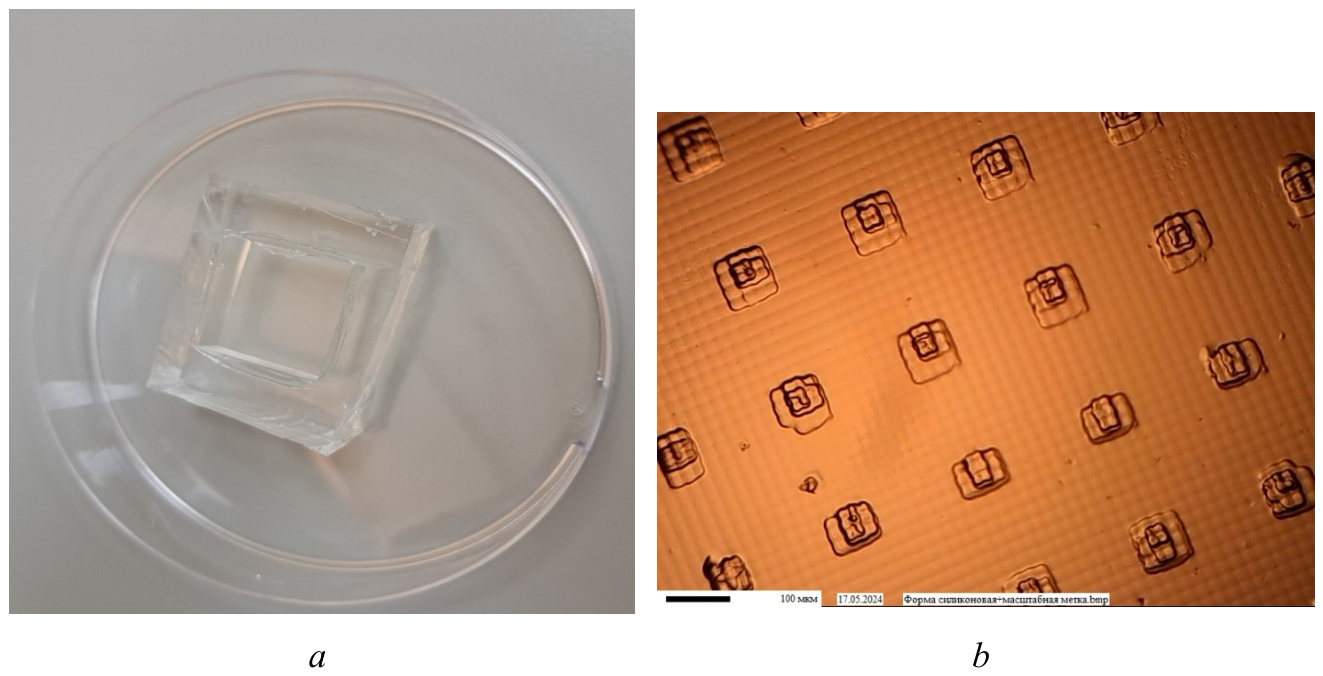
Figure 5.
Image of the PLGA patch relief: a – immediately after removal from the mold; b – and 7 days later.
Figure 5.
Image of the PLGA patch relief: a – immediately after removal from the mold; b – and 7 days later.
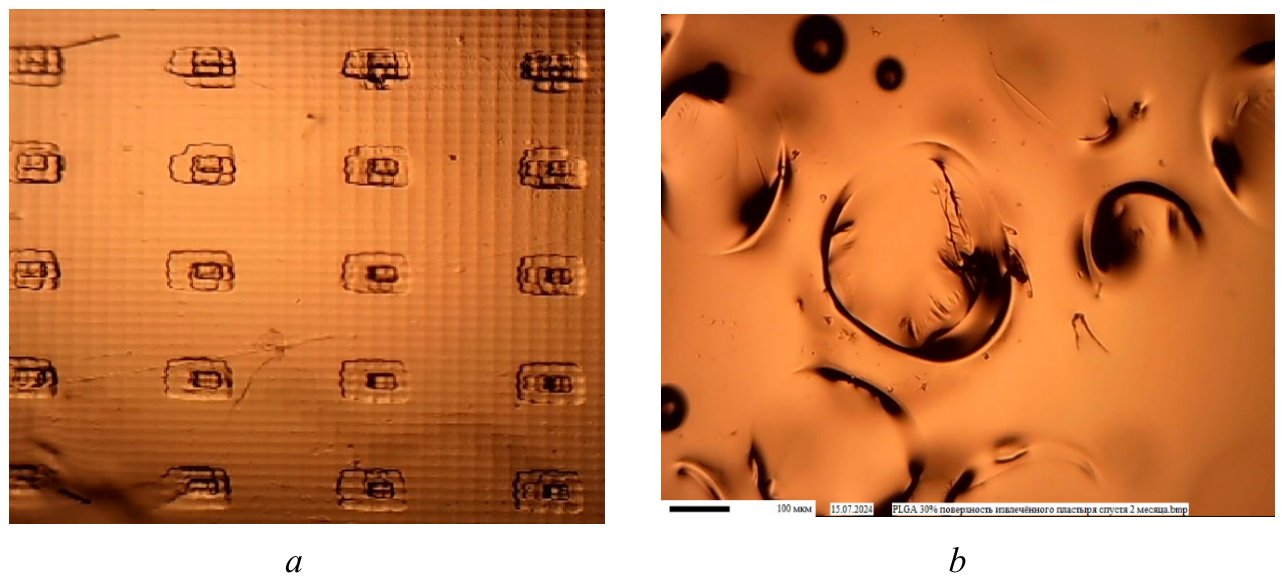
Figure 6.
Comparison of solubility in acetone: a – PLA; b – PDLLA.
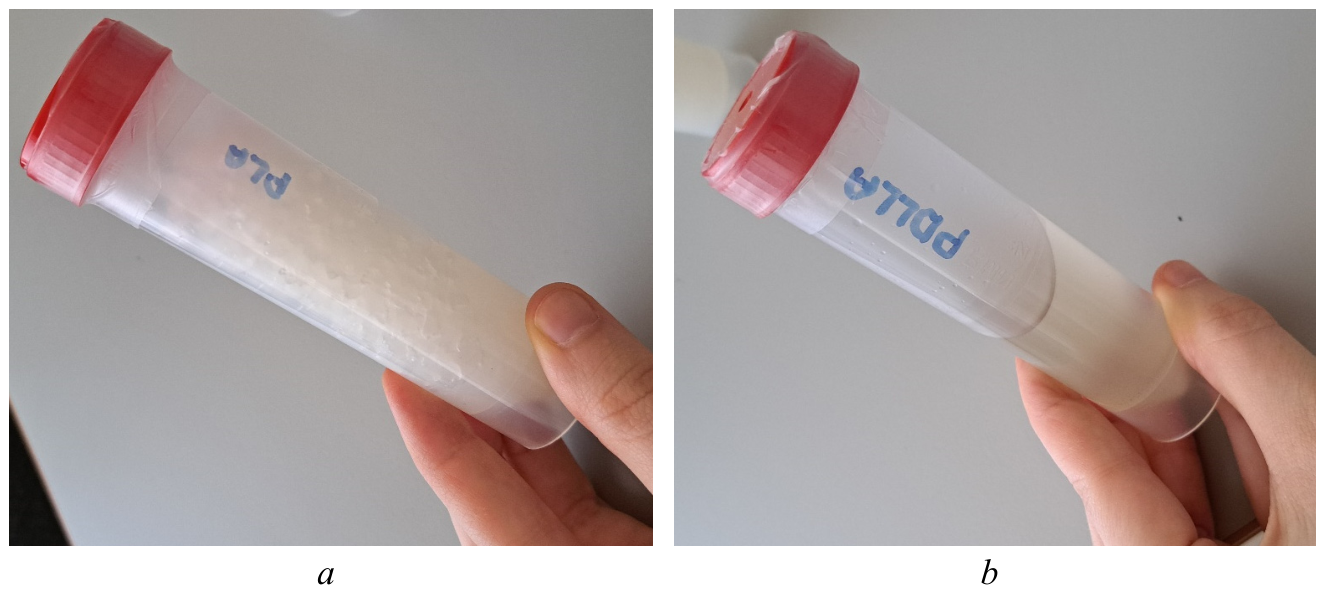
Figure 7.
Image of the relief on PDLLA: a – using a 30% solution and heating for 15 minutes; b –a 40% solution and heating for 30 minutes (b).
Figure 7.
Image of the relief on PDLLA: a – using a 30% solution and heating for 15 minutes; b –a 40% solution and heating for 30 minutes (b).
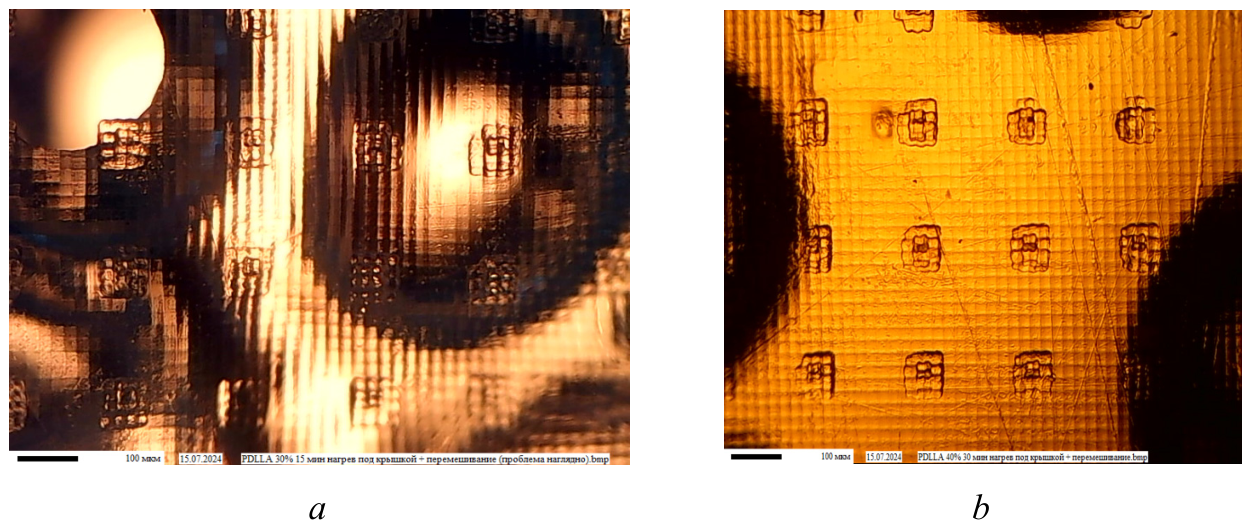
Figure 8.
Technology making microneedle patch.
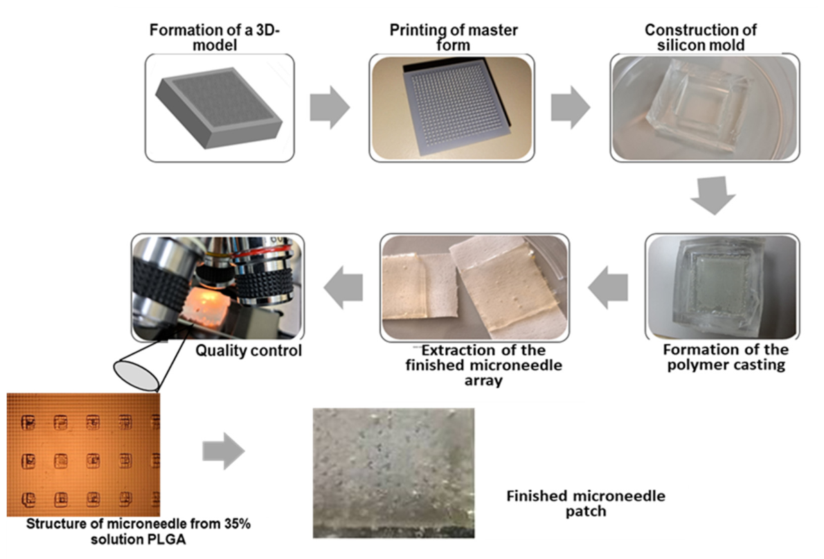
Figure 9.
Metformin (a), sibutramine (b) and tesofensine release kinetics.
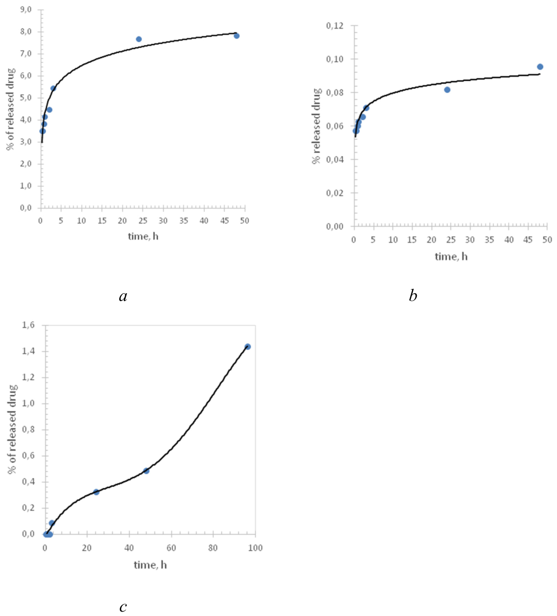
Figure 10.
Changes in animal body mass under various conditions: 1 - control – obesity; 2 – metformin patch; 3 - intragastric metformin; 4 - sibutramine patch; 5 –– intragastric sibutramin; 6 - tesofensine patch; 7 - intragastric tesofensine: 8- plaster mix; 9 – intragastric mix.
Figure 10.
Changes in animal body mass under various conditions: 1 - control – obesity; 2 – metformin patch; 3 - intragastric metformin; 4 - sibutramine patch; 5 –– intragastric sibutramin; 6 - tesofensine patch; 7 - intragastric tesofensine: 8- plaster mix; 9 – intragastric mix.
Disclaimer/Publisher’s Note: The statements, opinions and data contained in all publications are solely those of the individual author(s) and contributor(s) and not of MDPI and/or the editor(s). MDPI and/or the editor(s) disclaim responsibility for any injury to people or property resulting from any ideas, methods, instructions or products referred to in the content. |
© 2026 by the authors. Licensee MDPI, Basel, Switzerland. This article is an open access article distributed under the terms and conditions of the Creative Commons Attribution (CC BY) license (http://creativecommons.org/licenses/by/4.0/).
Copyright: This open access article is published under a Creative Commons CC BY 4.0 license, which permit the free download, distribution, and reuse, provided that the author and preprint are cited in any reuse.