

Submitted:
08 October 2026
Posted:
10 October 2026
You are already at the latest version
Abstract
Date palm (Phoenix dactylifera L.) is an economically important crop in Sudan, and symptoms of decline may be associated with diverse and poorly understood fungal communities. An unclassified agaricoid fungus was consistently isolated from basidiomata occurring on or near symptomatic date palms in an orchard in Khartoum State, Sudan. Affected palms exhibited chlorosis, reduced growth, drooping fronds, progressive browning, and desiccation. Six isolates (A–F) were examined based on their morphological characteristics and ITS1-5.8S-ITS2 sequences. Unlike the poroid basidiomata typical of Ganoderma, the isolates produced mushroom-like basidiomata with gills and a stipe. Several morphological characteristics also differed from the typical description of Hohenbuehelia, particularly the centrally positioned stipe and the free or narrowly adnate lamellae. BLASTn analysis showed only 83–89% ITS sequence identity with available Hohenbuehelia sequences. In the present ITS dataset, isolates A–F formed a strongly supported monophyletic group (bootstrap values of 99–100%) outside the sampled Hohenbuehelia and Ganoderma sequences. These results help clarify the phylogenetic position of the isolates within the taxa included in the current dataset but are insufficient for formal genus-level classification or the recognition of a new taxon. Furthermore, following 12 months of artificial inoculation, no consistent disease symptoms were observed, and the causal agents of date palm decline could not be determined. This study reports an unidentified agaricoid fungus associated with declining date palms and highlights the need for broader sampling of Agaricales, multilocus phylogenetic analyses, detailed morphological characterization, and carefully controlled pathogenicity tests before its taxonomic identity and ecological significance can be established.
Keywords:
date palm (Phoenix dactylifera)
; Ganoderma zonatum
; Hohenbuehelia spp
; BLASTn
; basidiospores
1. Introduction
The date palm (Phoenix dactylifera L.) is one of the oldest and most important fruit crops in Africa, particularly in northern Sudan [1,2]. It is affected by numerous diseases and decline syndromes that can adversely affect productivity [3,4,5,6,7]. Basal stem rot caused by Ganoderma spp. is an important disease of palms in several production areas [8,9,10,11]. It was first observed in Sudan at the Elbawga scheme in 2009 [12]. In the orchard investigated in the present study, affected palms showed pronounced yellowing and deterioration, accompanied by stunted growth, downward-drooping fronds, progressive browning, and desiccation. However, symptomatology alone was insufficient to determine the etiological agent, as similar decline symptoms may result from different biotic or abiotic factors. Therefore, morphological, cultural, and molecular characterization of the associated fungus was undertaken because unusual agaricoid basidiomata were observed on or near some of the symptomatic palms.
The Basidiomycota represents a large branch of the fungal tree, comprising more than 40,000 species and making up about a third of all the fungi described. This category includes important taxa such as fungi, plant pathogens such as smut and rust, and yeasts of commercial interest. Moreover, this collection is important for ecological systems as it allows for important nutrient cycling [13]. Mushrooms are a group of fungi belonging to the class Agaricomycetes in the order Agaricales (Underw) [14], which comprises gilled mushrooms. Agaricales is known to be one of the most diverse orders in the phylum Basidiomycota. The taxonomic classification of Agaricales is founded on phylogenetic relationships and distinct morphological features of its suborders, emphasizing the great diversity and specializations typical of this fungal order. A phylogenomic study of Agaricales revealed the strong phylogenetic affinity between the families Pleurotaceae and Pterulaceae, and the suborder Pleurotineae was established [15]. The systematics of Agaricales are complicated by unclear phylogenetic relationships, especially for the group of the Pleurotineae [16]. Hohenbuehelia spp. is primarily characterized by a spathulate reniform or flabelliform cap, usually with a gelatinous layer under the cap surface, decurrent gills, reduced or absent stalk, ellipsoid basidiospores and robust cystidia. Phylogenetic studies using 25S rDNA have shown that Hohenbuehelia is distinct from Pleurotus, thus confirming its status as a monophyletic group in the Pleurotaceae family [17]. Further it was proposed that the genera Hohenbuehelia and Pleurotus could be distinguished by the presence of a gelatinous layer between context and cuticle. This feature is present in Hohenbuehelia species but absent in Pleurotus [18]. The genus Hohenbuehelia has a wide variation in distribution and is mainly known for its significant role as a decomposer in various ecosystems [19,20]. Species of the genus Hohenbuehelia have been found on decaying branches, rotting wood, logs and sometimes on the bark of living trees or on herbaceous stems [21,22]. The fungus is known for its predatory activity on nematodes. It has been identified as a predatory fungus [23]. Some Hohenbuehelia species have an asexual phase in which the mycelium produces adhesive knobs to catch nematodes [24,25,26,27]. However, Barron [28] explained that Hohenbuehelia and its anamorph species decompose wood naturally, producing ligninases and cellulases, while nematode predation is a subset to get nitrogen supplement
The present study aimed to identify an unclassified agaricoid fungus associated with the decline of date palm (P. dactylifera) trees in Khartoum State, Sudan. We evaluated the macro- and microscopic characteristics of the recovered material and compared them to selected morphologically relevant reference taxa and evaluated ITS1–5.8S–ITS2 sequence affinities using phylogenetic analyzes. ITS is inadequate for formal taxonomic circumscription at the genus level in many Basidiomycota. Molecular analyzes were used as a preliminary framework to evaluate if the isolates were nested within the sampled reference taxa and not to erect a new genus or species. The study also examined whether artificial inoculation reproduced the decline symptoms observed in the field.
2. Materials and Methods
We examined symptomatic palms and associated basidiomata following a report on the declining date palm (P. dactylifera) population in an orchard in Khartoum State (latitude 15.5974°, longitude 32.5356°). Basidiomata collected from the affected orchard were taken to Plant Pathology Laboratory, Faculty of Agriculture, University of Khartoum for isolation and study.
2.1. Isolation and Preliminary Identification of Fungi
The fungus was isolated from the basidioma samples collected. The fragments of basidioma (0.5 cm diameter) were washed gently in tap water and surface sterilized with 5.25 % sodium hypochlorite (NaOCl; Sigma-Aldrich, St. Louis, MO, USA), rinsed thoroughly in sterile distilled water, and then inoculated on potato dextrose agar supplemented with 0.05 g/L chloramphenicol (PDA; Merck KGaA, Darmstadt, Germany). The plates were incubated at 25 ± 2°C for 5 to 7 days. Cultures were always subcultured for the purpose of purification [29]. Colony appearance was assessed using the Mycological Color Chart [30,31]. A complete survey of 90 date palm (Phoenix dactylifera L.) trees was conducted in an orchard of about 1.5 feddans during several field trips from September to December 2022. 30–35% of palms studied displayed symptoms of decline, but all affected trees did not have conspicuous fungal basidiomata. During each survey visit, mature basidiomata collected on or near symptomatic palms were used for fungal isolation. Initially, more than six basidiomata were gathered from different trees and sites within the orchard. Some cultures, however, were later discarded because of contamination, loss of cultural purity, or inability to maintain viable pure cultures in the laboratory. Six pure cultures were preserved for morphological, cultural and molecular studies, which were named isolates A–F. Isolates were obtained from different date palm trees at varying places in the orchard with different ages of palms, so they are independent field collections and not technical subcultures from a single basidioma. No voucher specimens or live cultures have been deposited in an accredited herbarium or culture collection.
2.2. Macro- and Micromorphological Studies
Recently collected basidiomata were analyzed for macromorphological and micromorphological traits and dried in hot air at 50 °C [32]. Microscopic specimens were prepared with lactophenol and water. Structures were examined in a light compound microscope (Krüss OPTRONIC, Model MBL2000, Hamburg, Germany) under magnifications of 400× and 1000×. Twenty-five measurements were made for each structure. Spore size was measured in micrometers using a calibrated reticle and a stage micrometer. The micrographs were obtained using a Nikon Digital Sight DS-5M camera (Tokyo, Japan) attached to the microscope.
2.3. DNA Extraction and Amplification
Genomic DNA was extracted from pure cultures of isolates A–F following the method of CTAB as described by Stewart and Via (1993) [33]. The isolated DNA was submitted to Macrogen for further molecular analysis. PCR amplification and bidirectional Sanger sequencing of ITS1–5The 8S–ITS2 region was studied using universal fungal primers ITS1 and ITS4 by an external service [34]. Amplicon purification and sequencing was performed by the service provider using its standard laboratory protocols. The authors were not given the exact make-up of the PCR reaction, thermal-cycling conditions and the purification parameters, and could not infer them retrospectively.
2.4. DNA Sequencing and Sequence Similarity Search
The successfully amplified PCR products were sequenced directly in both directions via the sequencing service of Macrogen (Geumcheon-gu, Seoul, South Korea), Europe ac-cording to the company's fungal identification protocol. The ITS1–5.8S–ITS2 region was amplified using the universal fungal primers ITS1 (5′-TCCGTAGGTGAACCTGCGG-3′) and ITS4 (5′-TCCTCCGCTTATTGATATGC-3′). Amplified products were purified and subjected to bidirectional Sanger sequencing. Sequencing was repeated twice for confirmation, and the resulting sequences from both directions were assembled and edited using BioEdit sequence alignment editor v. 7.0 (Hall, T.A., Ibis Biosciences, Carlsbad, CA, USA). All the newly generated ITS sequences were deposited in the NCBI GenBank (https://blast.ncbi.nlm.nih.gov) with accession numbers PV985354–PV985359 (corresponding to isolates A–F). Sequence-similarity searches were carried out with the BLASTn tool (NCBI, Bethesda, MD, USA).
2.5. Phylogenetic Analyzes
The ITS1–5.8S–ITS2 sequences of isolates A–F were aligned with selected reference sequences obtained from GenBank using MUSCLE in MEGA12. Maximum-likelihood (ML) trees were inferred using the GTR + I + G model with 1000 bootstrap iterations treating gaps as missing data. Preliminary ITS analyzes shown in Figure 5, Figure 6 and Figure 7 used Pleurotus cystidiosus var. formosensis (MH862689) as outgroup. These tests were performed to determine whether isolates A–F could be assigned to the sampled Hohenbuehelia or Ganoderma sequences, based on the current data set, and not to give an official taxonomic structure to propose a new genus. The resulting phylogenetic structure needs to be interpreted with caution due to the low taxon sampling and the single-locus ITS data for a conclusive higher-level classification. During the review, the original alignment output files were unavailable and therefore sequence/alignment summary statistics, model-selection results, branch-collapse parameters, and type/ex-type validation are not reconstructed.
2.6. Artificial Inoculation Tests
An artificial inoculation experiment was conducted by sterilizing some sections of date palm trunks with 70% ethanol, then wounding and inserting colonized PDA plugs. The inoculation points were covered with damp cotton and sealed with Parafilm. Palms were monitored at three-month intervals over a 12-month period. The description given is not enough to incorporate all the elements required for a full pathogenicity assessment and hence the experiment is viewed as an artificial inoculation assay, rather than conclusive proof of pathogenicity.
2.7. Statistical Analysis and Analysis of Colony Growth
Colony proliferation was evaluated in standard 9 cm diameter Petri dishes with PDA. The radius of the colony was measured in cm from the point of inoculation to the edge of the growing colony after two and three weeks of incubation. Each isolate was used in four independent Petri dishes (n = 4); each Petri dish was considered as a single experimental unit. The isolates were analyzed statistically for colony radius measurements over a period of three weeks. Data was analyzed using IBM SPSS Statistics for Windows, Version 24.0 (IBM Corp., Armonk, NY, USA). The isolates were analyzed using one factor analysis of variance (ANOVA). Normality of ANOVA residuals was tested using Shapiro–Wilk test and homogeneity of variances was tested using Levene’s test. Both assumptions were met (Shapiro- Wilk: W=0.944, p=0.200; Levene’s test: F=0.450, p=0.808). Pairwise comparisons were made with Fisher’s least significant difference (LSD) at 0.05 significance level. The results are presented as mean ± standard deviation (SD).
3. Results
3.1. Symptomatology
Deteriorating date palm (Phoenix dactylifera) trees were observed in a private orchard in Khartoum State, Central Sudan. Symptoms were chlorosis, general decline, stunting, downward bending of fronds, followed by browning and drying out. Some palms showed symptoms on one side only, which then developed on other fronds (Figure 1). These field observations define symptomatic palms, but do not identify the causal agent of deterioration.
At the close of the rainy season, a white cottony mycelial growth was noticed around the trunks of trees and on the soil surfaces immediately adjacent to them. This growth of mycelia grew with time and showed up as dirty-white to beige and then brown basidiomata in the soil around some decaying palms and, in some cases, at the base of the trunk. The presence of these basidiomata suggests an association with the dying palms but does not in itself demonstrate infection or causation.
The field symptoms were initially thought to resemble decline syndromes associated with Ganoderma spp., however the basidiomata collected were agaricoid and gilled rather than poroid. Thus, morphological and molecular analyzes required comparisons with Hohenbuehelia and other agaricoid taxa. The current evidence suggests that rather than being classified in the genera Hohenbuehelia or Ganoderma, the fungus is an agaricoid associate that has not been classified.
3.2. Macromorphological Characteristics
Figure 2 illustrates the characteristic features of an epigeous, gilled fungus. Basidiomata have caps that are convex to subglobose, usually bell-shaped or dome-shaped in the later stages. The pileus diameters ranged from 1.5 to 3.0 cm in a hemispherical to campanulate shape that broadened slightly with maturity. The pilei surface was dry and finely fibrous to somewhat downy, with a pale whitish to light beige color that was a little darker toward the disk. Some caps had a pale brown color. The margins were concave at first, and then straight or a little elevated. Some specimens were finely striated in the direction of the underlying lamellae. Caps mature, desiccated, brown with concentric bands.
The lamellae / gills were loosely attached, attached to the stem at an almost right angle and occupied a large area of the surface of the stipe as shown in Figure 2. The lamellae were evenly spaced and symmetrical, radiating from the stipe with smooth, unbroken edges that were not serrated or branched. Dark brown to deep brown shades. The gills were slightly ventricose, i.e., gently arched outwards or somewhat bulging at mid-gill. The shorter lamellulae, inserted between the longer ones, contributed to the overall configuration and esthetic of the hymenophore. The stalks were slender, centrally attached and about 2-3 mm in diameter. The stipe was about 3-5 cm long. It was made of fiber, delicate, with a subtly narrowed base. Some of the stipes had a slight bend or curvature. Surface smoothly to finely striate, pale brownish to off-white at apex, darkening toward the base.
3.3. Cultural Characteristics
All fungal isolates developed white, fluffy aerial mycelium on potato dextrose agar (PDA), accompanied by reddish-brown pigmentation on the colony surface (Figure 3). Similar reddish-brown pigmentation was also observed on the reverse side of the colonies. Colony development after three weeks of incubation is presented in Table 1.
Colony radius for three weeks was significantly different between isolates (one-way ANOVA: F (5,18) = 12.786, p = 0.000021; Table 1). Isolates B (3.40 ± 0.29 cm) and F (3.33 ± 0.43 cm) produced the largest colony radii and were not statistically different from each other. Both had significantly larger colony radii than isolates A (2.23 ± 0.38 cm), C (2.05 ± 0.42 cm), D (2.15 ± 0.42 cm), and E (1.90 ± 0.28 cm). The latter four isolates were not significantly different from each other according to Fisher’s LSD test (α = 0.05). Values are expressed as mean ± standard deviation of four independent plates.
3.4. Micromorphological Characteristics
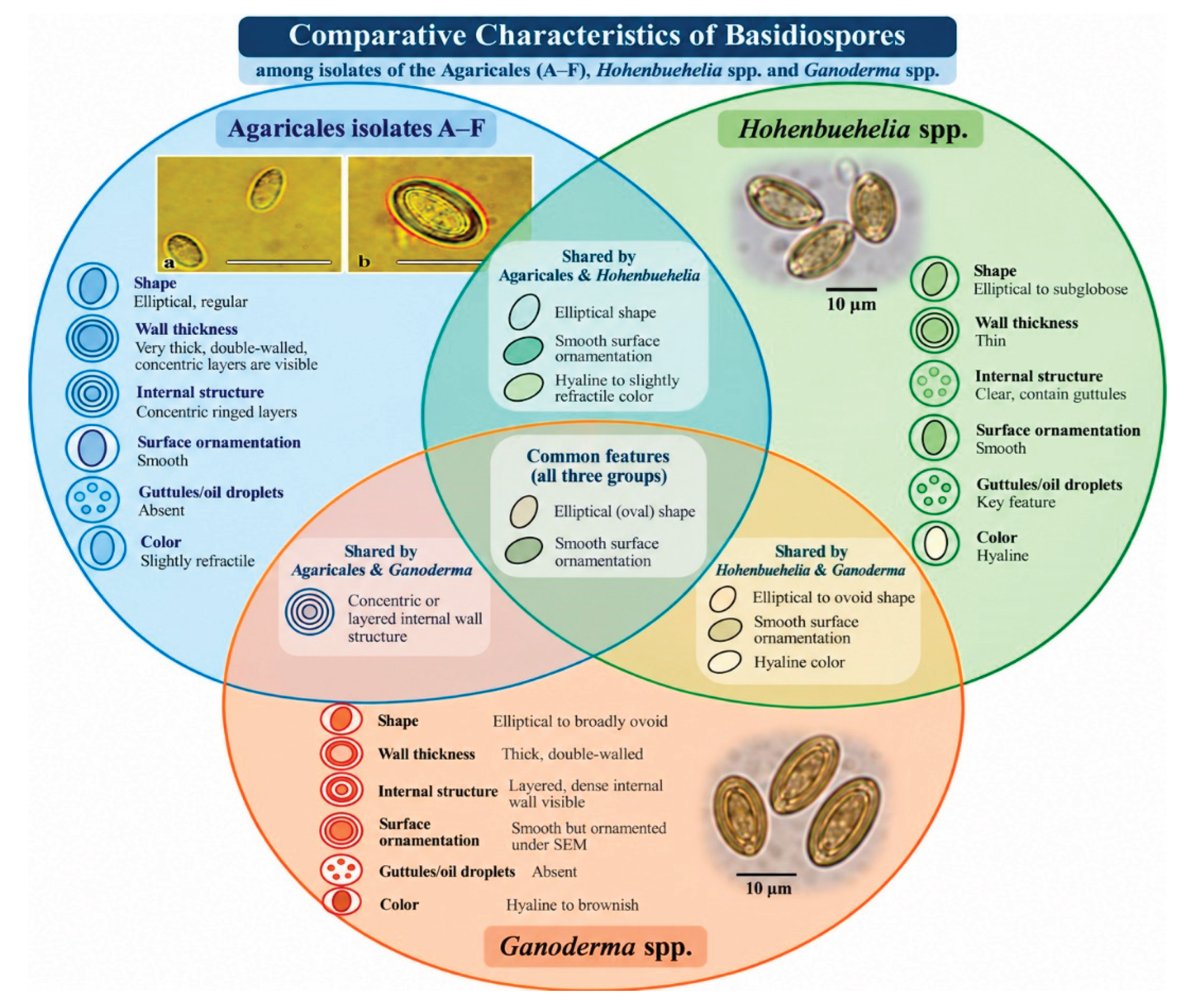
Microscopic observation showed that the basidiospores were smooth, thick-walled and with a clear internal wall structure (Figure 4a–b). Spores were ellipsoid to ovoid, sometimes subglobose, non-amyloid and non-dextrinoid, no obvious guttules observable under the microcopy used. Therefore, earlier ambiguous measurements ('2–9 × 3.4 µm') are not included as quantitative evidence, since there is no data on the original measurement of the basidiospore and calibrated image metadata to validate. The claim of concentric wall layering is also not supported as validation requires calibrated microscopy with sufficient precision.
The isolates produced hyaline, septate and smooth-walled hyphae of approximately 3–6 µm width, and no clamp connections were observed in the examined specimens (Figure 4c). Spherical, thick-walled structures (Fig. 4d) were also observed, but their identity as chlamydospores needs to be confirmed by further developmental or morphological characteristics.
3.5. Molecular Identification and Phylogenetic Analyses
The ITS1–5.8S–ITS2 sequences obtained for the isolates A–F were deposited in the NCBI GenBank under the accession numbers PV985354, PV985355, PV985356, PV985357, PV985358 and PV985359, respectively. The BLASTn analysis revealed an identity of 83–89% with the existing Hohenbuehelia reference sequences. Similarity values were described and were not used as a definitive benchmark at the genus level.
The internal branching pattern between isolates A-F shows the variability of ITS within the collected specimens. But the branch length does not tell us whether the cultures are one or more species. During the revision, a pairwise ITS identity/distance matrix could not be generated, as the original aligned sequence file was not available; therefore, not pairwise values were derived from tree branch lengths or BLAST percentages. Therefore, there is no species level inference.
The ITS positioning of isolates A–F was investigated by comparing their ITS1–5.8S–ITS2 sequences with selected Hohenbuehelia and Ganoderma references from GenBank and constructing exploratory maximum-likelihood trees in MEGA12. The outgroup was Pleurotus cystidiosus var. formosensis (MH862689). Node support was assessed using 1000 bootstrap replicates. In figures 5-7, isolates B and F were repeatedly found as a strongly supported sister pair, and A and E were also grouped together. C and D were placed in turn within the A-F cluster. These internal connections are described descriptively and are not used for defining species. The A–F assemblage was unique from the reference genera sampled in the current ITS datasets.
In the present ITS datasets, isolates A–F always formed a strongly supported monophyletic group (bootstrap 99–100 %) and did not cluster within the sampled Hohenbuehelia or Ganoderma sequences. In the combined analysis (Figure 7) the A-F clade was clearly distinct from both sampled genera and shows a topology more similar to the sampled Hohenbuehelia group than to Ganoderma. This is only documented for the current taxon sampling. It does not show that the isolates belong to Hohenbuehelia, nor does it confirm a new genus. Official taxonomic classification requires further sampling of Agaricales and multilocus data.
Figure 5.
Exploratory maximum-likelihood ITS phylogeny of isolates A–F (red circles) and sampled Hohenbuehelia reference sequences. Bootstrap support values are indicated at nodes; the scale bar represents 0.05 nucleotide substitutions per site.
Figure 5.
Exploratory maximum-likelihood ITS phylogeny of isolates A–F (red circles) and sampled Hohenbuehelia reference sequences. Bootstrap support values are indicated at nodes; the scale bar represents 0.05 nucleotide substitutions per site.
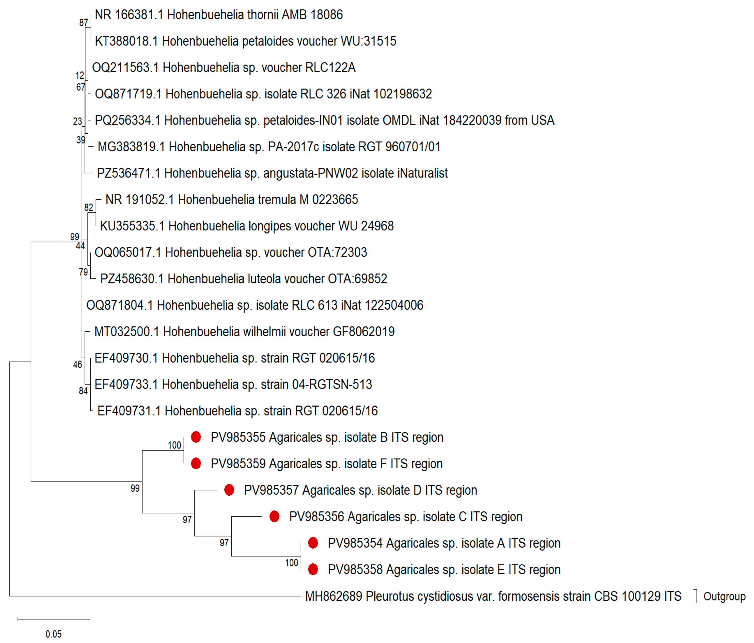
Figure 6.
Exploratory maximum-likelihood ITS phylogeny of isolates A–F (red circles) and sampled Ganoderma reference sequences. Bootstrap support values are indicated at nodes; the scale bar represents 0.05 nucleotide substitutions per site.
Figure 6.
Exploratory maximum-likelihood ITS phylogeny of isolates A–F (red circles) and sampled Ganoderma reference sequences. Bootstrap support values are indicated at nodes; the scale bar represents 0.05 nucleotide substitutions per site.
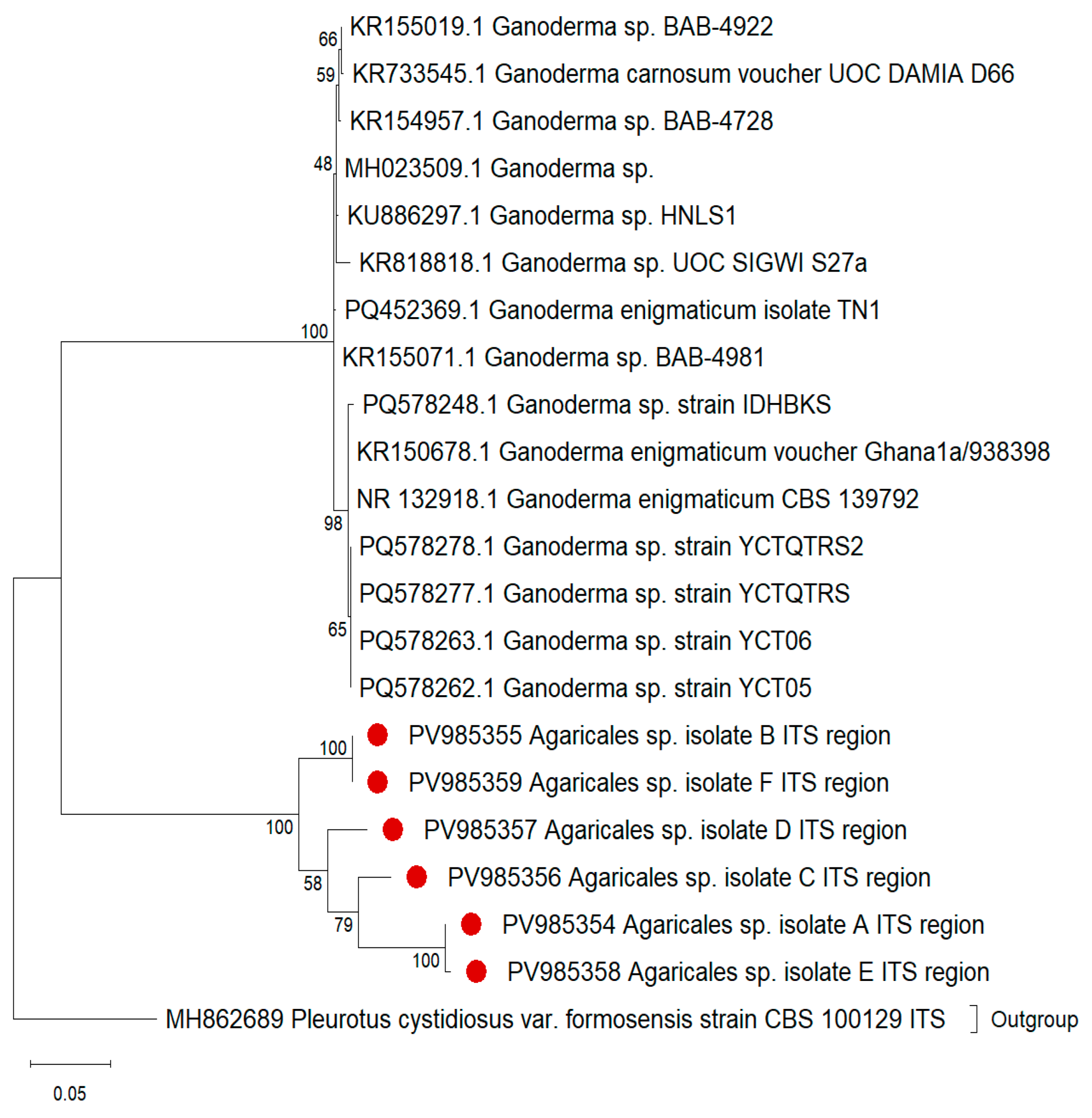
Figure 7.
Exploratory maximum-likelihood ITS phylogeny including isolates A–F (red circles) and the sampled Hohenbuehelia and Ganoderma reference sequences. Bootstrap support values are shown at nodes. The tree is used to describe placement within the sampled dataset and does not constitute formal genus-level assignment.
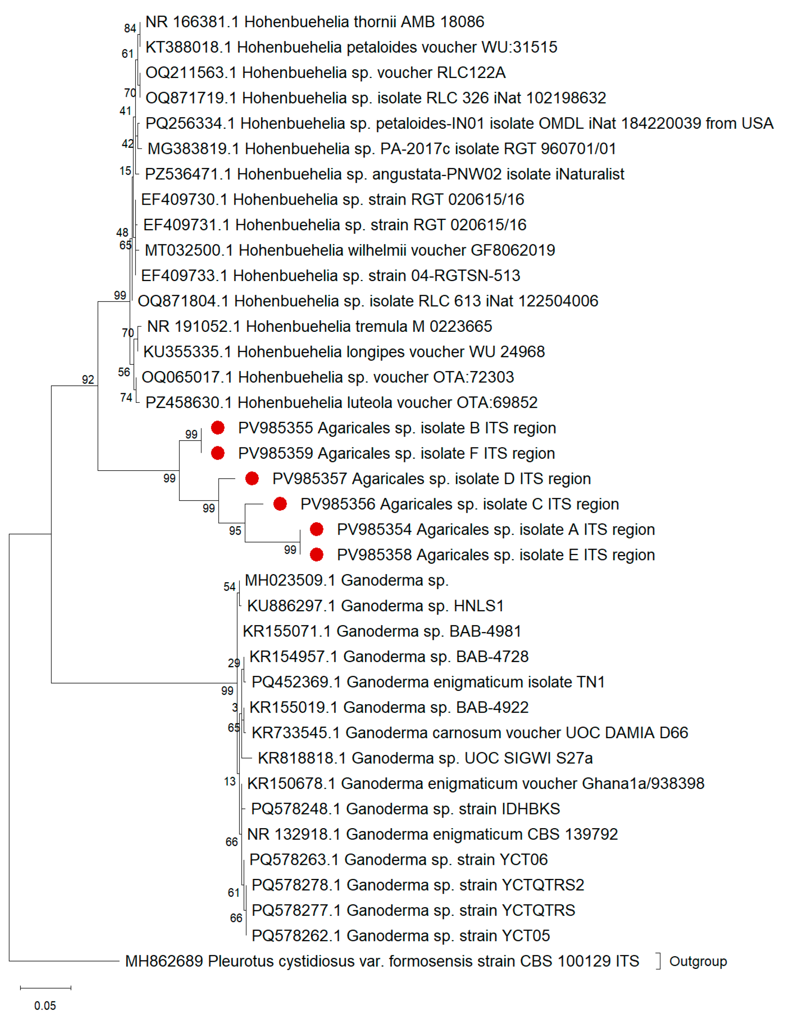
Figure 8.
Descriptive comparison of selected macromorphological features of isolates A–F with representative features of Hohenbuehelia and Ganoderma. The comparison is illustrative and does not imply formal placement of the isolates in either genus.
Figure 8.
Descriptive comparison of selected macromorphological features of isolates A–F with representative features of Hohenbuehelia and Ganoderma. The comparison is illustrative and does not imply formal placement of the isolates in either genus.
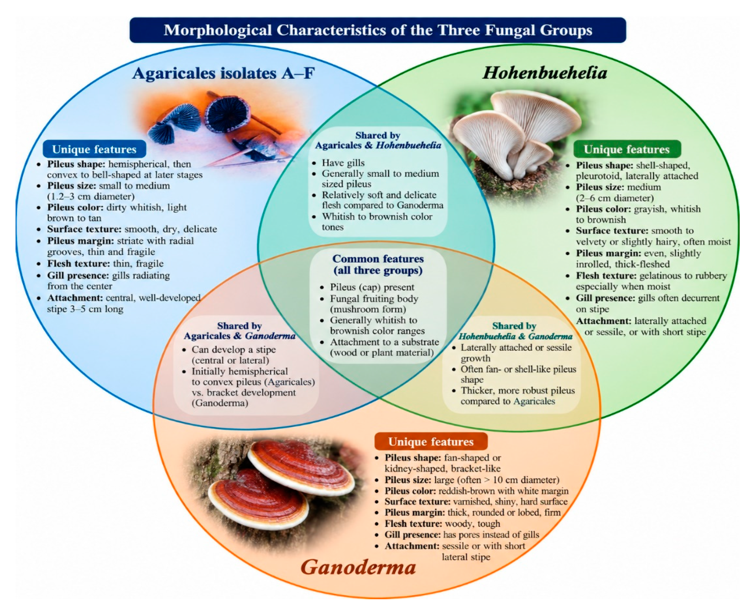
Figure 9.
Descriptive comparison of selected basidiospore features observed in isolates A–F with representative features reported for Hohenbuehelia and Ganoderma. Features requiring higher-resolution confirmation are interpreted cautiously.
Figure 9.
Descriptive comparison of selected basidiospore features observed in isolates A–F with representative features reported for Hohenbuehelia and Ganoderma. Features requiring higher-resolution confirmation are interpreted cautiously.
3.6. Artificial inoculation
In the 12-month artificial inoculation experiment, no consistent or diagnostic disease symptoms were observed at the inoculation sites. Therefore, the analysis could not confirm that the isolated fungus was the cause of observed decline symptoms in the field. The only evidence for an association with the disease at present is that the fungus was originally isolated from basidioma tissue associated with the decline of palms and not directly from the diseased internal tissue of the palms. The inoculation experiment should be well regulated according to the postulates of Koch, re-isolation and verification of the introduced fungus should be carried out.
4. Discussion
The recovered fungus collected cannot currently be reliably identified as Hohenbuehelia or Ganoderma. Its basidiomata are agaricoid with gills and a central stipe, unlike the usual pleurotoid, laterally stipe or sessile and decurrent lamellae of many Hohenbuehelia species. The specimens are basically different from Ganoderma in having lamellae instead of a poroid hymenophore. Both genera exhibit nematophagous behavior, with capturing nematodes through the production of adhesive knobs on their hyphae (the so-called predatory mode) or via germinated asexual conidia produced from denticles on their hyphae or in germinated basidiospores originating from the fruiting body or mushroom (the parasitoid mode, [35]). These results need to be compared in a broader context in Agaricales with Pleurotaceae and other agaricoid taxa with similar morphology, rather than restricting the analysis to Hohenbuehelia and Ganoderma only.
The observed symptoms on date palm were consistent with a preliminary diagnosis of Ganoderma zonatum as the causal agent [36,37]. In addition, the basidiospores illustrated in Figure 4a and Figure 4b closely resemble the characteristics of Ganoderma species, particularly the thick, double- or multilayered wall and the internal structure, as reported by Castillo et al. [8]. Some features of the spore wall observed were superficially similar to Ganoderma, but this resemblance should not obscure the significant differences in the basidioma. Ganoderma species are polyporous fungi with poroid hymenophore and can produce pileate, sessile, laterally stipitate or in some taxa, particularly stipitate basidiomata, thus the genus cannot be defined based on the absence of a distinct pileus and stipe. Conversely, the stipe, formed centrally and attached lamellae narrowly, are not the typical pleurotoid structure of Hohenbuehelia. The integrated character set enables the study of the specimens as an unclassified agaricoid fungus, pending further extensive morphological and molecular analysis.
The ITS indicated isolates A–F formed a well-supported monophyletic group (bootstrap 99–100%) that was distinct from the sampled sequences of Ganoderma or Hohenbuehelia. The topology of the A–F clade in the combined phylogenetic tree was closer to the sampled Hohenbuehelia clade, but still distinct. This topology should not be considered support for a genus level placement, given the single locus analysis and limited taxon sampling. The three exploratory trees were retained to show the behavior of the A–F grouping across different reference sets, but the combined tree is the core comparative analysis, and no definitive taxonomic conclusions are drawn from the individual trees.
The internal branching pattern of isolates A-F shows ITS variation in the collected specimens; however, branch lengths alone are not able to determine whether the cultures represent one or multiple species. During the revision, a pairwise ITS identity/distance matrix could not be generated, as the original aligned sequence file was not available; therefore, no pairwise values were derived from tree branch lengths or BLAST percentages. This avoids species level inference.
The BLASTn identity scores of 83–89% with existing Hohenbuehelia references indicate substantial divergence from the sequences currently available in the database. In this context, ITS sequence similarity above 90% has been suggested to support genus-level affiliation, whereas values below 90% may indicate potential novelty or a more distant relationship [38,39]. Thus, the observed 83–89% similarity provides an indication of a relatively distant relationship between the present isolates and the sampled Hohenbuehelia sequences. However, this interpretation should be made cautiously because Hohenbuehelia is itself a morphologically and ecologically diverse genus [17], and no universal ITS similarity threshold can be applied to delimit fungal genera. Moreover, ITS sequence identity alone is insufficient to justify the establishment of a new genus, as the observed divergence may also result from incomplete taxon sampling, sequence quality, database errors, or alignment limitations. Therefore, the BLAST results should be regarded as preliminary evidence of divergence from the sampled references rather than definitive evidence of taxonomic novelty. A reliable taxonomic assessment requires integration of morphological characteristics with broader taxon sampling and multilocus phylogenetic analyses. In particular, improved sampling of Agaricales, including sequences from relevant type or ex-type specimens, together with additional loci such as LSU, tef1-α, and rpb2, will be necessary before any formal taxonomic classification is proposed.
The radius of the colonies was significantly different between isolates (Table 1). B and F showed a larger mean colony radius than A, C, D and E under the conditions tested. Some growth clusters resemble features of the ITS topology, but no formal correlation of colony-growth phenotypes with ITS subclades has been investigated. Growth data should therefore not be taken as evidence of common genetic or physiological features of phylogenetic subgroups.
Artificial inoculation experiments did not induce consistent disease symptoms over a period of 12 months. This unfavorable result, as well as the removal of basidiomata rather than identification of the fungus in the affected internal tissue of the palm, indicates that the existence of a causal relationship in the decline of date palms cannot be established at present. The fungus may be a saprotroph, a secondary colonizer or some other associate of stressed palms; its pathogenicity is a hypothesis that needs to be proven and will require properly replicated controls, documentation of environmental conditions, re-isolation, molecular confirmation and perhaps longer experiments. Symptoms alone are insufficient to determine the cause, since different fungi may be involved in similar decline syndromes and cryptic or opportunistic taxa may be present in stressed plant tissues. Accurate identification of fungi involved in the deterioration of plants requires the integration of morphological characteristics, sequence-based information, appropriate taxon sampling and, when causation is implied, rigorously controlled pathogenicity tests. Consequently, the present work should be regarded as an initial description of an unclassified agaricoid fungus associated with declining date palms and not as evidence of a new taxon or a pathogen.
5. Conclusions
The present study reports an unclassified agaricoid fungus associated with date palm (Phoenix dactylifera L.) decline in Central Sudan. The isolates showed some morphological similarities with the taxa evaluated in the preliminary identification stage, but the centrally formed stipe free to narrowly adnate lamellae and other mentioned features do not support a definitive classification in Hohenbuehelia. ITS analyzes consistently placed isolates A–F in a strongly supported clade apart from the sampled Hohenbuehelia and Ganoderma sequences. However, limited taxon sampling and lack of a multilocus framework, as well as the single-locus ITS dataset, do not support recognition of a new genus or any formal taxon. The ITS identity of 83–89% with existing Hohenbuehelia references also shows divergence from the sampled sequences but is not used as a criterion for genus delimitation. The 12-months of artificial inoculation did not result in uniform symptoms of disease, and the fungus was isolated from associated basidiomata rather than identified in the affected internal palm tissue. The cause of palm decline was not established. The taxonomical placement and ecological role of these isolates needs to be resolved by greater sampling of the Agaricales, inclusion of type or ex-type reference sequences where available, multilocus analyzes involving LSU, tef1-α and rpb2, improved morphological documentation and strictly controlled inoculation and re-isolation experiments.
Author Contributions
Conceptualization, ASHA and MAES.; methodology, MAES and ASHA.; software, EAHM and MAES.; validation, KP., MAES., and ASHA.; formal analysis, EAHM. and MAES; investigation, AADA.; resources, AADA.; data curation, EAHM and AADA.; writing—original draft preparation, AADA and EAHM.; writing—review and editing, KP, EAHM, MAES, and ASHA; visualization, ASHA and MAES; supervision, ASHA.; project administration, ASHA and MAES. All authors have read and agreed to the published version of the manuscript.
Funding
This research received no external funding.
Data Availability Statement
All data supporting the findings of this study are available within the article.
Conflicts of Interest
The authors declare no conflicts of interest.
References
- Elshibli, S.; Korpelainen, H. Microsatellite Markers Reveal High Genetic Diversity in Date Palm (Phoenix Dactylifera L.) Germplasm from Sudan. Genetica 2008, 134, 251–260. [Google Scholar] [CrossRef] [PubMed]
- Mohammed, N.M.I.; Said, A.G.E. Date Palm (Phoenix Dactylifera) Seeds Germination. Cell Biol. Dev. 2018, 2. [Google Scholar] [CrossRef]
- Ziedan, E.-S.H.E.; Hashem, M.; Mostafa, Y.S.; Alamri, S. Management of Deleterious Effect of Fusarium Oxysporum Associated with Red Palm Weevil Infestation of Date Palm Trees. Agriculture 2022, 12, 71. [Google Scholar] [CrossRef]
- Alwahshi, K.J.; Saeed, E.E.; Sham, A.; Alblooshi, A.A.; Alblooshi, M.M.; El-Tarabily, K.A.; AbuQamar, S.F. Molecular Identification and Disease Management of Date Palm Sudden Decline Syndrome in the United Arab Emirates. IJMS 2019, 20, 923. [Google Scholar] [CrossRef] [PubMed]
- Mohammed, E.A.H.; Pál, K.; Abbo, A.S.H. Octanoic Fatty Acid Significantly Impacts the Growth of Foodborne Pathogens and Quality of Mabroom Date Fruits (Phoenix Dactylifera L.). In Proceedings of the IECHo 2025; MDPI, July 24 2025; p. 2. [Google Scholar]
- Alhudaib, K.A.; El-Ganainy, S.M.; Almaghasla, M.I.; Sattar, M.N. Characterization and Control of Thielaviopsis Punctulata on Date Palm in Saudi Arabia. Plants 2022, 11, 250. [Google Scholar] [CrossRef] [PubMed]
- Abhary, M.K.; Al-Baity, O.A. Occurrence and Distribution of Date Palm Phytoplasma Disease in Al-Madinah Region, KSA. J. Taibah Univ. Sci. 2018, 12, 266–272. [Google Scholar] [CrossRef]
- Castillo, S.Y.; Rodríguez, M.C.; González, L.F.; Zúñiga, L.F.; Mestizo, Y.A.; Medina, H.C.; Montoya, C.; Morales, A.; Romero, H.M.; Sarria, G.A. Ganoderma Zonatum Is the Causal Agent of Basal Stem Rot in Oil Palm in Colombia. JoF 2022, 8, 230. [Google Scholar] [CrossRef] [PubMed]
- Elkhateeb, W. The Precious Ganoderma Mushroom and Plant Diseases. PDD 2022, 1, 01–05. [Google Scholar] [CrossRef] [PubMed]
- Downer, A.J.; Uchida, J.Y.; Hodel, D.R.; Elliott, M.L. Lethal Palm Diseases Common in the United States. hortte 2009, 19, 710–716. [Google Scholar] [CrossRef]
- Adaskaveg, J.E.; Blanchette, R.A.; Gilbertson, R.L. Decay of Date Palm Wood by White-Rot and Brown-Rot Fungi. Can. J. Bot. 1991, 69, 615–629. [Google Scholar] [CrossRef]
- Elhassan, A.M.; Mahir, M.; Hamad, A.; Kh.Ali, A.A.M.E. Occurrence of Fungal Diseases and Their Importance on Date Palm in Sudan. Int.J.Curr.Microbiol.App.Sci 2017, 6, 16–22. [Google Scholar] [CrossRef]
- He, M.-Q.; Zhao, R.-L. Outline of Basidiomycota. In Encyclopedia of Mycology; Elsevier, 2021; pp. 310–319. ISBN 978-0-323-85180-0. [Google Scholar]
- McLaughlin, D.J.; Spatafora, J.W. (Eds.) Systematics and Evolution: Part A; Springer Berlin Heidelberg: Berlin, Heidelberg, 2014; ISBN 978-3-642-55317-2. [Google Scholar]
- Vizzini, A.; Alvarado, P.; Consiglio, G.; Marchetti, M.; Xu, J. Family Matters inside the Order Agaricales: Systematic Reorganization and Classification of Incertae Sedis Clitocyboid, Pleurotoid and Tricholomatoid Taxa Based on an Updated 6-Gene Phylogeny. Stud. Mycol. 2024, 107, 67–148. [Google Scholar] [CrossRef] [PubMed]
- Wang, G.-S.; Cai, Q.; Hao, Y.-J.; Bau, T.; Chen, Z.-H.; Li, M.-X.; David, N.; Kraisitudomsook, N.; Yang, Z.-L. Phylogenetic and Taxonomic Updates of Agaricales, with an Emphasis on Tricholomopsis. Mycology 2024, 15, 180–209. [Google Scholar] [CrossRef] [PubMed]
- Thorn, R.G.; Moncalvo, J.-M.; Reddy, C.A.; Vilgalys, R. Phylogenetic Analyses and the Distribution of Nematophagy Support a Monophyletic Pleurotaceae within the Polyphyletic Pleurotoid-Lentinoid Fungi. Mycologia 2000, 92, 241–252. [Google Scholar] [CrossRef]
- Xu, J.; Jiang, Y.; Wang, T.; Zhang, D.; Li, X.; Hosen, Md.I. Morphological Characteristics and Phylogenetic Analyses Revealed Four New Species of Agaricales from China. Front. Microbiol. 2023, 14, 1118525. [Google Scholar] [CrossRef] [PubMed]
- Læssøe, T.; Petersen, J. Fungi of Temperate Europe; Princeton University Press, 2019; Vol. 1, ISBN 978-0-691-18037-3. [Google Scholar]
- Phonemany, M.; Vadthanarat, S.; Raghoonundon, B.; Thongklang, N.; Raspé, O. Additions to Hohenbuehelia (Basidiomycota, Pleurotaceae): two new species and notes on H. tristis from northern Thailand. Mycokeys 2023, 99, 109–130. [Google Scholar] [CrossRef] [PubMed]
- Holec, J.; Zehnálek, P. Taxonomy of Hohenbuehelia Auriscalpium, H. Abietina, H. Josserandii, and One Record of H. Tremula. Czech. Mycol. 2020, 72, 199–220. [Google Scholar] [CrossRef]
- Vlasenko, V.A.; Turmunkh, D.; Vlasenko, A.V. New Combination in the Genus Hohenbuehelia (Pleurotaceae). Phytotaxa 2022, 555. [Google Scholar] [CrossRef]
- Thorn, R.G.; Barron, G.L. Carnivorous Mushrooms. Science 1984, 224, 76–78. [Google Scholar] [CrossRef] [PubMed]
- Shipley, S.M.; Barr, A.L.; Graf, S.J.; Collins, R.P.; McCloud, T.G.; Newman, D.J. Development of a Process for the Production of the Anticancer Lead Compound Pleurotin by Fermentation of Hohenbuehelia Atrocaerulea. J. IND MICROBIOL. BIOTECHNOL. 2006, 33, 463–468. [Google Scholar] [CrossRef] [PubMed]
- Bohni, N.; Schumpp, O.; Schnee, S.; Bertrand, S.; Gindro, K.; Wolfender, J. Targeted Isolation of Induced and Bioactive Metabolites from Fungal Co-Cultures. Planta Med. 2013, 79, s-0033-1351867. [Google Scholar] [CrossRef]
- Sandargo, B.; Thongbai, B.; Stadler, M.; Surup, F. Cysteine-Derived Pleurotin Congeners from the Nematode-Trapping Basidiomycete Hohenbuehelia Grisea. J. Nat. Prod. 2018, 81, 286–291. [Google Scholar] [CrossRef] [PubMed]
- Sandargo, B.; Thongbai, B.; Praditya, D.; Steinmann, E.; Stadler, M.; Surup, F. Antiviral 4-Hydroxypleurogrisein and Antimicrobial Pleurotin Derivatives from Cultures of the Nematophagous Basidiomycete Hohenbuehelia Grisea. Molecules 2018, 23, 2697. [Google Scholar] [CrossRef] [PubMed]
- Barron, G.L. Predatory Fungi, Wood Decay, and the Carbon Cycle. Biodiversity 2003, 4, 3–9. [Google Scholar] [CrossRef]
- Thorn, R.G.; Barron, G.L. Nematoctonus and the Tribe Resupinatae in Ontario, Canada. Mycotaxon 1986, 25, 321–453. [Google Scholar] [CrossRef]
- Rayner, R.W. A Mycological Colour Chart; Commonwealth Mycological Institute and British Mycological Society: Kew, UK, 1970. [Google Scholar]
- Teixeira, L.P.; Soares, T.d.P.F.; Oliveira, L.S.S.; Mathioni, S.M.; Ferreira, M.A. First Report of Leaf Spot Caused by Alternaria argyroxiphii on African Mahogany Trees (Khaya senegalensis). Phytopathol. Mediterr. 2017, 56, 502–510. [Google Scholar] [CrossRef]
- Phonemany, M.; Vadthanarat, S.; Raghoonundon, B.; Thongklang, N.; Raspé, O. Additions to Hohenbuehelia (Basidiomycota, Pleurotaceae): Two New Species and Notes on H. Tristis from Northern Thailand. MC 2023, 99, 109–130. [Google Scholar] [CrossRef] [PubMed]
- Stewart, C.N.; Via, L.E. A Rapid CTAB DNA Isolation Technique Useful for RAPD Fingerprinting and Other PCR Applications. BioTechniques 1993, 14, 748–750. [Google Scholar] [PubMed]
- White, T.J.; Bruns, T.; Lee, S.; Taylor, J. AMPLIFICATION AND DIRECT SEQUENCING OF FUNGAL RIBOSOMAL RNA GENES FOR PHYLOGENETICS. In PCR Protocols; Elsevier, 1990; pp. 315–322. ISBN 978-0-12-372180-8. [Google Scholar]
- Barron, G.L. The Nematode-Destroying Fungi; Canadian Biological Publications: Guelph, Canada, 1977; ISBN 978-0-920370-00-1. [Google Scholar]
- Santos, P.; Hodel, D. Ganoderma Basal Stem Rot on Palms in California. PALMARBOR 2025, 2, 1–26. [Google Scholar]
- Dhillon, B.; Chakrabarti, S. Ganoderma Zonatum Causes Butt Rot of Areca ( Dypsis Lutescens ) and Robellini ( Phoenix Roebelenii ) Palms in Florida. Plant Dis. 2023, 107, 2844. [Google Scholar] [CrossRef]
- Vu, D.; Groenewald, M.; De Vries, M.; Gehrmann, T.; Stielow, B.; Eberhardt, U.; Al-Hatmi, A.; Groenewald, J.Z.; Cardinali, G.; Houbraken, J.; et al. Large-Scale Generation and Analysis of Filamentous Fungal DNA Barcodes Boosts Coverage for Kingdom Fungi and Reveals Thresholds for Fungal Species and Higher Taxon Delimitation. Stud. Mycol. 2019, 92, 135–154. [Google Scholar] [CrossRef] [PubMed]
- Schoch, C.L.; Seifert, K.A.; Huhndorf, S.; Robert, V.; Spouge, J.L.; Levesque, C.A.; Chen, W. Fungal Barcoding Consortium; Fungal Barcoding Consortium Author List Nuclear Ribosomal Internal Transcribed Spacer (ITS) Region as a Universal DNA Barcode Marker for Fungi. In Proc. Natl. Acad. Sci. U.S.A.; Bolchacova, E., et al., Eds.; 2012; Volume 109, pp. 6241–6246. [Google Scholar] [CrossRef] [PubMed]
Figure 1.
Decline symptoms observed on date palm (Phoenix dactylifera) trees in the surveyed orchard: (a) browning and drying of fronds; (b) symptoms affecting one side of the crown; (c) downward drooping and desiccation of fronds.
Figure 1.
Decline symptoms observed on date palm (Phoenix dactylifera) trees in the surveyed orchard: (a) browning and drying of fronds; (b) symptoms affecting one side of the crown; (c) downward drooping and desiccation of fronds.
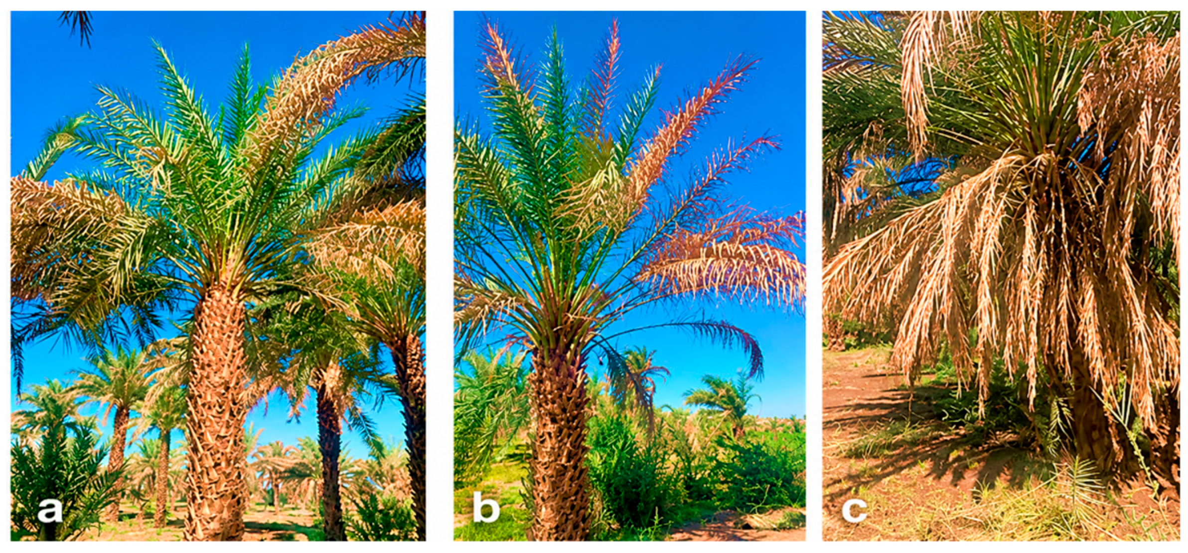
Figure 2.
An epigeous, gilled mushroom with convex to bell-shaped or dome-like, dry whitish pilei, showing moderately spaced lamellae which are radiated symmetrically from the stipe.
Figure 2.
An epigeous, gilled mushroom with convex to bell-shaped or dome-like, dry whitish pilei, showing moderately spaced lamellae which are radiated symmetrically from the stipe.
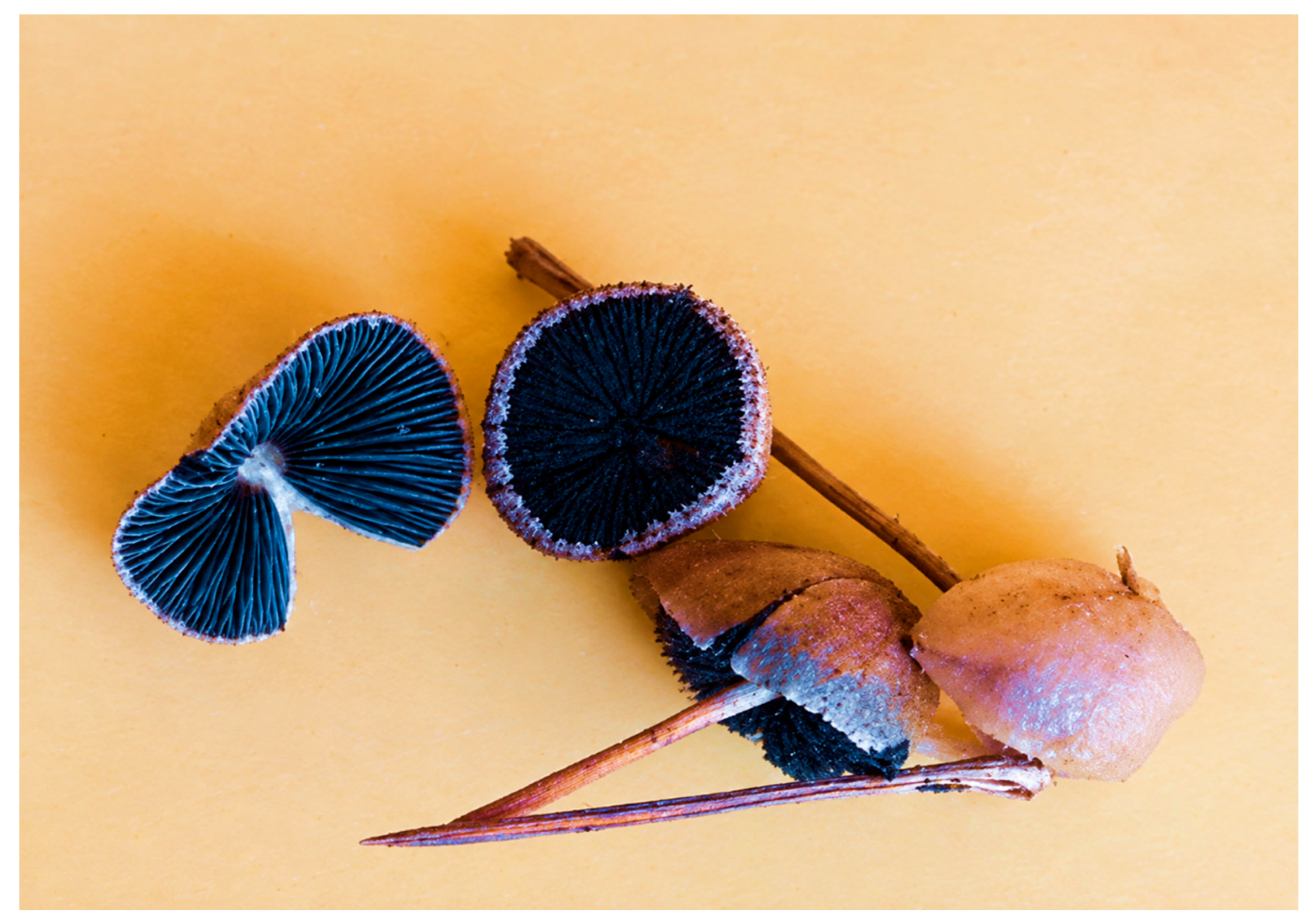
Figure 3.
Cultural characteristics of fungal isolates on potato dextrose agar (PDA): (a) isolate E, (b) isolate C, and (c) isolate F, showing white, fluffy aerial mycelium and reddish-brown pigmentation.
Figure 3.
Cultural characteristics of fungal isolates on potato dextrose agar (PDA): (a) isolate E, (b) isolate C, and (c) isolate F, showing white, fluffy aerial mycelium and reddish-brown pigmentation.
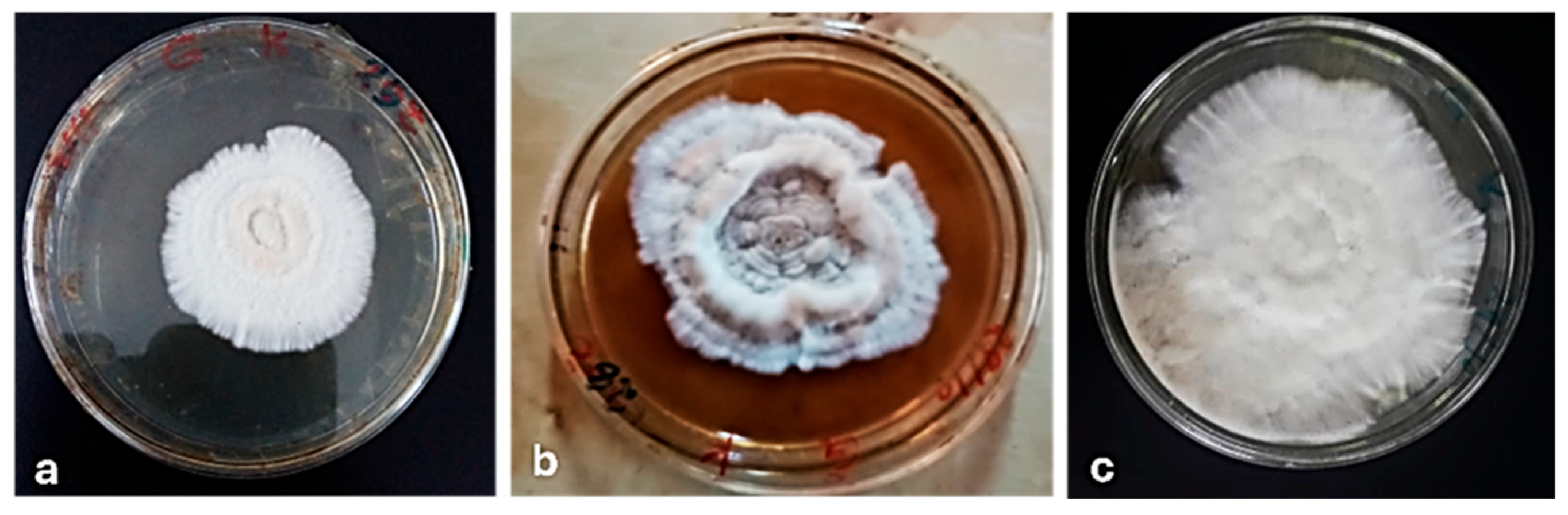
Figure 4.
Microscopic features of the unidentified agaricoid fungus. (a–b) Ellipsoid to ovoid, occasionally subglobose basidiospores; (c) hyaline septate hyphae; (d) rounded thick-walled structures requiring further characterization. Scale bars should be interpreted only where calibration from the original microscope system is available; no scale was estimated retrospectively from pixel dimensions.
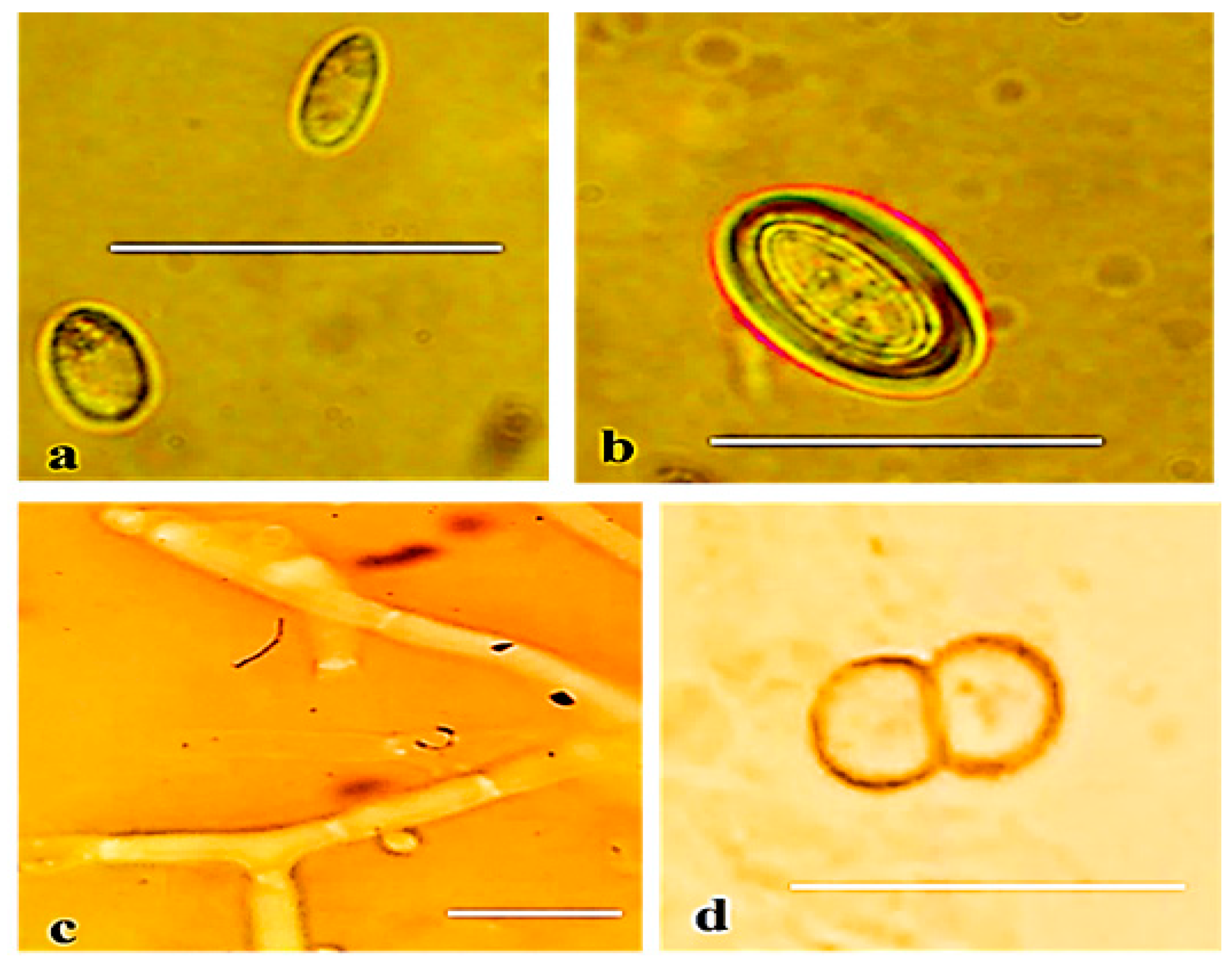

Table 1.
Mean colony radius (cm) of fungal isolates A–F cultured on potato dextrose agar (PDA) after three weeks of incubation. Values represent mean ± SD (n = 4). Different lowercase letters indicate significant differences according to Fisher’s LSD test at 0.05.
Table 1.
Mean colony radius (cm) of fungal isolates A–F cultured on potato dextrose agar (PDA) after three weeks of incubation. Values represent mean ± SD (n = 4). Different lowercase letters indicate significant differences according to Fisher’s LSD test at 0.05.
| No. | Isolates | Colony radius (cm) | n |
| 1 | Isolate A | 2.23 ± 0.38 b | 4 |
| 2 | Isolate B | 3.40 ± 0.29 a | 4 |
| 3 | Isolate C | 2.05 ± 0.42 b | 4 |
| 4 | Isolate D | 2.15 ± 0.42 b | 4 |
| 5 | Isolate E | 1.90 ± 0.28 b | 4 |
| 6 | Isolate F | 3.33 ± 0.43 a | 4 |
| ANOVA | F(5,18) = 12.786 | p = 0.000021 |
Values represent mean ± SD (n = 4). Different lowercase letters indicate significant differences among isolates according to Fisher’s least significant difference (LSD) test at = 0.05.
Disclaimer/Publisher’s Note: The statements, opinions and data contained in all publications are solely those of the individual author(s) and contributor(s) and not of MDPI and/or the editor(s). MDPI and/or the editor(s) disclaim responsibility for any injury to people or property resulting from any ideas, methods, instructions or products referred to in the content. |
© 2026 by the authors. Licensee MDPI, Basel, Switzerland. This article is an open access article distributed under the terms and conditions of the Creative Commons Attribution (CC BY) license (http://creativecommons.org/licenses/by/4.0/).
Copyright: This open access article is published under a Creative Commons CC BY 4.0 license, which permit the free download, distribution, and reuse, provided that the author and preprint are cited in any reuse.