

Submitted:
07 October 2026
Posted:
09 October 2026
You are already at the latest version
Abstract
Sex hormones shape reproductive tract immunity and influence host defense against viral infection. However, much of our understanding of hormone-mediated immunity derives from cisgender populations and systemic studies. Comparatively less is known about the effects of sustained hormonal change within reproductive tissues. This gap is particularly relevant to transgender men (TGM) taking testosterone-based gender-affirming hormone therapy (GAHT) and to menopausal women using menopause hormone therapy (MHT). Although biologically distinct, these populations provide complementary contexts for examining how sustained androgen exposure, ovarian hormone decline, and therapeutic hormone use influence reproductive tract immunity. In this review, we examine hormone-associated changes in reproductive tract epithelial, stromal, and immune cells including the reproductive tract microbiome and virome. We consider their implications for the acquisition, replication, persistence and pathogenesis of HIV-1, HPV, HSV and CMV. Understanding these tissue specific, hormone-immune-virus interactions may help identify gaps in viral prevention, screening, vaccination, and treatment efforts across diverse hormonal and reproductive contexts.
Keywords:
gender affirming hormone therapy (GAHT)
; menopause hormone therapy (MHT)
; sex-hormones
; menopause
; estrogen
; progesterone
; testosterone
; transgender men
; female reproductive tract (FRT)
1. Introduction
Sex hormones, including estrogens, progesterone, and androgens, substantially contribute to immune development, homeostasis, and function [1,2,3]. Through receptor-mediated signaling in immune, epithelial, stromal, and other non-immune cells, these hormones regulate innate and adaptive immune cell development and function [1,2,4]. Their effects vary with hormone concentration, receptor expression, tissue microenvironment, age, reproductive stage, and the duration and route of exposure [3,5,6,7,8]. Physiological endocrine changes and exogenous hormone administration can therefore generate distinct immune states with consequences for infection, vaccination, autoimmunity, and inflammatory disease [3,7,9,10,11]. Much of the current understanding of hormone-mediated immunity derives from cisgender populations [4,12]. Studies of endogenous testosterone variation, aging, hypogonadism, and testosterone replacement therapy in cisgender men provide evidence regarding androgen-mediated immune regulation [10,13,14,15]. Likewise, menstrual cycling, pregnancy, hormonal contraception, gynecological and endocrine conditions, and reproductive aging illustrate how hormonal fluctuations influence systemic and tissue specific immunity in cisgender women [8,16,17,18].Collectively, these hormonal contexts illustrate substantial endocrine heterogeneity related to sex and gender. Differences in age, reproductive stage, underlying conditions, and exogenous hormone use further contribute towards this heterogeneity [14,19,20,21]. Moreover, differences in baseline physiology, anatomy, receptor expression, tissue microenvironment, and duration of exposure limit the extent to which findings from one hormonal context can be extrapolated to another.
Amidst the broader context of this evidence base, transgender men (TGM) taking testosterone-based gender affirming hormone therapy (GAHT), and menopausal women who do or do not receive menopausal hormone therapy (MHT), represent distinct populations experiencing sustained hormonal change within reproductive tissues. Currently, the immunologic consequences of GAHT for TGM, and of MHT in post-menopausal women remain incompletely understood. Testosterone-based GAHT introduces sustained elevations in androgen exposure, generally suppresses hypothalamic-pituitary-ovarian activity, and reduces circulating estradiol concentrations [22,23]. Notably, ovarian suppression may be incomplete in some individuals [24]. The menopausal transition is characterized by progressive loss of ovarian follicular function and increasingly variable sex-steroid production, culminating in sustained reductions in circulating estradiol and progesterone following the final menstrual period [19,25,26,27]. MHT further modifies the postmenopausal hormonal environment, with effects dependent on formulation, dose and route of administration [28]. Systemic estrogen formulations increase circulating estrogen exposure, while low-dose vaginal estrogen primarily targets local genitourinary tissues with comparatively limited systemic absorption [29].
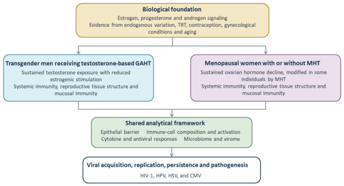
While the hormonal contexts of these populations are not biologically equivalent, biological data from one can complement our understanding of the other. Understanding the immunological consequences of GAHT, menopause and MHT in the reproductive tract requires direct examination of the respective tissues. Reproductive tissues contain specialized epithelial, stromal, microbial, and tissue-resident immune compartments with site-specific phenotypes and functions [30,31]. Hormone-mediated changes in epithelial barrier function, mucosal immunity, the microbiome, and viral target-cell availability may influence viral acquisition, replication, persistence, and local immune control [30,32,33] This review therefore examines how hormonal changes and exogenous hormone exposure shape reproductive tract epithelial structure and immunity in TGM taking testosterone-based GAHT and menopausal women +/- MHT. Presenting evidence from cisgender populations and experimental models as mechanistic context, we consider implications for risk/susceptibility to infection and pathogenesis by HIV-1, human papillomavirus (HPV), herpes simplex virus (HSV), and cytomegalovirus (CMV); in the process we distinguish population- and tissue-specific evidence from extrapolation across hormonal contexts. (Fig. 1).
2. Sex Hormones as Regulators of Reproductive Tract Immunity
Hormonal regulation of the female reproductive tract (FRT) occurs across anatomically and functionally distinct mucosal compartments [30,33]. The upper FRT, including the endometrium and endocervix, is predominantly lined by a single layer of columnar epithelium [30]. In contrast, the lower FRT comprising the ectocervix and vagina, is lined by a multilayered, non-keratinized stratified squamous epithelium that forms a physical barrier against pathogen entry [34,35]. These compartments also differ in mucus production, antimicrobial activity, microbiome and immune cell phenotypes [30,34,36]. Consequently, hormonal effects on FRT mucosal immunity are largely site and cell specific. In epithelial and stromal cells, hormone signaling can modify barrier function by regulating epithelial differentiation and junctional proteins, while also altering antimicrobial production and cytokine/chemokine secretion [37,38,39,40,41]. In immune cells, hormonal signaling can similarly regulate recruitment, localization, abundance, activation, and effector function [42,43]. These responses are further shaped by local steroid metabolism and receptor expression, such that circulating hormone concentrations may not directly reflect hormone activity within reproductive tissues [44,45]. Together, these mechanisms provide the basis through which changes in the hormonal environment can alter reproductive tract homeostasis and susceptibility or resistance to infection.
2.1. Estrogen
Estrogens, particularly 17β-estradiol (E2), regulate reproductive tract epithelium and mucosal immunity predominantly through estrogen receptor α (ERα) and estrogen receptor β (ERβ), with additional signaling mediated by the G-protein coupled estrogen receptor (GPER) [30,46]. ERα and ERβ are expressed by epithelial, stromal, and immune cells throughout the FRT [30,47]. E2 can therefore regulate mucosal immunity both directly through receptor mediated signaling and indirectly through paracrine interactions between tissue resident cells [48]. Notably, the resulting effects are dependent on anatomical site, hormone concentration, and cellular context.
E2 has prominent, but site-specific effects on reproductive epithelial structure and barrier function [37,49]. In the lower FRT, estrogen promotes maturation of the stratified squamous epithelium and increases epithelial glycogen accumulation [50,51]. Glycogen derived substrates can in turn support Lactobacillus dominant microbial communities and lactic acid production, linking estrogen dependent epithelial metabolism to the vaginal microbial environment [52,53,54]. In contrast, E2 has also been shown to increase the permeability of cervical epithelial barriers in ex vivo models. In cultured human cervical epithelial cells, E2 reduced transepithelial resistance and increased paracellular permeability in a time and dose responsive manner [37]. Subsequent studies further implicated estrogen dependent regulation of tight junction proteins in this reponse [49]. E2 also directly regulates epithelial innate immune function [38,41]. In ex vivo polarized primary human uterine epithelial cells, E2 increased secretion of secretory leukocyte protease inhibitor (SLPI) ~4-fold and increased human β-defensin-2 (HBD2) mRNA ~5-fold [38]. In the same model, E2 suppressed inflammatory responses to innate immune stimulation wherein LPS and poly(I:C) induced secretion of pro-inflammatory mediators, IL-6, IL-8, and macrophage migration inhibitory factor (MIF) was inhibited. Additionally, E2 also reduced IL-1β induced TNF-α, IL-8, and NF-κB mRNA expression by ~85%, 95%, and 70%, respectively [38]. Notably, these effects were site-specific, wherein E2 enhanced HBD2 and SLPI in uterine epithelial cells, while reducing HBD2 and elafin secretion by primary vaginal epithelial cells [38,41].
Hormonal regulation of reproductive immunity also extends to antiviral pathways. Interferon epsilon (IFNε), a constitutively expressed type I interferon (IFN-I) that is enriched in reproductive tract epithelium, contributes to basal mucosal antiviral defense [55]. IFNε expression varies with hormonal state, although its regulation differs between experimental models and reproductive tract compartments. In mice, estrogen induces reproductive tract IFNε expression, whereas studies of primary human uterine epithelial cells indicate that progesterone suppresses IFNε expression without a detected direct effect of E2 [55], [56]. In human reproductive tract epithelium, IFNε induces interferon stimulated genes (ISGs), including MX1, OAS2 and CXCL10 [56]. E2 can also modify other interferon-mediated antiviral pathways. In primary human uterine epithelial cells, E2 did not alter poly(I:C)-induced IFNβ or downstream MxA, OAS2, and PKR expression [57]. In contrast, E2 suppressed IFNλ1 induced expression of MxA, OAS2, and ISG15 through an ERα-dependent mechanism. Thus, estrogenic regulation of epithelial antiviral immunity appears to depend on the interferon pathway and cellular context which may not reflect uniform enhancement or suppression of antiviral responses. Together with estrogen-dependent regulation of antimicrobial peptides, protease inhibitors, cytokines, and chemokines, these pathways illustrate the active role of reproductive epithelial cells in mucosal immune defense.
E2 additionally regulates the endometrial chemokine expression relevant to immune cell recruitment. In human endometrial organoid cultures, E2 increased CXCL10 and CXCL11 mRNA expression by ~ 8.5 and 7.7-fold, respectively [42]. The estrogen receptor antagonist, ICI182780, prevented this induction [42]. These chemokines bind the CXCR3 receptor expressed by uterine and circulatory NK cells, which suggests that local E2 signaling could promote recruitment of CXCR3+ NK cells into the endometrium [42]. E2 can also directly modify lymphocyte effector function. In human endometrial CD8+ T cells, E2 suppressed cytotoxic activity and reduced perforin, granzyme A (GZMA), and granzyme B (GZMB) expression [43]. Estrogen can therefore simultaneously promote some chemotactic and antimicrobial pathways while restraining inflammatory or cytotoxic effector responses. Collectively, these findings demonstrate that estrogenic regulation of reproductive tract immunity cannot be characterized as uniformly pro-inflammatory or anti-inflammatory, or as uniformly protective or disruptive to mucosal barrier function. Instead, E2 coordinates epithelial differentiation and permeability, antimicrobial and antiviral defenses, chemokine signaling, and immune-cell recruitment and function in a manner that is site, concentration, and context dependent.
2.2. Progesterone and Progestins
Progesterone regulates reproductive tract function predominantly through the progesterone receptors (PRs) [58,59,60]. The major isoforms, PR-A and PR-B, mediate context dependent transcriptional responses in reproductive epithelial and stromal cells [58,61]. Within the reproductive tract epithelium, progesterone regulates differentiation and barrier associated pathways: in primary human endometrial epithelial cells, progesterone increased expression of the tight-junction proteins claudin 1, 3, 4, and 7, and occludin, while reducing transepithelial resistance [40]. These findings indicate that progesterone dependent upregulation of tight-junction proteins does not necessarily translate to increased epithelial barrier integrity.
Progesterone also modifies cytokine and chemokine secretion by reproductive epithelial and stromal cells thereby providing an indirect mechanism for regulating local immune cell recruitment and function [42,62]. In the ex vivo human endometrial tissue model described in Section 2.1, progesterone also increased expression of CXCL10 and CXCL11. In a separate study, progesterone also increased CXCL14 expression in human endometrial glandular epithelial cells, with CXCL14 independently promoting uterine NK cell chemotaxis [62]. Progesterone can also indirectly suppress endometrial CD8+ T cell cytotoxicity by increasing TGF-β production by endometrial epithelial cells [43]. Similar to E2, progesterone also regulates epithelial antimicrobial and antiviral pathways. In human vaginal epithelial cells, progesterone suppressed HBD2 production following LPS stimulation [41]. Progesterone also suppressed IFNE expression in uterine epithelial cells, consistent with cyclical variation in endometrial IFNε and PR dependent regulation of the IFNE promoter [56]. Thus, the immunological effects of progesterone reflect interactions between hormone responsive structural and immune cells.
Importantly, endogenous progesterone and synthetic progestins are not biologically interchangeable. Synthetic progestins differ in their binding affinities and activities at the PR, glucocorticoid receptor (GR), androgen receptor (AR), and the mineralocorticoid receptor (MR), which can result in distinct transcriptional and immunological effects [63,64]. This distinction is particularly relevant to medroxyprogesterone acetate (MPA), which exhibits substantial GR activity [64,65]. In differentiated human vaginal epithelial cultures, MPA increased epithelial permeability and inhibited cell-cycle progression, accompanied by altered expression of genes involved in cell division, cell-cell adhesion, and sterol biosynthesis. Neither progesterone nor E2 produced comparable effects [66]. In activated human peripheral blood mononuclear cells (PBMCs), MPA suppressed multiple cytokines and chemokines, including IFN-γ, IL-2, IL-4, IL-6, IL-12, TNF-α, and CCL3, altered activation-associated regulation of CCR5 and CXCR4, and increased HIV-1 replication [65]. In human cervical explants, MPA similarly increased CCR5 expression and R5 HIV-1 replication, whereas norethisterone did not produce comparable effects [67].
In vivo studies demonstrate an even more complex and tissue dependent response. Depot- medroxyprogesterone acetate (DMPA) use has been associated with alterations in reproductive tract cytokine profiles, epithelial integrity, and mucosal T cell phenotypes [68,69]. However, the direction and magnitude of these effects vary across studies and tissue compartments. In an ECHO randomized trial mucosal sub-study, DMPA-IM increased the frequency of cervical Th17-like cells, including activated CD38+CCR5+α4β7+ populations [70]. However, these changes were accompanied by proteomic signatures associated with enhanced mucosal barrier function [70]. At the clinical level, earlier observational studies reported associations between injectable hormonal contraceptive use and increased HIV-1 acquisition, although interpretation was limited by the potential for residual confounding [71]. Importantly, these associations were not confirmed as a substantial difference in HIV-1 acquisition in randomized clinical data. The ECHO trial, which randomized 7 829 African women to DMPA-IM, a copper intrauterine device or levonorgestrel implant, found no statistically significant differences in HIV-1 incidence among the three contraceptive methods [72].Thus, mechanistic evidence that MPA/DMPA alters epithelial integrity, immune cell phenotypes and HIV-1 related pathways do not directly translate to increased HIV-1 acquisition. These findings highlight the context-dependent immunological effects of synthetic progestins and reinforce that their biological activity cannot be directly extrapolated from endogenous progesterone.
2.3. Androgens and Testosterone
Androgens regulate cellular function predominantly through the androgen receptor (AR), which is expressed by reproductive tract epithelial and stromal cells as well as multiple immune cell populations [73]. Testosterone can signal directly through AR or undergo local conversion by 5α-reductase to the more potent, non-aromatizable androgen, dihydrotestosterone (DHT) [73,74]. Testosterone can also be converted to E2 by aromatase, allowing some testosterone-associated effects to occur through estrogen receptor signaling [74]. Consequently, responses to testosterone may reflect AR signaling, estrogenic signaling following aromatization, or interactions between these pathways. This distinction is particularly important when comparing testosterone with DHT or interpreting responses across tissues with different capacities for local steroid metabolism.
Androgen signaling can also directly modify innate immune cell function [75]. Monocytes and macrophages express AR, and respond to androgen exposure with changes in inflammatory signaling and cytokine production. Although androgens are frequently described as anti-inflammatory, these effects are context dependent. Testosterone has been shown to suppress selected inflammatory mediators in human monocyte-derived macrophages (MDMs), whereas physiological testosterone concentrations increased IL-12 and IL-1β producing monocytes in stimulated whole blood [75]. Androgen signaling can also regulate antiviral pathways, including IFN-I responses [76]. Human studies have demonstrated androgen-associated changes in IFN-I production and transcriptional programs in circulating pDCs and monocytes. Whether comparable effects occur within human reproductive tract myeloid populations remains poorly defined.
Direct evidence for androgen-mediated regulation of human reproductive tract epithelium is more limited. In a model of reconstructed human vaginal epithelium, testosterone produced a thinner epithelium and altered cornification relative to E2, while testosterone and DHT induced transcriptional programs distinct from the estrogen-dominant state [3,77]. Compared with E2, testosterone exposure resulted in 155 upregulated and 51 downregulated genes, whereas DHT resulted in 76 upregulated and 51 downregulated genes. Genes downregulated under androgen exposure were enriched for immune-related functions, including cytokine receptor activity and CX-C chemokine binding and receptor activity [77]. Importantly, these morphological changes were not accompanied by impaired barrier function. Testosterone and DHT increased transepithelial electrical resistance relative to pretreatment measurements, and neither treatment produced detectable FITC-dextran permeability [77]. Thus, androgen associated epithelial thinning does not necessarily equate functional barrier disruption.
Interpretation of androgen mediated immune effects requires consideration of the hormonal and experimental context. Physiological testosterone exposure, therapeutic testosterone administration, and experimental treatment with testosterone or DHT represent biologically distinct conditions. Differences in concentration, duration and route of exposure, aromatization, local steroid metabolism, receptor expression, age, and tissue site may produce different immune outcomes. Mechanistic studies using DHT are particularly useful for distinguishing AR-mediated effects from those arising through aromatization because DHT cannot be converted to E2, but they do not fully reproduce the biological effects of testosterone. Androgen-mediated reproductive tract immunity should therefore be interpreted according to the specific hormone, tissue, cellular target, and endocrine context being studied.
2.4. Hormone-Immune Interactions in Experimental Models and from Cisgender Individuals
Studies of cisgender individuals provide much of the evidence underlying the current understanding of sex hormone regulation of immunity [78]. In cisgender men, investigations of endogenous testosterone variation, hypogonadism, and testosterone replacement therapy provide insight into the systemic effects of physiological and therapeutic androgen exposure [10,13,14]. Collectively, these studies link testosterone to inflammatory mediators, immune-cell function, and responses to vaccination [10,14,79]. However, much of this evidence is derived from peripheral blood or systemic hormone measurements and therefore provides limited insight into androgen effects within reproductive tissues. In cisgender women, menstrual-cycle studies demonstrate that fluctuations in estrogen and progesterone coincide with changes in reproductive tract epithelial structure, soluble immune mediators, immune-cell trafficking, and microbial communities [30]. Studies on hormonal contraception provide additional evidence for the effects of exogenous estrogens and progestins, while gynecological and endocrine conditions characterized by dysregulated hormones illustrate the consequences of altered hormone signaling. Polyendocrine metabolic ovarian syndrome (PMOS), previously termed polycystic ovary syndrome (PCOS), provides context for endogenous hyperandrogenism, although accompanying metabolic and ovarian abnormalities complicate attribution of immune effects specifically to androgens [80,81,82,83]. Similarly, endometriosis illustrates the complex interactions among altered hormone regulation, progesterone resistance, and tissue inflammation [84]. Reproductive aging provides the principal comparative context for menopause-associated mucosal remodeling [30,33,48]. Declining ovarian hormone production occurs alongside changes in the epithelium of the reproductive tract, the composition of the vaginal microbiome, local inflammation, and systemic immune aging compared to pre- and peri-menopausal life stages [26,77,85]. Studies of menopausal hormone therapy (MHT) can help distinguish potentially hormone-responsive changes from those associated with aging, however; interpretation will be complicated by factors such as formulation, route, timing and duration of treatment, progestogen use, and baseline tissue state [7,28,86,87,88].
Experimental models provide greater control over hormone exposure and receptor signaling. Animal models using gonadectomy, hormone replacement, receptor disruption, or pharmacological inhibition have helped establish causal relationships between endocrine signaling, mucosal immunity, and susceptibility to infection [8,46,48,89]. However, differences in reproductive physiology, tissue architecture, immune organization, microbial ecology, hormonal regulation, and pathogen tropism can limit direct extrapolation to humans [90,91]. Human ex vivo tissues, organoids, and cell-culture systems complement these approaches by enabling controlled assessment of local hormone responses, while preserving different degrees of tissue-specific organization [90,92,93,94]. However, depending on the model, these systems may lack vascular and systemic immune interactions, endocrine feedback, physiological hormone fluctuations, immune cell recruitment or an intact microbiome [92,93,94,95,96]. Evidence from cisgender populations and experimental systems is therefore valuable for identifying hormone-responsive mechanisms but cannot substitute for direct studies of the populations of interest. TGM taking testosterone-based GAHT and menopausal women (+/-MHT) differ both from their cisgender or pre/peri-menopausal counterparts and from one another in age, baseline endocrine physiology, hormone exposure, and immune aging. Thus, evidence from cisgender or pre/peri-menopausal women should guide mechanistic hypotheses rather than be used to predict tissue-level outcomes in TGM taking testosterone-based GAHT and menopausal women (+/-MHT).
3. Testosterone-Based Gender Affirming Hormone Therapy (GAHT) and Reproductive Tract Remodeling in Transgender Men (TGM)
Testosterone-based GAHT produces a sustained shift in the endocrine environment of TGM. Increased androgen exposure, suppression of ovarian activity and reduced circulating estradiol contribute to this altered hormonal state [22,24,97]. However, the extent of ovarian suppression varies among individuals, and ovulatory activity may persist despite testosterone treatment and amenorrhea [22,24].These changes occur within reproductive tissues that remain responsive to androgens, estrogens, and progesterone, and retain the capacity for local steroid metabolism [73,98,99]. Testosterone-based GAHT therefore represents a distinct hormonal context in which increased androgen exposure occurs alongside changes in ovarian hormone production and signaling. Emerging evidence indicates that this altered hormonal environment is accompanied by remodeling across multiple reproductive tract compartments [100,101,102,103]. Changes have been reported in epithelial morphology, the cervicovaginal microbiome, soluble immune mediators, and local immune-cell populations [100,101,102,103], and are detailed below in sections 3.2-3.4. Collectively, these changes may have implications for mucosal barrier function, inflammation, antimicrobial and antiviral defense, and the availability and function of viral target cells. However, reproductive tract responses to testosterone-based GAHT remain incompletely characterized, particularly with respect to tissue specific immune changes [102,104]. Moreover, differences in testosterone formulation, dose, route and duration of treatment, residual ovarian activity, and concurrent other hormone or contraceptive use may contribute to heterogeneity between individuals and studies [22,24,97,105]. Understanding testosterone-associated reproductive tract remodeling therefore requires consideration of both the endocrine changes induced by GAHT and the resulting structural, microbial, and immunological tissue responses.
3.1. Hormonal Context and Treatment Characteristics
Testosterone-based GAHT can be administered using several formulations and dosing regimens [97]. Common preparations include the shorter-acting testosterone esters, testosterone cypionate and testosterone enanthate. These may be administered intramuscularly or subcutaneously, typically at 50-100 mg weekly or 100-200 mg every two weeks. Longer-acting testosterone undecanoate is administered intramuscularly at substantially longer intervals, while transdermal preparations, including testosterone gels and patches, are generally administered daily [97,106,107]. These regimens produce different patterns of testosterone exposure. Shorter-acting injectable formulations produce greater peak-to-trough variation in circulating testosterone concentrations across the dosing interval, whereas transdermal preparations generally provide more continuous exposure [107,108]. Treatment is individualized according to clinical response and circulating testosterone concentrations, such that formulation, dose, route, dosing interval, and duration of treatment may contribute to differences in hormone exposure between individuals and studies. This treatment heterogeneity is evident across studies examining the immunological effects of testosterone-based GAHT, which have included intramuscular testosterone undecanoate administered ~ every 12 weeks [3,109], intramuscular depot preparations administered four-weekly or three-monthly [110], and mixed testosterone esters administered every three weeks [111].
3.2. Systemic Immune Effects
Emerging studies indicate that testosterone-based GAHT modifies several components of systemic immunity in TGM. In longitudinal samples, circulating testosterone concentrations correlated positively with CXCL1 and TNF-α secretion by LPS stimulated PBMCs [112]. Testosterone treatment has also been associated with increased IL-6 secretion and endothelial adhesion molecules [109]. Longitudinal studies have also identified changes in innate antiviral signaling, including reduced TLR7-induced IFN-I production by pDCs and attenuation of IFN-I-associated transcriptional programs [3,76] changes occurred alongside enhanced TNF, IL-6 and IL-15 responses in stimulated monocytes, and increased IFN-γ associated responses by NK cells [3].
Changes in the circulating immune-cell composition have also been reported. Testosterone treatment was associated with reduced circulating frequencies of pDCs, CD8+ mucosa-associated invariant T (MAIT) cells and CD24+CD8+ central memory T cell populations [3]. In a cross-sectional study, trans males taking testosterone had higher proportions of CD4+ Tregs and lower proportions of CD11c+ B cells compared to controls assigned female at birth who were not taking testosterone [110]. A separate longitudinal study reported increased frequencies of circulating CD4+ Tregs, and reduced ratios of Th1:Treg and Th17:Treg, after 6 months of testosterone treatment [113]. More recently, six months of testosterone-based GAHT was found to be associated with increased circulating lymphocyte and monocyte counts and increased TLR8 and TLR10 transcript expression in transmasculine individuals [111]. Collectively, these studies demonstrate that testosterone-based GAHT can remodel circulating immune-cell composition, function, and innate immune signaling. Whether these systemic changes reflect corresponding changes within reproductive tract immune cell populations remains poorly defined.
3.3. Epithelial Changes of the Female Reproductive Tract
Reproductive tract tissues of TGM taking testosterone-based GAHT exhibit cytological changes consistent with epithelial remodeling. Cervical cytology studies have reported higher frequencies of small cells interpreted as atrophic parabasal-like cells and transitional cell metaplasia, among TGM taking testosterone compared to atrophic cisgender women [103]. These changes are consistent with altered maturation of the estrogen-responsive stratified squamous epithelium and demonstrate that sustained testosterone is associated with detectable remodeling of the lower reproductive tract. In a vaginal tissue study, samples from 16 TGM receiving intramuscular testosterone for at least 1 year were compared with those from 16 premenopausal and 16 postmenopausal cisgender women. Testosterone treated TGM exhibited loss of intermediate and superficial epithelial layers, depleted glycogen content, and reduced Ki-67 proliferation compared to both comparison groups [114]. In a prospective study of 26 TGM, 12 weeks of testosterone treatment was associated with increased clinical signs of vaginal atrophy and reduced epithelial maturation compared to pretreatment assessments [115]. However, structural or atrophy-associated changes should not necessarily be interpreted as evidence of impaired epithelial barrier function. As discussed above for a reconstructed human vaginal epithelial model, testosterone induced epithelial thinning was not accompanied by reduced transepithelial resistance, or increased permeability [77]. Whether the epithelial changes observed in TGM translate into altered tight-junctional organization, permeability, antimicrobial function, or pathogen access in vivo remains poorly defined. Together, these findings support that reproductive tract epithelial remodeling occurs in response to testosterone-based GAHT, however; to date the functional characterization of these changes remains very limited. Studies integrating epithelial morphology with measures of barrier integrity and mucosal immune function are needed to determine the consequences of this remodeling for reproductive tract homeostasis and susceptibility to viral infection.
3.4. Mucosal Immunological Changes
Emerging studies suggest that testosterone-based GAHT may be associated with immunological changes within the reproductive tract, although direct characterization remains limited [102]. Importantly, reproductive tract immunity cannot be inferred from circulating immune responses alone. Local immune populations are regulated by tissue-specific signals and interactions with epithelial, stromal, and resident microbial communities [30,102,116]. The following sections describe what is known regarding changes in innate and adaptive mucosal immunity in TGM taking testosterone-based GAHT.
3.4.1. Innate Immunity
Little is known about if and how innate mucosal immune function is impacted in TGM taking testosterone-based GAHT. In a study of transmasculine individuals receiving sustained testosterone therapy, vaginal concentrations of several soluble immune mediators differed from those of reproductive-age cisgender women. TNF-α and the chemokines CCL3 (MIP-1α), CCL4 (MIP-1β), and CCL5 (RANTES) were elevated, as was the anti-inflammatory cytokine IL-10, whereas IL-1α and CXCL10 (IP-10) were reduced. In contrast, IL-6 and IL-8 concentrations did not differ significantly between groups [102]. Thus, testosterone-based GAHT was associated with a distinct profile of mucosal innate immune mediators. Importantly, soluble mediator concentrations were associated with a distinct vaginal microbial composition. These distinct microbial communities differed in inflammatory profiles, characterized by variation in TNF-α, CCL5, IL-1α, IL-10, and CXCL10 concentrations [102]. These findings demonstrate a close relationship between the vaginal microbial and immune environments in TGM, although the relative contributions of testosterone exposure, epithelial changes, and microbial composition to these immune cell profiles remain unresolved. Moreover, soluble mediator measurements do not identify their cellular sources, and direct characterization of reproductive tract innate immune cell populations, including macrophages, DCs, and NK cells, remains limited. Consequently, whether testosterone-based GAHT alters the abundance, phenotype, localization, or antiviral function of these cell populations within the human reproductive tract remains poorly defined.
3.4.2. Adaptive Immunity
The effects of testosterone-based GAHT on adaptive immune cell populations within the reproductive tract remain poorly characterized. Although reproductive tract tissues contain abundant CD4+ and CD8+ T cells with important roles in mucosal immune surveillance and antiviral defense, few studies have examined how sustained testosterone-based GAHT alters their abundance, phenotype, localization, or function in TGM. Similarly, little is known about the effects of testosterone-based GAHT on reproductive tract B cells, plasma cells, or local antibody responses. As described above in Section 3.2, systemic studies demonstrate that testosterone-based GAHT can modify adaptive immune-cell populations [3,110,113]. However, whether these systemic changes are recapitulated within reproductive tract tissues remains unknown. This represents an important knowledge gap regarding susceptibility to reproductive tract viral infection. In particular, the effects of testosterone-based GAHT on the abundance and activation state of mucosal CD4+ T-cell populations expressing chemokine/homing HIV-1-associated receptors, including CCR5 and α4β7, remain insufficiently defined. Likewise, the consequences of testosterone-based GAHT for local cytotoxic CD8+ T-cell responses and B-cell-mediated humoral immunity have not been established. Direct phenotypic and functional studies of reproductive tract lymphocyte populations are therefore needed to determine how testosterone-based GAHT modifies adaptive mucosal immunity and whether these changes influence antiviral defense or viral target-cell availability.
4. Menopause and Menopause Hormone Therapy in Reproductive Tract Remodeling
The peri-menopausal and postmenopausal period are accompanied by substantial changes in reproductive tract tissue architecture and immunity [117,118,119]. Declining ovarian hormone production occurs alongside chronological/immune aging, thereby making it difficult to distinguish the relative contributions of endocrine changes and immunosenescence [117,118]. Age- and menopause- associated changes have been described across epithelial and immune compartments of the human FRT, including site-specific alterations in innate and adaptive immune protection with potential implications for HIV-1 susceptibility [117]. Less well defined is the extent to which MHT modifies these changes within reproductive tissue. Systemic and local MHT produce distinct patterns of hormone exposure, and may therefore differentially influence epithelial, microbial, and immune compartments [28,29,87,88,120]. Moreover, effects may depend on treatment formulation, dose, duration, timing relative to menopause, and concomitant progestogen use [28,63]. Understanding MHT-associated reproductive tract remodeling therefore requires distinguishing changes associated with menopause and aging from those modified by exogenous hormone exposure [117,118,121]. The following sections consider this distinction across systemic immunity, reproductive tract structure, and mucosal innate and adaptive immunity, with particular emphasis on areas in which the effects of MHT remain poorly characterized.
4.1. Endocrine Transition and Treatment Characteristics
Perimenopause is characterized by progressive loss of ovarian follicular function and increasing variability in ovarian hormone production, culminating in the final menstrual period (FMP) [25,26]. According to the STRAW+10 staging system, the transition encompasses early and late stages characterized by increasing menstrual-cycle variability and changes in circulating follicle-stimulating hormone (FSH) and E2 concentrations [25,26]. Following the FMP, the postmenopausal period is characterized by sustained ovarian insufficiency and low E2 concentrations [25,26]. Thus, studies across peri-menopausal and postmenopausal periods include women at biologically distinct reproductive stages and endocrine states. MHT further modifies the postmenopausal hormonal environment and can be administered using different formulations and routes [28]. Estrogen therapy can be administered systemically through oral preparations or transdermal patches, gels, and sprays, or locally through vaginal creams, tablets, inserts, and rings [28,29]. The use of MHT has also evolved substantially over time. MHT use declined following early reports from the Women’s Health Initiative (WHI), which raised concerns regarding cardiovascular disease, breast cancer, and dementia [122,123,124]. Subsequent reassessment has emphasized that MHT-associated benefits and risks vary with age, timing of initiation, formulation, and route of administration [28].Reflecting this evolving evidence base, in 2025 the FDA requested removal of cardiovascular disease, breast cancer, and probable dementia from boxed warnings for menopausal hormone therapies, with the first revised product labels approved in 2026 [125,126]. The boxed warning regarding endometrial cancer with systemic estrogen-alone MHT was retained. Accordingly, women with an intact uterus taking systemic estrogen generally require concomitant progestogen to protect against estrogen-induced endometrial hyperplasia [28].
Low-dose vaginal estrogen produces substantially lower systemic E2 exposure than systemic estrogen therapy, although absorption varies according to dose and formulation. In untreated post-menopausal women, basal circulating E2 concentrations are low, with concentrations ranging from undetectable to ~10.7 pg/ml [29] compared to the median range of 46.5-133 pg/ml in premenopausal women across the menstrual cycle [127]. Low-dose vaginal E2, reported systemic concentrations range from ~3.6-3.9 pg/mL with 4μg inserts, 4.6-14.8 pg/ml with 10μg preparations, and 7.1-22.7 pg/ml with 25μg preparations [29]. Serum E2 concentrations in postmenopausal women taking 2 mg or 1 mg E2 orally has been reported to be 107.6 pg/ml and 65.8 pg/ml respectively, whereas the serum concentrations of those taking conjugated estrogen in either 0.625 or 0.45 mg doses was 76.8 pg/ml and 60.1 pg/ml respectively [128]. These findings highlight the influence of dose and formulation on systemic hormone exposure, even among vaginal estrogen preparations. MHT therefore encompasses heterogeneous patterns of hormonal exposure. In addition to differences in estrogen formulation, dose, and route, systemic regimens may include different progestogens with distinct pharmacological and steroid-receptor activities [28,29,63].Consequently, treatment formulation, route, dose, duration, timing relative to menopause, and concomitant progestogen use should be considered when interpreting studies examining the immunological effects of MHT.
4.2. Systemic Immune Effects
Menopause is accompanied by changes in systemic immunity that occur in the context of declining ovarian hormone production and age associated immune remodeling [7,117,118,129]. MHT may modify some of these changes, although its effects vary according to treatment regimen and immune population examined [7,129,130]. In a recent prospective study of peri-menopausal and early post-menopausal women, 3 months of MHT significantly reduced the frequency of classical monocytes while increasing the frequency of intermediate monocytes, and reduced MCP-1 concentrations [7]. Oral MHT was also associated with increased circulating NK and B cells, whereas transdermal MHT was associated with increased CD4+ T-helper cells [7]. Earlier studies identified MHT-associated changes in lymphocyte function and cytokine production, although findings have been inconsistent [129,130]. Estrogen-progesterone therapy has been associated with reduced NK cell cytotoxicity and decreased IL-2 and IFN-γ production. In contrast, another study reported that MHT reversed decline in NK cell cytotoxicity without restoring T cell populations or IFN-γ production [130]. Greater circulating B-cell abundance and mitogen induced T-cell proliferation have also been reported among women taking MHT [131].Transdermal E2 reduced stimulated IL-6 production without corresponding changes in IFN-γ, IL-4, or IL-10; this effect was attenuated following the addition of MPA [132]. A longitudinal study reported reductions in PBMC production of IL-1β, IL-6, and TNF-α in total PBMC cultures after 6 months of MHT [133]. A randomized study also demonstrated route-dependent effects on circulating inflammatory markers; oral estrogen increased circulating CRP, while transdermal E2 had little or no effect, despite minimal corresponding changes in circulating IL-6 or TNF-α [134]. Together, these studies demonstrate that MHT can modify systemic innate and adaptive immune responses, although the direction and magnitude of these effects vary across treatment regimens and immune parameters. Importantly, systemic immune responses to MHT cannot be assumed to reflect corresponding changes within the reproductive tract. Whether these systemic changes are accompanied by restoration or modification of reproductive tract immunity requires direct tissue-specific investigation.
4.3. Hormone Associated Remodeling of the FRT Epithelium
Menopause is associated with structural changes throughout the lower FRT, including thinning and reduced maturation of the vaginal epithelium, reduced glycogen availability, and changes in cervical and vaginal epithelial function [121,135,136]. These changes contribute to genitourinary syndrome of menopause, previously known as vulvovaginal atrophy, atrophic vaginitis, or urogenital atrophy, and reflect, in part, reduced estrogenic stimulation of hormone-responsive tissues [137]. MHT, particularly local vaginal estrogen, can reverse several features of this epithelial remodeling [28,119,121]. Vaginal estrogen treatment promotes epithelial thickening and maturation, increasing the proportions of superficial and intermediate cells while reducing parabasal cells [119,138]. Local estrogen can also modify the underlying tissue architecture. In postmenopausal women treated with vaginal estrogen before prolapse surgery, epithelial thickness increased ~1.8-fold, accompanied by increased collagen synthesis and reduced matrix metalloproteinase activity [119]. These findings demonstrate that local estrogen can induce substantial structural remodeling within the post-menopausal vaginal tissue [119]. However, as discussed above, estrogen-induced epithelial thickening does not necessarily imply enhanced epithelial barrier function, because epithelial thickness and paracellular permeability are distinct properties. In cultured cervical epithelial cells derived from post-menopausal women, baseline paracellular permeability was lower than in cells from premenopausal women. Estrogen treatment increased paracellular permeability, although it remained below pre-menopausal levels [49]. Studies of vaginal-ectocervical epithelial cells have also linked menopause and estrogen associated differences in paracellular permeability to regulation of the actomyosin cytoskeleton and tight-junction-associated occludin [139]. Together, these studies demonstrate that post-menopausal reproductive tract epithelium remains responsive to estrogen and that MHT can modify structural and functional features of the mucosa. Much of the available evidence, however, has focused on vaginal atrophy, epithelial maturation, and clinical manifestations of genitourinary syndrome of menopause. The extent to which local or systemic MHT modifies epithelial junctional organization, antimicrobial and antiviral function, or pathogen access across different reproductive tract compartments remains less well characterized.
4.4. Mucosal Immunological Changes
Menopause and aging are associated with substantial changes in mucosal immunity throughout the FRT. These changes are tissue specific and affect epithelial immune function, soluble antimicrobial mediators, and innate and adaptive immune cell populations. Although these changes have been reviewed extensively [117,118], the concurrent effects of ovarian hormone loss and aging related immunosenescence remain difficult to distinguish. Here, we briefly summarize this background and focus on evidence that MHT modifies mucosal immune populations, soluble mediators, and the vaginal microbial environment.
4.4.1. Innate Immunity
Menopause and aging are associated with compartment-specific changes in innate mucosal immunity within the FRT, affecting epithelial antimicrobial activity, soluble mediators, and immune cell abundance and function [117]. Post-menopausal women have lower cervicovaginal SLPI concentrations than peri-menopausal women, while endometrial epithelial cells exhibit reduced SLPI secretion and antibacterial activity [38,140] . Vaginal CD1a+ antigen presenting cell (APC) density is also lower after menopause [121]. As highlighted by Wira et al. (2026), these changes do not represent uniform functional decline; although CD11c+ dendritic cell abundance decreases with age, selected endometrial DC functions are enhanced, including induction of naive T-cell proliferation and CD103 expression on proliferating CD8+ T cells [117,141,142]. By contrast, genital neutrophil numbers remain relatively stable, but aging is associated with delayed neutrophil extracellular trap release in response to HIV-1 exposure [143]. Innate lymphoid cells (ILCs) also show compartment specific changes with age, including declining ectocervical abundance and endometrial ILC3 enrichment [144]. These findings provide a basis for examining whether MHT modifies distinct components of innate mucosal immunity across reproductive tract compartments.
Observational studies demonstrate that vaginal estrogen can alter several of these parameters. Within a month of vaginal E2 treatment, several soluble cervicovaginal antimicrobial and inflammatory mediators shifted towards profiles more similar to those of pre-menopausal women. This was also observed in vaginal biopsies for vaginal CD1a+ APC density [121]. These changes occurred alongside reduced ex vivo HIV-1 replication in ectocervical tissue biopsies taken after approximately 1 month of treatment, suggesting that local estrogen exposure can modify multiple components of mucosal immune protection117. However, owing to the heterogeneity described earlier, MHT effects have not been shown to be uniformly anti-inflammatory. In vaginal tissue biopsies from post-menopausal women with severe pelvic organ prolapse, local estrogen treatment given prior to the biopsies was associated with increased expression of cytokines and chemokines involved in cell recruitment, including CCL2, CCL3, CCL4, CCL5, CXCL8, CXCL10, IL-1β, IL-6, TNF-α, and IFN-γ measured using real-time RT-PCR and Luminex assays [120]. CD45+ leukocyte and CD68+ macrophage infiltration were also observed [120]. Notably, these studies were conducted in the context of vaginal prolapse and tissue remodeling and may not be applicable to otherwise healthy post-menopausal women. Hormone-associated responses also differ by compartment and measurement [145]. Lower SLPI expression was quantified by real-time PCR in vaginal and ectocervical tissues from hormonally treated postmenopausal women, with no corresponding treatment effect in the endometrium [145]. This contrasts with the increased SLPI concentrations measured in cervicovaginal secretions following vaginal estrogen treatment, emphasizing that tissue expression and luminal concentrations are distinct endpoints [140,145]. Collectively, these studies support modification of innate mucosal immunity by MHT, without suggesting uniform suppression of inflammation or restoration of a pre-menopausal immune state.
4.4.2. Adaptive Immunity
Menopause and aging are associated with site-specific changes in the abundance, phenotype, and function of adaptive immune cells throughout the FRT [117,118]. Post-menopausal women have lower endometrial CD4+ T cell numbers and proportions than pre-menopausal women, with further declines associated with increasing age after menopause [8]. Aging also shifts endometrial CD4+ subset composition toward increased Th1 and decreased Th17 and Treg proportions, while stimulated IFN-γ production declines across endometrial and cervical CD4+ T cells [8]. The proportion of CD4+ T cells expressing the tissue residency marker, CD69, increases with age across these compartments, whereas cervical CD8+CD103+ tissue resident T cell proportions decline after menopause [8,141]. In the endometrium, CD8+ T cell proportions increase despite declining cell numbers, and cytotoxic activity increases with age after menopause [8,146]. Unconventional DN (CD4-CD8-) T cells also exhibit age-associated changes in cervical abundance and compartment-specific perforin production [142]. Beyond T cells, the organized lymphoid aggregates that contain B cells are absent after menopause, however, the effects of aging on local antibody responses remain poorly characterized [117,118].
Clinical studies indicate that MHT can modify selected adaptive immune parameters, although evidence for restoration remains limited. In healthy post-menopausal women, ~1 month of vaginal E2 treatment reduced vaginal epithelial CD3+ and CD8+ T cell densities, without corresponding changes in the lamina propria [121]. However, these measurements were expressed per unit tissue area and were accompanied by epithelial thickening. Therefore, they do not establish reduced recruitment or total T cell abundance. In women with severe pelvic organ prolapse, local vaginal estrogen use was associated with increased expression of T and B cell homing chemokines [120]. Earlier studies had also examined endometrial immune cells during systemic sequential MHT (continuous estrogen with cyclic progesterone administration), wherein greater CD3+ cell abundance and leukocyte proliferation was observed in HRT treated endometrium compared to the corresponding menstrual phase in untreated pre-menopausal women [147]. In post-menopausal women taking oral E2 with sequential trimegestone, endometrial CD3+ counts resembled those of proliferative and early secretory phases of the menstrual cycle in pre-menopausal women, and did not differ significantly across trimegestone doses [148].
In ex vivo culture, E2 directly suppressed endometrial CD8+ T cell cytotoxicity and reduced perforin and GZMA expression, whereas progesterone acted indirectly by increasing epithelial TGF-β secretion [43]. E2 also suppressed constitutive TNF-α secretion by isolated endometrial CD8+ T cells without significantly altering IL-6 or IFN-γ secretion [146]. In vitro E2 treatment increased PD-L1 expression on endocervical CD8+ T cells; the immunological implications were not assessed [149]. While these experimental studies demonstrate hormone responsiveness, responses to clinical MHT regimens have yet to be established. Meanwhile, the effect of MHT on antibody responses remains considerably sparse. A small, randomized study in menopausal women with HIV-1 and taking ART, found no significant changes in cervicovaginal IgA or IgG concentrations after 12 weeks of vaginal E2 [150].
4.4.3. Microbiome, Virome and Bacteriophages
As described in Section 4.3, estrogen promotes vaginal epithelial glycogen accumulation [51]. Glycogen breakdown products provide substrates for Lactobacillus growth, and higher free glycogen concentrations in vaginal fluid are associated with greater abundance of Lactobacillus and lower pH [52,54]. Menopause is frequently associated with reduced vaginal Lactobacillus abundance, increased bacterial diversity, and higher vaginal pH, although with substantial inter-individual variability [151].These microbial changes occur alongside the alterations in antimicrobial mediators and innate and adaptive immune cell populations summarized above [117,118].
Vaginal estrogen can modify bacterial communities and their metabolic profiles. In a secondary analysis of a randomized trial, 12 weeks of vaginal E2 increased the proportion of women with Lactobacillus or Bifidobacterium dominated communities, and increased vaginal lactate concentration [152]. Microbial and metabolic changes were greatest among women with diverse bacterial communities at baseline [152]. Although moisturizer and placebo formulations also lowered pH, there was no correlation with microbiota or metabolome changes, thereby suggesting that vaginal E2 effects extend beyond acidification [152]. Findings for systemic MHT vary across studies; in a cross-sectional study, oral MHT use was associated with greater vaginal Lactobacillus abundance and lower bacterial diversity compared to untreated post-menopausal women [151]. However, a separate small randomized trial of oral E2/dydrogesterone treatment found no significant differences in vaginal bacterial diversity after 12 weeks, despite improvements in vaginal atrophy and pH [153].
Noteworthy, the vaginal virome includes viruses infecting human cells and bacteriophages infecting resident bacteria [154]. Vaginal phage profiles, including sequences assigned to Lactobacillus, Gardnerella and Prevotella phages, have been linked to bacterial community composition and dynamics [154,155]. However, their relevance to menopause, MHT and mucosal immunity has not been characterized.
5. Implications for Viral Infection and Pathogenesis
The hormonal remodeling described in Section 3 and Section 4, involving changes in FRT epithelial architecture, immunity and microbial communities, may influence viral acquisition and subsequent pathology [117,118,156]. These effects may differ among FRT associated viruses including HIV-1, HPV, HSV, and CMV, due to differences in viral entry requirements, cellular tropism, replication strategies, and mechanisms of persistence [157,158,159,160]. The following sections examine virus specific findings in TGM taking testosterone as part of GAHT, and in menopausal women with or without MHT. Available studies remain limited and vary by both virus and study design. They encompass ex vivo tissue challenge, mucosal biomarker studies, microbiome investigations, clinical assessments of viral detection, or observations of viral persistence and disease progression [121,152,161]. Experimental studies provide mechanisms relevant to infection, while observational studies identify clinical associations. However, these findings do not necessarily establish that hormonal remodeling increases acquisition risk in vivo. Attributing viral outcomes specifically to GAHT, menopause or MHT requires consideration of age, viral exposure, and other clinical and behavioral factors.
5.1. Human Immunodeficiency Virus TYPE 1 (HIV-1)
5.1.1. HIV in TGM Taking Testosterone-Based GAHT
Understanding HIV-1 incidence and acquisition risk in TGM requires consideration of structural barriers to prevention and patterns of viral exposure [104]. Healthcare stigma and limited access to affirming sexual health services can impede HIV-1 testing and PrEP access. Substantial unmet PrEP needs have been documented among transmasculine individuals reporting sexual exposures associated with HIV-1 acquisition [162]. As described above in Section 3, testosterone associated changes in reproductive tract epithelium, mucosal immunity, and microbial communities warrant investigation as potential modifiers of genital susceptibility, although their contribution to acquisition remains unresolved. Limited representation in HIV-1 prevention research further restricts our understanding of how these biological changes interact with prevention strategies.
Population-level studies document HIV-1 burden among TGM, although without establishing an independent effect of testosterone exposure on HIV-1 acquisition. A U.S. systematic review and meta-analysis estimated laboratory-confirmed HIV-1 prevalence among TGM at 3.2%, while a global meta-analysis estimated standardized prevalence at 2.56% [163,164]. A New York City community-health-Centre study of 577 transmasculine individuals found HIV-1 prevalence of 2.8% among those screened. Most participants had received testosterone, but prevalence varied substantially according to the gender of their sexual partners [165]. In the longitudinal U.S. cohort (LEGACY), prescription GAHT was associated with lower HIV-1 seropositivity, and among participants living with HIV-1, viral non-suppression. However, the analysis included multiple GAHT regimens and could not isolate a testosterone-specific biological effect [166]. These studies were not designed to separate testosterone’s biological effects from other determinants of HIV-1 risk, including concurrent genital infections, sexual practices and exposure, retained reproductive anatomy, local hormone exposure, and variation in testosterone formulation, dose and duration.
Mucosal studies provide limited evidence concerning HIV-1 susceptibility and prevention pharmacology in TGM. Among oral PrEP users, 32 cisgender women were compared with 7 TGM taking testosterone and 6 TGM that were not on testosterone treatment. Cervicovaginal tenofovir concentrations were lower in testosterone treated TGM, but differences were not statistically significant. Genital inflammatory markers also did not differ significantly between groups [167]. These findings do not establish reduced PrEP protection. Additional pharmacokinetic evidence comes from the iMACT study, in which 39 TGM received testosterone and daily oral emtricitabine/tenofovir disoproxil fumarate or emtricitabine/tenofovir alafenamide. Co-administration did not significantly alter plasma testosterone or measured PrEP drug exposure in plasma, PBMCs, or rectal tissue. However, cervical tissue concentrations of tenofovir-diphosphate and emtricitabine-triphosphate were significantly lower during testosterone co-administration in the tenofovir alafenamide group [168]. This finding identifies a potential regimen and compartment specific pharmacokinetic interaction, but its implications for protection remain unresolved because HIV-1 acquisition was not assessed. Longitudinal studies integrating incident infections, exposure routes, hormone use, and prevention uptake are needed to distinguish these contributions and inform effective, affirming HIV-1 prevention. Further investigation should determine whether hormone-associated mucosal changes influence local PrEP pharmacology or protection, without assuming that altered cellular susceptibility translates into reduced prevention efficacy or increased vulnerability to HIV-1 infection in vivo. In a cross-sectional conference study, participants taking testosterone for ≥6 months had higher vaginal concentrations of IL-1α, IL-1β, IL-8, TNF-α, CCL3, and CCL20, and lower concentrations of SLPI, elafin, and HBD-2 than participants not taking testosterone [169]. Although an altered vaginal immune environment was observed in TGM; the comparison group consisted predominantly of cisgender women, and neither antimicrobial function nor HIV-1 susceptibility was directly assessed.
5.1.2. HIV in Menopausal Women With and Without MHT
Epidemiological studies have largely examined the timing of menopause among women living with HIV-1. Cohort studies suggest that early menopause may be more frequent in this population, although estimates vary and biochemical confirmation has been uncommon [170,171,172]. Earlier menopause in PLWH, may prolong exposure to estrogen deficiency, with implications for bone health, cardiovascular risk, genitourinary symptoms and quality of life. However, the implications to HIV-1, in the context of both PWLH and without, remains insufficiently characterized.
As described above and reviewed by Wira et al (2026), menopause associated immunological alterations in reproductive epithelium, immunity and microbial communities could modify HIV-1 susceptibility and protection. Ectocervical explants from post-menopausal women supported greater HIV-1 BaL replication than those from pre-menopausal women, with greater viral transcription and p24 release associated with an enhanced inflammatory response [121,173]. More recent experiments identified increased CCR5 expression on CD4+ T cells across the endometrium, endocervix and ectocervix; and increased integrin α4β7 expression in the endometrium and cervix [89]. Increased cellular infection frequency after menopause, was confined to the endometrium. However, controlling for CCR5 or α4β7 expression did not eliminate the increased susceptibility, suggesting additional determinants of cellular permissiveness [89]. A clinical study reported no significant differences in the frequency or intensity of cervicovaginal HIV-1 shedding between pre- and post-menopausal women living with HIV-1 [174]. Plasma viral load, vaginal pH, and cervicovaginal TNF-α concentrations were independently associated with shedding intensity [174]. Evidence concerning persistence includes a small longitudinal study in which inducible HIV-1 RNA in blood cells increased across reproductive stages in women taking suppressive ART [175]. The relationship between these systemic findings and reproductive tract reservoirs remains insufficiently characterized.
Estrogen treatment studies demonstrate hormone responsiveness of selected HIV-1 related endpoints. Approximately 1 month of vaginal E2 treatment in healthy post-menopausal women reduced ex vivo ectocervical HIV-1 replication toward levels observed in pre-menopausal women [121]. Separately, E2 treatment of isolated post-menopausal endometrial CD4+ T cells reduced HIV-1 infection frequency without reducing CCR5 or α4β7 expression [89]. These findings identify estrogen responsive mechanisms affecting tissue and cellular infection. Their relevance to protection during clinical MHT requires investigation. In women living with HIV-1, a randomized pilot study of vaginal E2 reported improvements in genitourinary symptoms and vaginal maturation. There were no significant differences in microbiome or mucosal immune mediators [150]. As discussed above, these responses illustrate the endpoint-specific effects of local estrogen. Further studies are needed to determine how local and systemic MHT influence HIV-1 persistence, shedding and acquisition. Menopause and vaginal estrogen also influence local ART pharmacology. Following vaginal tenofovir gel administration, post-menopausal women had lower tenofovir concentrations in vaginal tissue and fluid, with similar tissue concentrations of the active metabolite, tenofovir-diphosphate. Approximately two months of vaginal E2 modified several pharmacokinetic endpoints toward pre-menopausal levels [176]. These findings support assessment of drug distribution and activation alongside mucosal susceptibility when evaluating hormonal effects on HIV-1 prevention.
5.2. Human Papillomavirus (HPV)
5.2.1. HPV in TGM Taking Testosterone-Based GAHT
Studies investigating the effects of testosterone-based GAHT on Human Papilloma Virus (HPV) acquisition, persistence, and cervical carcinogenesis remain limited. Available evidence primarily concerns clinical screening and histological outcomes of HPV infection. Only a small number of laboratory studies examined responses to androgen signaling in HPV-associated cervical tissue models. In a cohort of transmasculine individuals taking testosterone for a median of 3 years, the presence of high-risk HPV (HR-HPV) strains was detected in ~19% of the 32 available HPV tests. Histological follow up identified high-grade squamous intraepithelial lesions (HSIL) among HR-HPV positive cases with abnormal cytology [161]. As described above, testosterone-associated epithelial atrophy can complicate cytological assessment. Cervical specimens from testosterone treated individuals lacked transformation zones, showed atrophy, or had scant cellularity compared to samples from untreated individuals [177]. These studies highlight challenges in obtaining and interpreting screening samples from TGM on GAHT regimens [161,177,178]. However, a subsequent study found the ability of cytology and HR-HPV screening to predict Cervical Intraepithelial Neoplasia Grade 2 or worse (CIN2+) among transmasculine patients taking GAHT during histological follow-up to be comparable [178]. A retrospective cohort of 2,095 transmasculine and gender-diverse individuals taking testosterone identified 5 cases of CIN2+ and no cervical cancers during a median follow up of ~1.7 years [179]. Notably, the incidence of CIN2+ was not significantly increased relative to the reference population [179].
A few multi-site studies have demonstrated the presence of HPV in cervical and anal compartments. A Brazilian study reported detection of HPV in ~38% of cervical and ~25% of anal samples from TGM [180]. Concurrent positivity at cervical and anal sites occurred in 11.6% of participants, demonstrating that HPV can infect multiple anatomical compartments within the same individual [180]. A separate study of transgender and gender-diverse people assigned female at birth found HPV in 30.5% of cervicovaginal samples and 39.6% of anal samples, with HR-HPV detected in 26.8% and 33.3%, respectively [181]. Among 44 participants for whom positive HPV cervicovaginal and anal results could be validly paired, only 9 (20.5%) were found to harbor the same HPV genotype at both sites. This suggests that genotypically distinct HPV reservoirs can coexist at different anatomical site and that infection at one site may only incompletely represent infection elsewhere [181]. Of note, HPV prevalence did not differ significantly by GAHT status [181].
In laboratory experiments using two HPV-transformed cervical cell lines, exposure to testosterone did not increase steady-state HPV-18 E7 protein levels under the conditions examined [182]. More recently, DHT treatment of androgen receptor-transfected W12 cells, an immortalized human cervical keratinocyte cell line naturally infected with HPV16, reduced nuclear FOXP4 expression, inhibited proliferation, and promoted squamous differentiation through an ELF3-dependent pathway [183]. These findings, thus, identified androgen-sensitive processes in cervical precancer models. Whether these pathways are of relevance to clinical HPV infection in TGM during testosterone-based GAHT requires additional investigations under conditions of physiological receptor expression, and in models that assess viral entry, productive replication, and persistence. Access of TGM patients to screening, and adequacy of samples remain important considerations for assessment of HR-HPV detection and cervical dysplasia in TGM. Longitudinal studies incorporating serial HPV genotyping, documented testosterone exposure, vaccination status, sexual exposure, screening history, and histological outcomes are needed to characterize potential hormonal effects on clearance, persistence, and progression.
5.2.2. HPV in Menopausal Women With and Without MHT
As described above, reproductive aging modifies the epithelial, immune and microbial environment in which HPV infection occurs [30,33,184]. Importantly, detection of HPV after menopause cannot automatically be interpreted as newly acquired infection. HPV acquired earlier in life can remain below the detection threshold, intermittently reactivate, or persist at low levels before becoming detectable again. Thus, an increase in HPV detection among older women does not necessarily indicate an increase in new HPV acquisition [185,186]. Longitudinal studies demonstrate that HPV remains prevalent and can persist after menopause. Among 105 women aged 45-64 years who were examined annually for 7 years, cumulative HPV prevalence was ~34%, HR-HPV was detected in 24%. and the reported persistent rate was ~16% of women [187]. An earlier study of post-menopausal women found HPV in ~14% of participants, including oncogenic types in 6% and persistent infection in 5.8% [188]. These observations demonstrate that menopause does not eliminate HPV and that persistent oncogenic infection remains clinically relevant during reproductive aging. Evidence for a causal hormonal contribution to HPV-associated carcinogenesis comes from experimental models. In HPV16 E6/E7 transgenic mice, chronic estrogen exposure promoted cervical dysplasia and carcinoma, whereas estrogen withdrawal reduced established disease [189]. Subsequent studies showed that ERα is required for estrogen-induced cervical carcinogenesis in this model [189,190], and implicated stromal ERα and the classical ERα DNA-binding pathway in disease development [189,190]. These experimental findings support estrogen as a cofactor in progression of established HPV-associated disease rather than evidence that menopause itself increases HPV acquisition.
The direct clinical evidence linking MHT to HPV acquisition, persistence, or clearance remains limited and mixed. Ferenczy et al. reported no convincing difference in the presence of HPV infection between postmenopausal women with and without hormone therapy [191]. In the Iowa component of the Postmenopausal Estrogen/Progestin Interventions Trial, Smith et al. found no significant difference in short-term changes in HPV detection between placebo and combination HRT groups [192]. Conversely, an observational study of 429 postmenopausal women found no overall increase in HPV detection among current or past HRT users, although longer duration of prior combined estrogen/progestin therapy was associated with greater HPV detection [193]. An earlier analysis from the same research program similarly reported an association between past HRT use and HPV detection with increasing duration [194]. These observational findings cannot establish causality and may reflect differences in age, sexual history, baseline HPV status, hormone formulation, duration of exposure, sampling methodology, and the distinction between transient detection and persistent infection.
Mechanistically, MHT may exert opposing effects on different stages of HPV pathogenesis. Restoration of estrogen signaling can increase epithelial maturation, restore glycogen availability, lower vaginal pH, and promote Lactobacillus-dominant microbial communities. These effects could theoretically improve the mucosal environment for immune-mediated viral clearance. In contrast, estrogen signaling through ERα can promote proliferation of HPV-transformed epithelium and accelerate HPV-associated carcinogenesis in experimental models [189,190,195]. Direct in vitro studies do not support a model in which estrogen or progesterone uniformly increases HPV oncogene expression. Ruutu et al. exposed HPV16+ CaSki and SiHa cervical carcinoma cells to E2 and progesterone and found no significant change in HPV16 E6/E7 transcription or hormone receptor expression [196]. Three-dimensional spheroid co-culture experiments subsequently showed that E2 and progesterone could decrease E6/E7 expression in CaSki spheroids but increase expression in SiHa spheroids, emphasizing the strong dependence of cellular context on hormone effects [197].
5.3. Herpes Simplex Virus (HSV)
5.3.1. HSV in TGM Taking Testosterone-Based GAHT
Evidence concerning genital HSV acquisition, shedding and recurrence in TGM taking GAHT remains sparse. A systematic review of laboratory-confirmed STIs in transgender populations identified 3 studies reporting HSV serology. Only 1 included TGM, reporting ~1.2% HSV seroprevalence without specifying HSV type [198]. Thus, existing epidemiologic data are insufficient to assess a testosterone-specific effect. Experimental studies of female genital tissue provide evidence that the epithelial compartment itself is hormonally responsive to HSV. Primary human cervical and endometrial epithelial cells are susceptible to HSV-2, and activation of epithelial TLR-3 signaling can reduce viral shedding [199]. In the same ex vivo system, E2 increased HSV-2 infection of endometrial epithelial cells, whereas progesterone reduced viral shedding, illustrating that hormone effects can differ according to tissue compartment and the specific stage of infection being measured [199]. A separate vaginal epithelial air–liquid interface model found that progesterone produced greater HSV-2 infection and replication than E2 and was associated with altered epithelial differentiation [200].
As described above, testosterone-based GAHT modifies epithelial and immune characteristics that could influence mucosal antiviral responses, including HSV. Longitudinal systemic studies have also identified attenuation of selected IFN-I associated responses during GHAT [3]. However, the existing systemic studies do not establish that these changes occur in genital mucosa, nor do they demonstrate altered HSV replication or shedding. Following primary mucosal infection, HSV establishes latency in sensory ganglia; subsequent reactivation depends on neuronal and immune mechanisms that are not equivalent to those governing initial epithelial infection [201]. Testosterone could theoretically influence recurrence through changes in systemic immune surveillance or genital inflammation, but there is currently no direct evidence that testosterone alters HSV latency within sensory ganglia or the frequency of genital reactivation. Studies combining documented hormone exposure with serial genital HSV sampling and local antiviral immune measurements are needed to assess acquisition, asymptomatic shedding, and symptomatic recurrence.
5.3.2. HSV in Menopausal Women With and Without MHT
Evidence linking menopause to genital HSV acquisition or recurrence is also limited. As described above, menopause is associated with substantial changes in the reproductive tract [30,33,184,202] that could influence local HSV susceptibility. Soluble anti-HSV activity appears to be preserved in some post-menopausal populations. In a study of cervicovaginal lavage (CVL) from asymptomatic women, CVL anti-HSV-1 or anti-HSV-2 activity was not significantly different between post-menopausal women and pre-menopausal women or among different hormonal groups [203]. Interestingly, other studies have identified an increase in anti-HSV-2 activity in some post-menopausal populations. In women participating in the Women's Interagency HIV Study, post-menopausal women living with HIV-1 had greater cervicovaginal anti-HSV-2 activity than premenopausal women. This activity correlated positively with inflammatory mediators including IL-6, IL-8, human neutrophil peptides 1–3, lactoferrin, and fibronectin [202]. The findings suggest that increased HSV-2 inhibition can occur in the setting of a more inflammatory mucosal environment and therefore should not automatically be interpreted as evidence of improved overall mucosal health. Experimental models of ovarian hormone depletion have demonstrated reduced genital epithelial integrity and increased susceptibility to HSV-2, while estrogen replacement restored barrier-associated characteristics in the ovariectomized mice [184]. The same study also found lower ectocervical cell adhesion molecules desmoglein-1 (DSG1) and desmocollin-1 (DSC1) in menopausal and postmenopausal women than in premenopausal women [184].
The effects of systemic and local MHT on genital HSV acquisition, recurrence, and shedding remain insufficiently characterized. Approximately one month of vaginal E2 treatment in healthy postmenopausal women produced no significant change in CVL HSV-2 inhibitory activity [121]. Experimental studies provide additional evidence of hormone associated mechanisms affecting HSV infection. In a murine model of primary genital HSV-2 infection, E2-treated ovariectomized mice were protected from infection at a viral dose that produced extensive disease and mortality in control and progesterone-treated animals. E2 treatment was associated with markedly reduced vaginal pathology and viral shedding, whereas progesterone-treated animals showed extensive infection and inflammatory responses. Combined E2 and progesterone treatment produced an intermediate phenotype, with delayed disease progression and prolonged viral shedding [9]. These findings indicate that the effects of estrogen and progesterone cannot necessarily be inferred by considering either hormone independently. Progesterone-dominant conditions have repeatedly been associated with increased genital HSV susceptibility in experimental systems.
Progesterone treatment increased susceptibility to HSV-2 and reduced antiviral immune responses in mice, and prolonged progesterone exposure impaired induction of protective mucosal immune responses following intravaginal immunization [204,205]. In another model, DMPA and levonorgestrel increased genital epithelial permeability, reduced expression of DSG-1α, and increased susceptibility to HSV-2, while intravaginal estrogen restored barrier function and prevented infection [206]. These findings are relevant to hormonal biology but should not be generalized directly to MHT, because synthetic contraceptive progestins and MHT progestogens can differ in receptor activity, tissue exposure, pharmacokinetics, and downstream signaling [63]. Conversely, estrogen may have stage-specific effects on HSV. In a murine model of latent HSV-1 corneal infection, E2 promoted viral reactivation through an ER-α-dependent mechanism [207]. However, its relevance to genital HSV recurrence during MHT remains unresolved.
5.4. Cytomegalovirus (CMV)
5.4.1. CMV in TGM Taking Testosterone-Based GAHT
No study identified in this review has directly measured genital CMV shedding, local CMV-specific immunity, or CMV replication in reproductive tract tissue before and after testosterone initiation. Limited experimental evidence also suggests that testosterone itself does not necessarily act directly on CMV replication [208]. Early studies found no enhancement of CMV replication following androgen treatment in human embryonic lung cells [209]. Similarly, in another early in vitro study, E2, progesterone, and testosterone produced little change in CMV replication in human embryonic lung cells or a primary cell line derived from endometrial stromal cells [208]. The combination of hormones delayed CMV-induced Fc-receptor expression, but the investigators concluded that hormonal effects on cervical CMV excretion were more likely to operate through mechanisms other than a direct effect on viral replication [208]. Additionally, CMV DNA can be detected intermittently in cervicovaginal secretions of otherwise healthy women, demonstrating that mucosal detection can occur without clinically apparent CMV disease [210,211]. Thus, future studies in TGM should evaluate longitudinal CMV DNA shedding rather than relying solely on seropositivity.
5.4.2. CMV in Menopausal Women With and Without MHT
The available evidence suggests that CMV shedding from the female genital tract is associated with age and immune status, with limited evidence suggesting hormonal modulation, but these variables are difficult to separate. Importantly, most available studies were not designed specifically to compare pre-menopausal and post-menopausal women, so they cannot establish menopause as a determinant of genital CMV replication [210,211,212,213]. CMV is frequently detectable in female genital secretions. In a study of 305 healthy young women, CMV DNA was detected in cervical samples from 11.5% of participants, while EBV DNA was detected in 10.5%; the investigators also detected HSV-1 and HSV-2 DNA in a smaller proportion of samples [210]. A separate population-based study in India detected CMV DNA in approximately 26% of cervical specimens and EBV DNA in approximately 20%. CMV detection was associated with younger age and visible cervical inflammation, whereas EBV detection was associated with older age and cervical intraepithelial lesions [211]. These observations demonstrate that herpesvirus DNA is frequently detectable at the cervix but also emphasize that detection is strongly influenced by population characteristics and local inflammation.
Older virologic studies similarly reported an age-related decline in genital CMV shedding. In a study of more than 1,000 women, CMV shedding from the genital tract declined progressively with age and was not detected in women ≥31 years of age in that particular cohort, whereas HSV shedding showed a different age pattern [214]. More direct evidence for hormonal regulation comes from longitudinal sampling across the menstrual cycle. In a study of 17 women living with HIV-1 who were seropositive for CMV, HSV-1, and HSV-2, CMV DNA was detected in 52% of cervical samples collected across the menstrual cycle. Cervical CMV shedding was significantly more frequent during the luteal phase, with an odds ratio of ~1.9 compared with the other phases [213]. This observation is relevant because the luteal phase is characterized by increased progesterone exposure, but the study population consisted entirely of women with HIV-1 and therefore cannot establish the same hormonal pattern in immunocompetent women. CMV contributes to chronic immune stimulation and immunosenescence, particularly with advancing age, but peripheral-blood CMV-specific immune alterations do not demonstrate increased viral replication in reproductive tissues. Conversely, genital CMV DNA detection may occur without evidence of systemic CMV disease [210,211].
Thus, the effects of menopause on CMV are likely to involve interactions among chronological aging, immune aging, latent viral burden, local mucosal immunity, and hormonal remodeling rather than estrogen deficiency alone. The effects of local or systemic MHT on genital CMV shedding, replication or CMV specific mucosal immunity remain poorly defined. As described above, MHT modifies circulating immune-cell subsets and cytokines [7], although it remains unknown if these modifications could potentially influence cervico-vaginal immunity and susceptibility to CMV. Local estrogen replacement can reverse several manifestations of genitourinary estrogen deficiency, including epithelial thinning and changes in the vaginal environment. These effects could theoretically modify the tissue context in which latent CMV undergoes reactivation [30,33,150,215].
CONCLUSIONS AND FUTURE CONSIDERATIONS Testosterone-based GAHT, menopause, and MHT provide distinct hormonal contexts in which changes in hormone exposure can modify reproductive tract epithelial function, mucosal immunity, and microbial communities. Across the studies reviewed, responses vary by tissue site, hormone exposure, age and the endpoints that were examined. Menopause-associated mucosal immune changes are increasingly characterized, while the number of studies investigating reproductive tract immunity during testosterone-based GAHT and MHT remain limited. Experimental findings identify hormone-responsive mechanisms relevant to HIV-1, HPV, HSV, and CMV, with comparatively more ex vivo studies investigating HIV-1 susceptibility after menopause. However, the clinical implications of these mechanisms for viral acquisition, persistence, reactivation, and disease progression in vivo remain incompletely defined. Thus, there is a need for future studies to prioritize longitudinal sampling across testosterone initiation, the menopausal transition, and initiation of local or systemic MHT. Detailed characterization of hormone formulation, dose, route, duration, circulating concentrations, residual ovarian activity, and local steroid metabolism will help distinguish treatment-associated responses from baseline differences and immune aging. Combined bacterial, virome, and metabolomic analyses could clarify how microbial changes interact with mucosal immunity, including the largely unexplored contributions of bacteriophages. Moreover, community involvement and inclusive recruitment will be essential to developing feasible studies and interpreting their findings within the healthcare experiences of TGM and menopausal women. These priorities can inform effective, affirming prevention, screening, and treatment across diverse hormonal contexts.
References
- Olsen, N.J.; Olson, G.; Viselli, S.M.; Gu, X.; Kovacs, W.J. Androgen receptors in thymic epithelium modulate thymus size and thymocyte development. Endocrinology 2001, 142, 1278–1283. [Google Scholar] [CrossRef]
- Grimaldi, C.M.; Cleary, J.; Dagtas, A.S.; Moussai, D.; Diamond, B. Estrogen alters thresholds for B cell apoptosis and activation. J. Clin. Invest 2002, 109, 1625–1633. [Google Scholar] [CrossRef]
- Lakshmikanth, T.; Consiglio, C.; Sardh, F.; Forlin, R.; Wang, J.; Tan, Z.; Barcenilla, H.; Rodriguez, L.; Sugrue, J.; Noori, P.; et al. Author Correction: Immune system adaptation during gender-affirming testosterone treatment. Nature 2024, 634, E5. [Google Scholar] [CrossRef]
- Seillet, C.; Laffont, S.; Trémollières, F.; Rouquié, N.; Ribot, C.; Arnal, J.F.; Douin-Echinard, V.; Gourdy, P.; Guéry, J.C. The TLR-mediated response of plasmacytoid dendritic cells is positively regulated by estradiol in vivo through cell-intrinsic estrogen receptor α signaling. Blood 2012, 119, 454–464. [Google Scholar] [CrossRef]
- Maret, A.; Coudert, J.D.; Garidou, L.; Foucras, G.; Gourdy, P.; Krust, A.; Dupont, S.; Chambon, P.; Druet, P.; Bayard, F.; et al. Estradiol enhances primary antigen-specific CD4 T cell responses and Th1 development in vivo. Essential role of estrogen receptor alpha expression in hematopoietic cells. Eur. J. Immunol. 2003, 33, 512–521. [Google Scholar] [CrossRef]
- Pudney, J.; Quayle, A.J.; Anderson, D.J. Immunological microenvironments in the human vagina and cervix: mediators of cellular immunity are concentrated in the cervical transformation zone. Biol. Reprod. 2005, 73, 1253–1263. [Google Scholar] [CrossRef]
- Averyanova, M.; Yureneva, S.; Kiseleva, V.; Yakushevskaya, O.; Iskusnykh, M.; Pavlova, A.; Elchaninov, A.; Fatkhudinov, T.; Mikhanoshina, N.; Ivanets, T.; et al. Effect of Menopausal Hormone Therapy on Cellular Immunity Parameters and Cytokine Profile. Biomedicines 2024, 12. [Google Scholar] [CrossRef]
- Shen, Z.; vom Steeg, L.G.; Patel, M.V.; Rodriguez-Garcia, M.; Wira, C.R. Impact of aging on the frequency, phenotype, and function of CD4+ T cells in the human female reproductive tract. Front. Immunol. 2024, 15–2024. [Google Scholar] [CrossRef]
- Gillgrass, A.E.; Fernandez, S.A.; Rosenthal, K.L.; Kaushic, C. Estradiol Regulates Susceptibility following Primary Exposure to Genital Herpes Simplex Virus Type 2, while Progesterone Induces Inflammation. J. Virol. 2005, 79, 3107–3116. [Google Scholar] [CrossRef]
- Furman, D.; Hejblum, B.P.; Simon, N.; Jojic, V.; Dekker, C.L.; Thiébaut, R.; Tibshirani, R.J.; Davis, M.M. Systems analysis of sex differences reveals an immunosuppressive role for testosterone in the response to influenza vaccination. Proc. Natl. Acad. Sci. 2014, 111, 869–874. [Google Scholar] [CrossRef]
- Polanczyk, M.J.; Carson, B.D.; Subramanian, S.; Afentoulis, M.; Vandenbark, A.A.; Ziegler, S.F.; Offner, H. Cutting Edge: Estrogen Drives Expansion of the CD4+CD25+ Regulatory T Cell Compartment. J. Immunol. 2004, 173, 2227–2230. [Google Scholar] [CrossRef]
- Enright, S.; Werstuck, G.H. Investigating the Effects of Sex Hormones on Macrophage Polarization. Int. J. Mol. Sci. 2024, 25. [Google Scholar] [CrossRef]
- Trumble, B.C.; Blackwell, A.D.; Stieglitz, J.; Thompson, M.E.; Suarez, I.M.; Kaplan, H.; Gurven, M. Associations between male testosterone and immune function in a pathogenically stressed forager-horticultural population. Am. J. Phys. Anthropol. 2016, 161, 494–505. [Google Scholar] [CrossRef]
- Malkin, C.J.; Pugh, P.J.; Jones, R.D.; Kapoor, D.; Channer, K.S.; Jones, T.H. The Effect of Testosterone Replacement on Endogenous Inflammatory Cytokines and Lipid Profiles in Hypogonadal Men. J. Clin. Endocrinol. Metab. 2004, 89, 3313–3318. [Google Scholar] [CrossRef]
- Kalinchenko, S.Y.; Tishova, Y.A.; Mskhalaya, G.J.; Gooren, L.J.; Giltay, E.J.; Saad, F. Effects of testosterone supplementation on markers of the metabolic syndrome and inflammation in hypogonadal men with the metabolic syndrome: the double-blinded placebo-controlled Moscow study. Clin. Endocrinol. (Oxf) 2010, 73, 602–612. [Google Scholar] [CrossRef]
- Wang, W.; Vilella, F.; Alama, P.; Moreno, I.; Mignardi, M.; Isakova, A.; Pan, W.; Simon, C.; Quake, S.R. Single-cell transcriptomic atlas of the human endometrium during the menstrual cycle. Nat. Med. 2020, 26, 1644–1653. [Google Scholar] [CrossRef]
- Chen, D.; Wang, W.; Wu, L.; Liang, L.; Wang, S.; Cheng, Y.; Zhang, T.; Chai, C.; Luo, Q.; Sun, C.; et al. Single-cell atlas of peripheral blood mononuclear cells from pregnant women. Clin. Transl. Med. 2022, 12, e821. [Google Scholar] [CrossRef]
- Tan, Y.; Flynn, W.F.; Sivajothi, S.; Luo, D.; Bozal, S.B.; Davé, M.; Luciano, A.A.; Robson, P.; Luciano, D.E.; Courtois, E.T. Single-cell analysis of endometriosis reveals a coordinated transcriptional programme driving immunotolerance and angiogenesis across eutopic and ectopic tissues. Nat. Cell Biol. 2022, 24, 1306–1318. [Google Scholar] [CrossRef]
- Randolph, J.F., Jr.; Zheng, H.; Sowers, M.R.; Crandall, C.; Crawford, S.; Gold, E.B.; Vuga, M. Change in follicle-stimulating hormone and estradiol across the menopausal transition: effect of age at the final menstrual period. J. Clin. Endocrinol. Metab. 2011, 96, 746–754. [Google Scholar] [CrossRef]
- Tepper, P.G.; Randolph, J.F., Jr.; McConnell, D.S.; Crawford, S.L.; El Khoudary, S.R.; Joffe, H.; Gold, E.B.; Zheng, H.; Bromberger, J.T.; Sutton-Tyrrell, K. Trajectory clustering of estradiol and follicle-stimulating hormone during the menopausal transition among women in the Study of Women's Health across the Nation (SWAN). J. Clin. Endocrinol. Metab. 2012, 97, 2872–2880. [Google Scholar] [CrossRef]
- Achilles, S.L.; Meyn, L.A.; Mhlanga, F.G.; Matubu, A.T.; Stoner, K.A.; Beamer, M.A.; Chirenje, Z.M.; Hillier, S.L. Zim CHIC: A cohort study of immune changes in the female genital tract associated with initiation and use of contraceptives. Am. J. Reprod. Immunol. 2020, 84, e13287. [Google Scholar] [CrossRef]
- Taub, R.L.; Ellis, S.A.; Neal-Perry, G.; Magaret, A.S.; Prager, S.W.; Micks, E.A. The effect of testosterone on ovulatory function in transmasculine individuals. Am. J. Obstet. Gynecol. 2020, 223, 229.e221–229.e228. [Google Scholar] [CrossRef]
- Defreyne, J.; Aers, X.P.; Collet, S.M.; Wiepjes, C.M.; Fisher, A.D.; Schreiner, T.; Den Heijer, M.; Kaufman, J.M.; T'Sjoen, G.G.R. Lower Serum Estradiol Levels in Assigned Female at Birth Transgender People with Initiation of Testosterone Therapy: Results from the European Network for the Investigation of Gender Incongruence. LGBT Health 2020, 7, 71–81. [Google Scholar] [CrossRef]
- Asseler, J.D.; Del Valle, J.S.; Chuva de Sousa Lopes, S.M.; Verhoeven, M.O.; Goddijn, M.; Huirne, J.A.F.; van Mello, N.M. One-third of amenorrheic transmasculine people on testosterone ovulate. Cell Rep. Med. 2024, 5, 101440. [Google Scholar] [CrossRef]
- Harlow, S.D.; Gass, M.; Hall, J.E.; Lobo, R.; Maki, P.; Rebar, R.W.; Sherman, S.; Sluss, P.M.; de Villiers, T.J. Executive summary of the Stages of Reproductive Aging Workshop +10: addressing the unfinished agenda of staging reproductive aging. Climacteric 2012, 15, 105–114. [Google Scholar] [CrossRef]
- Santoro, N.; Randolph, J.F., Jr. Reproductive hormones and the menopause transition. Obstet. Gynecol. Clin. North Am. 2011, 38, 455–466. [Google Scholar] [CrossRef]
- Hale, G.E.; Zhao, X.; Hughes, C.L.; Burger, H.G.; Robertson, D.M.; Fraser, I.S. Endocrine features of menstrual cycles in middle and late reproductive age and the menopausal transition classified according to the Staging of Reproductive Aging Workshop (STRAW) staging system. J. Clin. Endocrinol. Metab. 2007, 92, 3060–3067. [Google Scholar] [CrossRef]
- The 2022 hormone therapy position statement of The North American Menopause Society. Menopause 2022, 29, 767–794. [CrossRef]
- Santen, R.J.; Mirkin, S.; Bernick, B.; Constantine, G.D. Systemic estradiol levels with low-dose vaginal estrogens. Menopause 2020, 27, 361–370. [Google Scholar] [CrossRef]
- Wira, C.R.; Rodriguez-Garcia, M.; Patel, M.V. The role of sex hormones in immune protection of the female reproductive tract. Nat. Rev. Immunol. 2015, 15, 217–230. [Google Scholar] [CrossRef]
- Yüzen, D.; Arck, P.C.; Thiele, K. Tissue-resident immunity in the female and male reproductive tract. Semin Immunopathol. 2022, 44, 785–799. [Google Scholar] [CrossRef]
- Wessels, J.M.; Felker, A.M.; Dupont, H.A.; Kaushic, C. The relationship between sex hormones, the vaginal microbiome and immunity in HIV-1 susceptibility in women. Dis. Model Mech. 2018, 11. [Google Scholar] [CrossRef]
- Wira, C.R.; Fahey, J.V.; Rodriguez-Garcia, M.; Shen, Z.; Patel, M.V. Regulation of mucosal immunity in the female reproductive tract: the role of sex hormones in immune protection against sexually transmitted pathogens. Am. J. Reprod. Immunol. 2014, 72, 236–258. [Google Scholar] [CrossRef]
- Monin, L.; Whettlock, E.M.; Male, V. Immune responses in the human female reproductive tract. Immunology 2020, 160, 106–115. [Google Scholar] [CrossRef]
- Blaskewicz, C.D.; Pudney, J.; Anderson, D.J. Structure and function of intercellular junctions in human cervical and vaginal mucosal epithelia. Biol. Reprod. 2011, 85, 97–104. [Google Scholar] [CrossRef]
- Chen, C.; Song, X.; Wei, W.; Zhong, H.; Dai, J.; Lan, Z.; Li, F.; Yu, X.; Feng, Q.; Wang, Z.; et al. The microbiota continuum along the female reproductive tract and its relation to uterine-related diseases. Nat. Commun. 2017, 8, 875. [Google Scholar] [CrossRef]
- Gorodeski, G.I. Estrogen increases the permeability of the cultured human cervical epithelium by modulating cell deformability. Am. J. Physiol. 1998, 275, C888–899. [Google Scholar] [CrossRef]
- Fahey, J.V.; Wright, J.A.; Shen, L.; Smith, J.M.; Ghosh, M.; Rossoll, R.M.; Wira, C.R. Estradiol selectively regulates innate immune function by polarized human uterine epithelial cells in culture. Mucosal Immunol. 2008, 1, 317–325. [Google Scholar] [CrossRef]
- Coleman, K.D.; Ghosh, M.; Crist, S.G.; Wright, J.A.; Rossoll, R.M.; Wira, C.R.; Fahey, J.V. Modulation of hepatocyte growth factor secretion in human female reproductive tract stromal fibroblasts by poly (I:C) and estradiol. Am. J. Reprod. Immunol. 2012, 67, 44–53. [Google Scholar] [CrossRef]
- Someya, M.; Kojima, T.; Ogawa, M.; Ninomiya, T.; Nomura, K.; Takasawa, A.; Murata, M.; Tanaka, S.; Saito, T.; Sawada, N. Regulation of tight junctions by sex hormones in normal human endometrial epithelial cells and uterus cancer cell line Sawano. Cell Tissue Res. 2013, 354, 481–494. [Google Scholar] [CrossRef]
- Patel, M.V.; Fahey, J.V.; Rossoll, R.M.; Wira, C.R. Innate immunity in the vagina (part I): estradiol inhibits HBD2 and elafin secretion by human vaginal epithelial cells. Am. J. Reprod. Immunol. 2013, 69, 463–474. [Google Scholar] [CrossRef]
- Sentman, C.L.; Meadows, S.K.; Wira, C.R.; Eriksson, M. Recruitment of uterine NK cells: induction of CXC chemokine ligands 10 and 11 in human endometrium by estradiol and progesterone. J. Immunol. 2004, 173, 6760–6766. [Google Scholar] [CrossRef]
- Shen, Z.; Rodriguez-Garcia, M.; Patel, M.V.; Wira, C.R. Direct and Indirect endocrine-mediated suppression of human endometrial CD8+T cell cytotoxicity. Sci. Rep. 2021, 11, 1773. [Google Scholar] [CrossRef]
- Huhtinen, K.; Desai, R.; Ståhle, M.; Salminen, A.; Handelsman, D.J.; Perheentupa, A.; Poutanen, M. Endometrial and endometriotic concentrations of estrone and estradiol are determined by local metabolism rather than circulating levels. J. Clin. Endocrinol. Metab. 2012, 97, 4228–4235. [Google Scholar] [CrossRef]
- Gibson, D.A.; Simitsidellis, I.; Collins, F.; Saunders, P.T.K. Endometrial Intracrinology: Oestrogens, Androgens and Endometrial Disorders. Int. J. Mol. Sci. 2018, 19. [Google Scholar] [CrossRef]
- Hewitt, S.C.; Winuthayanon, W.; Korach, K.S. What's new in estrogen receptor action in the female reproductive tract. J. Mol. Endocrinol. 2016, 56, R55–71. [Google Scholar] [CrossRef]
- Pelletier, G.; El-Alfy, M. Immunocytochemical localization of estrogen receptors alpha and beta in the human reproductive organs. J. Clin. Endocrinol. Metab. 2000, 85, 4835–4840. [Google Scholar] [CrossRef]
- Wira, C.R.; Fahey, J.V.; Ghosh, M.; Patel, M.V.; Hickey, D.K.; Ochiel, D.O. Sex hormone regulation of innate immunity in the female reproductive tract: the role of epithelial cells in balancing reproductive potential with protection against sexually transmitted pathogens. Am. J. Reprod. Immunol. 2010, 63, 544–565. [Google Scholar] [CrossRef]
- Gorodeski, G.I.; Pal, D. Involvement of estrogen receptors alpha and beta in the regulation of cervical permeability. Am. J. Physiol. Cell Physiol. 2000, 278, C689–696. [Google Scholar] [CrossRef]
- Amabebe, E.; Anumba, D.O.C. The Vaginal Microenvironment: The Physiologic Role of Lactobacilli. Front. Med. 2018, Volume 5 - 2018. [Google Scholar] [CrossRef]
- Ayehunie, S.; Islam, A.; Cannon, C.; Landry, T.; Pudney, J.; Klausner, M.; Anderson, D.J. Characterization of a Hormone-Responsive Organotypic Human Vaginal Tissue Model: Morphologic and Immunologic Effects. Reprod. Sci. 2015, 22, 980–990. [Google Scholar] [CrossRef]
- Mirmonsef, P.; Hotton, A.L.; Gilbert, D.; Burgad, D.; Landay, A.; Weber, K.M.; Cohen, M.; Ravel, J.; Spear, G.T. Free glycogen in vaginal fluids is associated with Lactobacillus colonization and low vaginal pH. PLoS ONE 2014, 9, e102467. [Google Scholar] [CrossRef]
- Mirmonsef, P.; Hotton, A.L.; Gilbert, D.; Gioia, C.J.; Maric, D.; Hope, T.J.; Landay, A.L.; Spear, G.T. Glycogen Levels in Undiluted Genital Fluid and Their Relationship to Vaginal pH, Estrogen, and Progesterone. PLoS ONE 2016, 11, e0153553. [Google Scholar] [CrossRef]
- Spear, G.T.; French, A.L.; Gilbert, D.; Zariffard, M.R.; Mirmonsef, P.; Sullivan, T.H.; Spear, W.W.; Landay, A.; Micci, S.; Lee, B.H.; et al. Human α-amylase present in lower-genital-tract mucosal fluid processes glycogen to support vaginal colonization by Lactobacillus. J. Infect. Dis. 2014, 210, 1019–1028. [Google Scholar] [CrossRef]
- Fung, K.Y.; Mangan, N.E.; Cumming, H.; Horvat, J.C.; Mayall, J.R.; Stifter, S.A.; De Weerd, N.; Roisman, L.C.; Rossjohn, J.; Robertson, S.A.; et al. Interferon-ε protects the female reproductive tract from viral and bacterial infection. Science 2013, 339, 1088–1092. [Google Scholar] [CrossRef]
- Bourke, N.M.; Achilles, S.L.; Huang, S.U.; Cumming, H.E.; Lim, S.S.; Papageorgiou, I.; Gearing, L.J.; Chapman, R.; Thakore, S.; Mangan, N.E.; et al. Spatiotemporal regulation of human IFN-ε and innate immunity in the female reproductive tract. JCI Insight 2022, 7. [Google Scholar] [CrossRef]
- Patel, M.V.; Ghosh, M.; Fahey, J.V.; Wira, C.R. Uterine epithelial cells specifically induce interferon-stimulated genes in response to polyinosinic-polycytidylic acid independently of estradiol. PLoS ONE 2012, 7, e35654. [Google Scholar] [CrossRef]
- Yudt, M.R.; Berrodin, T.J.; Jelinsky, S.A.; Hanna, L.A.; Brown, E.L.; Chippari, S.; Bhat, R.A.; Winneker, R.C.; Zhang, Z. Selective and opposing actions of progesterone receptor isoforms in human endometrial stromal cells. Mol. Cell Endocrinol. 2006, 247, 116–126. [Google Scholar] [CrossRef]
- Mote, P.A.; Balleine, R.L.; McGowan, E.M.; Clarke, C.L. Heterogeneity of progesterone receptors A and B expression in human endometrial glands and stroma. Hum. Reprod. 2000, 15 Suppl 3, 48–56. [Google Scholar] [CrossRef]
- Kaya, H.S.; Hantak, A.M.; Stubbs, L.J.; Taylor, R.N.; Bagchi, I.C.; Bagchi, M.K. Roles of progesterone receptor A and B isoforms during human endometrial decidualization. Mol. Endocrinol. 2015, 29, 882–895. [Google Scholar] [CrossRef]
- Zhang, X.L.; Zhang, D.; Michel, F.J.; Blum, J.L.; Simmen, F.A.; Simmen, R.C. Selective interactions of Kruppel-like factor 9/basic transcription element-binding protein with progesterone receptor isoforms A and B determine transcriptional activity of progesterone-responsive genes in endometrial epithelial cells. J. Biol. Chem. 2003, 278, 21474–21482. [Google Scholar] [CrossRef]
- Mokhtar, N.M.; Cheng, C.W.; Cook, E.; Bielby, H.; Smith, S.K.; Charnock-Jones, D.S. Progestin regulates chemokine (C-X-C motif) ligand 14 transcript level in human endometrium. Mol. Hum. Reprod. 2010, 16, 170–177. [Google Scholar] [CrossRef]
- Stanczyk, F.Z.; Hapgood, J.P.; Winer, S.; Mishell, D.R., Jr. Progestogens used in postmenopausal hormone therapy: differences in their pharmacological properties, intracellular actions, and clinical effects. Endocr. Rev. 2013, 34, 171–208. [Google Scholar] [CrossRef]
- Africander, D.; Verhoog, N.; Hapgood, J.P. Molecular mechanisms of steroid receptor-mediated actions by synthetic progestins used in HRT and contraception. Steroids 2011, 76, 636–652. [Google Scholar] [CrossRef]
- Huijbregts, R.P.; Helton, E.S.; Michel, K.G.; Sabbaj, S.; Richter, H.E.; Goepfert, P.A.; Hel, Z. Hormonal contraception and HIV-1 infection: medroxyprogesterone acetate suppresses innate and adaptive immune mechanisms. Endocrinology 2013, 154, 1282–1295. [Google Scholar] [CrossRef]
- Woods, M.W.; Zahoor, M.A.; Lam, J.; Bagri, P.; Dupont, H.; Verschoor, C.P.; Nazli, A.; Kaushic, C. Transcriptional response of vaginal epithelial cells to medroxyprogesterone acetate treatment results in decreased barrier integrity. J. Reprod. Immunol. 2021, 143, 103253. [Google Scholar] [CrossRef]
- Ray, R.M.; Maritz, M.F.; Avenant, C.; Tomasicchio, M.; Dlamini, S.; van der Spuy, Z.; Hapgood, J.P. The contraceptive medroxyprogesterone acetate, unlike norethisterone, directly increases R5 HIV-1 infection in human cervical explant tissue at physiologically relevant concentrations. Sci. Rep. 2019, 9, 4334. [Google Scholar] [CrossRef]
- Edfeldt, G.; Lajoie, J.; Röhl, M.; Oyugi, J.; Åhlberg, A.; Khalilzadeh-Binicy, B.; Bradley, F.; Mack, M.; Kimani, J.; Omollo, K.; et al. Regular Use of Depot Medroxyprogesterone Acetate Causes Thinning of the Superficial Lining and Apical Distribution of Human Immunodeficiency Virus Target Cells in the Human Ectocervix. J. Infect. Dis. 2022, 225, 1151–1161. [Google Scholar] [CrossRef]
- Bradley, F.; Franzén Boger, M.; Kaldhusdal, V.; Åhlberg, A.; Edfeldt, G.; Lajoie, J.; Bergström, S.; Omollo, K.; Damdimopoulos, A.; Czarnewski, P.; et al. Multi-omics analysis of the cervical epithelial integrity of women using depot medroxyprogesterone acetate. PLoS Pathog. 2022, 18, e1010494. [Google Scholar] [CrossRef]
- Bunjun, R.; Ramla, T.F.; Jaumdally, S.Z.; Noël-Romas, L.; Ayele, H.; Brown, B.P.; Gamieldien, H.; Harryparsad, R.; Dabee, S.; Nair, G.; et al. Initiating Intramuscular Depot Medroxyprogesterone Acetate Increases Frequencies of Th17-like Human Immunodeficiency Virus Target Cells in the Genital Tract of Women in South Africa: A Randomized Trial. Clin. Infect. Dis. 2022, 75, 2000–2011. [Google Scholar] [CrossRef]
- Heffron, R.; Donnell, D.; Rees, H.; Celum, C.; Mugo, N.; Were, E.; de Bruyn, G.; Nakku-Joloba, E.; Ngure, K.; Kiarie, J.; et al. Use of hormonal contraceptives and risk of HIV-1 transmission: a prospective cohort study. Lancet Infect. Dis. 2012, 12, 19–26. [Google Scholar] [CrossRef]
- HIV incidence among women using intramuscular depot medroxyprogesterone acetate, a copper intrauterine device, or a levonorgestrel implant for contraception: a randomised, multicentre, open-label trial. Lancet 2019, 394, 303–313. [CrossRef]
- Ito, K.; Suzuki, T.; Akahira, J.; Moriya, T.; Kaneko, C.; Utsunomiya, H.; Yaegashi, N.; Okamura, K.; Sasano, H. Expression of androgen receptor and 5alpha-reductases in the human normal endometrium and its disorders. Int. J. Cancer 2002, 99, 652–657. [Google Scholar] [CrossRef]
- Swerdloff, R.S.; Dudley, R.E.; Page, S.T.; Wang, C.; Salameh, W.A. Dihydrotestosterone: Biochemistry, Physiology, and Clinical Implications of Elevated Blood Levels. Endocr. Rev. 2017, 38, 220–254. [Google Scholar] [CrossRef]
- Posma, E.; Moes, H.; Heineman, M.J.; Faas, M.M. The effect of testosterone on cytokine production in the specific and non-specific immune response. Am. J. Reprod. Immunol. 2004, 52, 237–243. [Google Scholar] [CrossRef]
- Benjamin Grünhagel, M.B.; Hagen, Sven Hendrik; Ziegler, Susanne M.; Henseling, Florian; Glau, Laura; Thiele, Rebecca-Jo; Pujantell, Maria; Sivayoganathan, Varshi; Padoan, Benedetta; Claussen, Janna M.; Düsedau, Arne; Hennesen, Jana; Bunders, Madeleine J.; Bonn, Stefan; Tolosa, Eva; Krebs, Christian F.; Dorn, Christoph; Altfeld, Marcus. Reduction of IFN-I responses by plasmacytoid dendritic cells in a longitudinal trans men cohort. Elsevier BV 2023, 26, 108209. [Google Scholar] [CrossRef]
- Montgomery, S.; Ingalls, R.R. Epithelial Changes in the Testosterone-Dominant Vagina: Implications for Menopause, Transgender Care, and Beyond. Cells 2026, 15. [Google Scholar] [CrossRef]
- Klein, S.L.; Marriott, I.; Fish, E.N. Sex-based differences in immune function and responses to vaccination. Trans. R. Soc. Trop. Med. Hyg. 2015, 109, 9–15. [Google Scholar] [CrossRef]
- Ainslie, R.J.; Simitsidellis, I.; Kirkwood, P.M.; Gibson, D.A. RISING STARS: Androgens and immune cell function. J. Endocrinol. 2024, 261. [Google Scholar] [CrossRef]
- de Medeiros, S.F.; Yamamoto, M.M.W.; de Medeiros, M.A.S.; Yamamoto, A.; Barbosa, B.B. Polycystic ovary syndrome and risks for COVID-19 infection: A comprehensive review: PCOS and COVID-19 relationship. Rev. Endocr. Metab. Disord. 2022, 23, 251–264. [Google Scholar] [CrossRef]
- Sanchez-Garrido, M.A.; Tena-Sempere, M. Metabolic dysfunction in polycystic ovary syndrome: Pathogenic role of androgen excess and potential therapeutic strategies. Mol. Metab. 2020, 35, 100937. [Google Scholar] [CrossRef]
- Chan, J.L.; Masini, I.; Pisarska, M.D. Polyendocrine metabolic ovarian syndrome (PMOS)/polycystic ovary syndrome (PCOS): current and future trends. J. Clin. Invest 2026, 136. [Google Scholar] [CrossRef]
- Teede, H.J.; Khomami, M.B.; Morman, R.; Laven, J.S.E.; Joham, A.E.; Costello, M.F.; Patil, M.; Rees, D.A.; Berry, L.; Cree, M.G.; et al. Polyendocrine metabolic ovarian syndrome, the new name for polycystic ovary syndrome: a multistep global consensus process. The Lancet 2026, 407, 2329–2339. [Google Scholar] [CrossRef]
- Chen, H.; Malentacchi, F.; Fambrini, M.; Harrath, A.H.; Huang, H.; Petraglia, F. Epigenetics of Estrogen and Progesterone Receptors in Endometriosis. Reprod. Sci. 2020, 27, 1967–1974. [Google Scholar] [CrossRef]
- Muhleisen, A.L.; Herbst-Kralovetz, M.M. Menopause and the vaginal microbiome. Maturitas 2016, 91, 42–50. [Google Scholar] [CrossRef]
- Goldštajn, M.; Mikuš, M.; Ferrari, F.A.; Bosco, M.; Uccella, S.; Noventa, M.; Török, P.; Terzic, S.; Laganà, A.S.; Garzon, S. Effects of transdermal versus oral hormone replacement therapy in postmenopause: a systematic review. Arch. Gynecol. Obstet. 2023, 307, 1727–1745. [Google Scholar] [CrossRef]
- Shen, J.; Song, N.; Williams, C.J.; Brown, C.J.; Yan, Z.; Xu, C.; Forney, L.J. Effects of low dose estrogen therapy on the vaginal microbiomes of women with atrophic vaginitis. Sci. Rep. 2016, 6, 24380. [Google Scholar] [CrossRef]
- Geng, L.; Huang, W.; Jiang, S.; Zheng, Y.; Zhou, Y.; Zhou, Y.; Hu, J.; Li, P.; Tao, M. Effect of Menopausal Hormone Therapy on the Vaginal Microbiota and Genitourinary Syndrome of Menopause in Chinese Menopausal Women. Front Microbiol. 2020, 11, 590877. [Google Scholar] [CrossRef]
- Vom Steeg, L.G.; Shen, Z.; Collins, J.; Patel, M.V.; Barr, F.D.; Hopkins, D.C.; Ochsenbauer, C.; Wira, C.R. Increases in the susceptibility of human endometrial CD4(+) T cells to HIV-1 infection post-menopause are not dependent on greater viral receptor expression frequency. Front Immunol. 2024, 15, 1506653. [Google Scholar] [CrossRef]
- Boger, M.F.; Kaldhusdal, V.; Broliden, K. Novel Approaches for Investigating Host Responses to HIV Infection in Ex Vivo Human Genital Tissues. Am. J. Reprod. Immunol. 2026, 95, e70240. [Google Scholar] [CrossRef]
- Stejskalová, A.; Vankelecom, H.; Sourouni, M.; Ho, M.Y.; Götte, M.; Almquist, B.D. In vitro modelling of the physiological and diseased female reproductive system. Acta Biomater. 2021, 132, 288–312. [Google Scholar] [CrossRef]
- Merbah, M.; Introini, A.; Fitzgerald, W.; Grivel, J.C.; Lisco, A.; Vanpouille, C.; Margolis, L. Cervico-vaginal tissue ex vivo as a model to study early events in HIV-1 infection. Am. J. Reprod. Immunol. 2011, 65, 268–278. [Google Scholar] [CrossRef]
- Grivel, J.C.; Margolis, L. Use of human tissue explants to study human infectious agents. Nat. Protoc. 2009, 4, 256–269. [Google Scholar] [CrossRef]
- Alzamil, L.; Nikolakopoulou, K.; Turco, M.Y. Organoid systems to study the human female reproductive tract and pregnancy. Cell Death Differ. 2021, 28, 35–51. [Google Scholar] [CrossRef]
- Chumduri, C.; Turco, M.Y. Organoids of the female reproductive tract. J. Mol. Med. (Berl) 2021, 99, 531–553. [Google Scholar] [CrossRef]
- Heremans, R.; Jan, Z.; Timmerman, D.; Vankelecom, H. Organoids of the Female Reproductive Tract: Innovative Tools to Study Desired to Unwelcome Processes. Front Cell Dev. Biol. 2021, 9, 661472. [Google Scholar] [CrossRef]
- Hembree, W.C.; Cohen-Kettenis, P.T.; Gooren, L.; Hannema, S.E.; Meyer, W.J.; Murad, M.H.; Rosenthal, S.M.; Safer, J.D.; Tangpricha, V.; T’Sjoen, G.G. Endocrine Treatment of Gender-Dysphoric/Gender-Incongruent Persons: An Endocrine Society Clinical Practice Guideline. J. Clin. Endocrinol. Metab. 2017, 102, 3869–3903. [Google Scholar] [CrossRef]
- Mertens, H.J.; Heineman, M.J.; Koudstaal, J.; Theunissen, P.; Evers, J.L. Androgen receptor content in human endometrium. Eur. J. Obstet. Gynecol. Reprod. Biol. 1996, 70, 11–13. [Google Scholar] [CrossRef]
- Berman, J.R.; Almeida, F.G.; Jolin, J.; Raz, S.; Chaudhuri, G.; Gonzalez-Cadavid, N.F. Correlation of androgen receptors, aromatase, and 5-alpha reductase in the human vagina with menopausal status. Fertil. Steril. 2003, 79, 925–931. [Google Scholar] [CrossRef]
- Feil, K.; Pabst, L.; Reider, S.; Schuchter, S.; Ciresa-König, A.; Toth, B. The vaginal microbiome of transgender men receiving gender-affirming hormonal therapy in comparison to that of cisgender women. Sci. Rep. 2024, 14, 21526. [Google Scholar] [CrossRef]
- Van Gerwen, O.T.; Aaron, K.J.; Kay, E.S.; Siwakoti, K.; Pontius, A.; Richter, S.; Sherman, Z.A.; Graves, K.J.; Tamhane, A.; Elnaggar, J.H.; et al. The Impact of Testosterone Therapy on the Vaginal Microbiota of Transgender Men and Nonbinary People: A Prospective Study. J. Infect. Dis. 2025, 232, 649–658. [Google Scholar] [CrossRef]
- Monari, B.; Wilcox, H.; Haywood, P.; Gajer, P.; Rojas-Vargas, J.; Zuanazzi, D.; Rutt, L.; Shouldice, A.; Parmar, R.; Waetjen, L.E.; et al. The vaginal microbiota, symptoms, and local immune correlates in transmasculine individuals using sustained testosterone therapy. bioRxiv 2025. [Google Scholar] [CrossRef]
- Williams, M.P.A.; Kukkar, V.; Stemmer, M.N.; Khurana, K.K. Cytomorphologic findings of cervical Pap smears from female-to-male transgender patients on testosterone therapy. Cancer Cytopathol. 2020, 128, 491–498. [Google Scholar] [CrossRef]
- Scott, R.K.; Marzinke, M.A.; Prodger, J.L.; Powell, A.M.; Attalla, A.E.; Ghosh, M. Special Issue: New Horizons in HIV and Reproductive Health Research: HIV Acquisition and Reproductive Health in Key Populations. Am. J. Reprod. Immunol. 2026, 96, e70280. [Google Scholar] [CrossRef]
- Obedin-Maliver, J.; Snow, A.; Ragosta, S.; Fix, L.; Hastings, J.; Lubensky, M.E.; Capriotti, M.R.; Dastur, Z.; Flentje, A.; Lunn, M.R.; et al. Contraceptive use among transgender men and gender diverse individuals in the United States: Reasons for use, non-use, and methods used for pregnancy prevention. Contraception 2025, 141, 110719. [Google Scholar] [CrossRef]
- Coleman, E.; Radix, A.E.; Bouman, W.P.; Brown, G.R.; de Vries, A.L.C.; Deutsch, M.B.; Ettner, R.; Fraser, L.; Goodman, M.; Green, J.; et al. Standards of Care for the Health of Transgender and Gender Diverse People, Version 8. Int. J. Transgend Health 2022, 23, S1–s259. [Google Scholar] [CrossRef]
- Madsen, M.C.; Heijer, M.D.; Pees, C.; Biermasz, N.R.; Bakker, L.E.H. Testosterone in men with hypogonadism and transgender males: a systematic review comparing three different preparations. Endocr. Connect 2022, 11. [Google Scholar] [CrossRef]
- Ichihara, K.; Masumori, N.; Fujii, S.; Toda, T. Pharmacokinetics of Testosterone Enanthate After Intramuscular Injection for Transgender Men. Androgens 2020, 1, andro.2020.0002. [Google Scholar] [CrossRef]
- Iannantuoni, F.; Salazar, J.D.; Martinez de Marañon, A.; Bañuls, C.; López-Domènech, S.; Rocha, M.; Hurtado-Murillo, F.; Morillas, C.; Gómez-Balaguer, M.; Víctor, V.M. Testosterone administration increases leukocyte-endothelium interactions and inflammation in transgender men. Fertil. Steril. 2021, 115, 483–489. [Google Scholar] [CrossRef]
- White, A.A.; P., T.; Coenen, I.; Bickendorf, X.; Moore, J.K.; Strauss, P.; Saunders, L.A.; Chaplyn, G.; Siafarikas, A.; Lin, A.; French, M.; Tjiam, C.; Strickland, D.; Leffler, J. Plasma testosterone concentration is correlated with circulating immune cell abundance in transgender young people on gender-affirming hormone treatment. Front. Immunol. 2025, Volume 16. [Google Scholar] [CrossRef]
- Cihan, H.; Güngör, Ö.; Kocabas, G.Ü.; Köroglu, E.P.; Gülpinar, K.; Pariltay, E.; Ardeniz, Ö.; T’Sjoen, G.; Leffler, J.; Winzeler, B.; et al. Impact of testosterone-based gender-affirming hormone therapy on toll-like receptor transcript levels and peripheral blood leukocyte counts in transmasculine individuals. Front. Immunol. 2026, Volume 17 - 2026. [Google Scholar] [CrossRef]
- Sellau, J.; Groneberg, M.; Fehling, H.; Thye, T.; Hoenow, S.; Marggraff, C.; Weskamm, M.; Hansen, C.; Stanelle-Bertram, S.; Kuehl, S.; et al. Androgens predispose males to monocyte-mediated immunopathology by inducing the expression of leukocyte recruitment factor CXCL1. Nat. Commun. 2020, 11, 3459. [Google Scholar] [CrossRef]
- Henze, L.; Will, N.; Lee, D.; Haas, V.; Casar, C.; Meyer, J.; Stein, S.; Mangler, F.; Steinmann, S.; Poch, T.; et al. Testosterone affects female CD4+ T cells in healthy individuals and autoimmune liver diseases. JCI Insight 2025, 10. [Google Scholar] [CrossRef]
- Baldassarre, M.; Giannone, F.A.; Foschini, M.P.; Battaglia, C.; Busacchi, P.; Venturoli, S.; Meriggiola, M.C. Effects of long-term high dose testosterone administration on vaginal epithelium structure and estrogen receptor-α and -β expression of young women. Int. J. Impot Res. 2013, 25, 172–177. [Google Scholar] [CrossRef]
- Panichaya, P.; Panyakhamlerd, K.; Suwan, A.; Ariyasriwatana, C.; Visedthorn, S.; Chanchaem, P.; Klomkliew, P.; Payungporn, S.; Nilgate, S.; Chatsuwan, T.; et al. Characteristics of the Vaginal Microbiome Before and After Testosterone Treatment in Transgender Men. Transgender Health 2026, 11, 55–64. [Google Scholar] [CrossRef]
- Wira, C.R.; Rodriguez-Garcia, M.; Shen, Z.; Patel, M.; Fahey, J.V. The role of sex hormones and the tissue environment in immune protection against HIV in the female reproductive tract. Am. J. Reprod. Immunol. 2014, 72, 171–181. [Google Scholar] [CrossRef]
- Wira, C.R.; Patel, M.V.; Faber, G.; Fu, B.; Rodriguez-Garcia, M. The Effects of Sex Hormones and Aging on the Mucosal Immune System in the Human Female Reproductive Tract and Implications for HIV Acquisition. Am. J. Reprod. Immunol. 2026, 95, e70269. [Google Scholar] [CrossRef]
- Rodriguez-Garcia, M.; Patel, M.V.; Shen, Z.; Wira, C.R. The impact of aging on innate and adaptive immunity in the human female genital tract. Aging Cell 2021, 20, e13361. [Google Scholar] [CrossRef]
- Rahn, D.D.; Good, M.M.; Roshanravan, S.M.; Shi, H.; Schaffer, J.I.; Singh, R.J.; Word, R.A. Effects of preoperative local estrogen in postmenopausal women with prolapse: a randomized trial. J. Clin. Endocrinol. Metab. 2014, 99, 3728–3736. [Google Scholar] [CrossRef]
- Tyagi, T.; Alarab, M.; Leong, Y.; Lye, S.; Shynlova, O. Local oestrogen therapy modulates extracellular matrix and immune response in the vaginal tissue of post-menopausal women with severe pelvic organ prolapse. J. Cell. Mol. Med. 2019, 23, 2907–2919. [Google Scholar] [CrossRef]
- Thurman, A.R.; Yousefieh, N.; Chandra, N.; Kimble, T.; Asin, S.; Rollenhagen, C.; Anderson, S.M.; Herold, B.C.; Freiermuth, J.L.; Starkman, B.S.; et al. Comparison of Mucosal Markers of Human Immunodeficiency Virus Susceptibility in Healthy Premenopausal Versus Postmenopausal Women. AIDS Res. Hum. Retroviruses 2017, 33, 807–819. [Google Scholar] [CrossRef]
- Hersh, A.L.; Stefanick, M.L.; Stafford, R.S. National Use of Postmenopausal Hormone TherapyAnnual Trends and Response to Recent Evidence. JAMA 2004, 291, 47–53. [Google Scholar] [CrossRef]
- Shumaker, S.A.; Legault, C.; Rapp, S.R.; Thal, L.; Wallace, R.B.; Ockene, J.K.; Hendrix, S.L.; Jones, B.N., III; Assaf, A.R.; Jackson, R.D.; et al. Estrogen Plus Progestin and the Incidence of Dementia and Mild Cognitive Impairment in Postmenopausal WomenThe Women's Health Initiative Memory Study: A Randomized Controlled Trial. JAMA 2003, 289, 2651–2662. [Google Scholar] [CrossRef]
- Investigators, W.G.f.t.W.s.H.I. Risks and Benefits of Estrogen Plus Progestin in Healthy Postmenopausal WomenPrincipal Results From the Women's Health Initiative Randomized Controlled Trial. JAMA 2002, 288, 321–333. [Google Scholar] [CrossRef]
- Administration, F.a.D. HHS Advances Women’s Health, Removes Misleading FDA Warnings on Hormone Replacement Therapy; 2025; FDA.gov. [Google Scholar]
- Administration, F.a.D. FDA Approv. Labeling Chang. To Menopausal Horm. Ther. Prod. 2026. [CrossRef]
- Farland, L.V.; Mu, F.; Eliassen, A.H.; Hankinson, S.E.; Tworoger, S.S.; Barbieri, R.L.; Dowsett, M.; Pollak, M.N.; Missmer, S.A. Menstrual cycle characteristics and steroid hormone, prolactin, and growth factor levels in premenopausal women. Cancer Causes Control 2017, 28, 1441–1452. [Google Scholar] [CrossRef]
- Kim, S.M.; Kim, S.E.; Lee, D.Y.; Choi, D. Serum estradiol level according to dose and formulation of oral estrogens in postmenopausal women. Sci. Rep. 2021, 11, 3585. [Google Scholar] [CrossRef]
- Stopińska-Głuszak, U.; Waligóra, J.; Grzela, T.; Głuszak, M.; Jóźwiak, J.; Radomski, D.; Roszkowski, P.I.; Malejczyk, J. Effect of estrogen/progesterone hormone replacement therapy on natural killer cell cytotoxicity and immunoregulatory cytokine release by peripheral blood mononuclear cells of postmenopausal women. J. Reprod. Immunol. 2006, 69, 65–75. [Google Scholar] [CrossRef]
- Yang, J.H.; Chen, C.D.; Wu, M.Y.; Chao, K.H.; Yang, Y.S.; Ho, H.N. Hormone replacement therapy reverses the decrease in natural killer cytotoxicity but does not reverse the decreases in the T-cell subpopulation or interferon-gamma production in postmenopausal women. Fertil. Steril. 2000, 74, 261–267. [Google Scholar] [CrossRef]
- Porter, V.R.; Greendale, G.A.; Schocken, M.; Zhu, X.; Effros, R.B. Immune effects of hormone replacement therapy in post-menopausal women. Exp. Gerontol. 2001, 36, 311–326. [Google Scholar] [CrossRef]
- BERG, G.; EKERFELT, C.; HAMMAR, M.; LINDGREN, R.; MATTHIESEN, L.; ERNERUDH, J. Cytokine Changes in Postmenopausal Women Treated with Estrogens: a Placebo-controlled Study. Am. J. Reprod. Immunol. 2002, 48, 63–69. [Google Scholar] [CrossRef]
- Bernard-Poenaru, O.; Roux, C.; Blanque´, R.; Gardner, C.; de Vernejoul, M.C.; Cohen-Solal, M.E. Bone-Resorbing Cytokines from Peripheral Blood Mononuclear Cells after Hormone Replacement Therapy: A Longitudinal Study. Osteoporos. Int. 2001, 12, 769–776. [Google Scholar] [CrossRef]
- Lacut, K.; Oger, E.; Le Gal, G.; Blouch, M.T.; Abgrall, J.F.; Kerlan, V.; Scarabin, P.Y.; Mottier, D. Differential effects of oral and transdermal postmenopausal estrogen replacement therapies on C-reactive protein. Thromb. Haemost. 2003, 90, 124–131. [Google Scholar] [CrossRef]
- Mirmonsef, P.; Modur, S.; Burgad, D.; Gilbert, D.; Golub, E.T.; French, A.L.; McCotter, K.; Landay, A.L.; Spear, G.T. Exploratory comparison of vaginal glycogen and Lactobacillus levels in premenopausal and postmenopausal women. Menopause 2015, 22, 702–709. [Google Scholar] [CrossRef]
- Gorodeski, G.I. Effects of menopause and estrogen on cervical epithelial permeability. J. Clin. Endocrinol. Metab. 2000, 85, 2584–2595. [Google Scholar] [CrossRef]
- Portman, D.J.; Gass, M.L. Genitourinary syndrome of menopause: new terminology for vulvovaginal atrophy from the International Society for the Study of Women's Sexual Health and the North American Menopause Society. Menopause 2014, 21, 1063–1068. [Google Scholar] [CrossRef]
- Simon, J.; Nachtigall, L.; Gut, R.; Lang, E.; Archer, D.F.; Utian, W. Effective treatment of vaginal atrophy with an ultra-low-dose estradiol vaginal tablet. Obstet. Gynecol. 2008, 112, 1053–1060. [Google Scholar] [CrossRef]
- Gorodeski, G.I. Estrogen modulation of epithelial permeability in cervical-vaginal cells of premenopausal and postmenopausal women. Menopause 2007, 14, 1012–1019. [Google Scholar] [CrossRef]
- Shimoya, K.; Zhang, Q.; Temma, K.; Kimura, T.; Tsujie, T.; Tsutsui, T.; Wasada, K.; Kanzaki, T.; Koyama, M.; Murata, Y. Secretory leukocyte protease inhibitor levels in cervicovaginal secretion of elderly women. Maturitas 2006, 54, 141–148. [Google Scholar] [CrossRef]
- Rodriguez-Garcia, M.; Fortier, J.M.; Barr, F.D.; Wira, C.R. Aging impacts CD103(+) CD8(+) T cell presence and induction by dendritic cells in the genital tract. Aging Cell 2018, 17, e12733. [Google Scholar] [CrossRef]
- Parthasarathy, S.; Shen, Z.; Carrillo-Salinas, F.J.; Iyer, V.; Vogell, A.; Illanes, D.; Wira, C.R.; Rodriguez-Garcia, M. Aging modifies endometrial dendritic cell function and unconventional double negative T cells in the human genital mucosa. Immun. Ageing 2023, 20, 34. [Google Scholar] [CrossRef]
- Moreno de Lara, L.; Werner, A.; Borchers, A.; Carrillo-Salinas, F.J.; Marmol, W.; Parthasarathy, S.; Iyer, V.; Vogell, A.; Illanes, D.; Abadía-Molina, A.C.; et al. Aging dysregulates neutrophil extracellular trap formation in response to HIV in blood and genital tissues. Front Immunol. 2023, 14, 1256182. [Google Scholar] [CrossRef]
- Werner, A.; Moreno de Lara, L.; Holmes, A.; Parthasarathy, S.; Moldovan, G.E.; Borchers, A.; Carrillo-Salinas, F.J.; Fortier, J.M.; Iyer, V.; Vogell, A.; et al. Spatial and functional diversity of innate lymphoid cells in the human female genital tract may contribute to antiviral responses to HIV. Nat. Immunol. 2025, 26, 2030–2044. [Google Scholar] [CrossRef]
- Kumar, R.; Vicari, M.; Gori, I.; Achtari, C.; Fiche, M.; Surbeck, I.; Damnon, F.; Canny, G.O. Compartmentalized secretory leukocyte protease inhibitor expression and hormone responses along the reproductive tract of postmenopausal women. J. Reprod. Immunol. 2011, 92, 88–96. [Google Scholar] [CrossRef]
- Shen, Z.; Patel, M.V.; Rodriguez-Garcia, M.; Wira, C.R. Aging beyond menopause selectively decreases CD8+ T cell numbers but enhances cytotoxic activity in the human endometrium. Immun. Ageing 2022, 19, 55. [Google Scholar] [CrossRef]
- Habiba, M.A.; Bell, S.C.; Al-Azzawi, F. The effect of hormone replacement therapy on the number and the proliferation index of endometrial leukocytes. Hum. Reprod. 1999, 14, 3088–3094. [Google Scholar] [CrossRef]
- Wahab, M.; Thompson, J.; Al-Azzawi, F. The distribution of endometrial leukocytes and their proliferation markers in trimegestone-treated postmenopausal women compared to the endometrium of the natural cycle: a dose-ranging study. Hum. Reprod. 1999, 14, 1201–1206. [Google Scholar] [CrossRef]
- Shen, Z.; Rodriguez-Garcia, M.; Patel, M.V.; Barr, F.D.; Wira, C.R. Menopausal status influences the expression of programmed death (PD)-1 and its ligand PD-L1 on immune cells from the human female reproductive tract. Am. J. Reprod. Immunol. 2016, 76, 118–125. [Google Scholar] [CrossRef]
- Murphy, K.; Gromisch, M.; Connolly, J.; Wang, T.; McWalters, J.; Atrio, J.; Mahant, A.M.; Gera, S.; Colanta, A.; Cajigas, A.; et al. Impact of Vaginal Estradiol on the Genitourinary Syndrome of Menopause, Vaginal Microbiome and Mucosal Immune Mediators in Women With HIV. Clin. Infect. Dis. 2026, 82, e710–e719. [Google Scholar] [CrossRef]
- Lan, Y.; Jin, B.; Zhang, Y.; Huang, Y.; Luo, Z.; Su, C.; Li, J.; Ma, L.; Zhou, J. Vaginal microbiota, menopause, and the use of menopausal hormone therapy: a cross-sectional, pilot study in Chinese women. Menopause 2024, 31, 1014–1023. [Google Scholar] [CrossRef]
- Srinivasan, S.; Hua, X.; Wu, M.C.; Proll, S.; Valint, D.J.; Reed, S.D.; Guthrie, K.A.; LaCroix, A.Z.; Larson, J.C.; Pepin, R.; et al. Impact of Topical Interventions on the Vaginal Microbiota and Metabolome in Postmenopausal Women: A Secondary Analysis of a Randomized Clinical Trial. JAMA Netw. Open 2022, 5, e225032. [Google Scholar] [CrossRef]
- Panyakhamlerd, K.; Rungruxsirivorn, T.; Panichaya, P.; Nimitpanya, P.; Payungporn, S.; Ariyasriwatana, C.; Suwan, A.; Visedthorn, S.; Ruengket, P.; Tanprasertkul, C. The effect of hormone therapy on vaginal microbiota in women with genitourinary syndrome of menopause: A double-blind, randomized, placebo-controlled trial. Maturitas 2026, 209, 108963. [Google Scholar] [CrossRef]
- Jakobsen, R.R.; Haahr, T.; Humaidan, P.; Jensen, J.S.; Kot, W.P.; Castro-Mejia, J.L.; Deng, L.; Leser, T.D.; Nielsen, D.S. Characterization of the Vaginal DNA Virome in Health and Dysbiosis. Viruses 2020, 12. [Google Scholar] [CrossRef]
- Hugerth, L.W.; Krog, M.C.; Vomstein, K.; Du, J.; Bashir, Z.; Kaldhusdal, V.; Fransson, E.; Engstrand, L.; Nielsen, H.S.; Schuppe-Koistinen, I. Defining Vaginal Community Dynamics: daily microbiome transitions, the role of menstruation, bacteriophages, and bacterial genes. Microbiome 2024, 12, 153. [Google Scholar] [CrossRef]
- Rodriguez-Garcia, M.; Connors, K.; Ghosh, M. HIV Pathogenesis in the Human Female Reproductive Tract. Curr. HIV/AIDS Rep. 2021, 18, 139–156. [Google Scholar] [CrossRef]
- Deng, H.; Liu, R.; Ellmeier, W.; Choe, S.; Unutmaz, D.; Burkhart, M.; Marzio, P.D.; Marmon, S.; Sutton, R.E.; Hill, C.M.; et al. Identification of a major co-receptor for primary isolates of HIV-1. Nature 1996, 381, 661–666. [Google Scholar] [CrossRef]
- Kines, R.C.; Thompson, C.D.; Lowy, D.R.; Schiller, J.T.; Day, P.M. The initial steps leading to papillomavirus infection occur on the basement membrane prior to cell surface binding. Proc. Natl. Acad. Sci. U S A 2009, 106, 20458–20463. [Google Scholar] [CrossRef]
- Croen, K.D.; Ostrove, J.M.; Dragovic, L.J.; Smialek, J.E.; Straus, S.E. Latent herpes simplex virus in human trigeminal ganglia. Detection of an immediate early gene "anti-sense" transcript by in situ hybridization. N Engl. J. Med. 1987, 317, 1427–1432. [Google Scholar] [CrossRef]
- Kondo, K.; Kaneshima, H.; Mocarski, E.S. Human cytomegalovirus latent infection of granulocyte-macrophage progenitors. Proc. Natl. Acad. Sci. U S A 1994, 91, 11879–11883. [Google Scholar] [CrossRef]
- Lin, L.H.; Zhou, F.; Elishaev, E.; Khader, S.; Hernandez, A.; Marcus, A.; Adler, E. Cervicovaginal cytology, HPV testing and vaginal flora in transmasculine persons receiving testosterone. Diagn. Cytopathol. 2022, 50, 518–524. [Google Scholar] [CrossRef]
- Golub, S.A.; Fikslin, R.A.; Starbuck, L.; Klein, A. High Rates of PrEP Eligibility but Low Rates of PrEP Access Among a National Sample of Transmasculine Individuals. J. Acquir Immune Defic. Syndr. 2019, 82, e1–e7. [Google Scholar] [CrossRef]
- Becasen, J.S.; Denard, C.L.; Mullins, M.M.; Higa, D.H.; Sipe, T.A. Estimating the Prevalence of HIV and Sexual Behaviors Among the US Transgender Population: A Systematic Review and Meta-Analysis, 2006-2017. Am. J. Public Health 2019, 109, e1–e8. [Google Scholar] [CrossRef]
- Stutterheim, S.E.; van Dijk, M.; Wang, H.; Jonas, K.J. The worldwide burden of HIV in transgender individuals: An updated systematic review and meta-analysis. PLoS ONE 2021, 16, e0260063. [Google Scholar] [CrossRef]
- Radix, A.E.; Larson, E.L.; Harris, A.B.; Chiasson, M.A. HIV prevalence among transmasculine individuals at a New York City Community Health Centre: a cross-sectional study. J. Int. AIDS Soc. 2022, 25 Suppl 5, e25981. [Google Scholar] [CrossRef]
- Reisner, S.L.; Pletta, D.R.; Mayer, K.H.; Deutsch, M.B.; Poteat, T.; Potter, J.; Wirtz, A.L.; Harris, A.; Campbell, J.; Keuroghlian, A.S.; et al. HIV seropositivity and viral non-suppression in transgender, non-binary, and gender-diverse people in primary care receiving gender-affirming hormone therapy in the USA between 2013 and 2019 (LEGACY): an observational, longitudinal, cohort study. Lancet HIV 2025, 12, e283–e292. [Google Scholar] [CrossRef]
- Hastie, E.; Srivatsa, M.S.; Gianella, S.; Cottrell, M.; Forsyth, K.; Porrachia, M.; Burke, L.; Morris, S.; Rawlings, S.A.; Karris, M.; et al. Brief Report: Genital Microbiome, Inflammation, and Tenofovir Levels in Transgender Men and Cisgender Women Taking Oral PrEP. J. Acquir Immune Defic. Syndr. 2024, 97, 477–481. [Google Scholar] [CrossRef]
- Hiransuthikul, A.; Thammajaruk, N.; Kerr, S.; Janamnuaysook, R.; Nonenoy, S.; Hongchookiat, P.; Trichavaroj, R.; Tawon, Y.; Boonruang, J.; Teeratakulpisarn, N.; et al. Exploring potential drug-drug interactions between masculinizing hormone therapy and oral pre-exposure prophylaxis (F/TDF and F/TAF) among transgender men (iMACT study): a randomized, open-label pharmacokinetic study in Thailand. J. Int. AIDS Soc. 2025, 28, e26445. [Google Scholar] [CrossRef]
- Jacobson, M.K.J.; Capozzi, E. Vaginal Immune Dysregulation in Transgender/Non-Binary Individuals on Masculinizing Hormone Therapy. in CROI Conference, Denver, CO, 2024. [Google Scholar]
- Van Ommen, C.E.; King, E.M.; Murray, M.C.M. Age at menopause in women living with HIV: a systematic review. Menopause 2021, 28, 1428–1436. [Google Scholar] [CrossRef]
- Bullington, B.W.; Edmonds, A.; Ramirez, C.; Rahangdale, L.; Neal-Perry, G.; Konkle-Parker, D.; Weiss, D.J.; Moran, C.; Golub, E.T.; Cejtin, H.; et al. Premature and early menopause among US women with or at risk for HIV. Menopause 2022, 29, 741–747. [Google Scholar] [CrossRef]
- Hachfeld, A.; Atkinson, A.; Stute, P.; Calmy, A.; Tarr, P.E.; Darling, K.; Babouee Flury, B.; Polli, C.; Sultan-Beyer, L.; Abela, I.A.; et al. Women with HIV transitioning through menopause: Insights from the Swiss HIV Cohort Study (SHCS). HIV Med. 2022, 23, 417–425. [Google Scholar] [CrossRef]
- Rollenhagen, C.; Asin, S.N. Enhanced HIV-1 replication in ex vivo ectocervical tissues from post-menopausal women correlates with increased inflammatory responses. Mucosal Immunol. 2011, 4, 671–681. [Google Scholar] [CrossRef]
- Melo, K.C.; Melo, M.R.; Ricci, B.V.; Segurado, A.C. Correlates of human immunodeficiency virus cervicovaginal shedding among postmenopausal and fertile-aged women. Menopause 2012, 19, 150–156. [Google Scholar] [CrossRef]
- Gianella, S.; Rawlings, S.A.; Dobrowolski, C.; Nakazawa, M.; Chaillon, A.; Strain, M.; Layman, L.; Caballero, G.; Scully, E.; Scott, B.; et al. Sex Differences in Human Immunodeficiency Virus Persistence and Reservoir Size During Aging. Clin. Infect. Dis. 2022, 75, 73–80. [Google Scholar] [CrossRef]
- Thurman, A.R.; Chandra, N.; Yousefieh, N.; Kimble, T.; Anderson, S.M.; Cottrell, M.; Sykes, C.; Kashuba, A.; Schwartz, J.L.; Doncel, G.F. Differences in Local and Systemic TFV PK Among Premenopausal Versus Postmenopausal Women Exposed to TFV 1% Vaginal Gel. J. Acquir Immune Defic. Syndr. 2018, 78, 82–92. [Google Scholar] [CrossRef]
- Chen, D.; Erroba, J.; Liu, S.; Hodes, T.; Holderness, H.; Huguet, N.; Milano, C. The Effects of Testosterone on Cervicovaginal Cytology in Transgender and Gender-Diverse Individuals. J. Am. Board Fam. Med. 2024, 37, 1009–1013. [Google Scholar] [CrossRef]
- Bolten, K.; Burns, H.; Scott, C.; Smart, K.; Mittal, A.; De Haan, G.; Jou, J.; Bruegl, A. Cytology and High-Risk Human Papillomavirus Testing Performance for Cervical Cancer Screening in Transmasculine Patients. J. Womens Health (Larchmt) 2026, 35, 670–676. [Google Scholar] [CrossRef]
- Vestering, A.; van Vugt, W.L.J.; Berner, A.M.; Snijders, M.L.H.; Heijer, M.D.; Groenman, F.A.; Huirne, J.A.F.; Wiepjes, C.M.; van Mello, N.M. Incidence of cervical intraepithelial neoplasia and cervical cancer in transmasculine and gender diverse individuals using testosterone: a retrospective, single-centre cohort study. EClinicalMedicine 2025, 80, 103037. [Google Scholar] [CrossRef]
- Costa, F.L.S.; Boldrini, N.A.T.; Caldeira, C.S.; Ferrugini, C.L.P.; Volpini, L.P.B.; Saldanha, F.G.C.; Soares, L.D.; Miranda, A.E. Prevalence of HPV infection and anal and cervical cytological abnormalities in transgender people at a referral service in Vitoria, Espirito Santo state, Brazil, between 2018 and 2021. Epidemiol. Serv. Saude 2024, 33, e2024279. [Google Scholar] [CrossRef]
- McIntosh, R.D.; Andrus, E.C.; Walline, H.M.; Sandler, C.B.; Goudsmit, C.M.; Moravek, M.B.; Stroumsa, D.; Kattari, S.K.; Brouwer, A.F. Prevalence and Determinants of Cervicovaginal, Oral, and Anal Human Papillomavirus Infection in a Population of Transgender and Gender Diverse People Assigned Female at Birth. LGBT Health 2024, 11, 437–445. [Google Scholar] [CrossRef]
- Selvey, L.A.; Dunn, L.A.; Tindle, R.W.; Park, D.S.; Frazer, I.H. Human papillomavirus (HPV) type 18 E7 protein is a short-lived steroid-inducible phosphoprotein in HPV-transformed cell lines. J. Gen. Virol. 1994, 75 Pt 7, 1647–1653. [Google Scholar] [CrossRef]
- Matsumoto, T.; Suzuki, T.; Nakamura, M.; Yamamoto, M.; Iizuka, T.; Ono, M.; Kagami, K.; Kasama, H.; Kanda, T.; Sakai, Y.; et al. Androgen promotes squamous differentiation of atypical cells in cervical intraepithelial neoplasia via an ELF3-dependent pathway. Cancer Med. 2023, 12, 10816–10828. [Google Scholar] [CrossRef]
- Quispe Calla, N.E.; Vicetti Miguel, R.D.; Aceves, K.M.; Huang, H.; Howitt, B.; Cherpes, T.L. Ovariectomized mice and postmenopausal women exhibit analogous loss of genital epithelial integrity. Tissue Barriers 2021, 9, 1865760. [Google Scholar] [CrossRef]
- Gravitt, P.E.; Winer, R.L. Natural History of HPV Infection across the Lifespan: Role of Viral Latency. Viruses 2017, 9. [Google Scholar] [CrossRef]
- Rositch, A.F.; Burke, A.E.; Viscidi, R.P.; Silver, M.I.; Chang, K.; Gravitt, P.E. Contributions of recent and past sexual partnerships on incident human papillomavirus detection: acquisition and reactivation in older women. Cancer Res. 2012, 72, 6183–6190. [Google Scholar] [CrossRef]
- Smith, E.M.; Johnson, S.R.; Ritchie, J.M.; Feddersen, D.; Wang, D.; Turek, L.P.; Haugen, T.H. Persistent HPV infection in postmenopausal age women. Int. J. Gynaecol. Obstet. 2004, 87, 131–137. [Google Scholar] [CrossRef]
- Smith, E.M.; Ritchie, J.M.; Levy, B.T.; Zhang, W.; Wang, D.; Haugen, T.H.; Turek, L.P. Prevalence and persistence of human papillomavirus in postmenopausal age women. Cancer Detect Prev. 2003, 27, 472–480. [Google Scholar] [CrossRef]
- Son, J.; Park, J.W.; Lambert, P.F.; Chung, S.H. Requirement of estrogen receptor alpha DNA-binding domain for HPV oncogene-induced cervical carcinogenesis in mice. Carcinogenesis 2014, 35, 489–496. [Google Scholar] [CrossRef]
- Chung, S.H.; Shin, M.K.; Korach, K.S.; Lambert, P.F. Requirement for stromal estrogen receptor alpha in cervical neoplasia. Horm. Cancer 2013, 4, 50–59. [Google Scholar] [CrossRef]
- Ferenczy, A.; Gelfand, M.M.; Franco, E.; Mansour, N. Human papillomavirus infection in postmenopausal women with and without hormone therapy. Obstet. Gynecol. 1997, 90, 7–11. [Google Scholar] [CrossRef]
- Smith, E.M.; Johnson, S.R.; Figuerres, E.J.; Mendoza, M.; Fedderson, D.; Haugen, T.H.; Turek, L.P. The frequency of human papillomavirus detection in postmenopausal women on hormone replacement therapy. Gynecol. Oncol. 1997, 65, 441–446. [Google Scholar] [CrossRef]
- Smith, E.M.; Levy, B.T.; Ritchie, J.M.; Jia, J.; Wang, D.; Haugen, T.H.; Turek, L.P. Is use of hormone replacement therapy associated with increased detection of human papillomavirus and potential risk of HPV-related genital cancers? Eur. J. Cancer Prev. 2002, 11, 295–305. [Google Scholar] [CrossRef]
- Kutza, J.; Smith, E.; Levy, B.; Jian, J.; Haugen, T.; Turek, L. Use of hormone replacement therapy (Hrt) And detection of human papillomavirus (Hpv) Dna in postmenopausal women. Ann. Epidemiol. 2000, 10, 465–466. [Google Scholar] [CrossRef]
- Chung, S.H.; Wiedmeyer, K.; Shai, A.; Korach, K.S.; Lambert, P.F. Requirement for estrogen receptor alpha in a mouse model for human papillomavirus-associated cervical cancer. Cancer Res. 2008, 68, 9928–9934. [Google Scholar] [CrossRef]
- Ruutu, M.; Wahlroos, N.; Syrjanen, K.; Johansson, B.; Syrjanen, S. Effects of 17beta-estradiol and progesterone on transcription of human papillomavirus 16 E6/E7 oncogenes in CaSki and SiHa cell lines. Int. J. Gynecol. Cancer 2006, 16, 1261–1268. [Google Scholar] [CrossRef]
- Ruutu, M.; Rautava, J.; Turunen, A.; Tirri, T.; Syrjanen, S. 17beta-estradiol and progesterone effect on human papillomavirus 16 positive cells grown as spheroid co-cultures. Cytotechnology 2018, 70, 235–244. [Google Scholar] [CrossRef]
- Van Gerwen, O.T.; Jani, A.; Long, D.M.; Austin, E.L.; Musgrove, K.; Muzny, C.A. Prevalence of Sexually Transmitted Infections and Human Immunodeficiency Virus in Transgender Persons: A Systematic Review. Transgend Health 2020, 5, 90–103. [Google Scholar] [CrossRef]
- MacDonald, E.M.; Savoy, A.; Gillgrass, A.; Fernandez, S.; Smieja, M.; Rosenthal, K.L.; Ashkar, A.A.; Kaushic, C. Susceptibility of human female primary genital epithelial cells to herpes simplex virus, type-2 and the effect of TLR3 ligand and sex hormones on infection. Biol. Reprod. 2007, 77, 1049–1059. [Google Scholar] [CrossRef]
- Lee, Y.; Dizzell, S.E.; Leung, V.; Nazli, A.; Zahoor, M.A.; Fichorova, R.N.; Kaushic, C. Effects of Female Sex Hormones on Susceptibility to HSV-2 in Vaginal Cells Grown in Air-Liquid Interface. Viruses 2016, 8. [Google Scholar] [CrossRef]
- Nicoll, M.P.; Proenca, J.T.; Efstathiou, S. The molecular basis of herpes simplex virus latency. FEMS Microbiol. Rev. 2012, 36, 684–705. [Google Scholar] [CrossRef]
- Murphy, K.; Keller, M.J.; Anastos, K.; Sinclair, S.; Devlin, J.C.; Shi, Q.; Hoover, D.R.; Starkman, B.; McGillick, J.; Mullis, C.; et al. Impact of reproductive aging on the vaginal microbiome and soluble immune mediators in women living with and at-risk for HIV infection. PLoS ONE 2019, 14, e0216049. [Google Scholar] [CrossRef]
- Chappell, C.A.; Isaacs, C.E.; Xu, W.; Meyn, L.A.; Uranker, K.; Dezzutti, C.S.; Moncla, B.J.; Hillier, S.L. The effect of menopause on the innate antiviral activity of cervicovaginal lavage. Am. J. Obstet. Gynecol. 2015, 213, 204 e201–206. [Google Scholar] [CrossRef]
- Gillgrass, A.E.; Ashkar, A.A.; Rosenthal, K.L.; Kaushic, C. Prolonged exposure to progesterone prevents induction of protective mucosal responses following intravaginal immunization with attenuated herpes simplex virus type 2. J. Virol. 2003, 77, 9845–9851. [Google Scholar] [CrossRef]
- Kaushic, C.; Ashkar, A.A.; Reid, L.A.; Rosenthal, K.L. Progesterone increases susceptibility and decreases immune responses to genital herpes infection. J. Virol. 2003, 77, 4558–4565. [Google Scholar] [CrossRef]
- Quispe Calla, N.E.; Vicetti Miguel, R.D.; Boyaka, P.N.; Hall-Stoodley, L.; Kaur, B.; Trout, W.; Pavelko, S.D.; Cherpes, T.L. Medroxyprogesterone acetate and levonorgestrel increase genital mucosal permeability and enhance susceptibility to genital herpes simplex virus type 2 infection. Mucosal Immunol. 2016, 9, 1571–1583. [Google Scholar] [CrossRef]
- Vicetti Miguel, R.D.; Sheridan, B.S.; Harvey, S.A.; Schreiner, R.S.; Hendricks, R.L.; Cherpes, T.L. 17-beta estradiol promotion of herpes simplex virus type 1 reactivation is estrogen receptor dependent. J. Virol. 2010, 84, 565–572. [Google Scholar] [CrossRef]
- Mackowiak, P.A.; Haley, M.L.; Marling-Cason, M.; Tiemens, K.M.; Luby, J.P. Effect of human sex hormones on cytomegalovirus growth and Fc receptor expression. J. Lab Clin. Med. 1987, 110, 427–432. [Google Scholar]
- Tanaka, J.; Ogura, T.; Kamiya, S.; Sato, H.; Yoshie, T.; Ogura, H.; Hatano, M. Enhanced replication of human cytomegalovirus in human fibroblasts treated with dexamethasone. J. Gen. Virol. 1984, 65 Pt 10, 1759–1767. [Google Scholar] [CrossRef]
- Berntsson, M.; Dubicanac, L.; Tunback, P.; Ellstrom, A.; Lowhagen, G.B.; Bergstrom, T. Frequent detection of cytomegalovirus and Epstein-Barr virus in cervical secretions from healthy young women. Acta Obstet. Gynecol. Scand. 2013, 92, 706–710. [Google Scholar] [CrossRef]
- Silver, M.I.; Paul, P.; Sowjanya, P.; Ramakrishna, G.; Vedantham, H.; Kalpana, B.; Shah, K.V.; Gravitt, P.E. Shedding of Epstein-Barr virus and cytomegalovirus from the genital tract of women in a periurban community in Andhra Pradesh, India. J. Clin. Microbiol. 2011, 49, 2435–2439. [Google Scholar] [CrossRef]
- Schoenfisch, A.L.; Dollard, S.C.; Amin, M.; Gardner, L.I.; Klein, R.S.; Mayer, K.; Rompalo, A.; Sobel, J.D.; Cannon, M.J. Cytomegalovirus (CMV) shedding is highly correlated with markers of immunosuppression in CMV-seropositive women. J. Med. Microbiol. 2011, 60, 768–774. [Google Scholar] [CrossRef]
- Mostad, S.B.; Kreiss, J.K.; Ryncarz, A.; Chohan, B.; Mandaliya, K.; Ndinya-Achola, J.; Bwayo, J.J.; Corey, L. Cervical shedding of herpes simplex virus and cytomegalovirus throughout the menstrual cycle in women infected with human immunodeficiency virus type 1. Am. J. Obstet. Gynecol. 2000, 183, 948–955. [Google Scholar] [CrossRef]
- Knox, G.E.; Pass, R.F.; Reynolds, D.W.; Stagno, S.; Alford, C.A. Comparative prevalence of subclinical cytomegalovirus and herpes simplex virus infections in the genital and urinary tracts of low-income, urban women. J. Infect. Dis. 1979, 140, 419–422. [Google Scholar] [CrossRef]
- Collins-McMillen, D.; Buehler, J.; Peppenelli, M.; Goodrum, F. Molecular Determinants and the Regulation of Human Cytomegalovirus Latency and Reactivation. Viruses 2018, 10. [Google Scholar] [CrossRef]
Disclaimer/Publisher’s Note: The statements, opinions and data contained in all publications are solely those of the individual author(s) and contributor(s) and not of MDPI and/or the editor(s). MDPI and/or the editor(s) disclaim responsibility for any injury to people or property resulting from any ideas, methods, instructions or products referred to in the content. |
© 2026 by the authors. Licensee MDPI, Basel, Switzerland. This article is an open access article distributed under the terms and conditions of the Creative Commons Attribution (CC BY) license.
Copyright: This open access article is published under a Creative Commons CC BY 4.0 license, which permit the free download, distribution, and reuse, provided that the author and preprint are cited in any reuse.