

Submitted:
06 October 2026
Posted:
09 October 2026
You are already at the latest version
Abstract
RNA editing via adenosine-to-inosine (A-to-I) conversion requires adenosine deaminases acting on RNA (ADAR) and provides a reversible, transcript-level alternative to permanent DNA genome editing. Current strategies rely on the recruitment of endogenous human ADARs with synthetic guide RNAs (arRNAs, AIMers) instead of delivering exogenous Cas-fusion proteins, which reduces immunogenicity and delivery issues. There is a central paradox in this approach, because double-stranded RNA (dsRNA) is required for ADAR to catalyze deamination, but mammalian cells have evolved cytosolic sensors (e.g., MDA5, RIG-I, PKR, OAS/RNase L) and endosomal Toll-like receptors (TLR3, TLR7/8) specifically to detect and destroy dsRNA as a hallmark of viral infection. The guide-target duplexes used for editing thus have the potential to induce interferon production, translational arrest, or transcript degradation. This review discusses the overlapping structure of the editing demands of ADAR with the activation thresholds of immune sensors and summarizes chemical and topological evasion strategies. We discuss evidence that, although internal mismatches are deleterious, position-specific 2′-fluoro ribose modification, terminal dephosphorylation, distal base modification, and RNA circularization can support efficient editing while reducing innate immune activation. We also review delivery routes, off-target considerations, the clinical candidate WVE-006, and computational approaches to guide design.
Keywords:
ADAR
; site-directed RNA editing
; guide RNA engineering
; innate immune evasion
; MDA5
; RIG-I
; chemical modifications
; circular RNA
1. Introduction
1.1. RNA Editing as a Reversible Alternative to DNA Editing
Permanent editing platforms, such as the CRISPR-Cas nucleases, are in fact clinically burdened by their inherent off-target cleavage and unintended chromosomal translocation properties, which are permanently incorporated into patient’s genome [1,2]. In contrast, programmable RNA editing offers a completely different therapeutic strategy as it focuses on editing pre-mRNA or mRNA, which are present in the cell for a short time and do not involve changing the genomic DNA [1,3]. Since the cellular transcripts are continually replaced from an intact DNA template, interventions at the transcript level are inherently reversible, dosage controllable, and appropriate for diseases, which require temporary or adjustable modulation [1,3]. The major platform for targeted transcript recoding is site-directed adenosine-to-inosine (A-to-I) editing, which is mediated by adenosine deaminases acting on RNA (ADAR) [1]. Targeted A-to-I deamination can effectively repair pathogenic G-to-A point mutations as well as add premature stop codons or alter alternative splicing also avoiding permanent genomic lesions, because inosine is read as guanosine by the cellular translational and splicing machines [1,2].
1.2. ADAR Biology: Physiological Role in Self/Non-Self RNA Discrimination
Endogenous ADAR enzymes catalyze the hydrolytic deamination of adenosine to inosine within double-stranded RNA (dsRNA) substrates [4,5]. There are three members of the ADAR family in mammalian genomes: two are catalytically active (ADAR1 and ADAR2) and one is catalytically inactive (ADAR3) [6,7]. The standard domain architecture consists of amino-terminal tandem double-stranded RNA-binding domains (dsRBDs) that recognize duplex geometry, followed by a carboxy-terminal catalytic deaminase domain [6,8]. There are two isoforms of ADAR1: p110, which is constitutively expressed mainly in the nucleus; and p150, which is induced by interferon [5,7]. It is unique that ADAR1p150 has an extended N-terminus with a Z-DNA/RNA-binding (Zα) domain and a functional nuclear export signal, which enables ADAR1p150 to survey the cytoplasm where innate immune surveillance is mainly active [5,7]. In terms of function, the evolutionary role of ADAR1 can be described as the deamination of a wide range of endogenous duplexes, mostly inverted-repeat Alu elements (IR-Alus) in noncoding regions of cellular pre-mRNAs [5,7]. One of the important functions of ADAR1 is to help destabilize these self-complementary structures, preventing aberrant activation of host transcripts of the cytosolic sensors that detect RNA and triggering an inflammatory response [5,7].
1.3. Recruiting Endogenous Enzymes for Therapeutic Intervention
Early site-directed RNA editing approaches involved the exogenously introduced recombinant proteins, which include catalytically impaired Cas13 with ADAR deaminase domains (dCas13-ADAR) [2,9]. There are several critical issues with expressing foreign bacterial proteins, however, such as limitations on packaging of adeno-associated virus (AAV) vectors, host anti-Cas humoral and cellular immunity, and significant off-target deamination of the host transcriptome due to the over-expression of unconstrained deaminase enzymes [2,10]. To overcome these obstacles, the field has shifted its focus to the abundant endogenous ADAR enzymes found in human tissues [2,9]. In this approach, a completely synthetic antisense oligonucleotide, called an ADAR-recruiting RNA (arRNA) or AIMer, hybridizes with the target transcript to form a localized duplex [2,10]. The synthetic duplex contains a cytidine mismatch opposite the target adenosine that recruits endogenous ADAR for targeted deamination without adding foreign protein machinery [9,11].
1.4. The Central Paradox: ADAR Function Versus Cytosolic dsRNA Sensing
To recruit endogenous ADAR, an antisense oligonucleotide is introduced that hybridizes with the target transcript to form an artificial dsRNA helix [7,12]. In humans, however, this absolute structural requirement renders an acute molecular contradiction: The mammalian cell has, during evolution, developed a set of pattern recognition receptors (PRRs) (including MDA5, RIG-I, PKR and OAS/RNase L) which recognize the presence of cytosolic dsRNA as a clear hallmark of viral replication [12,13]. Once activated, these sensors trigger vigorous defense pathways such as type I interferon transcription, global inhibition of protein synthesis and direct enzymatic degradation of cellular RNA [12,13]. Biologically, endogenous ADAR1 is there exactly to prevent host duplexes from triggering these sensors [6,14]. Therapeutic guide recruitment, on the other hand, sets out to deliberately replicate the precise molecular ‘signature’ that cellular surveillance is programmed to target and destroy [12]. The major molecular engineering problem for clinical translation of RNA editing is to resolve this conflict and design guide RNAs that can be recruited by ADAR without being recognized by innate sensors (Figure 1).
1.5. Central Question and Scope of the Review
Previous studies reviewed the mechanisms of ADAR [7,15,16,17], the innate immune recognition has been considered a peripheral caveat. In this review, the focus is on the biophysical and immunological interactions between synthetic guide RNAs and cellular surveillance systems. The research question is: What is the rational design of guide RNAs to achieve high editing efficiency for the target and complete lack of innate immune activation? To answer this question, we dissect the molecular mechanisms of RNA sensing, map the structural and chemical evasion strategies, and examine topological circRNA platforms, and synthesizing the design rules governing clinical translation.
2. Programmable ADAR-Mediated RNA Editing: Mechanisms and Guide Design
2.1. Endogenous ADAR Recruitment Strategies: arRNAs and AIMers
Protein-free site-directed RNA editing is the use of synthetic, antisense RNA oligonucleotides (sASO) that recruit native cellular ADAR enzymes without providing exogenous RNA editing enzymes [10,11,15]. These single-stranded molecules are variably called ADAR-recruiting RNAs (arRNAs), edit-directing oligonucleotides (EDOs), or AIMers, and vary in length from short (20–30 nt) heavily modified oligonucleotides to longer (70–120 nt) structured constructs [9,10,11]. Endogenous recruitment does not rely on an exogenous protein payload, completely avoids foreign-protein immunogenicity, fully circumvents packaging limits of viral vectors and significantly reduces deamination of the transcriptome at off-target sites [9,10].
2.2. Guide-RNA Architectures: Linear, Hairpin, and Circular Formats
There are several different structural frameworks used for engineered guide RNAs. The simplest form is linear antisense oligonucleotides, which form a simple duplex with a target pre-mRNA or mRNA molecule [18,19]. To introduce conditional control, it was necessary to "hide" the recruitment sequence inside an intramolecular hairpin on the strand-displacement platforms, SPRING, until bound by the target, where the deaminase recruitment domain is then exposed [20]. Likewise, rationally engineered allosteric RNA switches can link ADAR editing to cellular signals such as microRNAs, metabolites or protein markers [21]. Therapeutically, such architectures have been very successful in targeting pathogenic nonsense mutations in inherited retinal diseases, including TRPM1, FAM161A, KIZ, and USH2A [18,19].Moreover, circular guide RNAs (circ-arRNAs) have been identified as the most topologically closed constructs which are resistant to exonuclease and have specific immunological properties [18].
2.3. Determinants of On-Target Editing Efficiency
The efficiency of ADAR deamination depends on structural features within the defined guide-target duplex. The length of the duplex is a major determinant: 20-30 nt oligonucleotides are sufficient for recruitment [22,23] and strand-displacement designs demonstrate that the 25 nt antisense domain has optimal deamination kinetics [20]. At the edit site, ADAR requires an ‘orphan base’ opposite the target adenosine [22,23]. An A-C mismatch is far more favorable in ADAR than a mismatch of any other kind, with the orphan cytidine stabilizing through hydrogen bonding with a conserved catalytic glutamate (E488 in ADAR2; E1008 in ADAR1) in the catalytic pocket [22,23]. Non-canonical bases, such as N-3-uridine (N3U), also contribute to base-flipping dynamics without adding Watson-Crick pairing penalties [22]. ADAR also exhibits a strong 5-prime nearest neighbour preference (U > A > C > G), resulting in poor editing efficiency within 5′-guanosine contexts [22,23]. These same duplex parameters overlap with the structural triggers recognized by innate immune sensors, as detailed in Section 3 and Section 4.
3. Innate Immune Recognition of Double-Stranded RNA
3.1. MDA5: Long dsRNA Sensing and MAVS Signaling
Melanoma differentiation-associated protein 5 (MDA5) recognizes cytosolic dsRNA by forming cooperative filaments along the RNA backbone, with each monomer covering ~14 base pairs [7,24]. For filament stabilization and ATP-dependent proofreading to function, MDA5 requires long, uninterrupted duplexes of more than 100-500 base pairs in length such as unedited inverted Alu repeats (~280 bp/arm) [17,24]. When bound, MDA5 N-terminal caspase activation and recruitment domains (CARDs) form helical filaments that serve as a nucleation site for the mitochondrial antiviral-signaling protein (MAVS) [7,12]. Upon activation, MAVS also brings together TBK1 complexes and IKK complexes to promote nuclear translocation of IRF3 and NF-κB, leading to robust expression of type I interferon (IFN-α/β) and pro-inflammatory cytokines [12,13,14]. Physiologically, ADAR1p150 deaminates endogenous dsRNAs, generating I-U wobble pair that stalls duplex continuity, shorten the length of the MDA5 filament by an average of 43% and prevent autoinflammation [14]. Unmodified synthetic guide RNAs with long continuous duplexes share these pathogen signatures and are vulnerable to activation of potent MDA5 [12,14].
3.2. RIG-I: 5′-Triphosphate and Blunt-End Recognition
The main cytosolic sensor of short double-stranded RNAs (<500 bp) is the retinoic acid-inducible gene I (RIG-I) [25]. RIG-I has high affinity for duplex ends, binding to blunt-ended double-stranded termini containing 5′-triphosphate (5′-ppp) or 5′-diphosphate groups, which are structures characteristic of viral replication intermediates, but absent in mature host transcripts [25]. In short dsRNAs (<500 bp), RIG-I/dsRNA complexes dissociate in an ATP-dependent manner, releasing the N-terminal CARDs to promote MAVS filamentation, stimulating type I interferon expression [25]. In the case of synthetic guide RNAs, the in vitro transcription (IVT) process leaves terminal 5′-ppp groups, making unpurified guide transcripts immediate ligands for the activation of RIG-I regardless of their length [25].
3.3. Endosomal Toll-like Receptors (TLR3, TLR7/8)
Toll-like receptors 3, 7, and 8 are present in endolysosomal membranes and are responsible for detecting internalized extracellular nucleic acids [26,27]. TLR3 has a length-dependent recognition mechanism for dsRNA, with a minimum duplex length of 40-50 base pairs being necessary for receptor dimerization and downstream TRIF activation [27]. TLR7 and TLR8, on the other hand, recognize single-stranded RNA (ssRNA), specifically uridine-rich and GU-rich sequences, as well as nucleolytic degradation products [27,28]. Single-stranded overhangs at the end of the guide RNA and unhybridized domains that recruit ADAR are particularly relevant to TLR7/8 surveillance in therapeutic guide RNAs [27]. On activation, TLRs activate two pathways, TRIF-dependent and MyD88-dependent pathways, leading to activation of NF-κB, IRF3, and IRF7, which in turn activate interferon and inflammatory cytokine production [26,27]. Endosomal immune evasion is important for endocytic delivery via lipid nanoparticles, in particular, because endosomal TLRs are encountered in this context [26,27]. The insertion of modified nucleosides (such as pseudouridine or 2’-O-methyl) is sufficient to block TLR activation, because these modifications block endolysosomal nuclease processing to generate agonist fragments [28,29].
3.4. PKR: Dimerization, eIF2α Phosphorylation, and Translational Shutdown
Protein kinase R (PKR) operates via a post-translational mechanism that halts cellular protein synthesis [13,30]. PKR features tandem N-terminal dsRNA-binding motifs and a C-terminal kinase domain [30]. While PKR binds duplexes as short as 16–18 bp, functional activation requires back-to-back dimerization and autophosphorylation (at Thr446/Thr451), which demands a minimum continuous dsRNA length of ~33 bp (reaching maximal activation at ~80 bp) [30]. Sub-33 bp duplexes fail to activate PKR and instead serve as competitive inhibitors [30]. Once activated, PKR phosphorylates the α-subunit of eukaryotic translation initiation factor 2 (eIF2α) at Ser51, sequestering the guanine nucleotide exchange factor eIF2B and inducing immediate global shutdown of cap-dependent translation accompanied by stress granule formation [30]. Beyond antiviral defense, nuclear PKR is transiently activated during normal splicing by exonic hairpins (such as helix S1 in β-globin exon 1) to support spliceosome assembly [31]. In the cytosol, ADAR1p150 independently prevents PKR activation by competitively binding duplexes to block kinase dimerization [32]. Unintended PKR activation by a synthetic guide-target duplex therefore precipitates global translational arrest, a severe cellular failure mode [29,30].
3.5. OAS/RNase L: Direct Enzymatic RNA Degradation
The 2′-5′-oligoadenylate synthetase (OAS) family is activated by the presence of dsRNA in the cytosol and catalyzes the formation of 2′-5′-linked oligoadenylates (2-5A) from ATP [32,33]. Synthesized 2-5A forms an allosteric second messenger that reversibly binds latent monomeric RNase L, causing it to dimerize and become an endoribonuclease that cleaves single-stranded RNA at UN^N motifs [32,33,34]. Each OAS isoform has different activation requirements. OAS1 is activated by short duplexes (WWN9WG consensus motifs) and OAS3 by longer dsRNAs (>50 bp) [33,34,35]. Central A-C mismatches and inosine substitutions (I-U, I-C) are not sufficient to turn off OAS1 activation. Instead, they can actually promote OAS1 sensing by increasing helical flexibility and naturally favoring the active bent conformation of the duplex [36]. OAS3-dsRNA complexes also form biomolecular foci (dRIFs) which promote the concentration of 2-5A production, thereby speeding up RNase L dimerization [33]. This represents a distinct and highly destructive failure mode for therapeutic RNA editing. While other sensors inhibit translation or launch transcriptional cascades, active RNase L directly cleaves single-stranded RNA, causing the rapid degradation of both the synthetic guide and the target transcript [34,35,36].

Table 1.
Innate Immune Sensors of Double-Stranded and Structured RNA.
| Sensor | Subcellular Location | Structural Trigger & Threshold | Signalling Cascade | Functional Consequence |
| MDA5 | Cytosol [24] | Long dsRNA (>100–500 bp for stable filamentation; optimal ~0.5–7 kb) [14,24] | CARD filamentation → MAVS → TBK1 / IRF3 [7,14] | Type I interferon (IFN-α/β) & ISG transcription [12,14] |
| RIG-I | Cytosol [24] | Short dsRNA (<500 bp) with blunt ends bearing 5′-ppp or 5′-pp [24,25] | ATP-driven dissociation → CARDs → MAVS → IRF3 [24,25] | Type I interferon (IFN-β) & pro-inflammatory signaling [24,25] |
| TLR3 | Endosomes [26,27] | dsRNA; minimum length ~40–50 bp for receptor dimerization [27] | Receptor dimerization → TRIF → NF-κB / IRF3 [26,27] | Type I interferon & pro-inflammatory cytokine expression [26,27] |
| TLR7/8 | Endosomes [26,27] | ssRNA; U-rich & GU/AU-rich motifs, single-stranded overhangs [26,27] | Endolysosomal cleavage → MyD88 → NF-κB / IRF5 / IRF7 [26,27] | Type I interferon & pro-inflammatory cytokine expression [26,27] |
| PKR | Cytosol & Nucleus [30,31] | dsRNA; binds ≥16–18 bp, requires ≥33 bp for dimerization (optimal ~80 bp) [30] | Autophosphorylation → eIF2α Ser51 phosphorylation → eIF2B arrest [13,30] | Global cap-dependent translational shutdown; stress granules [13,30] |
| OAS / RNase L | Cytosol [32,34] | dsRNA; OAS1: ≥18 bp (WWN9WG motif, mismatch-tolerant); OAS3: >50 bp [32,33,36] | 2-5A synthesis → RNase L dimerization in dRIF condensates [32,34] | Direct enzymatic degradation of guide RNA, target mRNA, & rRNA [34,35] |
Abbreviations: CARD, caspase activation and recruitment domain; dRIF, dsRNA-induced foci; eIF2α, eukaryotic translation initiation factor 2 alpha; eIF2B, eukaryotic translation initiation factor 2B; IFN, interferon; IRF, interferon regulatory factor; ISG, interferon-stimulated gene; MAVS, mitochondrial antiviral-signaling protein; MDA5, melanoma differentiation-associated protein 5; MyD88, myeloid differentiation primary response 88; NF-κB, nuclear factor kappa B; OAS, 2′-5′-oligoadenylate synthetase; PKR, protein kinase R; RIG-I, retinoic acid-inducible gene I; RNase L, ribonuclease L; TBK1, TANK-binding kinase 1; TLR, Toll-like receptor; TRIF, TIR-domain-containing adapter-inducing interferon-β.
4. Guide-RNA Structural Determinants of Immune Recognition
4.1. Duplex Length Thresholds: ADAR Requirements Versus Immune Triggers
Synthetic guide-target duplexes of 20 to 30 base pairs are needed for efficient site-directed ADAR editing, to facilitate dsRBD engagement and catalytic base deamination [21,22]. Comparing this ideal editing window with the activation thresholds of innate sensors reveals an inevitable overlap. The short duplex sensors are positioned directly in the therapeutic editing window (TEW) while MDA5 and TLR3 require extended duplexes (>100–500 bp) [21]. In particular, OAS1 is active on duplexes as short as 18 bp [32,36] while RIG-I sense short duplexes < 500 bp [23,24]. A typical 20-30 bp guide duplex would then be in the activation window of OAS1 and RIG-I (Figure 2). Distinguishing an active guide from an immunogenic one requires length tuning to be coupled with structural and chemical engineering (Section 5 and Section 6).
4.2. Mismatches and Wobble Pairs: Balancing Sensor Disruption and Catalysis
ADAR deamination naturally requires a central mismatch: an orphan cytidine opposite the target adenosine facilitates base flipping into the active site and hydrogen bonding with catalytic glutamate residues [22,23]. Introducing additional mismatches, bulges, or wobble pairs throughout the guide-target duplex has been investigated as a strategy to evade immune sensors by disrupting regular A-form helical geometry [35,36]. Sensors like MDA5 and PKR rely on continuous A-form geometry for cooperative assembly; introducing helical defects effectively impairs their multimerization [31,36]. However, mismatch engineering is not universally protective. In OAS1, non-canonical inosine substitutions (I-U, I-C) and A-C mismatches fail to prevent activation and can even accelerate it by increasing helical flexibility and predisposing the RNA to adopt the bent, active-conformation state [36]. Mismatch placement is also strictly constrained near the editing locus: modifications at the −1 or −2 positions (such as locked nucleic acids or 4′-C-methyl modifications) severely impair deamination by impeding required sugar re-puckering or causing steric clashes with ADAR residues [37,38]. Any additional mismatches must therefore be placed carefully, disrupting sensor assembly without compromising active-site dynamics.
4.3. Terminal Chemistry: Blunt Ends, Overhangs, and RIG-I Vulnerabilities
Terminal chemistry represents a major determinant of RIG-I activation. The RIG-I sensor is acutely triggered by blunt-ended duplexes that carry 5’-triphosphate (5’-ppp) or 5’-diphosphate ends. These specific terminal groups frequently emerge as byproducts during in vitro enzymatic transcription [20,24,39]. ADAR, on the other hand, completely ignores these 5’-phosphate structures during catalytic deamination [37]. Converting 5′-ppp termini to 5′-hydroxyl or 5′-monophosphate groups via phosphatase treatment eliminates RIG-I recognition [39]. Similarly, solid-phase chemical synthesis yields oligonucleotides lacking 5′-ppp moieties, entirely avoiding this activation trigger. Extending this concept, covalently closing the RNA into a circular molecule completely eliminates free ends, conferring resistance to both RIG-I sensing and exonuclease decay (Section 6) [39].
5. Chemical Modification Strategies for Immune Evasion
5.1. 2′-O-Methyl and 2′-Fluoro Ribose Modifications: Balancing Stability and Catalysis
Modification of the ribose 2’ position is a common strategy for developing nuclease-resistant and less immunogenic oligonucleotide therapeutics [38,40]. Replacing the native 2′-hydroxyl (2′-OH) with 2′-O-methyl (2′-OMe) or 2′-fluoro (2′-F) eliminates the free hydroxyls required for activating endosomal TLRs (TLR3/7/8) and cytosolic sensors [38,40]. The effects of the 2′-OMe and 2′-F nucleosides, however, on the catalytic activity of ADAR are quite different, due to their distinct positions [37,38]. Within the region contacted by human ADAR2’s dsRBDs (specifically positions +11 to +13 relative to the edit site), 2′-F substitution accelerates deamination, yielding up to a six-fold rate increase at positions +12 and +13 [38]. In crystal structures, 2′-F is observed to have a canonical C3′-endo conformation that is essential for A-form helix stability. This arrangement also provides stability of the A-form helix and provides favourable contacts (including with Asn241) which lead to productive enzyme dimerization [38]. Conversely, 2′-OMe substitution at these same positions hinders editing because of steric conflicts between the large methyl group and the backbone of the sugar [38]. This rate increase only occurs for ADAR2 deamination and not for ADAR1 [38]. Modification tolerance is also heavily restricted near the edit locus (−1, −2); here, unhindered ribose pucker transitions are required [37].
5.2. Phosphorothioate Linkages: Balancing Nuclease Resistance Against Protein Binding and Toxicity
Typically, backbone modifications that replace a non-bridging oxygen with sulfur (i.e., phosphorothioate or PS linkages) are routinely added to prevent exonuclease degradation and extend the intracellular half-life of the molecules [40,41]. However, the substitution of sulfur atoms leads to higher backbone hydrophobicity and alters the distribution of negative charge, resulting in non-specific binding to cellular proteins [41]. Promiscuous protein binding leads to changes in the intracellular trafficking, sequestration of essential paraspeckle and splicing factors (such as SFPQ, NONO, nucleolin), and changes in the integrity of the nucleolus [41,42]. Single-stranded oligonucleotides that contain the entire PS backbone quickly form nuclear inclusions that resemble fibrils, which disrupt Cajal bodies and activate apoptosis [42]. Highly PS-modified oligonucleotides additionally dysregulate the DNA damage response (DDR) by binding core repair enzymes (DNA-PKcs, ATM, PARP1) to create active nuclear ‘PS bodies’ on chromatin that mistakenly prime ATM signaling and cell cycle arrest in the absence of actual DNA damage [43]. To avoid these toxicities, guide designs should restrict PS linkages to 2–3 terminal positions, balancing nuclease resistance with cellular tolerability [42,43].
5.3. Nucleobase Modifications (Pseudouridine, m⁵C) for TLR Evasion
Modified nucleosides naturally found in cellular RNA, such as pseudouridine (Ψ), N1-methylpseudouridine (m1Ψ) and 5-methylcytidine (m5C), potently inhibit Toll-like receptor (TLR3, TLR7/8) activation in dendritic cells and macrophages [44]. For mRNA vaccines, m1Ψ is used to downregulate innate sensing and improve translation [44]. Pseudouridine interferes with TLR activation by blocking the endolysosomal nucleases (RNase T2 and PLD3/4) from cleaving nucleosides that bind TLR7/8 to trigger activation [29,30]. The use of base modifications such as Ψ or m1Ψ in guide RNAs represents a validated strategy to inhibit endosomal TLR surveillance [28,44]. These base modifications affect the immune-evasion axis but do not change ribose pucker, thus not interfering with the ADAR deamination kinetics when these are incorporated in the distal guide regions [37].
5.4. Spatial Patterning: Protecting Immunostimulatory Motifs While Preserving the Editing Core
Modifying guide RNA uniformly is counterproductive since individual chemical modifications have opposing effects that depend on their position in the guide RNA [37,38,40]. Current guide design is based on the concept of ‘spatial patterning’ in which the oligonucleotide is divided into several functional zones [38,45]. Heavy modification in the terminal and distal regions (2′-OMe and terminal PS linkages) is, however, observed to be exonuclease resistant and inhibits TLR recognition without being in contact with the catalytic machinery of ADAR [45]. The catalytic ‘editing bubble’ (positions -3 to 0, where the adenosine to be edited is at position 0) and auxiliary dsRBD contact positions (+11 to +13) are strictly controlled [37]. Position-specific 2′-F is used to force a C3′-endo pucker and promote ADAR2 dimerization, and bulky 2′-OMe and LNA modifications are not incorporated within the editing bubble to allow base flipping to occur [37,38]. This multi-zone architecture achieves metabolic stability and immune silence at the periphery while maintaining peak deamination kinetics at the core (Figure 3) [38,45].
Table 2.
Chemical Modification Strategies for Guide-RNA Immune Evasion and Catalytic Maintenance.
| Modification Type | Immune Sensor / Mechanism Evaded | Impact on ADAR Editing Kinetics | Design Recommendation |
| 2′-O-Methyl (2′-OMe) | Eliminates free 2′-OH; suppresses endosomal TLR3/7/8 & cytosolic sensors; resists nucleases [38,40] | Inhibits editing near active site (−1/−2) & at +12/+13 due to steric hindrance; position +11 is neutral [37,38] | Deploy in distal & terminal flanks; strictly exclude from editing bubble and +12/+13 positions |
| 2′-Fluoro (2′-F) | Eliminates native 2′-OH groups, reducing immune sensor recognition [38,40] | Adopts C3′-endo pucker; drives ADAR2-specific 6-fold deamination acceleration at +12/+13 [38] | Position specifically at +12 and +13 dsRBD contact sites to gain efficiency and immune silence together |
| Phosphorothioate (PS) Backbone | Confers exonuclease resistance; does not directly evade sensors. Promotes protein binding & toxicity [41,42,43] | Uniform modification reduces editing and causes severe cytotoxicity via DDR dysregulation [40,43] | Restrict to minimal terminal linkages (2–3 PS bonds); avoid heavy or uniform backbone substitution |
| Modified Bases (Ψ, m1Ψ, m⁵C) | Suppresses TLR3/7/8 by blocking endolysosomal processing by RNase T2 & PLD3/4 [28,29,44] | Acts on immune axis; minimal direct perturbation on ADAR catalytic base-flipping pocket [29,37] | Incorporate in distal/overhang single-stranded regions to eliminate endosomal TLR activation |
Abbreviations: 2′-F, 2′-fluoro; 2′-OH, 2′-hydroxyl; 2′-OMe, 2′-O-methyl; DDR, DNA damage response; dsRBD, double-stranded RNA binding domain; m1Ψ, N1-methylpseudouridine; m5C, 5-methylcytidine; PLD, phospholipase D; PS, phosphorothioate; RNase, ribonuclease; TLR, Toll-like receptor; Ψ, pseudouridine.
6. Structural Evasion Strategies: Circular Guide RNAs
6.1. Bypassing Terminal Sensors and Exonucleases
As described in Section 4.3, RIG-I activation requires very specifically terminal duplex signatures, where blunt-ended dsRNA has a 5′-triphosphate or 5′-diphosphate group [24,25]. Likewise, the major exoribonucleases of cells require the presence of free 5′ or 3′ ends to start degradation [39]. Circular RNAs (circRNAs) are RNAs that have a covalently closed loop structure with no free ends [39,46]. By eliminating free ends, circularization simultaneously resolves both vulnerabilities: covalently closed circRNAs evade terminal RIG-I surveillance and completely resist exonuclease decay without requiring terminal chemical blocking groups [39,46]. For guide-RNA applications the antisense sequence is inserted into a circular backbone (circ-arRNAs), which results in the selective hybridization with the target transcripts, while maintaining the topologically protected editing substrate [47].
6.2. Manufacturing-Induced Immunogenicity and the Critical Role of Purification
Synthetic circular RNAs are usually produced by in vitro transcription and then circularization using ribozyme enzymes [47]. Circularization reactions are in themselves incomplete, resulting in a mixture of target circRNAs, unreacted linear precursors, nicked intermediates and aberrant double-stranded IVT by-products [47]. This introduces an acute manufacturing irony: the residual linear species retain exposed duplexes, and 5′-triphosphate ends that potently stimulate of RIG-I, MDA5, and TLR3/8 [46]. This is why an unpurified circular RNA preparation will give rise to significant secretion of interferon, which will cancel out the immunological effect of circularization [46]. While RNase R digestion breaks down many linear RNAs, it is insufficient on its own because highly structured linear species can resist enzymatic decay. If immunogenic linear molecules are present, high-performance liquid chromatography (HPLC), combined with phosphatase treatment of the molecules to remove 5′-triphosphates, is essential to separate circular molecules from linear molecules [46]. Circular RNA is not intrinsically non-immunogenic but rather depends on the purity of manufacturing [46].
6.3. In Vivo Pharmacokinetics and Sustained Editing Kinetics
Covalently closed and circular guide RNAs are especially resistant to cellular nucleases and exhibit more than a 2.5-fold longer half-life than linear guide RNAs in the intracellular space [48]. Therapeutically, circular RNAs can remain active for more than 42 hours after administration while linear transcripts are quickly cleared within hours [39]. The engineered circRNAs are able to sustain hybridization with target transcripts like mutant TP53 and CTNNB1 when used for site-directed RNA editing. In vivo, the circRNAs were shown to be delivered through lipid nanoparticles to achieve durable phenotypic correction in a mouse model of Hurler syndrome, where the amount of (α-L-iduronidase) activity was restored without eliciting general off-target editing or systemic inflammatory response [48]. Circularization thus represents a viable topological alternative to spatial chemical patterning, provided rigorous purification is enforced (Section 6.2).
7. The Central Biophysical Trade-Off: Editing Efficiency Versus Immune Evasion
7.1. Conflicting Design Constraints: Shared Ligand Properties
The structural parameters needed for the deamination of ADAR compare directly with innate immune surveillance, suggesting a direct molecular conflict. The 20–30 bp duplex is required for the binding and catalytic base flipping by dsRBDs [38]. These editing windows fully overlap with cytosolic sensors: OAS1 binds to duplexes as short as ~18 bp [36,38]; PKR binds to duplexes as short as ~33 bp [30]; RIG-I binds to duplexes of less than 500 bp [24,26]. The length of duplex is thus not enough to differentiate an editing-competent guide from an immunostimulatory ligand (Figure 2). One such conflict occurs in helical geometry: while A-form geometry is required to position the dsRBDs and stabilize the catalytic core for ADAR binding, the same A-form geometry is required for the obligate A-form binding by the dsRBDs of PKR, OAS1, and MDA5 in order to support dimerization and filament nucleation. Lastly, standard in vitro transcription results in the production of 5′-triphosphates, which are not very useful for recruitment of ADAR but can be very useful for the activation of RIG-I [24,25]. By structural coincidence, an unmodified guide-target duplex designed to bind ADAR is an optimized ligand for multiple innate immune sensors (Figure 4).
7.2. Independent Levers: Decoupling Catalytic Recruitment from Sensor Activation
Although there is structural similarity, certain molecular characteristics permit the dissociation of ADAR recruitment from innate immune recognition. First, the position-specific chemistry of ribose (such as 2′-fluoro) provides a dual advantage, as it can simultaneously remove native 2′-hydroxyls targeted by immune sensors while introducing a C3′-endo pucker that enhances the rates of the ADAR2 deamination reactions up to 6-fold [38]. Second, terminal chemistry offers an orthogonal evasion axis: terminal dephosphorylation or chemical synthesis of 5′-hydroxyl ends will not compromise the efficiency of editing and would completely eliminate the RIG-I sensing function [37,39]. Third, distal base modifications (e.g., pseudouridine) inhibit endosomal TLR3/7/8 sensing without disturbing base-flipping within the active site [37]. Finally, circularization bypasses the recognition of terminal RIG-I and exonucleases [39]. These orthogonal levers will enable the developer to control for immune silencing without compromising on the efficiency of editing.
7.3. Limits of Current Evidence: Empirical Findings Versus Extrapolations
The integrated multi-axis guidelines are based on a synthesis of separate studies, while individual parameters within this framework are based on direct measurements of the empirical data, such as the six-fold enhancement of the ADAR2 activity by the addition of 2′-F modification at +12/+13 [38] or the precise length threshold for the activation of PKR (around 33 bp) [30] and OAS1 (≥18 bp) [32]. To date, there is no published study that has analyzed a guide RNA containing both 2′-F (+12/+13) and terminal dephosphorylation, while the quantification of ADAR editing in the same cellular system compared to all five sensor pathways (MDA5, RIG-I, PKR, OAS1, TLR3/7/8). Furthermore, most of the essential immunological data was derived from viral RNA or synthetic homopolymer (e.g., poly(I:C)) rather than short ADAR guide duplexes [30]. The validation of these integrated design rules in therapeutic human cell models is an important next step in the field.
8. Delivery, Specificity and Translational Considerations
8.1. Delivery Modalities: Lipid Nanoparticles Versus GalNAc Conjugates
The intracellular pathway of a therapeutic guide RNA is dictated by the delivery platform, and hence the type of immune sensors encountered [49,50]. Oligonucleotides can be encapsulated in lipid nanoparticles (LNPs) with ionizable lipids for systemic delivery and endocytic uptake in a variety of tissues [49,50]. In acidic endosomes, with a typical escape efficiency below 1%, guide RNAs are directly encountered by endosomal TLR3 and TLR7/8 prior to cytosolic escape [49,50]. By contrast, N-acetylgalactosamine (GalNAc) conjugates bind to asialoglycoprotein receptors (ASGPR) on hepatocytes and therefore can be easily delivered subcutaneously and are highly effective for liver gene editing [49]. Both LNPs and GalNAc conjugates enter endosomes before their release into the cytosol [50] although GalNAc conjugates do not require foreign nanoparticle formulation lipids. Both these delivery systems then must overcome endosomal surveillance, therefore, the chemical evasion strategies outlined in Section 5.3 are required.
8.2. Distinguishing Transcriptome-Wide Bystander Edits from Immune Activation
The ‘off-target’ effects in the case of ADAR therapeutics are divided into two mechanistically distinct categories and must be examined separately. The first is catalytic off-target editing (‘bystander editing’), whereby ADAR deaminates the non-target adenosines in the guide-target duplex, or in the entire transcriptome [51]. Using transcriptome-wide profiling, thousands of off-target A-to-I edits were identified in the mRNA, circular RNA, and lncRNA populations [51]. The second phenomenon is immune-activation off-targeting, where the guide duplex is recognized by pattern recognition receptors, inducing inflammatory signaling or translational arrest [11]. The two axes are orthogonal – a guide RNA can be extremely immunogenic but still perfectly catalytically specific (no bystander editing), or extremely non-immunogenic but catalytically promiscuous (off-target deamination) [51]. Spatial chemical patterning and terminal engineering (Section 5 and Section 6) specifically address the immune-activation axis, while optimization of duplex length and mismatch placement helps to minimize bystander editing [22].
8.3. Clinical Proof-of-Concept: WVE-006 and Emerging Pipelines
Endogenous ADAR recruitment has made it to the clinical validation stage in humans [52]. There are preclinical pipelines that have shown therapeutic lipid-lowering in LNP mediated editing of B4GALT1 in hepatocytes [52]. The clinical benchmark is a GalNAc-conjugated, chemically patterned AIMer currently being evaluated in the RestorAATion-2 clinical trial (NCT06405633) for alpha-1 antitrypsin deficiency (AATD) (WVE-006, Wave Life Sciences). Subcutaneously injected WVE-006 is used to recruit endogenous ADAR to correct the pathogenic G-to-A point mutation (E342K) in SERPINA1 mRNA, thereby restoring production of wild-type (WT) M-AAT protein [17]. Initial clinical data showed that the 200 mg single dose was well tolerated in ‘ZZ’ homozygous AATD patients, raising the total circulating AAT to physiological levels (~11 µM) and with >60% functional M-AAT [17]. These early results indicate that chemically patterned oligonucleotides designed to recruit endogenous ADAR can produce therapeutic RNA correction in humans without dose-limiting innate immune toxicity. Later company updates reported total AAT of 11.9 µM (M-AAT 7.2 µM) after repeat 200 mg dosing, with a favorable safety profile to date [53], and, in May 2026, M-AAT proportions resembling those of heterozygous MZ individuals across biweekly and monthly dosing [54]. These results come from company press releases on an ongoing open-label study and have not yet been peer reviewed.
9. Future Perspectives
9.1. Quantitative Multi-Sensor Profiling of Therapeutic Guide Duplexes
Future research is needed to develop quantitative activation thresholds for the guide RNAs, moving from a qualitative design heuristic to a quantitative predictive engineering. There is a clear need for systematic dose-response characterization across all 5 sensor pathways (MDA5, RIG-I, PKR, OAS1, TLR3/7/8) with bona fide ADAR guide duplexes. There should also be empirical combinatorial testing in the field in parallel with the rules for spatial patterning provided in Table 3, which will be tested in matched cellular models containing terminal dephosphorylation, position-specific 2′-F (+12/+13), and distal base modifications. Addressing these obvious gaps of evidence will help move guide engineering into an exact science that relies on accurate numbers.
9.2. Predictive Computational and Machine-Learning Frameworks
Site-directed RNA editing is moving from a blind screening towards a rational, structure-informed approach [22]. Going forward, machine-learning (ML) architectures should be trained on multi-sensor immunological data as well as editing efficiency data. It will be possible to optimize the guide in the in-silico sense only when predictive models are developed that can consider sequence-dependent bystander edit liability, innate sensor activation, and deamination kinetics simultaneously and in complex sequence contexts. A structural biophysics approach combined with multi-objective computational design will enable next generation RNA editing therapeutics to deliver high-efficiency, minimally immunostimulatory transcript recoding by design. Non-ADAR effectors may also merit testing for immune compatibility; for example, an evolved bacterial tRNA adenosine deaminase (TadA8e) has been reported to deliver CRISPR-guided A-to-I editing with 88% lower off-target effects than ADAR-overexpression platforms [55].
10. Conclusion
Programmable RNA editing by ADAR is a promising reversible therapeutic RNA repair strategy, but the use of double-stranded RNA substrates leads to an inherent conflict with host innate immune surveillance. The length of the duplex, the helical regularity and the terminal chemistry necessary for efficient recruitment of ADAR is naturally similar to what is recognized by MDA5, RIG-I, PKR and OAS/RNase L for PAMPs. But this design overlap is not complete. Current evidence suggests that catalytic recruitment of ADAR can be modulated largely independently of innate immune activation by incorporating terminal 5′ dephosphorylation, auxiliary position-specific 2′-fluoro modification (+12/+13), distal nucleobase modification (e.g., pseudouridine) and circular RNA architectures. Early clinical results with WVE-006 provide initial evidence that chemically optimized guide RNAs can be effective in humans without dose-limiting immunogenicity. Systematic multi-sensor profiling and computational models will continue to improve the platform of guide-RNA engineering, which will enable precision genetic medicine in a safer, less immunostimulatory manner.
Author Contributions
Conceptualization: Muhammad Hamza. Investigation and literature review: Muhammad Hamza, Farhan Ali, Shaukat Aman, Hamna Siddique. Visualization and figure generation: Muhammad Hamza, Farhan Ali. Writing – original draft: Muhammad Hamza. Writing – review and editing: Farhan Ali, Shaukat Aman, Hamna Siddique. All authors read and approved the final manuscript.
Funding
The authors declare that no funds, grants, or other support were received during the preparation of this manuscript.
References
- Bertoli, M.; La Via, L.; Barbon, A. ADAR therapeutics as a new tool for personalized medicine. Genes 2025, vol. 16(no. 1), art. no. 77. [Google Scholar] [CrossRef] [PubMed]
- Qu, L.; Yi, Z.; Zhu, S.; Wang, C.; Cao, Z.; Zhou, Z. Programmable RNA editing by recruiting endogenous ADAR using engineered RNAs. Nat. Biotechnol. 2019, vol. 37(no. 9), 1059–1069. [Google Scholar] [CrossRef] [PubMed]
- Makkar, S. K. Advances in RNA-based therapeutics: current breakthroughs, clinical translation, and future perspectives. Front. Genet. 2025, vol. 16, 1675209. [Google Scholar] [CrossRef] [PubMed]
- Fisher, J.; Beal, P. A. Structural perspectives on adenosine to inosine RNA editing by ADARs. Mol. Ther. Nucleic Acids 2024, vol. 35(no. 3), 102284. [Google Scholar] [CrossRef] [PubMed]
- Hu, S.; Li, J. B. Biological roles and regulation of ADAR-mediated RNA editing. Curr. Opin. Genet. Dev. 2024, vol. 86, 102188. [Google Scholar] [CrossRef]
- Ashley, J.; Deng, P.; Beal, P. A. Structure and function of mammalian ADAR deaminases. Curr. Issues Mol. Biol. 2024, vol. 46(no. 3), 2145–2165. [Google Scholar] [CrossRef] [PubMed]
- Deng, P.; Khan, A.; Jacobson, D.; Samelson, A. J.; Beal, P. A. Structural basis of double-stranded RNA editing by ADAR enzymes. Mol. Cell 2025, vol. 85(no. 4), 720–735.e6. [Google Scholar] [CrossRef] [PubMed]
- Rehwinkel, J.; Mehdipour, P. Detection of double-stranded RNA by the innate immune system. Trends Cell Biol. 2025, vol. 35(no. 3), 198–212. [Google Scholar] [CrossRef] [PubMed]
- Borkiewicz, L. Site-directed RNA editing using endogenous ADAR enzymes: Recent advances and challenges. Int. J. Mol. Sci. vol. 27(no. 4), 1820, 2026. [CrossRef] [PubMed]
- Katrekar; Chen, G.; Wurster, E. C.; Sarker, A.; Chaterji, S.; Mali, P. Oligonucleotide-directed adenosine deamination with endogenous ADAR. Nat. Biotechnol. 2022, vol. 40(no. 6), 938–945. [Google Scholar] [CrossRef] [PubMed]
- Aquino-Jarquin, G. Novel engineered guide RNAs for site-directed RNA editing. Mol. Ther. Nucleic Acids 2020, vol. 19, 1065–1072. [Google Scholar] [CrossRef] [PubMed]
- Liu, Y.; Choi, Y. Cytosolic double-stranded RNA sensing and innate immune responses. Front. Immunol. 2026, vol. 17, 1189452. [Google Scholar] [CrossRef]
- Hu, B.; Deng, T.; Zhang, L.; Sun, T.; Li, J. B. ADAR1 prevents autoinflammation by suppressing PKR and MDA5 activation. bioRxiv 2023, 2023.08.14.553289. [Google Scholar] [CrossRef] [PubMed]
- Sun, T.; Yu, P.; Liu, Z.; Li, J. B. ADAR1-mediated RNA editing regulates MDA5 filament length and prevents autoinflammatory disease. bioRxiv 2022, 2022.11.03.515042. [Google Scholar] [CrossRef]
- Zhang, X.; Wang, L.; Liu, H. Advances in programmable RNA editing technologies for gene therapy. Nucleic Acids Res. 2025, vol. 53(no. 3), 1245–1262. [Google Scholar] [CrossRef] [PubMed]
- Hou, Z.; Zhang, Y.; Mali, P. Engineering CRISPR-Cas13 systems for precision transcriptomic intervention. Nucleic Acids Res. 2026, vol. 54(no. 1), 210–228. [Google Scholar] [CrossRef] [PubMed]
- Rainaldi; Sgromo, A.; Montellese, C. Clinical advances in site-directed RNA editing therapeutics. Stem Cells Transl. Med. 2025, vol. 14(no. 2), e13421. [Google Scholar] [CrossRef]
- Bellingrath, J. S.; Gießl, A.; Schneider, N. Therapeutic RNA editing for inherited retinal diseases. Front. Mol. Neurosci. 2023, vol. 16, 1159336. [Google Scholar] [CrossRef]
- Schneider, N.; Bellingrath, J. S.; Gießl, A. Restoring vision in inherited retinal dystrophies via site-directed RNA editing. Mol. Ther. Nucleic Acids 2024, vol. 35(no. 2), 102145. [Google Scholar] [CrossRef] [PubMed]
- Ai, Y.; Zhang, J.; Wang, X. Conditional site-directed RNA editing via strand-displacement-activated guide RNAs (SPRING). Mol. Ther. Nucleic Acids 2025, vol. 36(no. 1), 102280. [Google Scholar] [CrossRef]
- Qin, P.; Li, X.; Zhou, Y. Rationally designed allosteric RNA switches for conditional ADAR recruitment. Nucleic Acids Res. 2024, vol. 52(no. 14), 8450–8465. [Google Scholar] [CrossRef] [PubMed]
- Lu, J.; Monteleone, L. R.; Salvador, C.; Beal, P. A. Chemical and architectural optimization of antisense oligonucleotides recruiting endogenous ADAR. Nucleic Acids Res. 2024, vol. 52(no. 18), 11200–11215. [Google Scholar] [CrossRef] [PubMed]
- Salvador, C.; Monteleone, L. R.; Beal, P. A. Quantitative biochemical assays for site-directed RNA editing. Methods Enzymol. 2025, vol. 698, 145–172. [Google Scholar] [CrossRef] [PubMed]
- Peisley, et al. Cooperative assembly and dynamic disassembly of MDA5 filaments for viral RNA recognition. Proc. Natl. Acad. Sci. U. S. A. 2012, vol. 109(no. 49), E3340–E3349. [Google Scholar] [CrossRef]
- Im, J.; Kim, K. H.; Lee, S.; Kim, D. Length-dependent activation and ATP-driven dissociation of RIG-I from double-stranded RNA. Sci. Rep. 2023, vol. 13(no. 1), 12045. [Google Scholar] [CrossRef]
- Gosu, V.; Basith, S.; Kwon, O. P.; Choi, S. Computational insights into ligand recognition by Toll-like receptors TLR3, TLR7, and TLR8. Molecules 2012, vol. 17(no. 11), 13578–13590. [Google Scholar] [CrossRef]
- Dondalska; Raftery, N.; Ruzive, S.; Pfaller, C. Toll-like receptor sensing of double-stranded and single-stranded nucleic acids in the endosome. Int. J. Mol. Sci. 2022, vol. 23(no. 15), 8201. [Google Scholar] [CrossRef] [PubMed]
- Cerneckis, J.; Shi, P. Mechanisms of modified nucleoside evasion of endolysosomal Toll-like receptors. Trends Mol. Med. 2025, vol. 31(no. 2), 142–156. [Google Scholar] [CrossRef] [PubMed]
- Karikó, K.; Buckstein, M.; Ni, H.; Weissman, D. Suppression of RNA recognition by Toll-like receptors: the impact of nucleoside modification and the evolutionary origin of RNA. Immunity 2005, vol. 23(no. 2), 165–175. [Google Scholar] [CrossRef] [PubMed]
- Dauber, B.; Wolff, T. Activation of protein kinase PKR and evasion by RNA viruses. Viruses 2009, vol. 1(no. 3), 523–544. [Google Scholar] [CrossRef] [PubMed]
- Ilan, M.; Brody, Y.; Neufeld, N.; Bialik, S.; Kimchi, A. Transient activation of nuclear PKR by structured exonic RNA hairpins orchestrates early spliceosome assembly. Cell Res. 2017, vol. 27(no. 10), 1245–1262. [Google Scholar] [CrossRef] [PubMed]
- Donovan, J.; Dufner, M.; Korennykh, A. Structural basis for cytosolic double-stranded RNA surveillance by OAS proteins. Proc. Natl. Acad. Sci. U. S. A. 2015, vol. 112(no. 13), 3953–3958. [Google Scholar] [CrossRef]
- Schwartz, S. L.; Park, S.; Korennykh, A. Length and sequence requirements for double-stranded RNA activation of human OAS1. Nucleic Acids Res. 2020, vol. 48(no. 17), 9780–9794. [Google Scholar] [CrossRef] [PubMed]
- Cusic, M. E.; Burke, J. M. OAS3 and dsRNA biomolecular condensates drive RNase L activation in antiviral defense. Sci. Signal. 2024, vol. 17(no. 832), eadd5631. [Google Scholar] [CrossRef]
- Karasik, D.; Guydosh, N. R. The OAS-RNase L pathway: Molecular mechanisms of ribonuclease activation and cellular decay. WIREs RNA 2024, vol. 15(no. 2), e1832. [Google Scholar] [CrossRef] [PubMed]
- Schwartz, S. L.; Conn, G. L.; Korennykh, A. Inosine substitutions and non-canonical base pairs modulate OAS1 activation dynamics. Proc. Natl. Acad. Sci. U. S. A. 2022, vol. 119(no. 24), e2122119119. [Google Scholar] [CrossRef]
- Jauregui-Matos, D. A.; Monteleone, L. R.; Beal, P. A. Positional effects of ribose sugar puckering on ADAR deamination kinetics. Nucleic Acids Res. 2024, vol. 52(no. 10), 5890–5904. [Google Scholar] [CrossRef] [PubMed]
- Campbell, S.; Monteleone, L. R.; Salvador, C.; Beal, P. A. Position-specific 2′-fluoro modifications promote ADAR2 dimerization and deamination rates. ACS Chem. Biol. 2025, vol. 20(no. 3), 542–553. [Google Scholar] [CrossRef]
- Wesselhoeft, R. A.; Kowalski, P. S.; Parker-Hale, F. C.; Huang, Y.; Bisaria, N.; Anderson, D. G. RNA circularization diminishes immunogenicity and confers extended stability. Mol. Cell 2019, vol. 74(no. 3), 508–520.e4. [Google Scholar] [CrossRef] [PubMed]
- Basila, M.; Kelley, M. L.; Busch, A. Minimal 2′-O-methyl and phosphorothioate modification patterns preserve oligonucleotide efficacy and reduce cellular toxicity. PLoS ONE 2017, vol. 12(no. 11), e0188673. [Google Scholar] [CrossRef] [PubMed]
- Crooke, S. T.; Wang, S.; Vickers, T. A.; Shen, W.; Liang, X. H. Cellular mechanisms and protein-binding liabilities of phosphorothioate-modified antisense oligonucleotides. Nucleic Acids Res. 2020, vol. 48(no. 10), 5235–5253. [Google Scholar] [CrossRef] [PubMed]
- Flynn, R. A.; Pedram, K.; Malaker, S. A.; Batista, P. J.; Smith, B. A.; Johnson, A. G. Heavily phosphorothioate-modified oligonucleotides alter nuclear architecture and induce nucleolar stress. Front. Genet. 2022, vol. 13, 854921. [Google Scholar] [CrossRef]
- Hjelmgren, C.; Malmgren, C.; Shen, W.; Crooke, S. T. Phosphorothioate oligonucleotides trigger aberrant DNA damage response signaling via nuclear body formation. Nat. Commun. vol. 17, 145, 2026. [CrossRef]
- Morais, P.; Adachi, H.; Yu, Y. T. The critical contribution of pseudouridine to mRNA COVID-19 vaccines. Front. Cell Dev. Biol. 2021, vol. 9, 789427. [Google Scholar] [CrossRef] [PubMed]
- Järver, P.; Coursindel, T.; Andaloussi, S. E.; Godfrey, C.; Wood, M. J.; Gait, M. J. Spatial chemical modification profiling of steric-blocking oligonucleotides. Sci. Rep. 2018, vol. 8(no. 1), 13245. [Google Scholar] [CrossRef]
- Tai, L.; Chen, L. Covalently closed circular RNA: Immunogenicity, synthesis, and purification. J. Mol. Cell Biol. 2023, vol. 15(no. 4), mjad025. [Google Scholar] [CrossRef] [PubMed]
- Jiang, L.; Kjems, J. High-resolution chromatographic purification and quality control of circular RNA therapeutics. bioRxiv 2025, 2025.01.12.632145. [Google Scholar] [CrossRef]
- Zhen, C.; Zhang, X.; Wei, Y.; Liu, H. Durable in vivo correction of metabolic disease via circular ADAR-recruiting RNAs (circ-arRNAs). Med-X 2026, vol. 3, e20260012. [Google Scholar] [CrossRef]
- Debacker, J.; Voutila, J.; Catley, M.; Ferrara, D.; Habib, N. Delivery of oligonucleotides to the liver and beyond. Mol. Ther. 2020, vol. 28(no. 8), 1759–1771. [Google Scholar] [CrossRef] [PubMed]
- Zhang, M. M.; Bahal, R.; Rasmussen, T. P.; Manautou, J. E.; Zhong, X. B. Targeted delivery of therapeutic oligonucleotides: Comparison of lipid nanoparticles and ligand conjugates. Front. Pharmacol. 2022, vol. 13, 934256. [Google Scholar] [CrossRef] [PubMed]
- Li, Q.; Yu, P.; Zhang, L.; Li, J. B. Comprehensive transcriptomic profiling of bystander A-to-I and C-to-U edits induced by programmable deaminases. J. Mol. Cell Biol. 2023, vol. 15(no. 2), mjac078. [Google Scholar] [CrossRef] [PubMed]
- De Chiara, B.; Monteleone, L. R.; Sgromo, A. Therapeutic modulation of serum lipids via lipid nanoparticle-mediated hepatic RNA base editing. Mol. Ther. 2025, vol. 33(no. 2), 412–425. [Google Scholar] [CrossRef] [PubMed]
- Wave Life Sciences, “Wave Life Sciences announces positive update from ongoing RestorAATion-2 trial of WVE-006 in alpha-1 antitrypsin deficiency,” press release, Sep. 3, 2025. Available online: https://ir.wavelifesciences.com/node/12371/pdf.
- Wave Life Sciences, “Wave Life Sciences announces positive update on RestorAATion-2 trial: WVE-006 (GalNAc-RNA editing) achieves MZ-like phenotype across both biweekly and monthly dosing,” press release, May 18, 2026. Available online: https://wavelifesciences.gcs-web.com/node/13016.
- Yan, H.; Tang, W. Programmed RNA editing with an evolved bacterial adenosine deaminase. Nat. Chem. Biol. 2024, vol. 20(no. 10), 1361–1370. [Google Scholar] [CrossRef] [PubMed]
Figure 1.
Immunological Paradox: Dual functional roles of Therapeutic Guide RNAs. A single guide-target duplex (~20-30 bp, A-form geometry) is used for two competing molecular functions. On the therapeutic axis (left), the duplex recruits endogenous ADAR via its dsRBDs and active-site glutamate residues, which modifies the target adenosine to inosine (read as guanosine by the ribosome), leading to the clinical correction. On the immunological axis (right), the same duplex structure exposes a " pathogen-associated molecular pattern " triggering both cytosolic (MDA5, RIG-I, PKR, OAS/RNase L) and endosomal (TLR3/7/8) sensors, with the potential for massive induction of type I interferon, global translational arrest and/or catalytic destruction of transcripts.
Figure 1.
Immunological Paradox: Dual functional roles of Therapeutic Guide RNAs. A single guide-target duplex (~20-30 bp, A-form geometry) is used for two competing molecular functions. On the therapeutic axis (left), the duplex recruits endogenous ADAR via its dsRBDs and active-site glutamate residues, which modifies the target adenosine to inosine (read as guanosine by the ribosome), leading to the clinical correction. On the immunological axis (right), the same duplex structure exposes a " pathogen-associated molecular pattern " triggering both cytosolic (MDA5, RIG-I, PKR, OAS/RNase L) and endosomal (TLR3/7/8) sensors, with the potential for massive induction of type I interferon, global translational arrest and/or catalytic destruction of transcripts.
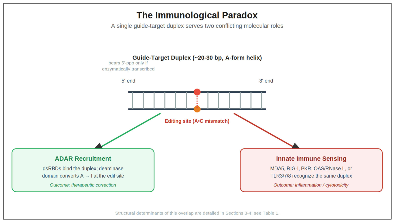
Figure 2.
Duplex-Length Overlap Between ADAR Editing and Immune Sensor Activation. Log-scale schematic comparing the duplex-length threshold of ADAR deamination against innate immune sensors. The shaded green band (20–30 bp) indicates the minimal functional window required for efficient ADAR recruitment and catalytic base flipping. This window falls directly within the sensitivity threshold of OAS1 (≥18 bp), abuts the activation threshold of PKR (dimerization at ≥33 bp, optimal at ~80 bp), and lies fully within the length-sensitive activation range of RIG-I (<500 bp). While it safely avoids the length thresholds of endosomal TLR3 (40–50 bp) and cytosolic MDA5 (>100–500 bp), duplex length alone cannot separate an editing-competent guide from an immunostimulatory one.
Figure 2.
Duplex-Length Overlap Between ADAR Editing and Immune Sensor Activation. Log-scale schematic comparing the duplex-length threshold of ADAR deamination against innate immune sensors. The shaded green band (20–30 bp) indicates the minimal functional window required for efficient ADAR recruitment and catalytic base flipping. This window falls directly within the sensitivity threshold of OAS1 (≥18 bp), abuts the activation threshold of PKR (dimerization at ≥33 bp, optimal at ~80 bp), and lies fully within the length-sensitive activation range of RIG-I (<500 bp). While it safely avoids the length thresholds of endosomal TLR3 (40–50 bp) and cytosolic MDA5 (>100–500 bp), duplex length alone cannot separate an editing-competent guide from an immunostimulatory one.
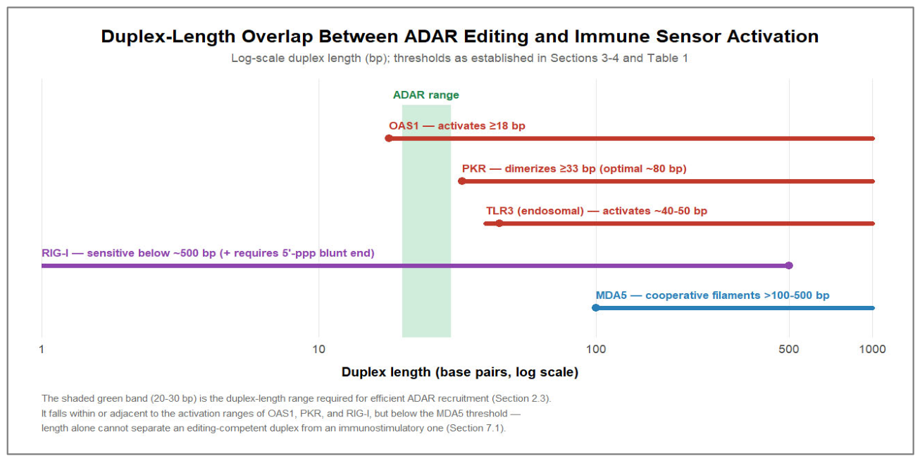
Figure 3.
Chemical Modification Map of the Guide Strand: Spatial Patterning of 2′-OMe, 2′-F, and PS Linkages. Nucleotide-level spatial architecture balancing ADAR catalytic efficiency with innate immune evasion. The guide strand is partitioned into three distinct functional zones: (1) Terminal and distal flanks (green circles) are heavily modified with 2′-O-methyl (2′-OMe) and phosphorothioate (PS) linkages to resist nuclease degradation and ablate endosomal TLR3/7/8 sensing; (2) The central ‘editing bubble’ spanning positions −3 to 0 opposite the target adenosine (red circle, forming an A•C mismatch) remains unmodified or minimally modified (open circles) to preserve sugar re-puckering and active-site base flipping into ADAR’s catalytic pocket; (3) The auxiliary dsRBD-contact interface at positions +11 to +13 (blue circles) is selectively modified with 2′-fluoro (2′-F), which enforces a C3′-endo pucker and accelerates ADAR2 deamination rates up to six-fold by stabilizing functional enzyme dimerization. Position numbering follows Campbell et al. [38] and Jauregui-Matos et al. [37]; the dsRBD contact is asymmetric along the 3′ flank.
Figure 3.
Chemical Modification Map of the Guide Strand: Spatial Patterning of 2′-OMe, 2′-F, and PS Linkages. Nucleotide-level spatial architecture balancing ADAR catalytic efficiency with innate immune evasion. The guide strand is partitioned into three distinct functional zones: (1) Terminal and distal flanks (green circles) are heavily modified with 2′-O-methyl (2′-OMe) and phosphorothioate (PS) linkages to resist nuclease degradation and ablate endosomal TLR3/7/8 sensing; (2) The central ‘editing bubble’ spanning positions −3 to 0 opposite the target adenosine (red circle, forming an A•C mismatch) remains unmodified or minimally modified (open circles) to preserve sugar re-puckering and active-site base flipping into ADAR’s catalytic pocket; (3) The auxiliary dsRBD-contact interface at positions +11 to +13 (blue circles) is selectively modified with 2′-fluoro (2′-F), which enforces a C3′-endo pucker and accelerates ADAR2 deamination rates up to six-fold by stabilizing functional enzyme dimerization. Position numbering follows Campbell et al. [38] and Jauregui-Matos et al. [37]; the dsRBD contact is asymmetric along the 3′ flank.
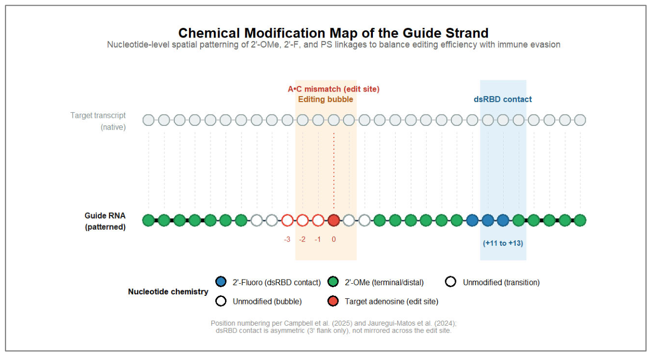
Figure 4.
The Central Biophysical Trade-Off: Composite Innate Immune Activation vs. ADAR Editing Efficiency across Duplex Lengths. Schematic of basic engineering trade-off controlled by guide duplex length. The blue curve shows ADAR editing efficiency, which is optimal around the 20–30 bp range necessary for clamping and catalyzing the conversion of adenosine to inosine (A-to-I) [20,22]. The red curve shows composite innate immune activation, from sequential onsets of OAS1 (≥18 bp), PKR dimerization (~33 bp) and endosomal TLR3 (40–50 bp). The therapeutic window (shaded green band, 20-30 bp) represents the region of maximum ADAR activity but intersects an increasing immune-activation curve, requiring chemical spatial patterning or circularization (Section 5 and Section 6) to prevent immune signalling.
Figure 4.
The Central Biophysical Trade-Off: Composite Innate Immune Activation vs. ADAR Editing Efficiency across Duplex Lengths. Schematic of basic engineering trade-off controlled by guide duplex length. The blue curve shows ADAR editing efficiency, which is optimal around the 20–30 bp range necessary for clamping and catalyzing the conversion of adenosine to inosine (A-to-I) [20,22]. The red curve shows composite innate immune activation, from sequential onsets of OAS1 (≥18 bp), PKR dimerization (~33 bp) and endosomal TLR3 (40–50 bp). The therapeutic window (shaded green band, 20-30 bp) represents the region of maximum ADAR activity but intersects an increasing immune-activation curve, requiring chemical spatial patterning or circularization (Section 5 and Section 6) to prevent immune signalling.
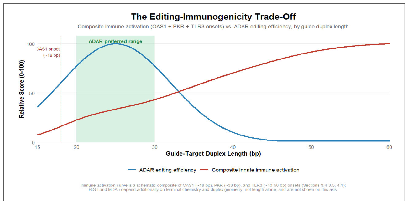
Table 3.
Multi-Parameter Guide-RNA Design Matrix.
| Design Parameter | Impact on ADAR Catalysis | Impact on Innate Sensors | Design Recommendation |
| Duplex Length (20–30 bp) | Required for efficient dsRBD binding & deamination [20,22] | Within OAS1 (≥18 bp) & RIG-I (<500 bp) windows; below MDA5 [24,30,32] | Maintain 20–30 bp; combine with spatial chemical patterning or circularization |
| Helical Geometry (A-form) | Essential for dsRBD positioning & catalytic stability [22,38] | Obligate ligand for PKR dimerization, OAS1, & MDA5 filamentation [14,30,36] | Preserve A-form near edit site; introduce modifications distally to disrupt multimerization |
| Internal Mismatches / Bulges | Inhibitory near active site (−1/−2); tolerated distally [37,38] | Disrupts MDA5/PKR; OAS1 tolerates or is stimulated by A-C / I-U pairs [36] | Deploy cautiously; exclude from immediate editing bubble (−3 to 0) |
| Terminal 5′ Phosphorylation | Neutral; ADAR does not engage terminal 5′ phosphates [37] | 5′-ppp potently activates RIG-I → MAVS → type I IFN [24,25,39] | Mandatory removal via phosphatase, solid-phase synthesis (5′-OH), or circularization |
| 2′-Fluoro at dsRBD (+12/+13) | Enforces C3′-endo pucker; accelerates ADAR2 deamination up to 6-fold [38] | Eliminates native 2′-OH groups required for sensor recognition [38,40] | Prioritize at +12/+13; improves catalysis and immune evasion together |
| 2′-O-Methyl at dsRBD (+12/+13) | Causes steric clashes, reducing deamination rates [37,38] | Suppresses TLR3/7/8; enhances metabolic stability [38,40] | Exclude from dsRBD contacts and editing bubble; restrict to distal/terminal flanks |
| Phosphorothioate Linkages | Heavily modified backbones impair deamination and cause toxicity [40,43] | Uniform PS causes protein sequestration, nuclear bodies, & DDR arrest [41,42,43] | Restrict to 2–3 terminal linkages for exonuclease protection; avoid uniform PS |
| Nucleobase Analogs (Ψ, m1Ψ) | Minimal impact on catalytic base-flipping pocket [29,37] | Suppresses TLR3/7/8 by blocking endolysosomal processing into agonists [28,29,44] | Incorporate in distal regions, especially single-stranded overhangs and recruitment loops |
| Circularization (circ-arRNA) | Supports sustained target hybridization and high editing efficiency [48] | Ablates terminal RIG-I sensing; unpurified linear IVT byproducts trigger IFN [39,46,47] | Adopt as topological evasion platform; enforce rigorous HPLC-SEC and RNase R purification |
Abbreviations: bp, base pairs; DDR, DNA damage response; dsRBD, double-stranded RNA binding domain; HPLC-SEC, high-performance liquid chromatography size-exclusion chromatography; IFN, interferon; MAVS, mitochondrial antiviral-signaling protein; MDA5, melanoma differentiation-associated protein 5; m1Ψ, N1-methylpseudouridine; OAS1, 2′-5′-oligoadenylate synthetase 1; PKR, protein kinase R; PS, phosphorothioate; RIG-I, retinoic acid-inducible gene I; RNase, ribonuclease; TLR, Toll-like receptor; Ψ, pseudouridine.
Disclaimer/Publisher’s Note: The statements, opinions and data contained in all publications are solely those of the individual author(s) and contributor(s) and not of MDPI and/or the editor(s). MDPI and/or the editor(s) disclaim responsibility for any injury to people or property resulting from any ideas, methods, instructions or products referred to in the content. |
© 2026 by the authors. Licensee MDPI, Basel, Switzerland. This article is an open access article distributed under the terms and conditions of the Creative Commons Attribution (CC BY) license (http://creativecommons.org/licenses/by/4.0/).
Copyright: This open access article is published under a Creative Commons CC BY 4.0 license, which permit the free download, distribution, and reuse, provided that the author and preprint are cited in any reuse.