

Submitted:
07 October 2026
Posted:
09 October 2026
You are already at the latest version
Abstract
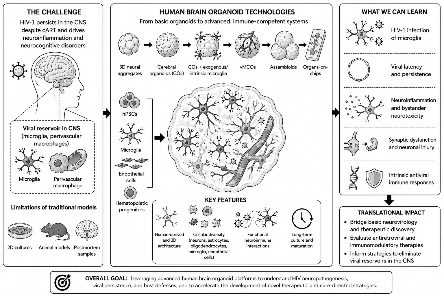
The persistence of human immunodeficiency virus type 1 (HIV-1) in the central nervous system (CNS) remains a major barrier to a cure and a key driver of HIV-associated neurocognitive disorders (HAND). For decades, progress in understanding HIV-1 neuropathogenesis has been limited by the lack of experimental models capable of recapitulating the complexity of the human brain. The development of three-dimensional (3D) brain organoids derived from human induced pluripotent stem cells (iPSCs) has significantly advanced the field by providing a physiologically relevant and clinically translatable platform for studying HIV-1 infection in the CNS. This review summarizes the evolution of brain organoid technologies, from self-organizing cultures to immune-competent organoids and multi-region assembloids. We highlight their contributions to understanding HIV-1 neuroinflammation, neuronal injury, and CNS reservoir establishment. We also discuss emerging advances integrating organoids with bioengineering and single-cell omics approaches to overcome current limitations and accelerate the development of therapeutic and curative strategies targeting HIV-1 persistence in the CNS.
Keywords:
brain organoids
; NeuroHIV
; HIV-1
; microglia
; vascularized microglia-containing cerebral organoids
; assembloids
; organoids-on-chip
1. Introduction
Despite the success of combination antiretroviral therapy (cART), HIV-1 persists in anatomical sanctuaries. The central nervous system (CNS) represents one of the most formidable of these reservoirs [1]. HIV-1 infiltrates the CNS early in the course of infection, establishing a long-lived latent reservoir primarily within resident microglia and perivascular macrophages [2]. HIV-1 persistence in these cellular sanctuaries, through either low-level replication or latency, enables viruses to evade immune surveillance and therapeutic intervention. Consequently, the CNS reservoir remains a major contributor to the pathogenesis of HIV-associated neurocognitive disorders (HAND), a spectrum of conditions ranging from asymptomatic neurocognitive impairment to severe dementia, even among individuals with effectively suppressed plasma viral loads [3].
For decades, progress in understanding the molecular mechanisms underlying HIV-1 neuropathogenesis has been limited by the lack of physiologically relevant experimental models. Elucidating how relatively restricted viral infection leads to widespread neuroinflammation, synaptic dysfunction, and neuronal injury has remained a significant challenge [4]. Historically, NeuroHIV research relied heavily on postmortem specimens, surgical samples, and fetal tissues, all of which provide limited opportunities for experimental manipulation and longitudinal investigation. Although animal models have yielded important insights, they fail to fully replicate the complexity of human brain architecture, cellular composition, and HIV-1 disease progression. Likewise, conventional two-dimensional (2D) cell culture systems cannot reproduce the intricate three-dimensional (3D) cytoarchitecture, cellular heterogeneity, and dynamic cell-cell interactions that define the human brain microenvironment [5].
The emergence of 3D brain organoid technology derived from human pluripotent stem cells (hPSCs) has revolutionized the study of human neurobiology and disease. By exploiting the dual capacity of hPSCs for unlimited self-renewal and differentiation, researchers can generate complex, self-organizing neural tissues under defined 3D culture conditions. These organoids recapitulate key structural and cellular features of the developing human brain and contain diverse neural cell populations, including neurons, astrocytes, oligodendrocytes, and, in more advanced models, microglia [6,7]. Importantly, brain organoids are not merely static cellular aggregates; they are dynamic tissues that undergo progressive maturation and can be maintained in culture for months or even years, enabling the investigation of developmental and pathological processes over extended periods [8].
Beyond modeling human brain development, brain organoids have demonstrated significant utility as preclinical platforms. Studies have demonstrated that human iPSC-derived 3D brain organoids provide more physiologically relevant platforms than conventional 2D cultures and many animal models for predicting drug-induced neurotoxicity. Benchmarking studies reported high specificity and moderate sensitivity for detecting neurotoxic compounds, while subsequent reviews have highlighted the superior ability of brain organoids to recapitulate human-specific cytoarchitecture, cellular diversity, and neurotoxic responses, supporting their application in preclinical safety assessment and drug development [9]. As a result, they provide a unique and physiologically relevant platform for studying the multifaceted interactions between HIV-1 and the human brain, including viral infection, neuroinflammation, cellular dysfunction, and reservoir persistence [10].
In this review, we summarize the development of human brain organoids and their application to NeuroHIV research. We describe the key developments of these models and discuss their limitations and highlight advanced technologies that may further enhance their use in understanding HIV-1 neuropathogenesis and developing curative strategies.
2. The Technological Evolution of Brain Organoids
The development of in vitro brain organoids to recapitulate the complexity of the human brain reflects progressive and joint efforts by the investigators (Figure 1 and Table 1). This evolution was enabled by the transition from 2D cultures to 3D systems that exploit the intrinsic self-organizing capacity of pluripotent stem cells. Early studies demonstrated that embryonic stem cells cultured in suspension could spontaneously generate polarized neural tissues [11,12,13]. In 2008, Eiraku et al. showed that mouse embryonic stem cells could form cortical-like structures resembling early corticogenesis [12]. This concept was extended by Lancaster et al. in 2013, who generated cerebral organoids from hPSCs containing multiple brain regions, including the cerebral cortex, ventral forebrain, and hippocampus [14]. Around the same time, Kadoshima et al. demonstrated the formation of cortical tissues that reproduced key features of fetal brain development, including layered organization and human-specific outer radial glia [15]. Together, these studies established brain organoids as powerful models for investigating human neurodevelopment and neurological disease [16].
A major advantage of human brain organoids is their ability to retain donor-specific genetic backgrounds, enabling patient-derived disease modeling [17]. However, early brain organoids lacked vasculature, stromal components, and tissue-resident immune cells, particularly microglia. This limitation is especially relevant for NeuroHIV research because microglia are the principal immune cells of the CNS and a major target of HIV-1 infection [5,18,19]. To address this gap, several strategies have been developed to generate microglia-containing organoids (MCOs), including: (1) intrinsic microglial differentiation, (2) exogenous microglial incorporation, (3) co-culture with hematopoietic progenitors, (4) vascularized microglia-containing organoids, (5) assembloid technologies and (6) organ-on-chip platforms.
2.1. Intrinsic Generation of Microglia within Organoids
One approach generates neuroectodermal and mesodermal lineages simultaneously from a common progenitor population. Using this strategy, Ormel et al. demonstrated the spontaneous emergence of microglia-like cells that closely resembled adult microglia transcriptionally and functionally [20]. Alternative protocols have since been developed, including transcription factor-guided differentiation by Cakir et al. and simplified methods reported by Bodnar et al. [21,22]. Because microglia develop alongside neural cells, these models achieve high developmental integration. However, unguided differentiation can result in variability between organoids, necessitating rigorous quality control of stem cell lines, culture conditions, and extracellular matrices. Synthetic hydrogels may help improve reproducibility by providing more standardized culture environments [7,23]. Because microglia arise concurrently with neural development, they become broadly distributed throughout the organoid parenchyma and establish early interactions with neural progenitors and differentiating neurons, more closely resembling the developmental colonization of the human fetal brain than exogenous incorporation approaches.
2.2. Exogenous Incorporation and Spatial Integration of Microglia
An alternative strategy introduces mature microglia into pre-formed organoids. Abud et al. demonstrated that iPSC-derived microglia successfully integrated into neural tissues, adopted ramified morphologies, and displayed phagocytic and inflammatory functions characteristic of native microglia [34]. These cells can also be genetically engineered to model disease-specific mechanisms [35]. Importantly, Dos Reis et al. first used this platform to study HIV-1 infection, showing that incorporated microglia supported persistent low-level viral replication and produced proinflammatory cytokines such as TNF-α and IL-1β, closely resembling NeuroHIV pathology in vivo [36].
Unlike intrinsically generated microglia, exogenously introduced microglia typically enter mature organoids from the surrounding culture medium and initially localize to the outer cortical-like layers before gradually migrating inward [37]. Their distribution is therefore often heterogeneous and may remain biased toward the organoid periphery, depending on the timing of incorporation, organoid maturity, and culture duration [24,34,36]. Although these cells can infiltrate neural tissue and respond to local environmental cues, their spatial organization does not fully recapitulate the widespread, developmentally coordinated colonization observed during embryonic microgliogenesis. Consequently, neuron–microglia interactions and regional immune surveillance may differ from those observed in organoids in which microglia arise intrinsically during organoid development.
Despite these limitations, exogenous incorporation remains a practical and highly reproducible strategy for generating immune-competent brain organoids, particularly for studies requiring genetically engineered microglia, rapid model establishment, or controlled manipulation of microglial abundance.
2.3. Co-Culture with Hematopoietic Progenitors
Another strategy incorporates hematopoietic progenitor cells (HPCs) rather than mature microglia. In these systems, HPCs are introduced into developing organoids, where they migrate and differentiate into microglia under cytokine stimulation, including IL-3, IL-34, M-CSF, and GM-CSF. This approach more closely mimics the developmental colonization of the CNS by microglia and allows control over microglial abundance within organoids. Song et al. demonstrated efficient microglial incorporation into dorsal forebrain organoids through co-culture [38]. Similarly, Wörsdörfer et al. generated vascularized organoids by combining mesodermal progenitors with neural organoids and treating them with PDGF or VEGF, enabling microglial infiltration [28]. These models have subsequently been used to study microglial responses to viral infections, including Zika and dengue viruses [37,39].
More recently, Narasipura et al. developed a co-culture system in which hematopoietic progenitors were integrated into nascent organoids, generating cerebral organoid-derived microglia (CO-iMg) at physiologically relevant levels (~7%) with molecular and functional features resembling resident human microglia [26]. Despite these advances, most protocols achieve only partial microglial integration, and long-term survival remains challenging. Further optimization is needed to better understand how developmental origin and local environmental cues shape microglial identity and function.
2.4. Vascularized Microglia-Containing Organoids
A major limitation of conventional cerebral organoids is the lack of vascularization, which restricts oxygen and nutrient diffusion and limits long-term maturation. To overcome this challenge, researchers have developed vascularized brain organoids by incorporating blood-brain barrier (BBB) endothelial cells, mesodermal progenitors, or angiogenic signaling pathways during organoid development [28,40,41]. These advances have led to the emergence of vascularized microglia-containing cerebral organoids (vMCOs), which model both neurovascular and neuroimmune interactions. By supporting the co-development of neural cells, microglia, and vascular-like structures, vMCOs exhibit improved viability, enhanced cellular maturation, and more physiologically relevant inflammatory responses. Endothelial cells contribute to neuronal maturation and microglial functional development through direct cellular interactions and the secretion of trophic and signaling factors [17,30,42]. These vascular-derived cues promote neuronal network formation and support the acquisition of brain-specific microglial phenotypes associated with homeostatic surveillance and immune responsiveness [20,37,39]. By incorporating vascular-like structures, vMCOs more closely recapitulate the neurovascular and neuroimmune microenvironment of the human brain [22,28,40]. Given that HIV-1 enters the CNS through the neurovascular interface and establishes reservoirs within brain-resident myeloid cells, vMCOs provide a promising platform for investigating viral entry across the BBB, microglial infection, and HIV-associated neurovascular dysfunction.
2.5. From Organoids to Assembloids
The development of region-specific organoids representing the cortex, midbrain, thalamus, cerebellum, and striatum has expanded the ability to model human brain development in vitro. However, many neurological processes depend on interactions between multiple brain regions. To address this limitation, researchers have fused distinct organoids to create assembloids, which model interregional communication and neuronal migration during brain development [29,43,44]. These systems have successfully reproduced processes such as tangential interneuron migration and cortico-striatal connectivity [45,46,47,48]. Although assembloids provide a more physiologically relevant model of neural circuitry, their application to viral neuropathogenesis remains largely unexplored. Incorporating microglia into assembloid systems may offer a powerful platform for studying viral replication, neuroinflammation, and region-specific CNS pathology.
2.6. Organoids-on-Chip
While conventional organoids capture many aspects of brain development, they lack vascularization and physiological fluid flow. Organoids-on-chip address these limitations by integrating organoids with microfluidic devices or 3D-printed scaffolds that provide continuous perfusion, nutrient delivery, and waste removal, thereby better mimicking in vivo tissue environments [49]. These platforms have enabled studies of microglial recruitment and neuroimmune interactions and support high-throughput drug screening and real-time monitoring [30,32,33,50]. Despite their promise, organoids-on-chip [51,52] remain technically complex and costly, and standardized protocols are still lacking. Furthermore, ethical concerns surrounding advanced neural organoids and chimeric models may pose challenges for broader adoption and clinical translation.
3. Modeling HIV-1 Infection and Neuroinflammation in Brain Organoids
MCOs or CO-iMg have emerged as powerful models for studying HIV-1 infection in the human CNS (Table 2). Studies have shown that MCOs express the key HIV-1 entry factors CD4, CCR5, and CXCR4 and support productive infection, as evidenced by viral protein expression, viral RNA production, and proviral DNA integration [24,25,53,54]. These in vitro observations are consistent with clinical findings that microglia are the primary CNS target and reservoir of HIV-1.
Beyond modeling HIV-1 infection, MCOs have been successfully used to study the neuroinflammation and the mechanisms underlying HAND. HIV-infected microglia mount a robust inflammatory response that extends to uninfected bystander cells. Single-cell RNA sequencing studies revealed widespread transcriptional changes in neural progenitors and mature neurons, including reduced neurotransmitter transporter expression and increased expression of senescence- and apoptosis-related genes [55]. HIV-induced neuroinflammation causes synaptic loss, dendritic spine degeneration, and neuronal apoptosis, key pathological features of HAND [26]. These models have also proven valuable for assessing the efficacy of cART in a CNS-relevant context. cART effectively suppresses viral replication and partially restores neuronal function in HIV-infected organoids, highlighting the utility of brain organoid models for evaluating antiretroviral therapies and identifying novel NeuroHIV interventions [53].
Importantly, immune-competent brain organoids provide a unique platform for investigating HIV-1 persistence within the CNS reservoir. Studies in microglial models have shown that latency regulation differs from that in peripheral CD4+ T cells and monocytes. For example, activation of Toll-like receptor 3 (TLR3) effectively reverses HIV latency in microglia, whereas several latency-reversing agents active in peripheral reservoirs exhibit limited efficacy in CNS-resident myeloid cells [56]. These findings highlight the biological distinctiveness of the CNS reservoir and underscore the need for brain-specific models to evaluate cure-directed strategies [57]. In addition to modeling viral persistence, recent studies have revealed that microglia-containing cerebral organoids possess intrinsic antiviral defense mechanisms. Xiao et al. demonstrated that TLR3 activation induced robust expression of type I and type III interferons, interferon-stimulated genes, and antiviral chemokines in microglia-containing cerebral organoids [54]. This coordinated innate immune response significantly suppressed HIV-1 replication, indicating that neural and immune cells within organoids can mount effective antiviral defenses. Together, these findings establish immune-competent brain organoids as a versatile platform for investigating HIV-1 infection, viral persistence, and host antiviral immunity within the CNS, while facilitating the development of novel therapeutic and cure-directed strategies.
4. Consistency and Reproducibility of Brain Organoid Models
A major challenge in applying brain organoids to NeuroHIV research is ensuring experimental consistency and reproducibility. Considerable variability can arise from differences in induced pluripotent stem cell (iPSC) donor genetic background, cell line quality, differentiation efficiency, culture conditions, extracellular matrices, and batch-to-batch variation [58]. These factors can influence organoid size, cellular composition, maturation state, and susceptibility to HIV-1 infection, thereby complicating comparisons among studies.
Recent advances have substantially improved reproducibility through standardized differentiation protocols, quality-control criteria, automated bioreactor systems, and chemically defined culture media [58,59]. The use of isogenic iPSC lines, multiple biological replicates, and rigorous molecular characterization, including single-cell transcriptomics and immunophenotyping, further enhances experimental reliability, establishing that individual organoids can reproducibly recapitulate highly constrained developmental trajectories and complex cellular diversity [7,60]. Furthermore, uniform aggregate assembly methods have enabled the controlled, highly reproducible incorporation of the microglial immune component, which is critical for establishing standardized, immune-competent models of neuroinflammation [61]. In addition, cryopreservation of neural organoids or early-stage neuroectoderm has emerged as a practical strategy for reducing batch variation and improving experimental consistency across laboratories [58]. As standardized manufacturing and validation protocols continue to evolve, brain organoid models are expected to become increasingly robust and reproducible platforms for investigating HIV persistence, neuroinflammation, and therapeutic interventions.
5. Future Directions in Organoid Engineering and Genomics
Despite rapid advances, current brain organoid models still lack several key features of the CNS, including functional vasculature, complete immune cell populations, and robust myelination. Additional challenges include batch-to-batch variability, limited maturation beyond fetal developmental stages, and difficulties in modeling chronic neurodegenerative processes [13,62,63].
A major limitation of currently used brain organoids is the absence of physiological perfusion, which restricts nutrient delivery and waste removal, resulting in hypoxia and the formation of necrotic cores. Strategies such as organoid slicing and microfluidic organ-on-chip platforms have improved tissue viability and promoted maturation by enhancing oxygenation and nutrient exchange [64,65]. Integration of organoids with microfluidic systems may also facilitate more physiologically relevant BBB models, creating new opportunities to investigate HIV-1 trafficking into the CNS and evaluate the penetration and efficacy of therapeutic agents.
Although the incorporation of microglia has substantially increased the utility of brain organoids for NeuroHIV research, other important immune cell populations, including T cells, perivascular macrophages, and infiltrating monocytes, remain largely absent. Future model systems will likely combine advances in assembloid technology, organ-on-chip engineering, and stem cell biology to generate more comprehensive neuroimmune platforms that better recapitulate the cellular complexity of the CNS.
As organoid models become increasingly sophisticated, high-resolution analytical approaches will be essential for evaluating cellular composition, developmental fidelity, and disease-associated alterations. Single-cell transcriptomics, epigenomics, and large-scale reference resources such as the Human Cell Atlas [66,67] provide powerful frameworks for benchmarking organoid fidelity, characterizing cellular heterogeneity, and identifying molecular pathways involved in HIV-associated neuropathogenesis [68,69,70]. Together, these technological and analytical advances are expected to improve model reproducibility, enhance translational relevance, and accelerate the development of therapies aimed at eliminating HIV reservoirs and persistent infection within the CNS.
6. Conclusions
Human iPSC-derived brain organoids have emerged as physiologically relevant platforms for studying NeuroHIV and have significantly advanced our understanding of HIV-1 infection, neuroinflammation, and neuronal injury within the CNS. As highlighted throughout this review, a variety of organoid-based models have been developed, each offering distinct advantages and limitations (Figure 1). Consequently, no single model is optimal for all applications, and researchers must carefully balance biological complexity, reproducibility, scalability, and experimental tractability when selecting an appropriate system.
Ongoing advances in bioengineering, including the development of vascularized organoids, organ-on-chip technologies, and increasingly sophisticated neuroimmune co-culture systems, are expected to further enhance the physiological relevance of these models. In parallel, high-resolution single-cell and spatial genomics approaches will provide unprecedented insights into cellular heterogeneity, host-virus interactions, and disease-associated molecular pathways. Together, these innovations will improve model fidelity and analytical power, enabling more accurate investigation of HIV neuropathogenesis and CNS viral persistence. Ultimately, continued refinement of brain organoid technologies will accelerate efforts to elucidate the mechanisms underlying HAND and facilitate the development of therapeutic strategies aimed at eliminating HIV-1 reservoirs within the CNS.
Author Contributions
Conceptualization, Q.-H.X., X.W., A.M.A. and W.-Z.H.; Investigation, Q.-H.X., J.M., R.P., Y.P. and W.-Z.H.; Writing—Original Draft Preparation, Q.-H.X. and X.W.; Writing—Review and Editing, Q.-H.X., X.W., J.M., R.P., Y.P., A.M.A. and W.-Z.H.; Supervision, W.-Z.H.
Funding
This work was supported by the National Institutes of Health (grant numbers DA058536 and MH134402). The funder was not involved in the study design, collection, analysis, and interpretation of data, writing of this article or the decision to submit it for publication.
Data Availability Statement
No new data were created or analyzed in this study. Data sharing is not applicable to this article.
Acknowledgments
During the preparation of this manuscript, the authors employed ChatGPT for spell checking, improving linguistic clarity and readability, and assisting with manuscript formatting and bibliographic organization. Following use of this tool, the authors critically reviewed and revised the text as necessary and assume full responsibility for the final content of the publication.
Conflicts of Interest
The authors declare that they have no known competing financial interests or personal relationships that could have appeared to influence the work reported in this paper.
References
- Saylor, D.; Dickens, A.M.; Sacktor, N.; Haughey, N.; Slusher, B.; Pletnikov, M.; Mankowski, J.L.; Brown, A.; Volsky, D.J.; McArthur, J.C. HIV-associated neurocognitive disorder—pathogenesis and prospects for treatment. Nat. Rev. Neurol. 2016, 12, 234–248. [Google Scholar] [CrossRef]
- Wallet, C.; De Rovere, M.; Van Assche, J.; Daouad, F.; De Wit, S.; Gautier, V.; Mallon, P.W.G.; Marcello, A.; Van Lint, C.; Rohr, O.; et al. Microglial Cells: The Main HIV-1 Reservoir in the Brain. Front. Cell. Infect. Microbiol. 2019, 9, 362. [Google Scholar] [CrossRef]
- Ellis, R.J.; Marquine, M.J.; Kaul, M.; Fields, J.A.; Schlachetzki, J.C.M. Mechanisms underlying HIV-associated cognitive impairment and emerging therapies for its management. Nat. Rev. Neurol. 2023, 19, 668–687. [Google Scholar] [CrossRef]
- Waight, E.; Zhang, C.; Mathews, S.; Kevadiya, B.D.; Lloyd, K.C.K.; Gendelman, H.E.; Gorantla, S.; Poluektova, L.Y.; Dash, P.K. Animal models for studies of HIV-1 brain reservoirs. J. Leukoc. Biol. 2022, 112, 1285–1295. [Google Scholar] [CrossRef]
- Sarkar, P.; Wang, X.; Hu, W.; Zhu, J.; Ho, W.Z. Human Microglia Models for NeuroHIV. Viruses 2025, 17, 641. [Google Scholar] [CrossRef]
- Amiri, A.; Coppola, G.; Scuderi, S.; Wu, F.; Roychowdhury, T.; Liu, F.; Pochareddy, S.; Shin, Y.; Safi, A.; Song, L.; et al. Transcriptome and epigenome landscape of human cortical development modeled in organoids. Science 2018, 362, eaat6720. [Google Scholar] [CrossRef]
- Velasco, S.; Kedaigle, A.J.; Simmons, S.K.; Nash, A.; Rocha, M.; Quadrato, G.; Paulsen, B.; Nguyen, L.; Adiconis, X.; Regev, A.; et al. Individual brain organoids reproducibly form cell diversity of the human cerebral cortex. Nature 2019, 570, 523–527. [Google Scholar] [CrossRef]
- Marx, V. Reality check for organoids in neuroscience. Nat. Methods. 2020, 17, 961–964. [Google Scholar] [CrossRef]
- Wang, Q.; Cohen, J.D.; Yukawa, T.; Estrella, H.; Leonard, C.; Nunes, J.; Choi, C.; Lewis, L.; Baker, K.S.; Kuga, K.; et al. Assessment of a 3D neural spheroid model to detect pharmaceutical-induced neurotoxicity. ALTEX 2022, 39, 560–582. [Google Scholar] [CrossRef]
- Premeaux, T.A.; Mediouni, S.; Leda, A.; Furler, R.L.; Valente, S.T.; Fine, H.A.; Nixon, D.F.; Ndhlovu, L.C. Next-Generation Human Cerebral Organoids as Powerful Tools To Advance NeuroHIV Research. mBio 2021, 12, e0068021. [Google Scholar] [CrossRef]
- Eiraku, M.; Takata, N.; Ishibashi, H.; Kawada, M.; Sakakura, E.; Okuda, S.; Sekiguchi, K.; Adachi, T.; Sasai, Y. Self-organizing optic-cup morphogenesis in three-dimensional culture. Nature 2011, 472, 51–56. [Google Scholar] [CrossRef]
- Eiraku, M.; Watanabe, K.; Matsuo-Takasaki, M.; Kawada, M.; Yonemura, S.; Matsumura, M.; Wataya, T.; Nishiyama, A.; Muguruma, K.; Sasai, Y. Self-organized formation of polarized cortical tissues from ESCs and its active manipulation by extrinsic signals. Cell Stem Cell 2008, 3, 519–532. [Google Scholar] [CrossRef]
- Kelava, I.; Lancaster, M.A. Stem Cell Models of Human Brain Development. Cell Stem Cell 2016, 18, 736–748. [Google Scholar] [CrossRef]
- Lancaster, M.A.; Renner, M.; Martin, C.A.; Wenzel, D.; Bicknell, L.S.; Hurles, M.E.; Homfray, T.; Penninger, J.M.; Jackson, A.P.; Knoblich, J.A. Cerebral organoids model human brain development and microcephaly. Nature 2013, 501, 373–379. [Google Scholar] [CrossRef]
- Kadoshima, T.; Sakaguchi, H.; Nakano, T.; Soen, M.; Ando, S.; Eiraku, M.; Sasai, Y. Self-organization of axial polarity, inside-out layer pattern, and species-specific progenitor dynamics in human ES cell–derived neocortex. Proc. Natl. Acad. Sci. USA 2013, 110, 20284–20289. [Google Scholar] [CrossRef]
- Buonfiglioli, A.; Kübler, R.; Missall, R.; De Jong, R.; Chan, S.; Haage, V.; Wendt, S.; Lin, A.J.; Mattei, D.; Graziani, M.; et al. A microglia-containing cerebral organoid model to study early life immune challenges. Brain Behav. Immun. 2025, 123, 1127–1146. [Google Scholar] [CrossRef]
- Bennett, F.C.; Bennett, M.L.; Yaqoob, F.; Mulinyawe, S.B.; Grant, G.A.; Hayden Gephart, M.; Plowey, E.D.; Barres, B.A. A Combination of Ontogeny and CNS Environment Establishes Microglial Identity. Neuron 2018, 98, 1170–1183.e8. [Google Scholar] [CrossRef]
- Lawson, L.J.; Perry, V.H.; Dri, P.; Gordon, S. Heterogeneity in the distribution and morphology of microglia in the normal adult mouse brain. Neuroscience 1990, 39, 151–170. [Google Scholar] [CrossRef]
- Olah, M.; Raj, D.; Brouwer, N.; De Haas, A.H.; Eggen, B.J.; Den Dunnen, W.F.; Biber, K.P.; Boddeke, H.W. An optimized protocol for the acute isolation of human microglia from autopsy brain samples. Glia 2012, 60, 96–111. [Google Scholar] [CrossRef]
- Ormel, P.R.; Vieira de Sá, R.; van Bodegraven, E.J.; Karst, H.; Harschnitz, O.; Sneeboer, M.A.M.; Johansen, L.E.; van Dijk, R.E.; Scheefhals, N.; Berdenis van Berlekom, A.; et al. Microglia innately develop within cerebral organoids. Nat. Commun. 2018, 9, 4167. [Google Scholar] [CrossRef]
- Bodnar, B.; Zhang, Y.; Liu, J.; Lin, Y.; Wang, P.; Wei, Z.; Saribas, S.; Zhu, Y.; Li, F.; Wang, X.; et al. Novel Scalable and Simplified System to Generate Microglia-Containing Cerebral Organoids From Human Induced Pluripotent Stem Cells. Front. Cell. Neurosci. 2021, 15, 682272. [Google Scholar] [CrossRef]
- Cakir, B.; Tanaka, Y.; Kiral, F.R.; Xiang, Y.; Dagliyan, O.; Wang, J.; Lee, M.; Greaney, A.M.; Yang, W.S.; duBoulay, C.; et al. Expression of the transcription factor PU.1 induces the generation of microglia-like cells in human cortical organoids. Nat. Commun. 2022, 13, 430. [Google Scholar] [CrossRef]
- Below, C.R.; Kelly, J.; Brown, A.; Humphries, J.D.; Hutton, C.; Xu, J.; Lee, B.Y.; Cintas, C.; Zhang, X.; Hernandez-Gordillo, V.; et al. A microenvironment-inspired synthetic three-dimensional model for pancreatic ductal adenocarcinoma organoids. Nat. Mater. 2022, 21, 110–119. [Google Scholar] [CrossRef]
- Dos Reis, R.S.; Sant, S.; Keeney, H.; Wagner, M.C.E.; Ayyavoo, V. Modeling HIV-1 neuropathogenesis using three-dimensional human brain organoids (hBORGs) with HIV-1 infected microglia. Sci. Rep. 2020, 10, 15209. [Google Scholar] [CrossRef]
- Gumbs, S.B.H.; Berdenis van Berlekom, A.; Kübler, R.; Schipper, P.J.; Gharu, L.; Boks, M.P.; Ormel, P.R.; Wensing, A.M.J.; de Witte, L.D.; Nijhuis, M. Characterization of HIV-1 Infection in Microglia-Containing Human Cerebral Organoids. Viruses 2022, 14, 829. [Google Scholar] [CrossRef]
- Narasipura, S.D.; Zayas, J.P.; Ash, M.K.; Reyes, A.F.; Shull, T.; Gambut, S.; Szczerkowski, J.L.A.; McKee, C.; Schneider, J.R.; Lorenzo-Redondo, R.; et al. Inflammatory responses revealed through HIV infection of microglia-containing cerebral organoids. J. Neuroinflammation 2025, 22, 36. [Google Scholar] [CrossRef]
- Ji, Y.; Chen, X.; Wang, Z.; Meek, C.J.; McLean, J.L.; Yang, Y.; Yuan, C.; Rochet, J.C.; Liu, F.; Xu, R. Alzheimer’s disease patient brain extracts induce multiple pathologies in novel vascularized neuroimmune organoids for disease modeling and drug discovery. Mol. Psychiatry 2025, 30, 4558–4575. [Google Scholar] [CrossRef]
- Wörsdörfer, P.; Dalda, N.; Kern, A.; Krüger, S.; Wagner, N.; Kwok, C.K.; Henke, E.; Ergün, S. Generation of complex human organoid models including vascular networks by incorporation of mesodermal progenitor cells. Sci. Rep. 2019, 9, 15663. [Google Scholar] [CrossRef]
- Birey, F.; Andersen, J.; Makinson, C.D.; Islam, S.; Wei, W.; Huber, N.; Fan, H.C.; Metzler, K.R.C.; Panagiotakos, G.; Thom, N.; et al. Assembly of functionally integrated human forebrain spheroids. Nature 2017, 545, 54–59. [Google Scholar] [CrossRef]
- Ao, Z.; Cai, H.; Wu, Z.; Song, S.; Karahan, H.; Kim, B.; Lu, H.C.; Kim, J.; Mackie, K.; Guo, F. Tubular human brain organoids to model microglia-mediated neuroinflammation. Lab Chip 2021, 21, 2751–2762. [Google Scholar] [CrossRef]
- Fredrikson, J.P.; Roth, D.M.; Cosgrove, J.A.; Sener, G.; Crow, L.A.; Eckenstein, K.; Wu, L.; Hosseini, M.; Thomas, G.; Eksi, S.E.; et al. Engineering neuronal networks in granular microgels to innervate bioprinted cancer organoids on-a-chip. Lab Chip 2025, 25, 3467–3481. [Google Scholar] [CrossRef]
- Liu, J.; Li, Q.; Zhu, H.; Li, A.; Geng, B.; Li, Z.A.; Tian, T. Suspension Culture With Uniform Shear Stress in Brain Organoids-on-a-Chip for Modelling Alzheimer’s Disease. Adv. Healthc. Mater. 2026, 15, e03966. [Google Scholar] [CrossRef]
- Saorin, G.; Caligiuri, I.; Rizzolio, F. Microfluidic organoids-on-a-chip: The future of human models. Semin. Cell Dev. Biol. 2023, 144, 41–54. [Google Scholar] [CrossRef]
- Abud, E.M.; Ramirez, R.N.; Martinez, E.S.; Healy, L.M.; Nguyen, C.H.H.; Newman, S.A.; Yeromin, A.V.; Scarfone, V.M.; Marsh, S.E.; Fimbres, C.; et al. iPSC-Derived Human Microglia-like Cells to Study Neurological Diseases. Neuron 2017, 94, 278–293.e9. [Google Scholar] [CrossRef]
- Lin, Y.-T.; Seo, J.; Gao, F.; Feldman, H.M.; Wen, H.-L.; Penney, J.; Cam, H.P.; Gjoneska, E.; Raja, W.K.; Cheng, J.; et al. APOE4 causes widespread molecular and cellular alterations associated with Alzheimer’s disease phenotypes in human iPSC-derived brain cell types. Neuron 2018, 98, 1141–1154.e7. [Google Scholar] [CrossRef]
- Dos Reis, R.S.; Sant, S.; Ayyavoo, V. Three-Dimensional Human Brain Organoids to Model HIV-1 Neuropathogenesis. Methods Mol. Biol. 2023, 2610, 167–178. [Google Scholar] [CrossRef]
- Xu, R.; Boreland, A.J.; Li, X.; Erickson, C.; Jin, M.; Atkins, C.; Pang, Z.P.; Daniels, B.P.; Jiang, P. Developing human pluripotent stem cell-based cerebral organoids with a controllable microglia ratio for modeling brain development and pathology. Stem Cell Rep. 2021, 16, 1923–1937. [Google Scholar] [CrossRef]
- Song, L.; Yuan, X.; Jones, Z.; Vied, C.; Miao, Y.; Marzano, M.; Hua, T.; Sang, Q.-X.A.; Guan, J.; Ma, T.; et al. Functionalization of brain region-specific spheroids with isogenic microglia-like cells. Sci. Rep. 2019, 9, 11055. [Google Scholar] [CrossRef]
- Fagerlund, I.; Dougalis, A.; Shakirzyanova, A.; Gómez-Budia, M.; Pelkonen, A.; Konttinen, H.; Ohtonen, S.; Fazaludeen, M.F.; Koskuvi, M.; Kuusisto, J.; et al. Microglia-like cells promote neuronal functions in cerebral organoids. Cells 2022, 11, 124. [Google Scholar] [CrossRef]
- Cakir, B.; Xiang, Y.; Tanaka, Y.; Kural, M.H.; Parent, M.; Kang, Y.J.; Chapeton, K.; Patterson, B.; Yuan, Y.; He, C.S.; et al. Engineering of human brain organoids with a functional vascular-like system. Nat. Methods. 2019, 16, 1169–1175. [Google Scholar] [CrossRef]
- Shi, Y.; Sun, L.; Wang, M.; Liu, J.; Zhong, S.; Li, R.; Li, P.; Guo, L.; Fang, A.; Chen, R.; et al. Vascularized human cortical organoids (vOrganoids) model cortical development in vivo. PLoS Biol. 2020, 18, e3000705. [Google Scholar] [CrossRef]
- Zhu, H.; Qiao, X.; Liu, W.; Wang, C.; Zhao, Y. Microglia Play an Essential Role in Synapse Development and Neuron Maturation in Tissue-Engineered Neural Tissues. Front. Neurosci. 2020, 14, 586452. [Google Scholar] [CrossRef]
- Bagley, J.A.; Reumann, D.; Bian, S.; Lévi-Strauss, J.; Knoblich, J.A. Fused cerebral organoids model interactions between brain regions. Nat. Methods. 2017, 14, 743–751. [Google Scholar] [CrossRef]
- Xiang, Y.; Tanaka, Y.; Patterson, B.; Kang, Y.-J.; Govindaiah, G.; Roselaar, N.; Cakir, B.; Kim, K.-Y.; Lombroso, A.P.; Hwang, S.-M.; et al. Fusion of Regionally Specified hPSC-Derived Organoids Models Human Brain Development and Interneuron Migration. Cell Stem Cell 2017, 21, 383–398.e7. [Google Scholar] [CrossRef]
- Andersen, J.; Revah, O.; Miura, Y.; Thom, N.; Amin, N.D.; Kelley, K.W.; Singh, M.; Chen, X.; Thete, M.V.; Walczak, E.M.; et al. Generation of Functional Human 3D Cortico-Motor Assembloids. Cell 2020, 183, 1913–1929.e26. [Google Scholar] [CrossRef]
- Miura, Y.; Li, M.Y.; Birey, F.; Ikeda, K.; Revah, O.; Thete, M.V.; Park, J.Y.; Puno, A.; Lee, S.H.; Porteus, M.H.; et al. Generation of human striatal organoids and cortico-striatal assembloids from human pluripotent stem cells. Nat. Biotechnol. 2020, 38, 1421–1430. [Google Scholar] [CrossRef]
- Onesto, M.M.; Kim, J.I.; Pasca, S.P. Assembloid models of cell-cell interaction to study tissue and disease biology. Cell Stem Cell 2024, 31, 1563–1573. [Google Scholar] [CrossRef]
- Xiang, Y.; Tanaka, Y.; Cakir, B.; Patterson, B.; Kim, K.Y.; Sun, P.; Kang, Y.J.; Zhong, M.; Liu, X.; Patra, P.; et al. hESC-Derived Thalamic Organoids Form Reciprocal Projections When Fused with Cortical Organoids. Cell Stem Cell 2019, 24, 487–497.e7. [Google Scholar] [CrossRef]
- Park, S.E.; Georgescu, A.; Huh, D. Organoids-on-a-chip. Science 2019, 364, 960–965. [Google Scholar] [CrossRef]
- Wang, H.; Zhu, W.; Xu, C.; Su, W.; Li, Z. Engineering organoids-on-chips for drug testing and evaluation. Metabolism 2025, 162, 156065. [Google Scholar] [CrossRef]
- Russell, T.; Dirar, Q.; Li, Y.; Chiang, C.; Laskowitz, D.T.; Yun, Y. Cortical spheroid on perfusable microvascular network in a microfluidic device. PLoS ONE 2023, 18, e0288025. [Google Scholar] [CrossRef]
- Salmon, I.; Grebenyuk, S.; Abdel Fattah, A.R.; Rustandi, G.; Pilkington, T.; Verfaillie, C.; Ranga, A. Engineering neurovascular organoids with 3D printed microfluidic chips. Lab Chip 2022, 22, 1615–1629. [Google Scholar] [CrossRef]
- Donadoni, M.; Cakir, S.; Bellizzi, A.; Swingler, M.; Sariyer, I.K. Modeling HIV-1 infection and NeuroHIV in hiPSCs-derived cerebral organoid cultures. J. Neurovirol. 2024, 30, 362–379. [Google Scholar] [CrossRef]
- Xiao, Q.-H.; Wang, X.; Sarkar, P.; Wang, X.-L.; Song, L.; Ling, B.; Hu, W.-H.; Ho, W.-Z. TLR3 activation of microglia-containing cerebral organoid induces antiviral factors against HIV-1 infection. Front. Pharmacol. 2026, 17, 1835457. [Google Scholar] [CrossRef]
- Kong, W.; Frouard, J.; Xie, G.; Corley, M.J.; Helmy, E.; Zhang, G.; Schwarzer, R.; Montano, M.; Sohn, P.; Roan, N.R.; et al. Neuroinflammation generated by HIV-infected microglia promotes dysfunction and death of neurons in human brain organoids. PNAS Nexus 2024, 3, pgae179. [Google Scholar] [CrossRef]
- Alvarez-Carbonell, D.; Garcia-Mesa, Y.; Milne, S.; Das, B.; Dobrowolski, C.; Rojas, R.; Karn, J. Toll-like receptor 3 activation selectively reverses HIV latency in microglial cells. Retrovirology 2017, 14, 9. [Google Scholar] [CrossRef]
- Blanco, A.; Coronado, R.A.; Arun, N.; Ma, K.; Dar, R.D.; Kieffer, C. Monocyte to macrophage differentiation and changes in cellular redox homeostasis promote cell type-specific HIV latency reactivation. Proc. Natl. Acad. Sci. USA 2024, 121, e2313823121. [Google Scholar] [CrossRef]
- Sandoval, S.O.; Cappuccio, G.; Kruth, K.; Osenberg, S.; Khalil, S.M.; Mendez-Albelo, N.M.; Padmanabhan, K.; Wang, D.; Niciu, M.J.; Bhattacharyya, A.; et al. Rigor and reproducibility in human brain organoid research: Where we are and where we need to go. Stem Cell Rep. 2024, 19, 796–816. [Google Scholar] [CrossRef]
- Di Stefano, J.; Di Marco, F.; Cicalini, I.; FitzGerald, U.; Pieragostino, D.; Verhoye, M.; Ponsaerts, P.; Van Breedam, E. Generation, interrogation, and future applications of microglia-containing brain organoids. Neural Regen. Res. 2025, 20, 3448–3460. [Google Scholar] [CrossRef]
- Atamian, A.; Cordon-Barris, L.; Quadrato, G. Taming human brain organoids one cell at a time. Semin. Cell Dev. Biol. 2021, 111, 23–31. [Google Scholar] [CrossRef]
- Rittenhouse, A.; Krall, C.; Plotkin, J.; Alam El Din, D.M.; Kincaid, B.; Laird, J.; Smirnova, L. Microglia-containing neural organoids as brain microphysiological systems for long-term culture. Front. Cell. Neurosci. 2025, 19, 1616470. [Google Scholar] [CrossRef]
- Hong, H.; Jun, Y.; Yoon, S.-B.; Park, S.; Lee, J.; Jang, J.W.; Nam, H.J.; Cho, H. Manufacturing Uniform Cerebral Organoids for Neurological Disease Modeling and Drug Evaluation. Biomater. Res. 2024, 28, 0104. [Google Scholar] [CrossRef]
- Wu, S.-R.; Nowakowski, T.J. Exploring human brain development and disease using assembloids. Neuron 2025, 113, 1133–1150. [Google Scholar] [CrossRef]
- Aili, Y.; Maimaitiming, N.; Wang, Z.; Wang, Y. Brain organoids: A new tool for modelling of neurodevelopmental disorders. J. Cell. Mol. Med. 2024, 28, e18560. [Google Scholar] [CrossRef]
- Chartrain, N.A.; Pryzhkova, M.V.; Candelaria, J.I.; Gilchrist, K.H.; Jordan, P.W. An Efficient Organoid Cutting Method for Long-Term Culture and High-Throughput Analyses. Tissue Eng. Regen. Med. 2025, 22, 675–690. [Google Scholar] [CrossRef]
- Ament, S.A.; Campbell, R.R.; Lobo, M.K.; Receveur, J.P.; Agrawal, K.; Borjabad, A.; Byrareddy, S.N.; Chang, L.; Clarke, D.; Emani, P.; et al. The single-cell opioid responses in the context of HIV (SCORCH) consortium. Mol. Psychiatry 2024, 29, 3950–3961. [Google Scholar] [CrossRef]
- Green, A.A.; Vashist, T.D.; Jakhmola, S.; Chen, X.; Baidwan, G.; Buchanan, J.; Tiwari, S.K.; Griffin, E.; Howell, A.; Lee, Y.; et al. Cell-type specific impact of opioid use disorder and HIV on the human forebrain and cerebellum. bioRxiv 2026. [Google Scholar] [CrossRef]
- Charles, S.; Jackson-Holmes, E.; Sun, G.; Zhou, Y.; Siciliano, B.; Niu, W.; Han, H.; Nikitina, A.; Kemp, M.L.; Wen, Z.; et al. Non-Invasive Quality Control of Organoid Cultures Using Mesofluidic CSTR Bioreactors and High-Content Imaging. Adv. Mater. Technol. 2025, 10, 2400473. [Google Scholar] [CrossRef]
- He, Z.; Dony, L.; Fleck, J.S.; Szałata, A.; Li, K.X.; Slišković, I.; Lin, H.C.; Santel, M.; Atamian, A.; Quadrato, G.; et al. An integrated transcriptomic cell atlas of human neural organoids. Nature 2024, 635, 690–698. [Google Scholar] [CrossRef]
- Luo, C.; Lancaster, M.A.; Castanon, R.; Nery, J.R.; Knoblich, J.A.; Ecker, J.R. Cerebral Organoids Recapitulate Epigenomic Signatures of the Human Fetal Brain. Cell Rep. 2016, 17, 3369–3384. [Google Scholar] [CrossRef]
Figure 1.
Evolution of human stem cell–derived brain models for HIV neuropathogenesis. Schematic showing the progression from 2D cell cultures to neurospheres, cerebral organoids, microglia-containing and vascularized organoids, assembloids/organ-on-chip systems, and advanced HIV–brain models. Increasing model complexity improves physiological relevance by incorporating diverse brain cell types, neuroimmune interactions, and BBB-like structures, facilitating studies of HIV infection, viral persistence, neuroinflammation, and therapeutic development.
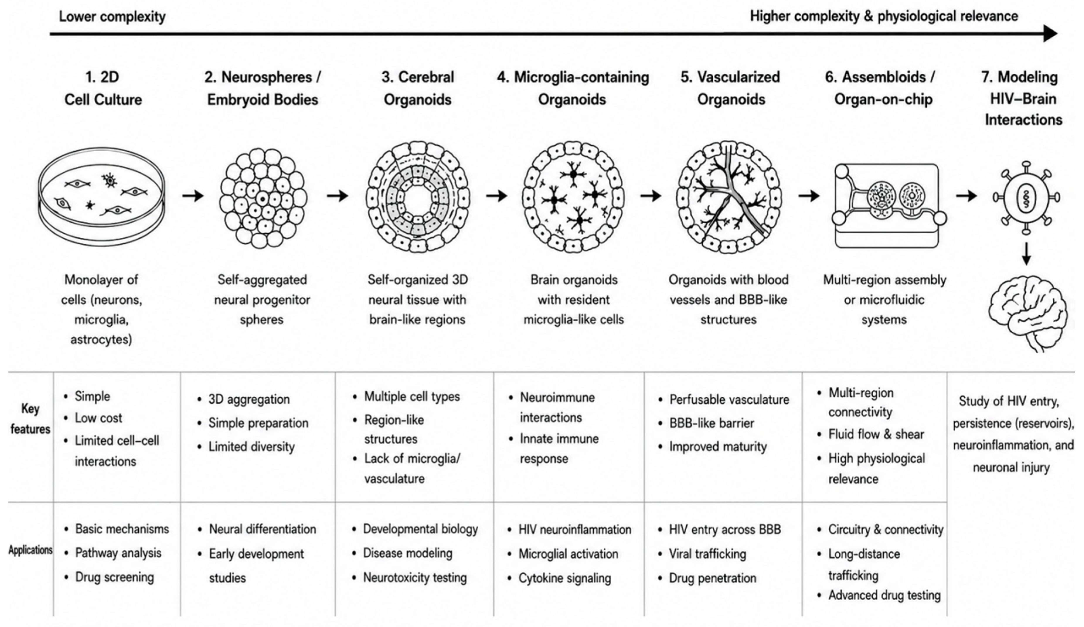

Table 1.
Overview of Human Cerebral Organoid Models for Neurological Research.
| Model | Key Innovation | Major Strengths | Major Limitations |
|---|---|---|---|
| 3D Neural Aggregates | Spontaneous self-organization of ESCs in suspension culture | First demonstration of self-organized cortical tissue formation [12]. | Limited complexity; not a standardized organoid model |
| Cerebral Organoids (CO) | Guided self-organization of human PSCs into brain-like structures | Models human neurodevelopment and regional brain organization [14]. | Lack microglia and BBB components |
| CO + Exogenous Mature Microglia | Incorporation of mature microglia into preformed organoids | Enables HIV infection and neuroinflammation studies [24]. | Limited developmental integration |
| CO + Intrinsic Microglia | Co-development of neural and immune lineages | Physiological microglial maturation and interactions [20,25]. | High batch-to-batch variability |
| CO + Hematopoietic Progenitors | In situ differentiation of microglia from progenitor cells | Controlled and physiologically relevant microglial incorporation [26]. | Limited immune-cell diversity |
| vMCOs | Emergence of endothelial-marker–expressing cells | Vascular spheroid and organoid fusion, long-term survival [22,27,28] | BBB incompleteness |
| Assembloid | Fusion of region-specific organoids | Models neuronal migration and interregional circuitry [29] | Technically demanding and less standardized |
| Organoids-on-Chip | Integration with microfluidic perfusion systems | Improved maturation, nutrient exchange, and reproducibility [30,31,32,33] | High cost and technical complexity |
Abbreviations: 3D, three-dimensional; BBB, blood-brain barrier; CO, cerebral organoids; ESCs, embryonic stem cells; HIV, human immunodeficiency virus; PSCs, pluripotent stem cells; vMCOs, vascularized microglia-containing cerebral organoids.
Table 2.
Key Findings from HIV-1 Infection Studies in Brain Organoids.
| Model | Research Question | Major Findings |
|---|---|---|
| Brain organoids with exogenously added HIV-infected microglia [24] |
Can HIV neuropathogenesis be modeled in human brain organoids? | HIV-infected microglia induce chronic neuroinflammation characterized by TNF-α and IL-1β production [24]. |
| Cerebral organoids with intrinsically grown microglia [25] |
Which CNS cells support HIV replication? | HIV replication restricted to CD4+CCR5+ microglia [25]. |
| MCOs and choroid plexus organoids [55] |
How do microglia propagate neuroinflammatory signaling? | HIV-infected microglia trigger CXCL10, CCL2 neuroinflammation and cause bystander neuronal dysfunction and death [55]. |
| CO-iMg [26] |
What is the impact of HIV infection on neurons? | HIV infection impairs neuronal function and promotes neurodegeneration [26]. |
| MCOs [53] |
Can cART mitigate HIV-associated neuronal injury? | cART suppresses viral replication and reduces HIV-induced cell death [53]. |
| MCOs [54] |
Can intrinsic antiviral immunity suppress HIV infection in brain organoids? | TLR3-mediated IFN responses suppress HIV replication [54] |
Abbreviations: cART, combination antiretroviral therapy; CNS, central nervous system; CO-iMg, cerebral organoid-derived microglia; HIV, human immunodeficiency virus; IFN, interferon; IL-1β, interleukin-1β; MCOs, microglia-containing organoids; TLR3, Toll-like receptor 3; TNF-α, tumor necrosis factor-α.
Disclaimer/Publisher’s Note: The statements, opinions and data contained in all publications are solely those of the individual author(s) and contributor(s) and not of MDPI and/or the editor(s). MDPI and/or the editor(s) disclaim responsibility for any injury to people or property resulting from any ideas, methods, instructions or products referred to in the content. |
© 2026 by the authors. Licensee MDPI, Basel, Switzerland. This article is an open access article distributed under the terms and conditions of the Creative Commons Attribution (CC BY) license (http://creativecommons.org/licenses/by/4.0/).
Copyright: This open access article is published under a Creative Commons CC BY 4.0 license, which permit the free download, distribution, and reuse, provided that the author and preprint are cited in any reuse.