
Submitted:
06 October 2026
Posted:
09 October 2026
You are already at the latest version
Abstract
Acyl-CoA thioesterase 9 (ACOT9), a member of the type II hot-dog fold thioesterase superfamily, play important roles in regulating the balance between intracellular acyl-CoA and free fatty acids. Although its functions have been extensively studied in vertebrates, its evolutionary diversification and physiological roles in Diptera, particularly in the economically important insect black soldier fly (Hermetia illucens), remain largely unclear. In this study, ACOT9 homologs were identified from 93 representative dipteran species, and H. illucens was used as a model to investigate phylogenetic re-lationships, synteny, domain architecture, expression profiles, and RNA interference (RNAi)-based functional analysis. The results showed that ACOT9 underwent lineage-specific duplications in the suborder Brachycera, giving rise to four paralogs (ACOT9.1–9.4), accompanied by lineage-specific gene losses. Synteny and conserved domain analyses suggested potential functional divergence among these paralogs. In contrast, the ancestral single-copy ACOT9 retained a catalytically con-served domain (e.g., d1lo7a) shared between vertebrates and insects. We cloned the full-length open reading frame (ORF) of H. illucens ACOT9. Transcript abundance peaked on day 12 of larval devel-opment (pre-pupation), with higher expression in males than in females, and predominant localiza-tion in the Malpighian tubules. RNAi-mediated knockdown in 6- to 7-day-old larvae significantly altered fatty acid composition: total saturated fatty acids increased by 13.35%, while monounsatu-rated and polyunsaturated fatty acids decreased by 5.39% and 14.27%, respectively, with relative abundances of lauric acid and linoleic acid changing by more than 10%. Transcriptomic analysis revealed that ACOT9 knockdown significantly upregulated genes involved in de novofatty acid synthesis (FAS1/2/3, ACC, MAT, KAS, ACP) and acyl-CoA activation (ACSM1/2, ACSL4/6), down-regulated the lipolysis gene LIP, and induced expression of the mitochondrial β-oxidation trans-porter genes CPT1/2. These findings indicate that ACOT9 deficiency triggers compensatory lipid synthesis and β-oxidation activation, ultimately leading to saturated fatty acid accumulation. In summary, this study elucidates the evolutionary diversification of ACOT9 in Diptera and demon-strates its pivotal role in lipid homeostasis in H. illucens, providing important insights into insect thioesterase function and endogenous lipid metabolic networks.
Keywords:
acyl-CoA thioesterase 9
; diptera
; fatty acid metabolism
; gene duplication
; Hermetia illucens
; RNA interference
1. Introduction
Acyl-CoA thioesterases (ACOTs) constitute a conserved enzyme superfamily distributed across prokaryotes and eukaryotes that exert central regulatory roles in lipid metabolism[1]. These enzymes catalyze the hydrolysis of acyl-CoA thioester bonds to generate free fatty acids and coenzyme A (CoA), thereby dynamically balancing intracellular pools of acyl-CoA and free fatty acids to sustain systemic lipid homeostasis [2-3]. To date, 15 vertebrate ACOT family members have been identified and classified based on structural and sequence features into two distinct subfamilies: type I α/β-hydrolase fold proteins (ACOT1–6) and type II “hotdog” fold thioesterases (ACOT7–15)[4-5].
ACOT9 belongs to the type II hot-dog fold thioesterase subfamily and is a mitochondrially localized thioesterase that has been primarily characterized in animal models. This enzyme has unique capability of medium-chain acyl-CoA recognition: unlike most thioesterases with narrow substrate specificity, ACOT9 can hydrolyze both short-chain (C2–C5) and medium-to-long-chain (C12–C16) acyl-CoAs, exhibiting particularly high catalytic activity toward medium-chain substrates such as lauroyl-CoA (C12-CoA). This property stems from its "double hot-dog" hexameric architecture, which enables non-competitive binding of short- and medium-chain substrates and generates a synergistic catalytic effect upon their coexistence. Under dual-substrate conditions, the hydrolytic efficiency of ACOT9 toward medium-chain acyl-CoAs increases by 6- to 20-fold compared with single-substrate conditions [6-7]. The role of ACOT9 thus extends beyond the classical fatty acid β-oxidation pathway, it also participates in clearing various amino acid-derived CoA esters, and involved in shaping tissue fatty acid profiles [8]. Consequently, ACOT9 is recognized as uniquely capable of medium-chain acyl-CoA recognition and cooperative catalysis, endowing it with an irreplaceable functional position within the mitochondrial thioesterase family.
The black soldier fly (Hermetia illucens, Diptera: Stratiomyidae) is a sustainable industrial insect promoted by the Food and Agriculture Organization (FAO) of the United Nations, distinguished by rapid growth, short reproductive cycles, low rearing costs, and high crude protein and lipid contents [9-10]. In 2025, China’s Ministry of Agriculture and Rural Affairs officially incorporated black soldier fly larvae into the National Feed Ingredient Catalogue to validate its feed utilization value. Compared with conventional plant and animal lipid feedstocks, dried black soldier fly larvae contain over 35% crude lipids dominated by medium-chain fatty acids (MCFAs), principally lauric acid, which can account for more than 30% of total fatty acids and reach up to 60% under optimized feeding regimes [11]. Despite the industrial relevance of its unique MCFAs-enriched phenotype, the molecular genetic mechanisms governing medium-chain fatty acid biosynthesis and accumulation in H. illucens remain uncharacterized. Given that ACOT family proteins serve as pivotal switches governing acyl-CoA metabolic flux and ACOT9 exhibits robust catalytic activity toward medium-chain acyl-CoA substrates, we hypothesized that ACOT9 plays a key role in regulating MCFAs accumulation in black soldier fly. However, no systematic characterization of ACOT9 has been reported in this species, leaving a critical gap in understanding the genetic basis of its MCFA-rich phenotype.
Herein, we systematically retrieved ACOT9 homologs from representative dipteran genomes to analyze gene copy number variation, protein tertiary structure divergence, synteny conservation, and phylogenetic relationships to uncover the genetic basis of functional differentiation among ACOT9 paralogs. Integrative experiments including gene cloning, bioinformatic characterization, spatiotemporal expression profiling, and RNAi-mediated gene silencing were further conducted to elucidate the regulatory roles of ACOT9 in fatty acid metabolism and fatty acid profile remodeling in H. illucens. This work provides fundamental molecular data to decipher the genetic basis of MCFAs accumulation in black soldier fly larvae.
2. Materials and Methods
2.1. Retrieval of ACOT9 Nucleotide and Protein Sequences
Whole-genome assemblies of 93 dipteran insect species were downloaded from the NCBI public database. The amino acid sequence of ACOT9 was used as the query for local tBLASTn searches (E-value cutoff = 10-5). Raw blast hits were filtered via the SOLAR v0.9.19 Perl script with thresholds of ≥75% identity and ≥40% similarity [11], followed by gene structure prediction using GenWise v2.4.1 [12]. Flanking genomic regions spanning 3,000 bp upstream and downstream of each predicted ACOT9 locus were extracted, and Perl scripts were applied to complete full-length nucleotide and protein sequences of all ACOT9 homologs.
2.2. Phylogenetic Reconstruction and Conserved Domain Prediction of ACOT9
Amino acid sequences of ACOT9 homologs retrieved from 93 dipteran species, together with outgroup sequences from Homo sapiens, Mus musculus, and Danio rerio, were subjected to multiple sequence alignment using MAFFT software[13]. A neighbor-joining phylogenetic tree was constructed in MEGA 11 with 1,000 bootstrap replicates for statistical support [11], and tree visualization was performed with FigTree v1.4.4(http://tree.bio.ed.ac.uk/software/figtree/). Conserved functional domains of ACOT9 proteins were predicted via the SMART online tool (http://smart.embl-heidelberg.de/); core functional site mutagenesis and docking analyses were not performed due to limited available functional residue data in the PDB database.
2.3. Synteny Analysis of Genomic Loci Flanking ACOT9
To evaluate the evolutionary conservation of genomic regions harboring ACOT9, the full-length ACOT9 and its adjacent flanking genes were set as the reference template. Genomic intervals spanning 100 kb upstream and downstream of ACOT9 in representative dipteran species were extracted, and all genes within these intervals were subjected to pairwise homology comparisons to identify conserved syntenic blocks. The IBS 2.0 online tool [14] was utilized for synteny block visualization.
2.4. Black Soldier Fly Rearing, Sample Collection, and Spatiotemporal Expression Profiling of ACOT9
Wild-type black soldier fly eggs were provided by the Institute of Animal Science, Guangdong Academy of Agricultural Sciences. Eggs were hatched under controlled conditions (28–30 °C, 60–70% relative humidity, photoperiod L8:D16). Newly hatched 3-day-old larvae were inoculated into PVC rearing boxes (0.45 m × 0.3 m × 0.3 m) at a density of 100 larvae per 500 g artificial diet until adult eclosion. The diet formulation consisted of 26% wheat bran, 2% brewer’s yeast, 1% casein, 1% plant soybean oil, 0.1% mineral mixture, 1% cholesterol, and 70% distilled water.
Whole-body samples were collected at embryonic stage, 3-, 6-, 9-, and 12-day larval stages, pupal stage, and male/female adult stages. Dissections of pre-pupae were performed to isolate fat body, Malpighian tubules, and midgut tissues. All samples were snap-frozen and stored at −80 °C. Four biological replicates with three individual larvae/tissues per replicate were prepared for each sampling timepoint.
Total RNA was extracted using the TRIzOL Up Plus RNA Kit (TransGen Biotech, Beijing), followed by DNase I digestion to eliminate genomic DNA contamination. RNA integrity and purity were validated via agarose gel electrophoresis and NanoDrop One spectrophotometry. Purified RNA was reverse-transcribed into complementary DNA (cDNA). Specific primers targeting ACOT9 (Forward: ACCTGTTCCAGTTTCGTCCT; Reverse: GGAACAATGGCTGGACACTG, GenBank accession XM_038066561) and the reference gene ef1α (Forward: TATGGTTGTCGTCTTCGCC; Reverse: GGATGGTTGAGCACGATGA, GenBank accession XM_038071208) were designed for quantitative real-time PCR (qRT-PCR). qRT-PCR reactions were conducted on a CFX Connect Real-Time PCR System using PerfectStart™ Green qPCR SuperMix (TransGen Biotech). The 20 μL reaction system contained 10 μL 2× qPCR SuperMix, 1 μL cDNA template, 0.4 μL each forward/reverse primer, and 8.2 μL nuclease-free water. Thermal cycling parameters were as follows: initial denaturation at 94 °C for 30 s, followed by 40 cycles of 95 °C for 15 s, 60 °C for 15 s, and 72 °C for 10 s. Relative mRNA expression levels were calculated using the 2−ΔΔCt method.
2.5. Full-Length Cloning and Bioinformatic Characterization of ACOT9
Full-length amplification primers were designed via Primer Premier 5.0 based on the open reading frame (ORF) sequence of ACOT9 retrieved in Section 2.1 (Forward: 5’-TGTTGGCGTACTTTGAGCG-3’; Reverse: 5’-TCTTAAAGGACAGGAGGAGCT-3’).Full-length PCR amplification was performed using cDNA synthesized from 6-day-old larvae as the template. Amplification products were separated on 1% agarose gels, and target fragments were purified with the SanPrep Column DNA Gel Extraction Kit. Purified DNA concentration and purity were quantified using a NanoDrop One spectrophotometer. Recovered amplicons were ligated into the pEASY®-T3 cloning vector, and ligation products were transformed into Trans5α competent Escherichia coli cells. Transformants were spread on LB solid medium containing ampicillin, supplemented with an equal-volume mixture of 500mM IPTG and 20 mg/mL X-gal, for blue-white screening. White single colonies were picked for Sanger sequencing, and strains with fully matched target sequences were cultured in liquid LB medium and preserved at −80 °C with 50% glycerol.
A comprehensive suite of bioinformatic tools was applied to characterize the deduced ACOT9 protein sequence: ProtParam(http://web.expasy.org/protparam/) for physicochemical property calculation, SignalP(http://www.cbs.dtu.dk/services/SignalP/) for signal peptide prediction, TMHMM (http://www.cbs.dtu.dk/services/TMHMM/)for transmembrane domain identification, Euk-multi-Loc(http://www.csbio.sjtu.edu.cn/bioinf/euk-multi-2/) for subcellular localization prediction, NetPhos(http://www.cbs.dtu.dk/services/NetPhos/) for phosphorylation site prediction, GlycoMod(https://web.expasy.org/glycomod/)for O-glycosylation(https://npsa-prabi.ibcp.fr/cgi-bin/npsa_automat.pl?page=npsa_sopma.htmL) site identification, and SOPMA for secondary structure prediction.
2.6. Synthesis, Delivery, and RNAi Knockdown of ACOT9 siRNA
Targeted siRNA duplexes were designed and synthesized based on the full-length ORF and conserved-domain sequence of ACOT9 using an online siRNA design platform (Tianyi Huayu Gene Technology Co., Ltd., Wuhan, China). Lyophilized siRNA powder was dissolved in RNase-free water to prepare a 3 μg/μL stock solution, which was mixed with lipid-nanoparticle (LNP) transfection reagent to prepare the working transfection solution. Pre-experiments were performed to screen the optimal siRNA sequence (sense strand: 5′-CCUUUGACAUACdTdT-3′; antisense strand: 5′-UUAGGCUGUAUGUCAAAGGdTdT-3′) and interference dosage, achieving a 59% gene-silencing efficiency for ACOT9 (Li Z.H., unpublished data). The scrambled negative-control siRNA (si-NC) shared identical GC content and chemical modifications with siACOT9, and BLAST analysis confirmed that it had no predicted off-target sites in the Hermetia illucens genome.
For the formal RNAi assay, two experimental groups were set up: si-NC negative-control group, and 3 μg/μL siACOT9 treatment group. six-day-old larvae were anesthetized on ice, and a fixed volume of corresponding working solution was microinjected into the intersegmental membrane between the 7th and 8th abdominal somites (Figure 1). Four biological replicates containing 80 healthy larvae each were established for each group. After injection, larvae were reared according to the feeding protocol described in Section 2.4 for 48 h. Whole-body larval samples were then collected and stored at −80 °C for fatty-acid profiling and quantification of lipid-metabolism-related gene expression.
2.7. qRT-PCR Quantification of Lipid Metabolism-Related Genes
Total RNA extraction, reverse transcription, and qRT-PCR amplification were performed on RNAi-treated larval samples following the protocols detailed in Section 2.2. The 20 μL reaction system contained 10 μL 2× qPCR SuperMix, 1 μL cDNA template, 0.4 μL each forward/reverse primer, and 8.2 μL nuclease-free water. Thermal cycling conditions consisted of a preheating step at 94 °C for 30 s, followed by 40 cycles of 94 °C for 5 s, 60 °C for 15 s, and 72 °C for 10 s. Relative gene expression levels were normalized to the ef1α reference gene using the 2−ΔΔCt method. The expression stability of ef1α in RNAi-treated larvae was validated prior to quantification. Complete primer sequences for all target lipid metabolism genes are listed in Table 1.
2.8. Crude Fat and Fatty Acid Composition Analysis
Larval samples collected pre- and post-RNAi were vacuum freeze-dried and pulverized, and subjected to crude fat extraction using the Soxhlet extraction method. Crude fat extracts were subjected to saponification and fatty acid methyl ester (FAME) derivatization following the Chinese national standard GB 5009.168-2016. Mixed fatty acid reference standards were used for chromatographic peak identification, and the relative percentage of each fatty acid component was calculated via peak area normalization.
2.9. Statistical Analysis
All raw data were processed in SPSS 26.0 software. For multi-group comparisons (developmental-stage and tissue expression profiles), one-way ANOVA followed by Duncan’s post-hoc test was applied. For two-group comparisons (siACOT9 vs negative control assays), independent-samples t-test was used. Normality and homogeneity-of-variance assumptions were examined prior to statistical testing.
3. Results
3.1. Copy Number Variation of ACOT9 Gene Across Diptera
In total, 119 ACOT9 homologs were identified from whole-genome assemblies of 93 dipteran species, exhibiting copy numbers ranging from 0 to 4 (Table 2). Five species (5.38%) displayed complete ACOT9 gene loss; 65 species (69.89%) possessed a single ACOT9 copy, representing the dominant genotype; 20 species (21.51%) harbored two paralogs, while only one species carried three copies and two species contained four ACOT9 paralogs. The average ACOT9 copy number across all 93 dipteran taxa was 1.25.
Within the suborder Nematocera, Diogma glabrata, Bibio marci, and Smittia aterrima possessed two ACOT9 copies, whereas gene loss was detected in Symmerus nobilis and Diadocidia ferruginosa. All remaining nematoceran species maintained a single ACOT9 paralog. For Brachycera, all Tabanomorpha species retained one ACOT9 copy except Atherix ibis, which exhibited gene loss. Multiple ACOT paralogs were widespread within Muscomorpha and Asilomorpha: gene loss was observed in Megaselia abdita and Phytomyza melanocephala, while species including Calliphora vicina, Glossina fuscipes, Drosophila simulans, and Drosophila yakuba underwent ACOT9 duplication, confirming recurrent gene amplification events during dipteran evolution (Figure 2).
Intrafamilial copy number divergence was also evident. Within the family Anthomyiidae, Eustalomyia histrio carried a single ACOT9 copy while Leucophora obtusa possessed two paralogs. The Drosophilidae exhibited extreme copy variation: Drosophila gunungcola had one copy, D. yakuba contained three paralogs, and D. simulans harbored four ACOT9 subtypes. Collectively, these data demonstrate pervasive ACOT9 copy number polymorphism across Diptera, with single-copy status representing the ancestral conserved state, while lineage-specific gene loss and duplication generated hierarchical variation at the suborder, family, and species levels (Figure 3).
3.2. Phylogenetic Analysis Based on ACOT9 Amino Acid Sequences
A circular phylogenetic tree was constructed using full-length ACOT9 protein sequences from representative Diptera (Figure 4). The tree resolved two primary monophyletic clades corresponding to Nematocera (Clade I) and Brachycera, with Brachycera further subdivided into three well-supported independent subclades (Clades II, III, IV).
All Tabanomorpha species except Atherix ibis retained single-copy ACOT9 sequences that clustered tightly within Clade II together with Asilomorpha, forming reciprocal sister lineages. Partial Asilomorpha species exhibited ACOT9 duplication: paralogs from Condylostylus longicornis, Sciapus platypterus, and Empis stercorea fell within Clade II but formed distinct sub-branches indicative of substantial sequence divergence between duplicated copies. Muscomorpha taxa exclusively occupied Clades III and IV, with species carrying 2–4 ACOT9 paralogs displaying clear subtype-based clustering: ancestral ACOT9/ACOT9.1 sequences formed one subclade, while derived ACOT9.2/3/4 paralogs grouped into a separate monophyletic lineage.
3.3. Synteny Analysis of Genomic Regions Flanking ACOT9 Loci
Synteny comparisons of the 100 kb genomic intervals upstream and downstream of ACOT9 were performed across representative dipteran taxa (Figure 5). Conserved syntenic blocks of adjacent genes flanking ACOT9 were detected in most species, yet no universal gene arrangement was shared across all lineages, with partial loss of syntenic genes observed in several taxa.
In taxa harboring multiple ACOT paralogs such as Condylostylus longicornis and Drosophila yakuba, ACOT9.1 and ACOT9.2 were tightly linked on the same chromosome or scaffold. Distinct genomic partitioning patterns were identified in Drosophila simulans and D. yakuba: ACOT9.2/3/4 formed a tandem array within a single syntenic block, whereas ACOT9.1 translocated to a separate chromosomal interval, indicating large-scale genomic rearrangement following gene duplication in drosophilid lineages.
3.4. Conserved Domain Architecture of Dipteran ACOT9 Proteins
Conserved domain prediction was performed on ACOT9 protein sequences from representative animal taxa (Figure 6). All ACOT9 proteins contained a core hydrolase domain, with the d1lo7a fold representing the universal conserved catalytic domain present in all single-copy ACOT9 orthologs; a subset of species additionally harbored the 4IEJD hydrolase variant. In Muscomorpha and drosophilid species with duplicated ACOT paralogs, derived ACOT9.2/3/4 subtypes acquired two novel hydrolase folds (2QQ2L and 3SPSIF), resulting in divergent domain architectures between ancestral and duplicated paralogs and confirming functional specialization following gene duplication. Purple vertical bars in the schematic denote low-complexity protein regions with lineage-specific distribution patterns across multi-copy taxa. Cross-kingdom comparisons revealed nearly identical domain organization between H. illucens ACOT9 and vertebrate ACOT9 orthologs from human and zebrafish, demonstrating deep evolutionary conservation of the core lipid hydrolysis function of ACOT9 across vertebrate and dipteran lineages.
3.5. Spatiotemporal Expression Profiling of ACOT9 in Hermetia Illucens
qRT-PCR quantification of ACOT9 transcript levels was conducted across nine developmental timepoints and three pre-pupal tissues (Figure 7). ACOT9 exhibited constitutive expression throughout all life stages. Larval expression levels increased gradually and peaked at day 12 prior to pre-pupal transition, followed by a progressive decline during late larval development. Adult expression profiling revealed significantly higher ACOT9 abundance in male adults relative to females. Tissue-specific expression in pre-pupae demonstrated maximal transcript accumulation in Malpighian tubules, intermediate expression in fat body, and minimal expression in the midgut.
3.6. Full-Length Cloning and Bioinformatic Characterization of ACOT9
PCR amplification using gene-specific primers yielded a 1,347 bp full-length ORF of ACOT9. Gel purification, vector ligation, and colony PCR screening confirmed successful construction of positive recombinant clones. Sanger sequencing verified that the ORF encodes a 448-amino-acid polypeptide mapped to the genomic scaffold LR899014.1 (coordinates 41610125–41619099) with a TGA termination codon. Physicochemical parameter calculations (Table 3) predicted a molecular mass of 51,658.69 Da (51.66 kDa) and theoretical pI of 9.04; an instability index of 49.65 classified ACOT9 as an unstable protein, with a negative grand average of hydropathicity (GRAVY = −0.292) indicating overall hydrophilicity.
SignalP 5.0 detected no canonical N-terminal secretory signal peptide, and TMHMM 2.0 predicted the absence of transmembrane helices. Subcellular localization via Euk-multi-Loc dualistically targeted ACOT9 to mitochondria and cytoplasm. SOPMA secondary structure prediction identified 38.17% α-helices, 0.89% β-turns, and 44.87% random coils. Post-translational modification analysis via DictyOGlyc 1.1 identified a single O-glycosylation site at residue 90, while NetPhos 3.1 predicted 47 total phosphorylation sites (23 serine, 20 threonine, 4 tyrosine). Homology modeling using human ACOT9 (Q9Y305) as the template revealed highly conserved global tertiary folding between human and black soldier fly ACOT9, with structural divergence limited to disordered C-terminal flexible loops (Figure 8).
3.7. Functional Effects of RNAi-Mediated ACOT9
Changes in larval fat content and fatty acid profiles followed ACOT9 knockdown. After 48 h of ACOT9 gene interference, the crude fat content of larvae slightly decreased, dropping from 18.65 ± 0.25 % in the negative control to 17.85 ± 0.05 % (P < 0.05).
GC-MS profiling of fatty acid composition was performed on larvae treated with 3 μg/μL siACOT9-LNP for 48 h (Table 4). Relative to negative control larvae, total saturated fatty acid (SFA) proportions increased by 13.35%, while monounsaturated fatty acids (MUFAs) and polyunsaturated fatty acids (PUFAs) decreased by 5.39% and 14.27%, respectively. Among SFAs, C10:0, C12:0, C14:0, and C18:0 content was significantly elevated, with lauric acid (C12:0) exhibiting the largest increase of 10.39%; palmitic acid (C16:0) showed no statistically significant variation. All detected unsaturated fatty acids (C16:1n-7, C18:1n-9, C18:2n-6, C18:3n-3) were significantly reduced, with linoleic acid (C18:2n-6) displaying the most substantial decline of 12.51%.
3.8. Transcriptional Remodeling of Lipid Metabolism Genes Following ACOT9 Knockdown
qRT-PCR quantification of key lipid regulatory genes revealed widespread transcriptional rewiring after siRNA injection (Figure 9). All core genes governing type II de novo fatty acid synthesis (FAS1/2/3, ACC, MAT, KAS, ACP) were significantly upregulated (P < 0.05). Both medium-chain acyl-CoA synthetase paralogs ACSM1 and ACSM2 were induced upon ACOT9 knockdown, although ACSM1 exhibited a far greater magnitude of up-regulation relative to its negative control than ACSM2. Similarly, long-chain fatty acid-activating genes ACSL4 and ACSL6 were significantly up-regulated; the induction amplitude of ACSL4 was much higher than that of ACSL6. Within fatty acid catabolic pathways, the lipase-encoding gene LIP was markedly downregulated, whereas mitochondrial fatty acid transporters CPT1 and CPT2 exhibited significant transcriptional elevation. Collectively, these transcriptional changes demonstrate that knockdown of ACOT9 activates the complete de novo fatty acid synthesis program and enhances medium-/long-chain fatty acid activation, while suppressing triacylglycerol lipolysis and compensatorily upregulating mitochondrial β-oxidation transport machinery to alleviate intracellular acyl-CoA accumulation.
4. Discussion
By integrating comparative genomics, phylogenetics, and in vivo RNAi, the present study provides the first systematic characterization of ACOT9 across Diptera, with a detailed functional dissection in the black soldier fly (Hermetia illucens). The findings provide genetic evidence for understanding the evolutionary diversification of ACOT9 in dipteran insects and the molecular regulatory mechanism underlying medium-chain fatty acid enrichment in black soldier fly.
4.1. Lineage-Specific Duplication and Functional Diversification of ACOT9 in Diptera
Genome-wide surveys across 93 dipteran species revealed that the single-copy state of ACOT9 represents the ancestral condition, with lineage-specific gene losses and duplications generating substantial copy-number variation. The average copy number of 1.25 and the predominance of single-copy taxa imply persistent purifying selection on ACOT9 in most lineages. Nevertheless, recurrent duplications in Brachy-cera—especially within Muscomorpha—generated up to four paralogs (ACOT9.1–9.4). This expansion pattern mirrors the birth-and-death dynamics documented in other in-sect lipid-metabolism gene families, such as fatty acyl-CoA reductases and desaturases [15], indicating that ACOT9 amplification in higher Diptera is likely driven by ecological or metabolic specialization.
Phylogenetic reconstruction resolved a clear separation between Nematocera and Brachycera, with further divergence of ancestral ACOT9/ACOT9.1 and derived ACOT9.2/3/4 clades within Muscomorpha. This topology argues against simple redun-dancy among duplicated copies. Consistent with this, synteny analysis demonstrated that ACOT9.2/3/4 paralogs frequently reside in tandem arrays, whereas ACOT9.1 has been subject to chromosomal rearrangement and repositioning, as observed in Drosophila simulans and D. yakuba. Such genomic shuffling is known to alter cis-regulatory landscapes and dosage constraints, providing a mechanistic basis for sub- or neofunctionalization [16].
4.2. Conserved Catalytic Core Alongside Divergent Domain Architectures
Structural comparison revealed that the d1lo7a catalytic domain is universally conserved in single-copy ACOT9 orthologs across insects and vertebrates. This supports deep evolutionary conservation of the core thioesterase reaction—hydrolytic cleavage of acyl-CoA into free fatty acid and CoA—over approximately 500 million years of divergence. The nearly identical domain architecture between H. illucens ACOT9 and its human and mouse counterparts further suggests that the mitochondrial lipid-homeostatic role of ACOT9 is an ancestral trait rather than a vertebrate-specific acquisition.
In contrast, derived paralogs in Muscomorpha acquired additional hydrolase folds (e.g., 2QQ2L, 3SPSIF) and lineage-specific low-complexity regions absent in ancestral copies. Similar domain innovation of specific genes, such as has been reported in insect, where duplicated members often evolve altered substrate specificity or tissue distribution [17-18]. Our data therefore update the current understanding of ACOT9 evolution: the protein is characterized by a conserved catalytic core flanked by a highly plastic peripheral architecture, offering a compelling model for studying how enzyme families reconcile metabolic constraint with evolutionary experimentation.
4.3. Expression Profiling in Larval Lipid Remodeling and Excretory Tissue Metabolism
In H. illucens, ACOT9 transcript levels peaked on day 12 of larval development, immediately preceding the prepupal transition when larvae cease feeding and initiate metabolic reorganization for metamorphosis. This temporal dynamics aligns with the reports that lipid-metabolic genes reach maximal activity during late larval feeding stages in dipterans and lepidopterans [19].
ACOT9 expression also exhibited significant sexual dimorphism, with higher transcript abundance in males than in females, implying sex-specific lipid allocation strategies. In Drosophila, males preferentially mobilize lipids to support flight and motility, whereas females channel lipids into reproductive storage [20]; Notably, ACOT9 was predominantly expressed in Malpighian tubules rather than the fat body, the primary lipid storage organ in insects. Accumulating evidence indicates that Malpighian tubules participate in active lipid metabolism, xenobiotic detoxification, and intracellular CoA homeostasis [21]. These results expand our current understanding of insect acyl-CoA buffering systems and highlight the involvement of excretory tissues in lipid regulation.
4.4. ACOT9 Knockdown Reshapes Fatty Acid Composition Toward Saturated and Medium-Chain Accumulation
RNAi-mediated ACOT9 silencing induced a substantial shift in larval fatty acid profiles: total SFAs increased by 13.35%, while MUFAs and PUFAs decreased by 5.39% and 14.27%, respectively, with lauric acid (C12:0) rising over 10% and linoleic acid (C18:2n-6) declining by ~12.5%. In vertebrates, ACOT9 localizes to mitochondria and couples acyl-CoA hydrolysis to β-oxidation [11]; however, in vivo functional data for insect ACOT9 paralogs have been lacking. Our results thus provide the first evidence that ACOT9is a key regulator of fatty acid homeostasis in an insect.
Contrary to the expectation that ACOT9 loss would cause acyl-CoA accumulation and free fatty acid depletion, transcriptomic profiling revealed a coordinated compensatory response. Core de novo lipogenesis genes (FAS1/2/3, ACC, MAT, KAS, ACP) were markedly upregulated, as were medium-chain acyl-CoA synthetases ACSM1 and ACSM2. Notably, ACSM1 showed a far greater induction than ACSM2, consistent with its preferential activation of endogenous C4–C12 SFAs and raising the possibility that intracellular medium-chain [22]. SFAs accumulation is a primary metabolic consequence of ACOT9 knockdown—a model requiring biochemical validation. Concurrently, LIP was downregulated, while mitochondrial transporters CPT1/2 and long-chain activators ACSL4/6 were induced, together reflecting an integrated rewiring of lipolysis, β-oxidation, and acyl-CoA trafficking to restore lipid balance [23].
These transcriptional responses reveal that ACOT9 knockdown triggers multi-layered metabolic reprogramming rather than a passive loss-of-function phenotype. The concurrent rise in C12:0 and fall in PUFAs indicates that endogenous ACOT9 normally channels medium-chain acyl-CoA toward mitochondrial oxidation and CoA recycling; its disruption redirects flux toward medium-chain FFA accumulation at the expense of PUFA desaturation. We therefore establish ACOT9 as an active gatekeeper of MCFAs versus PUFAs balance in insects.
5. Conclusions
In conclusion, based on the evolutionary trajectory of ACOT9 across Diptera and functional investigation of its role in lipid metabolism in the black soldier fly (Hermetia illucens), the present study revealed that ACOT9 has experienced lineage-specific duplication and gene loss, with recurrent amplification in Brachycera giving rise to the ACOT9.1–9.4 paralogous cluster. In H. illucens, ACOT9 exhibits a conserved catalytic architecture shared with vertebrates, yet its spatiotemporal expression is coupled to late larval development and is predominantly localized to Malpighian tubules, pointing to a tissue-enriched role in acyl-CoA buffering. RNAi-mediated silencing demonstrated that ACOT9 may act as a key node linking de novo lipogenesis, acyl-CoA activation, lipolysis, and mitochondrial β-oxidation: its knockdown triggers a compensatory metabolic reprogramming that shifts larval fatty acid profiles toward saturated and medium-chain enrichment, most notably elevating lauric acid (C12:0) while suppressing polyunsaturated fatty acid pools, suggesting that ACOT9 contributes to fatty acid homeostasis in H. illucens. Further investigation into the enzymatic properties of recombinant H. illucens ACOT9 and evaluation of gene-targeted molecular breeding strategies for improving larval lipid quality are warranted.
Author Contributions
Yu-Ting Lin and Ze-Hong Li: Investigation, Software, Formal analysis, Writing-original draft; Kai Zhang: Data curation, Validation; Ren-Bao Yang and Gui-Chun Huang: Formal analysis; Jia-Lin Luo: Visualization; Rishen Liang: Supervision, Writing – review & editing; Meng Zhou: Conceptualization, Resources, Project administration, Writing, review & editing. All authors have read and approved the final version of the manuscript.
Data Availability Statement
The data presented in this study are available from the corresponding author upon reasonable request.
Acknowledgments
This study was supported by National Key R&D Pro-gram of China (2023YFD2402000 and 2023YFD2401705).
Conflicts of Interest
The authors declare no conflicts of interest.
References
- Benjamin, T. Caswell.; Caio, Cesar de. C.; Hung, Nguyen.; Monikrishna, R.; Tin, N.; David, C. C. Thioesterase enzyme families: Functions, structures, and mechanisms. Protein Sci 2021, 31(3): 652-676. [CrossRef]
- Bekeova, C.; Anderson-Pullinger, L.; Boye, K.; Boos, F.; Sharpadskaya, Y.; Herrmann, J. M.; Seifert, E.L. Multiple mitochondrial thioesterases have distinct tissue and substrate specificity and CoA regulation, suggesting unique functional roles. J Bio chem 2019, 294(50): 19034-19047. [CrossRef]
- Bai, L.; Yang, P.; Han, B.; Kong, L. Progress of the acyl-Coenzyme A thioester hydrolase family in cancer. Front Oncol 2024, 14, 1374094. [CrossRef]
- Brocker. C.; Carpenter. C.; Nebert, D. W.; Vasiliou, V. Evolutionary divergence and functions of the human acyl-CoA thioesterase gene (ACOT) family. Human genomics, 2010, 4(6), 411. [CrossRef]
- Hunt MC, Yamada J, Maltais LJ, et al. A revised nomen-clature for mammalian acyl-CoA thioesterases/hydro-lases. Lipid Res, 2005, 46(9): 2029-2032.
- Tillander, V.; Arvidsson, N. E.; Reilly, J. Acyl-CoA thioesterase 9 (ACOT9) in mouse may provide a novel link between fatty acid and amino acid metabolism in mitochondria[J]. Cell Mol Life Sci, 2014, 71(5):933-948.
- Tillander, V. Peroxisomal and mitochondrial enzymes involved in lipid metabolism: studies on function and regulation. Doctor's thesis, Karolinska Institutet, Stockholm, Sweden, 2013.
- Steensels, S.; Qiao, J.; Zhang, Y. Acyl-coenzyme A thioesterase 9 traffics mitochondrial short-chain fatty acids toward de novo lipogenesis and glucose production in the liver. Hepatology, 2020, 72(3), 857-872.
- Ursada, R.; Bagastyo A Y.; Tay, C. Y.; Purnomo, A.; Warmadewanthi, I.D.A.A. Bioconversion of sludge waste by black soldier fly (Hermetia illucens) larvae: A review of the potential use of food and beverage industry sludge as a rearing substrate. Clean Waste Syst 2026,14, 100500.
- Barrett, M.; Chia, S.Y.; Fischer, B.; Tomberlin, J.K. Welfare considerations for farming black soldier flies, Hermetia illucens (Diptera: Stratiomyidae): a model for the insects as food and feed industry. J Insects Food Feed 2023, 9, 119–148. [CrossRef]
- Giannetto, A.; Oliva, S.; Ceccon Lanes, C.F.; de Araújo Pedron, F.; Savastano, D.; Baviera, C.; Parrino, V.; Lo Paro, G.; Spanò, N.C.; Cappello, T.; Maisano, M.; Mauceri, A.; Fasulo, S. Hermetia illucens (Diptera: Stratiomydae) larvae and prepupae: Biomass production, fatty acid profile and expression of key genes involved in lipid metabolism. J Biotechnol 2020, 307, 44–54. [CrossRef]
- Yu, X.J.; Zheng, H.K.; Wang, J.; Wang, W.; Su, B. Detecting lineage-specific adaptive evolution of brain-expressed genes in human using rhesus macaque as outgroup. Genomics 2006, 88, 745–751. [CrossRef]
- Birney, E.; Clamp, M.; Durbin, R. GeneWise and Genomewise. Genome Res 2004, 14, 988–995. [CrossRef]
- Yamada, K.D.; Tomii, K.; Katoh, K. Application of the MAFFT sequence alignment program to large data-reexamination of the usefulness of chained guide trees. Bioinformatics 2016, 32, 3246–3251. [CrossRef]
- Finet, C.; Slavik, K.; Pu, J.; Carroll, S.B.; Chung, H. Birth-and-Death Evolution of the Fatty Acyl-CoA Reductase (FAR) Gene family and diversification of cuticular hydrocarbon synthesis in Drosophila. Genome Biol Evol 2019, 11, 1541–1551. [CrossRef]
- Lucek, K.; Giménez, M. D.; Joron, M.; Rafajlović, M.; Searle, J.; Walden, N.; Westram, A.; Faria, R. The impact of chromosomal rearrangements in speciation: From micro- to macroevolution. CSH Perspect Biol 2023, 15(11) . [CrossRef]
- Kaur, K.; Kaur, A.; Shelly, S.; Bala, B.; Kaur, A. Xenobiotic detoxification in insects: regulatory mechanisms and evolutionary roles of cytochrome P450. J Adv Zoo 2024. [CrossRef]
- Colinet, D.; Haon, M.; Drula, E.; Boyer, M.; Grisel, S.; Belliardo, C.; Koutsovoulos, G. D.; Berrin, J. G.; Danchin, E. G. J. Functional carbohydrate-active enzymes acquired by horizontal gene transfer from plants in the whitefly Bemisia tabaci. Genome Bio Evol 2025, 17(2). [CrossRef]
- Giannetto, A.; Oliva, S.; Lanes, C.F.C.; Pedron, F.A.; Savastano, D.; Baviera, C.; Maisano, M. Hermetia illucens larvae and prepupae: biomass production, fatty acid profile and expression of key genes involved in lipid metabolism. J Biotechnol 2020, 307, 44–54.
- Dähn, S.; Wagner, A. E. Drosophila melanogaster as a model organism to investigate sex specific differences. Sci Rep 2025, 15, 19648. [CrossRef]
- Silva, J. R.; Amaral, D. T.; Viviani, V. R. Comparison of the Malpighian tubules and fat body transcriptional profiles of Zophobas morio larvae (Coleoptera: Tenebrionidae), Com Biochem Physiol -D, 2019, 29, 95-105.
- van der Sluis, R.; Erasmus, E. Xenobiotic/medium chain fatty acid: CoA ligase – a critical review on its role in fatty acid metabolism and the detoxification of benzoic acid and aspirin. Expert Opin Drug Met 2016, 12(10), 1169–1179.
- Ding, K.; Liu, C.; Li, L.; Yang, M.; Jiang, N.; Luo, S.; Sun, L. Acyl-CoA synthase ACSL4: an essential target in ferroptosis and fatty acid metabolism. Chin Med J 2023, 136(21) . [CrossRef]
Figure 1.
Schematic diagram showing the microinjection site in the black soldier fly larvae.The red circle marks the injection site at the intersegmental membrane of the 7th and 8th abdominal segments.
Figure 1.
Schematic diagram showing the microinjection site in the black soldier fly larvae.The red circle marks the injection site at the intersegmental membrane of the 7th and 8th abdominal segments.
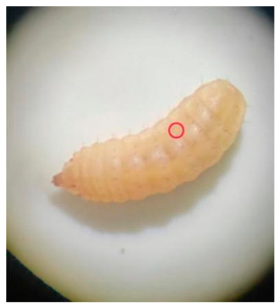
Figure 2.
Identification of ACOT9 gene copy numbers in individual dipteran species.
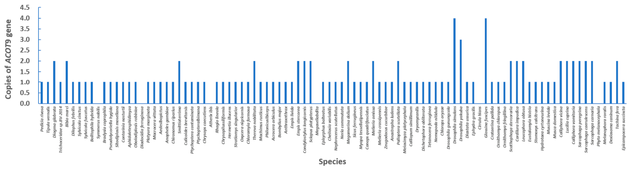
Figure 3.
Average ACOT9 gene copy numbers across the top 10 dipteran families with available genomic data.
Figure 3.
Average ACOT9 gene copy numbers across the top 10 dipteran families with available genomic data.
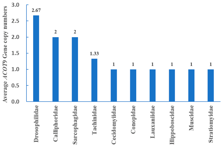
Figure 4.
Phylogenetic relationships among Dipteran insect ACOT9 amino acid sequences. Four clades are marked by different colors: green for Clade I (Nematocera), purple for Clade II (Tabanomorpha and Asilomorpha), light blue for Clade III (ancestral ACOT9/ACOT9.1 of Muscomorpha), dark blue for Clade IV (derived ACOT9.2/3/4 of Muscomorpha). Representative Brachycera ACOT9 genes were categorized into three main clades.
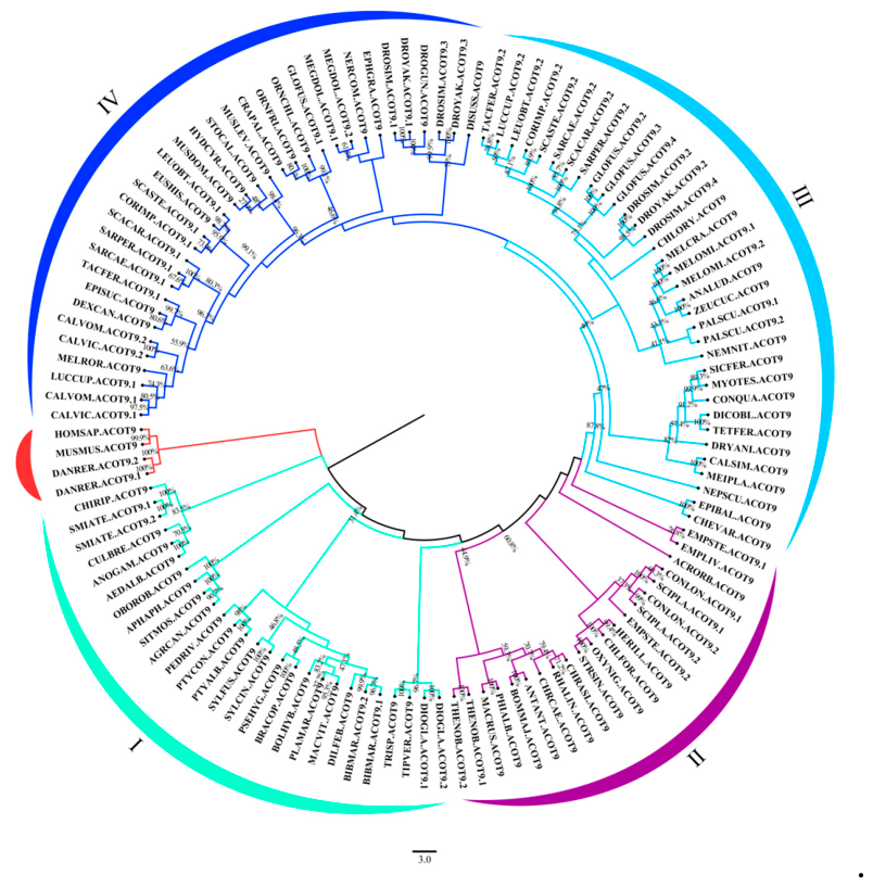
Figure 5.
Synteny visualization of conserved gene clusters flanking ACOT9 loci in representative Diptera. Each colored oval represents a distinct orthologous gene locus.
Figure 5.
Synteny visualization of conserved gene clusters flanking ACOT9 loci in representative Diptera. Each colored oval represents a distinct orthologous gene locus.
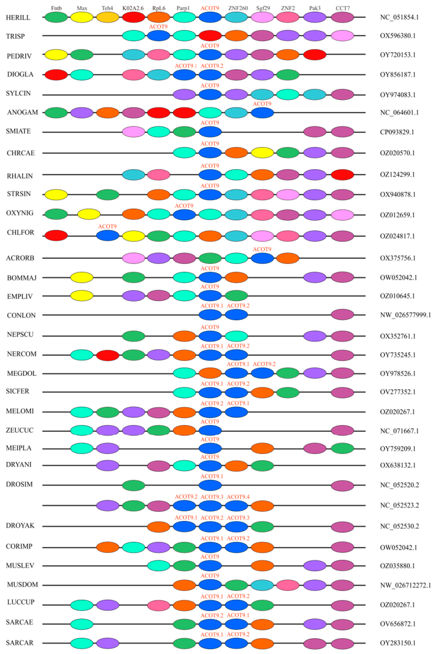
Figure 6.
The domains featured in ACOT9 genes of Dipteran insects. Gray horizontal lines represent full-length amino acid sequences; orange boxes indicate the universal conserved d1lo7a core hydrolase domain; red boxes denote lineage/subtype-specific hydrolase folds (4IEJD, 2QQ2L, 3SPSIF); purple vertical lines mark low-complexity regions. Human and mouse ACOT9 sequences serve as vertebrate outgroups on the left side of the panel.
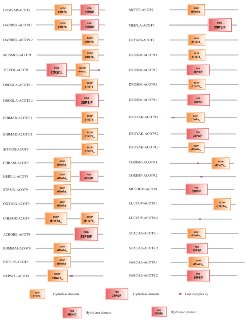
Figure 7.
Relative spatiotemporal expression levels of ACOT9 in Hermetia illucens. A: Developmental stage expression profiles; B: Tissue-specific expression in pre-pupae. EB: Embryo; 3d/6d/9d/12d/15d: 3–15-day-old larvae; PP: Pre-pupa; FA: Female adult; MA: Male adult; FB: Fat body; MT: Malpighian tubules; MB: Midgut. Different lowercase letters above bars indicate statistically significant intergroup differences (P < 0.05).
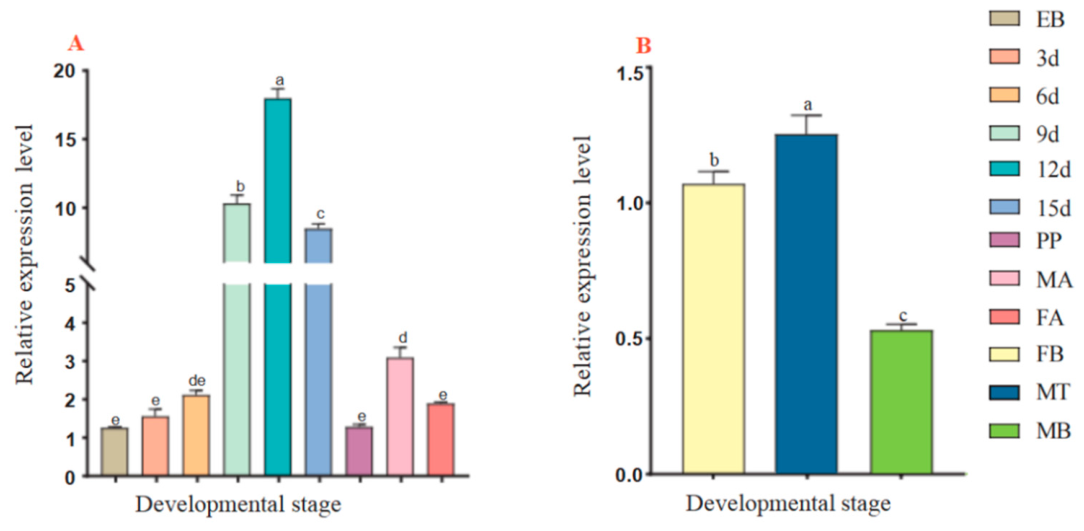
Figure 8.
Predicted tertiary structures of human and black soldier fly ACOT9 proteins. A: Human ACOT9 (UniProt Q9Y305); B: ACOT9 from Hermetia illucens. Conserved core hydrolase domains are colored blue; unstructured flexible terminal regions are highlighted in orange.
Figure 8.
Predicted tertiary structures of human and black soldier fly ACOT9 proteins. A: Human ACOT9 (UniProt Q9Y305); B: ACOT9 from Hermetia illucens. Conserved core hydrolase domains are colored blue; unstructured flexible terminal regions are highlighted in orange.
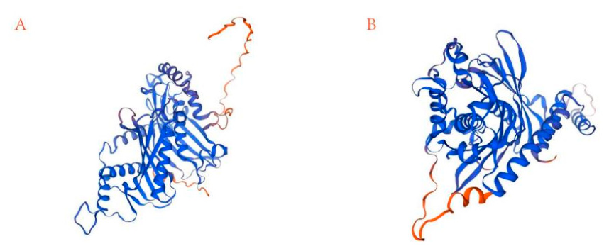
Figure 9.
Relative expression levels of lipid metabolism regulatory genes after ACOT9 RNAi silencing. Orange bars: Negative control group; gray bars: 3 μg/μL siACOT9 interference group. Asterisks denote significant intergroup differences (** P < 0.01, *** P < 0.001). Gene abbreviations: ACC acetyl-CoA carboxylase; MAT malonyl-CoA transferase; KAS β-ketoacyl-ACP synthase; ACP acyl carrier protein; FAS1/2/3 fatty acid synthase paralogs; LIP triacylglycerol lipase; CPT1/2 carnitine palmitoyltransferase 1/2; ACSM1/2 medium-chain acyl-CoA synthetase paralogs; ACSL4/6 long-chain acyl-CoA synthetase paralogs.
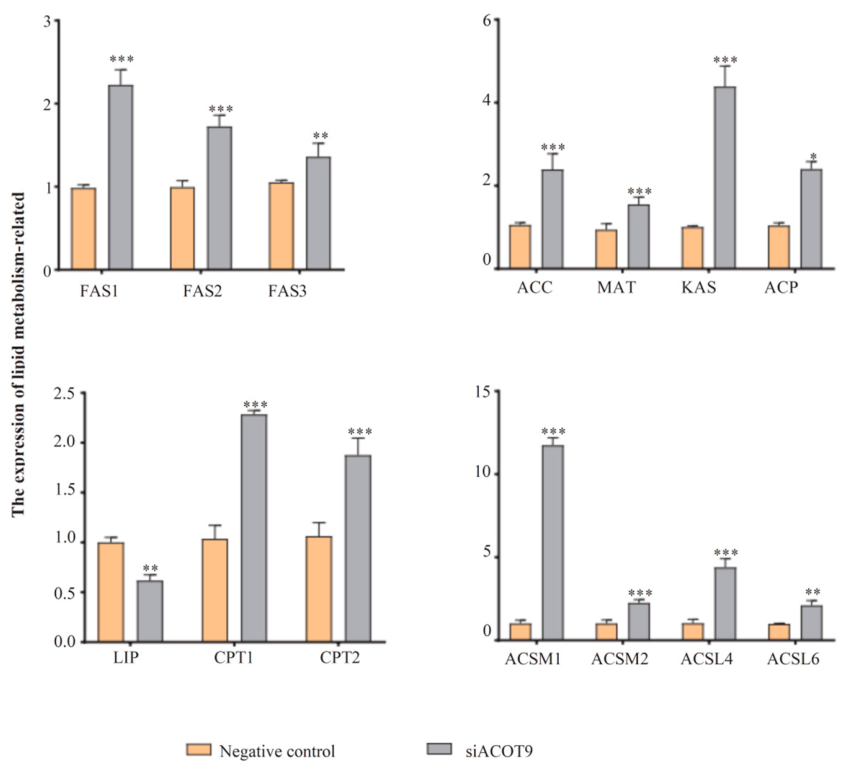

Table 1.
Primer sequences for qRT-PCR detection of lipid metabolic genes.
| Gene Name | Forward Primer (5’–3’) | Reverse Primer (5’–3’) |
| CPT1 | TGAACCGTGGAGACTGTCAA | TTAAATTCTCGCCAGCCACG |
| CPT2 | CTCCATACTGCCAACCATGC | GTGGTCCTGAGCCTGTTTTG |
| LIP | GGGAATTCAGTTGGCACGAA | GTGCATTGCTCGGATTTTGC |
| ACSM1 | GCAGAACGGCACCAAGTTTA | GACCTATCACAGCGGCTTTG |
| ACSM2 | GAGGAAAGATGCACCGTCAC | ATCCGGAAGACTTTGGCTCA |
| ACSL4 | CTGGCATCTTCTTTCGGTCG | AATGCGCTACACCATCATCG |
| ACSL6 | GTATCTCGGCCAGTTCCAGA | ATACGCACCGACACGAAATG |
| ACC | CGGGTGACAAGCAGAAAACA | CACGCTCTTGACTCTCTCTCCA |
| ACP | GATGCTCCATCGACAATGCG | GGACGCGTTCACGAATTTCT |
| MAT | CCGCATTGCAAGGTGATAGC | CCACTCACTGGCAACGATCT |
| KAS | GCGAAGGTTTTGTGATGGGT | AAGCTCCGGAACCATCTTCA |
| FAS1 | TTGTGCCTATGCCGATGGTT | TTGTGGCAAGCAGCAATCAC |
| FAS2 | ATCAATCGGGCGAAACCCTT | ACACTACGGGGGTTTCTGAC |
| FAS3 | AAGCGTGGTACAATGTCGGT | ACGTACCGAAGGAGTAGCCT |
*CPT1/2, carnitine palmitoyltransferase 1/2; LIP, triacylglycerol lipase; ACSM1/2, acyl-CoA synthetase medium-chain family member 1/2; ACSL4/6, acyl-CoA synthetase long-chain family member 4/6; ACC, acetyl-CoA carboxylase; ACP, acyl carrier protein; MAT, malonyl-CoA-acyl-carrier-protein transacylase; KAS, β-ketoacyl-ACP synthase; FAS1/2/3, fatty acid synthase 1/2/3.
Table 2.
Summary of ACOT9 copy number distribution among 93 dipteran species.
| Parameter | Value |
| Total valid dipteran species | 93 |
| Species with zero ACOT9 copies (gene loss) | 5 |
| Species with one ACOT9 copy | 65 |
| Species with two ACOT9 copies | 20 |
| Species with three ACOT9 copies | 1 |
| Species with four ACOT9 copies | 2 |
| Mean ACOT9 copy number across all species | 1.25 |
Table 3.
Basic physicochemical and bioinformatic parameters of ACOT9 protein.
| Parameter | Value |
| Total amino acid residues | 448 |
| Genomic scaffold ID | LR899014.1 |
| Gene start coordinate | 41610125 |
| Gene end coordinate | 41619099 |
| Termination codon | TGA |
| Molecular weight (Da) | 51658.69 |
| Theoretical isoelectric point (pI) | 9.04 |
| Instability index | 49.65 |
| Aliphatic index | 86.18 |
| Grand average of hydropathicity (GRAVY) | −0.292 |
| Predicted subcellular localization | Mitochondria, Cytoplasm |
Table 4.
Fatty acid composition (% total fatty acids) of negative control and siACOT9-treated black soldier fly larvae.
Table 4.
Fatty acid composition (% total fatty acids) of negative control and siACOT9-treated black soldier fly larvae.
| Fatty Acids | Negative Control | 3 μg/μL siACOT9 Group |
| C10:0 | 0.78±0.05 | 1.61±0.01*** |
| C12:0 | 31.87±1.81 | 42.26±0.36*** |
| C14:0 | 5.74±0.12 | 6.44±0.10* |
| C16:0 | 10.69±0.43 | 11.52±0.44 |
| C16:1n-7 | 1.54±0.02 | 0.86±0.07*** |
| C18:0 | 3.09±0.21 | 3.67±0.14** |
| C18:1n-9 | 11.45±0.57 | 6.73±0.39*** |
| C18:2n-6 | 28.33±0.72 | 15.82±1.39*** |
| C18:3n-3 | 3.47±0.02 | 1.70±0.20* |
| Other fatty acids | 2.42±0.04 | 5.30±0.91** |
| Total SFA | 52.16±1.33 | 65.51±0.41** |
| Total MUFA | 12.99±0.59 | 7.60±0.41*** |
| Total PUFA | 31.80±0.70 | 17.53±1.59*** |
Other fatty acids: Fatty acids with a relative content below 0.5%; Independent samples t-test: * P < 0.05, ** P < 0.01, *** P < 0.001.
Disclaimer/Publisher’s Note: The statements, opinions and data contained in all publications are solely those of the individual author(s) and contributor(s) and not of MDPI and/or the editor(s). MDPI and/or the editor(s) disclaim responsibility for any injury to people or property resulting from any ideas, methods, instructions or products referred to in the content. |
© 2026 by the authors. Licensee MDPI, Basel, Switzerland. This article is an open access article distributed under the terms and conditions of the Creative Commons Attribution (CC BY) license (http://creativecommons.org/licenses/by/4.0/).
Copyright: This open access article is published under a Creative Commons CC BY 4.0 license, which permit the free download, distribution, and reuse, provided that the author and preprint are cited in any reuse.