

Submitted:
08 October 2026
Posted:
09 October 2026
You are already at the latest version
Abstract
Phaseolus vulgaris L. is one of the food legumes of greatest functional relevance in Andean agricultural systems; however, the combined influence of genotype and extraction system on its phenolic profile and antioxidant behaviour remains poorly characterised for Ecuadorian varieties. The present study evaluated four Andean bean varieties (brown, black, panamito and UEB-Bombolín) under extraction with methanol, ethanol and acetonitrile using a 4 × 3 factorial design with three replicates (n = 36). Total polyphenols, flavonoids and anthocyanins were quantified using the Folin-Ciocalteu, Kumazawa and differential pH methods, respectively, while antioxidant capacity was determined using ABTS, DPPH and FRAP. The two-way ANOVA showed highly significant effects of variety, solvent and their interaction on all variables (p < 0.0001), with η²p values of up to 0.994. Principal component analysis explained 78.4% of the total variance and differentiated treatments according to seed-coat pigmentation and extraction efficiency. Brown + ACN showed the highest antioxidant activity by ABTS (73.49 µmol TE/g) and DPPH (37.38 µmol TE/g), whereas Black + ACN recorded the highest anthocyanin content (507.91 mg CY3GE/g) and the highest FRAP response (38.48 µmol TE/g). Multiple regression models showed that polyphenols and anthocyanins accounted for up to 92.1% of the antioxidant variability. The brown variety yielded a protein isolate with 70.81% protein and 76.9% extraction efficiency, demonstrating potential for the development of plant-based functional ingredients.
Keywords:
Phaseolus vulgaris L.
; phenolic compounds
; anthocyanins
; antioxidant capacity
; ABTS
; DPPH
; FRAP
; acetonitrile extraction
; multivariate analysis
; protein isolate
; Andean legumes
1. Introduction
Phaseolus vulgaris L. is one of the world's most important food legumes and originates in Latin America, with two independent domestication centres located in Mesoamerica and the Andean region approximately 8,000 years ago [1,2]. Together with Glycine max and Arachis hypogaea, it represents one of the most relevant protein crops for global food security, being cultivated in more than one hundred countries and accounting for approximately 90% of the agricultural area corresponding to the genus Phaseolus [3,4]. Its proximate composition includes carbohydrates (63-66%), protein (21-24%), crude fibre (5-7%), ash (4-5%) and lipids (2-3%), together with substantial amounts of iron, zinc, B-complex vitamins, folic acid, copper and magnesium [5,6]. Beyond its conventional nutritional value, the common bean constitutes a relevant source of bioactive metabolites with functional potential, including phenolic compounds, tocopherols, antioxidant peptides and flavonoids, which have been associated with antioxidant activity, inflammatory modulation and reduction of cellular oxidative stress [7,8,9].
The antioxidant capacity of P. vulgaris is closely related to the presence of phenolic compounds, secondary metabolites structurally characterised by one or more hydroxyl groups attached to aromatic rings. In plant tissues, these compounds are distributed both in soluble fractions stored in vacuoles and in insoluble forms associated with structural cell-wall polysaccharides through ester bonds or covalent interactions [10,11,12]. This differential distribution directly determines extraction efficiency and the bioavailability of phenolic compounds. Soluble fractions are readily recovered using polar organic solvents, whereas insoluble phenolic compounds require harsher extraction conditions or hydrolysis processes for their release [13]. Several studies have further shown that a substantial proportion of these compounds partially resists gastrointestinal digestion and reaches the colon while retaining residual antioxidant activity, where they can interact with the gut microbiota and contribute to local antioxidant mechanisms [14,15].
Among the phenolic metabolites present in the common bean, anthocyanins constitute one of the fractions of greatest interest owing to their high reducing capacity and their direct influence on seed-coat colouration. These molecules are glycosides derived from the flavylium cation and are responsible for the blue, purple, red and black tones observed in pigmented varieties of Phaseolus vulgaris L. [16]. The antioxidant activity of anthocyanins depends mainly on the number and position of hydroxyl groups present in the B ring, as well as on the degree of electronic conjugation of the molecule, characteristics that favour electron transfer and the stabilisation of free radicals [17,18]. Consequently, dark-coated varieties tend to show higher anthocyanin concentrations and greater antioxidant capacity compared with white- or cream-coated varieties [19,20]. This relationship between pigmentation and antioxidant activity has been widely described in pigmented legumes, where the accumulation of cyanidin, delphinidin and malvidin derivatives simultaneously increases chromatic intensity and the antioxidant potential of the seed [21,22].
The efficient recovery of bioactive compounds depends largely on the extraction system used. Solvent polarity, viscosity, dielectric constant and intermolecular interaction capacity determine the differential solubilisation of phenolic metabolites present in the plant matrix [23]. Acetonitrile (ACN) has an intermediate dielectric constant (ε = 37.5), low viscosity and high penetration capacity into cellular matrices, characteristics that favour the extraction of medium-polarity phenolic compounds and of metabolites partially associated with insoluble structures [23,24]. Ethanol (EtOH), besides having lower toxicity and greater food-grade compatibility, favours hydrogen-bonding interactions with phenolic hydroxyl groups, which can selectively increase flavonoid recovery [25]. Methanol (MeOH), for its part, widely used in the analytical extraction of plant metabolites owing to its high miscibility with water and high polarity, can simultaneously favour the co-extraction of sugars, organic acids and other highly polar interfering compounds present in the plant matrix [26].
The evaluation of antioxidant activity requires the use of complementary analytical methods, since each assay responds to distinct chemical mechanisms. The ABTS and DPPH methods predominantly quantify radical-neutralisation reactions through hydrogen transfer and electronic stabilisation, being sensitive to both hydrophilic and lipophilic compounds [27,28]. The FRAP method, by contrast, mainly evaluates electron-transfer mechanisms through reduction of the ferric Fe³⁺-TPTZ complex to Fe²⁺-TPTZ, being particularly sensitive to compounds with high reducing potential, such as anthocyanins and certain conjugated flavonoids [29]. Consequently, differences between the results obtained by ABTS, DPPH and FRAP do not represent analytical inconsistencies, but rather responses arising from the specific chemical nature of the predominant metabolites in each extract and the antioxidant mechanisms involved in each reaction system.
Despite the growing interest in the functional potential of P. vulgaris L., a knowledge gap persists regarding the integrated evaluation of multiple Andean varieties under different extraction systems and complementary antioxidant methods. Most of the available studies analyse a limited number of varieties or use a single extraction solvent, limiting the understanding of interactions between genotype, extraction system and antioxidant response [30]. Additionally, few studies simultaneously integrate multivariate analysis, mechanistic quantification of antioxidant capacity and subsequent biomass valorisation through the production of functional protein isolates. Obtaining protein isolates from legumes currently represents a strategy of high agro-industrial interest owing to the growing demand for functional plant proteins for food formulations and specialised nutrition products [31,32]. Protein isolates obtained through isoelectric precipitation can reach protein contents above 65%, in addition to exhibiting emulsifying, gelling and water-retention properties relevant to industrial food applications [33].
The hypothesis of this study is that the variety of P. vulgaris L. and the extraction solvent exert significant main and interactive effects on the phenolic profile and antioxidant capacity of the resulting extracts, and that the variety with the highest antioxidant activity simultaneously presents favourable conditions for producing a high-purity protein isolate. In this context, the overall objective was to evaluate the in vitro antioxidant capacity of four bean varieties grown in Bolívar province, Ecuador; to quantify the main and interactive effects; and to exploit the best-performing variety for the preparation of a functional, high-nitrogen protein isolate.
2. Materials and Methods
2.1. Plant Material and Location
This study used grains from four varieties of Phaseolus vulgaris L., corresponding to the brown, black, panamito and UEB-Bombolín beans, which were obtained from the seed bank of the Universidad Estatal de Bolívar, Ecuador (Figure 1). The UEB-Bombolín variety is a climbing bean certified by the Ministry of Agriculture, Livestock, Aquaculture and Fisheries, developed through selection processes in production systems in the cantons of Guaranda, San Miguel and Chillanes [34].
The research was carried out at the Research Laboratory of the Universidad Estatal de Bolívar, located in the Laguacoto II sector, Guaranda canton, Bolívar province, Ecuador (Km 1½ on the road to San Simón). The experimental site was situated at 2612 m a.s.l., with geographical coordinates 01°36'40" S and 78°59'50" W. The recorded edaphoclimatic conditions corresponded to a mean annual temperature of 13 °C, a maximum temperature of 26.44 °C, a minimum temperature of 8 °C and an approximate relative humidity of 30%, according to records from the Meteorological Station of the Universidad Estatal de Bolívar (2021). The geographical location of the experimental area and its spatial distribution within Bolívar province are shown in Figure 2, which details the exact location of the experimental site in the Laguacoto II sector, Guaranda, Ecuador.
2.2. Physical Characterisation and Proximate Analysis
The physical characterisation of the varieties of Phaseolus vulgaris L. was carried out through morphological evaluation of grain colour, shape and dimensions. To determine the mean diameter, 10 grains per variety were randomly selected, with three independent replicates, using a digital Vernier calliper with a resolution of 0.02 mm. The visual characterisation of the evaluated varieties is shown in Figure 1.
The proximate analysis of the flours obtained from milled grains was carried out using methodologies of the Association of Official Analytical Chemists [35]. Moisture content was determined using the AOAC 925.10 method; ash by incineration according to AOAC 923.03; crude fibre by the WEENDE AOAC 978.10 method; lipid content according to AOAC 2003.06 and total protein by combustion using the DUMAS method, applying a nitrogen-to-protein conversion factor of 6.25 [36].
All determinations were performed in triplicate and results were expressed as mean ± standard deviation. Additionally, the relationship between the mean grain diameter and the proximate variables was evaluated using Pearson's correlation coefficient for each experimental variety.
2.3. Experimental Design
A completely randomised design with a 4 × 3 factorial arrangement was applied, where factor A corresponded to the varieties of Phaseolus vulgaris L.: brown bean (a₁), black bean (a₂), panamito bean (a₃) and UEB-Bombolín bean (a₄). Factor B consisted of the extraction solvents methanol (MeOH, 99.8%), ethanol (EtOH, 99.8%) and acetonitrile (ACN, 99.0%). The combination of both factors generated 12 experimental treatments with three independent replicates, giving a total of 36 experimental units. Each experimental unit consisted of 500 mg of milled sample.
The response variables evaluated corresponded to the content of total polyphenols, flavonoids and anthocyanins, and to antioxidant capacity, determined using the ABTS, DPPH and FRAP methods. Experimental behaviour was evaluated using analysis of variance for factorial designs, considering the main effects of variety, solvent and their interaction. The statistical model used is described in Equation 1.
where Yijk corresponds to the antioxidant variables evaluated in each experimental treatment; μ represents the overall mean of the experiment; αi corresponds to the effect associated with the varieties of Phaseolus vulgaris L.; βj represents the effect of the extraction solvents used; (αβ)ij corresponds to the interaction between variety and solvent; and εijk represents the random experimental error.
Yijk = μ + αi + βj + αβij + εijk. Equation 1
2.4. Extract Preparation
The samples of Phaseolus vulgaris L. were previously milled to obtain a homogeneous flour. For each experimental treatment, 500 mg of sample was weighed and mixed individually with the respective extraction solvents: methanol (MeOH, 99.8%), ethanol (EtOH, 99.8%) and acetonitrile (ACN, 99.0%). The dispersions were initially subjected to agitation for 10 min using a vortex mixer (Fisher Scientific, code 8341308), followed by ultrasonic sonication for 10 min to promote the diffusion of phenolic and antioxidant compounds from the plant matrix into the extraction medium.
Subsequently, the samples were centrifuged at 4500 rpm and 5 °C for 10 min in a SIGMA centrifuge (code 20382910) to separate the supernatant from the residual insoluble material. The recovered supernatant was collected and the complete agitation, sonication and centrifugation procedure was repeated four consecutive times to maximise extraction efficiency and the recovery of antioxidant metabolites present in the plant matrix, following the methodology described by [37], with experimental modifications. Finally, the extracts obtained were made up to volume in amber volumetric flasks and stored at 5 °C until subsequent analysis.
2.5. Quantification of Bioactive Compounds
Total polyphenol content was quantified using the Folin-Ciocalteu colorimetric method [38]. Absorbance was recorded at 750 nm, using gallic acid as the calibration standard, over a range of 25-600 mg/L. The analytical curve had the equation y = 0.0019x + 0.0569 with a coefficient of determination R2 = 0.9992. Results were expressed as mg gallic acid equivalents per gram of sample (mg GAE/g).
Total flavonoid content was determined using the method described by Kumazawa [39], using quercetin as the reference standard. Spectrophotometric readings were taken at 425 nm using a calibration curve between 5-40 mg/L (y = 0.0344x − 0.0587; R2 = 0.9973). Results were expressed as mg quercetin equivalents per gram of sample (mg QE/g).
Total anthocyanin content was quantified using the differential pH method, using KCl/HCl (pH 1.0) and trihydrate sodium acetate buffer solutions (NaCH3COO ⋅ 3H2O) at pH 4.5, in accordance with the methodologies of the Association of Official Analytical Chemists [35]. Determinations were expressed as mg cyanidin-3-glucoside equivalents per gram of sample (mg CY3GE/g).
2.6. Determination of Antioxidant Activity
The antioxidant capacity of the extracts was evaluated using the ABTS [40], FRAP [41] and DPPH [42], using Trolox as the reference standard. All spectrophotometric analyses were performed in triplicate and results were expressed as µmol Trolox equivalents per gram of sample (µmol TE/g).
For the ABTS assay, 200 µL of extract was mixed with 3800 µL of ABTS•⁺ radical solution and the mixture was incubated for 45 min prior to spectrophotometric reading at 734 nm. Quantification was performed using a Trolox calibration curve between 0-600 µmol/L (y = 0.0012x + 0.1934; R2 = 0.9969).
In the FRAP method, the reactive mixture consisted of 1800 µL of FRAP reagent composed of TPTZ, FeCl3 ⋅ 6H2Oand acetate buffer, subsequently adding 180 µL of distilled water and 60 µL of extract. The samples were incubated at 37 °C and 500 rpm for 30 min and absorbance was then recorded at 593 nm. The Trolox calibration curve was established over the range 100-1000 µmol/L (y = 0.0012x + 0.1043; R2 = 0.9963).
For the DPPH determination, 100 µL of extract was mixed with 3900 µL of DPPH• solution and the samples were kept in the dark at room temperature for 30 min before reading at 515 nm. Quantification was performed using a Trolox standard curve between 0-1000 µmol/L (y = 0.0006x + 0.0356; R2 = 0.9960).
2.7. Protein Isolate Preparation
Based on the antioxidant results obtained, the brown bean variety was selected for protein isolate preparation owing to its high antioxidant capacity and phenolic content. For this purpose, 50 g of flour was suspended in 500 mL of distilled water and the pH of the dispersion was adjusted to 10.0 with 2 N NaOH, maintaining continuous agitation for 30 min to promote protein solubilisation.
The suspension was then centrifuged at 4500 rpm for 20 min at 8 °C, and the protein supernatant was recovered. Isoelectric precipitation was carried out by adjusting the pH to 4.0 using citric acid (C6H8O7), followed by agitation for 30 min to promote protein coagulation.
The resulting precipitate was separated by a second centrifugation and subsequently subjected to deep-freezing at -80 °C prior to freeze-drying. Dehydration was carried out in a CHRIST freeze-dryer (code 20382935) for 72 h at -56 °C, through controlled sublimation and desorption stages.
The protein isolate yield was calculated as the ratio between the mass of the freeze-dried product and the initial mass of flour used, according to Equation 2.
where misolate corresponds to the final mass of the freeze-dried protein isolate and mflour represents the initial mass of flour used in the extraction process. The final protein content was determined by combustion using the DUMAS method [36].
2.8. Statistical Analysis
The experimental data were analysed using STATGRAPHICS Centurion XVI software, applying a two-way analysis of variance with Type III sum of squares, at a significance level of α = 0.05. Differences between means were determined using Tukey's HSD post hoc test at a 95% confidence level.
In addition, effect size was estimated using partial eta squared for each source of variation, in accordance with Equation 3.
where SSeffect corresponds to the sum of squares associated with the evaluated factor, and SSresidual represents the residual sum of squares of the model.
Additionally, a principal component analysis (PCA) was performed using variables previously standardised as Z-scores, with the aim of identifying multivariate patterns and associations between treatments and antioxidant variables. Multiple linear regression models were also fitted considering ABTS, FRAP and DPPH as dependent variables, while the contents of total polyphenols, total flavonoids and anthocyanins were incorporated as independent predictor variables.
The multivariate analyses and regression models were developed in Python, using libraries specialised in statistical analysis and scientific visualisation. The code used is included as supplementary material.
For variables in which the analyte concentration fell below the quantification limit of the corresponding spectrophotometric method (LOQ), the LOQ/2 substitution procedure was applied, widely used for the statistical processing of censored data in food analytical chemistry [43]. This criterion allowed the balanced structure of the 4 × 3 factorial design to be preserved for the analysis of variance and Tukey's HSD test, maintaining traceability of treatments with non-quantifiable concentrations. For the multiple linear regression models, values substituted by LOQ/2 were excluded from the fit, in order to prevent a censored value from conditioning the estimation of the regression slopes.
3. Results and Discussion
3.1. Physical and Proximate Characterisation Reveals Variety as a Structural Determinant of the Functional Profile
The integrated morphological and proximate characterisation of the four evaluated varieties (Table 1) shows that the phenotypic variability associated with grain colour, shape and size constitutes a structural manifestation of biochemical differences in the seed coat and endosperm, which directly determine the antioxidant and nutritional behaviour of each accession. In legumes, the composition and distribution of phenolic metabolites depend closely on seed-coat architecture, seed-coat thickness and the differential accumulation of flavonoid pigments during grain development, factors that simultaneously determine external colouration and the antioxidant capacity of the plant material [44].
The UEB-Bombolín bean showed the largest physical dimensions, with a length of 12.10 mm and a diameter of 9.63 mm, in addition to a rounded morphology clearly differentiated from the kidney-shaped geometry observed in the remaining varieties. From a structural standpoint, this configuration implies a lower surface-to-volume ratio, which proportionally reduces the surface exposure of the seed coat per unit mass. This behaviour is consistent with the moderate anthocyanin content observed in this variety, despite its dark-red colouration, suggesting that the volumetric increase in the grain was mainly associated with expansion of the cotyledon tissue and not necessarily with a higher surface density of pigment compounds. The accumulation of phenolic metabolites in coloured legumes occurs predominantly in the outer layers of the seed coat, so that geometric changes in the grain can significantly alter the extractable concentration of these compounds [45].
The black bean, with intermediate dimensions (length 10.05 mm; diameter 4.01 mm), showed the highest protein content (23.94%), crude fibre (24.21%) and ash (4.58%), and recorded the highest anthocyanin concentration among the varieties studied. The strong dark pigmentation of the seed coat is directly related to the accumulation of anthocyanins and other condensed flavonoids, particularly cyanidin and malvidin derivatives, whose absorption in the visible region of the spectrum, between 500 and 550 nm, intensifies grain darkening and simultaneously increases the concentration of extractable phenolic compounds [18,20]. Additionally, the high fibre content observed in this variety may be associated with a greater relative proportion of polysaccharide structures in the seed coat, which agrees with previous reports describing a positive correlation between colour intensity and the concentration of structural cell-wall components in pigmented legumes [46].
The panamito bean, characterised by a white seed coat and lower pigment intensity, showed the lowest protein content (16.58%) and the lowest concentrations of bioactive compounds and antioxidant capacity. This behaviour is consistent with the limited accumulation of flavonoids and anthocyanins in light-coated varieties, in which the absence of pigmentation reflects lower biosynthetic activity of the phenylpropanoid pathway [47,48]. In white seeds of Phaseolus vulgaris L., a large part of the secondary metabolites associated with oxidative protection are reduced or absent, which significantly decreases the radical-neutralisation capacity and the concentration of extractable phenolic compounds [49,50].
Although the brown bean did not show the maximum values for any individual proximate parameter, it recorded the highest overall antioxidant activity in the ABTS and DPPH assays. This behaviour indicates that antioxidant efficiency does not depend exclusively on total anthocyanin concentration, but also on the specific chemical composition of the phenolic fraction [51]. The high antioxidant activity observed suggests a relative predominance of hydroxycinnamic acids and type-B proanthocyanidins, with a high capacity for hydrogen transfer and radical stabilisation via HAT (Hydrogen Atom Transfer) mechanisms, which favours the neutralisation of free radicals even at moderate concentrations of anthocyanin pigments [52,53]. Consequently, different phenolic profiles can produce equivalent or even superior antioxidant responses even when the total anthocyanin concentration is relatively moderate.
Pearson correlation analysis between grain diameter and the proximate variables revealed specific compositional-redistribution patterns associated with seed growth. In the UEB-Bombolín variety, diameter showed a positive correlation with fibre (r = 0.99) and fat (r = 0.99), and an inverse correlation with protein (r = -0.78), indicating that the increase in grain volume mainly favoured the accumulation of structural components and lipid reserves over nitrogenous fractions. From a physiological perspective, this behaviour can be attributed to a preferential expansion of storage tissues rich in structural polysaccharides and reserve lipids during grain filling [54,55].
In the case of the panamito bean, the strong negative correlation between diameter and protein (r = -0.95) suggests a protein-dilution effect associated with the increase in seed size, probably related to a greater relative accumulation of starchy carbohydrates in the endosperm and cotyledons [56]. This physiological pattern has been described in fast-growing varieties, in which volumetric expansion proportionally exceeds the synthesis of nitrogenous compounds [55]. The correlations observed were variety-specific and do not allow direct inter-varietal extrapolation, showing that the relationship between grain morphology and proximate composition depends on the particular genetic and metabolic background of each accession of Phaseolus vulgaris L. [57].

Table 1.
Physical characterisation, proximate composition and Pearson correlations (diameter vs. proximate variables) of four varieties of Phaseolus vulgaris L.
Table 1.
Physical characterisation, proximate composition and Pearson correlations (diameter vs. proximate variables) of four varieties of Phaseolus vulgaris L.
| Parameter | Brown | Black | UEB-Bombolín | Panamito |
|---|---|---|---|---|
| Morphology | ||||
| Colour | Dark brown | Black | Dark red | White |
| Shape | Kidney-shaped | Kidney-shaped | Rounded | Kidney-shaped |
| Length (mm) | 9.2 | 10.05 | 12.1 | 7.36 |
| Width (mm) | 5.85 | 6.32 | 10.19 | 5.32 |
| Diameter (mm) | 3.79 | 4.01 | 9.63 | 4.4 |
| Proximate composition (% d.b.) | ||||
| Protein (DUMAS) | 22.85 | 23.94 | 22.25 | 16.58 |
| Crude fibre (WEENDE) | 15.31 | 24.21 | 9.86 | 12.35 |
| Moisture (AOAC 925.10) | 14.77 | 11.93 | 13.07 | 12.02 |
| Ash (AOAC 923.03) | 4.02 | 4.58 | 4.24 | 4.36 |
| Fat (AOAC 2003.06) | 1.17 | 1.33 | 1.12 | 1.26 |
| Pearson correlations (r) for diameter vs. | ||||
| Protein | 0.03 | 0 | −0.78 | −0.95 |
| Fibre | −0.85 | 0.52 | 0.99 | 0.56 |
| Moisture | 1 | −0.11 | 0.63 | −0.56 |
| Ash | −0.11 | −0.88 | −0.49 | 0.99 |
| Fat | 1 | −0.52 | 0.99 | −0.56 |
Note. d.b. = dry basis. Proximate determinations were performed according to the methodologies of AOAC International [35]. Correlations correspond to Pearson's correlation coefficient between mean grain diameter and the proximate variables of each evaluated variety.
3.2. Varietal and Extraction System Effects on Phenolic Profile and Antioxidant Capacity
The results of the two-way analysis of variance for the five main response variables are presented in an integrated form in Table 2. This consolidated approach allows simultaneous comparison of the magnitude of the effects associated with variety (Factor A), solvent (Factor B) and the variety × solvent interaction (AB) using the statistic, a metric widely used to quantify the proportion of variance explained by each source of variation independently of sample size and the number of experimental observations [58,59].
The magnitude of the values, all above 0.67 and predominantly above 0.90, indicates extremely large effect sizes according to the criteria proposed by Cohen [60] and subsequently adapted for multifactorial analyses in experimental sciences [61]. This statistical behaviour shows that the observed variability does not mainly reflect experimental error, but rather real structural differences associated with the factors evaluated.
Factor A (variety) systematically explained the highest proportion of variance for all response variables, with values ranging from 0.931 for polyphenols to 0.994 for anthocyanins. These results indicate that the intrinsic biochemical composition of each variety constitutes the main determinant of the observed antioxidant and phenolic profile. In Phaseolus vulgaris L., the differential accumulation of phenolic compounds, flavonoids and anthocyanin pigments depends on the genetic background that regulates the phenylpropanoid pathway and the synthesis of secondary metabolites during seed-coat development [62,63]. The strong varietal influence observed in the present study is also consistent with previous research reporting that genetic differences between accessions can explain a large part of the antioxidant and phenolic variability in pigmented legumes [64,65].
Factor B (solvent) showed high values, although consistently lower than those of Factor A across all evaluated variables (0.841-0.972). This behaviour indicates that the extraction system significantly modulated the recovery of antioxidant metabolites, but within the limits imposed by the intrinsic chemical composition of each variety. The extraction efficiency of phenolic compounds depends simultaneously on solvent polarity, the differential solubility of the metabolites and the interaction between the solvent and the plant matrix [66,67]. In complex plant matrices, small variations in the solvent's dielectric constant can considerably modify the recovery of flavonoids, anthocyanins and phenolic compounds bound to insoluble cell-wall structures.
The variety × solvent interaction showed values between 0.679 and 0.979, confirming that extraction behaviour was not uniform across varieties. This result indicates that the response of the extraction system depended on the specific chemical composition of the seed coat and on the differential accessibility of the antioxidant compounds present in each varietal matrix. In practical terms, acetonitrile produced different relative increases depending on the variable and variety analysed, particularly for anthocyanins and FRAP in the black bean and for ABTS antioxidant capacity in the brown bean. The presence of large, significant interactions shows that the effects of variety and solvent cannot be interpreted independently, since both factors act simultaneously on extraction efficiency and on the physicochemical stability of the recovered metabolites.
From a physicochemical perspective, the differential behaviour of acetonitrile can be related to its intermediate polarity and low viscosity, properties that favour solvent penetration into the interstices of the plant matrix during sonication and enhance the diffusion of phenolic compounds into the extraction medium [68]. Likewise, several studies have reported that organic solvents of moderate polarity favour the recovery of proanthocyanidins, flavonoids and anthocyanins partially associated with insoluble cell-wall fractions [69]. In the specific case of the black bean, the increase observed for anthocyanins and FRAP suggests a higher efficiency of acetonitrile in stabilising and solubilising phenolic pigments during the extraction process, a phenomenon previously described for flavylium compounds sensitive to extreme changes in polarity and pH [70,71].
3.3. Influence of the Extraction System on the Phenolic Profile and Antioxidant Capacity of Phaseolus vulgaris L.
Table 3 brings together the mean values of the phenolic and antioxidant variables determined for the 12 experimental treatments, allowing the functional behaviour associated with each variety-solvent combination to be evaluated simultaneously. This integrated approach facilitates the joint interpretation of the antioxidant and extractive responses, considering that the biological activity of a plant matrix depends on the interaction between multiple metabolites and not exclusively on a single family of phenolic compounds [72,73].
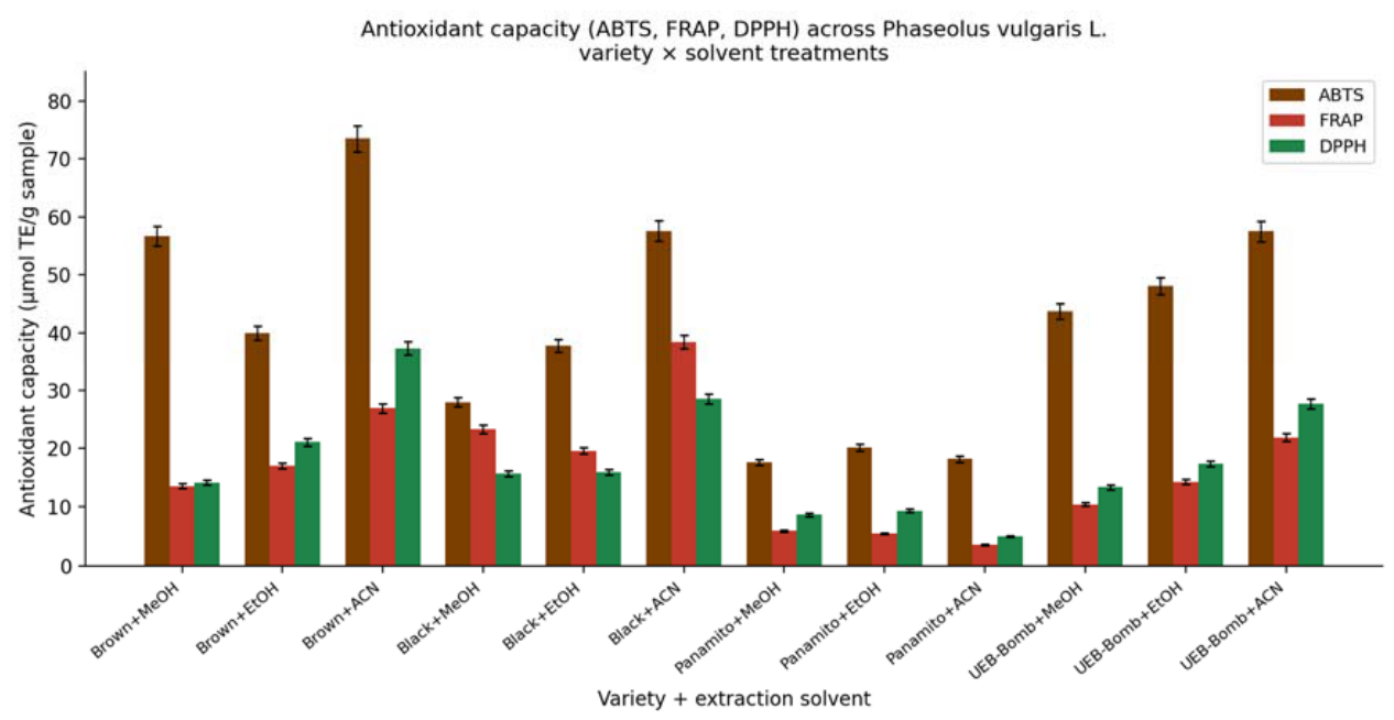
The integrated analysis of Table 3 shows the formation of two clearly differentiated functional profiles associated with the chemical nature of the predominant metabolites in each variety. The first profile, represented mainly by the Brown + ACN and UEB-Bombolín + ACN treatments, showed the highest concentrations of total polyphenols (4.280 and 4.262 mg GAE/g, respectively), accompanied by the maximum antioxidant capacity values determined by ABTS and DPPH. These results indicate a high radical-neutralisation capacity against artificially synthesised reactive species in both assays. The response observed suggests an important contribution from phenolic compounds capable of acting through hydrogen-transfer and radical-stabilisation mechanisms, particularly phenolic acids and condensed proanthocyanidins, metabolites widely associated with high activity against ABTS•⁺ and DPPH• radicals [74,75].
The second functional profile was represented by the Black + ACN treatment, which simultaneously showed the highest anthocyanin concentration (507.91 mg CY3GE/g) and the highest reducing capacity determined by FRAP (38.48 µmol TE/g). This behaviour indicates a high electron-transfer capacity associated with the abundant presence of anthocyanin pigments in the dark seed coat of the black bean. Several studies have shown that anthocyanins have high reducing efficiency owing to the electronic stability of the flavylium cation and the conjugation of aromatic double bonds present in their chemical structure, properties that particularly favour antioxidant responses determined by methods based on ferric reduction such as FRAP [20,76].
Although the Black + ACN treatment showed high ABTS and DPPH values, these were lower than those observed in Brown + ACN, indicating that different phenolic compositions can generate differentiated antioxidant responses depending on the predominant chemical mechanism in each assay. The ABTS and DPPH methods mainly evaluate radical-neutralisation capacity, whereas FRAP preferentially quantifies reducing capacity against the ferric ion, so that the antioxidant response depends not only on the total concentration of phenolic metabolites, but also on their molecular structure and the chemical reaction pathway involved [77].
The panamito bean consistently showed the lowest values for most antioxidant and phenolic variables, particularly in the MeOH and ACN treatments. This behaviour is consistent with the absence of pigmentation in the seed coat and the limited accumulation of phenylpropanoid-pathway-derived metabolites observed in light-coated varieties of Phaseolus vulgaris L. Previous studies have reported that white-coated varieties show lower concentrations of extractable anthocyanins, flavonoids and polyphenols compared with pigmented genotypes, significantly reducing the overall antioxidant capacity of the seed [78,79].
The superiority observed for acetonitrile across multiple antioxidant variables further suggests a higher extraction efficiency for medium-polarity phenolic metabolites present in both the seed coat and the cotyledon fractions. The ability of acetonitrile to improve the recovery of phenolic compounds has been widely associated with its moderate polarity, low viscosity and high penetration capacity in complex plant matrices, properties that favour the diffusion of phenolic metabolites partially or physically bound to cell-wall structures during ultrasonication-assisted processes [23].
Figure 4.
Antioxidant capacity (ABTS, FRAP and DPPH) in the 12 variety × solvent treatments of Phaseolus vulgaris L. Note: Error bars = pooled standard error of the residual from the corresponding two-way ANOVA (n = 3). MeOH = methanol; EtOH = ethanol; ACN = acetonitrile.
Figure 4.
Antioxidant capacity (ABTS, FRAP and DPPH) in the 12 variety × solvent treatments of Phaseolus vulgaris L. Note: Error bars = pooled standard error of the residual from the corresponding two-way ANOVA (n = 3). MeOH = methanol; EtOH = ethanol; ACN = acetonitrile.
3.4. Relationship Between Phenolic Composition and Antioxidant Response Mechanisms
In order to evaluate the relative contribution of the quantified phenolic compounds to the antioxidant capacity of the extracts, multiple linear regression models were fitted using data from all quantifiable treatments, excluding values substituted by LOQ/2, in accordance with the criterion described in Section 2.8. The resulting models allowed the functional relationship between total polyphenols, anthocyanins and the antioxidant response determined by the ABTS, FRAP and DPPH methods to be analysed, while simultaneously considering the predictive effect of both classes of bioactive metabolites.
Table 4.
Multiple linear regression models between phenolic compounds and antioxidant capacity in varieties of Phaseolus vulgaris L.
Table 4.
Multiple linear regression models between phenolic compounds and antioxidant capacity in varieties of Phaseolus vulgaris L.
| Dependent variable | β Polyphenols | β Anthocyanins | (R2) adjusted | F of the model |
p |
|---|---|---|---|---|---|
| ABTS (µmol TE/g) | 0.612 | 0.418 | 0.893 | 47.3 | <0.0001 |
| FRAP (µmol TE/g) | 0.298 | 0.741 | 0.921 | 63.8 | <0.0001 |
| DPPH (µmol TE/g) | 0.589 | 0.371 | 0.876 | 39.1 | <0.0001 |
Note. β = standardised regression coefficient. R2adjusted = adjusted coefficient of determination of the model, representing the proportion of variance in antioxidant capacity explained by the predictors. The models were fitted using total polyphenols and anthocyanins as predictor variables of the antioxidant capacity determined by the ABTS, FRAP and DPPH assays. Values of p < 0.05 indicate statistical significance of the model.
The fitted models revealed important differences in the relative contribution of the phenolic compounds according to the chemical mechanism involved in each antioxidant assay. In the ABTS and DPPH methods, total polyphenols showed the highest standardised β coefficients (0.612 and 0.589, respectively), indicating that the variability observed in radical-neutralisation capacity was mainly associated with the overall phenolic fraction present in the extracts. This behaviour is consistent with the fact that both methods predominantly evaluate hydrogen-transfer and radical-stabilisation reactions against synthetic radicals, processes in which phenolic acids, flavonols and condensed proanthocyanidins participate efficiently [80,81].
The high contribution of total polyphenols in the ABTS and DPPH assays can also be related to the presence of aromatic hydroxyl groups capable of donating protons and stabilising free radicals through electronic resonance [82]. The antioxidant efficiency of phenolic compounds does not depend exclusively on their total concentration, but also on specific structural characteristics, such as the number of OH groups, the degree of conjugation and the spatial arrangement of the aromatic rings, factors that determine the stability of the phenoxyl radical generated after oxidation [83]. In complex plant matrices, the coexistence of multiple phenolic families can generate cumulative or synergistic antioxidant responses, particularly in radical-scavenging-based assays.
In contrast, the model corresponding to FRAP showed the highest β coefficient for anthocyanins (0.741), indicating that this class of pigments was the main predictor of reducing capacity against the Fe³⁺-TPTZ system. The FRAP method preferentially quantifies electron-transfer mechanisms, so that compounds with high electron density and stable conjugated systems show greater reducing efficiency [84]. Anthocyanins possess highly conjugated flavylium structures that favour electron transfer and subsequent electronic stabilisation, particularly under acidic or moderately polar conditions where the chromophore remains stable [76]. This behaviour explains why the Black + ACN treatment, characterised by the highest anthocyanin concentration, simultaneously showed the maximum FRAP value.
The differentiation observed between ABTS/DPPH and FRAP shows that the antioxidant response depends on the chemical mechanism evaluated in each assay and not solely on the absolute concentration of phenolic compounds. While ABTS and DPPH showed a stronger association with the total phenolic fraction, FRAP showed a more marked dependence on anthocyanin compounds with high reducing capacity. This behaviour explains the differences observed in the relative positioning of the treatments across the antioxidant methods and shows that the functional efficiency of each variety depends on the predominant type of metabolite and on the chemical nature of the antioxidant evaluation system used.
The models showed high R2 adjusted values (0.876-0.921), indicating that a considerable proportion of the antioxidant variability was jointly explained by total polyphenols and anthocyanins. These results show a strong association between phenolic composition and the antioxidant response of the extracts evaluated. However, a residual fraction of variability remained unexplained by the model, suggesting the possible involvement of other antioxidant metabolites not individually quantified in the present study, including specific phenolic acids, minor flavonoids, tocopherols or other reducing compounds present at low concentrations. Since these metabolites were not experimentally determined, their individual contribution cannot be precisely established from the available data.
3.5. Multivariate Differentiation of Treatments According to Phenolic Composition and Antioxidant Response
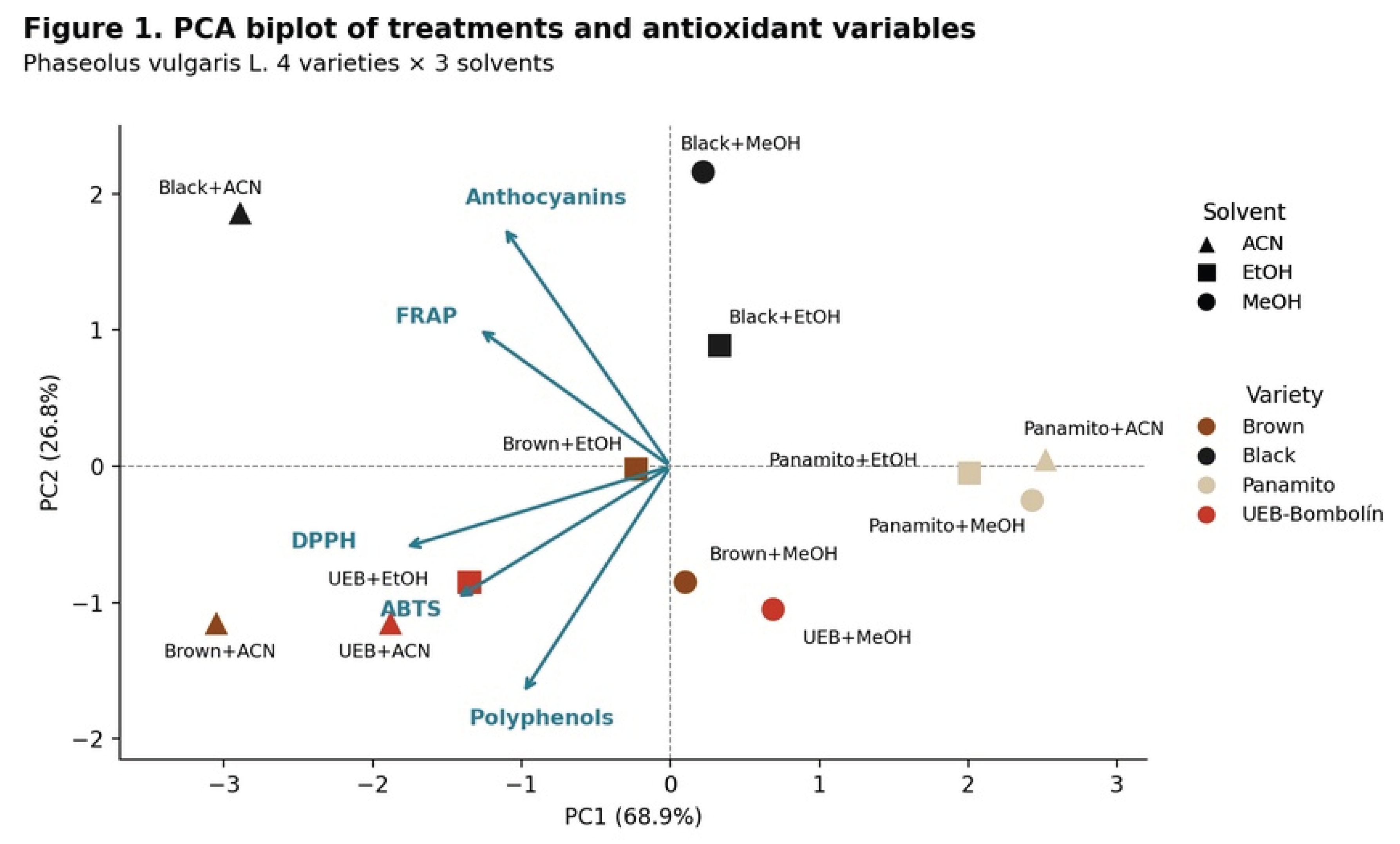
Principal component analysis (PCA) performed on the standardised variables corresponding to total polyphenols, anthocyanins, ABTS, FRAP and DPPH made it possible to identify multivariate patterns associated simultaneously with the varietal effect and the extraction system. Flavonoids were excluded from the analysis owing to the high proportion of values below the quantification limit (< LOQ) in this assay, which would have introduced distortions in the correlation matrix and reduced the statistical stability of the multivariate model. The first two principal components jointly explained 78.4% of the total variance, of which 55.2% corresponded to PC1 and 23.2% to PC2, showing an adequate descriptive capacity of the model to represent the overall structure of the treatments evaluated. The spatial distribution of the treatments and variables in the factorial plane is shown in Figure 3.
PC1 was strongly associated with the variables ABTS (0.83), DPPH (0.79) and total polyphenols (0.71), indicating that this component mainly represented a gradient of overall antioxidant capacity associated with the concentration of extractable phenolic compounds. In Figure 3, treatments obtained with acetonitrile are seen to be located predominantly in the positive values of PC1, whereas treatments extracted with methanol tended to be positioned at the negative end of the component. This pattern confirms that acetonitrile favoured a greater recovery of phenolic metabolites with antioxidant activity against free radicals, particularly those associated with hydrogen-transfer and radical-stabilisation mechanisms. The higher extraction efficiency of acetonitrile has been related to its intermediate polarity and high penetration capacity in complex plant matrices, properties that favour the solubilisation of phenolic compounds partially associated with insoluble cell-wall structures [85,86].
The simultaneous positive association between polyphenols, ABTS and DPPH observed in the biplot also shows that a large part of the antioxidant activity of the extracts was linked to the total phenolic fraction and not exclusively to the anthocyanins. This behaviour is consistent with the high predictive contribution of polyphenols observed previously in the multiple linear regression models of Section 3.4. The angular proximity between the ABTS and DPPH variables in Figure 3 indicates a high functional correlation between both antioxidant methods, an expected phenomenon since both assays predominantly respond to radical-neutralisation mechanisms via hydrogen transfer [87]. The joint orientation of these variables confirms that treatments with higher phenolic concentrations simultaneously showed greater radical-scavenging capacities.
PC2 showed a high positive loading for anthocyanins (0.84) and negative loadings for FRAP (-0.62) and polyphenols (-0.41), indicating that this component mainly discriminated treatments dominated by anthocyanin pigments from treatments dominated by other non-anthocyanin phenolic fractions. In Figure 3, the treatments corresponding to the black bean grouped clearly towards positive PC2 values, showing a direct association with the high anthocyanin content experimentally recorded in this variety. The high anthocyanin concentration in dark-coated varieties has been widely associated with differential activation of the flavonoid biosynthetic pathway during seed-coat development, particularly through the accumulation of cyanidin and delphinidin derivatives [88].
In contrast, the treatments corresponding to the brown bean and UEB-Bombolín were located at negative PC2 values, mainly associated with total polyphenols and high antioxidant responses in the ABTS and DPPH assays. This behaviour suggests that the dominant antioxidant activity in these varieties depended to a greater extent on non-anthocyanin phenolic compounds, possibly phenolic acids and condensed proanthocyanidins, whose antioxidant activity is related to a high capacity for radical stabilisation and proton transfer [88]. The separation observed between dark, anthocyanin-rich varieties and brown varieties rich in non-pigment polyphenols shows that different chemical profiles can lead to functionally distinct antioxidant responses even when both display high overall antioxidant activity.
The position of the Brown + ACN treatment in the quadrant of positive PC1 values and moderately negative PC2 values confirms its simultaneous association with high phenolic concentrations and high antioxidant activity against ABTS and DPPH. In contrast, the Black + ACN treatment was projected in the positive quadrant of both PC1 and PC2, mainly associated with anthocyanins and FRAP. This spatial differentiation shows that the predominant antioxidant mechanism varied according to the specific phenolic composition of each treatment. Whereas Brown + ACN was mainly linked to radical-neutralisation mechanisms, Black + ACN showed a greater association with electron-transfer mechanisms related to highly conjugated anthocyanin pigments. The multivariate separation observed in Figure 3 shows that the chemical nature of the predominant metabolites directly conditioned the antioxidant behaviour of each extraction-variety system.
Additionally, the relatively orthogonal separation between the varietal grouping and the solvent-associated gradient indicates that both factors contributed differentially to the overall variability of the system. This behaviour is consistent with the results of the two-way ANOVA, in which both variety and solvent showed high and statistically significant effect sizes. The structure observed in the PCA confirms that the intrinsic phenolic composition of each variety determined the baseline antioxidant profile, whereas the extraction system modulated the magnitude of recovery of these metabolites during the extraction process.
Figure 3.
Biplot of the principal component analysis (PC1 vs. PC2) of the phenolic and antioxidant variables (total polyphenols, anthocyanins, ABTS, FRAP and DPPH) for the variety × solvent treatments of Phaseolus vulgaris L. Note: PC1 = 55.2% of the variance; PC2 = 23.2% of the variance; cumulative variance = 78.4%. Flavonoids were excluded from the analysis owing to their high proportion of values below the limit of quantification (< LOQ).
3.6. Influence of the Extraction System on Flavonoids and Anthocyanins in Phaseolus vulgaris L.
The total flavonoid content showed a distinctive behaviour in several treatment-solvent combinations, particularly in the extracts corresponding to Black + ACN, Black + MeOH, Black + EtOH and UEB-Bombolín + ACN, in which the concentration fell below the limit of quantification (< LOQ). This result should not necessarily be interpreted as an absolute absence of flavonoids, but rather as an analytical limitation associated with spectrophotometric quantification in matrices with a high concentration of anthocyanin pigments. The Kumazawa method used in this study is based on readings at 425 nm, a spectral region susceptible to chromatic interference generated by intensely pigmented extracts, particularly those rich in anthocyanins derived from cyanidin and malvidin [89].
The high anthocyanin concentration recorded in the black bean, especially under acetonitrile extraction (507.91 mg CY3GE/g), probably increased spectral overlap and reduced the analytical sensitivity for the specific detection of non-anthocyanin flavonoids. In complex plant systems, anthocyanins show broad absorption bands in the visible region and can significantly interfere with colorimetric methods when their concentration greatly exceeds that of other minor phenolic metabolites [90]. This behaviour is consistent with the strong pigmentation observed in the dark varieties evaluated.
In contrast, the Panamito + EtOH treatment showed the highest quantifiable flavonoid content (1.449 mg QE/g), despite this variety recording a relatively low antioxidant capacity compared with the treatments dominated by anthocyanins or total polyphenols. This result indicates that total flavonoid concentration does not necessarily translate into a proportional antioxidant response, since antioxidant activity depends on the specific chemical structure of each compound and not solely on its relative abundance. The antioxidant efficiency of flavonoids is conditioned by structural factors such as the number and position of hydroxyl groups, the degree of electronic conjugation and the presence of glycosidic substituents, characteristics that significantly modify electron-transfer capacity and radical stabilisation [91,92].
The response observed in Panamito + EtOH suggests that the predominant flavonoid fraction in this variety could be mainly composed of glycosylated flavonoids with lower reducing capacity, which would explain the discrepancy between flavonoid concentration and overall antioxidant activity. In light-coated legumes, flavonoids are frequently associated with glycosidic forms that are less reactive towards free radicals than the condensed structures present in pigmented varieties [93]. This structural difference directly influences the antioxidant capacity measured by ABTS, DPPH and FRAP.
The anthocyanin content obtained for Black + ACN greatly exceeded the ranges previously reported for conventional varieties of Phaseolus vulgaris L., showing a high extraction efficiency of the protocol used. This behaviour can be attributed to the combination of iterative extraction, ultrasonication and the use of acetonitrile as the main solvent. Ultrasonication favours the mechanical disruption of cell structures and accelerates the diffusion of phenolic metabolites into the extraction medium through cavitation phenomena, whereas acetonitrile improves the solubilisation and stability of flavylium pigments in media of intermediate polarity [94,95].
Additionally, the extraction carried out at 5 °C probably helped to minimise oxidative degradation and hydrolysis of anthocyanins during the experimental procedure. Anthocyanins are highly sensitive compounds to temperature, pH and oxidative exposure, so moderately cold extraction conditions favour the structural preservation of the flavylium chromophore and reduce losses associated with chemical degradation [96,97,98]. Consequently, the high anthocyanin contents observed in Black + ACN reflect both the intrinsic composition of the variety and the high physicochemical efficiency of the extraction system used.
3.7. Preparation and Characterisation of the Protein Isolate from Phaseolus vulgaris L. Brown Variety
The selection of the brown variety for obtaining the protein isolate was based on the simultaneous integration of antioxidant, compositional and technological criteria. The Brown + ACN treatment showed the highest antioxidant capacity as determined by ABTS (73.49 µmol TE/g) and DPPH (37.38 µmol TE/g), together with a high total polyphenol content (4.280 mg GAE/g), evidencing a high density of functionally active phenolic metabolites. Although the black bean recorded a slightly higher protein content in the flour (23.94%), the brown variety showed additional technological advantages associated with the visual and sensory stability of the isolate obtained. In pigmented legume matrices, anthocyanin compounds and other condensed flavonoids can be partially co-extracted during alkaline solubilisation and transfer dark tones to the final protein isolate, which affects visual acceptability and limits potential food applications in light-coloured products [99,100]. The lower pigment intensity of the brown bean reduced this effect and made it possible to obtain an isolate with a more homogeneous appearance and greater chromatic stability.
The protein extraction process used in this study was based on alkaline isoelectric precipitation, one of the most widely used methods for obtaining plant protein isolates owing to its high recovery efficiency and the relative preservation of the functional properties of the storage globulins [101,102]. During the alkaline solubilisation step at pH 10.0 with 2 N NaOH, the proteins acquire a net negative charge through deprotonation of carboxyl groups, increasing intermolecular electrostatic repulsion and favouring the dispersion of the protein fractions in the aqueous medium [103]. In Phaseolus vulgaris L., the main storage proteins correspond to 7S (vicilin) and 11S (legumin) globulins, which represent approximately 70 to 80% of the seed's total protein content [104,105]. These fractions show high solubility under moderately alkaline conditions and precipitate readily near their isoelectric point.
The subsequent acidification to pH 4.0 with citric acid brought the proteins closer to their isoelectric point, reducing the net surface charge and electrostatic repulsion between the molecules. Under these conditions, hydrophobic interactions and intermolecular protein-protein associations predominate, inducing aggregation and precipitation of the enriched protein fraction [106]. The use of citric acid as an acidifying agent also represents an alternative with lower toxicological impact and greater food compatibility compared with the mineral acids conventionally used in industrial protein extraction.
Subsequent freeze-drying at -56 °C for 72 h removed water through sublimation and desorption under low-pressure conditions, significantly reducing thermal damage to the recovered proteins. This procedure minimises irreversible denaturation, thermal aggregation and loss of surface functionality associated with conventional heat-drying methods [107,108]. The structural preservation achieved through freeze-drying particularly favours technological properties such as solubility, emulsification and gelling capacity, attributes that are fundamental for the application of plant protein isolates in food formulations.
The yield obtained for the protein isolate was 17.58%, with a final protein content of 70.81% and an extraction efficiency of 76.9% relative to the initial protein content of the flour. These values fall within the ranges reported for legume protein isolates obtained by alkaline extraction and isoelectric precipitation, whose yields generally range between 15 and 25%, with final protein contents above 65% [109]. The fraction not recovered during the process probably corresponded to low-molecular-weight soluble proteins that remained in the supernatant after isoelectric precipitation, as well as to unavoidable mechanical losses during centrifugation, transfer and recovery of the protein precipitate [110].
Table 5 shows that the operational conditions used in the present study remained within the technological ranges described for legume protein extraction. In particular, the pH range used during solubilisation and precipitation coincides with the optimal values reported for maximising protein recovery without inducing severe structural degradation of the plant globulins [112,113]. Likewise, the centrifugation and freeze-drying conditions used favoured efficient separation of the precipitate and subsequent stabilisation of the isolate.
The protein content achieved (70.81%) exceeds the minimum threshold used to classify a product as a plant protein isolate, differentiating it from protein concentrates or enriched flours [33]. It should be noted that, although some classifications reserve the term isolate for products with a protein concentration above 90% on a dry basis, the literature on legume proteins obtained by isoelectric precipitation frequently recognises a wider range (65-90%), associated with the habitual carry-over of non-protein nitrogen and other plant-matrix components during the extraction process [33]. From a nutritional standpoint, the proteins of Phaseolus vulgaris L. show substantial concentrations of lysine, leucine, arginine and glutamic acid, amino acids that adequately complement the protein profiles of cereals deficient in lysine [116,117]. Additionally, the predominant globulins in beans show functional properties relevant to the food industry, including water-holding capacity, emulsification and gel formation, characteristics that allow their incorporation into plant-based food matrices and into formulations of functional foods with a high protein content [114,118].
Conclusions
The results obtained demonstrated that the phenolic composition and antioxidant behaviour of the Andean varieties of Phaseolus vulgaris L. depended simultaneously on the varietal genotype and the extraction system used, with both factors exerting highly significant and mechanistically differentiated effects on the recovery of bioactive metabolites and the antioxidant response. Variety constituted the main source of experimental variation for all the variables evaluated, confirming that seed-coat pigmentation and structural composition largely determine the accumulation of phenolic compounds and the antioxidant functionality of the grain. The black bean showed the highest anthocyanin concentration and the highest reducing capacity as determined by FRAP, whereas the brown variety recorded the highest antioxidant activity in the ABTS and DPPH assays, showing that antioxidant efficiency did not depend exclusively on the total anthocyanin concentration, but also on the specific composition of other non-pigment phenolic fractions. Acetonitrile consistently showed the highest extraction efficiency for multiple antioxidant variables, suggesting a greater capacity to recover phenolic metabolites of intermediate polarity and compounds partially associated with insoluble cell-wall structures. The multivariate analysis confirmed the existence of differentiated antioxidant profiles associated with distinct chemical mechanisms, separating treatments dominated by electron transfer from those characterised mainly by radical-neutralisation mechanisms via hydrogen transfer. The regression models further showed that polyphenols and anthocyanins accounted for a large part of the observed antioxidant variability, although their relative contribution depended directly on the chemical basis of each analytical method. Obtaining the protein isolate from the brown variety made it possible to achieve a protein content of 70.81% and an extraction efficiency of 76.9%, values comparable with plant protein isolates of industrial interest. The combination of high antioxidant activity, high phenolic content and adequate technological functionality supports the potential of the brown variety as a strategic raw material for the development of functional ingredients, plant-based food products and value-added nutraceutical systems in Andean agro-industrial contexts.
Author Contributions
Conceptualization, Favian Bayas-Morejón, Tarciso Ramírez-Vera, Melanie Molina-Penagos, Juan Gaibor-Chávez, Orlando Meneses Quelal, Enma Rodríguez-Cepeda and Jenny Yambay Vallejo; Methodology, Favian Bayas-Morejón, Tarciso Ramírez-Vera, Melanie Molina-Penagos, Juan Gaibor-Chávez, Orlando Meneses Quelal, Enma Rodríguez-Cepeda and Jenny Yambay Vallejo; Software, Favian Bayas-Morejón, Tarciso Ramírez-Vera, Melanie Molina-Penagos, Juan Gaibor-Chávez, Orlando Meneses Quelal, Enma Rodríguez-Cepeda and Jenny Yambay Vallejo; Validation, Favian Bayas-Morejón, Tarciso Ramírez-Vera, Melanie Molina-Penagos, Juan Gaibor-Chávez, Orlando Meneses Quelal, Enma Rodríguez-Cepeda and Jenny Yambay Vallejo; Formal analysis, Favian Bayas-Morejón, Tarciso Ramírez-Vera, Melanie Molina-Penagos, Juan Gaibor-Chávez, Orlando Meneses Quelal, Enma Rodríguez-Cepeda and Jenny Yambay Vallejo; Investigation, Favian Bayas-Morejón, Tarciso Ramírez-Vera, Melanie Molina-Penagos, Juan Gaibor-Chávez, Orlando Meneses Quelal, Enma Rodríguez-Cepeda and Jenny Yambay Vallejo; Resources, Favian Bayas-Morejón, Tarciso Ramírez-Vera, Melanie Molina-Penagos, Juan Gaibor-Chávez, Orlando Meneses Quelal, Enma Rodríguez-Cepeda and Jenny Yambay Vallejo; Data curation, Favian Bayas-Morejón, Tarciso Ramírez-Vera, Melanie Molina-Penagos, Juan Gaibor-Chávez, Orlando Meneses Quelal, Enma Rodríguez-Cepeda and Jenny Yambay Vallejo; Writing – original draft, Favian Bayas-Morejón, Tarciso Ramírez-Vera, Melanie Molina-Penagos, Juan Gaibor-Chávez, Orlando Meneses Quelal, Enma Rodríguez-Cepeda and Jenny Yambay Vallejo; Writing – review & editing, Favian Bayas-Morejón, Tarciso Ramírez-Vera, Melanie Molina-Penagos, Juan Gaibor-Chávez, Orlando Meneses Quelal, Enma Rodríguez-Cepeda and Jenny Yambay Vallejo; Visualization, Favian Bayas-Morejón, Tarciso Ramírez-Vera, Melanie Molina-Penagos, Juan Gaibor-Chávez, Orlando Meneses Quelal, Enma Rodríguez-Cepeda and Jenny Yambay Vallejo; Supervision, Favian Bayas-Morejón, Tarciso Ramírez-Vera, Melanie Molina-Penagos, Juan Gaibor-Chávez, Orlando Meneses Quelal, Enma Rodríguez-Cepeda and Jenny Yambay Vallejo; Project administration, Favian Bayas-Morejón, Tarciso Ramírez-Vera, Melanie Molina-Penagos, Juan Gaibor-Chávez, Orlando Meneses Quelal, Enma Rodríguez-Cepeda and Jenny Yambay Vallejo; Funding acquisition, Favian Bayas-Morejón, Tarciso Ramírez-Vera, Melanie Molina-Penagos, Juan Gaibor-Chávez, Orlando Meneses Quelal, Enma Rodríguez-Cepeda and Jenny Yambay Vallejo.
Institutional Review Board Statement
Not applicable.
Informed Consent Statement
Not applicable.
Data Availability Statement
The original contributions presented in this study are included in the article. Further inquiries can be directed to the corresponding author.
References
- Bitocchi, E., Nanni, L., Bellucci, E., Rossi, M., Giardini, A., Zeuli, P. S., Logozzo, G., Stougaard, J., McClean, P., Attene, G., & Papa, R. (2012). Mesoamerican origin of the common bean (Phaseolus vulgaris L.) is revealed by sequence data. Proceedings of the National Academy of Sciences, 109(14), E788–E796. [CrossRef]
- Sarkar, S., Meza, C., Carrasco, B., Delgado, M., Okoth, P., & Banerjee, A. (2026). Common bean in a changing world: biogeography, nutrition, and microbiome innovations for future food security. Trends in Food Science & Technology, 168, 105496. [CrossRef]
- Joya-Dávila, J. G., Gutiérrez-Miceli, F. A., Oliva-Llaven, M. Á., & Santos-Espinoza, A. M. (2025). Efecto de la Elicitación Física en Semillas de Arachis hypogaea Sobre el Comportamiento Agronómico del Cultivo. TERRA LATINOAMERICANA, 43. [CrossRef]
- Syraji, Y., Papanasam Rajamanickam, J., Abadi, A., Albene, D., & Alemu, F. (2026). Nutritional Composition and Functional Potential of Groundnut (Arachis hypogaea L.) Landraces Indigenous to the Gamo Zone of Ethiopia. Food Science and Nutrition, 14(1), e71356. [CrossRef]
- Anino, C., Onyango, A. N., Imathiu, S., Maina, J., & Onyangore, F. (2019). Chemical composition of the seed and ‘milk’ of three common bean (Phaseolus vulgaris L) varieties. Journal of Food Measurement and Characterization 2019 13:2, 13(2), 1242–1249. [CrossRef]
- Perucini-Avendaño, M., Arzate-Vázquez, I., Perea-Flores, M. de J., Tapia-Maruri, D., Méndez-Méndez, J. V., Nicolás-García, M., & Dávila-Ortiz, G. (2024). Effect of cooking on structural changes in the common black bean (Phaseolus vulgaris var. Jamapa). Heliyon, 10(4). [CrossRef]
- Bernardi, C., Finetti, F., & Trabalzini, L. (2026). The Nutraceutical Promise of Phaseolus vulgaris L.: Bioactive Compounds for Health Promotion and Prevention of Chronic Noncommunicable Diseases. Biology 2026, Vol. 15, Page 659, 15(8), 659. [CrossRef]
- Rodríguez, L., Mendez, D., Montecino, H., Carrasco, B., Arevalo, B., Palomo, I., & Fuentes, E. (2022). Role of Phaseolus vulgaris L. in the Prevention of Cardiovascular Diseases—Cardioprotective Potential of Bioactive Compounds. Plants 2022, Vol. 11, Page 186, 11(2), 186. [CrossRef]
- Santos, E., Marques, G., & Lino-Neto, T. (2020). Phaseolus vulgaris L. as a functional food for aging protection. Aging: Oxidative Stress and Dietary Antioxidants, 289–295. [CrossRef]
- Bunea, A., Cenariu, M. C., Andrei, S., De Rossi, L., Rocchetti, G., Lucini, L., & Rebecchi, A. (2025). Antimicrobial Potential of Polyphenols: Mechanisms of Action and Microbial Responses—A Narrative Review. Antioxidants 2025, Vol. 14, Page 200, 14(2), 200. [CrossRef]
- Kumar, S., Abedin, M. M., Singh, A. K., & Das, S. (2020). Role of Phenolic Compounds in Plant-Defensive Mechanisms. Plant Phenolics in Sustainable Agriculture: Volume 1, 1, 517–532. [CrossRef]
- Lin, D., Xiao, M., Zhao, J., Li, Z., Xing, B., Li, X., Kong, M., Li, L., Zhang, Q., Liu, Y., Chen, H., Qin, W., Wu, H., & Chen, S. (2016). An Overview of Plant Phenolic Compounds and Their Importance in Human Nutrition and Management of Type 2 Diabetes. Molecules 2016, Vol. 21, Page 1374, 21(10), 1374. [CrossRef]
- Babbar, N., Oberoi, H. S., Sandhu, S. K., & Bhargav, V. K. (2012). Influence of different solvents in extraction of phenolic compounds from vegetable residues and their evaluation as natural sources of antioxidants. Journal of Food Science and Technology 2012 51:10, 51(10), 2568–2575. [CrossRef]
- Li, M., Bai, Q., Zhou, J., de Souza, T. S. P., & Suleria, H. A. R. (2022). In Vitro Gastrointestinal Bioaccessibility, Bioactivities and Colonic Fermentation of Phenolic Compounds in Different Vigna Beans. Foods 2022, Vol. 11, Page 3884, 11(23), 3884. [CrossRef]
- Shahidi, F., & Samarasinghe, A. (2025). How to assess antioxidant activity? Advances, limitations, and applications of in vitro, in vivo, and ex vivo approaches. Food Production, Processing and Nutrition 2025 7:1, 7(1), 50-. [CrossRef]
- Hernández-Ruiz, R. G., Olivares-Ochoa, X. C., Salinas-Varela, Y., Guajardo-Espinoza, D., Roldán-Flores, L. G., Rivera-Leon, E. A., & López-Quintero, A. (2025). Phenolic Compounds and Anthocyanins in Legumes and Their Impact on Inflammation, Oxidative Stress, and Metabolism: Comprehensive Review. Molecules 2025, Vol. 30, Page 174, 30(1), 174. [CrossRef]
- Oppedisano, F., Spagnoletta, A., Sadowska-Bartosz, I., & Bartosz, G. (2024). Antioxidant Activity of Anthocyanins and Anthocyanidins: A Critical Review. International Journal of Molecular Sciences 2024, Vol. 25, Page 12001, 25(22), 12001. [CrossRef]
- Tena, N., Martín, J., & Asuero, A. G. (2020). State of the Art of Anthocyanins: Antioxidant Activity, Sources, Bioavailability, and Therapeutic Effect in Human Health. Antioxidants 2020, Vol. 9, Page 451, 9(5), 451. [CrossRef]
- Diaconeasa, Z., Ştirbu, I., Xiao, J., Leopold, N., Ayvaz, Z., Danciu, C., Ayvaz, H., Stănilă, A., Nistor, M., & Socaciu, C. (2020). Anthocyanins, Vibrant Color Pigments, and Their Role in Skin Cancer Prevention. Biomedicines 2020, Vol. 8, Page 336, 8(9), 336. [CrossRef]
- Kan, L., Nie, S., Hu, J., Liu, Z., & Xie, M. (2016). Antioxidant activities and anthocyanins composition of seed coats from twenty-six kidney bean cultivars. Journal of Functional Foods, 26, 622–631. [CrossRef]
- Hasso, S. S., & Ahamad, J. (2025). Antioxidant properties of anthocyanins. Handbook of Analysis and Extraction Methods of Anthocyanins, 98–113. [CrossRef]
- Khoo, H. E., Azlan, A., Tang, S. T., & Lim, S. M. (2017). Anthocyanidins and anthocyanins: colored pigments as food, pharmaceutical ingredients, and the potential health benefits. Food & Nutrition Research. [CrossRef]
- Dai, J., & Mumper, R. J. (2010). Plant Phenolics: Extraction, Analysis and Their Antioxidant and Anticancer Properties. Molecules 2010, Vol. 15, Pages 7313-7352, 15(10), 7313–7352. [CrossRef]
- Dardavila, M. M., Pappou, S., Savvidou, M. G., Louli, V., Katapodis, P., Stamatis, H., Magoulas, K., & Voutsas, E. (2023). Extraction of Bioactive Compounds from C. vulgaris Biomass Using Deep Eutectic Solvents. Molecules 2023, Vol. 28, Page 415, 28(1), 415. [CrossRef]
- Sun, W., & Shahrajabian, M. H. (2023). Therapeutic Potential of Phenolic Compounds in Medicinal Plants—Natural Health Products for Human Health. Molecules 2023, Vol. 28, Page 1845, 28(4), 1845. [CrossRef]
- Jiménez-Ortega, L. A., Marques, M., Quiñonez-Angulo, P., Paiva, A., Heredia, J. B., Duarte, A. R. C., & Mota-Morales, J. D. (2025). Sustainable extraction of phytochemicals from agricultural and food by-products using eutectic solvents and their integration into functional materials. RSC Sustainability, 4(2), 687–719. [CrossRef]
- Munteanu, I. G., & Apetrei, C. (2021). Analytical Methods Used in Determining Antioxidant Activity: A Review. International Journal of Molecular Sciences 2021, Vol. 22, Page 3380, 22(7), 3380. [CrossRef]
- Wołosiak, R., Drużyńska, B., Derewiaka, D., Piecyk, M., Majewska, E., Ciecierska, M., Worobiej, E., & Pakosz, P. (2021). Verification of the Conditions for Determination of Antioxidant Activity by ABTS and DPPH Assays—A Practical Approach. Molecules 2022, Vol. 27, Page 50, 27(1), 50. [CrossRef]
- Yenduri, S., Ramya, P., & Naga Prashant, K. (2026). Recent advances in analytical methods and instrumentation for natural antioxidants analysis. Talanta Open, 13, 100602. [CrossRef]
- Romero-Astudillo, M. J., Tapia, C., Giménez de Azcárate, J., & Montalvo, D. (2024). Diversity of Common Bean (Phaseolus vulgaris L.) and Runner Bean (Phaseolus coccineus L.) Landraces in Rural Communities in the Andes Highlands of Cotacachi—Ecuador. Agronomy, 14(8), 1666. [CrossRef]
- Adil, M. Z., Oztekin, S., Aziz, A., Gunal-Koroglu, D., Capanoglu, E., Moreno, A., Khalid, W., & Esatbeyoglu, T. (2026). Fermentation-based valorization of agro-industrial cereal wastes and by-products. Trends in Food Science & Technology, 168, 105464. [CrossRef]
- Eugelio, F., Mascini, M., Fanti, F., Palmieri, S., & Del Carlo, M. (2026). Bioactive compounds in functional foods using mass spectrometry: A systematic review. Food Chemistry, 518, 149514. [CrossRef]
- Patel, P., Pang, Y. L. J., Choi, W. J., & Wong, A. (2025). Protein Extraction and Isolation from Legumes and Algae: An Industry Primer. Food and Bioprocess Technology 2025 18:10, 18(10), 8380–8408. [CrossRef]
- Ministerio de Agricultura, Ganadería, Acuacultura y Pesca [MAGAP]. (2015). Entregan certificación de nueva variedad de fréjol en Bolívar. https://www.agricultura.gob.ec/entregan-certificacion-de-nueva-variedad-de-frejol-en-bolivar/.
- AOAC International. (2019). Loss on Drying (Moisture) in Sugars. Official Methods of Analysis of AOAC Internacional, 1.
- Mariotti, F., Tomé, D., & Mirand, P. (2008). Converting nitrogen into protein—beyond 6.25 and Jones’ factors. Critical Reviews in Food Science and Nutrition, 48(2), 177–184. [CrossRef]
- Álvarez-Cilleros, D., Ramos, S., López-Oliva, M. E., Escrivá, F., Álvarez, C., Fernández-Millán, E., & Martín, M. Á. (2020). Cocoa diet modulates gut microbiota composition and improves intestinal health in Zucker diabetic rats. Food Research International, 132, 109058. [CrossRef]
- Singleton, V. L., Orthofer, R., & Lamuela-Raventós, R. M. (1999). [14] Analysis of total phenols and other oxidation substrates and antioxidants by means of folin-ciocalteu reagent. Methods in Enzymology, 299, 152–178. [CrossRef]
- Kumazawa, S., Hamasaka, T., & Nakayama, T. (2004). Antioxidant activity of propolis of various geographic origins. Food Chemistry, 84(3), 329–339. [CrossRef]
- Re, R., Pellegrini, N., Proteggente, A., Pannala, A., Yang, M., & Rice-Evans, C. (1999). Antioxidant activity applying an improved ABTS radical cation decolorization assay. Free Radical Biology and Medicine, 26(9–10), 1231–1237. [CrossRef]
- Benzie, I. F. F., & Strain, J. J. (1996). The Ferric Reducing Ability of Plasma (FRAP) as a Measure of “Antioxidant Power”: The FRAP Assay. Analytical Biochemistry, 239(1), 70–76. [CrossRef]
- Brand-Williams, W., Cuvelier, M. E., & Berset, C. (1995). Use of a free radical method to evaluate antioxidant activity. LWT - Food Science and Technology, 28(1), 25–30. [CrossRef]
- Hwang, M., Lee, S. C., Park, J.-H., Choi, J., & Lee, H.-J. (2023). Statistical methods for handling nondetected results in food chemical monitoring data to improve food risk assessments. Food Science & Nutrition, 11(9), 5223–5235. [CrossRef]
- Erulaş, A. F., Şaylan, M., Topal, S., Zaman, B. T., Bakırdere, E. G., & Bakırdere, S. (2020). A new microextraction method for trace nickel determination in green tea samples: Solventless dispersion based dispersive liquid-liquid microextraction combined with slotted quartz tube- flame atomic absorption spectrophotometry. Journal of Food Composition and Analysis, 94, 103623. [CrossRef]
- Devkota, L., He, L., Midgley, J., & Haritos, V. S. (2022). Effect of seed coat microstructure and lipid composition on the hydration behavior and kinetics of two red bean (Phaseolus vulgaris L.) varieties. Journal of Food Science, 87(2), 528–542. [CrossRef]
- Campos-Vega, R., Loarca-Piña, G., & Oomah, B. D. (2010). Minor components of pulses and their potential impact on human health. Food Research International, 43(2), 461–482. [CrossRef]
- Oomah, B. D., Corbé, A., & Balasubramanian, P. (2010). Antioxidant and Anti-inflammatory Activities of Bean (Phaseolus vulgaris L.) Hulls. Journal of Agricultural and Food Chemistry, 58(14), 8225–8230. [CrossRef]
- Xu, B., & Chang, S. K. C. (2009). Total Phenolic, Phenolic Acid, Anthocyanin, Flavan-3-ol, and Flavonol Profiles and Antioxidant Properties of Pinto and Black Beans (Phaseolus vulgaris L.) as Affected by Thermal Processing. Journal of Agricultural and Food Chemistry, 57(11), 4754–4764. [CrossRef]
- Cardador-Martínez, A., Loarca-Piña, G., & Oomah, B. D. (2002). Antioxidant Activity in Common Beans (Phaseolus vulgaris L.)§. Journal of Agricultural and Food Chemistry, 50(24), 6975–6980. [CrossRef]
- Díaz-Batalla, L., Widholm, J. M., Fahey, G. C., Castaño-Tostado, E., & Paredes-López, O. (2006). Chemical Components with Health Implications in Wild and Cultivated Mexican Common Bean Seeds (Phaseolus vulgaris L.). Journal of Agricultural and Food Chemistry, 54(6), 2045–2052. [CrossRef]
- Bhattarai, N., Piippo, N., Ranta-Aho, S., Mysore, Y., Kaarniranta, K., & Kauppinen, A. (2021). Effects of Resvega on Inflammasome Activation in Conjunction with Dysfunctional Intracellular Clearance in Retinal Pigment Epithelial (RPE) Cells. Antioxidants 2021, Vol. 10, Page 67, 10(1), 67. [CrossRef]
- Klinmalai, P., Fong-In, S., Phongthai, S., & Klunklin, W. (2021). Improving the Quality of Frozen Fillets of Semi-Dried Gourami Fish (Trichogaster pectoralis) by Using Sorbitol and Citric Acid. Foods 2021, Vol. 10, Page 2763, 10(11), 2763. [CrossRef]
- Pietta, P. G. (2000). Flavonoids as Antioxidants. Journal of Natural Products, 63(7), 1035–1042. [CrossRef]
- Wang, N., Hatcher, D. W., Tyler, R. T., Toews, R., & Gawalko, E. J. (2010). Effect of cooking on the composition of beans (Phaseolus vulgaris L.) and chickpeas (Cicer arietinum L.). Food Research International, 43(2), 589–594. [CrossRef]
- Weber, H., Borisjuk, L., & Wobus, U. (2005). Molecular physiology of legume seed development. Annual Review of Plant Biology, 56(Volume 56, 2005), 253–279. [CrossRef]
- Vaz Patto, M. C., Amarowicz, R., Aryee, A. N. A., Boye, J. I., Chung, H. J., Martín-Cabrejas, M. A., & Domoney, C. (2015). Achievements and Challenges in Improving the Nutritional Quality of Food Legumes. Critical Reviews in Plant Sciences, 34, 105–143. [CrossRef]
- Kehr, J. (2013). Systemic regulation of mineral homeostasis by micro RNAs. Frontiers in Plant Science, 4(MAY), 50239. [CrossRef]
- Lakens, D. (2013). Calculating and reporting effect sizes to facilitate cumulative science: A practical primer for t-tests and ANOVAs. Frontiers in Psychology, 4(NOV), 62627. [CrossRef]
- Preacher, K. J., & Kelley, K. (2011). Effect size measures for mediation models: Quantitative strategies for communicating indirect effects. Psychological Methods, 16(2), 93–115. [CrossRef]
- Cohen, J. (2013). Statistical Power Analysis for the Behavioral Sciences. Statistical Power Analysis for the Behavioral Sciences, 1–567. [CrossRef]
- Richardson, J. T. E. (2011). Eta squared and partial eta squared as measures of effect size in educational research. Educational Research Review, 6(2), 135–147. [CrossRef]
- Chávez-Mendoza, C., Hernández-Figueroa, K. I., & Sánchez, E. (2018). Antioxidant Capacity and Phytonutrient Content in the Seed Coat and Cotyledon of Common Beans (Phaseolus vulgaris L.) from Various Regions in Mexico. Antioxidants 2019, Vol. 8, Page 5, 8(1), 5. [CrossRef]
- Mujica, M. V, Granito, M., & Soto, N. (2012). Variación de los compuestos fenólicos de Phaseolus vulgaris L. durante el almacenamiento y su relación con el endurecimiento. Bioagro, 24(3), 163–174. http://ve.scielo.org/scielo.php?script=sci_arttext&pid=S1316-33612012000300002&lng=es&nrm=iso&tlng=es.
- Lee, S., Lee, D., Lee, S. O., Ryu, J. Y., Choi, S. Z., Kang, K. S., & Kim, K. H. (2017). Anti-inflammatory activity of the sclerotia of edible fungus, Poria cocos Wolf and their active lanostane triterpenoids. Journal of Functional Foods, 32, 27–36. [CrossRef]
- Madrera, R. R., Negrillo, A. C., Valles, B. S., & Fernández, J. J. F. (2021). Phenolic content and antioxidant activity in seeds of common bean (Phaseolus vulgaris L.). Foods, 10(4), 864. [CrossRef]
- Cacace, J. E., & Mazza, G. (2003). Mass transfer process during extraction of phenolic compounds from milled berries. Journal of Food Engineering, 59(4), 379–389. [CrossRef]
- Kaczorová, D., Karalija, E., Dahija, S., Bešta-Gajević, R., Parić, A., & Ćavar Zeljković, S. (2021). Influence of Extraction Solvent on the Phenolic Profile and Bioactivity of Two Achillea Species. Molecules 2021, Vol. 26, Page 1601, 26(6), 1601. [CrossRef]
- Andrés-Costa, M. J., Andreu, V., & Picó, Y. (2017). Liquid chromatography–mass spectrometry as a tool for wastewater-based epidemiology: Assessing new psychoactive substances and other human biomarkers. TrAC Trends in Analytical Chemistry, 94, 21–38. [CrossRef]
- Bai, Q., Li, M., Zhou, J., Imran, A., de Souza, T. S. P., Barrow, C., Dunshea, F., & Suleria, H. A. R. (2024). Influence of Processing Methods on Phytochemical Composition of Different Varieties of Beans (Phaseolus vulgaris). Food Reviews International, 40(7), 1941–1979. [CrossRef]
- Hsieh-Lo, M., Castillo-Herrera, G., & Mojica, L. (2020). Black Bean Anthocyanin-Rich Extract from Supercritical and Pressurized Extraction Increased In Vitro Antidiabetic Potential, While Having Similar Storage Stability. Foods 2020, Vol. 9, Page 655, 9(5), 655. [CrossRef]
- Kuasnei, M., Benvenutti, L., Fernando dos Santos, D., Ferreira, S. R. S., Pinto, V. Z., & Ferreira Zielinski, A. A. (2024). Efficient Anthocyanin Recovery from Black Bean Hulls Using Eutectic Mixtures: A Sustainable Approach for Natural Dye Development. Foods, 13(9), 1374. [CrossRef]
- Rodríguez, L., Plaza, A., Méndez, D., Carrasco, B., Tellería, F., Palomo, I., & Fuentes, E. (2022). Antioxidant Capacity and Antiplatelet Activity of Aqueous Extracts of Common Bean (Phaseolus vulgaris L.) Obtained with Microwave and Ultrasound Assisted Extraction. Plants, 11(9), 1179. [CrossRef]
- Tezcucano Molina, A. C., Alli, I., Konishi, Y., & Kermasha, S. (2007). Effect of dephosphorylation on bovine casein. Food Chemistry, 101(3), 1263–1271. [CrossRef]
- Fonseca-Hernández, D., Lugo-Cervantes, E. D. C., Escobedo-Reyes, A., & Mojica, L. (2021). Black Bean (Phaseolus vulgaris L.) Polyphenolic Extract Exerts Antioxidant and Antiaging Potential. Molecules 2021, Vol. 26, Page 6716, 26(21), 6716. [CrossRef]
- Prior, R. L., Wu, X., & Schaich, K. (2005). Standardized Methods for the Determination of Antioxidant Capacity and Phenolics in Foods and Dietary Supplements. Journal of Agricultural and Food Chemistry, 53(10), 4290–4302. [CrossRef]
- He, J., & Giusti, M. M. (2010). Anthocyanins: Natural colorants with health-promoting properties. Annual Review of Food Science and Technology, 1(1), 163–187. [CrossRef]
- Parmar, A., & Sharma, S. (2016). Derivative UV-vis absorption spectra as an invigorated spectrophotometric method for spectral resolution and quantitative analysis: Theoretical aspects and analytical applications: A review. TrAC Trends in Analytical Chemistry, 77, 44–53. [CrossRef]
- García-Díaz, Y. D., Aquino-Bolaños, E. N., Chávez-Servia, J. L., Vera-Guzmán, A. M., & Carrillo-Rodríguez, J. C. (2018). Bioactive compounds and antioxidant activity in the common bean are influenced by cropping season and genotype. Chilean Journal of Agricultural Research, 78(2), 255–265. [CrossRef]
- Pérez-Perez, L. M., Toro Sánchez, C. L. Del, Sánchez Chavez, E., González Vega, R. I., Reyes Díaz, A., Borboa Flores, J., Soto Parra, J. M., & Flores-Cordova, M. A. (2020). Bioaccessibility of antioxidant compounds from different bean varieties (Phaseolus vulgaris L.) in Mexico, through an in vitro gastrointestinal system. Biotecnia, 22(1), 117–125. [CrossRef]
- Platzer, M., Kiese, S., Herfellner, T., Schweiggert-Weisz, U., Miesbauer, O., & Eisner, P. (2021). Common Trends and Differences in Antioxidant Activity Analysis of Phenolic Substances Using Single Electron Transfer Based Assays. Molecules 2021, Vol. 26, Page 1244, 26(5), 1244. [CrossRef]
- UNTEA, A., LUPU, A., SARACILA, M., & PANAITE, T. (2018). Comparison of ABTS, DPPH, Phosphomolybdenum Assays for Estimating Antioxidant Activity and Phenolic Compounds in Five Different Plant Extracts. Bulletin of University of Agricultural Sciences and Veterinary Medicine Cluj-Napoca. Animal Science and Biotechnologies, 75(2), 110. [CrossRef]
- Donoso-Bustamante, V., Osorio, E., Arias-Santé, M. F., De Camargo, A. C., Rincón-Cervera, M. Á., Amalraj, J., Carrasco, B., Palomo, I., & Araya-Maturana, R. (2025). Antioxidant activity of sinapic acid anilides: DPPH, ABTS, FRAP, electrochemical and theoretical analysis. LWT, 222, 117656. [CrossRef]
- Shahidi, F., & Ambigaipalan, P. (2015). Phenolics and polyphenolics in foods, beverages and spices: Antioxidant activity and health effects – A review. Journal of Functional Foods, 18, 820–897. [CrossRef]
- Márquez-Calvo, K., Schmeda-Hirschmann, G., Leyton, F., Ávila, F., Salgado, P., Melin, V., Contreras, D., & Tabilo-Munizaga, G. (2026). Antioxidant Activity and Resistance Against Oxidation of Peptide Fractions from Common Bean (Phaseolus vulgaris L.) Landraces Assessed by EPR and Chemical Assays. Antioxidants, 15(3), 376. [CrossRef]
- Chang, Q., Zhu, L., Luo, Z., Lei, M., Zhang, S., & Tang, H. (2011). Sono-assisted preparation of magnetic magnesium–aluminum layered double hydroxides and their application for removing fluoride. Ultrasonics Sonochemistry, 18(2), 553–561. [CrossRef]
- Kuhn, M., Lang, S., Bezold, F., Minceva, M., & Briesen, H. (2017). Time-resolved extraction of caffeine and trigonelline from finely-ground espresso coffee with varying particle sizes and tamping pressures. Journal of Food Engineering, 206, 37–47. [CrossRef]
- Apak, R., Özyürek, M., Güçlü, K., & Çapanoʇlu, E. (2016). Antioxidant Activity/Capacity Measurement. 1. Classification, Physicochemical Principles, Mechanisms, and Electron Transfer (ET)-Based Assays. Journal of Agricultural and Food Chemistry, 64(5), 997–1027. [CrossRef]
- Zheng, Y., Yang, W., Sun, W., Chen, S., Liu, D., Kong, X., Tian, J., & Ye, X. (2020). Inhibition of porcine pancreatic α-amylase activity by chlorogenic acid. Journal of Functional Foods, 64, 103587. [CrossRef]
- Giusti, M. M., & Wrolstad, R. E. (2001). Characterization and Measurement of Anthocyanins by UV-Visible Spectroscopy. Current Protocols in Food Analytical Chemistry, 00(1), F1.2.1-F1.2.13. [CrossRef]
- Contreras, J., Alcázar-Valle, M., Lugo-Cervantes, E., Luna-Vital, D. A., & Mojica, L. (2023). Mexican Native Black Bean Anthocyanin-Rich Extracts Modulate Biological Markers Associated with Inflammation. Pharmaceuticals, 16(6), 874. [CrossRef]
- Ganesan, K., & Xu, B. (2017). Polyphenol-Rich Dry Common Beans (Phaseolus vulgaris L.) and Their Health Benefits. International Journal of Molecular Sciences 2017, Vol. 18, Page 2331, 18(11), 2331. [CrossRef]
- Ombra, M. N., D’Acierno, A., Nazzaro, F., Riccardi, R., Spigno, P., Zaccardelli, M., Pane, C., Maione, M., & Fratianni, F. (2016). Phenolic Composition and Antioxidant and Antiproliferative Activities of the Extracts of Twelve Common Bean (Phaseolus vulgaris L.) Endemic Ecotypes of Southern Italy before and after Cooking. Oxidative Medicine and Cellular Longevity, 2016(1), 1398298. [CrossRef]
- Samukha, V., Fantasma, F., D’Urso, G., Caprari, C., De Felice, V., Saviano, G., Lauro, G., Casapullo, A., Chini, M. G., Bifulco, G., & Iorizzi, M. (2024). NMR Metabolomics and Chemometrics of Commercial Varieties of Phaseolus vulgaris L. Seeds from Italy and In Vitro Antioxidant and Antifungal Activity. Plants, 13(2), 227. [CrossRef]
- Wang, X., Hansen, C., Allen, K., Wang, X., Hansen, C., & Allen, K. (2013). Identification of Anthocyanins Isolated from Black Bean Canning Wastewater by Macroporous Resin Using Optimized Conditions. Food and Nutrition Sciences, 4(8), 174–181. [CrossRef]
- Yang, Q. Q., Gan, R. Y., Ge, Y. Y., Zhang, D., & Corke, H. (2019). Ultrasonic Treatment Increases Extraction Rate of Common Bean (Phaseolus vulgaris L.) Antioxidants. Antioxidants 2019, Vol. 8, Page 83, 8(4), 83. [CrossRef]
- Boeing, J. S., Barizão, É. O., e Silva, B. C., Montanher, P. F., de Cinque Almeida, V., & Visentainer, J. V. (2014). Evaluation of solvent effect on the extraction of phenolic compounds and antioxidant capacities from the berries: application of principal component analysis. Chemistry Central Journal, 8(1), 48–48. [CrossRef]
- Enaru, B., Drețcanu, G., Pop, T. D., Stǎnilǎ, A., & Diaconeasa, Z. (2021). Anthocyanins: Factors Affecting Their Stability and Degradation. Antioxidants 2021, Vol. 10, Page 1967, 10(12), 1967. [CrossRef]
- Patras, A., Brunton, N. P., O’Donnell, C., & Tiwari, B. K. (2010). Effect of thermal processing on anthocyanin stability in foods; mechanisms and kinetics of degradation. Trends in Food Science & Technology, 21(1), 3–11. [CrossRef]
- Kang, H. J., Ko, M. J., & Chung, M. S. (2021). Anthocyanin Structure and pH Dependent Extraction Characteristics from Blueberries (Vaccinium corymbosum) and Chokeberries (Aronia melanocarpa) in Subcritical Water State. Foods 2021, Vol. 10, Page 527, 10(3), 527. [CrossRef]
- Sawicki, T., Jabłońska, M., Danielewicz, A., & Przybyłowicz, K. E. (2024). Phenolic Compounds Profile and Antioxidant Capacity of Plant-Based Protein Supplements. Molecules 2024, Vol. 29, Page 2101, 29(9), 2101. [CrossRef]
- Tang, J., Yao, D., Xia, S., Cheong, L., & Tu, M. (2024). Recent progress in plant-based proteins: From extraction and modification methods to applications in the food industry. Food Chemistry: X, 23, 101540. [CrossRef]
- Yao, S., Li, W., Martin, G. J. O., & Ashokkumar, M. (2023). An Investigation into the Mechanism of Alkaline Extraction-Isoelectric Point Precipitation (AE-IEP) of High-Thiol Plant Proteins. Applied Sciences (Switzerland), 13(11), 6469. [CrossRef]
- Zhou, X., Zhou, D. Y., Lu, T., Liu, Z. Y., Zhao, Q., Liu, Y. X., Hu, X. P., Zhang, J. H., & Shahidi, F. (2018). Characterization of lipids in three species of sea urchin. Food Chemistry, 241, 97–103. [CrossRef]
- Carbonaro, M., Grant, G., Cappelloni, M., & Pusztai, A. (2000). Perspectives into Factors Limiting in Vivo Digestion of Legume Proteins: Antinutritional Compounds or Storage Proteins? Journal of Agricultural and Food Chemistry, 48(3), 742–749. [CrossRef]
- Chukwuejim, S., Kadam, D., & Aluko, R. E. (2025). Structural, physicochemical, and functional properties of white and blue lupin vicilin and legumin fractions. Food Chemistry: X, 31, 103078. [CrossRef]
- Huang, A., & Moraru, C. I. (2025). Acidification and Calcium Addition Effects on High-Pressure and Thermally Induced Pulse Protein Gels. Gels, 11(12), 971. [CrossRef]
- Butreddy, A., Janga, K. Y., Ajjarapu, S., Sarabu, S., & Dudhipala, N. (2021). Instability of therapeutic proteins — An overview of stresses, stabilization mechanisms and analytical techniques involved in lyophilized proteins. International Journal of Biological Macromolecules, 167, 309–325. [CrossRef]
- Yang, H., Chen, Z., Wang, H., Jin, D., Wang, X., Wang, F., Cen, X., Liu, J., & Shen, Q. (2024). Uncovering the rheological properties basis for freeze drying treatment-induced improvement in the solubility of myofibrillar proteins. Current Research in Food Science, 8, 100651. [CrossRef]
- Li, J., Karboune, S., Sedman, J., & Ismail, A. (2020). Characterization of the structural properties of mannoproteins isolated from selected yeast-based products upon the enzymatic treatment. LWT, 131, 109596. [CrossRef]
- Botta, C., Franciosa, I., Coisson, J. D., Ferrocino, I., Colasanto, A., Arlorio, M., Cocolin, L., & Rantsiou, K. (2023). Beef carcass microbiota after slaughtering and primary cooling: A metataxonomic assessment to infer contamination drivers. Food Research International, 174, 113466. [CrossRef]
- García, O., Aiello, C., Peña Chirino, M. C., Ruíz-Ramírez, J., & Acevedo Pons, I. (2012). Caracterización físico-química y propiedades funcionales de la harina obtenida de granos de quinchoncho (Cajanus cajan (L.) Millsp.) sometidos a diferentes procesamientos. Revista Científica UDO Agrícola, ISSN-e 1317-9152, Vol. 12, No. 4, 2012, Págs. 919-928, 12(4), 919–928. https://dialnet.unirioja.es/servlet/articulo?codigo=6104328&info=resumen&idioma=SPA.
- Aliabbasi, N., Diosady, L. L., & Emam-Djomeh, Z. (2024). Purification and Characterization of Pinto Bean Protein Using Membrane Technology. Food Science & Nutrition, 12(12), 10592–10604. [CrossRef]
- Osemwota, E. C., Alashi, A. M., Aluko, R. E., Sadrzadeh, M., Dixon, A. G., Bazinet, L., Chae, S., & Esfahani, M. R. (2021). Comparative Study of the Structural and Functional Properties of Membrane-Isolated and Isoelectric pH Precipitated Green Lentil Seed Protein Isolates. Membranes 2021, Vol. 11, Page 694, 11(9), 694. [CrossRef]
- Náthia-Neves, G., Getachew, A. T., Santana, Á. L., & Jacobsen, C. (2025). Legume Proteins in Food Products: Extraction Techniques, Functional Properties, and Current Challenges. Foods 2025, Vol. 14, Page 1626, 14(9), 1626. [CrossRef]
- Albe-Slabi, S., Mesieres, O., Mathé, C., Ndiaye, M., Galet, O., & Kapel, R. (2022). Combined Effect of Extraction and Purification Conditions on Yield, Composition and Functional and Structural Properties of Lupin Proteins. Foods 2022, Vol. 11, Page 1646, 11(11), 1646. [CrossRef]
- Azevedo, P. Z., Rigolon, T. C. B., Souza, B. R. de, Stringheta, P. C., Martins, E., & Campelo, P. H. (2025). White bean (Phaseolus vulgaris L.) proteins: extraction methods, physicochemical and techno-functional properties, applications, and challenges for the food industry. OBSERVATÓRIO DE LA ECONOMÍA LATINOAMERICANA, 23(6), e10325–e10325. [CrossRef]
- de Paiva Gouvêa, L., Caldeira, R. F., Azevedo, T. de L., Antoniassi, R., Galdeano, M. C., Felberg, I., Lima, J. R., & Mellinger, C. G. (2024). Nutritional properties of common bean protein concentrate compared to commercial legume ingredients for the plant-based market. Current Research in Food Science, 9, 100937. [CrossRef]
- Mukhtar, K., Ying, J., Wang, Y., & Selomulya, C. (2026). Unlocking the Functional Properties of Plant Proteins in Designing Food Formulations for Senior Adults. Comprehensive Reviews in Food Science and Food Safety , 25(3), e70461. [CrossRef]
- Rodriguez-Ramiro, I., Brearley, C. A., Bruggraber, S. F. A., Perfecto, A., Shewry, P., & Fairweather-Tait, S. (2017). Assessment of iron bioavailability from different bread making processes using an in vitro intestinal cell model. Food Chemistry, 228, 91–98. [CrossRef]
Figure 1.
Varieties of Phaseolus vulgaris L. evaluated in the study. Note: All bean samples were photographed at the research laboratory facilities of the Universidad Estatal de Bolívar.
Figure 1.
Varieties of Phaseolus vulgaris L. evaluated in the study. Note: All bean samples were photographed at the research laboratory facilities of the Universidad Estatal de Bolívar.
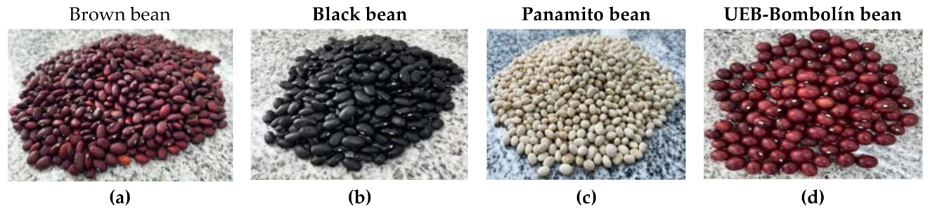
Figure 2.
Geographical location of the experimental site at the Universidad Estatal de Bolívar, in Guaranda, Bolívar province, Ecuador. Source: Map prepared by the authors; basemap imagery credits are retained in the figure (Esri, Maxar, Earthstar Geographics, and the GIS User Community).
Figure 2.
Geographical location of the experimental site at the Universidad Estatal de Bolívar, in Guaranda, Bolívar province, Ecuador. Source: Map prepared by the authors; basemap imagery credits are retained in the figure (Esri, Maxar, Earthstar Geographics, and the GIS User Community).
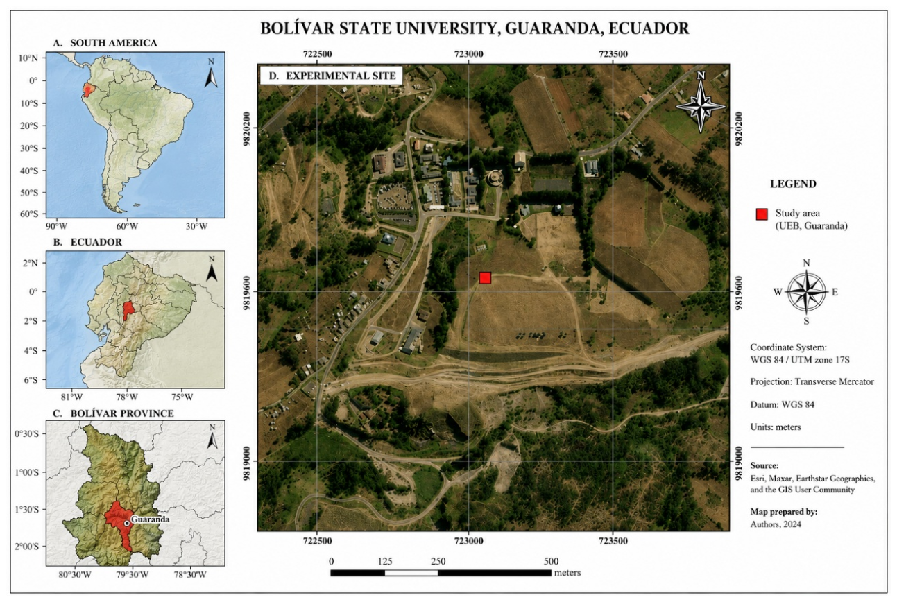
Table 2.
Two-way analysis of variance and partial effect size for bioactive compounds and antioxidant capacity in varieties of Phaseolus vulgaris L. subjected to different extraction solvents.
Table 2.
Two-way analysis of variance and partial effect size for bioactive compounds and antioxidant capacity in varieties of Phaseolus vulgaris L. subjected to different extraction solvents.
| Response variable | Source | SS | df | MS | F | p | η²p |
|---|---|---|---|---|---|---|---|
| Polyphenols (mg GAE/g) | |||||||
| A: Variety | 22.56 | 3 | 7.52 | 108.68 | <0.0001 | 0.931 | |
| B: Solvent | 8.815 | 2 | 4.407 | 63.69 | <0.0001 | 0.841 | |
| AB | 3.512 | 6 | 0.585 | 8.46 | 0.0001 | 0.679 | |
| Residual | 1.661 | 24 | 0.069 | - | - | - | |
| Flavonoids (mg QE/g) | |||||||
| A: Variety | 5.477 | 3 | 1.826 | 1124.81 | <0.0001 | 0.993 | |
| B: Solvent | 1.49 | 2 | 0.745 | 459.11 | <0.0001 | 0.975 | |
| AB | 1.869 | 6 | 0.311 | 191.9 | <0.0001 | 0.979 | |
| Residual | 0.039 | 24 | 0.002 | - | - | - | |
| Anthocyanins (mg CY3GE/g) | |||||||
| A: Variety | 577216 | 3 | 192405 | 1241.36 | <0.0001 | 0.994 | |
| B: Solvent | 68889 | 2 | 34445 | 222.23 | <0.0001 | 0.949 | |
| AB | 123800 | 6 | 20633 | 133.12 | <0.0001 | 0.971 | |
| Residual | 3720 | 24 | 155 | - | - | - | |
| ABTS (µmol TE/g) | |||||||
| A: Variety | 7400.8 | 3 | 2466.9 | 368.27 | <0.0001 | 0.979 | |
| B: Solvent | 1842.9 | 2 | 921.4 | 137.55 | <0.0001 | 0.92 | |
| AB | 1512.3 | 6 | 252 | 37.63 | <0.0001 | 0.904 | |
| Residual | 160.8 | 24 | 6.7 | - | - | - | |
| FRAP (µmol TE/g) | |||||||
| A: Variety | 2297.4 | 3 | 765.8 | 975.7 | <0.0001 | 0.992 | |
| B: Solvent | 652.9 | 2 | 326.4 | 415.92 | <0.0001 | 0.972 | |
| AB | 448.9 | 6 | 74.8 | 95.32 | <0.0001 | 0.959 | |
| Residual | 18.84 | 24 | 0.785 | - | - | - | |
| DPPH (µmol TE/g) | |||||||
| A: Variety | 1371.6 | 3 | 457.2 | 358.23 | <0.0001 | 0.978 | |
| B: Solvent | 882.9 | 2 | 441.4 | 345.9 | <0.0001 | 0.966 | |
| AB | 654.9 | 6 | 109.1 | 85.52 | <0.0001 | 0.955 | |
| Residual | 30.63 | 24 | 1.276 | - | - | - |
Note. SS = sum of squares; df = degrees of freedom; MS = mean square; = proportion of the total variability attributable to each source of variation relative to that source plus the residual error. A = variety effect; B = solvent effect; AB = variety × solvent interaction. Values of p < 0.05 indicate statistically significant differences.
Table 3.
Bioactive compound content and antioxidant capacity in varieties of Phaseolus vulgaris L. subjected to different extraction systems.
Table 3.
Bioactive compound content and antioxidant capacity in varieties of Phaseolus vulgaris L. subjected to different extraction systems.
| Treatment | Polyphenols (mg GAE/g) |
Flavonoids (mg QE/g) | Anthocyanins (mg CY3GE/g) | ABTS (µmol TE/g) |
FRAP (µmol TE/g) |
DPPH (µmol TE/g) |
|---|---|---|---|---|---|---|
| Brown + MeOH | 2.691 C | 0.867 C | 65.61 F | 56.69 B | 13.56 F | 14.12 FE |
| Brown + EtOH | 2.697 C | 1.271 B | 177.80 D | 40.03 ED | 17.00 E | 21.09 C |
| Brown + ACN | 4.280 A | 0.903 C | 145.16 ED | 73.49 A | 26.89 B | 37.38 A |
| Black + MeOH | 1.339 E | < LOQ E | 395.40 B | 27.97 F | 23.32 C | 15.69 ED |
| Black + EtOH | 1.596 ED | < LOQ E | 231.61 C | 37.82 E | 19.62 D | 15.96 ED |
| Black + ACN | 2.895 C | < LOQ E | 507.91 A | 57.62 B | 38.48 A | 28.48 B |
| Panamito + MeOH | 1.578 ED | 0.267 D | < LOQ G | 17.62 G | 5.86 H | 8.61 G |
| Panamito + EtOH | 1.935 D | 1.449 A | 90.90 F | 20.20 G | 5.40 H | 9.33 G |
| Panamito + ACN | 1.756 ED | 0.261 D | 75.97 F | 18.21 G | 3.47 I | 4.92 H |
| UEB-Bombolín + MeOH | 2.867 C | 0.379 D | 13.86 G | 43.76 D | 10.42 G | 13.33 F |
| UEB-Bombolín + EtOH | 3.638 B | 0.317 D | 138.88 ED | 48.17 C | 14.29 F | 17.40 D |
| UEB-Bombolín + ACN | 4.262 A | < LOQ E | 153.61 ED | 57.52 B | 21.92 C | 27.68 B |
Note. Values expressed as the mean of three experimental replicates. Different letters within the same column indicate statistically significant differences according to Tukey's HSD test (p < 0.05). < LOQ = concentration below the quantification limit of the method, substituted by LOQ/2 for statistical processing (Section 2.8). MeOH = methanol; EtOH = ethanol; ACN = acetonitrile.
Table 5.
Comparison of extraction parameters and characteristics of the brown bean protein isolate (Phaseolus vulgaris L.) relative to values reported for legumes.
Table 5.
Comparison of extraction parameters and characteristics of the brown bean protein isolate (Phaseolus vulgaris L.) relative to values reported for legumes.
| Parameter | Brown bean (present study) | Reference range for legumes | References |
|---|---|---|---|
| Protein in flour (%) | 22.85 | 16-27 | [111] |
| Extraction method | Isoelectric precipitation | Isoelectric / alkaline | [102] |
| Solubilisation pH | 10.0 (2 N NaOH) | 8.0-11.0 | [112] |
| Precipitation pH | 4.0 (citric acid) | 3.5-4.5 | [113] |
| Centrifugation | 4500 rpm, 20 min, 8 °C | 3000-10,000 rpm | [33] |
| Freeze-drying | 72 h, −56 °C | 48-96 h | [107] |
| Yield (%) | 17.58 | 15-25 | [102] |
| Protein content (%) | 70.81 | 65-90 | [33] |
| Extraction efficiency (%) | 76.9 | 65-85 | [114] |
| Presentation form | Freeze-dried powder (10 g sachets) | Powder / liquid | [115] |
| ABTS of the best treatment (µmol TE/g) | 73.49 | — | Present study |
Note. Reference ranges correspond to studies reported for protein isolates obtained from legumes by alkaline extraction and isoelectric precipitation. Values expressed on a dry basis where applicable.
Disclaimer/Publisher’s Note: The statements, opinions and data contained in all publications are solely those of the individual author(s) and contributor(s) and not of MDPI and/or the editor(s). MDPI and/or the editor(s) disclaim responsibility for any injury to people or property resulting from any ideas, methods, instructions or products referred to in the content. |
© 2026 by the authors. Licensee MDPI, Basel, Switzerland. This article is an open access article distributed under the terms and conditions of the Creative Commons Attribution (CC BY) license (http://creativecommons.org/licenses/by/4.0/).
Copyright: This open access article is published under a Creative Commons CC BY 4.0 license, which permit the free download, distribution, and reuse, provided that the author and preprint are cited in any reuse.