

Submitted:
05 October 2026
Posted:
08 October 2026
You are already at the latest version
Abstract
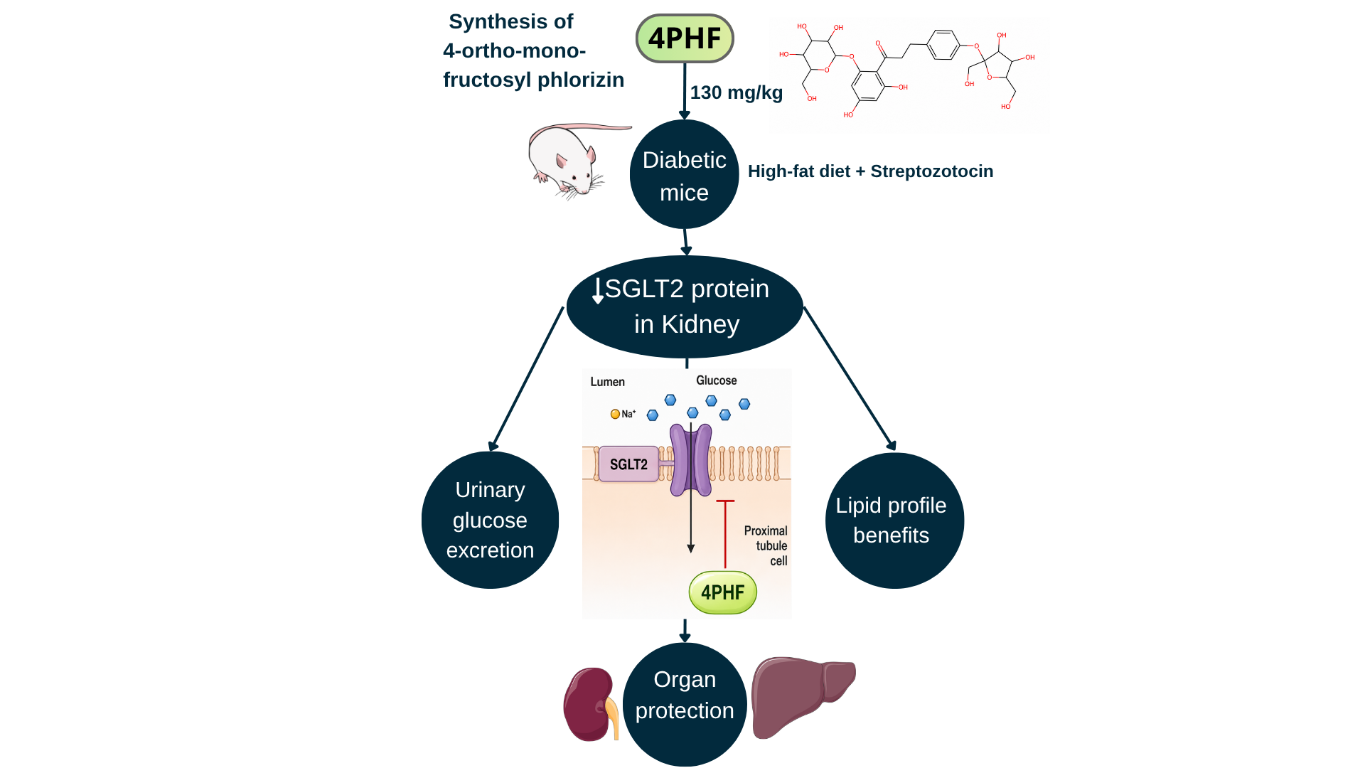
Diabetes mellitus (DM) is a chronic metabolic disorder characterized by persistent hyperglycemia, which, together with associated metabolic abnormalities, contributes to progressive damage in multiple organ systems. The development of effective and safe therapeutic alternatives remains a major research priority. Phlorizin is a dihydrochalcone that inhibits sodium-glucose cotransporter 1 and 2 (SGLT1 and SGLT2). 4‐ortho‐mono‐fructosyl phlorizin (4PHF) was synthesized from phlorizin by transfructosylation with a levansucrase from Paraburkholderia phymatum (KesL) using sucrose as a donor. The in vivo antidiabetic effect of 4PHF was evaluated using a murine model of experimental diabetes (high-fat diet followed by a single streptozotocin dose). Groups were treated as follows for 28 days: Non-Diabetic control group received only distilled water; a group receiving 4PHF, and a group treated with a SGLT2 inhibitor, dapagliflozin. 4PHF exhibited a tendency to reduce blood glucose levels, increased urinary glucose excretion, improved lipid profile by lowering triglycerides and total cholesterol. Histologically, 4PHF showed clear hepatoprotective and renoprotective effects. Immunohistochemistry revealed a marked decrease in SGLT2 expression and in the number of SGLT2-positive tubules in treated animals, suggesting that the treatment may modulate SGLT2 inhibition. Overall, 4PHF emerges as a promising therapeutic candidate for diabetes mellitus, as it reduces renal SGLT2 expression, improves metabolic dysfunction, and attenuates diabetes-induced hepatic and renal damage.
Keywords:
biocatalysis
; fructosylated derivative of phlorizin
; phlorizin fructoside
; SGLT2 inhibition
1. Introduction
Diabetes mellitus (DM) is a chronic metabolic disorder characterized by persistent hyperglycemia resulting from impaired insulin secretion, insulin resistance, or a combination of both mechanisms [1]. The disease is also associated with the progressive dysfunction of pancreatic β-cells, which contributes to the deterioration of glycemic control over time [1,2]. DM comprises several clinical categories, including type 1 diabetes, type 2 diabetes (T2DM), gestational diabetes, and less common forms such as monogenic diabetes and secondary diabetes. Among these, T2DM is the most prevalent, accounting for approximately 90% of all diabetes cases worldwide [1]. Over the past decade, the global burden of T2DM has increased dramatically, making it a major public health problem due to its strong association with long-term complications affecting the cardiovascular system, kidneys, liver, and overall metabolic homeostasis [3,4]. Currently, an estimated 589 million adults aged 20–79 years live with diabetes mellitus, representing approximately 11.1% of the global adult population, and this figure is projected to rise to nearly 852.5 million by 2050 [5].
The treatment for T2DM involves lifestyle changes, oral medications, and insulin therapy. Medications help to maintain blood sugar levels under control, but they can cause weight gain, risks of low blood sugar, and insufficient protection against cardiovascular, liver, and kidney complications, among others. This has led to the development of antidiabetic molecules that can regulate blood sugar levels while providing additional metabolic and cardiovascular benefits [6]. Sodium-glucose cotransporters (SGLTs), particularly SGLT1 and SGLT2, play a critical role in glucose homeostasis by mediating glucose reabsorption in the intestine and kidneys, respectively [7,8]. SGLT2, expressed in the proximal renal tubules, mediates ~90% of glucose reabsorption from tubular fluid; its inhibition blocks renal glucose reabsorption, increases urinary glucose excretion, and lowers blood glucose levels. Pharmacologically inhibiting these transporters has proven effective in improving glycemic control, leading to the development of selective SGLT2 inhibitors (gliflozins), which are now widely used in the clinical management of T2DM [8]. Recent studies have shown that SGLT-2 inhibitors exert beneficial effects on renal and cardiovascular function and display hepatoprotective properties, including reductions in hepatic steatosis and improvements in the lipid profile [9].
One of the first compounds identified as an SGLT inhibitor was phlorizin, a naturally occurring dihydrochalcone described in the 19th century as a dual SGLT1/SGLT2 inhibitor with marked antidiabetic activity [10,11]. However, despite its efficacy, its clinical application has been limited by its low water solubility, low oral bioavailability, and rapid metabolic degradation [12].
To overcome these pharmacokinetic limitations, 4-ortho-mono-fructosyl phlorizin (4PHF); a novel fructosylated derivative of phlorizin, was obtained via enzymatic biocatalysis. This structural modification was previously designed to improve phlorizin solubility and bioavailability [13,14], with the aim of retaining its known SGLT2 inhibitory activity [12].
In the context of T2DM, comprehensive evaluation of biochemical and histopathological parameters is essential to characterize the therapeutic potential of new antidiabetic agents [15]. Historically, T2DM treatment has primarily focused on lowering blood glucose levels. However, more recent therapeutic approaches have shifted toward addressing the underlying pathophysiological disturbances associated with the disease, particularly by reducing cardiorenal risk and promoting organ protection [9]. Furthermore, the liver and kidneys are key target organs in the pathophysiology of T2DM [16]. The liver plays a central role in glucose and lipid metabolism and is highly susceptible to metabolic dysregulation, while the kidneys are crucial in glucose reabsorption and are a primary target of diabetic complications such as nephropathy [2,17].
Therefore, in this study the effects of 4PHF were assessed in a murine model, emphasizing its potential benefits on metabolic abnormalities and the histological protection of the liver and kidneys. Furthermore, 4PHF modulation of SGLT2 renal tissue expression was accomplished to obtain mechanistic information of glucose reabsorption inhibition. Consequently, the present study aimed to evaluate the antidiabetic efficacy of 4PHF in an experimental diabetic mouse model induced by a high-fat diet (HFD) and streptozotocin (STZ) by assessing its effects on glycemia, urinary glucose excretion, lipid profile, hepatic and renal histopathology, and renal SGLT2 expression.
2. Materials and Methods
All reagents were obtained from Sigma-Aldrich-Merck (Darmstadt, Germany), unless otherwise specified.
2.1. Enzymatic Synthesis and Purification of 4-Ortho-Mono-Fructosyl Phlorizin (4PHF)
2.1.1. Enzymatic Synthesis and Purification
Biocatalysis was carried out following previously reported methodologies [13,18]. In brief, the reaction mixture contained fructosyltransferase from Paraburkholderia phymatum (KesL) (0.5 U/mL), kindly provided by the Institute of Biotechnology (IBT, Cuernavaca, Morelos, Mexico), sucrose (500 mM) as the fructosyl donor, and 99% pure phlorizin (25 mM) in acetate buffer (5 mM, pH 5.8) as the acceptor substrate. The final reaction volume was 500 mL. The reaction was incubated at 37 °C for 24 h and subsequently terminated by heating in a water bath, at 90 °C for 10 min.
2.1.2. HPLC-UV Analysis
After the reaction was stopped, phlorizin bioconversion was determined from the relative decrease in substrate concentration, quantified by high-performance liquid chromatography with ultraviolet detection (HPLC–UV), according to the following equation: Bioconversion (%) = [(C₀ − Cf)/C₀] × 100, where C₀ and Cf represent the initial and final phlorizin concentrations, respectively [14]. A solution of the lyophilized sample was prepared in water at a final concentration of 10 mg/L and analyzed using an Agilent Technologies (Santa Clara, CA, USA) Infinity 1260 HPLC system equipped with a Phenomenex (Torrance, CA, USA) Luna C18 column (5 μm, 100 Å, 150 mm × 4.6 mm) and an Agilent G4212 60007 UV detector (6 MPa). Chromatographic separation was achieved using a binary gradient of 0.05% formic acid in water (solvent A) and acetonitrile with 0.05% formic acid (solvent B) at a flow rate of 0.8 mL/min and a column temperature of 40 °C, gradient elution conditions: 90% A-10% B (min 0), 50% A-50% B (1-20 min), 5% A-95% B (20-25 min), 90% A-10% B (25-30 min). Total injection time was 30 min [14]. Analyses were performed in triplicate. Residual phlorizin was detected at 280 nm and quantified using an external calibration curve (0.05–1.0 mM; R² ≥ 0.99) prepared with a phlorizin standard.
2.1.3. UPLC-ESI-QTOF-MS/MS Analysis
The final reaction mixture was analyzed by ultra-performance liquid chromatography coupled to electrospray ionization quadrupole time-of-flight tandem mass spectrometry (UPLC–ESI–QTOF–MS/MS) and subsequently purified by flash chromatography using a Flash Manager C-615 system (BÜCHI, Switzerland).
Chromatographic analysis of the reaction products was performed using a Waters Acquity UPLC H-Class system (Waters, Milford, MA, USA) equipped with a degasser, quaternary pump, autosampler, and column heater. Separation was achieved at 40 °C using Acquity UPLC BEH C18 column (2.1 × 100 mm, 1.7 μm) from Waters, with an injection volume of 10 μL. The mobile phase consisted of (A) water containing 0.3% (v/v) formic acid and (B) acetonitrile containing 0.3% (v/v) formic acid, which were delivered at a flow rate of 0.3 mL/min. The gradient program was as follows: 0–1 min, 95% A; 1–11 min, linear decrease from 95% to 30% A; 11–12 min, return to 95% A; and 12–15 min, re-equilibration at 95% A. Mass spectrometric analysis was conducted using a Acquity Xevo G2-XS QToF mass spectrometer from Waters, equipped with an electrospray ionization (ESI) source operating in negative ion mode. Spectra were acquired over a mass range of m/z 50–1000. The operating parameters were set as previously reported: capillary voltage 2.5 kV, cone voltage 40 V, source temperature 100 °C, desolvation temperature 250 °C, and collision energy 6 eV [19].
2.1.4. Flash Chromatography
The products obtained from the enzymatic reaction with the highest conversion were purified by flash chromatography using a Flash Manager C-615 system (BÜCHI, Switzerland), equipped with a dual A/B pumping system and coupled to a C-660 fraction collector (BÜCHI, Switzerland). The separation was performed on a FlashPure EcoFlex C18 column (20 g capacity). Before loading the sample, the column was equilibrated with 50 mL of Milli-Q ultrapure water at a flow rate of 3 mL/min for 5 min, followed by conditioning with 50 mL of 100 % acetonitrile under the same flow rate and time condition, then sugars were eluted with water during 15 min at 3 mL/min, followed by phenolic fructosides elution with a gradient from 0 to 20 % of acetonitrile (30 min at 3 mL/min), subsequently a gradient from 20 to 30 % of acetonitrile was applied (30 min at 3 mL/min).
The purification process was monitored by thin-layer chromatography (TLC). Chromatographic spots corresponding to UV-absorbing compounds, including 4PHF, were visualized under UV illumination at 280 nm. Carbohydrate-containing compounds were subsequently detected by derivatization with an orcinol-based reagent under acidic conditions, followed by heating for color development [19].
2.2. In Vivo Antidiabetic Activity of 4PHF
2.2.1. Ethical Approval and Animals
All experimental procedures were conducted in accordance with the guidelines of the Internal Committee for the Care and Use of Laboratory Animals (CICUAL) (No. 2024-0C2). Animal handling followed the recommendations of the Official Mexican Standard NOM-062-ZOO-1999 [20].
Twenty-seven male CD1 mice (5 weeks old, 28 ± 4.76 g) were used in this study and obtained from the Bioterio Morelos (Morelos, Mexico). The animals were housed under controlled environmental conditions (12:12 h light–dark cycle, 22 °C, constant humidity) with free access to food and water.
2.2.2. HFD/STZ-Induced Experimental Diabetes Model
To induce DM a slightly modified HFD/STZ model was used [21]. Twenty-one mice were fed ad libitum with a HFD (4.6 kcal/g and 45% kcal from fat) acquired from Dyets, #102238 (Bethlehem, PA, USA) for 28 consecutive days. On day 29, a single intraperitoneal injection of STZ at a dose of 150 mg/kg body weight was performed to induce diabetes. STZ solution was freshly prepared in 0.1 M citrate buffer (pH 4.8) and administered following a 4 h fasting period. The overall mortality rate during diabetes induction was 15%. To reduce the risk of fatal hypoglycemia following STZ administration mice received 10% sucrose solution instead of drinking water for 24 h after STZ injection. Moreover, mortality rates above 20% have been proposed as indicative of excessive experimental severity, requiring implementation of additional animal welfare measures [22].
Hyperglycemia was confirmed 72 h after STZ administration by determining blood glucose levels from tail vein blood using a portable glucometer One Touch, Lifescan (Malvern, PA, USA). Animals with fasting blood glucose levels ≥150 mg/dL were considered diabetic [22]. Fasting blood glucose was subsequently monitored weekly for 28 days under 4 h fasting conditions.
The six remaining animals were included in the negative control group that received a standard diet (3.4 kcal/g, 14.1% energy from fat) that was obtained from Safe (Augy, France).
2.2.3. Experimental Design and Treatments
Diabetic mice were randomly assigned to four experimental groups (n = 6 per group). Mice in each experimental group were housed in polycarbonate cages of appropriate dimensions [20]: the negative control group comprising healthy mice (NC), the diabetic control group (DC) that were administered distilled water orally; the positive control group, comprising of diabetic control mice treated orally with dapagliflozin (10 mg/kg) (DAPA) (AstraZeneca®); and a group including diabetic mice treated orally with 4PHF (130 mg/kg). The dose was selected based on a previous study in which a medium dose of phlorizin (60 mg/kg) was administered to rats [23]. The equivalent mouse dose was calculated using the standard body surface area-based conversion factors [24]. Treatments were administered once daily by oral gavage for 28 consecutive days, as previously reported [23]. Body weight was recorded weekly throughout the experimental period. Food and water consumption were determined over a 24-h period by calculating the difference between the amounts provided and the respective amounts remaining.
2.2.4. Assessment of Urinary Glucose Excretion, Blood Sampling and Biochemical Analyses
On day 26 of treatment, mice were individually placed in metabolic cages and spontaneously eliminated urine was collected over a 6-hour period to assess glucose levels in the urine using the RANDOX glucose kit (Crumlin, UK) [25]. On day 29, following completion of the treatment period, fasting serum glucose was evaluated with a portable glucometer as described previously. Then, the animals were euthanized by cervical dislocation, and whole blood samples were collected and centrifuged at 1,500 ×g for 10 min at room temperature to obtain serum for biochemical analyses. Serum samples were frozen at -20°C and processed within 1 week. Serum levels of total cholesterol, triglycerides, high-density lipoprotein cholesterol (HDL), and low-density lipoprotein cholesterol (LDL) were quantified using commercial enzymatic kits from RANDOX, according to the manufacturer's instructions. All measurements were performed in duplicate [21,26]. Alterations in biochemical parameters have been previously described in HFD/STZ- induced diabetes model (26).
2.2.5. Histological Analysis of the Liver and Kidneys
Liver and kidney tissues were fixed in 4% paraformaldehyde, dehydrated through graded ethanol solutions (70%, 80%, 96%, and 100%), and embedded in paraffin blocks at 56–60 °C. Tissue sections (4 μm thick) were obtained using a microtome. Five liver sections per animal were stained with hematoxylin and eosin (H&E), whereas five kidney sections per animal were stained with periodic acid–Schiff (PAS) and counterstained with hematoxylin. Measurements obtained from the five sections were averaged to generate a single value per animal for each tissue. Histological evaluation was performed using a BestScope B2048T optical microscope (Beijing, China) equipped with an EP200 digital camera at magnifications of 400×.
Liver histopathology was evaluated using a semi-quantitative scoring system [27], where scores were assigned as follows: 0 (normal), 1 (mild alterations), 2 (moderate alterations), and 3 (severe alterations). The parameters assessed included hepatocellular degeneration, inflammatory infiltration, cytoplasmic vacuolization, vascular congestion, and sinusoidal alterations, and five H&E-stained sections per animal were analyzed.
Renal histopathology was assessed in PAS-stained sections using a semi-quantitative scoring system adapted from Fioretto and Mauer [28], scoring tubular atrophy, tubular dilation, PAS-positive tubular staining, glomerular damage, and inflammatory infiltration using the same 0–3 scale, with five renal sections analyzed per animal.
2.2.6. Immunohistochemical Detection of SGLT2 in Renal Cortex
Immunohistochemical analysis of SGLT2 expression was performed on five renal sections per animal following a previously described protocol [29], with minor modifications. Sections were deparaffinized, rehydrated through graded ethanol and xylol solutions (100, 90, 80, 70%), and incubated with a blocking solution containing 0.1 M PBS, 5% bovine serum albumin, and Triton X-100 0.5% for 2 h at 4 °C. They were then incubated overnight at 4 °C with a rabbit anti-SGLT2 primary antibody (ab37296) diluted at a 1:500 ratio obtained from ABCAM (Waltham, MA, USA).
After washing, the sections were incubated with a biotinylated goat anti-rabbit IgG (BA-100) from Vector Laboratories (Newark, CA, United States) for 2 h at room temperature, followed by incubation with the avidin–biotin–peroxidase complex (ABC, Vector Laboratories) for 1 h. Immunoreactivity was visualized using a 3,3′-diaminobenzidine (DAB) solution (0.05%) in the presence of hydrogen peroxide.
SGLT2 expression was quantified as the percentage of SGLT2-positive tubular area per field, following modified methods described by Wang et al. (2017) [30]. Five renal cortical fields per animal were analyzed using ImageJ software 2.17.0-0 (Bethesda, MD, USA). Following the evaluation of immunoreactivity, tissue sections were counterstained with PAS to improve visualization.
2.3. Statistical Analysis
All results are presented as mean ± standard error of the mean (SEM). Data were analyzed using one-way ANOVA for comparisons among independent groups and repeated-measures ANOVA for longitudinal data, using GraphPad Prism software (version 8.0.1). Post hoc comparisons were performed using Dunnett’s multiple -comparison test, p < 0.05 was considered statistically significant, and the DC group considered as the reference.
3. Results
3.1. Biocatalytic Synthesis, Purification, and Characterization of Fructosylated Phlorizin Derivatives
HPLC–UV analysis was used to determine phlorizin bioconversion at the end of the reaction and to assess the chromatographic purity of both the individual 4PHF-containing fractions and the final pooled preparation after lyophilization. The enzymatic reaction resulted in a bioconversion of 81.98 % as determined by HPLC-UV from the relative decrease in substrate concentration between the beginning and the end of the reaction.
UPLC–ESI–QTOF–MS/MS analysis of the final reaction mixture in negative-ion mode revealed multiple chromatographic peaks tentatively assigned to residual phlorizin and fructosylated phlorizin derivatives (Figure S1 and Table S1). As reported in Table S1, the peak at 8.83 min exhibited an ion at m/z 481.197, tentatively assigned to the formate adduct of phlorizin, [C₂₁H₂₄O₁₀ + HCOO]⁻. A peak at 7.94 min exhibited an ion at m/z 643.222, tentatively assigned to the formate adduct of monofructosylated phlorizin, [C₂₇H₃₄O₁₅ + HCOO]⁻. Additional peaks at 7.92 and 7.41 min, with ions at m/z 643.225 and 643.212, respectively, were tentatively associated with monofructosylated species. Their possible isomeric relationships require further structural characterization. Peaks at 7.51 and 6.82 min exhibited ions at m/z 805.249 and 805.253, respectively, provisionally assigned to the formate adducts of difructosylated phlorizin, [C₃₃H₄₄O₂₀ + HCOO]⁻. These preliminary assignments suggest the formation of mono- and difructosylated products during the enzymatic reaction.
The reaction mixture was subsequently purified by flash chromatography, and fractions 5–25 were collected. TLC analysis revealed UV-absorbing spots and positive staining with an orcinol-based reagent, supporting the presence of carbohydrate-containing compounds in these fractions (Figure S2). Fractions selected on the basis of their TLC profiles were further analyzed by HPLC–UV to assess their composition and chromatographic purity.
Fractions 5–25 were pooled, concentrated by rotary evaporation, and lyophilized to obtain the preparation administered to the animals. HPLC–UV analysis of the reconstituted lyophilized preparation at 280 nm revealed a predominant peak assigned to 4PHF at 12.7 min, a peak corresponding to residual phlorizin at 15.8 min, and additional peaks assigned to other fructosylated derivatives of phlorizin (Figure 1), the chemical structures of phlorizin and 4PHF are also included. The phlorizin standard exhibited a single sharp peak at approximately 15.8 min (Figure S3), whereas the analyzed fractions displayed a predominant peak at approximately 12.7 min, assigned to 4PHF. The structure of 4PHF was previously elucidated by nuclear magnetic resonance spectroscopy (18), and the corresponding nuclear magnetic resonance spectra are presented in Figure S4 and Figure S5. The relative peak areas were 94.50% for 4PHF, 2.11% for residual phlorizin, and 3.39% for the other identified fructosylated derivatives (Table 1). These percentages represent relative chromatographic peak areas rather than mass fractions. Thus, the preparation administered to the animals consisted predominantly of 4PHF, with residual phlorizin and minor amounts of other fructosylated derivatives.
3.2. Effects of 4PHF on Body Weight, Food Intake, and Water Consumption
Once diabetes was confirmed in male CD-1 mice showing fasting hyperglycemia (≥150 mg/dL), the treatments were initiated. The body weight of the NC group increased 20.58 % by study completion, whereas the DC group exhibited a reduction in mean body weight (−7.33 %) by study completion (Figure 2a). In contrast, the 4PHF-treated group showed a moderate decrease in body weight (−3.67 %), whereas mice in the DAPA group maintained relatively stable body weight, with a slight increase of 0.40 %. However, these differences were not statistically significant among the groups.
Water intake varied among the experimental groups (Figure 2b). The DC group had a daily average water intake of 31.32 ± 5.41 mL, which was apparently higher than that of the NC group (15.33 ± 0.52 mL); however, this difference did not reach statistical significance. Similarly, water intake in the 4PHF-treated group (18.58 ± 1.15 mL) and the DAPA-treated group (17.63 ± 1.98 mL) did not differ significantly from that of the DC group.
Average food intake was significantly higher in the DC group than in the NC group (76.31 ± 4.30 kcal/day vs. 52.32 ± 3.89 kcal/day, respectively; p < 0.01; Figure 2c). In contrast, food intake in the 4PHF- and DAPA-treated groups (71.02 ± 5.01 and 70.57 ± 4.30 kcal/day, respectively), did not differ significantly from that of the DC group.
3.3. Effects of 4PHF on Fasting Blood Glucose Levels
Weekly monitoring of fasting blood glucose levels revealed sustained hyperglycemia in the DC group throughout the experimental period, reaching approximately 460 mg/dL on day 28, corresponding to a modest reduction of 19.11% from baseline values. In contrast, the group treated with 4PHF exhibited a progressive reduction in blood glucose levels, reaching values of approximately 315–325 mg/dL at the end of the study, which represents a 36.44% decrease (p < 0.05 vs. DC). Treatment with dapagliflozin resulted in a more pronounced hypoglycemic effect, with glucose levels of approximately 220–230 mg/dL by day 28, corresponding to a 56.68% reduction (p < 0.01 vs. DC) (Table 2).
3.4. Effects of 4PHF on Urinary Glucose Excretion
Urinary glucose levels differed significantly among the experimental groups (Figure 4). As expected, the NC group showed minimal glucose excretion, whereas the DC group exhibited a significant increase (p < 0.001), reaching approximately 70 mg/dL. In contrast, treatment with 4PHF resulted in a significant increase in urinary glucose excretion, reaching values of approximately 190 mg/dL (p < 0.001 vs. DC). A more pronounced renal glucose excretion was observed in the dapagliflozin-treated group, which showed urinary glucose levels of approximately 270 mg/dL (p < 0.001 vs. DC), which is significantly higher than those observed in the 4PHF-treated group (p < 0.001).
3.5. Effects of 4PHF on Lipid Profile
Serum lipid profile analysis revealed significant differences between the diabetic DC group and the treated groups (Figure 5). Specifically, the DC group showed a significant increase in total cholesterol levels when compared to the NC group (205.16 ± 3.86 mg/dL vs. 73.27 ± 1.36 mg/dL; p < 0.001), respectively, while treatment with 4PHF significantly reduced them to 93.28 ± 4.84 mg/dL compared to DC, interestingly cholesterol levels in 4PHF-treated group and NC group were similar. The DAPA group showed a similar response (p < 0.001 vs. DC).
In addition, serum triglyceride concentrations were also significantly elevated (p < 0.01) in DC mice at 191.5 ± 14.18 mg/dL when compared to the NC group (105.17 ±10.23). 4PHF treatment significantly reduced triglyceride concentrations compared to DC (91.83 ± 8.70 vs. 191.5 ± 14.18; p < 0.001). Similarly, the DAPA group exhibited lower triglyceride levels (p < 0.001 vs. DC).
Regarding HDL, the DC group exhibited a significant decrease (p < 0.05) vs. NC group, 36.93 ± 2.97 mg/dL and 41.36 ± 0.57 mg/dL, respectively. The HDL levels were significantly increased in the 4PHF-treated group (53.39 ± 3.58 mg/dL) compared to the DC group (36.93 ± 2.97 mg/dL) (p < 0.001). Likewise, the DAPA group also showed higher HDL levels (p < 0.001 vs. DC). Furthermore, the LDL levels of the DC group were higher than NC (64.44 ± 2.61 mg/dL and 44.03 mg/dL ± 2.46, respectively; p < 0.01). The 4PHF group significantly reduced LDL levels (42.76 ± 2.80 mg/dL) relative to the DC animals (64.44 ± 2.61 mg/dL) (p < 0.001), and this effect was also observed in the DAPA group (p < 0.001 vs. DC).
3.6. Liver Weight and Hepatic Histopathological Evaluation
Liver weight did not differ significantly among the groups (Figure 6a). However, the liver weights of the 4PHF and DAPA-treated groups were numerically closer to those of the NC group than those of the DC group.
Histological examination of liver sections stained with hematoxylin and eosin (H&E) revealed significant pathological alterations in the DC group in contrast to NC group, including extensive vacuolization, vascular congestion, and hepatic architecture disruption (Figure 7). In contrast, both DAPA- and 4PHF-treated groups exhibited preserved hepatic morphology, with reduced structural alterations and more organized hepatic parenchyma relative to the DC group. Semi-quantitative analysis demonstrated significant reductions in vacuolization, vascular congestion, hepatocellular degeneration, inflammatory infiltration, and sinusoidal alterations in the 4PHF group when compared to the DC group (p < 0.05 to p < 0.001). The DAPA group also showed significant improvements (p < 0.01 to p < 0.001) when compared to DC animals. Notably, 4PHF showed effects comparable to DAPA across all evaluated parameters.
3.7. Renal Weight and Histopathological Evaluation
The weight of the left and right kidneys did not differ significantly among the groups as previously shown in Figure 6b and Figure 6c. However, the left kidney weight of the 4PHF group was numerically closer to those of the NC group than those of the DC group.
PAS-stained renal sections from the DC group revealed significant differences compared with the NC group (p < 0.01 to p < 0.001) with respect to tubular and glomerular alterations, including tubular dilation, accumulation of PAS-positive material, glomerular damage, inflammatory infiltration, and tubular atrophy (Figure 8). Treatment with 4PHF significantly attenuated these pathological features, with semi-quantitative scoring indicating significant reductions in tubular dilation (p < 0.01), PAS-positive staining (p < 0.001), glomerular damage (p < 0.01), inflammation/infiltration (p < 0.001), and tubular atrophy (p < 0.001) compared to DC animals. The magnitude of renal protection observed in the 4PHF-treated group was comparable to that of the DAPA group.
3.8. Effects of 4PHF on Renal SGLT2 Expression
Immunohistochemical analysis revealed significantly higher and intense SGLT2 immunoreactivity in the renal cortex of DC mice, with widespread staining in proximal tubules (Figure 9) relative to the NC group. In contrast, the group exhibited a marked reduction in SGLT2 staining intensity, and quantitative analysis demonstrated a significant decrease in SGLT2-positive area in 4PHF-treated animals compared to the NC group (p < 0.001). The 4PHF treatment reduced SGLT2 expression to approximately 7%, while treatment with DAPA reduced it further to about 5%.
The number of SGLT2-positive tubules per field in the 4PHF-treated group showed an intermediate reduction that did not reach statistical significance, whereas DAPA treatment reduced them when compared to NC mice (p < 0.01).
4. Discussion
The present study employed an integrated strategy combining enzymatic biocatalysis for the generation of fructosylated phlorizin derivatives with subsequent biological evaluation in an experimental diabetic mouse model induced by HFD and STZ. The biocatalytic results demonstrate that the KesL enzyme efficiently catalyzed fructosyl transfer reactions, generating a mixture of mono-, di-, and higher-order fructosylated derivatives as shown previously [18]. The formation of multiple fructosylated species is consistent with the transfructosylation activity reported for fructosyltransferases [13,31] and reflects the capacity of KesL to utilize phlorizin as an acceptor substrate. Although heterogeneous, the resulting product mixture was effectively enriched by flash chromatography, thereby allowing subsequent biological evaluation. The chromatographic complexity observed for the fructosylated products compared with the precursor compound further supports successful enzymatic modification [31]. The presence of a predominant derivative suggests preferential fructosylation at specific hydroxyl positions, consistent with previous reports describing regioselective enzymatic fructosylation of polyphenolic compounds [32]. The predominant fructosylated fraction, designated as 4PHF, represents a structurally modified derivative of phlorizin with potentially improved biological properties.
Fructosylation can modify the physicochemical properties of phenolic compounds relative to their native forms. In the present study, the enzymatic reaction achieved a phlorizin bioconversion of 81.98 %, reflecting substrate consumption rather than the specific yield of 4PHF. Following purification, the resulting preparation exhibited a chromatographic purity of 94.5% for 4PHF, compared with approximately 74.5% 4PHF in the enriched fraction reported in our previous work [18]. The higher purity achieved in this study was attributed to optimization of the purification and enrichment procedure. Nevertheless, further optimization of the reaction conditions is needed to minimize the formation of secondary products.
Comparable enzymatic transfructosylation efficiencies have been reported in the literature; for instance, levansucrase-catalyzed reactions using different bacterial systems achieved maximum yields of 58% with Vibrio natriegens LS2, 36% with Novosphingobium aromaticivorans LS3, and 18% with Pseudomonas graminis LS4 [33]. Other modifications such as galactosylation of several phenolic compounds, including phlorizin via permeabilized cells from Pantoea anthophila have been reported to reach conversion yields of up to 41.8% [19]. These studies highlight the versatility of enzymatic glycosylation strategies for modifying phenolic compound structures, and other studies have demonstrated that the versatility of enzymatic fructosylation of phenolic acceptors is highly dependent on the catalytic system employed and the reaction conditions [31].
4PHF has been reported to exhibit activity against key therapeutic targets implicated in T2DM pathogenesis, including dipeptidyl peptidase-4 and the insulin receptor [34]. More recently, our research group demonstrated that 4PHF inhibits α-amylase in vitro assays, while in silico analyses suggested a slightly stronger predicted binding affinity toward α-glucosidase than toward α-amylase [18]. The organoprotective effects observed in the present animal study may therefore be associated with the biological activities previously reported in vitro and in silico. It is possible to affirm that 4PHF has a dual effect by inhibiting enzymes responsible for carbohydrate digestion, and the prevention of renal glucose reabsorption. Although the precise mechanisms remain to be elucidated, our findings collectively support the potential of modified phlorizin derivatives such as 4PHF to exhibit improved biological properties. It has been reported that some chalcone derivatives show better biological activities [35], particularly antidiabetic activity, as determined by a method quantifying glucose consumption by adipocytes [36].
The present study demonstrates that the fructosylated phlorizin derivative 4PHF exerts a multifaceted pharmacological profile in an experimental diabetic mouse model induced by HFD and STZ. Although its antihyperglycemic activity was moderate compared with dapagliflozin, treatment consistently improved fasting blood glucose while producing marked increases in urinary glucose excretion, significant improvements in lipid metabolism, attenuation of hepatic and renal injury, and reduced renal SGLT2 expression. These findings highlight the functional relevance of the structural modification achieved through fructosylation and suggest that the biological activity of 4PHF extends beyond glucose lowering alone.
The partial preservation of body weight observed in 4PHF-treated animals is consistent with improved metabolic stability under chronic hyperglycemic conditions. In uncontrolled T2DM, persistent hyperglycemia promotes proteolysis and lipolysis, leading to progressive weight loss [37]. The ability of 4PHF to attenuate this effect without altering food intake suggests an improvement in systemic metabolic efficiency rather than appetite suppression, and similar neutral effects on food intake have been reported following phlorizin administration in diabetic rodent models [38].
Dyslipidemia is a central metabolic abnormality in DM and contributes significantly to cardiovascular risk and the progression of diabetic complications [39]. In the present study, 4PHF significantly reduced serum total cholesterol, triglycerides, and LDL concentrations while increasing HDL levels, indicating an overall improvement in metabolic homeostasis. These changes are particularly noteworthy because the lipid profile achieved after 4PHF treatment approached that reported for healthy male ICR (CD-1) mice, especially for total cholesterol, while triglyceride concentrations were markedly reduced and HDL levels were partially restored [40]. Although direct comparisons between independent studies should be interpreted cautiously due to differences in experimental protocols, these findings suggest that 4PHF effectively counteracts diabetes-associated dyslipidemia and promotes the recovery of a near-physiological lipid profile. Similar lipid-lowering effects have previously been reported for phlorizin and related flavonoids in diabetic models [23]. Improvements in lipid homeostasis have been associated with reduced glucotoxicity and enhanced insulin sensitivity, which contribute to normalization of hepatic lipid metabolism [41,42]. Furthermore, Zhang et al. demonstrated that phlorizin administration in HFD/STZ-induced diabetic rats significantly reduced fasting blood glucose, total cholesterol, triglycerides, and LDL-C while increasing HDL-C, accompanied by attenuation of hepatic lipid accumulation and restoration of pancreatic morphology, emphasizing that simultaneous correction of hyperglycemia and dyslipidemia is essential for limiting diabetes progression [23].
Interestingly, the favorable biochemical profile observed after 4PHF treatment was accompanied by a significant increase in urinary glucose excretion. Under diabetic conditions, chronic hyperglycemia induces adaptive SGLT2 overexpression in proximal tubular cells, thereby enhancing renal glucose reabsorption and perpetuating metabolic imbalance [12,43]. The ability of 4PHF to promote urinary glucose excretion suggests that the inhibitory activity of its phlorizin precursor is preserved [11], although with lower potency than the selective SGLT2 inhibitor dapagliflozin. Additionally, metabolic improvements associated with SGLT2 inhibition have been reported by Tahara et al., who demonstrated that a high dose of ipragliflozin not only improved hyperglycemia but also significantly reduced serum triglyceride levels from 194 ± 5.9 mg/dL in untreated HFD/STZ-induced diabetic mice to 128 ± 11 mg/dL in the treated group. In the present study, serum triglyceride levels in diabetic mice were comparable (191.5 ± 5.8 mg/dL); however, treatment with 4PHF resulted in an even greater reduction, reaching 91.8 ± 3.5 mg/dL, which is lower than the levels reported for ipragliflozin in the aforementioned study. A similar trend was observed for total plasma cholesterol. Tahara et al. reported that untreated diabetic mice exhibited total plasma cholesterol levels of 146 ± 8 mg/dL, which decreased to 117 ± 5 mg/dL following treatment with a high dose of ipragliflozin. Likewise, in the present study, untreated diabetic mice exhibited elevated total cholesterol levels (205.2 ± 3.8 mg/dL), which were markedly reduced to 93.28 ± 2.0 mg/dL following 4PHF treatment [26]. Our findings are consistent with those reported by Tahara et al. (2013), supporting the notion that SGLT2 inhibition by phlorizin derivatives contributes to the improvement of diabetes- and obesity-associated metabolic abnormalities.
Importantly, the renal histopathological improvements observed following 4PHF treatment appeared proportionally greater than the reduction in fasting blood glucose, suggesting that its renoprotective activity may not be exclusively explained by glycemic control. The combined administration of HFD and STZ has previously been reported to induce pronounced structural alterations in the kidneys [44]. Tubular dilation, PAS-positive material accumulation, inflammatory cell infiltration, and tubular atrophy are hallmarks of early diabetic nephropathy [45]. The significant attenuation of these lesions indicates that 4PHF confers substantial renal protection. Previous studies have also demonstrated that phlorizin attenuates renal injury and oxidative stress in experimental diabetes [46]. However, because oxidative stress biomarkers and inflammatory cytokines were not evaluated in the present study, the contribution of these mechanisms remains speculative and should be investigated in future studies.
Hepatic protection represents another important finding of this work. T2DM-associated liver injury is driven by glucotoxicity, lipid accumulation, oxidative stress, and inflammatory activation [47]. The significant reduction in hepatocellular degeneration, vacuolization, vascular congestion, and inflammatory cell infiltration observed after 4PHF administration suggests improved hepatic metabolic homeostasis. Together with the favorable changes in serum lipid profile, these observations are consistent with previous reports describing hepatoprotective effects of phlorizin and flavonoid-derived compounds [23], as well as with the improvements in hepatic steatosis reported for both phlorizin and SGLT2 inhibitors in experimental diabetes.
Immunohistochemical analysis further demonstrated that 4PHF significantly reduced renal SGLT2 overexpression, a pathological adaptation commonly observed in diabetic complications [30]. While dapagliflozin acts as a potent and selective SGLT2 inhibitor, 4PHF appeared to moderately reduce SGLT2 expression without complete suppression. This regulatory profile may partially explain the moderate antihyperglycemic effect associated with substantial metabolic and tissue protection observed in the present study.
Because several molecular targets have already been identified for phlorizin and related dihydrochalcones [48], additional studies evaluating oxidative stress and inflammatory mediators, and other signaling pathways will be required to fully elucidate the mechanism of action of 4PHF. On the other hand, the hypoglycemic activity of SGLT2 inhibitors is largely independent of pancreatic insulin secretion [42]. A previous study evaluated the effects of phlorizin on insulin-stimulated glucose uptake in MKR mice and reported no improvement [49]. In the present study, circulating insulin concentrations were not measured. However, the assessment of serum insulin levels together with insulin resistance would provide valuable insight into the metabolic effects of phlorizin derivative 4PHF in the treatment of experimental diabetes.
A previous study reported that male mice are more frequently used than female mice in experimental diabetes models [50]. Consistent with this trend, the present study included only male mice, highlighting the need for future studies to incorporate female mice to address sex-related differences in diabetes and treatment response.
5. Conclusions
The biocatalytic synthesis and purification of 4PHF was successfully achieved, and its biological activity was demonstrated in an experimental diabetic mouse model induced by HFD and STZ. Treatment with 4PHF significantly improved dyslipidemia, increased urinary glucose excretion, and diabetes-associated liver and kidney damage when compared to diabetic control group. Furthermore, 4PHF emerges as a promising therapeutic candidate for diabetes mellitus by reducing renal SGLT2 expression, improving metabolic dysfunction, and attenuating diabetes-associated hepatic and renal injury. These findings provide a solid experimental basis for future studies aimed at elucidating the underlying molecular mechanisms of 4PHF and evaluating its potential to prevent diabetes-related complications.
Supplementary Materials
The following supporting information can be downloaded at the website of this paper posted on Preprints.org.
Author Contributions
Conceptualization, E.P.-C. and J.A.; Investigation, O.R.T.-G., C.O.-C., S.G.C.-G., J.G.T.-T., A.H.-G. and I.M.S.-H.; data curation, A.S.G.-G., E.P.-C., O.R.T.-G., J.G.T.-T.; formal analysis, O.R.T.-G., A.H.-G. and I.M.S.-H., A.S.G.-G., and E.P.-C.; funding acquisition, E.P.-C. and J.A.; methodology, C.O.-C., A.S.G.-G., O.R.T.-G., S.G.C.-G., E.P.-C.; validation, O.R.T.-G.; visualization, A.S.G.-G.; writing—review and editing, O.R.T.-G., A.S.G.-G. and E.P.-C. All authors have read and agreed to the published version of the manuscript.
Funding
This research was funded by the COECYTJAL project FODECIJAL 10233-2022. This research was also supported by a doctoral scholarship granted to Omar Ricardo Torres-González (CVU 926677) by the Secretaría de Ciencia, Humanidades, Tecnología e Innovación (SECIHTI, Mexico), within the postgraduate program “Ciencias de la Innovación Biotecnológica” in Centro de Investigación y Asistencia en Tecnología y Diseño del Estado de Jalisco, A.C. (CIATEJ).
Institutional Review Board Statement
The animal study protocol was approved by the Internal Committee for the Care and Use of Laboratory Animals of Centro de Investigación y Asistencia en Tecnología y Diseño del Estado de Jalisco (protocol code 2024-0C2. Approval date: February 28, 2024).
Data Availability Statement
Data will be made available on request.
Acknowledgments
The authors would like to thank PhD. Mario Eduardo Flores-Soto from the Centro de Investigación Biomédica de Occidente (CIBO), Instituto Mexicano del Seguro Social (IMSS), for his valuable scientific support and guidance. We also acknowledge PhD. Angélica Yanet Nápoles Medina (CIBO, IMSS), for her technical assistance and contributions during the development of this study.
Conflicts of Interest
The authors declare that they have no known competing financial interests or personal relationships that could have influenced the work reported in this study.
Abbreviations
The following abbreviations are used in this manuscript:
| 4PHF | 4-ortho-mono-fructosyl phlorizin |
| DAPA | Dapaglifozin |
| HDL | High-density lipoprotein cholesterol |
| HFD | High fat diet |
| LDL | Low-density lipoprotein cholesterol |
| SGLT1 | Sodium-glucose cotransporter 1 |
| SGLT2 | Sodium-glucose cotransporter 2 |
| STZ | Streptozotocin |
| T2DM | Type 2 diabetes mellitus |
| TLC | Thin-layer chromatography |
| UPLC | Ultra performance liquid chromatography |
References
- Goyal, R.; Singhal, M.; Jialal, I. Type 2 diabetes. In StatPearls; StatPearls Publishing: Treasure Island, FL, USA, 2025; pp. 1–2. Available online: https://www.ncbi.nlm.nih.gov/books/NBK513253/ (accessed on 30 September 2025).
- Guo, H.; Pan, L.; Wu, Q.; Wang, L.; Huang, Z.; Wang, J.; Wang, L.; Fang, X.; Dong, S.; Zhu, Y.; et al. Type 2 Diabetes and the Multifaceted Gut-X Axes. Nutrients 2025, 17, 2708. [Google Scholar] [CrossRef] [PubMed]
- DeFronzo, R.A.; Ferrannini, E.; Groop, L.; Henry, R.R.; Herman, W.H.; Holst, J.J.; Hu, F.B.; Kahn, C.R.; Raz, I.; Shulman, G.I.; Simonson, D.C.; Testa, M.A.; Weiss, R. Type 2 diabetes mellitus. Nat. Rev. Dis. Primers 2015, 1, 15019. [Google Scholar] [CrossRef] [PubMed]
- Padhi, S.; Nayak, A.K.; Behera, A. Type II diabetes mellitus: A review on recent drug-based therapeutics. Biomed. Pharmacother. 2020, 131, 110708. [Google Scholar] [CrossRef] [PubMed]
- International Diabetes Federation. Diabetes Atlas, 11th ed.; International Diabetes Federation: Brussels, Belgium, 2025; Available online: https://diabetesatlas.org/media/uploads/sites/3/2025/04/IDF_Atlas_11th_Edition_2025-1.pdf (accessed on 19 September 2025).
- Stanimirova, D.; Petrova, G.; Mitkova, Z. Effectiveness of SGLT2 Inhibitors in Type 2 Diabetes: A Systematic Integrative Review of Reviews and Comparative Effectiveness Studies (2020–2025). Pharmacy 2026, 14, 47. [Google Scholar] [CrossRef] [PubMed]
- Alicic, R.Z.; Rooney, M.T.; Tuttle, K.R. Diabetic kidney disease: Challenges, progress, and possibilities. Clin. J. Am. Soc. Nephrol. 2017, 12, 2032–2045. [Google Scholar] [CrossRef] [PubMed]
- Yang, Y.; Chen, S.; Pan, H.; Zou, Y.; Wang, B.; Wang, G.; Zhu, H. Safety and efficiency of SGLT2 inhibitor combining with insulin in subjects with diabetes: A systematic review and meta-analysis. Medicine 2017, 96, e6944. [Google Scholar] [CrossRef] [PubMed]
- Tartau, C.G.; Boboc, I.K.S.; Mititelu-Tartau, L.; Bogdan, M.; Buca, B.R.; Pavel, L.L.; Amalinei, C. Exploring the Protective Effects of Traditional Antidiabetic Medications and Novel Antihyperglycemic Agents in Diabetic Rodent Models. Pharmaceuticals 2025, 18, 670. [Google Scholar] [CrossRef] [PubMed]
- Tian, L.; Cao, J.; Zhao, T.; Liu, Y.; Khan, A.; Cheng, G. The bioavailability, extraction, biosynthesis and distribution of natural dihydrochalcone: Phloridzin. Int. J. Mol. Sci. 2021, 22, 962. [Google Scholar] [CrossRef] [PubMed]
- Ghezzi, C.; Loo, D.D.F.; Wright, E.M. Physiology of renal glucose handling via SGLT1, SGLT2 and GLUT2. Diabetologia 2018, 61, 2087–2097. [Google Scholar] [CrossRef] [PubMed]
- Martus, G.; Bergling, K.; Öberg, C.M. Dual SGLT1/SGLT2 inhibitor phlorizin reduces glucose transport in experimental peritoneal dialysis. Perit. Dial. Int. 2023, 43, 145–150. [Google Scholar] [CrossRef] [PubMed]
- Herrera-González, A.; Núñez-López, G.; Morel, S.; Amaya-Delgado, L.; Sandoval, G.; Gschaedler, A.; Remaud-Simeon, M.; Arrizon, J. Functionalization of natural compounds by enzymatic fructosylation. Appl. Microbiol. Biotechnol. 2017, 101, 5223–5234. [Google Scholar] [CrossRef] [PubMed]
- Herrera-González, A.; Núñez-López, G.; Núñez-Dallos, N.; Amaya-Delgado, L.; Sandoval, G.; Remaud-Simeon, M.; Morel, S.; Arrizon, J.; Hernández, L. Enzymatic synthesis of phlorizin fructosides. Enzym. Microb. Technol. 2021, 147, 109783. [Google Scholar] [CrossRef] [PubMed]
- Elkhalifa, A.M.E.; Nazar, M.; Ali, S.I.; Khursheed, I.; Taifa, S.; Ahmad Mir, M.; Shah, I.H.; Malik, M.; Ramzan, Z.; Ahad, S.; et al. Novel Therapeutic Agents for Management of Diabetes Mellitus: A Hope for Drug Designing against Diabetes Mellitus. Life 2024, 14, 99. [Google Scholar] [CrossRef] [PubMed]
- Lu, X.; Xie, Q.; Pan, X.; Zhang, R.; Zhang, X.; Peng, G.; Zhang, Y.; Shen, S.; Tong, N. Type 2 diabetes mellitus in adults: Pathogenesis, prevention and therapy. Signal Transduct. Target. Ther. 2024, 9, 262. [Google Scholar] [CrossRef] [PubMed]
- Młynarska, E.; Czarnik, W.; Dzieża, N.; Jędraszak, W.; Majchrowicz, G.; Prusinowski, F.; Stabrawa, M.; Rysz, J.; Franczyk, B. Type 2 diabetes mellitus: New pathogenetic mechanisms, treatment and the most important complications. Int. J. Mol. Sci. 2025, 26, 1094. [Google Scholar] [CrossRef] [PubMed]
- Torres-González, O.R.; Arrizon, J.; Herrera-González, A.; Olvera-Carranza, C.; Sánchez-Hernández, I.M.; Padilla-Camberos, E.; González-Garibay, A.S. 4-O-Mono-Fructosyl Phlorizin-Enriched Fraction and Its Interaction with Carbohydrate Digestive Enzymes: In Vitro and In Silico Studies. Appl. Sci. 2026, 16, 2072. [Google Scholar] [CrossRef]
- Briceño, J.R.; Torres-Tolentino, J.G.; González-Ávila, M.; Amaya-Delgado, L.; López-Roa, R.; Herrera-González, A.; Arrizon, J. Enzymatic galactosylation of phenolic compounds by permeabilized cells of Pantoea anthophila. Appl. Microbiol. Biotechnol. 2026, 110, 10. [Google Scholar] [CrossRef] [PubMed]
- Secretaría de Agricultura; Ganadería; Desarrollo Rural; Pesca y Alimentación (SAGARPA). Norma Oficial Mexicana NOM-062-ZOO-1999, Especificaciones técnicas para la producción, cuidado y uso de los animales de laboratorio; Gobierno de México: Ciudad de México, México, 1999; Available online: https://www.gob.mx/cms/uploads/attachment/file/203498/NOM-062-ZOO-1999_220801.pdf (accessed on 3 January 2024).
- Tahara, A.; Matsuyama-Yokono, A.; Shibasaki, M. Effects of antidiabetic drugs in high-fat diet and streptozotocin-nicotinamide-induced type 2 diabetic mice. Eur. J. Pharmacol. 2011, 655(1-3), 108–16. [Google Scholar] [CrossRef] [PubMed]
- Furman, B.L. Streptozotocin-induced diabetic models in mice and rats. Curr. Protoc. Pharmacol. 2015, 70, 5.47.1–5.47.20. [Google Scholar] [CrossRef] [PubMed]
- Zhang, W.; Chen, S.; Fu, H.; Shu, G.; Tang, H.; Zhao, X.; Chen, Y.; Huang, X.; Zhao, L.; Yin, L.; Lv, C.; Lin, J. Hypoglycemic and hypolipidemic activities of phlorizin from Lithocarpus polystachyus Rehd in diabetic rats. Food Sci. Nutr. 2021, 9, 1989–1996. [Google Scholar] [CrossRef] [PubMed]
- Nair, A.B.; Jacob, S. A Simple Practice Guide for Dose Conversion between Animals and Human. J. Basic Clin. Pharm. 2016, 7, 27–31. [Google Scholar] [CrossRef] [PubMed]
- Tahara, A.; Kurosaki, E.; Yokono, M.; Yamajuku, D.; Kihara, R.; Hayashi, Y.; Yamamoto, K.; Uchida, M.; Noda, M.; Ohta, A. Pharmacological profile of ipragliflozin (ASP1941), a novel selective SGLT2 inhibitor, in vitro and in vivo. Naunyn-Schmiedeberg's Arch. Pharmacol. 2012, 385, 423–436. [Google Scholar] [CrossRef] [PubMed]
- Tahara, A.; Kurosaki, E.; Yokono, M.; Yamajuku, D.; Kihara, R.; Hayashizaki, Y.; Takasu, T.; Imamura, M.; Li, Q.; Tomiyama, H.; Kobayashi, Y.; Noda, A.; Sasamata, M.; Shibasaki, M. Effects of SGLT2 Selective Inhibitor Ipragliflozin on Hyperglycemia, Hyperlipidemia, Hepatic Steatosis, Oxidative Stress, Inflammation, and Obesity in Type 2 Diabetic Mice. Eur. J. Pharmacol. 2013, 715, 246–255. [Google Scholar] [CrossRef] [PubMed]
- Kleiner, D.E.; Brunt, E.M.; Van Natta, M.; Behling, C.; Contos, M.J.; Cummings, O.W.; Ferrell, L.D.; Liu, Y.C.; Torbenson, M.S.; Unalp-Arida, A.; Yeh, M.; McCullough, A.J.; Sanyal, A.J. Nonalcoholic Steatohepatitis Clinical Research Network. Design and validation of a histological scoring system for nonalcoholic fatty liver disease. Hepatology 2005, 41, 1313–1321. [Google Scholar] [CrossRef] [PubMed]
- Fioretto, P.; Mauer, M. Histopathology of diabetic nephropathy. Semin. Nephrol. 2007, 27, 195–207. [Google Scholar] [CrossRef] [PubMed]
- Paulsen, I.M.; Dimke, H.; Frische, S. A single simple procedure for dewaxing, hydration and heat-induced epitope retrieval for immunohistochemistry in formalin-fixed paraffin-embedded tissue. Eur. J. Histochem. 2015, 59, 2532. [Google Scholar] [CrossRef] [PubMed]
- Wang, X. X.; Levi, J.; Luo, Y.; Myakala, K.; Herman-Edelstein, M.; Qiu, L.; Wang, D.; Peng, Y.; Grenz, A.; Lucia, S.; Dobrinskikh, E.; D'Agati, V.D.; Koepsell, H.; Kopp, J.B.; Rosenberg, A.Z.; Levi, M. SGLT2 Protein Expression Is Increased in Human Diabetic Nephropathy: SGLT2 protein inhibition decreases renal lipid accumulation, inflammation, and the development of nephropathy in diabetic mice. J. Biol. Chem. 2017, 292(13), 5335–5348. [Google Scholar] [CrossRef] [PubMed]
- Wong, M.; Karboune, S. Levansucrases: a review of their catalytic properties, product characterization and applications in food systems. Crit. Rev. Food Sci. Nutr. 2025, 65(34), 8937–8957. [Google Scholar] [CrossRef] [PubMed]
- Núñez-López, G.; Herrera-González, A.; Hernandez, L.; Amaya-Delgado, L.; Sandoval, G.; Gschaedler, A.; Arrizon, J.; Remaud-Simeon, M.; Morel, S. Fructosylation of phenolic compounds by levansucrase from Gluconacetobacter diazotrophicus. Enzym. Microb. Technol. 2019, 122, 19–25. [Google Scholar] [CrossRef] [PubMed]
- Wong, M.; Liu, L.; Karboune, S. Investigating the Potential of Phenolic Compounds and Carbohydrates as Acceptor Substrates for Levansucrase-Catalyzed Transfructosylation Reaction. Chembiochem 2024, 25(10), e202400107. [Google Scholar] [CrossRef] [PubMed]
- Damian-Medina, K.; Herrera-González, A.; Figueroa-Yáñez, L.J.; Arrizon, J. Enzymatic Fructosylation of Phenolic Compounds: A New Alternative for the Development of Antidiabetic Drugs. Molecules 2024, 29, 3072. [Google Scholar] [CrossRef] [PubMed]
- Narwal, S.; Devi, B.; Dhanda, T.; Kumar, S.; Tahlan, S. Exploring chalcone derivatives: synthesis and their therapeutic potential. J. Mol. Struct. 2024, 1303, 137554. [Google Scholar] [CrossRef]
- Hsieh, C.T.; Hsieh, T.J.; El-Shazly, M.; Chuang, D.W.; Tsai, Y.H.; Yen, C.T.; Chang, F.R. Synthesis of chalcone derivatives as potential anti-diabetic agents. Bioorg. Med. Chem. Lett. 2012, 22(12), 3912–3915. [Google Scholar] [CrossRef] [PubMed]
- Attrill, E.H.; Scharapow, O.; Perera, S.; Mayne, S.; Sumargo, N.; Ross, R.M.; Richards, S.M.; Sutherland, B.A.; Premilovac, D. Controlled induction of type 2 diabetes in mice using high-fat diet and osmotic mini-pump infused streptozotocin. Sci. Rep. 2025, 15, 8812. [Google Scholar] [CrossRef] [PubMed]
- Strikić, D.; Vujević, A.; Perica, D.; Leskovar, D.; Paponja, K.; Pećin, I.; Merćep, I. Importance of dyslipidaemia treatment in individuals with type 2 diabetes mellitus—A narrative review. Diabetology 2023, 4, 538–552. [Google Scholar] [CrossRef]
- Chen, X.; Zhou, Y.; Yang, J.; Yang, R.; Xue, S.; Wang, Q.; Niu, W. Animal Model Screening for Hyperlipidemic ICR Mice. Int. J. Mol. Sci. 2025, 26, 2142. [Google Scholar] [CrossRef] [PubMed]
- Kabir, M.; Bergman, R.N.; Porter, J.; Stefanovski, D.; Paszkiewicz, R.L.; Piccinini, F.; Woolcott, O.O.; Yang, H.; Gopaul, V.S.; Stiles, L.; Kolka, C.M. Dapagliflozin prevents abdominal visceral and subcutaneous adipose tissue dysfunction in the insulin-resistant canine model. Obesity 2023, 31, 1798–1811. [Google Scholar] [CrossRef] [PubMed]
- Bechmann, L.E.; Emanuelsson, F.; Nordestgaard, B.G.; Benn, M. SGLT2 inhibition increases total, LDL, and HDL cholesterol and lowers triglycerides: Meta-analyses of 60 randomized trials. Atherosclerosis 2024, 394, 117236. [Google Scholar] [CrossRef] [PubMed]
- Vallon, V. The mechanisms and therapeutic potential of SGLT2 inhibitors in diabetes mellitus. Annu. Rev. Med. 2015, 66, 255–270. [Google Scholar] [CrossRef] [PubMed]
- Zhao, Q.; Li, H.; Liu, D.; Zhou, B.; Gong, C.; Chen, L.; Liao, F. The SGLT2 inhibitor dapagliflozin suppresses endothelial cell pyroptosis mediated by the NF-κB/NLRP3 pathway through downregulation of CTSB. Biochem. Pharmacol. 2025, 236, 116857. [Google Scholar] [CrossRef] [PubMed]
- Glastras, S.J.; Chen, H.; Teh, R.; McGrath, R.T.; Chen, J.; Pollock, C.A.; Wong, M.G.; Saad, S. Mouse models of diabetes, obesity and related kidney disease. PLoS ONE 2016, 11(8), e0162131. [Google Scholar] [CrossRef] [PubMed]
- Nespoux, J.; Vallon, V. SGLT2 inhibition and kidney protection. Clin. Sci. 2018, 132, 1329–1339. [Google Scholar] [CrossRef] [PubMed]
- Osorio, H.; Coronel, I.; Arellano, A.; Pacheco, U.; Bautista, R.; Franco, M.; Escalante, B. Sodium–glucose cotransporter inhibition prevents oxidative stress in the kidney of diabetic rats. Oxid. Med. Cell. Longev. 2012, 542042. [Google Scholar] [CrossRef] [PubMed]
- Ueta, K.; O’Brien, T.P.; McCoy, G.A.; Kim, K.; Healey, E.C.; Farmer, T.D.; Donahue, E.P.; Condren, A.B.; Printz, R.L.; Shiota, M. Glucotoxicity targets hepatic glucokinase in Zucker diabetic fatty rats. Am. J. Physiol. Endocrinol. Metab. 2014, 306, E1225–E1238. [Google Scholar] [CrossRef] [PubMed]
- Marulkar, V.S.; Bhatia, N.M. Chalcone and derived natural products: versatile scaffolds for multiple targets in treatment of Type 2 diabetes. Phytochem. Rev. 2025, 24, 2869–2906. [Google Scholar] [CrossRef]
- Zhao, H.; Yakar, S.; Gavrilova, O.; Sun, H.; Zhang, Y.; Kim, H.; Setser, J.; Jou, W.; LeRoith, D. Phloridzin improves hyperglycemia, but not hepatic insulin resistance, in a transgenic mouse model of type 2 diabetes. Diabetes 2004, 53, 2901–2909. [Google Scholar] [CrossRef] [PubMed]
- Florez-Vargas, O.; Brass, A.; Karystianis, G.; Bramhall, M.; Stevens, R.; Cruickshank, S.; Nenadic, G. Bias in the reporting of sex and age in biomedical research on mouse models. Elife 2016, 5, e13615. [Google Scholar] [CrossRef] [PubMed]
Figure 1.
Representative HPLC chromatogram of purified 4-ortho-mono-fructosyl phlorizin obtained by enzymatic fructosylation of phlorizin. The major peak corresponds to 4PHF at 12.7 min, while residual phlorizin was detected at 15.8 min. Chemical structures of 4PHF and phlorizin are shown. Analysis was performed at 280 nm.
Figure 1.
Representative HPLC chromatogram of purified 4-ortho-mono-fructosyl phlorizin obtained by enzymatic fructosylation of phlorizin. The major peak corresponds to 4PHF at 12.7 min, while residual phlorizin was detected at 15.8 min. Chemical structures of 4PHF and phlorizin are shown. Analysis was performed at 280 nm.
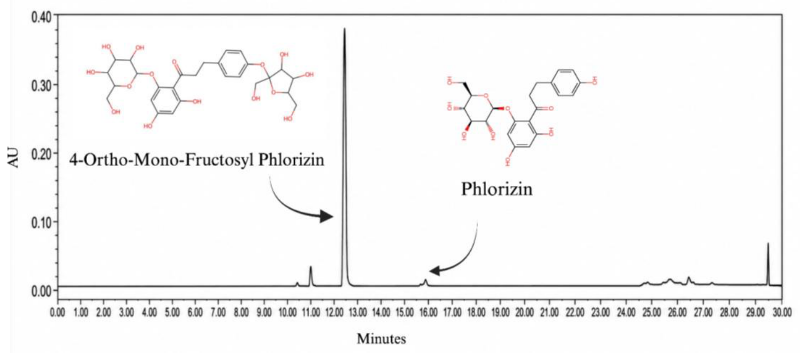
Figure 2.
Effects of 4PHF on body weight, water intake, and food intake in high-fat diet/streptozotocin-induced diabetic CD-1 mice during the 28-day treatment period. (a) Body weight is presented as mean ± SEM (n = 6/group) and was analyzed by one-way ANOVA followed by Dunnett’s multiple-comparisons test. (b) Average water intake during the study, expressed as mL/100 g body weight per day (c) Average food intake, expressed as kcal/100 g body weight per day. NC, negative control; DC, diabetic control; DAPA, dapagliflozin-treated diabetic mice, and 4PHF, 4-ortho-mono-fructosyl phlorizin-treated diabetic mice. **p < 0.01.
Figure 2.
Effects of 4PHF on body weight, water intake, and food intake in high-fat diet/streptozotocin-induced diabetic CD-1 mice during the 28-day treatment period. (a) Body weight is presented as mean ± SEM (n = 6/group) and was analyzed by one-way ANOVA followed by Dunnett’s multiple-comparisons test. (b) Average water intake during the study, expressed as mL/100 g body weight per day (c) Average food intake, expressed as kcal/100 g body weight per day. NC, negative control; DC, diabetic control; DAPA, dapagliflozin-treated diabetic mice, and 4PHF, 4-ortho-mono-fructosyl phlorizin-treated diabetic mice. **p < 0.01.
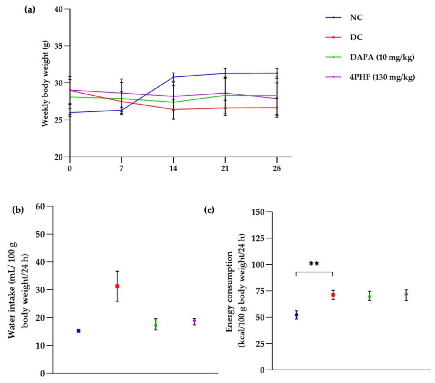
Figure 4.
Effect of 4PHF on urinary glucose excretion in CD-1 mice with diabetes induced by a high-fat diet and streptozotocin. Urinary glucose concentration was determined on day 26 of treatment. Data are presented as the mean ± SEM (n = 6 mice per group). Statistical analysis was performed using one-way ANOVA followed by Dunnett's multiple comparison test. ***p < 0.001 vs. DC. NC, negative control; DC, diabetic control; DAPA, dapagliflozin; 4PHF, group treated with 4-ortho-mono-fructosyl phlorizin.
Figure 4.
Effect of 4PHF on urinary glucose excretion in CD-1 mice with diabetes induced by a high-fat diet and streptozotocin. Urinary glucose concentration was determined on day 26 of treatment. Data are presented as the mean ± SEM (n = 6 mice per group). Statistical analysis was performed using one-way ANOVA followed by Dunnett's multiple comparison test. ***p < 0.001 vs. DC. NC, negative control; DC, diabetic control; DAPA, dapagliflozin; 4PHF, group treated with 4-ortho-mono-fructosyl phlorizin.
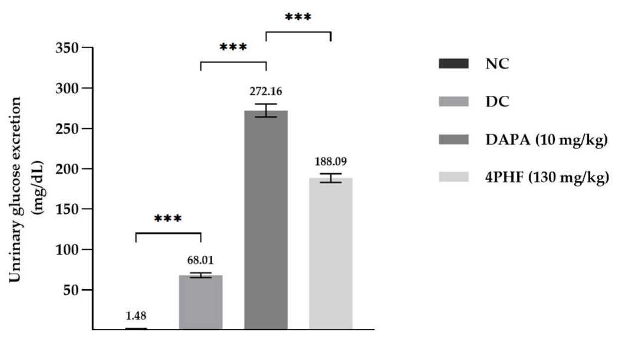
Figure 5.
Effects of 4PHF on the serum lipid profile of high-fat diet/streptozotocin-induced diabetic CD-1 mice. Serum levels of (a) total cholesterol, (b) triglycerides, (c) high-density lipoprotein cholesterol (HDL-C), and (d) low-density lipoprotein cholesterol (LDL-C) were determined in the negative control (NC), diabetic control (DC), dapagliflozin-treated diabetic (DAPA), and 4-ortho-mono-fructosyl phlorizin-treated diabetic (4PHF). Data are expressed as the mean ± SEM (n = 6 mice per group). Statistical analysis was performed using one-way ANOVA followed by Dunnett's multiple-comparisons test. *p < 0.05, **p < 0.01, and ***p < 0.001.
Figure 5.
Effects of 4PHF on the serum lipid profile of high-fat diet/streptozotocin-induced diabetic CD-1 mice. Serum levels of (a) total cholesterol, (b) triglycerides, (c) high-density lipoprotein cholesterol (HDL-C), and (d) low-density lipoprotein cholesterol (LDL-C) were determined in the negative control (NC), diabetic control (DC), dapagliflozin-treated diabetic (DAPA), and 4-ortho-mono-fructosyl phlorizin-treated diabetic (4PHF). Data are expressed as the mean ± SEM (n = 6 mice per group). Statistical analysis was performed using one-way ANOVA followed by Dunnett's multiple-comparisons test. *p < 0.05, **p < 0.01, and ***p < 0.001.
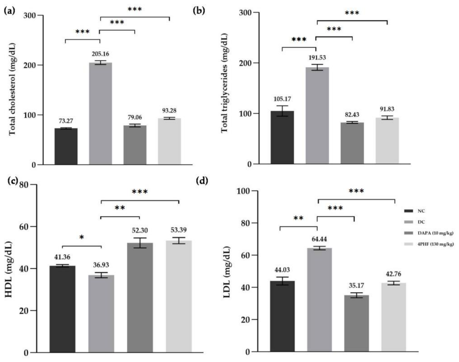
Figure 6.
Effects of 4PHF on relative liver weight and absolute kidney weights in high-fat diet/streptozotocin-induced diabetic CD-1 mice. (a) Relative liver weight, calculated individually as liver weight (g)/final body weight on day 28 (g) × 100; (b) Relative left kidney weight; and (c) Relative right kidney weight. Data are presented as the mean ± SEM (n = 6 mice per group). Statistical analysis was performed using one-way ANOVA followed by Dunnett's multiple-comparisons test. No statistically significant differences were detected among the groups (p > 0.05). NC, negative control; DC, diabetic control; DAPA, dapagliflozin; 4PHF, 4-ortho-mono-fructosyl phlorizin-treated diabetic.
Figure 6.
Effects of 4PHF on relative liver weight and absolute kidney weights in high-fat diet/streptozotocin-induced diabetic CD-1 mice. (a) Relative liver weight, calculated individually as liver weight (g)/final body weight on day 28 (g) × 100; (b) Relative left kidney weight; and (c) Relative right kidney weight. Data are presented as the mean ± SEM (n = 6 mice per group). Statistical analysis was performed using one-way ANOVA followed by Dunnett's multiple-comparisons test. No statistically significant differences were detected among the groups (p > 0.05). NC, negative control; DC, diabetic control; DAPA, dapagliflozin; 4PHF, 4-ortho-mono-fructosyl phlorizin-treated diabetic.
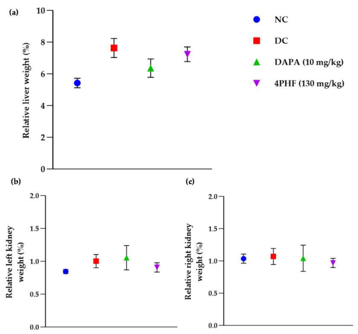
Figure 7.
Effects of 4PHF on liver histopathology in high-fat diet/streptozotocin-induced diabetic CD-1 mice. Representative photomicrographs of hematoxylin and eosin (H&E)-stained liver sections from the (a) negative control (NC), (b) diabetic control (DC), (c) dapagliflozin-treated diabetic (DAPA, 10 mg/kg), and (d) 4-ortho-mono-fructosyl phlorizin-treated diabetic (4PHF, 130 mg/kg) groups. Original magnification, ×400. Histopathological alterations were semiquantitatively scored on a scale from 0 to 3 and included (e) hepatocellular vacuolization, (f) vascular congestion, (g) hepatocellular degeneration, (h) inflammatory infiltration, and (i) sinusoidal alterations. Five liver sections were evaluated per animal, and the resulting scores were averaged to obtain one value per animal. Data are presented as the mean ± SEM (n = 6 mice per group). Statistical analysis was performed using one-way ANOVA followed by Dunnett’s multiple-comparisons test. *p < 0.05, **p < 0.01, and ***p < 0.001.
Figure 7.
Effects of 4PHF on liver histopathology in high-fat diet/streptozotocin-induced diabetic CD-1 mice. Representative photomicrographs of hematoxylin and eosin (H&E)-stained liver sections from the (a) negative control (NC), (b) diabetic control (DC), (c) dapagliflozin-treated diabetic (DAPA, 10 mg/kg), and (d) 4-ortho-mono-fructosyl phlorizin-treated diabetic (4PHF, 130 mg/kg) groups. Original magnification, ×400. Histopathological alterations were semiquantitatively scored on a scale from 0 to 3 and included (e) hepatocellular vacuolization, (f) vascular congestion, (g) hepatocellular degeneration, (h) inflammatory infiltration, and (i) sinusoidal alterations. Five liver sections were evaluated per animal, and the resulting scores were averaged to obtain one value per animal. Data are presented as the mean ± SEM (n = 6 mice per group). Statistical analysis was performed using one-way ANOVA followed by Dunnett’s multiple-comparisons test. *p < 0.05, **p < 0.01, and ***p < 0.001.
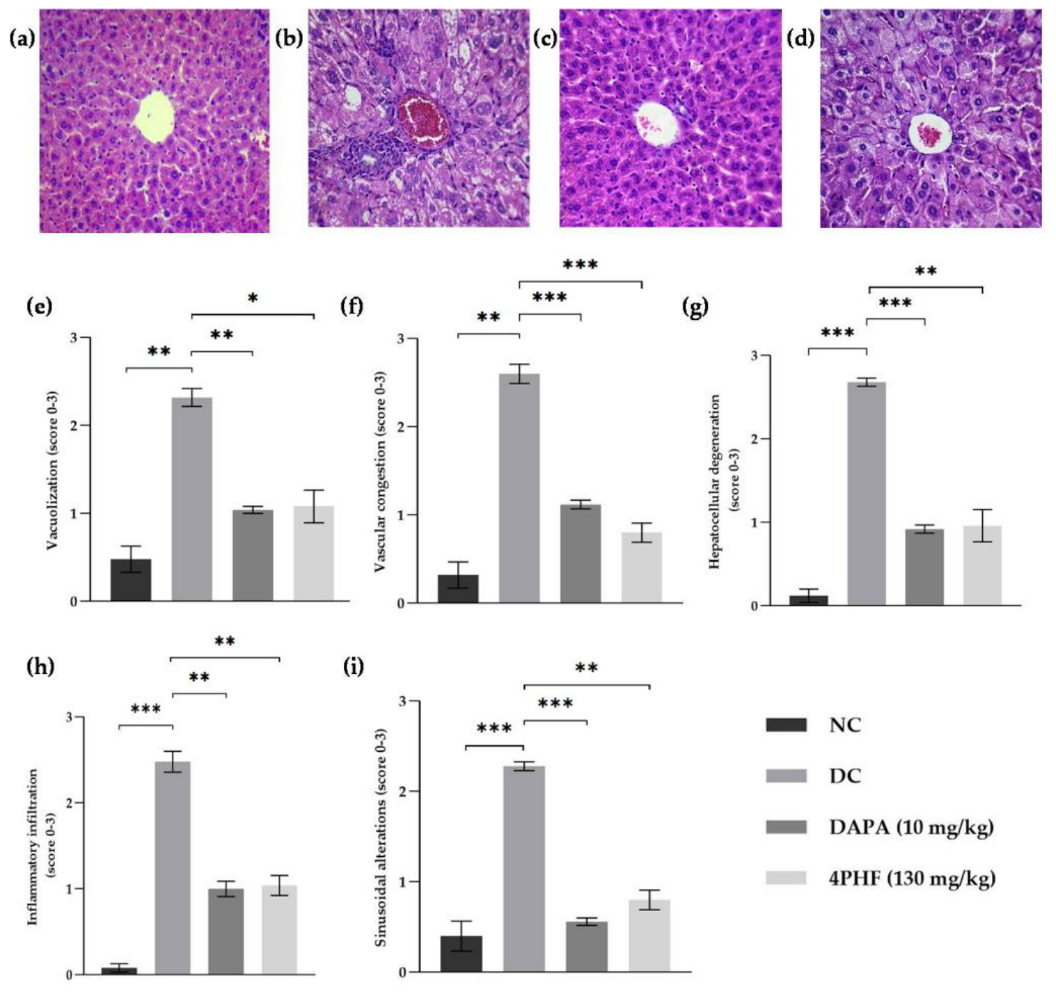
Figure 8.
Effects of 4PHF on renal histopathology in high-fat diet/streptozotocin-induced diabetic CD-1 mice. Representative photomicrographs of periodic acid–Schiff (PAS)-stained kidney sections from the (a) negative control (NC), (b) diabetic control (DC), (c) dapagliflozin-treated diabetic (DAPA), and (d) 4-ortho-mono-fructosyl phlorizin-treated diabetic (4PHF). Original magnification, ×400. Renal histopathological alterations were semiquantitatively scored on a scale from 0 to 3 and included (e) tubular dilation, (f) accumulation of PAS-positive material, (g) glomerular damage, (h) inflammatory infiltration, and (i) tubular atrophy. Five kidney sections were evaluated per animal, and the resulting scores were averaged to obtain one value per animal. Data are presented as the mean ± SEM (n = 6 mice per group). Statistical analysis was performed using one-way ANOVA followed by Dunnet's multiple-comparisons test. **p < 0.01 and ***p < 0.001.
Figure 8.
Effects of 4PHF on renal histopathology in high-fat diet/streptozotocin-induced diabetic CD-1 mice. Representative photomicrographs of periodic acid–Schiff (PAS)-stained kidney sections from the (a) negative control (NC), (b) diabetic control (DC), (c) dapagliflozin-treated diabetic (DAPA), and (d) 4-ortho-mono-fructosyl phlorizin-treated diabetic (4PHF). Original magnification, ×400. Renal histopathological alterations were semiquantitatively scored on a scale from 0 to 3 and included (e) tubular dilation, (f) accumulation of PAS-positive material, (g) glomerular damage, (h) inflammatory infiltration, and (i) tubular atrophy. Five kidney sections were evaluated per animal, and the resulting scores were averaged to obtain one value per animal. Data are presented as the mean ± SEM (n = 6 mice per group). Statistical analysis was performed using one-way ANOVA followed by Dunnet's multiple-comparisons test. **p < 0.01 and ***p < 0.001.
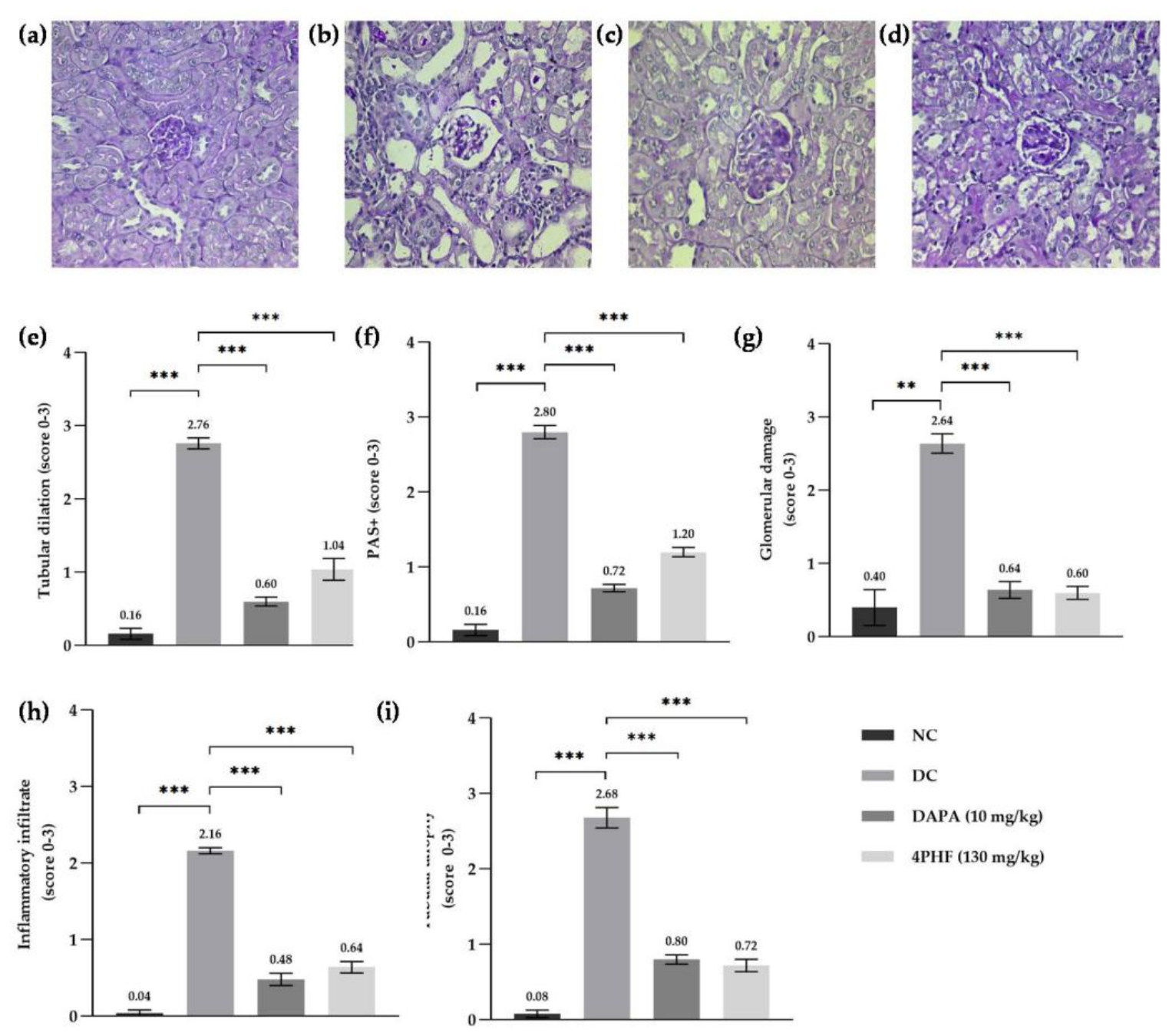
Figure 9.
Effects of 4PHF on renal SGLT2 expression in high-fat diet/streptozotocin-induced diabetic CD-1 mice. Representative photomicrographs of immunohistochemically stained renal cortical sections from the (a) negative control (NC), (b) diabetic control (DC), (c) dapagliflozin-treated diabetic (DAPA), and (d) 4-ortho-mono-fructosyl phlorizin-treated diabetic (4PHF). Original magnification, ×400. Brown staining indicates SGLT2 immunoreactivity. Quantitative analysis included (e) the percentage of SGLT2-positive area per field and (f) the number of SGLT2-positive renal tubules per field. Data are presented as the mean ± SEM (n = 6 mice per group). Statistical analysis was performed using one-way ANOVA followed by Dunnett's multiple-comparisons test. **p < 0.01 and ***p < 0.001.
Figure 9.
Effects of 4PHF on renal SGLT2 expression in high-fat diet/streptozotocin-induced diabetic CD-1 mice. Representative photomicrographs of immunohistochemically stained renal cortical sections from the (a) negative control (NC), (b) diabetic control (DC), (c) dapagliflozin-treated diabetic (DAPA), and (d) 4-ortho-mono-fructosyl phlorizin-treated diabetic (4PHF). Original magnification, ×400. Brown staining indicates SGLT2 immunoreactivity. Quantitative analysis included (e) the percentage of SGLT2-positive area per field and (f) the number of SGLT2-positive renal tubules per field. Data are presented as the mean ± SEM (n = 6 mice per group). Statistical analysis was performed using one-way ANOVA followed by Dunnett's multiple-comparisons test. **p < 0.01 and ***p < 0.001.
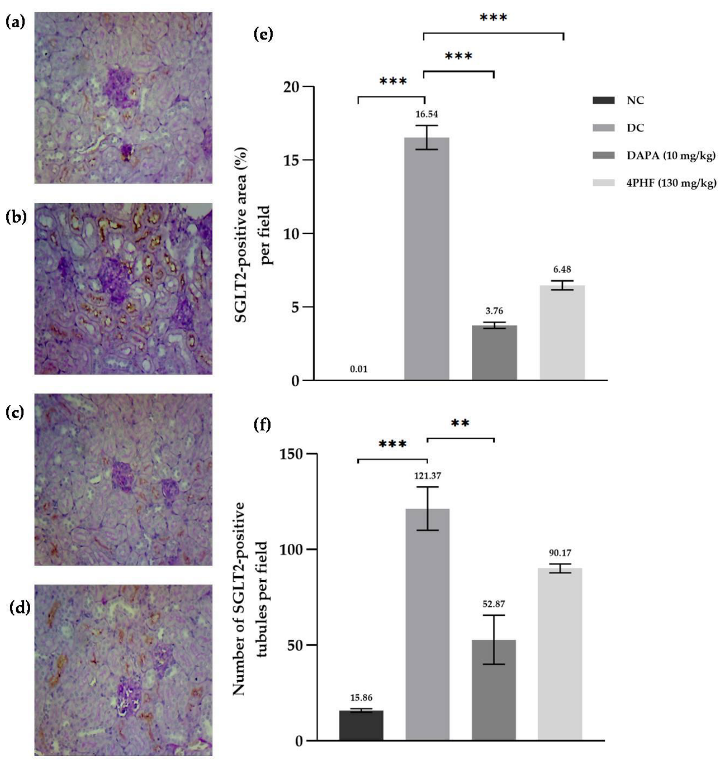

Table 1.
HPLC–UV composition of the final lyophilized 4PHF preparation administered to the animals.
| Component | Retention time (min) | Integrated peak area (AU·s)* | Relative peak area (%) |
|---|---|---|---|
| 4-O-Phlorizin monofructoside (4PHF) | 12.7 | 1876104 | 94.5 |
| Residual phlorizin | 15.8 | 41,884 | 2.11 |
| Other identified fructosylated derivatives | 11.4 | 67293 | 3.39 |
| Total | 1,985,040 | 100.0 |
Table 2.
Effect of 4PHF on fasting serum glucose levels in high-fat diet/streptozotocin-induced diabetic CD-1 mice.
Table 2.
Effect of 4PHF on fasting serum glucose levels in high-fat diet/streptozotocin-induced diabetic CD-1 mice.
| Group | Day 0 | Day 7 | Day 14 | Day 21 | Day 28 | Overall reduction (%) |
|---|---|---|---|---|---|---|
| NC | 127.80 ± 5.10*** | 128.80 ± 3.61*** | 127.80 ± 2.37*** | 126.30 ± 3.88*** | 125.50 ± 3.36** | 0.78 ± 5.80 |
| DC | 563.30 ± 24.30 | 534.00 ± 37.27 | 497.00 ± 37.81 | 516.50 ± 47.61 | 452.00 ± 40.30 | 19.10 ± 17.99 |
| DAPA (10 mg/kg) | 466.80 ± 14.51 | 294.60 ± 30.90* | 273.00 ± 39.92* | 276.40 ± 24.60* | 209.10 ± 15.67** | 56.68 ± 2.82* |
| 4PHF (130 mg/kg) | 512.50 ± 32.05 | 482.00 ± 33.84 | 417.80 ± 38.32 | 352.80 ± 28.90 | 327.30 ± 39.89 | 34.44 ± 5.77 |
NC, negative control; DC, diabetic control; DAPA, dapagliflozin group; 4PHF, 4-O-monofructoside phlorizin group. Overall reduction represents the percentage decrease in serum glucose from day 0 to day 28. Data are presented as the mean ± SEM. *p < 0.05, **p < 0.01, and **p < 0.001 vs. DC.
Disclaimer/Publisher’s Note: The statements, opinions and data contained in all publications are solely those of the individual author(s) and contributor(s) and not of MDPI and/or the editor(s). MDPI and/or the editor(s) disclaim responsibility for any injury to people or property resulting from any ideas, methods, instructions or products referred to in the content. |
© 2026 by the authors. Licensee MDPI, Basel, Switzerland. This article is an open access article distributed under the terms and conditions of the Creative Commons Attribution (CC BY) license (http://creativecommons.org/licenses/by/4.0/).
Copyright: This open access article is published under a Creative Commons CC BY 4.0 license, which permit the free download, distribution, and reuse, provided that the author and preprint are cited in any reuse.