

Submitted:
05 October 2026
Posted:
08 October 2026
You are already at the latest version
Abstract
The methylation pathway is a major player in epigenetics, with sophisticated regulatory control dictating which genes are expressed through a complex mechanism of hypo- and hypermethylation of cytosine bases in DNA. In this paper, we show that 5-formylcytosine (5fC), a rare and poorly understood modified base, has powerful capabilities to influence DNA mutations and repair and alter the structure of nucloeosomes, as well as introducing DNA-protein cross-links. Ten-eleven translocation (TET) enzymes are responsible for removing methyl groups from cytosine bases, through sequential oxidation steps, and we argue that 5fC (an intermediary in TET enzyme modifications) is highly enriched in deuterium due to the high deuterium kinetic isotope effect (KIE) of TET enzymes. The presence of a deuteron impedes the step that converts 5fC to 5-carboxylcytosine (5caC). Furthermore, we suggest that, similar to trimethylamine oxide (TMAO), 5fC serves as a marker for deuterium overload in the one-carbon (1C) units carried by methionine in the methylation pathway and through methyl tetrahydrofolate (CH3-THF) and its derivatives. The gut microbiome is responsible for stripping deuterium out of carriers of 1C units, via hydrogen gas recycling. Nutrients such as choline, L-carnitine, and betaine supply 1C units bound to nitrogen atoms which feed into the 1C pool via microbial enzymes. The anaerobic archaea play an essential role, and, when they are under-represented, deuterium becomes enriched in the 1C units. This ultimately increases the number of 5fC bases in the DNA, leading to an accelerated rate of aging, associated with increased risk to infection, autoimmune disease, cardiovascular disease, and cancer. Oxidative stress due to deuterium overload in the ATPase nanomotors leads to suppression of TET enzyme activity, which induces hypermethylation of promoters of tumor suppressive enzymes, facilitating cancer progression. In this paper, we present a perspective for a new pathogenesis avenue due to a dysregulated methylome.
Keywords:
methylation pathway
; formylcytosine
; deuterium
; cancer
; ten eleven translocation
; oxidative stress
; gut microbes
; ATPase nanomotors
1. Introduction
The methylome, consisting of complex enzymatic machinery, that either methylates or demethylates DNA, alters, or, in essence, controls, the expression of genes. The final methylation pattern produced by the methylome processing defines the aging of the DNA and ultimately, the age of the mammalian organism. Specific studies investigating immune function link the DNA methylation patterns to a) aging, b) autoimmunity and c) cancer [1]. Moreover, the hypomethylation of DNA regions, and thus the probable accumulation of oxidized 5-methylcytosine products, and the increases in methylation at cytosine guanine dinucleotide CpG islands in regions of tumour suppressor genes, contributes to the autoimmune reactions and carcinogenicity which associate with the aging of DNA [2].
In this paper, we first present a perspective/hypothesis that the methylation pathway normally carries methyl groups that are highly depleted in deuterium. This is primarily due to the activities of the gut microbiome, which continuously “scrub” deuterium from the methyl groups, through hydrogen gas recycling. Methylated amines in nutrients such as choline, betaine, and L-carnitine are processed by anaerobic archaea to produce formaldehyde and methane gas, both of which feed into the methylation pathway via methylene-tetrahydrofolate (CH2-THF) [3]. In previous work, we have argued that small gases containing hydrogen are naturally depleted in deuterium, in part because deuterium, being heavier, tends to remain behind in the aqueous medium [4,5].
Trimethylamine oxide (TMAO) is a liver metabolite derived from trimethylamine (TMA), a microbial product of the metabolism of nutrients containing trimethylated nitrogen atoms. Anaerobic archaea are able to fully metabolize TMA into formaldehyde and ammonia through several dehydrogenase enzymes. Because formaldehyde is a small hydrogen-containing gas, it is likely low in deuterium, especially if it is derived through metabolism of TMA (also a gas) by anaerobic archaea [5]. Furthermore, archaeal TMA dehydrogenase (TMADH) is a flavoprotein with a high deuterium kinetic isotope effect (KIE) (~8.6), due to vibrationally assisted hydrogen tunneling [6,7]. This implies that the TMA that is left unmetabolized by the archaea becomes enriched in deuterium.
The formaldehyde then supplies a one-carbon (1C) unit to THF, to produce CH2-THF, also through microbial enzymes. The human enzyme CH2-THF reductase (MTHFR) provides a third proton that is also likely to be 1H rather than 2H, because it is a flavoprotein, and deuterons are inefficient in the proton tunneling that flavoproteins exploit [8,9]. The deuterium-enriched TMA that is left behind becomes TMAO through the action of the liver enzyme, flavin monooxygenase 3 (FOM3).
Elevated serum TMAO is a risk factor for many chronic diseases [10]. It has been argued that TMAO is a signaling molecule that indicates both gut dysbiosis and excess deuterium in the methylation pathway. Because the methyl groups are ultimately metabolized in the mitochondria, providing protons to the mitochondrial water, it is very important that they carry very little deuterium, in order to maintain the proper functioning of the ATPase nanomotors, which are highly sensitive to deuterium [3].
Deuterium disrupts the function of ATPase nanomotors, causing an increase in reactive oxygen species (ROS) and inefficiencies in ATP production. Uracil-DNA glycosylase (UNG) is the enzyme that is responsible for base excision repair for misincorporated uracil, that comes about following cytosine deamination due to oxidative stress. Ung-/- cells accumulate huge excesses of uracil in their DNA [11]. UNG has large primary and secondary deuterium KIEs [12], so deuterium loading in mitochondrial water would significantly impair the ability to repair mitochondrial DNA damage. This issue is compounded by the increased release of ROS from deuterium-loaded ATPase nanomotors [3].
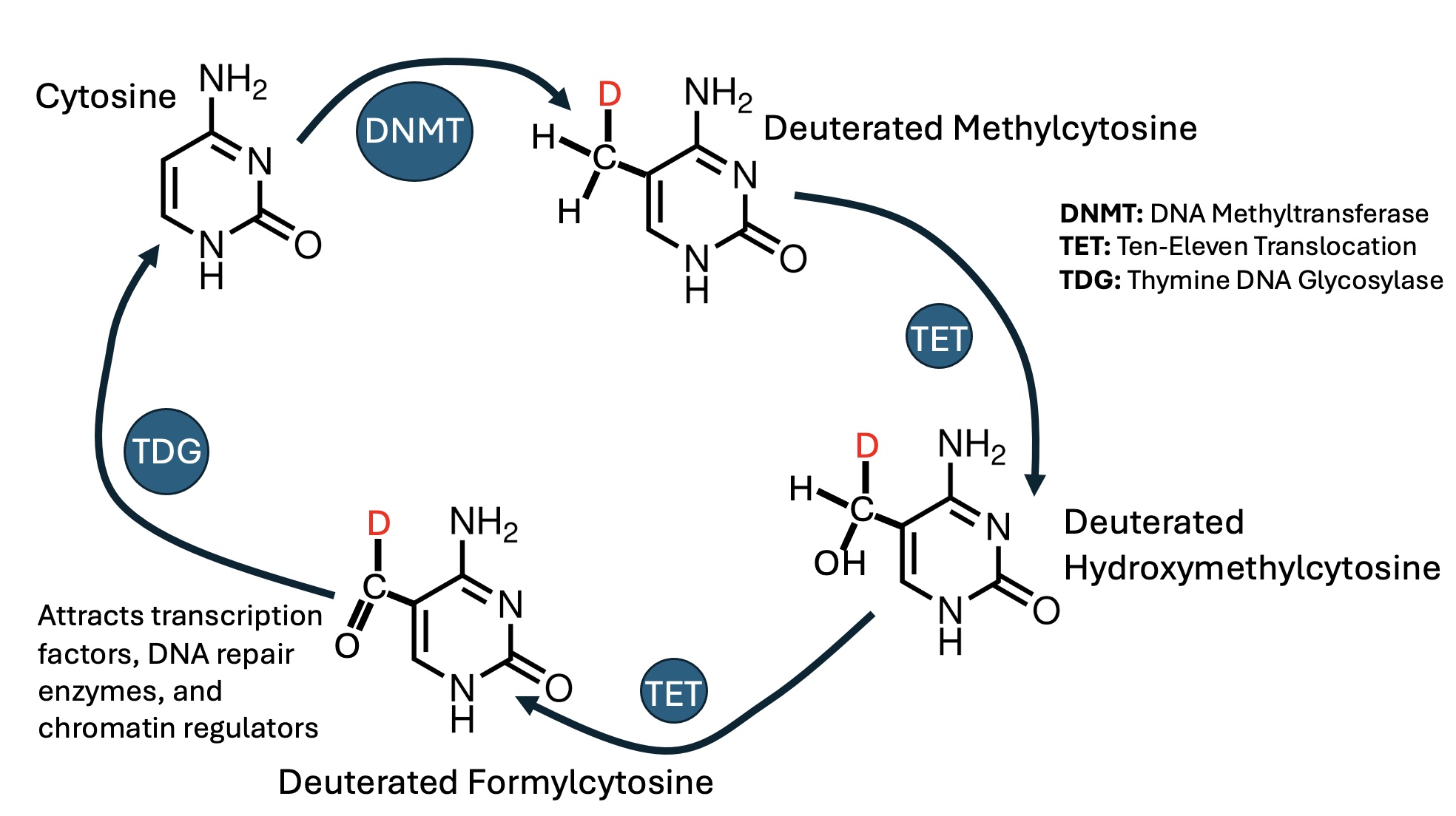
Our focus here is on DNA methylation and demethylation, paying particular attention to 5-formylcytosine, a rare modified base produced through the action of ten-eleven translocation (TET) enzymes on 5-methylcytosine (5mC). TET enzymes are responsible for removing methyl groups from DNA, which they achieve in sequential oxidation steps, producing first 5-hydroxymethylcytosine (5hmC), then 5-formylcytosine (5fC), and finally 5-carboxylcytosine (5caC). 5hmC is now recognized as a stable and functionally significant epigenetic mark with distinct genomic distributions. It serves as a crucial biomarker and regulator in cancer progression and therapy response [13]. 5fC plays a powerful role in recruiting DNA repair enzymes to the DNA [14], in influencing cross-links between DNA and proteins such as the histones [15], and in regulating nucleosome positioning [16]. We make the case that 5fC, like TMAO, is an indicator of excess deuterium in the methylation pathway.
TET enzymes exhibit a high primary deuterium KIE, typically around 9 [17]. This means that removing a deuteron is 9 times slower than removing a proton. A protein in the same class as TET enzymes, expressed by gut microbes but not by human cells, is an enzyme called taurine:α-ketoglutarate dioxygenase (TauD). Both enzymes are dioxygenases—they split O2 apart into two reactive oxygen atoms, and both oxygens are inserted into substrates. TauD’s reaction begins with extracting the proton from the C1 carbon in taurine. If the C1 hydrogen (1H) atom in taurine is replaced with deuterium (2H), the reaction slows down by a factor of 30-37-fold [18]. Quantum-mechanical tunneling plays a critical role, because deuterons are much less capable than protons of passing through the tunnel, due to their bulkiness. The taurine that does not get metabolized by TauD is discarded through the feces. A deuterium-rich molecule is excreted through such a mechanism [19]. The extent to which TET enzymes behave similarly is unknown.
2. A Crucial Role for Serine Hydroxymethyltransferase
Serine hydroxymethyltransferase (SHMT) is a critical enzyme in the methylation pathway. This enzyme reversibly converts glycine to serine, obtaining the hydroxymethyl group from CH2-THF. There are two isoforms of SHMT, SHMT1 expressed in the cytoplasm, and SHMT2 expressed primarily in the mitochondria. Genetic defects in SHMT2 are lethal in embryos of mice at day 13.5, but studies that involve selectively knocking out SHM2 in the adult liver using the “Cre-LoxP” system have shown that it primarily operates in the direction that converts glycine to serine. Its knockout leads to an eight-fold increase in serum glycine levels [20]. SHMT2 is significantly upregulated in more than 85% of cancer types compared to normal tissue [21]. Transcriptional activation of SHMT2 is regulated by the oncogenic transcription factor c-Myc [22].
If we assume that the CH2- unit in CH2-THF has normally been scrubbed of deuterium, then the hydroxymethyl group of serine will also be low in deuterium. Serine can be converted to pyruvate in a single step using serine dehydratase as the catalyst. The two protons on the hydroxymethyl group are preserved in the pyruvate that is produced. So, this pathway from glycine to serine to pyruvate becomes an excellent source of fuel for the citric acid cycle that can deliver low-deuterium protons to the mitochondrial water, in both the liver and the tumor cells.
SHMT2 also catalyzes another important reaction that converts methenyl-THF to 5-formyl-THF, also known as folinic acid and the prescription drug leucovorin [23]. Folinic acid is a naturally occurring, reduced and active form of folate that bypasses standard enzymatic conversion steps required by synthetic folic acid [24]. It can deliver methylfolate to the brain even when the folate receptors are disabled [25].
3. Excess Mitochondrial Succinate Suppresses TET Enzyme Activity
Succinate dehydrogenase (SDH) is a 4-subunit complex in the inner membrane of mitochondria, and it is the only enzyme complex that participates in both the citric acid cycle and the electron transport chain [26]. It oxidizes succinate to fumarate, and, as Complex II, it passes electrons from succinate or FADH2 directly to coenzyme Q [27]. Succinate dehydrogenase deficiency results in the accumulation of its substrate, succinate. Succinate acts as a mitochondrial retrograde signal that inhibits histone demethylases and TET enzymes, through product feedback inhibition, since succinate is also a product of TET enzyme activity [28,29]. Defective mutated versions of SDH are often present in cancer cells, and this leads to suppression of TET enzymes and associated dramatic loss of 5hmC in promoters and enhancers of tumor suppressor genes [30].
A paper published by Cramer-Morales et al. in 2020 revealed that erythroid precursor differentiation from embryonic stem cells is significantly impaired if the mitochondrial enzyme manganese superoxide dismutase (MnSOD;SOD2) is defective [31]. SOD2 plays an essential role in converting superoxide to hydrogen peroxide, to protect the mitochondria from ROS. Normally, naive embryonic stem cells maintain a high ratio of α-ketoglutarate to succinate to facilitate DNA and histone demethylation, which is required for pluripotency [32,33].
The Cramer-Morales et al. study genetically disrupted the SOD2 gene using CRISPR/Cas9 technology in a human erythroleukemia cell line, and they found that the cells’ ability to differentiate towards an erythroid phenotype was severely impaired. Importantly, these cells exhibited suppressed succinate dehydrogenase activity along with significantly reduced TET enzyme activity associated with a decrease in 5hmC and a corresponding increase in 5mC.
The authors attributed their failure to differentiate to a failure to complete the necessary global DNA demethylation program required for erythrocyte maturation [31]. More broadly, it can be anticipated that increased mitochondrial oxidative stress due to a high deuterium load would also result in reduced expression of succinate dehydrogenase in many cell types, with concomitant suppression of TET enzyme activity and associated DNA hypermethylation in critical control regions [34].
Figure 1 depicts the sequence of events that we propose lead to both deupleted nutrient supply to mitochondria under healthy conditions and pathways that lead to oncogenesis due to excess deuterium in the methylation pathway, when the gut microbiome is disrupted.
4. The Methylome and DNA Aging.
Chromatin organization modulates gene transcription and alters which genes get expressed through epigenetic mechanisms, tied directly to methylation patterns. Oncogenesis is associated with profoundly altered DNA methylation patterns. It has been known since the 1980s that cancer is associated with widespread hypomethylation [35,36]. However, paradoxically, cancer cells show aberrant hypermethylation in certain control regions, especially those associated with tumor suppressor genes [37].
In relation to DNA methylation and aging, numerous recent studies indicate that there are specific methylation profiles that correlate with the aging of human DNA. The recent study of M Jacques et al. showed that there are specific epigenetic modifications (methylation patterns) that accumulate with age, and these show differences depending on the specific tissue [38]. Overall, 17 different tissues including blood were investigated in that study. Specific age-related increases and decreases in methylation were identified using sophisticated gene co-expression network analysis. It is worth noting that the enhanced methylation with age was related to forebrain development and cell adhesion molecular functions whilst hypomethylation was related to synaptic signalling and immune functioning such as nuclear factor kappa-light-chain-enhancer of activated B cells (NF-kB), which indicated a decrease in regulatory control of neuronal and immune pathways with age. In the blood tissue, the hypomethylated areas were associated with homeostatic regulatory control whilst in the brain tissue the hypomethylated regions were associated with development and morphogenesis control.
There were several unique common epigenetic patterns that were associated with age across various tissues. Enriched regions that concerned cell adhesion (via plasma adhesion molecules) were observed in adipose, brain and blood tissues, indicating the loss of cellular interactions due to age. However, overall, most tissues were related to hypermethylation during aging, and this indicates that, during aging, epigenetic gene silencing occurs that affects gene expression. This can be explained by the suppression of TET enzymes due to oxidative stress generated by ATPase nanomotors in the presence of excess deuterium.
The study by M Jacques et al. concludes that there is a tissue-specific variation in methylome remodelling that occurs with aging [38]. This may be happening not at random, but in order for the organism to avoid disease. In any case, however, the epigenetic modifications during aging change, and the methylation remodelling that occurs is not random. Hypermethylation occurring during aging will enhance errors in the DNA methylome. It will particularly affect the regulatory control of NF-kB immune function and cause phenotypic dysfunction in various organs that is observed with aging [39]. Relating to cancer, the specific patterns that are over-methylated with aging associate with carcinogenesis, whereas other hypomethylated CpG regions that occur during aging relate to low cancer emergence [40]. Furthermore, specific cancer risk genes’ methylation patterns associate with carcinogenesis during aging.
Other studies on methylome changes in blood show that bivalent promoters (containing both activating and repressive histone modifications) become methylated with aging [41]. Although it was once thought that this will produce gene silencing, in cancer, it rather means activation of genes that regulate apoptosis, pluripotency and senescence [42]. Also, during aging, the Polycomb regions (areas of the DNA that are silenced by the Polycomb family of proteins) become increasingly methylated [41]. Importantly, the hypermethylation of Polycomb regions in immune (CD4) cells associates with autoimmunity [43].
A considerable hypomethylation pattern also occurs with aging [44], and this was also observed and recorded in the recent M Jacques et al. study [38]. In the K Seale et al. study, it was shown that blood monocytes lose methylation with aging in some DNA regions, but in a more regular way than they gain methylation in other DNA regions [41]. The hypomethylation would involve demethylation of these particular regions [45], and this is dangerous for the epigenetic regulation control, as it usually affects the regions that transcribe repetitive elements, such as LINE1 (Long Interspersed Nuclear Element-1), which otherwise remain dormant in expression due to their hypermethylated promoter regions. Genome-wide hypomethylation of Line-1 and Alu retroelements in cell-free DNA derived from blood, but not in cellular DNA, is an epigenetic biomarker of human aging [46]. The expression of these repetitive elements is implicated in various diseases, including cancer [45].
Active demethylation involves the use of TET enzyme catalytic machinery which hydroxylates the 5mC to form 5-hydroxymethylcytosine (5hmC) which is further oxidized to form 5fC [47]. Although it was once thought that 5fC was only a transient demethylation intermediate compared to 5hmC, occurring and accumulating only in early embryonic development, and then with levels dropping during the lifespan, recent experimental evidence proved the opposite. In the pioneering work of M Bachman et al., the researchers used deuterated methyl-13CD3 L-methionine to feed human cell cultures and animals to detect their DNA methylation—demethylation processes at various stages of development (lifespan) [48]. Remarkably, it was found that the 5fC (expected to be scarce) remained relatively abundant in older or more slowly proliferating DNA, like the brain tissue (that contained slowly proliferating cells). The brain tissue retained the most stable 5fC in its DNA structure, beginning from early development and spanning throughout, in later developmental stages.
In conclusion, the role of the 5fC oxidized intermediate may be more important in epigenetic regulation than was once thought, and it may have important implications in the overall gene expression during aging.
In the following section we focus specifically on the demethylation intermediate 5fC’s role in aging and disease.
5. The Role of 5-Formylcytosine During Aging and Disease.
It has been widely established that the intermediate TET enzyme demethylation product, 5-hydroxymethylcytosine (5hmC) has a stable presence in adult mammalian DNA. The work of M. Wagner et al. published in 2015 showed that, although after embryonic development the levels of 5hmC drop, they retain a steady concentration presence in human and animal brain tissues in post-developmental phases [49]. However, in these scientific experiments, the levels of 5fC were found to be dropping considerably with age after early developmental stages, and thus it had been considered by the researchers to be a transient epigenetic modification, lost in the progress of aging, rather than a stable intermediate of the demethylation process like 5hmC [49].
Nevertheless, the experiments in the study of M Bachman et al. led to the conclusion that 5fC can remain a stable DNA modification at later stages in life in human cells and mammalian tissues [48], following a pattern of presence that is distinct from 5hmC [48]. The question arising is on what is the essence of 5fC presence in adulthood as an epigenetic regulator of the human methylome? This is particularly important nowadays, as the proper functioning of TET enzyme demethylation has recently become the target of investigation for cancer immunotherapy efficiency [50].
During early embryonic development, the oxidized formation of 5fC has an important role in epigenetic reprogramming by activating gene expression across various genomic regions. The 5fC activation of transcription is associated with the excision capability of thymine DNA glycosylase [51]. In embryos, the regions that are enriched with 5fC are located on Polymerase III (PolIII) target genes, and the epigenetic regulatory role of 5fC is on recruiting Pol III and tRNA expression [52].
Recent experimental evidence provides more insights on the role of 5fC on the nucleosome [53]. 5fC promotes the occupancy by the nucleosome (histone octamers) at DNA regions near transcription start sites and enhancers, thereby activating gene transcription of these genes. Due to the formyl group, 5fC is capable of forming reversible Schiff base links with the lysines of histone proteins, and these influence the structural organisation of nucleosomes [15]. Another way that 5fC regulates transcription is by forming cross-links that bind to polymerases, transcription factors, and other DNA binding proteins, and in this way slows down transcription in human cells.
Importantly, these DNA-peptide cross-links due to 5fC are poorly recognised and removed by the nucleotide excision repair system (NER) [53], and this can constitute 5fC-induced peptide-DNA lesions which are candidates for mutagenesis [54]. Overall, the physicochemical properties of 5fC can be important for later stages in life because, in the study of T Furuhata et al., the presence of only one unit of 5fC per nucleosome was enough to change the nucleosome organisation and increase its occupancy [16].
Moreover, another capability that 5fC has on DNA structural rearrangement is on the enhancement of DNA flexibility. Due to its formyl group, which is a strong electron withdrawing group, 5fC has a reduced affinity for the N3 ring nitrogen for accepting protons. This weakens the Watson-Crick base pairing capacity of the DNA, and thus the DNA becomes less rigid. Finally, as the work of Furuhata has recently shown, 5fC has a strong influence on nucleosome positioning, i.e., 5fC follows the signal of intrinsic DNA sequences, through the protein-DNA cross-links and nucleobase stacking (DNA-to-RNA aromatic ring nucleobase attraction) that are formed due to its unique chemical properties [16].
In summary, the chemical properties of 5fC alter the nucleosome organisation, and this can induce nucleosome repositioning which can alter the gene expression of gene promoter enhancers or silence particular DNA regions. Relating to the aging process of DNA, 5fC can be a catalytic factor, as, during aging, a considerable nucleosome reorganisation occurs [55], Moreover, during aging, the nucleosome histone occupancy is increased in various tissue cells, including neural stem cells, especially in promoter regions near proinflammatory genes [56]. Finally, nucleosome repositioning occurs in the whole genome of cancer cells. Particularly for breast cancer, there is a considerable gain in nucleosome occupancy. The heaviest nucleosome regions in breast cancer cells are at CpG islands of promoters that encode DNA binding proteins [57]. Therefore, plausibly, future research may prove the 5fC energetic contribution to aging, autoimmunity and cancer, as 5fC, as a molecule, has the chemical properties to contribute to the DNA alterations that occur during these conditions.
6. The DNA Methylation Demethylation Machinery and the Possible Impacts of a Deuterated Methyl Group Transfer, in Cancer, Aging, and Other Diseases
The methylation and demethylation of cytosines on the DNA (methylome) constitute a complex epigenetic control of the gene’s expression that, when in balance, preserves health. The process, called the methylome, is operated by sets of enzymes that either methylate or demethylate the DNA nucleosides (specifically, cytosine bases), creating the landscape where other nuclear enzymes, such as the polymerases and the DNA repair enzymes, operate to preserve or restore the DNA sequence that is necessary for a healthy outcome. Imbalances in the methylome operations are strongly linked to diseases like cancer, autoimmunity and premature aging [58]. The concepts in the following subsections aim to identify possible dysregulations of the methylome that can arise due to the deuteration of the cytosine nucleosides of human DNA.
6.1. The Methylome and Relations to Disease
The final pattern of DNA methylation (due to the methylome) provides an archaic driven epigenetic modification profile which greatly interacts with other numerous genomic activities that, altogether, finally offer the great diversity of mammalian DNA [59]. For both humans and other mammals, the erasure and re-establishment of the DNA methylation patterns greatly affects the embryonic developmental stages and beyond. These are particularly important to be preserved in balance. Their imbalance greatly contributes to the development of mammalian and human embryonic and adult diseases [60]. Amongst the most prevalent epigenetic modifications (alterations) of DNA, namely the DNA methylations, the histone modifications and the modifications of microRNAs, all act synergistically to regulate the expression of genes.
DNA methylations are the most frequent to regulate the gene expression and are highly related to disease outcomes, as in the case of autoimmunity (e.g., in multiple sclerosis) [61]. The main orchestration of the DNA methylation machinery is conducted by the DNA methylating enzymes called DNA methyltransferases (DNMTRs). These enzymes transfer a methyl group to carbon 5 of the nitrogenous base cytosine to form 5mC [62], restricted to cytosine bases that are followed immediately by guanine (CpG sequences). In general, it is important to maintain the unmethylated form of cytosine in the CpG-rich regions located in the promoters of the genes, as this allows for an active transcription, whereas the hyper-methylations of the cytosines in the CpG regions leads to gene silencing [63].
It is not only cytosine that gets methylated, but also, methylated forms of guanosine and uracil (which is essentially a methylated form of thymidine) exist normally, as in plant cells [64], whereas for human DNA, a methylated form of guanine signifies an important DNA lesion, a candidate for mutagenesis. A base-excision repair (BER) mechanism is readily employed to restore these regional DNA defects [65]. Nevertheless, either a methylation or a demethylation event can affect gene transcription and therefore, the expression of neighbouring genes [66]. Moreover, both these epigenetic modifications act in concordance with other epigenetic events that alter gene expression, like those of the histone modifications and the action and function of the microRNAs. Undoubtedly, recent experimental evidence indicates that the rate of methylation or demethylation of cytosine pyrimidine bases in the CpG-rich promoter regions has an important impact on the emergence of disease. Indeed, an increased rate of DNA methylation in the promoter regions can result in the failure of tumour suppressor genes to express their proteins that otherwise protect against cancer [67]. In this regard, the role of DNMTRs becomes of great importance for the modulation of cancer emergence [68].
Moreover, another set of enzymes, this time the demethylases that remove the methyl group from methylated cytosines, are also very important for the emergence of cancer. The TET enzymes, which are 5mC oxidases, constitute a dioxygenase family of proteins. In a series of reactions, they oxidise 5mC to 5hmC, which is consecutively converted to 5fC, and finally to 5caC, also by the TET enzymatic reactions [69].
Overactive demethylation by the TET enzyme family is implicated in cancer development, especially in leukaemia cell growth [70]. Due to the continuous and repeated oxidation (iterative oxidation) of 5mC, by, e.g., TET1, to complete their hypomethylation tasks [71]. In the hypermethylated regions of the DNA, the oxidized regions are recognised as lesions on the DNA, and are actively excised. The lesions are thus repaired by the mediated action of thymidine DNA glycosylase (TDG) [72]. TDG activity has also been implicated in the development of oncogenesis [73]. Moreover, there is growing evidence that another group of methyltransferases, the methyltransferase-like (METTL) family of enzymes which is genomically distinct from the DNMTRs, is also implicated in tumour progression [74].
Methylations that drive towards oncogenesis occur not only on DNA but also on histones and on RNA sequences. These methylations also change gene expression. Intriguingly, a member of the METTL family of methyltransferases is thiol methyltransferase 1B (TMT1B), also known as Methyltransferase-like 7B (METTL7B) or Associated with Lipid Droplet Protein 1 (ALDI), which recently has been explored and found to activate cancer [75].
Yet another methyltransferase, the rat liver catechol O-methyltransferase (COMT) has some time ago been explored for its ability to transfer deuterated methyl groups [76]. The kinetic isotope effects of this methyltransferase are exceptionally intriguing. The rate of methylation was increased when the methyl group that was transferred had been deuterated—a reverse deuterium kinetic isotope effect (KIE), which is highly unusual in nature. Since the methylated catechol is a metabolite that is excreted in the urine, its methylation helps in removing deuterated methyl groups from the 1C pool.
6.2. Association Between DNA Methylation and Aging
Apart from the fact that an increased DNA methylation rate is important for the development of cancer [68], the rate of DNA methylation also holds the genetic keys for aging of the DNA and aging-related diseases [58]. In this regard, the developed cancer can be fundamentally viewed as an aging-related disease [77]. Moreover, the accelerated rate of DNA methylation has been especially regarded as a hallmark of cardiovascular disease and its related disorders, hypertension and diabetes. Accelerated DNA methylations ultimately define the biological aging of the heart tissue [78]. Furthermore, the acceleration of DNA methylations that ultimately define aging also define the mortality rate of the human population and render the variations between the chronological age and the biological age of an individual [79].
A remarkable fact that must be mentioned is the impact that the COVID-19 pandemic has had on accelerated epigenetic aging. By utilizing various epigenetic clock models, Gupta et al. found a striking marker for increased aging in association with COVID-19 infection. Those with severe COVID-19 had markedly higher age acceleration compared to milder cases. Furthermore, the immune cell composition of COVID-19 patients showed a significant decrease in B cells, CD4+ T cells, and natural killer cells, along with an increase in monocytes, compared to healthy controls. The immune dysregulations in the human population infected with SARS-CoV-2 was linked to increased demethylations associated with various epigenetic aging clocks, and therefore with the overall gene expression, in severely ill patients during COVID-19 [80].
7. The Impact of a Deuterated Methyl Group Transfer by Methyltransferases
The influence that a deuterated methyl group has on the kinetics of methyltransferases becomes significant for the emergence of disease. Normally, aging influences the rate of DNA methylations on the DNA. However, the changes in the DNA methylations rate seem to have a particular impact on aging and therefore on the emergence of cancer and autoimmunity [81]. Furthermore, accelerated methylations on DNA are linked to the epigenetic age acceleration seen in neurodevelopmental disease. In particular, a scientific school of thought defining neuronal disease links age acceleration due to accelerated methylations on DNA to the psychiatric disorders of depression and suicide [82].
Therefore, the impact of a deuterated methyl group to increase the reaction rate of a methyltransferase as in the case of COMT is considered significant, especially if other methyltransferases in the same family, such as the DNMTRs, or demethylating enzymes like the TETs, are affected by a similar deuterated methyl group. S-adenosylmethionine (SAM) is the molecule that most methyltransferases, including COMT, DNMTRs, and METTL, use as the source of the methyl group they transfer. Relevant studies have shown that SAM can easily be modified and become deuterated in the human organism [83]. Moreover, the transfer of deuterated methyl groups to SAM in the human liver has been shown to be feasible [84].
7.1. The Biological Impact of a Deuterated Formyl Group on 5-Formylcytosine and the Disease Outcomes
At this level, an important question is raised as to what can be the fate of a deuterated methyl group added to the DNA molecule during methylation by the DNMTRs. During the demethylating process operated by the TET enzymes, there is a chance for deuterium to substitute for the protium on the methyl group. The TET enzymes show a high kinetic isotope effect [17], and therefore, during the oxidation of 5mC to 5fC, the deuterium atom that has exchanged with a hydrogen atom will resist removal. The oxidized derivatives (the demethylated nucleosides of 5mC) produced by the TETs constitute important biological markers for mammalian pathogenesis, due to their super-excited state dynamics, that may have a considerable impact on DNA photo-damage and their potential for repair [85].
Due to these oxidized nucleosides, destructive singlet oxygen radicals are generated. The impact of a singlet oxygen on the DNA can be severe, as reactive oxygen species can cause more DNA damage events, driving inflammation and carcinogenesis [86]. Therefore, a deuterated and therefore more robust and stable form of an oxidized nucleoside, such as that of a deuterated 5fC, can destabilise the DNA methylation/demethylation balance and alter the epigenomic influence on the gene expression and subsequently enhance pathogenesis [87].
The thymidine DNA glycosylase (TDG) system of enzymatic excision of oxidized nucleosides from the DNA involves a highly specific catalytic mechanism that can be influenced by the deuterated nucleoside. A deuterated 5fC will form and alter the already weaker hydrogen bonds that a non-deuterated 5fC has with the guanosine base pair [88], and these hydrogen bonds constitute the basis of TDG recognition to excise the oxidized DNA lesion. A defect on excising the oxidized DNA lesions will generate more oxidized DNA lesions, such as the oxidized guanosine lesions, and dismantle the DNA repair mechanisms to correct these genetic defects [89]. This will render the DNA more susceptible to photo-damage and to the creation of mutagenic photo-lesions. This will finally lead to DNA pro-cancerous conditions [90], due to increased photo-damage.
Furthermore, related studies on the deuterated formyl groups in similar compounds have shown that similar deuterations in 5fC confer greater metabolic stability and resistance to enzymatic breakdown [91]. Hence, a deuterated 5fC will have a greater chance to react with its protein counterparts at the sites of the oxidized DNA lesions. This has the potential to affect the efficacy of important proteins like polymerase β (Pol β) and AP endonuclease 1 to perform their delicate catalysis on DNA synthesis and their final DNA repair processes [92].
5fC is a highly electrophilic oxidized molecule that readily reacts with amines, like the lysine residues of other proteins. This DNA cross-linking with proteins that constitute important transcription factors and/or histone proteins can affect the replication of the DNA, rendering mutagenesis a more plausible event [93]. Apart from the histones and transcription factors, other nuclear proteins like the DNA polymerases and DNA repair proteins can get involved in these cross-links and ultimately get trapped in these structures. The final outcome of these DNA-protein cross-links can be disastrous for the cells, as they ultimately affect the chromatin structure [94].
Again, going back to the previously mentioned premature aging process due to increased methylation, in addition, the oxidized e.g., guanosines confer age-related lesions on the DNA that are unable through the DNA repair mechanisms to be restored by mitosis [95]. Cancer [96], aging and the emergence of neurodegeneration may be inevitable outcomes, due to the same DNA formyl-oxidized lesions and their cross-linking with nuclear proteins. Cancer and neurodegeneration, although of diverging pathophysiology [97], may share common aetiological conditions that have as their origin a disbalanced epigenetic control system [98]. It has been extensively reviewed that reactive oxygen species (ROS) complicate the DNA damage response mechanisms, and this can have as origin the development of neurodegeneration due to impaired mitochondrial function [99]. The oxidized 5fC nucleoside and the oxidized guanine, especially when their clearance by the DNA excision and repair mechanisms is dysregulated, can destabilize the whole of the methylome homeostasis and affect the epigenome with unimaginable biological and health consequences [100].
Figure 2 schematizes the various ways in which deuterated 5fC may impact methylation pathways to ultimately cause disease.
8. TET2, Clonal Hematopoiesis, and Acute Myeloid Leukemia
Hematopoiesis is the continuous biological process by which the body produces new blood cells to replace old ones and maintain a steady supply. TET2 plays a critical role in hematopoiesis, as evidenced by the fact that somatic mutations in the TET2 gene are frequent in a variety of human hematogical malignancies, such as leukemia, meylodysplastic syndrome, and malignant lymphoma [101].
Age-related clonal hematopoiesis (ARCH) is a common condition where blood stem cells in older adults acquire genetic mutations that cause them to multiply into large groups of identical cells, without causing an active blood cancer. It is also known as clonal hematopoiesis of indeterminate potential (CHIP) [102]. It is specifically defined as a >2% variant allele frequency in the peripheral blood of a somatic mutation in a myeloid malignancy-associated gene. CHIP is a potent risk factor for the development of cardiovascular disease, pulmonary disease, type 2 diabetes, and hematological malignancies. Even after the onset of these malignancies, CHIP is associated with an increase in adverse outcomes [103]. It results from somatic mutations in human hematopoietic stem cells and progenitor cells that accumulate with aging.
The most common mutations are found in DNMT3A and TET2, clearly implicating DNA methylation disturbances in the process. TET2 loss-of-function mutations are present in one third of cases of CHIP [103]. Some of these somatic mutations lead to clonal expansion of the progeny of the mutated cells. CHIP can be readily detected through DNA sequencing, and it is increasingly common as people age. The expansion of a TET2-mutated clone was observed in 10% of those over 65 years of age, but in only 1% of those younger than 50 years old [104].
When a hematopoietic stem cell acquires a spontaneous, random somatic mutation in TET2, that single cell and its progeny gain a competitive fitness advantage over normal cells. This selective edge allows the mutated cell clone to gradually expand and dominate the blood cell population over time, especially during aging or under inflammatory stress conditions [105]. Somatic loss-of-function mutations in the TET2 gene drive myeloid malignancies by disrupting normal epigenetic regulation and locking hematopoietic stem cells into continuous self-renewal [106]. Following the loss of TET2, stem cells get locked into an altered gene expression profile that keeps them in a state of continuous self-renewal rather than proper differentiation.
In 1971, Alfred P Knudson published his foundational “two-hit” model of cancer bsed on observations on 48 cases of neuroblastoma [107]. The two-hit model of leukemogenesis explains how CHIP transforms into acute myeloid leukemia (AML). The first hit (founder mutation) is a mutation in TET2 or DNMMT3A. It alters the global methylation landscape of the cell, and it grants the mutated stem cell a subtle fitness advantage, leading to clonal hematopoiesis. The bone marrow is now primed for the second hit [108].
Loss-of-function mutations of TET2 are frequently detected in small clones of hematopoietic cells in healthy persons, especially those >50 years old. The frequency increases during aging, reaching 50% in centenarians [108]. Once a proliferative or differentiation-blocking mutation lands in a stem cell already rewired by a DNMT3A or TET2 mutation, the transition from benign clonal hematopoiesis to acute leukemia is rapid and devastating. The epigenetic rewiring from the first hit prevents the cell from shutting down the chaotic growth signals of the second hit [108].
Chronic oxidative stress actively damages DNA to create TET2 mutations, while TET2 deficiency in turn alters cellular metabolism and fuels a pro-inflammatory environment that generates further oxidative damage. This positive feedback loop plays a critical role in CHIP and the development of myeloid malignancies like Acute Myeloid Leukemia (AML) [103].
9. 5fC Recruits Multiple Regulatory Proteins to the DNA Site
A screening study on modified DNA bases identified specific reader proteins for 5hmC and 5fC. The findings demonstrate that 5fC and 5hmC recruit distinct regulatory and repair factors, supporting the model that these oxidized modified bases act as stable epigenetic signals and transcription controllers in their own right rather than being mere transient demethylation intermediates [14]. It appears that, while being much less common than 5hmC, 5fC has much stronger capabilities to recruit regulatory proteins to the DNA site. These authors wrote in the abstract: “Only a few proteins were identified with a preference for 5hmC (such as RPL26, PRP8 and the DNA mismatch repair protein MHS6), but proteins with a strong preference for 5fC were more numerous, including transcriptional regulators (FOXK1, FOXK2, FOXP1, FOXP4 and FOXI3), DNA repair factors (TDG and MPG) and chromatin regulators (EHMT1, L3MBTL2) and all components of the NuRD complex” [14]. In the following subsections, we will examine each of these groups in turn.
9.1. FOX Proteins
The genes FOXK1, FOXK2, FOXP1, FOXP4, and FOXI3 all belong to the large Forkhead box (FOX) family of transcription factors. These proteins share a highly conserved DNA-binding domain and play fundamental roles in regulating gene expression, cellular growth, and metabolism [109]. They preferentially bind 5fC compared to other DNA bases, and in this way they act as “readers” of the epigenome. They play key roles in chromatin remodeling and transcriptional regulation.
Both FOXK1 and FOXK2 are often overexpressed in tumors, and they are linked to more rapid tumor progression [110]. FOXP1 and FOXP4 also play a role in cancer cells. FOXP1 frequently acts as an oncogene in various B-cell lymphomas. High expression of FOXP1 is strongly associated with disease progression, aggressive lymphoma subtypes (like the activated B-cell-like subtype of DLBCL), and poor clinical outcomes [111]. FOXP1 directly represses the transcription of pro-apoptotic (cell-death) genes, allowing malignant B-cells to survive and multiply unchecked [112]. FOXP4 has been shown to promote migration and invasion in breast cancer [113]. It also contributes to thyroid cancer progression by downregulating certain tumor-suppressor genes [114].
FOXI3 plays an important role during embryonic development, to promote the formation of teeth, hair, and craniofacial bones. Curiously, it is elevated in association with prostate cancer, and it plays a critical role in inducing cancer cell migration to the bones (metastasis) [115].
9.2. DNA Repair Factors
The obvious reason for 5fC to attract DNA repair factors is that they are needed to replace the 5fC with an unmodified cytosine base. TDG (thymine-DNA glycosylase) actively removes 5fC, as the first step in base excision repair, leaving behind an abasic site, which is then restored as a cytosine base by BER enzymes.
MPG (N-methylpurine DNA glycosylase) is another enzyme in the class of glycosylases that assists with DNA repair, but it works primarily on other DNA lesions such as alkylated bases that can result from modifications induced by toxic exposures such as chemotherapy drugs or environmental toxicants, or other altered nucleotides due to oxidative exposures. Since oxidative stress is a consequence of excess deuterium in mitochondrial water, it might be expected that 5fC, which accumulates due to excess deuterium in the methylation pathway, might serve as a marker for oxidative stress.
9.3. Chromatin Regulators
EHMT1 and L3MBTL2 are called chromatin regulators, which are factors involved in chromatin compaction and gene silencing. EHMT1 (euchromatic histone methyltransferase 1) binds to DNA-associated histones and methylates them. This supports tight packing of chromatin, making certain genes inaccessible to the machinery that reads them. EHMT1 plays myriad roles in cancer metastasis, stemness and drug resistance [116].
L3MBTL2 (histone methyl-lysine binding protein 2) is a central regulator of chromatin structure in the cell nucleus. L3MBTL2 serves as a critical missing link in the DNA repair process. When DNA strands break, L3MBTL2 is recruited to the damaged site and helps coordinate the sequential recruitment of repair proteins like RNF8 and RNF168 [10]. L3MBTL2 acts as an essential component of an atypical Polycomb Repressive Complex 1 (PRC1), It is critical for both early embryonic development and cell cycle regulation. Following DNA double-strand breaks, L3MBTL2 is recruited by MDC1 and ubiquitylated by the RNF8 enzyme. This critical modification allows for the subsequent recruitment of RNF168, orchestrating the DNA repair pathway [117].
MDC1 (Mediator of DNA Damage Checkpoint 1) is a nuclear scaffold protein that plays a central role in the DNA damage response. It acts as a master scaffold protein that sits at the core of the DNA damage response. It detects and binds to double-strand DNA breaks and recruits other proteins that promote phosphorylation and oligomerization of MDC1, forming large DNA Damage foci. These foci then recruit other proteins involved in repairing DNA double-strand breaks [118]. It is logical that 5fC would recruit these proteins that can help orchestrate DNA repair mechanisms, since the abasic sites they create will increase the risk of double-strand breaks.
9.4. The NuRD Complex
The NuRD (Nucleosome Remodeling and Deacetylase) complex is a multi-protein transcription corepressor. It is a direct “reader” of 5fC-marked DNA loci. It physically binds to the 5fC loci to couple ATP-dependent chromatin remodeling with histone deacetylation. By removing acetyl groups from histones and altering chromatin accessibility, NuRD tightly packs DNA, making it inaccessible to the transcription machinery and effectively repressing gene expression. Thus, rather than just marking a site for silencing, 5fC actively recruits repressive complexes to fine-tune transcriptional programs at specific loci [119].
The WNT signaling pathway is abnormally hyperactive in nearly 90% of cases of colorectal cancer, making it a primary driver of the disease [120]. In colon cancer cells, the NuRD complex cooperates with DNMTs to support hypermethylation of the promoters of several negative regulators of the WNT pathway, disabling their expression [121]. Thus, NuRD can facilitate the excess methylation of promoter regions in the affected genes, turning them off.
Table 1 summarizes the major classes of proteins that preferentially bind to 5fC to effect modifications to DNA and to histones.
10. An Important Role for EZH2 in DNA Hypermethylation
Chronic inflammation activates the NF-κB pathway, which directly upregulates the expression of Enhancer of Zeste Homolog 2 (EZH2), which maintains an inflammatory microenvironment [122]. EZH2 is one of the most important epigenetic regulatory mechanisms in the cell. This protein acts as a bridge between two major gene-silencing mechanisms. It methylates histones directly and subsequently recruits enzymes that cause DNA hypermethylation [123]. Hypermethylation of the promoters for multiple genes expressing tumor repressor proteins directly leads to tumor progression, and this is one of the most critical epigenetic mechanisms driving cancer development and progression [124].
EZH2 is often mutated in lymphoma and other hematological malignancies [125]. EZH2 also mediates immune escape in cancer cells by downregulating immune recognition and activation, upregulating immune checkpoints and generally creating an immunosuppressive tumor microenvironment [126]. Finally, EZH2 upregulates the inflammatory response in neurons, and it is elevated in association with neuropathic pain [127].
In our view, chronic inflammation is a direct consequence of too much deuterium in the ATPase nanomotors. The increased production of H2O2 associated with inflammation is a strategy to supply abundant 1H to the mitochondrial waters, in the absence of an adequate supply coming from the anaerobic archaea via H2 recycling [3,5]. H2O2 produced outside the cell diffuses into the mitochondria, where glutathione peroxidase [128] and peroxiredoxin [129] are ready to convert it into two molecules of water that would be rich in 1H, because catalase, the enzyme that converts superoxide to hydrogen peroxide, has a high deuterium KIE (around 3.6) [130]. The reactive oxygen species that are an inherent feature of the inflammatory response contribute to the oxidative stress that induces increased expression of EZH2.
11. Conclusions
In this paper, we have presented the argument that the one-carbon units carried in methylation pathways are normally highly depleted in deuterium, primarily as a direct consequence of microbial metabolism involving hydrogen gas recycling. In previous work, we proposed that TMAO, a powerful marker associated with multiple pathologies, may be an indicator not only of gut dysbiosis but also of excess deuterium in the methylation pathway. Here, our primary focus is on TET enzymes and their potential role in signaling excess deuterium in the methylation pathway via the accumulation of deuterated 5fC in DNA, and the potential for oncogensis as a consequence.
We have presented a theory that a deuterium-enriched metabolic environment can lead to impaired demethylation of DNA by the mitochondrial TET enzymes and subsequent alterations in epigenetic methylation patterns, leading to disease. 5fC is a stable modification of cytosine bases formed during demethylation processes, likely with increasing frequency associated with aging. Because TET enzymes have a high deuterium KIE, 5fC is likely to be enriched in deuterium. We hypothesize that a deuterated 5fC base can disable the TDG excision mechanisms of oxidized DNA lesions and ultimately impair DNA repair processes. The persistence of deuterated 5fC can lead to a reorganization of the nucleosome structure. This will affect the binding of DNA binding proteins and transcription factors and will ultimately alter the overall DNA expression. Serious implications are raised for the normal aging process, chronic inflammation, increased potential for autoimmunity, neurodevelopmental defects, and carcinogenesis. Further studies are urgently needed to identify and evaluate the potential impact of deuterated 5fC and its implications in pathological models.
Abbreviations
1C: One-carbon; 1H: Protium; 2H: Deuterium; 5CaC: 5-Carboxylcytosine; 5fC:5-Formylcytosine; 5hmC: 5-Hydroxymethylcytosine; 5mC: 5-Methylcytosine; ALDI: Associated with Lipid Droplet Protein 1; AML: Acute Myeloid Leukemia; ARCH: Age-Related Clonal Hematopoiesis; BER: Base Excision Repair; CH2-THF: Methylenetetrahydrofolate; CH3-THF: Methyltetrahydrofolate; CHIP: Clonal Hematopoiesis of Indeterminate Potential; COMT: Catechol O-methyltransferase; DNMTR: DNA Methyltransferase; EHMT1: Euchromatic Histone MethylTransferase 1; EZH2: Enhancer of Zeste Homolog 2; FOM3: Flavin monooxygenase 3; FOX: Large forkhead box; KIE: Kinetic Isotope Effect; L3MBTL2: Histone methyl-lysine binding protein 2; LINE1: Long Interspersed Nuclear Element-1; MDC1: Mediator of DNA Damage Checkpoint 1; METTL7B: Methyltransferase-Like 7B; METTL: Methyltransferase-like; MPG: N-Methylpurine DNA glycosylase; MTHFR: Methylene tetrahydrofolate reductase; MnSOD: Manganese superoxide dismutase; NER: Nucleotide Excision Repair system; NF-kB: Nuclear Factor Kappa-light-chain-enhancer of activated B cells; NuRD: Nucleosome remodeling and deacetylase; PRC1: Polycomb Repressive Complex 1; Pol β: Polymerase β; PolII: Polymerase III; ROS: Reactive Oxygen Species; SAM: S-Adenosyl Methionine; SDH: Succinate dehydrogenase; SHMT: Serine hydroxymethyl transferase; TDG: Thymidine DNA Glycosylase; TET: Ten-Eleven Translocation; TMA: Trimethylamine; TMADH: Trimethylamine dehydrogenase; TMAO: Trimethylamine oxidase; TMT1B: Thiol Methyltransferase 1B; TauD: Taurine:α-ketoglutarate dioxygenase; UNG: Uracil-DNA glycosylase.
Author Contributions
SS and AMK both contributed substantially to the conceptualization and the writing of the first draft and multiple iterations of refinements.
Acknowledgments
This research was funded in part by Quanta Computer, Inc., in Taoyuan City, Taiwan, under grant number 6950759.
Disclosures
The authors declare that there are no conflicts of interest to disclose.
Generative AI
The authors declare that generative AI was not used in the creation of this manuscript.
References
- Agrawal A, Tay J, Yang GE, Agrawal S, Gupta S. Age-associated epigenetic modifications in human DNA increase its immunogenicity. Aging (Albany NY). 2010 Mar 20;2(2):93-100. [CrossRef]
- Richardson BC. Role of DNA methylation in the regulation of cell function: autoimmunity, aging and cancer. J Nutr. 2002 Aug;132(8 Suppl):2401S-2405S. [CrossRef]
- Seneff S, Boros LG. The essential role of hydrogen gas recycling by gut microbes in reducing deuterium load in host mitochondria: is trimethylamine oxide a deuterium sensor? Metabolomics 2026;22:63. [CrossRef]
- Seneff S. Are small hydrogen-containing gas molecules essential for maintaining low deuterium in mitochondrial water? Preprints 3 Jul 2026. [CrossRef]
- Seneff S. Do small hydrogen-containing gas molecules support low deuterium in mitochondrial water? Frontiers in Microbiology. 2026. In Press.
- Basran J, Sutcliffe MJ, Scrutton NS. Deuterium isotope effects during carbon-hydrogen bond cleavage by trimethylamine dehydrogenase. Implications for mechanism and vibrationally assisted hydrogen tunneling in wild-type and mutant enzymes. Journal of Biological Chemistry 2001; 276(27): 24581-7. [CrossRef]
- Wanninayake US, Subedi B, Fitzpatrick PF. pH and deuterium isotope effects on the reaction of trimethylamine dehydrogenase with dimethylamine. Archives of Biochemistry and Biophysics 2019; 676: 108136. [CrossRef]
- Sutcliffe MJ, Scrutton NS. A new conceptual framework for enzyme catalysis: Hydrogen tunneling coupled to enzyme dynamics in flavoprotein and quinoprotein enzymes. Eur J Biochem 2002; 269(13): 3096-3102. [CrossRef]
- Klinman JP. An integrated model for enzyme catalysis emerges from studies of hydrogen tunneling. Chem Phys Lett. 2009 Mar 26;471(4-6):179-193. [CrossRef]
- Constantino-Jonapa LA, Espinoza-Palacios Y, Escalona-Montaño AR, Hernández-Ruiz P, Amezcua-Guerra LM, Amedei A, et al. Contribution of trimethylamine n-oxide (TMAO) to chronic inflammatory and degenerative diseases. Biomedicines. 2023 Feb 2;11(2):431. [CrossRef]
- Krokan HE, Otterlei M, Nilsen H, Kavli B, Skorpen F, Andersen S, et al. Properties and functions Progress in Nucleic Acid Research and Molecular Biology 2001; 68: 365-86. [CrossRef]
- Werner RM, Stivers JT. Kinetic isotope effect studies of the reaction catalyzed by uracil DNA glycosylase: evidence for an oxocarbenium ion-uracil anion intermediate. Biochemistry 2000; 39(46): 14054-64. [CrossRef]
- Kharat SS, Sharan SK. 5-Hydroxymethylcytosine: a key epigenetic mark in cancer and chemotherapy response. Epigenetics Chromatin. 2025 Nov 17;18(1):73. [CrossRef]
- Iurlaro M, Ficz G, Oxley D, Raiber EA, Bachman M, Booth MJ, et al. A screen for hydroxymethylcytosine and formylcytosine binding proteins suggests functions in transcription and chromatin regulation. Genome Biol. 2013;14(10):R119. [CrossRef]
- Raiber EA, Portella G, Martínez Cuesta S, Hardisty R, Murat P, Li Z, Iurlaro M, et al. 5-Formylcytosine organizes nucleosomes and forms Schiff base interactions with histones in mouse embryonic stem cells. Nat Chem. 2018 Dec;10(12):1258-1266. [CrossRef]
- Furuhata T, Lin Y, Hino G, Okamoto A. 5-Formylcytosine functions as a chemical regulator of nucleosome positioning. Nat Commun 2026; 17: 6988. [CrossRef]
- Jonasson NSW, Daumann LJ. 5-Methylcytosine is oxidized to the natural metabolites of TET enzymes by a biomimetic iron(IV)-oxo complex. Chemistry. 2019 Sep 18;25(52):12091-12097. Erratum in: Chemistry. 2019 Nov 22;25(65):15004. doi: 10.1002/chem.201904427. [CrossRef]
- Price JC, Barr EW, Glass TE, Krebs C, Bollinger JM, Jr. Evidence for hydrogen abstraction from C1 of taurine by the high-spin Fe(IV) intermediate detected during oxygen activation by taurine:alpha-ketoglutarate dioxygenase (TauD). J Am Chem Soc. 2003 Oct 29;125(43):13008-9. [CrossRef]
- Seneff S, Kyriakopoulos AM. Taurine prevents mitochondrial dysfunction and protects mitochondria from reactive oxygen species and deuterium toxicity. Amino Acids 2025; 57: 6. [CrossRef]
- McBride MJ, Hunter CJ, Zhang Z, TeSlaa T, Xu X, Ducer GS, et al. Glycine homeostasis requires reverse SHMT flux. Cell Metab. 2024 Jan 2;36(1):103-115.e4. [CrossRef]
- Zeng Y, Zhang J, Xu M, Chen F, Zi R, Yue J, et al. Roles of mitochondrial serine hydroxymethyltransferase 2 (SHMT2) in human carcinogenesis. J Cancer 2021; 12(19): 5888-5894. [CrossRef]
- Nikiforov MA, Chandriani S, O’Connell B, Petrenko O, Kotenko I, Beavis A, et al. A functional screen for Myc-responsive genes reveals serine hydroxymethyltransferase, a major source of the one-carbon unit for cell metabolism. Mol Cell Biol. 2002 Aug;22(16):5793-8e00. [CrossRef]
- Stover P, Schirch V. Serine hydroxymethyltransferase catalyzes the hydrolysis of 5,10-methenyltetrahydrofolate to 5-formyltetrahydrofolate. J Biol Chem. 1990 Aug 25;265(24):14227-33.
- Scaglione F, Panzavolta G. Folate, folic acid and 5-methyltetrahydrofolate are not the same thing. Xenobiotica. 2014 May;44(5):480-8. [CrossRef]
- Frye RE, Slattery J, Delhey L, Furgerson B, Strickland T, Tippett M, et al. Folinic acid improves verbal communication in children with autism and language impairment: a randomized double-blind placebo-controlled trial. Mol Psychiatry. 2018 Feb;23(2):247-256. [CrossRef]
- Rutter J, Winge DR, Schiffman JD. Succinate dehydrogenaseAssembly, regulation and role in human disease. Mitochondrion. 2010 pJun;10(4):393-401. [CrossRef]
- Banerjee R, Purhonen J, Kallijärvi J. The mitochondrial coenzyme Q junction and complex III: biochemistry and pathophysiology. FEBS J. 2022 Nov;289(22):6936-6958. [CrossRef]
- Laukka T, Mariani CJ, Ihantola T, Cao JZ, Hokkanen J, Kaelin WG, et al. Fumarate and succinate regulate expression of hypoxia-inducible genes via TET enzymes. Journal of Biological Chemistry 2016 Feb 19;291(8):425665. [CrossRef]
- Xiao M, Yang H, Xu W, Ma S, Lin H, Zhu H, et al. Inhibition of α-KG-dependent histone and DNA demethylases by fumarate and succinate that are accumulated in mutations of FH and SDH tumor suppressors. Genes & Development 2012 Jun 15;26(12):132638. [CrossRef]
- Hoekstra AS, de Graaff MA, Briaire-de Bruijn IH, Ras C, Seifar RM, van Minderhout I, et al. Inactivation of SDH and FH cause loss of 5hmC and increased H3K9me3 in paraganglioma/pheochromocytoma and smooth muscle tumors. Oncotarget. 2015 Nov 17;6(36):38777-88. [CrossRef]
- Cramer-Morales KL, Heer CD, Mapuskar KA, Domann FE. Succinate accumulation links mitochondrial MnSOD depletion to aberrant nuclear DNA methylation and altered cell fate. J Exp Pathol (Wilmington). 2020;1(2):60-70.
- Carey BW, Finley LW, Cross JR, Allis CD, Thompson CB. Intracellular α-ketoglutarate maintains the pluripotency of embryonic stem cells. Nature 2015 Feb;518(7539):4136. [CrossRef]
- Hwang IY, Kwak S, Lee S, Kim H, Lee SE, Kim JH, et al. Psat1-dependent fluctuations in α-ketoglutarate affect the timing of ESC differentiation. Cell Metabolism 2016 Sep 13;24(3):494501. [CrossRef]
- Rasmussen KD, Helin K. Role of TET enzymes in DNA methylation, development, and cancer. Genes Dev. 2016 Apr 1;30(7):733-50. [CrossRef] [PubMed] [PubMed Central]
- Feinberg AP, Vogelstein B. Hypomethylation distinguishes genes of some human cancers from their normal counterparts. Nature 1938; 301: 89-92.
- Feinberg AP, Gehrke CW, Kuo KC, Ehrlich, M. Reduced genomic 5-methylcytosine content in human colonic neoplasia. Cancer Res 1988; 48: 1159-1161.
- Jones PA, Baylin SB. The fundamental role of epigenetic events in cancer. Nature Reviews 2002; 3: 415-428. [CrossRef]
- Jacques M, Seale K, Voisin S, Lysenko A, Grolaux R, Jones-Freeman B, et al. Meta-analysis of DNA methylation aging signatures in 17 human tissues. Nat Aging 2026; 6: 1501–1515. [CrossRef]
- Ashapkin VV, Kutueva LI, Vanyushin BF. Aging as an epigenetic phenomenon. Curr Genomics. 2017 Oct;18(5):385-407. [CrossRef]
- Wang Y, Zhang J, Xiao X, Liu H, Wang F, Li S, et al. The identification of age-associated cancer markers by an integrative analysis of dynamic DNA methylation changes. Sci Rep. 2016 Mar 7;6:22722. [CrossRef]
- Seale K, Teschendorff A, Reiner AP, Voisin S, Eynon N. A comprehensive map of the aging blood methylome in humans. Genome Biol. 2024 Sep 6;25(1):240. [CrossRef]
- O’Geen H, Mihalovits A, Brophy BD, Yang H, Miller MW, Lee CJ, et al. De-novo DNA Methylation of bivalent promoters induces gene activation through PRC2 displacement. bioRxiv [Preprint]. 2025 Feb 8:2025.02.07.636872. [CrossRef]
- Gilbert KM, Blossom SJ, Reisfeld B, Erickson SW, Vyas K, Maher M, et al. Trichloroethylene-induced alterations in DNA methylation were enriched in polycomb protein binding sites in effector/memory CD4+ T cells. Environ Epigenet. 2017 Jul;3(3):dvx013. [CrossRef]
- Wilson VL, Smith RA, Ma S, Cutler RG. Genomic 5-methyldeoxycytidine decreases with age. J Biol Chem. 1987 Jul 25;262(21):9948-51.
- Pappalardo XG, Barra V. Losing DNA methylation at repetitive elements and breaking bad. Epigenetics Chromatin. 2021 Jun 3;14(1):25. [CrossRef]
- Erichsen L, Beermann A, Arauzo-Bravo MJ, Hassan M, Dkhil MA, Al-Quraishy S, et al. Genome-wide hypomethylation of LINE-1 and Alu retroelements in cell-free DNA of blood is an epigenetic biomarker of human aging. Saudi J Biol Sci. 2018 Sep;25(6):1220-1226. [CrossRef]
- Zheng K, Lyu Z, Chen J, Chen G. 5-Hydroxymethylcytosine: far beyond the intermediate of DNA demethylation. Int J Mol Sci. 2024 Nov 2;25(21):11780. [CrossRef]
- Bachman M, Uribe-Lewis S, Yang X, Burgess HE, Iurlaro M, Reik W, et al. 5-Formylcytosine can be a stable DNA modification in mammals. Nat Chem Biol. 2015 Aug;11(8):555-7. [CrossRef]
- Wagner M, Steinbacher J, Kraus TF, Michalakis S, Hackner B, Pfaffeneder T, et al. Age-dependent levels of 5-methyl-, 5-hydroxymethyl-, and 5-formylcytosine in human and mouse brain tissues. Angew Chem Int Ed Engl. 2015 Oct 12;54(42):12511-4. [CrossRef]
- López-Moyado IF, Ko M, Hogan PG, Rao A. TET enzymes in the immune system: from DNA demethylation to immunotherapy, inflammation, and cancer. Annu Rev Immunol. 2024 Jun;42(1):455-488. [CrossRef]
- Raiber EA, Beraldi D, Ficz G, Burgess HE, Branco MR, Murat P, et al. Genome-wide distribution of 5-formylcytosine in embryonic stem cells is associated with transcription and depends on thymine DNA glycosylase. Genome Biol. 2012 Aug 17;13(8):R69. [CrossRef]
- Parasyraki E, Mallick M, Hatch V, Vastolo V, Musheev MU, Karaulanov E, et al. 5-Formylcytosine is an activating epigenetic mark for RNA Pol III during zygotic reprogramming. Cell. 2024 Oct 17;187(21):6088-6103.e18. [CrossRef]
- Ji S, Park D, Kropachev K, Kolbanovskiy M, Fu I, Broyde S, Essawy M, Geacintov NE, et al. 5-Formylcytosine-induced DNA-peptide cross-links reduce transcription efficiency, but do not cause transcription errors in human cells. J Biol Chem. 2019 Nov 29;294(48):18387-18397. [CrossRef]
- Schärer OD. Nucleotide excision repair in eukaryotes. Cold Spring Harb Perspect Biol. 2013 Oct 1;5(10):a012609. [CrossRef]
- Shtumpf M, Jeong S, Bikova M, Mamayusupova H, Ruje L, Teif VB. Aging clock based on nucleosome reorganisation derived from cell-free DNA. Aging Cell. 2024 May;23(5):e14100. [CrossRef]
- Chen Y, Bravo JI, Son JM, Lee C, Benayoun BA. Remodeling of the H3 nucleosomal landscape during mouse aging. Transl Med Aging. 2020;4:22-31. [CrossRef]
- Jacob DR, Guiblet WM, Mamayusupova H, Shtumpf M, Ciuta I, Ruje L, et al. Nucleosome reorganisation in breast cancer tissues. Clin Epigenetics. 2024 Apr 1;16(1):50. [CrossRef]
- Johnson AA, Akman K, Calimport SR, Wuttke D, Stolzing A, de Magalhães JP. The role of DNA methylation in aging, rejuvenation, and age-related disease. Rejuvenation Res. 2012 Oct;15(5):483-94. [CrossRef]
- Smith ZD, Hetzel S, Meissner A. DNA methylation in mammalian development and disease. Nat Rev Genet. 2025 Jan;26(1):7-30. [CrossRef]
- Greenberg MVC, Bourc’his D. The diverse roles of DNA methylation in mammalian development and disease. Nat Rev Mol Cell Biol. 2019 Oct;20(10):590-607. [CrossRef]
- Kiselev IS, Kulakova OG, Boyko AN, Favorova OO. DNA methylation as an epigenetic mechanism in the development of multiple sclerosis. Acta Naturae. 2021 Apr-Jun;13(2):45-57. [CrossRef]
- Moore LD, Le T, Fan G. DNA methylation and its basic function. Neuropsychopharmacology. 2013 Jan;38(1):23-38. [CrossRef]
- Angeloni A, Bogdanovic O. Sequence determinants, function, and evolution of CpG islands. Biochem Soc Trans. 2021 Jun 30;49(3):1109-1119. [CrossRef]
- Inamdar NM, Zhang X-Y, Brough CL, Gardiner WE, Bisaro DM, Ehrlich M. Transfection of heteroduplexes containing uracil-guanine or thymine-guanine mispairs into plant cells. Plant Molecular Biology 20: 123-131, 1992. [CrossRef]
- Lee S, Bowman BR, Ueno Y, Wang S, Verdine GL. Synthesis and structure of duplex DNA containing the genotoxic nucleobase lesion N7-methylguanine. J Am Chem Soc. 2008 Sep 3;130(35):11570-1. [CrossRef]
- Lei Y, Huang YH, Goodell MA. DNA methylation and de-methylation using hybrid site-targeting proteins. Genome Biol. 2018 Nov 6;19(1):187. [CrossRef]
- Wajed SA, Laird PW, DeMeester TR. DNA methylation: an alternative pathway to cancer. Ann Surg. 2001 Jul;234(1):10-20. [CrossRef]
- Lakshminarasimhan R, Liang G. The role of DNA methylation in cancer. Adv Exp Med Biol. 2016;945:151-172. [CrossRef]
- Pastor WA, Aravind L, Rao A. TETonic shift: biological roles of TET proteins in DNA demethylation and transcription. Nat Rev Mol Cell Biol. 2013 Jun;14(6):341-56. [CrossRef]
- Aanniz T, El Fessikh M, Touhtouh J, Aboulaghras S, El Omari N, Khalid A, et al. TET enzymes: Involvement in cancer development and therapeutical perspectives. Biochim Biophys Acta Gene Regul Mech. 2025 Dec;1868(4):195118. [CrossRef]
- Prasasya RD, Caldwell BA, Liu Z, Wu S, Leu NA, Fowler JM, et al. Iterative oxidation by TET1 is required for reprogramming of imprinting control regions and patterning of mouse sperm hypomethylated regions. Dev Cell. 2024 Apr 22;59(8):1010-1027.e8. [CrossRef]
- Kohli RM, Zhang Y. TET enzymes, TDG and the dynamics of DNA demethylation. Nature. 2013 Oct 24;502(7472):472-9. [CrossRef]
- Zhou JX, Shao ZY, Zhang L et al. Targeting thymine DNA glycosylase induces synthetic lethality in p53-deficient cancers. Nat Chem Biol 2026; 22: 1109–1119. [CrossRef]
- Denford SE, Wilhelm BT. Defining the elusive oncogenic role of the methyltransferase TMT1B. Front Oncol. 2023 Jun 29;13:1211540. [CrossRef]
- Guo E, Li L, Yang J, Zhou Y, Bai L, Zhu W, Hu Q, et al. HOXB4/METTL7B cascade mediates malignant phenotypes of hepatocellular carcinoma through TKT m6A modification. Biol Direct. 2025 Mar 5;20(1):26. [CrossRef]
- Hegazi MF, Borchardt RT, Schowen RL. .alpha.-Deuterium and carbon-13 isotope effects for methyl transfercatalyzed by catechol O-methyltransferase. SN2-like transition state. Journal American Chemical Society 1 July 1979; 101(15): 4359–4365. [CrossRef]
- Sedrak MS, Cohen HJ. The Aging-Cancer Cycle: Mechanisms and Opportunities for Intervention. J Gerontol A Biol Sci Med Sci. 2023 Jul 8;78(7):1234-1238. [CrossRef]
- Topriceanu CC, Dev E, Ahmad M, Hughes R, Shiwani H, Webber M, et al. Accelerated DNA methylation age plays a role in the impact of cardiovascular risk factors on the human heart. Clin Epigenetics. 2023 Oct 18;15(1):164. [CrossRef]
- Chen Y, Cheng X, Ji S. DNA methylation and prediction of biological age. Front Mol Biosci. 2026 Jan 12;12:1734464. [CrossRef]
- Gupta MK, Vadde R. DNA methylation-based epigenetic clocks highlight immune-driven aging acceleration in COVID-19 across diverse populations. Biogerontology. 2025 Dec 2;27(1):12. [CrossRef]
- Richardson B. Impact of aging on DNA methylation. Ageing Res Rev. 2003 Jul;2(3):245-61. [CrossRef]
- Francis A, Dwivedi Y. DNA methylation-derived age acceleration reveals neurodevelopmental and stress-linked pathways in depression and suicide. J Affect Disord. 2026 Sep 1;408:121881. [CrossRef]
- Klepacki J, Brunner N, Schmitz V, Klawitter J, Christians U, Klawitter J. Development and validation of an LC-MS/MS assay for the quantification of the trans-methylation pathway intermediates S-adenosylmethionine and S-adenosylhomocysteine in human plasma. Clin Chim Acta. 2013 Jun 5;421:91-7. [CrossRef]
- Pynn CJ, Henderson NG, Clark H, Koster G, Bernhard W, Postle AD. Specificity and rate of human and mouse liver and plasma phosphatidylcholine synthesis analyzed in vivo. J Lipid Res. 2011 Feb;52(2):399-407. [CrossRef]
- Wang X, Yu Y, Zhou Z, Liu Y, Yang Y, Xu J, et al. Ultrafast intersystem crossing in epigenetic DNA nucleoside 2′-deoxy-5-formylcytidine. J Phys Chem B. 2019 Jul 11;123(27):5782-5790. [CrossRef]
- Agnez-Lima LF, Melo JT, Silva AE, Oliveira AH, Timoteo AR, Lima-Bessa KM, et al. DNA damage by singlet oxygen and cellular protective mechanisms. Mutat Res Rev Mutat Res. 2012 Jul-Sep;751(1):15-28. [CrossRef]
- Borchiellini M, Ummarino S, Di Ruscio A. The bright and dark side of DNA methylation: a matter of balance. Cells. 2019 Oct 12;8(10):1243. [CrossRef]
- Dai Q, Sanstead PJ, Peng CS, Han D, He C, Tokmakoff A. Weakened N3 hydrogen bonding by 5-formylcytosine and 5-carboxylcytosine reduces their base-pairing stability. ACS Chem Biol. 2016 Feb 19;11(2):470-7. [CrossRef]
- Shafirovich V, Geacintov NE. Excision of oxidatively generated guanine lesions by competitive DNA repair pathways. Int J Mol Sci. 2021 Mar 7;22(5):2698. [CrossRef]
- Spivak G, Hanawalt PC. Photosensitive human syndromes. Mutat Res. 2015 Jun;776:24-30. [CrossRef]
- Meng Y, Shu B, Zhang J, Rao H, Zhou Z, Wang Z, et al. Photocatalyst-free photochemical deuteration via H/D exchange with D2O. Nat Commun. 2025 Jul 22;16(1):6744. [CrossRef]
- Vasil’eva IA, Moor NA, Lavrik OI. Effect of human XRCC1 protein oxidation on the functional activity of its complexes with the key enzymes of DNA base excision repair. Biochemistry (Mosc). 2020 Mar;85(3):288-299. [CrossRef]
- Ji S, Fu I, Naldiga S, Shao H, Basu AK, Broyde S, et al. 5-Formylcytosine mediated DNA-protein cross-links block DNA replication and induce mutations in human cells. Nucleic Acids Res. 2018 Jul 27;46(13):6455-6469. Erratum in: Nucleic Acids Res. 2018 Oct 12;46(18):9892. doi: 10.1093/nar/gky809. [CrossRef]
- Barker S, Weinfeld M, Murray D. DNA-protein crosslinks: their induction, repair, and biological consequences. Mutat Res. 2005 Mar;589(2):111-35. [CrossRef]
- Izzotti A, Cartiglia C, Taningher M, De Flora S, Balansky R. Age-related increases of 8-hydroxy-2′-deoxyguanosine and DNA-protein crosslinks in mouse organs. Mutat Res. 1999 Dec 13;446(2):215-23. [CrossRef]
- Wu FY, Lee YJ, Chen DR, Kuo HW. Association of DNA-protein crosslinks and breast cancer. Mutat Res. 2002 Apr 25;501(1-2):69-78. [CrossRef]
- Houck AL, Seddighi S, Driver JA. At the crossroads between neurodegeneration and cancer: a review of overlapping biology and its implications. Curr Aging Sci. 2018;11(2):77-89. [CrossRef]
- Li L, Chen R, Zhang H, Li J, Huang H, Weng J, et al. The epigenetic modification of DNA methylation in neurological diseases. Front Immunol. 2024 Sep 23;15:1401962. [CrossRef]
- Rahman MA, Khatun MT, Mim AK, Khandocar MP, Rahman A, Hasan MH, et al. Oxidative stress in DNA damage and neurodegenerative diseases: unveiling the mechanisms and therapeutic opportunities. Cell Biochem Biophys. 2025 Dec;83(4):4233-4252. [CrossRef]
- Gruber DR, Toner JJ, Miears HL, Shernyukov AV, Kiryutin AS, Lomzov AA, et al. Oxidative damage to epigenetically methylated sites affects DNA stability, dynamics and enzymatic demethylation. Nucleic Acids Res. 2018 Nov 16;46(20):10827-10839. [CrossRef]
- Nakajima H, Kunimoto H. TET2 as an epigenetic master regulator for normal and malignant hematopoiesis. Cancer Sci. 2014 Sep;105(9):1093-9. [CrossRef]
- Shlush LI. Age-related clonal hematopoiesis. Blood. 2018 Feb 1;131(5):496-504. [CrossRef]
- McClatchy J, Strogantsev R, Wolfe E, Lin HY, Mohammadhosseini M, Davis BA, Eden C, et al. Clonal hematopoiesis related TET2 loss-of-function impedes IL1-mediated epigenetic reprogramming in hematopoietic stem and progenitor cells. Nat Commun. 2023 Dec p7;14(1):8102. [CrossRef]
- Genovese G, Khler AK, Handsaker RE, Lindberg J, Rose SA, Bakhoum SF, et al. Clonal hematopoiesis and blood-cancer risk inferred from blood DNA sequence. N Engl J Med. 2014 Dec 25;371(26):2477-87. [CrossRef]
- Yang Y, Cathelin S, Liu ACH, Subedi A, Maher A, Hosseini M, et al. TET2 deficiency increases the competitive advantage of hematopoietic stem and progenitor cells through upregulation of thrombopoietin receptor signaling. Nat Commun. 2025 Mar 10;16(1):2384. [CrossRef]
- Moran-Crusio K, Reavie L, Shih A, Abdel-Wahab O, Ndiaye-Lobry D, Lobry C, et al. Tet2 loss leads to increased hematopoietic stem cell self-renewal and myeloid transformation. Cancer Cell. 2011 Jul 12;20(1):11-24. [CrossRef]
- Knudson AG Jr. Mutation and cancer: statistical study of retinoblastoma. Proc Natl Acad Sci U S A. 1971 Apr;68(4):820-3. [CrossRef]
- Joshi K, Zhang L, Breslin S J P, Kini AR, Zhang J. Role of TET dioxygenases in the regulation of both normal and pathological hematopoiesis. J Exp Clin Cancer Res. 2022 Oct 7;41(1):294. [CrossRef]
- Jackson BC, Carpenter C, Nebert DW, Vasiliou V. Update of human and mouse forkhead box (FOX) gene families. Hum Genomics. 2010 Jun;4(5):345-52. [CrossRef]
- Liu Y, Ding W, Ge H, Ponnusamy M, Wang Q, Hao X, Wu W, et al. FOXK transcription factors: Regulation and critical role in cancer. Cancer Lett. 2019 Aug 28;458:1-12. [CrossRef]
- Gascoyne DM, Banham AH. The significance of FOXP1 in diffuse large B-cell lymphoma. Leuk Lymphoma. 2017 May;58(5):1037-1051. [CrossRef]
- van Keimpema M, Grüneberg LJ, Mokry M, van Boxtel R, Koster J, Coffer PJ, et al. FOXP1 directly represses transcription of proapoptotic genes and cooperates with NF-κB to promote survival of human B cells. Blood. 2014 Nov 27;124(23):3431-40. [CrossRef]
- Ma T, Zhang J. Upregulation of FOXP4 in breast cancer promotes migration and invasion through facilitating EMT. Cancer Manag Res. 2019 Apr 8;11:2783-2793. [CrossRef]
- Zhou T, Ma N, Zhang YL, Chen XH, Luo X, Zhang M, et al. Transcription factor FOXP4 inversely governs tumor suppressor genes and contributes to thyroid cancer progression. Heliyon. 2024 Jan 7;10(2):e23875. [CrossRef]
- Mukherjee A, Hollern DP, Byrd WA, Rayburn TS, Williams OG, Knight C, et al. FOXI3 promotes migration and proliferation in prostate cancer bone metastases, modulated by FGF8. Front Oncol. 16:1819598. [CrossRef]
- Nachiyappan A, Gupta N, Taneja R. EHMT1/EHMT2 in EMT, cancer stemness and drug resistance: emerging evidence and mechanisms. FEBS J. 2022 Mar;289(5):1329-1351. [CrossRef]
- Nowsheen S, Aziz K, Aziz A, Deng M, Qin B, Luo K, et al. L3MBTL2 orchestrates ubiquitin signalling by dictating the sequential recruitment of RNF8 and RNF168 after DNA damage. Nat Cell Biol. 2018 Apr;20(4):455-464. [CrossRef]
- Coster G, Goldberg M. The cellular response to DNA damage: a focus on MDC1 and its interacting proteins. Nucleus. 2010 Mar-Apr;1(2):166-78. [CrossRef]
- Millard CJ, Varma N, Saleh A, Morris K, Watson PJ, Bottrill AR, et al. The structure of the core NuRD repression complex provides insights into its interaction with chromatin. Elife. 2016 Apr 21;5:e13941. [CrossRef]
- Cai Y, Geutjes EJ, de Lint K, Roepman P, Bruurs L, Yu LR, et al. The NuRD complex cooperates with DNMTs to maintain silencing of key colorectal tumor suppressor genes. Oncogene. 2014 Apr 24;33(17):2157-68. [CrossRef]
- Schatoff EM, Leach BI, Dow LE. Wnt signaling and colorectal cancer. Curr Colorectal Cancer Rep. 2017 Apr;13(2):101-110. [CrossRef]
- Iannetti A, Ledoux AC, Tudhope SJ, Sellier H, Zhao B, Mowla S, et al. Regulation of p53 and Rb links the alternative NF-κB pathway to EZH2 expression and cell senescence. PLoS Genet. 2014 Sep 25;10(9):e1004642. [CrossRef]
- Chen YH, Hung MC, Li LY. EZH2: a pivotal regulator in controlling cell differentiation. Am J Transl Res. 2012;4(4):364-75.
- Marei HE. Epigenetic regulators in cancer therapy and progression. npj Precision Oncology 2025; 9:206. [CrossRef]
- Wang X, Brea LT, Yu J. Immune modulatory functions of EZH2 in the tumor microenvironment: implications in cancer immunotherapy. Am J Clin Exp Urol. 2019 Apr 25;7(2):85-91.
- Kang N, Eccleston M, Clermont PL, Latarani M, Male DK, Wang Y, et al. EZH2 inhibition: a promising strategy to prevent cancer immune editing. Epigenomics. 2020 Aug;12(16):1457-1476. [CrossRef]
- Weng HR, Taing K, Chen L, Penney A. EZH2 methyltransferase regulates neuroinflammation and neuropathic pain. Cells. 2023 Mar 31;12(7):1058. [CrossRef]
- Mailloux RJ. Mitochondrial antioxidants and the maintenance of cellular hydrogen peroxide levels. Oxid Med Cell Longev. 2018 Jul 2;2018:7857251. [CrossRef]
- Stancill JS, Corbett JA. Hydrogen peroxide detoxification through the peroxiredoxin/thioredoxin antioxidant system: A look at the pancreatic -cell oxidant defense. Vitam Horm. 2023;121:45-66. [CrossRef]
- Fee JA, Bull C. Steady-state kinetic studies of superoxide dismutases. Saturative behavior of the copper- and zinc-containing protein. J Biol Chem. 1986 Oct 5;261(28):13000-5.
Figure 1.
Schematic of the microbial processes that help supply mitochondria with 1H and the processes that, according to our hypothesis, take place following excess deuterium load in the methylation pathway, due to a disrupted microbiome, that ultimately can lead to cancer.
Figure 1.
Schematic of the microbial processes that help supply mitochondria with 1H and the processes that, according to our hypothesis, take place following excess deuterium load in the methylation pathway, due to a disrupted microbiome, that ultimately can lead to cancer.
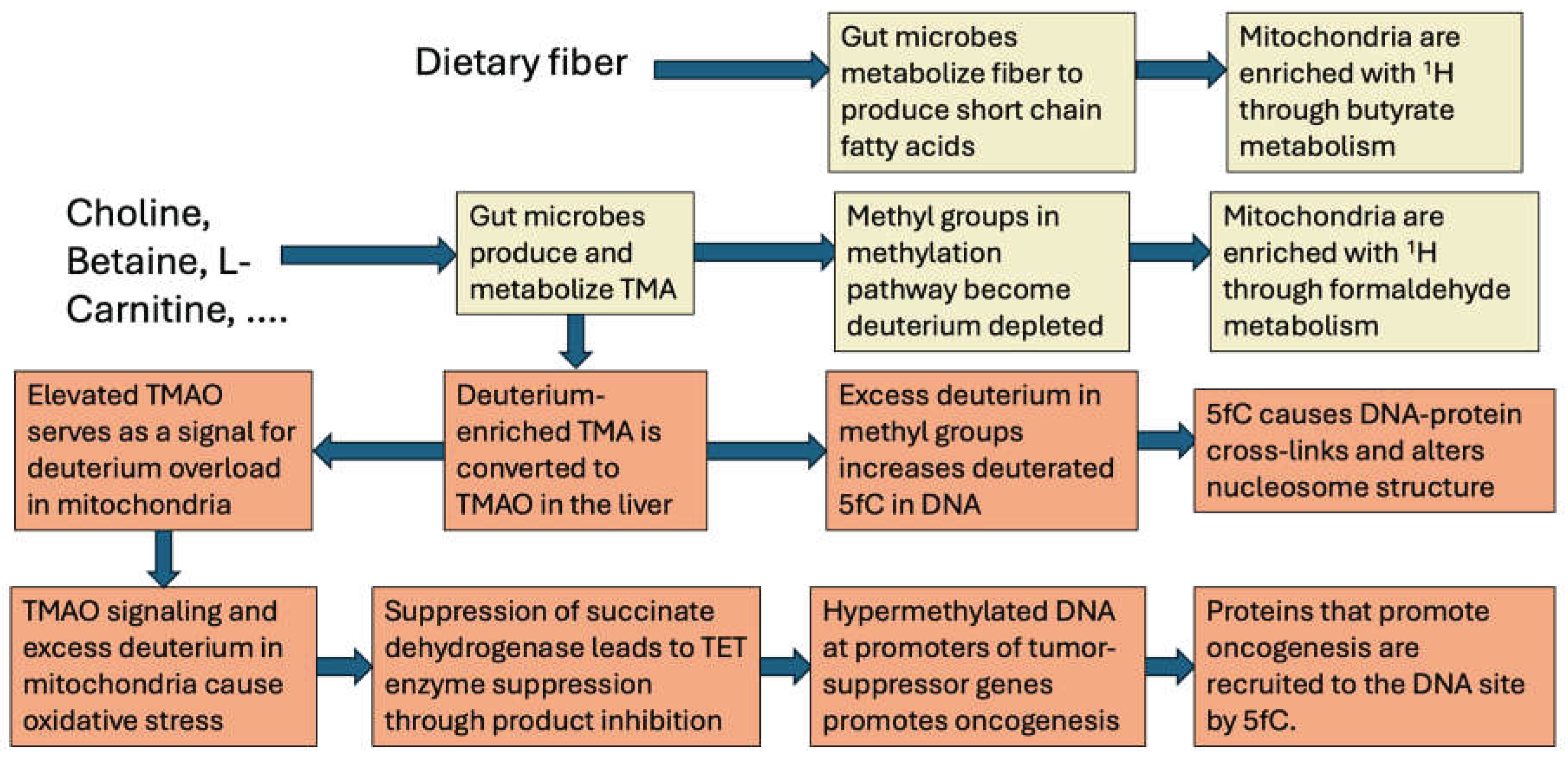
Figure 2.
Schematic of the impact of deuterated 5fC on the methylome, the nucleosome, gene expression, and disease outcomes.
Figure 2.
Schematic of the impact of deuterated 5fC on the methylome, the nucleosome, gene expression, and disease outcomes.
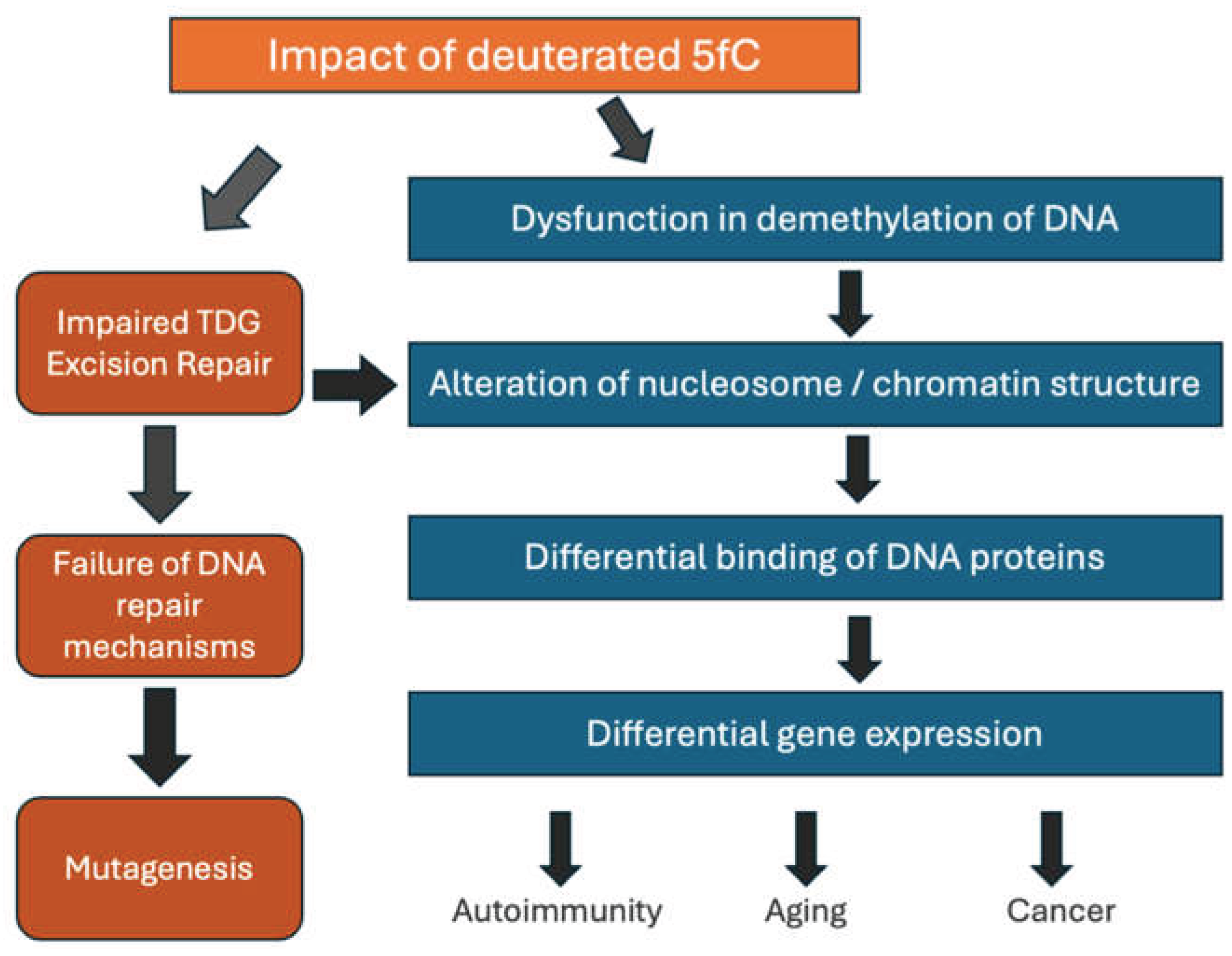

Table 1.
Tabulation of the key protein classes that preferentially bind to 5fC to effect multiple modifications to DNA and to histones.
Table 1.
Tabulation of the key protein classes that preferentially bind to 5fC to effect multiple modifications to DNA and to histones.
| Protein Class | Example Members | Biological Role |
|---|---|---|
| Transcriptional Regulators | FOXK1, FOXK2, FOXP1, FOXP4 and FOXI3 | This Forkhead box family of transcription factors preferentially bind 5fC compared to other DNA bases. They play key roles in chromatin remodeling and transcriptional regulation. All have been associated with tumor progression. |
| DNA Repair Factors | TDG and MPG | Glycosylases that assist with DNA repair. They are needed to restore cytosine from 5fC. |
| Chromatin Regulators | EHMT1, L3MBTL2 | EHMT1 binds to and methylates DNA-associated histones. It plays myriad roles in cancer metastasis, stemness and drug resistance. L3MBTl2 is recruited upon DNA strand breaks and it in turn recruits repair proteins. |
| NuRD Complex | A prominent multi-subunit protein complex | Uniquely couples ATP-dependent chromatin remodeling with histone deacetylation to repress transcription. NuRD is a direct reader of 5fC-marked DNA loci. It tightly packs DNA, effectively repressing gene expression. |
Disclaimer/Publisher’s Note: The statements, opinions and data contained in all publications are solely those of the individual author(s) and contributor(s) and not of MDPI and/or the editor(s). MDPI and/or the editor(s) disclaim responsibility for any injury to people or property resulting from any ideas, methods, instructions or products referred to in the content. |
© 2026 by the authors. Licensee MDPI, Basel, Switzerland. This article is an open access article distributed under the terms and conditions of the Creative Commons Attribution (CC BY) license (http://creativecommons.org/licenses/by/4.0/).
Copyright: This open access article is published under a Creative Commons CC BY 4.0 license, which permit the free download, distribution, and reuse, provided that the author and preprint are cited in any reuse.