

Submitted:
27 October 2026
Posted:
28 October 2026
You are already at the latest version
Abstract
The evolution of biological coordination is often framed as a progression toward specialized nervous systems. Here, we propose the Evolutionary Information Layering Framework, arguing that complexity emerges not by replacing ancestral mechanisms, but through their retention, specialization, and integration. We posit that pre-neural regulatory layers—including extracellular matrix (ECM) remodeling, mechanical forces, and endogenous bioelectric states—provided foundational organization later co-opted by neural systems. This hypothesis generates testable predictions: under neural disruption, non-neural layers (e.g., bioelectric gradients) sustain tissue coordination. By bridging evolutionary developmental biology (evo-devo), systems biology, and bioelectricity, this framework redefines nervous systems as specialized amplification layers embedded in an ancient, multi-scale regulatory architecture.
Keywords:
evolutionary biology
; biological coordination
; nervous system evolution
; bioelectricity
; extracellular matrix
; systems biology
1. Introduction
The emergence of nervous systems represents one of the major transitions in animal evolution. Neural systems enabled rapid communication between distant body regions and supported increasingly complex interactions with the environment (Jekely et al. 2015; Liebeskind et al. 2017). However, a persistent question in evolutionary biology remains: to what extent did biological coordination predate nervous systems?
Current evidence suggests that complex neural systems did not emerge independently from previously unorganized biological states. Instead, neural evolution likely involved the modification and specialization of pre-existing mechanisms of cellular communication and regulation (Jekely et al. 2015; Liebeskind et al. 2017). Studies of early-diverging animals provide illustrative examples. Sponges (Amphimedon queenslandica) possess a repertoire of genes associated with neuronal processes—ion channels, syntaxins, and glutamate receptors—yet organize them into non-synaptic physiological networks rather than conventional nervous systems (Leys 2015). Ctenophores present a more complex case: their nervous systems employ distinct molecular toolkits, fueling debate over whether neural systems evolved once or multiple times (Jekely et al. 2015; Liebeskind et al. 2017; Moroz et al. 2014).
Research in developmental biology and systems biology has also revealed that extracellular matrix regulation, mechanical interactions, and bioelectric mechanisms contribute substantially to biological organization beyond direct neural control. ECM remodeling regulates cellular behavior during development and tissue repair (Bonnans et al. 2014; Hynes and Naba 2012). Bioelectric signaling—mediated by voltage-gated ion channels and gap junctions—contributes to pattern formation, regeneration, and tissue organization in species lacking conventional nervous systems (Levin 2012, 2020).
Despite advances in neural evolution and tissue biology, a unified model integrating ECM, bioelectricity, and neural signaling remains lacking. Prior frameworks often treat these as sequential innovations. This article proposes that biological complexity arises from information layering: ancestral molecular and tissue-level regulations persist, specialize, and integrate with emerging neural systems. The diversification of cell types and body-plan organization across animal phyla provides essential phylogenetic context for understanding this transition (Arendt et al. 2016; Nielsen 2015).
2. Ancient Regulatory Layers Before Nervous Systems
The evolution of multicellular organisms required mechanisms that allowed individual cells to coordinate their activities within increasingly complex biological structures. Before the emergence of specialized nervous systems, organisms already possessed diverse regulatory mechanisms that enabled environmental sensing, cellular communication, and maintenance of biological organization. These mechanisms should not be considered equivalent to neural information processing. Instead, they represent evolutionarily conserved forms of biological regulation that provided pre-existing capacities later incorporated into more specialized systems.
2.1. Molecular and Cellular Regulatory Systems
The earliest forms of biological coordination emerged from molecular interactions; however, the term “proto-neural” is not appropriate in this context. Cells monitor internal and external states through receptor signaling, metabolic feedback, and gene regulatory networks. These processes maintain physiological stability and enable adaptation without specialized neural structures. Crucially, these capacities did not vanish when nervous systems appeared. Calcium signaling—a universal eukaryotic mechanism for intracellular communication that predates multicellularity by over a billion years—operates both in neurons (synaptic transmission) and in non-neural contexts such as wound healing, ciliary beating, and oocyte fertilization (Jekely et al. 2015; Liebeskind et al. 2017). The evolution of multicellularity and the emergence of early metazoan cell-type diversity established the substrate on which these regulatory innovations were built (Brunet and King 2017; Sebé-Pedrós et al. 2017).
2.2. Tissue-Level Regulation and Extracellular Matrix Organization
Multicellularity introduced a coordination problem: how do genetically identical cells in a shared tissue agree on collective behavior? The ECM is central to this process, although it has often been regarded primarily as a structural support. ECM molecules—collagens, laminins, and fibronectin—interact with cells through biochemical signaling and mechanical force transmission. During development, ECM remodeling directs cell migration and differentiation (Bonnans et al. 2014; Hynes and Naba 2012). In wound healing, ECM stiffness gradients guide fibroblast recruitment, a process that operates independently of neural input in denervated tissues.
ECM provided a pre-existing spatial organization system that neural networks could later exploit. Neural crest cell migration during embryogenesis, for instance, depends on precisely timed ECM degradation and reassembly. The nervous system did not originate tissue guidance; it co-opted an evolutionarily ancient mechanism. From an evolutionary perspective, such tissue-level systems provided organizational scaffolds that were subsequently integrated with specialized communication systems.
2.3. Bioelectric Regulation as an Evolutionarily Conserved Mechanism
Electrical properties are not neural inventions. Membrane potentials, ion gradients, and electrical signaling mechanisms occur across diverse forms of life and are not restricted to nervous tissues (Levin 2012, 2020). Membrane potentials govern regeneration in Hydra and planarians, operating independently of synapses. This phylogenetic conservation extends beyond animals: electrical signaling is widespread and conserved across plants, fungi, and animals alike (Humphrey et al. 2022). This deeply conserved bioelectric layer likely provided a pre-neural template for rapid signaling. Neural systems later specialized this medium, but the ancestral bioelectric code persists in modern tissues. Therefore, bioelectric regulation should be understood as an ancient regulatory medium that was progressively incorporated during evolution, rather than a transitional stage immediately preceding nervous systems.
3. Nervous Systems as Evolutionary Amplification Layers
The emergence of nervous systems introduced a highly specialized level of biological organization. Nervous systems did not simply replace earlier regulatory mechanisms; rather, they can be viewed as specialized amplification systems that increased the speed, range, and coordination of biological responses (Fields et al. 2020). The evolutionary transition to nervous systems was built upon pre-existing mechanical and signaling frameworks. Tissue mechanics likely played a central role in shaping the first neural circuits (Rosenthal et al. 2018), and the earliest nervous systems emerged through the gradual integration of these elements (Burkhardt and Sprecher 2017).
3.1. Enhancement of Communication Speed
Before specialized neural systems, biological coordination depended on diffusion-limited mechanisms: molecular gradients, direct cell–cell contact, and slow electrical coupling. The evolution of nerve cells—with action potentials traveling at meters per second rather than millimeters per hour—fundamentally changed the temporal scale of organismal response. A key observation is that neural speed did not eliminate slower regulatory layers but rather amplified them. A bioelectric gradient that once slowly coordinated a few hundred cells in a sponge could, via neural integration, coordinate millions of cells across a bilaterian body within milliseconds. The older mechanism persisted; the neural layer added velocity and range. Consequently, nervous systems are best described as amplification layers rather than replacements.
3.2. Expansion of Spatial Coordination
As bilaterian bodies elongated and compartmentalized, local regulatory systems faced a scaling problem. A mechanical signal in the tail of a fish cannot diffuse to the brain fast enough to coordinate rapid behavior. Neural networks solved this through dedicated pathways—axons and synapses forming long-distance communication channels. Yet even in animals with complex brains, local regulation persists. Smooth muscle contraction in the gut operates via the enteric nervous system and intrinsic pacemaker cells, independently of central control. Tissue repair after spinal cord injury involves bioelectric and ECM remodeling in the absence of descending neural signals (Fields et al. 2020). Neural expansion of spatial coordination is thus real and significant, but it operates on top of, rather than instead of, local systems.
3.3. Development of Anticipatory Regulation
Perhaps the most distinctive neural contribution is temporal integration: the ability to use past states to constrain future ones. Recurrent neural circuits, synaptic plasticity, and memory-related processes enable forms of anticipatory regulation that go beyond reactive homeostasis. However, anticipation is not unique to neural tissue. The plant Mimosa pudica shows habituation—a simple form of non-neural memory. Bioelectric states in regenerating amphibian limbs “remember” positional information across days of tissue remodeling (Levin 2020). Neural systems contributed acceleration, amplification, and integration of anticipatory processes across longer timescales, rather than anticipation itself. This reframing places nervous systems within a continuum of biological prediction rather than as its sole origin.
4. Evolutionary Information Layering Framework
This article proposes the Evolutionary Information Layering Framework as a conceptual model for understanding how biological complexity may emerge through interactions among multiple regulatory systems. The framework does not assume a fixed hierarchical sequence. Evolutionary innovation often involves preservation, modification, and integration of pre-existing mechanisms. The four layers are summarized in Table 1 and schematically illustrated in Figure 1.
Four evolutionarily superimposed layers coordinate biological function from bottom to top: Layer 0 Bioelectric Gradients; Layer 1 ECM and Mechanical Forces; Layer 2 Peripheral Neuromodulation; Layer 3 Nervous System acting as an evolutionary amplifier rather than a replacement. Bidirectional arrows indicate that lower layers are retained and integrated across evolutionary transitions. The upward arrow denotes evolutionary emergence through retention, specialization, and integration. Right: Prediction 1 in planarian regeneration—neural disruption does not abolish tissue coordination because bioelectric and ECM dynamics persist (Tab 1).
The four EIL layers are presented from the most ancient (Layer 0) at the base to the most recently evolved (Layer 3) at the top. Each layer retains functional autonomy while being modulated and integrated by higher layers. No single layer is replaced by those above it; rather, higher layers increase the speed, range, and predictive capacity of biological coordination( Figure 1). Central Proposition:Biological complexity may emerge not primarily through replacement of ancestral regulatory systems, but through their retention, specialization, and integration into increasingly complex biological architectures.
4.1. Retention of Ancestral Regulatory Mechanisms
Evolutionary innovations frequently occur within existing biological contexts. Ancient regulatory mechanisms may be retained because they continue to provide important biological functions, even after new specialized systems emerge. ECM molecules such as collagen and laminin, ancient in metazoans, still dictate neural migration and repair (Bonnans et al. 2014; Hynes and Naba 2012). The framework emphasizes that evolutionary novelty (e.g., nervous systems) does not erase these layers; instead, neural function depends on their continuous operation. Disruption of neural activity reveals residual coordination by ECM and bioelectric states—a key prediction.
4.2. Specialization of Existing Mechanisms
Evolutionary complexity often arises when an existing mechanism is recruited for a new function—a process known as exaptation. Neural components are not newly invented molecules; they are old molecules in new arrangements. Voltage-gated sodium channels, for example, evolved from calcium-channel ancestors present in single-celled eukaryotes. Their concentration at axon initial segments and nodes of Ranvier represents a spatial specialization of a pre-existing electrical property. Similarly, the SNARE complex—essential for synaptic vesicle release—functions in non-neural secretion across all eukaryotes. Specialization does not require novelty at the molecular level; it requires architectural reorganization, where the same parts are reassembled into a faster, more integrated system.
4.3. Integration Across Regulatory Layers
The framework’s central claim is that no single layer operates in isolation. Integration is not a one-time evolutionary event but a continuous, dynamic process (Fields et al. 2020). A concrete example is found in zebrafish spinal cord regeneration, where serotonergic signaling (neural layer) interacts with bioelectric states (ion channel layer) and ECM remodeling (structural layer) to guide axon regrowth. Blocking any one layer results in partial compensation by the others, but full functional recovery requires all three. Biological complexity is not a linear hierarchy with nervous systems at the apex; rather, it is a distributed network in which molecular, cellular, mechanical, electrical, and neural components all remain functionally significant. This perspective aligns with top-down modeling approaches that seek to explain complex living systems above the molecular level (Pezzulo and Levin 2016).
5. Testable Predictions and Future Directions
A useful conceptual framework should generate experimentally testable hypotheses. The Evolutionary Information Layering Framework therefore proposes several specific, falsifiable predictions.
Prediction 1: Neural Disruption and Non-Neural Compensation
If nervous systems are a specialized amplification layer, disruption of neural activity should not eliminate all biological coordination. In denervated regeneration models, blocking action potentials may not completely halt patterning; endogenous bioelectric gradients and ECM remodeling sustain coarse-grained coordination (Levin 2012, 2020). This prediction is testable via pharmacological denervation combined with bioelectric imaging.
Prediction 2: Multi-Scale Coupling During Adaptation and Repair
The framework predicts that complex biological responses involve coupled interactions among multiple levels. Alterations in ECM stiffness during development are expected to produce measurable shifts in cellular voltage gradients, demonstrating direct layer integration (Bonnans et al. 2014; Hynes and Naba 2012; Levin 2012). Future studies combining molecular biology, biomechanics, electrophysiology, and computational approaches may clarify how these regulatory systems interact dynamically.
Prediction 3: Development and Regeneration as Evolutionary Windows
Development and regeneration provide opportunities to investigate interactions among ancestral and specialized regulatory systems. Embryogenesis demonstrates how multiple biological processes become coordinated during organism formation, whereas regeneration reveals how biological systems reconstruct complex structures after injury (Levin 2012, 2020). The model predicts that regenerating tissues will reactivate ancestral regulatory networks (e.g., pre-neural bioelectric circuits) before neural-specific genes are expressed, linking development to evolutionary layering. These processes may provide experimental models for examining how layered regulatory architectures operate across developmental and evolutionary timescales.
6. Discussion
The EIL framework does not diminish nervous systems; it places them within a deeper evolutionary context. Nervous systems are the latest and fastest layer in a regulatory stack extending to the dawn of multicellularity.The EIL framework does not resolve nervous-system origins. It reframes the question: complexity is not invented anew at each transition, but accumulated through retention, specialization, and integration. The three predictions offered here are the next empirical testbed. Clinical translation (postural demand, non-neural repair observables, motor decompensation) is deferred to the companion clinical series. Premature inclusion would weaken the theoretical claim.
This perspective carries three implications. First, disrupting neural activity should reveal—not eliminate—coordinated responses at other regulatory levels. Second, regenerative medicine may benefit from targeting non-neural layers to enhance neural repair. Third, differences among animal lineages may reflect different patterns of layer integration, not merely more or less neural complexity.
Limitations are clear. This framework is conceptual; it does not yet specify quantitative relationships among layers or predict relative contributions across taxa. Comparative genomic evidence has not resolved whether neural systems arose once or multiple times (Jekely et al. 2015; Liebeskind et al. 2017; Moroz et al. 2014). Future work combining comparative genomics, electrophysiology, and computational modeling is needed. Empirical validation of the framework is a key priority.
In closing, biological coordination is older, broader, and more distributed than the nervous system alone. Understanding how evolution layered new regulatory systems onto ancient ones may be essential for understanding not only where nervous systems came from, but also how biological complexity emerges.
Author Contributions
Conceptualization: Dayong Hu; Writing—original draft: Dayong Hu; Writing—review & editing: Dayong Hu.
Funding
This research received no external funding.
Institutional Review Board Statement
Not applicable. This article is a conceptual framework and does not report research involving human participants or animals.
Informed Consent Statement
Not applicable.
Data Availability Statement
No new datasets were generated or analyzed for this conceptual article.
Competing Interests
The author declares no competing interests.
AI-assisted writing statement
During preparation of this manuscript, ChatGPT (OpenAI, https://chat.openai.com) was used as an AI-assisted tool for literature-oriented discussion, conceptual organization, language editing, and manuscript drafting. The author critically evaluated and revised all generated material and takes full responsibility for the final content, scientific interpretation, and accuracy of the manuscript. ChatGPT is not an author of this manuscript.
Preprint statement
A preprint version of this manuscript was posted on Preprints.org (DOI: 10.20944/preprints202610.0474.v1). This submission represents the finalized version under peer review; the preprint is non-peer-reviewed and consistent with Springer Nature preprint policy.
References
- Arendt, D.; Musser, J.M.; Baker, C.V.H.; et al. The origin and evolution of cell types. Nat. Rev. Genet 2016, 17, 744–757. [Google Scholar] [CrossRef]
- Baluška, F.; Levin, M. On having no head: cognition in the planarian flatworm. Front Psychol. 2016, 7, 553. [Google Scholar] [CrossRef]
- Bonnans, C.; Chou, J.; Werb, Z. Remodelling the extracellular matrix in development and disease. Nat. Rev. Mol. Cell Biol. 2014, 15, 786–801. [Google Scholar] [CrossRef]
- Brunet, T.; King, N. The origin of animal multicellularity and cell differentiation. Dev. Cell 2017, 43, 124–140. [Google Scholar] [CrossRef]
- Burkhardt, P.; Sprecher, S.G. Evolution of the first nervous systems. Integr. Comp. Biol. 2017, 57, 35–42. [Google Scholar] [CrossRef]
- Davies, J.A.; Garrod, D.R.; Tucker, J.B. The rules of formation of the vertebrate body plan: a conceptual review. J. Anat. 2012, 221, 1–15. [Google Scholar] [CrossRef]
- Fields, C.; Bischof, J.; Levin, M. Morphological coordination: a common ancestral function unifying neural and non-neural signaling. Physiology 2020, 35, 16–30. [Google Scholar] [CrossRef]
- Hobert, O. Beyond the wiring diagram. eLife 2016, 5, e20084. [Google Scholar] [CrossRef]
- Humphrey, G.W.; Chittka, L.; Wackermann, J. Electrical signaling in plants and animals: a comparative review. J. Comp. Physiol. A 2022, 208, 257–272. [Google Scholar] [CrossRef]
- Hynes, R.O.; Naba, A. Overview of the matrisome—an inventory of extracellular matrix constituents and functions. Cold Spring Harb. Perspect. Biol. 2012, 4, a004903. [Google Scholar] [CrossRef]
- Jekely, G.; Keijzer, F.; Godfrey-Smith, P. An option space for early neural evolution. Philos. Trans. R Soc. Lond. B Biol. Sci. 2015, 370, 20150181. [Google Scholar] [CrossRef]
- Levin, M. Molecular bioelectricity in developmental biology: new tools and recent discoveries. BioEssays 2012, 34, 205–217. [Google Scholar] [CrossRef]
- Levin, M. The biophysics of regenerative repair suggests new perspectives on biological causation. BioEssays 2020, 42, 1900146. [Google Scholar] [CrossRef]
- Leys, S.P. Elements of a “nervous system” in sponges. J. Exp. Biol. 2015, 218, 581–591. [Google Scholar] [CrossRef]
- Liebeskind, B.J.; Hofmann, H.A.; Hillis, D.M.; Zakon, H.H. Evolution of animal neural systems. Annu Rev. Ecol. Evol. Syst. 2017, 48, 377–398. [Google Scholar] [CrossRef]
- Mathews, E.A.; Levin, M. Adopting a bauplan for the study of neural evolution. Front Ecol. Evol. 2017, 5, 178. [Google Scholar] [CrossRef]
- Moroz, L.L.; Kocot, K.M.; Citarella, M.R.; et al. The ctenophore genome and the evolutionary origins of neural systems. Nature 2014, 510, 109–114. [Google Scholar] [CrossRef]
- Musser, J.M.; Schippers, K.J.; Nickel, M.; et al. Profiling cellular diversity in sponges reveals an ancient complex nervous system. Science 2021, 374, eabe8998. [Google Scholar] [CrossRef]
- Nielsen, C. Animal evolution: interrelationships of the living phyla, 4th edn; CRC Press: Boca Raton, 2015. [Google Scholar]
- Pezzulo, G.; Levin, M. Top-down models in biology: explanation and control of complex living systems above the molecular level. J. R Soc. Interface 2016, 13, 20160555. [Google Scholar] [CrossRef]
- Rosenthal, S.M.B.; Miller, D.J.; Alford, P.W. The role of tissue mechanics in the evolution of nervous systems. Philos. Trans. R Soc. B 2018, 373, 20170370. [Google Scholar] [CrossRef]
- Sebé-Pedrós, A.; Chomsky, E.; Pang, K.; et al. Early metazoan cell type diversity, evolution, and development. Mol. Biol. Evol. 2017, 34, 2071–2087. [Google Scholar] [CrossRef]
Figure 1.
Schematic of the Evolutionary Information Layering (EIL) framework. Four evolutionarily superimposed layers coordinate biological function from bottom to top: Layer 0 Bioelectric Gradients; Layer 1 ECM and Mechanical Forces; Layer 2 Peripheral Neuromodulation; Layer 3 Nervous System. Bidirectional arrows indicate that lower layers are retained and integrated across evolutionary transitions. The upward arrow denotes evolutionary emergence through retention, specialization, and integration. Right: Prediction 1 in planarian regeneration.
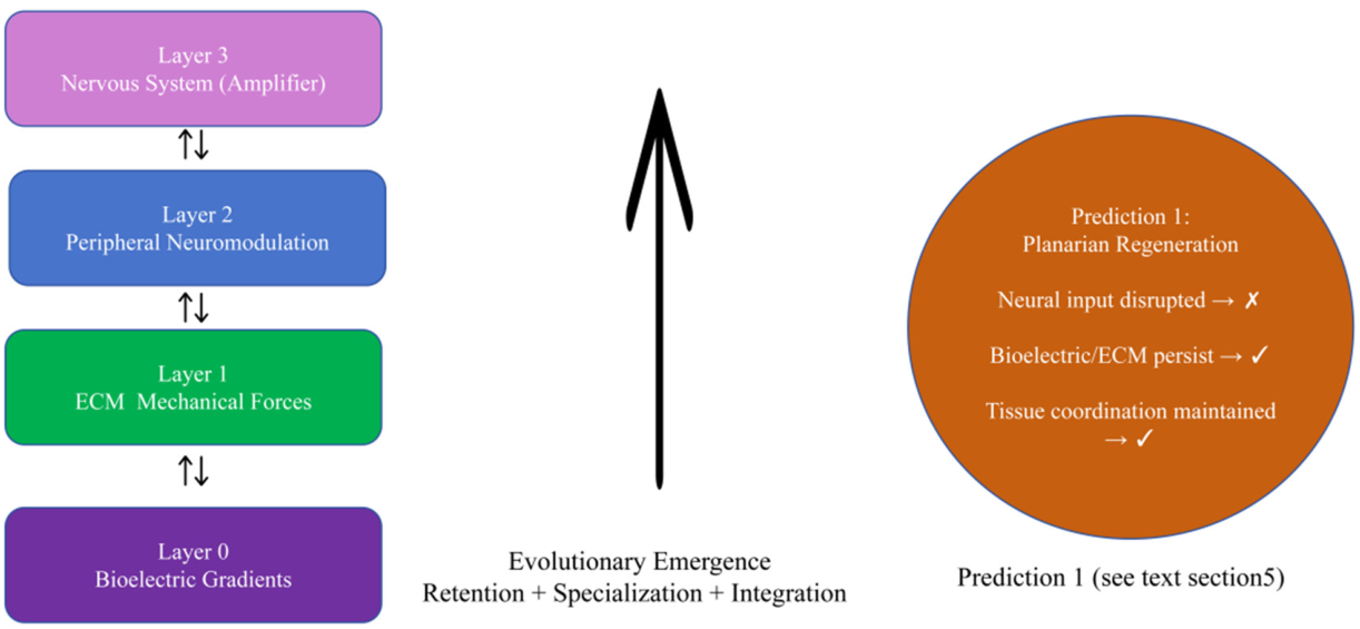

Table 1.
Evolutionary information-layering model of cross-scale biological coordination.
| Layer | Information substrate | Evolutionary origin | Timescale | Empirical examples | Experimental/clinical anchor |
| 0 | Bioelectric gradients (Vmem, ion flows) | Unicellular/early Metazoa | Hours–days | Hydra, planarian regeneration | Regenerative medicine, bioelectric patterning |
| 1 | ECM and mechanical forces (tensegrity, stiffness) | Multicellular/mesodermal | Minutes–days | ECM remodeling in development, DDH mechanics | Developmental orthopedics, mechanobiology |
| 2 | Peripheral neuromodulation (reflex, autonomic, somatosensory) | Bilaterian nervous systems | Milliseconds–seconds | Reflex arcs, enteric pacing | Neurorehabilitation |
| 3 | Central nervous system integration and amplification | Vertebrate brain expansion | Milliseconds–predictive | Motor learning, cortical mapping | Motor/cognitive rehabilitation |
Disclaimer/Publisher’s Note: The statements, opinions and data contained in all publications are solely those of the individual author(s) and contributor(s) and not of MDPI and/or the editor(s). MDPI and/or the editor(s) disclaim responsibility for any injury to people or property resulting from any ideas, methods, instructions or products referred to in the content. |
© 2026 by the author. Licensee MDPI, Basel, Switzerland. This article is an open access article distributed under the terms and conditions of the Creative Commons Attribution (CC BY) license.
Copyright: This open access article is published under a Creative Commons CC BY 4.0 license, which permit the free download, distribution, and reuse, provided that the author and preprint are cited in any reuse.