

Submitted:
03 October 2026
Posted:
07 October 2026
You are already at the latest version
Abstract
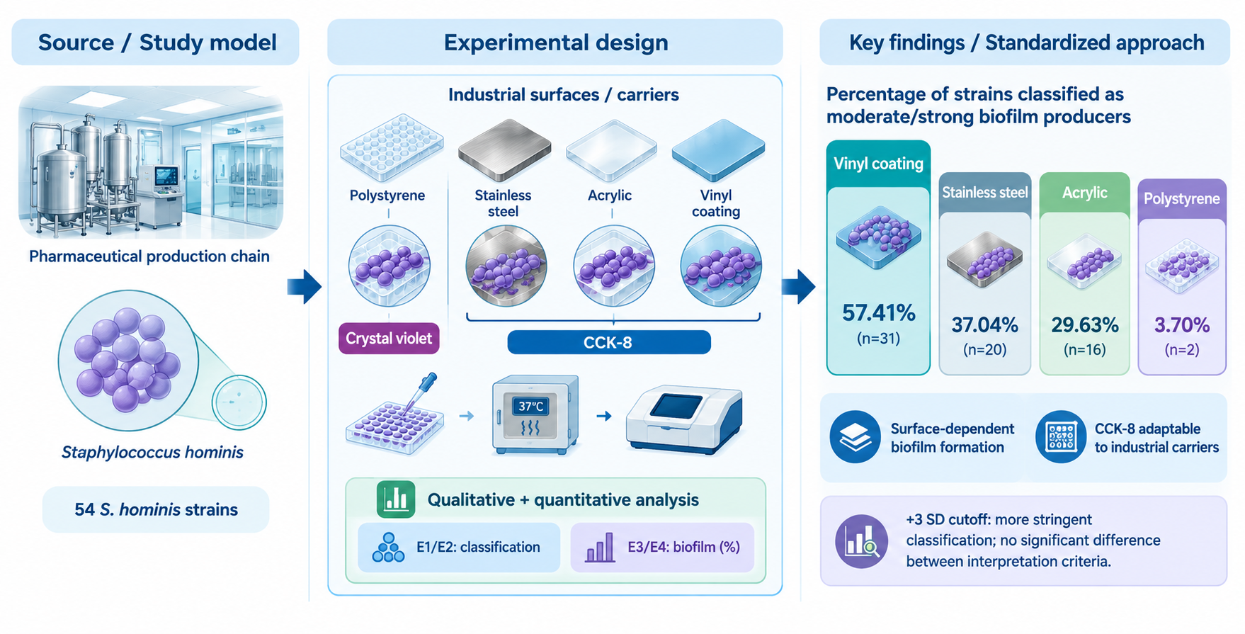
Biofilm formation by Staphylococcus hominis in pharmaceutical environments can promote microbial persistence on surfaces and hinder cleaning and disinfection, posing a risk to product quality and safety. This study aimed to standardize an in vitro adaptive methodology to evaluate biofilm for-mation on surfaces of interest to the pharmaceutical industry and analyze the impact of different interpretation criteria. Fifty-four S. hominis strains isolated between 2014 and 2022 from a pharma-ceutical production chain were evaluated. Biofilm formation was analyzed on stainless steel, acrylic, and vinyl coating using the Cell Counting Kit-8 (CCK-8) assay to quantify bacterial cell viability, as well as on polystyrene using the crystal violet method. Qualitative and quantitative interpretations applied different equations to define the negative control cutoff. Vinyl coating showed the highest formation, with 31 (57.41%) strains classified as moderate or strong producers, followed by stainless steel (n=20; 37.01%), acrylic (n=16; 29.63%), and polystyrene (n=2; 3.70%). Quantitative analysis identified surface-dependent hyper-producing strains, reaching maximum values from 49.75 to 61.60%. Applying three standard deviations to the negative control generated reclassifications without statistically significant differences between criteria. The proposed standardized method supports the CCK-8 assay as an adaptive methodology to evaluate biofilm formation by different bacterial strains and surfaces.
Keywords:
Staphylococcus hominis
; biofilm
; pharmaceutical industry
; cleaning and disinfection
1. Introduction
Biofilms constitute organized microbial communities formed by the adhesion of cells to biotic or abiotic surfaces and by the self-production of an extracellular matrix that enwraps and protects the microbial cells [1,2]. This matrix is composed of different components, including polysaccharides, proteins, lipids, and extracellular DNA, which play a fundamental role in the biofilm adhesion mechanism and in protecting the community against environmental stress and external agents [3].
Biofilm formation occurs through a process in which, initially, planktonic microbial cells reversibly adhere to the surface, influenced by nutrient availability, pH, surface material composition and properties, and temperature, in addition to being mediated by specialized structures such as pili, flagella, and fimbriae. Subsequently, cell attachment and aggregation take place, accompanied by the secretion of extracellular polymeric substances (EPS), which contribute to stabilizing the association of microorganisms with the substrate and rendering the adhesion irreversible. Subsequent cell multiplication and division promote the formation of microcolonies and the establishment of intercellular communication via quorum sensing, accompanied by increased EPS production. As this process progresses, the biofilm reaches maturation, characterized by the stabilization of the extracellular matrix and the development of a three-dimensional structure. Finally, bacterial cells disperse from the superficial layer of the biofilm, enabling the colonization of new surfaces [2,4].
Among the Gram-positive bacteria capable of forming biofilms, the genus Staphylococcus are widely distributed in the environment and in human microbiota [5]. Among the species of the genus, Staphylococcus hominis belongs to the coagulase-negative staphylococci (CoNS) group and can be isolated from regions such as the legs, arms, axillae, and head of the human body [5,6]. Although normally a commensal, S. hominis is considered an opportunistic pathogen associated with skin and soft tissue infections, endophthalmitis, endocarditis, and bacteremia, especially in immunocompromised or elderly individuals [7,8,9]. The species is also frequently isolated in environmental monitoring programs within the pharmaceutical manufacturing chain, with its presence being mainly related to contamination introduced by operators [10,11,12,13].
The ability of S. hominis and other CoNS to form biofilms contributes to the persistence of the species in different environments, and is relevant in both clinical and industrial contexts. In the hospital environment, this phenomenon is associated with the colonization of medical devices and persistent infections [14]. In pharmaceutical industry facilities, the presence of biofilms on surfaces, equipment, and pipelines can resist cleaning and disinfection procedures. Hence, the persistence of such microorganisms in the production environment pose a risk to product quality and safety [1,2,15,16,17].
Although in vitro biofilm quantification assays in polystyrene microplates are well established in the scientific literature [18,19], the standardization of protocols applicable to other types of surfaces remains a methodological challenge. Several authors have sought to adapt assays using carriers in order to simulate materials of industrial or clinical relevance [20,21] or to evaluate biofilm production qualitatively by scanning electron microscopy (SEM) [16,17]. However, such approaches employing alternative surfaces still lack standardization regarding execution and result interpretation [22].
Given the relevance of S. hominis in industrial environments, this study aimed to standardize an in vitro adaptative methodology for evaluating biofilm formation on surfaces of interest to the pharmaceutical industry that are not available in microplate format (stainless steel, acrylic, and vinyl coating), analyzing the impact of different equations on the qualitative and quantitative interpretation of the results.
2. Materials and Methods
2.1. Bacterial Strains
Fifty-four S. hominis strains were isolated between 2014 and 2022 from different stages of the production chain of an immunobiological manufacturing pharmaceutical industry facility located in the municipality of Rio de Janeiro, Brazil. Most strains were originated from environmental monitoring (n=32) and bioburden determination (n=12), followed by samples from aseptic process simulation (n=2), intermediate products (n=2), cleaning validation (n=2), growth promotion testing in culture media (n=1), sterility testing (n=1), water for injection (n=1), and raw material (n=1).
The strains were kept cryopreserved in 30% Difco™ Skim Milk (BD Biosciences, Le Pont de Claix, France) supplemented with 30% glycerol (Merck KGaA, Darmstadt, Germany) and stored in an ultra-low temperature freezer at <-70 °C. The strains were deposited at the Collection of Environment and Health Bacteria (Coleção de Bactérias do Ambiente e Saúde - CBAS) hosted at the Oswaldo Cruz Foundation (Fiocruz), Rio de Janeiro, Brazil. CBAS is affiliated with the World Federation for Culture Collections (WFCC) and registered as World Data Centre for Microorganisms (WDCM) 958.
2.2. Biofilm Formation Assay on Carriers
Biofilm formation was evaluated on stainless steel (SS), acrylic (AC), and vinyl coating (VC) carriers using the methodology described by Ourique et al. [22] and Jin et al. [23] with modifications. The strains were streaked onto Sheep Blood Agar (SBA; BioCen do Brasil, São Paulo, Brazil) and incubated at 37 °C for 24 h to check for purity. An isolated colony of each strain was inoculated into a sterile conical-bottom tube containing 5 mL of Brain Heart Infusion broth (BHI; Merck, Darmstadt, Germany) and incubated at 37 °C for 24 h under agitation at 150 rotations per minute (rpm). For assay controls, tubes containing uninoculated BHI broth were used as negative controls, while tubes containing the reference strains Pseudomonas aeruginosa American Type Culture Collection (ATCC) 27853 and Staphylococcus aureus ATCC 25923 were used as positive controls. Subsequently, 1 mL of the bacterial suspension of each strain was transferred into individual wells of sterile 24-well polystyrene microplates (Thermo Scientific, Roskilde, Denmark), each containing a sterile carrier of SS (Smart Imas LTDA, São Paulo, Brazil), AC (Hoppe Oficina Criativa, São Bento do Sul, Brazil), or VC (Tarkett Fademac, Rio de Janeiro, Brazil), all measuring 1 cm in diameter. The microplates were incubated at 22.5 °C for 48 h. Following incubation, the contents of the wells were aspirated using a sterile pipette, and the carriers were transferred to new 24-well microplates using sterile forceps. The carriers were submitted to five washes with phosphate-buffered saline (PBS; Gibco-BRL, Gaithersburg, USA) and maintained at room temperature inside a biological safety cabinet until completely dry.
For biofilm quantification, 0.5 mL of Cell Counting Kit-8 reagent (CCK-8; Merck, Darmstadt, Germany), previously diluted to 10% in PBS, was added to each well containing a carrier, and the microplates were incubated at 37 °C for 2 h. Afterwards, 200 µL of the contents from each well was transferred to a sterile 96-well polystyrene microplate (Thermo Scientific, Roskilde, Denmark), and the optical density (OD) was determined at 450 nanometers (nm) using a microplate spectrophotometer reader (bioMérieux, Reader 270, France). A schematic diagram of the methodology is shown in Figure 1.
Based on OD values, biofilm production was qualitatively classified [19] using two approaches to calculate the negative control (NC) value, according to Equations 1 (E1) and 2 (E2):
ODNC = x̄ of OD value of the BHI broth
ODNC = x̄ of OD value of the BHI broth +3 × σ
After, the following criteria was applied for strains classification: OD ≤ ODNC = no biofilm producer (NB); ODNC < ODsample ≤ 2×ODNC = weak biofilm producer (WB); 2×ODNC < ODsample ≤ 4×ODNC = moderate biofilm producer (MB); ODsample > 4×ODNC = strong biofilm producer (SB) [19].
Additionally, biofilm formation was quantified using an adapted equation proposed by Kadurugamuwa et al. [24], as described in Equations 3 (E3) and (E4).
Biolfim formation (%) = 100 x (ODsample - ODNC)
Where ODNC = x̄ of OD value of the BHI broth
Biolfim formation (%) = 100 x (ODsample - ODNC)
Where ODNC = x̄ of OD value of the BHI broth +3 × σ
2.3. Biofilm Formation Assay on Polystyrene
Biofilm formation on polystyrene (PO) surfaces was evaluated using the microplate crystal violet assay, following the methology described by Vasconcellos et al. [16]. Three independent experiments were performed in triplicate for each strain. The final result was determined from the median of the three independent experiment results, and the interpretation criteria were applied according to Stepanović et al. [19], with the ODNC calculated according to E1.
2.4. Statistical Analysis
Qualitative biofilm formation data (no biofilm producer, weak biofilm, moderate biofilm and strong biofilm) obtained using E1 for the four surfaces, and E2 for SS, AC and VC surfaces, were evaluated using the Kruskal-Wallis test for comparisons among surfaces, followed by the Mann-Whitney post hoc test for pairwise comparisons, applying the Bonferroni correction for multiple testing. The comparison between E1 and E2 for the surfaces SS, AC and VC was also performed using the Mann-Whitney test with Bonferroni correction in Microsoft® Excel® for Microsoft 365 (Version 2607).
Quantitative biofilm formation data expressed as percentages were subjected to descriptive statistical analysis, calculating the mean, median, minimum, maximum, and standard deviation in Microsoft® Excel® for Microsoft 365 (Version 2607). For inferential data analysis, the Friedman test was used to compare the biofilm percentages obtained for each strain across the three surfaces (SS, AC, and VC) using E3 and E4. When significant differences were observed, the Durbin-Conover post hoc test was applied. Analyses were conducted using jamovi statistical software (version 2.7) [25].
Pairwise comparisons of percentages between E3 and E4 for each surface were conducted using a two-sample t-test assuming equal variances in Microsoft® Excel® for Microsoft 365 (Version 2607).
For all inferential analyses, a significance level of 5% (p < 0,05) was adopted.
3. Results and Discussion
Biofilms represent one of the major challenges for Contamination Control Strategy (CCS) in pharmaceutical manufacturing environments [15]. So, studying biofilms in the pharmaceutical industry is crucial to prevent microbial contamination and ensure product safety. In the present study, biofilm formation on different surfaces was investigated in S. hominis strains, a commom microorganism found in pharmaceutical manufacturing enviroments (10-13).
The qualitative evaluation of biofilm formation capacity performed across the four surfaces, according to E1, demonstrated a predominance of weak biofilm producers on all four surfaces evaluated (Figure 2). The PO surface presented the lowest occurrence of biofilm formation, with 92.59% (n=50) of the strains classified as weak biofilm producers, and none as strong biofilm producer. These results were similar to reported by Lee et al. [26], who studied biofilm formation by Staphylococcus aureus (n=19) strains and observed lower formation on PO, which is a hydrophobic surface, compared to SS, which is a hydrophilic surface. In contrast, biofilm formation on the other surfaces showed an increase in the proportion of moderate biofilm producers and strong biofilm producers, corresponding to 29.63% (n=16) on AC, 37.04% (n=20) on SS, and reaching 57.41% (n=31) on VC.
AC is a hydrophobic polymeric substrate, and similarly, Silva et al. [27] observed that AC resin acts as a favorable substrate for bacterial attachment, highlighting that surface characteristics play a decisive role in colonization dynamics. Regarding VC, composed predominantly of polyvinyl chloride, this surface is described as hydrophobic and prone to displaying surface roughness [28]. Such properties may favor hydrophobic interactions between the surface and the staphylococcal cell wall, which also possesses a predominantly hydrophobic character [29], potentially contributing to the initial stage of adhesion and, consequently, to the higher proportion of moderate and strong biofilm-producing strains observed on VC.
However, surface hydrophobicity (in VC, AC, and PO) or hydrophilicity (in SS) alone may not fully explain biofilm development. Bacterial adhesion and biofilm formation are also influenced by the interaction among various surface physicochemical and topographical properties, including chemical composition, surface energy, and roughness. Furthermore, interaction forces between microbial cells and the material, such as electrostatic interactions, can influence this process [30].
A significant difference was observed among the four surfaces using the Kruskal-Wallis test (p= 1.53x10⁻⁸). However, this comparison presents the bias that PO was tested using the crystal violet method. Following pairwise comparisons between surfaces with Bonferroni adjustment, no significant differences were observed between SS and AC (p=1.00), or between SS and VC (p=0.11). All other surfaces showed significant differences from one another (p≤ 0.025).
In the CCK-8 assay performed in SS, AC and VC surfaces, the equation applied (E1 or E2) impacted the qualitative classification of biofilm formation across the three surfaces (Figure 2). The application of E2 promoted a shift of the strains toward lower intensity categories compared to E1. This effect was most evident on the AC surface, where E2 resulted in the reclassification of nine (16.67%) strains into the no biofilm producer category, whereas under E1 all strains demonstrated the ability to form biofilm. On the other hand, on the SS and VC surfaces, all strains remained classified as biofilm producers under both approaches. Notably, VC preserved 11 (20.37%) strains in the strong biofilm producer category regardless of the equation used. After statistical analysis, no significant differences were observed between E1 and E2 across all surfaces (p≥0.069).
The quantitative evaluation expressed as a percentage value obtained by E3 and E4 demonstrated consistent pattern of biofilm distribution, with minor variations (Figure 3). Following the Friedman test, significant differences were observed among the three surfaces (SS, AC, and VC) using E3 and E4 (p<0.001). Following multiple comparisons using the Durbin-Conover post hoc test, no significant difference was observed only when using E4 between the SS and VC surfaces (p=0.090). As also observed in qualitative analysis, VC showed higher levels of biofilm formation than the other surfaces, evidenced by superior medians and maximum values (Table 1). Despite the profile being concentrated in the lower ranges for most strains, the three surfaces revealed the presence of outliers, reaching expressive percentages of biofilm formation (Figure 3).
The descriptive analysis of the quantitative data confirmed VC as the surface most favorable for S. hominis biofilm formation, recording medians of 8.75% and 8.05% for E3 and E4, respectively, with maximum values exceeding 61% (Table 1). In contrast, AC surface exhibited the lowest production, with medians of 2.55% in E3 and 1.35% in E4. Across all surfaces, mean values were higher than medians due to the presence of hyper-producing strains. The application of E4 promoted only subtle changes in the descriptive statistical analysis compared to E3, and no significant difference was observed between them across all three tested surfaces (p≥0.47) (Table 1). The quantitative analysis offers a robust view of strain behavior, complementing qualitative classification into the four categories (no biofilm producer, weak biofilm, moderate biofilm, and strong biofilm producer).
The B7636, B086.17, and B608.20 strains exhibited a hyper-producing biofilm profile in SS, AC and VC, respectively, reaching the maximum percentage formation values recorded across both quantitative equations (E3 and E4).
Radar plots illustrating the percentage results obtained in E4 for these three strains are shown in Figure 4. Strain B7636 produced high biofilm formation exclusively on SS surface (58.69%), whereas B608.20 produced only on VC (61.20%). In contrast, B86.17 exhibited a capacity to form high biofilms on both SS (58.69%) and AC (49.75%) surfaces. These profiles corroborate the heterogeneity observed within S. hominis, demonstrating that although the species predominantly tends toward a weak biofilm producer profile, certain strains retain the potential to form strongly adherent biofilms.
The presence of hyper-producing strains explains why quantitative means exceeded the medians, and high σ values, acting as outliers in the data distribution. From a microbiological control perspective in the pharmaceutical industry, identifying isolates with such capacity can be critical, as biofilms represent persistent reservoirs that are difficult to sanitize, thereby increasing the risk of cross-contamination in manufacturing processes [2].
The capacity of S. hominis to form biofilm has previously been demonstrated in the literature with clinical strains. de Oliveira et al. [14] evalauted 20 S. hominis strains isolates from blood culture using an initial adhesion model on PO, and only one strain weakly adherent was identified, which exhibited small cellular clusters when tested on catheter segments and observed by SEM. Krósalik et al. [31] reported that one S. hominis strain isolated from a food contact surface formed biofilm on SS evaluated by SEM, which exhibited resistance to a disinfectant agent based on hydrogen peroxide, peracetic acid, and octanoic acid. These results align with the present study, corroborating the capacity of S. hominis to form biofilm on SS and PO.
Ourique et al. [22] studied biofilm formation by 50 Staphylococcus epidermidis strains isolated from a pharmaceutical industry facility in SS, AC, and VC surfaces using the CCK-8. According to the authors, the highest levels of biofilm formation were observed on VC and AC, with percentage range amplitudes from 1.3% to 57.6% on SS, 0.5% to 59.3% on VC, and 1.2% to 94.5% on AC. Although VC remained a permissive surface for biofilm formation in both species, S. hominis exhibited higher formation on SS than on AC, differing from the pattern described for S. epidermidis [22] and highlighting differences in adhesion dynamics between species of the same genus.
Despite these studies, literature regarding S. hominis biofilm formation on other surfaces of commonly found on pharmaceutical industry facilities remains scarce. Available studies focus on PO [32,33], whereas no experimental studies evaluating S. hominis behavior on SS, AC and VC under the operational and methodological conditions employed in this research were identified in the consulted literature. Thus, the present work expands available data by evaluating these three common industrial surfaces in a comprehensive study of 54 strains isolated from a pharmaceutical industry.
Another relevant aspect concerns the methodological differences between the CCK-8 assay and the conventional crystal violet assay. While crystal violet stains extracellular polymeric substances in addition to bacterial cells, CCK-8 measures the respiratory rate of bacteria present within biofilms. Besides eliminating mechanical cell removal methods for bacterial quantification in biofilms, the use of CCK-8 simplifies the process by dispensing steps such as fixation, drying, and dye solubilization [34]. Although employed in the present study on SS, AC, and VC surfaces, the CCK-8 assay is also an adaptive methodology, that can be applicable to PO microplates and other types of surfaces that can be miniaturized into carriers. Such applicability was demonstrated by Guimarães et al. [35], who evaluated biofilm formation on PO by Bacillus cereus sensu lato complex (n=50) strains. Although both methods presented a comparable overall distribution, with a predominance of weak biofilm producers in both crystal violet (78%, n=39) and CCK-8 (66%, n=33) assays, the CCK-8 assay exhibited higher sensitivity in detecting more metabolic activity, identifying 10% (n=5) as strong biofilm producers, whereas no strains were classified in this category by the crystal violet assay.
5. Conclusions
Overall, the results demonstrated that S. hominis possesses the capacity for biofilm formation across different surfaces commonly found in the pharmaceutical industry, displaying heterogeneous behavior among strains and surfaces. Higher biofilm occurrence was found on VC and SS surfaces compared to AC and PO.
The comparison between data interpretation approaches showed that the cutoff point from the NC does not significantly alter qualitative or quantitative interpretation (p≥0.47). However, the inclusion of three standard deviations provided a more stringent criterion that may reduce the likelihood of false-positive classification in biofilm formation and constitutes a more robust approach for evaluating biofilms on industrial surfaces. Furthermore, the methodology based on the CCK-8 assay proved to be adaptable to various types of industrial carriers, standing out as an efficient methodology for evaluating metabolism and cell viability in bacterial biofilms.
Supplementary Materials
The following supporting information can be downloaded at the website of this paper posted on Preprints.org.
Author Contributions
Conceptualization, J.V.H.R., B.A.V.P., L.V.d.C., H.K.T. and M.L.L.B.; methodology, J.V.H.R., B.A.V.P., D.M.O.R., J.C.d.L, P.A.S., L.V.d.C., and M.L.L.B.; validation, J.V.H.R., B.A.V.P., L.V.d.C., H.K.T. and M.L.L.B.; formal analysis, J.V.H.R., B.A.V.P., S.J.F., L.V.d.C., H.K.T. and M.L.L.B.; investigation, J.V.H.R., B.A.V.P., D.M.O.R., J.C.d.L, and P.A.S.,; resources, L.V.d.C., H.K.T. and M.L.L.B.; writing—original draft preparation, J.V.H.R., and B.A.V.P.; writing—review and editing, S.J.F., L.V.d.C., H.K.T. and M.L.L.B.; supervision, L.V.d.C. , H.K.T and M.L.L.B.; project administration, L.V.d.C. , H.K.T and M.L.L.B.; funding acquisition, L.V.d.C. , and M.L.L.B.
Funding
This study was financed in part by the Coordenação de Aperfeiçoamento de Pessoal de Nível Superior - Brasil (CAPES) - Finance Code 001. Marcelo Luiz Lima Brandão is grateful for the support from CNPq in the form of a Productivity in Research Fellowship (PQ-C) (Process: 304877/2024-7) and FAPERJ in the form of a Bolsa de Jovem Cientista do Nosso Estado (Process: E-26/204.245/2025-BOLSA N.º de Matrícula: 2005.03225.2). Paula Araújo de Souza acknowledges the support received from Fiocruz in the form of a Junior Postdoctoral Fellowship (Application number 641-20251230). Davi Marcelo Oliveira Reis acknowledges the support from CNPq in the form of an Institutional High School Scientific Initiation Fellowship (PIBIC-EM) (Process: 152767/2026-6).
Data Availability Statement
All other data are included within this article. The results obtained in each experiment are disposable in supplementary material.
Acknowledgments
The authors are grateful to the Instituto de Tecnologia em Imunobiológicos (Bio-Manguinhos) and Instituto Nacional de Controle de Qualidade em Saúde (INCQS) from Fiocruz, Universidade Federal do Rio de Janeiro (UFRJ), and Fundação Técnico-Educacional Souza Marques.
Conflicts of Interest
Stephen James Forsythe is Director of Foodmicrobe.com Ltd., the remaining authors declare that they have no conflicts of interest.
Abbreviations
The following abbreviations are used in this manuscript:
| AC | Acrylic |
| ATCC | American Type Culture Collection |
| BHI | Brain Heart Infusion |
| CBAS | Coleção de Bactérias do Ambiente e Saúde |
| CCK-8 | Cell Counting Kit-8 |
| CCS | Contamination Control Strategy |
| CoNS | Coagulase-negative staphylococci |
| E1 | Equation 1 |
| E2 | Equation 2 |
| E3 | Equation 3 |
| E4 | Equation 4 |
| EPS | Extracellular polymeric substances |
| IQR | Interquartile range |
| MB | Moderate biofilm producer |
| NB | No biofilm producer |
| NC | Negative control |
| nm | Nanometers |
| OD | Optical density |
| PBS | Phosphate saline buffer |
| PO | Polystyrene |
| rpm | Rotations per minute |
| SB | Strong biofilm producer |
| SBA | Sheep Blood Agar |
| SEM | Scanning electron microscopy |
| SS | Stainless steel |
| VC | Vinyl coating |
| WB | Weak biofilm producer |
| WDCM | World Data Centre for Microorganisms |
| WFCC | World Federation for Culture Collections |
References
- Shineh, G.; Mobaraki, M.; Perves Bappy, M.J.; Mills, D.K. Biofilm formation, and related impacts on healthcare, food processing and packaging, industrial manufacturing, marine industries, and sanitation–a review. Appl. Microbiol. 2023, 3, 629–665. [Google Scholar] [CrossRef]
- Silva-Santana, G.; Sales, F.L.S.; Aguiar, A.R.; Brandão, M.L.L. Pharmaceutical contamination by biofilms formed of the Burkholderia cepacia complex: public health risks. Processes 2025, 13, 1270. [Google Scholar] [CrossRef]
- Bamford, N.C.; MacPhee, C.E.; Stanley-Wall, N.R. Microbial primer: an introduction to biofilms - what they are, why they form and their impact on built and natural environments. Microbiology 2023, 169, 001338. [Google Scholar] [CrossRef] [PubMed]
- Sharma, S.; Mohler, J.; Mahajan, S.D.; Schwartz, S.A.; Bruggemann, L.; Aalinkeel, R. Microbial biofilm: a review on formation, infection, antibiotic resistance, control measures, and innovative treatment. Microorganisms 2023, 11, 1614. [Google Scholar] [CrossRef] [PubMed]
- Becker, K.; Heilmann, C.; Peters, G. Coagulase-negative staphylococci. Clin. Microbiol. Rev. 2014, 27, 870–926. [Google Scholar] [CrossRef] [PubMed]
- Kloos, W.E.; Musselwhite, M.S. Distribution and persistence of Staphylococcus and Micrococcus species and other aerobic bacteria on human skin. Appl. Microbiol. 1975, 30, 381–385. [Google Scholar] [CrossRef] [PubMed]
- Natsis, N.E.; Cohen, P.R. Coagulase-negative Staphylococcus skin and soft tissue infections. Am. J. Clin. Dermatol. 2018, 19, 671–677. [Google Scholar] [CrossRef] [PubMed]
- Uddin, O.; Hurst, J.; Alkayali, T.; Schmalzle, S.A. Staphylococcus hominis cellulitis and bacteremia associated with surgical clips. IDCases 2022, 27, e01436. [Google Scholar] [CrossRef] [PubMed]
- Aykut, A.; Sevik, M.O.; Şan, B.; Şahin, Ö. Staphylococcus hominis: a rare cause of endophthalmitis. Arq. Bras. Oftalmol. 2023, 86, 281–283. [Google Scholar] [CrossRef] [PubMed]
- Soares, G.; Amorim-Neto, D.P.; Cogo-Müller, K. Culturable microbiological profile of a non-sterile drugs pharmaceutical production environment. Braz. J. Pharm. Sci. 2024, 60, e23664. [Google Scholar] [CrossRef]
- Song, M.; Li, Q.; Liu, C.; Wang, P.; Qin, F.; Zhang, L.; et al. A comprehensive technology strategy for microbial identification and contamination investigation in the sterile drug manufacturing facility-a case study. Front. Microbiol. 2024, 15, 1327175. [Google Scholar] [CrossRef] [PubMed]
- Mattoso, J.M.V.; da Costa, L.V.; do Vale, B.A.; Falavina Dos Reis, C.; de Andrade, J.M.; Braga, L.M.P.D.S.; et al. Quantitative and qualitative evaluation of microorganism profile identified in bioburden analysis in a biopharmaceutical facility in Brazil: criteria for classification and management of results. PDA J. Pharm. Sci. Technol. 2025, 79, 125–156. [Google Scholar] [CrossRef] [PubMed]
- Li, Q.; Deng, D.; Dou, X.; Chen, X.; Duan, X.; Wang, T.; et al. Leveraging whole-genome sequencing for microbial contamination tracking and risk assessment in pharmaceutical manufacturing. Front. Microbiol. 2026, 17, 1807989. [Google Scholar] [CrossRef] [PubMed]
- de Oliveira, A.; Pinheiro-Hubinger, L.; Pereira, V.C.; Riboli, D.F.M.; Martins, K.B.; Romero, L.C.; Cunha, M.L.R.S.D. Staphylococcal biofilm on the surface of catheters: electron microscopy evaluation of the inhibition of biofilm growth by RNAIII inhibiting peptide. Antibiotics 2021, 10, 879. [Google Scholar] [CrossRef] [PubMed]
- Parenteral Drug Association (PDA). Technical Report No. 69: Bioburden and biofilm management in pharmaceutical manufacturing operations; PDA: Bethesda, MD, USA, 2015. [Google Scholar]
- Vasconcellos, L.; Silva, S.V.; da Costa, L.V.; de Miranda, R.V.D.S.L.; Dos Reis, C.M.F.; Braga, L.M.P.D.S.; Silva, C.; Conceição, G.; Mattoso, J.; Silva, I.B.; et al. Phenotypical and molecular characterization of Acinetobacter spp. isolated from a pharmaceutical facility. Lett. Appl. Microbiol. 2023, 76, ovad101. [Google Scholar] [CrossRef] [PubMed]
- Vasconcellos, L.; da Silva, S.V.; Costa, L.V.; de Miranda, R.V.S.L.; Silva, C.; Midlej, V.; et al. Phenotypical and molecular characterization of Pseudomonas spp. isolated from a pharmaceutical facility. Processes 2026, 14, 231. [Google Scholar] [CrossRef]
- Stepanović, S.; Vuković, D.; Dakic, I.; Savic, B.; Svabic-Vlahovic, M. A modified microtiter-plate test for quantification of staphylococcal biofilm formation. J. Microbiol. Methods 2000, 40, 175–179. [Google Scholar] [CrossRef] [PubMed]
- Stepanović, S.; Vuković, D.; Hola, V.; Di Bonaventura, G.; Djukić, S.; Cirković, I.; Ruzicka, F. Quantification of biofilm in microtiter plates: overview of testing conditions and practical recommendations for assessment of biofilm production by staphylococci. APMIS 2007, 115, 891–899. [Google Scholar] [CrossRef] [PubMed]
- Catania, A.M.; Di Ciccio, P.; Ferrocino, I.; Civera, T.; Cannizzo, F.T.; Dalmasso, A. Evaluation of the biofilm-forming ability and molecular characterization of dairy Bacillus spp. isolates. Front. Cell. Infect. Microbiol. 2023, 13, 1229460. [Google Scholar] [CrossRef] [PubMed]
- Matte, J.; Fonseca, S.; Robidoux, J.; Charette, S.J.; Cayer, M.P.; et al. Monitoring of Staphylococcus epidermidis biofilm formation on platelet storage bag surfaces. PLoS ONE 2025, 20, e0333558. [Google Scholar] [CrossRef] [PubMed]
- Ourique, J.M.P.L.; Guimarães, R.C.C.; Valadão, T.B.; de Miranda, R.V.S.L.; de Andrade, J.M.; da Costa, L.V.; Brandão, M.L.L. Biofilm characterization of Staphylococcus epidermidis strains on varied surfaces as a tool for contamination control strategy in a pharmaceutical industry facility. In Proceedings of the 9th International Symposium on Immunobiologicals, Rio de Janeiro, Brazil, 2025; Bio-Manguinhos: Rio de Janeiro, Brazil, 2025; p. 177. [Google Scholar] [CrossRef]
- Jin, T.; Pang, L.; Yue, T.; Niu, L.; Li, T.; Liang, Y.; Zhang, Y.; Yan, C.; Yang, B.; Zhang, C.; Xia, X. The role of DsbA and PepP genes in the environmental tolerance and virulence factors of Cronobacter sakazakii. Food Res. Int. 2024, 190, 114555. [Google Scholar] [CrossRef] [PubMed]
- Kadurugamuwa, J.L.; Sin, L.; Albert, E.; Yu, J.; Francis, K.; DeBoer, M.; Rubin, M.; Bellinger-Kawahara, C.; Parr, T.R., Jr.; Contag, P.R. Direct continuous method for monitoring biofilm infection in a mouse model. Infect. Immun. 2003, 71, 882–890. [Google Scholar] [CrossRef] [PubMed]
- The jamovi project. jamovi (Version 2.7) [Computer Software]. Available online: https://www.jamovi.org (accessed on 16 September 2026).
- Lee, J.-S.; Bae, Y.-M.; Lee, S.-Y.; Lee, S.-Y. Biofilm formation of Staphylococcus aureus on various surfaces and their resistance to chlorine sanitizer. J. Food Sci. 2015, 80, M2273–M2279. [Google Scholar] [CrossRef] [PubMed]
- Silva, R.V.D.R.; Jarros, I.C.; Del Bel Cury, A.A.; Sidhu, S.K.; Silva, S.; Negri, M.F.N.; Pascotto, R.C. Evaluation of biofilm formation on acrylic resin surfaces coated with silicon dioxide: An in situ study. Braz. Oral Res. 2022, 36, e007. [Google Scholar] [CrossRef] [PubMed]
- Loo, C.Y.; Young, P.M.; Lee, W.H.; Cavaliere, R.; Whitchurch, C.B.; Rohanizadeh, R. Superhydrophobic, nanotextured polyvinyl chloride films for delaying Pseudomonas aeruginosa attachment to intubation tubes and medical plastics. Acta Biomater. 2012, 8, 1881–1890. [Google Scholar] [CrossRef] [PubMed]
- Thewes, N.; Loskill, P.; Jung, P.; Peisker, H.; Bischoff, M.; Herrmann, M.; Jacobs, K. Hydrophobic interaction governs unspecific adhesion of staphylococci: A single cell force spectroscopy study. Beilstein J. Nanotechnol. 2014, 5, 1501–1512. [Google Scholar] [CrossRef] [PubMed]
- Li, P.; Yin, R.; Cheng, J.; Lin, J. Bacterial Biofilm Formation on Biomaterials and Approaches to Its Treatment and Prevention. Int. J. Mol. Sci. 2023, 24, 11680. [Google Scholar] [CrossRef] [PubMed]
- Królasik, J.; Zakowska, Z.; Krepska, M.; Klimek, L. Resistance of bacterial biofilms formed on stainless steel surface to disinfecting agent. Pol. J. Microbiol. 2010, 59, 281–287. [Google Scholar] [CrossRef]
- Mendoza-Olazarán, S.; Morfin-Otero, R.; Rodríguez-Noriega, E.; Llaca-Díaz, J.; Flores-Treviño, S.; González-González, G.M.; Villarreal-Treviño, L.; Garza-González, E. Microbiological and molecular characterization of Staphylococcus hominis isolates from blood. PLoS ONE 2013, 8, e61161. [Google Scholar] [CrossRef] [PubMed]
- Mendoza-Olazarán, S.; Morfín-Otero, R.; Villarreal-Treviño, L.; Rodríguez-Noriega, E.; Llaca-Díaz, J.; Camacho-Ortiz, A.; González, G.M.; Casillas-Vega, N.; Garza-González, E. Antibiotic susceptibility of biofilm cells and molecular characterisation of Staphylococcus hominis isolates from blood. PLoS ONE 2015, 10, e0144684. [Google Scholar] [CrossRef] [PubMed]
- Wang, L.; Zhang, K.; Zhang, K.; Zhang, J.; Fu, J.; Li, J.; Wang, G.; Qiu, Z.; Wang, X.; Li, J. Antibacterial activity of Cinnamomum camphora essential oil on Escherichia coli during planktonic growth and biofilm formation. Front. Microbiol. 2020, 11, 561002. [Google Scholar] [CrossRef] [PubMed]
- Guimarães, A.L.S.S.; Tiburcio, A.B.L.S.; de Miranda, R.V.S.L.; de Souza, P.A.; Ribeiro, J.V.H.; Reis, D.M.O.; da Costa, V.S.V.; da Costa, L.V.; Brandão, M.L.L. Evaluating of biofilm formation by Bacillus cereus group strains isolated from an immunobiological pharmaceutical facility on polystyrene surface. In Proceedings of the 10th International Symposium on Immunobiologicals, Rio de Janeiro, Brazil, 2026; Bio-Manguinhos: Rio de Janeiro, Brazil, 2026; p. 231. [Google Scholar] [CrossRef] [PubMed]
Figure 1.
Schematic diagram for the quantification of biofilm formation on carriers using the CCK-8 assay. SBA: sheep blood agar; BHI: brain heart infusion; rpm: revolutions per minute; SS: stainless steel; AC: acrylic; VC: vinyl coating; PBS: phosphate-buffered saline; CCK-8: cell counting kit-8; nm: nanometers; rpm: rotations per minute. Source: Adapted from Ourique et al. [22] and Jin et al. [23]. Prepared using Gemini Notebook generative AI tool.
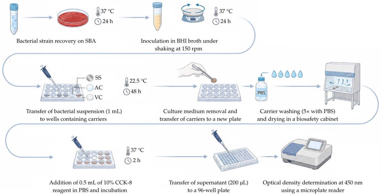
Figure 2.
Qualitative distribution of biofilm formation by S. hominis (n=54) on polystyrene, stainless steel, acrylic, and vinyl coating carriers using Equations 1 and 2. PO: polystyrene; SS: stainless steel; AC: acrylic; VC: vinyl coating; E1: Equation 1; E2: Equation 2. Note: Evaluation on the PO surface was performed using crystal violet, and on SS, AC, and VC surfaces was used CCK-8.
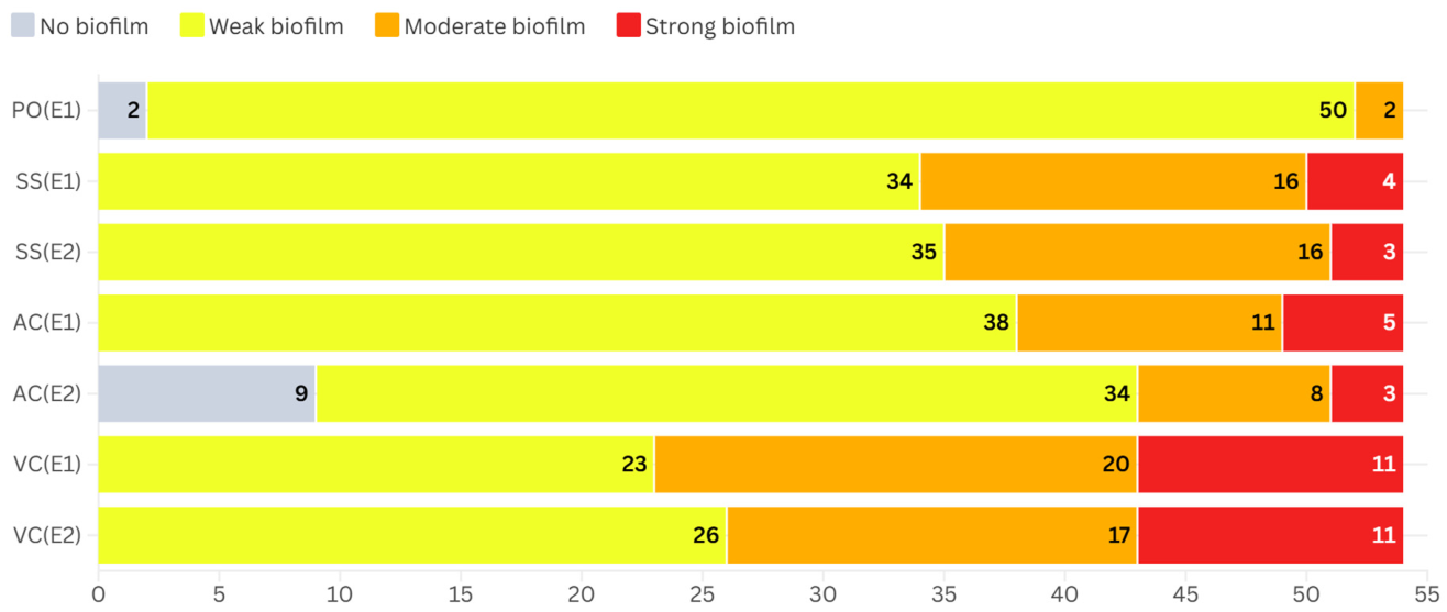
Figure 3.
Boxplot comparison of percentage values of biofilm formation by S. hominis (n=54) across the three different surfaces and the two equations evaluated with the CCK-8 assay. The central line indicates the median, while the box edges represent the first and third quartiles (interquartile range, IQR). Whiskers extend to the 1.5x IQR, and individual data points are plotted outside the whiskers as outliers. SS: stainless steel; AC: acrylic; VC: vinyl coating; E: equation.
Figure 4.
Biofilm formation percentage using Equation 4 (%) by the three strains that showed the maximum biofilm formation in each surface tested (B7636, B86.17, and B608.20). Each radar plot represents an individual strain evaluated across the tested surfaces. SS: stainless steel; AC: acrylic; VC: vinyl coating.
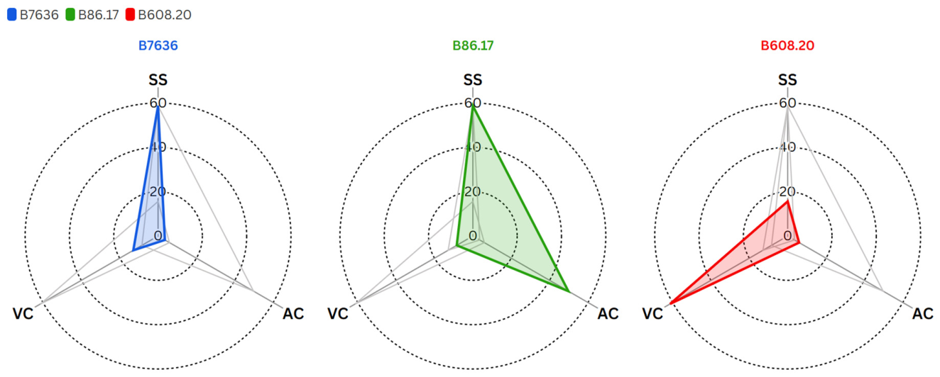

Table 1.
Descriptive statistical analysis of biofilm formation by S. hominis strains (n=54) using Equations 3 and 4, evaluated with CCK-8.
Table 1.
Descriptive statistical analysis of biofilm formation by S. hominis strains (n=54) using Equations 3 and 4, evaluated with CCK-8.
| Surface | Parameter (%) | Equation 3 | Equation 4 | t-test (p-value) |
|---|---|---|---|---|
| Stainless steel | Minimum | 0.80 | 0.59 | 0.93 |
| Maximum | 58.90 | 58.69 | ||
| Mean | 9.39 | 9.21 | ||
| Median | 5.25 | 5.09 | ||
| Standard deviation | 11.83 | 11.82 | ||
| Acrylic | Minimum | 0.10 | 0 | 0.47 |
| Maximum | 51.70 | 49.75 | ||
| Mean | 7.06 | 5.71 | ||
| Median | 2.55 | 1.35 | ||
| Standard deviation | 9.84 | 9.66 | ||
| Vinyl coating | Minimum | 2.30 | 1.50 | 0.80 |
| Maximum | 61.60 | 61.20 | ||
| Mean | 13.02 | 12.42 | ||
| Median | 8.75 | 8.05 | ||
| Standard deviation | 12.12 | 12.13 |
Disclaimer/Publisher’s Note: The statements, opinions and data contained in all publications are solely those of the individual author(s) and contributor(s) and not of MDPI and/or the editor(s). MDPI and/or the editor(s) disclaim responsibility for any injury to people or property resulting from any ideas, methods, instructions or products referred to in the content. |
© 2026 by the authors. Licensee MDPI, Basel, Switzerland. This article is an open access article distributed under the terms and conditions of the Creative Commons Attribution (CC BY) license (http://creativecommons.org/licenses/by/4.0/).
Copyright: This open access article is published under a Creative Commons CC BY 4.0 license, which permit the free download, distribution, and reuse, provided that the author and preprint are cited in any reuse.