

Submitted:
02 October 2026
Posted:
06 October 2026
You are already at the latest version
Abstract
Chromatin is dynamically folded into a hierarchical three-dimensional (3D) genome architecture that plays essential roles in gene regulation, DNA replication, and genome stability. Chromosome conformation capture (3C)-based technologies, particularly Hi-C and Micro-C, integrate chromatin fragmentation, proximity ligation, and high-throughput sequencing to map genome-wide chromatin interactions and have greatly advanced our understanding of spatial genome organization. These approaches have revealed three major levels of chromatin architecture: chromatin compartments, topologically associating domains (TADs), and chromatin loops, enhancer-promoter (E-P) interactions are frequently constrained within TADs, providing a structural framework for precise transcriptional control. Key architectural proteins, including CCCTC-binding factor (CTCF), cohesin, and the Mediator complex, are central to the establishment and maintenance of chromatin loops and TAD boundaries. In addition, lineage-specific transcription factors contribute to the dynamic organization of chromatin and the regulation of gene expression in a tissue- and developmental stage-specific manner. Genomic structural variations (SVs), including deletions, duplications, inversions, translocations, and complex chromosomal rearrangements, can disrupt TAD boundaries, alter chromatin looping and E-P interactions, and rewire higher-order genome organization, thereby promoting tumor initiation and progression. In this review, we summarize major advances in understanding 3D genome architecture, discuss current models and molecular mechanisms of chromatin folding, review chromatin interaction technologies, and highlight emerging links among structural genome variation, architectural protein dysfunction, chromatin dysregulation, and cancer development.
Keywords:
three-dimensional (3D) genome architecture
; topologically associating domains (TADs)
; enhancer-promoter (E-P) interactions
; architectural proteins
; Hi-C
; Micro-C
; Single-cell 3D genomics
; structural variants
; cancer
1. Introduction
The three-dimensional (3D) organization of chromosomes plays a critical role in nuclear compartmentalization and in bringing distal genomic elements into close spatial proximity. Understanding how chromosomes fold provides important insights into the relationships among chromatin architecture, gene regulation, cellular function, disease pathogenesis, and tumorigenesis. Recent advances in computational modeling, machine learning, high-resolution chromatin interaction technologies, and imaging-based approaches have further enabled multiscale characterization of genome organization and its functional consequences, providing an increasingly integrated view of genome structure and function [1].
Over the past two decades, advances in chromosome conformation capture (3C)-based technologies have transformed the study of genome organization and revealed fundamental principles governing chromatin folding, enhancer-promoter (E-P) connection, and long-range gene regulation [2,3,4,5,6,7,8,9,10]. These studies have demonstrated that the genome is organized into a hierarchy of structural features, including chromatin compartments, topologically associating domains (TADs), and chromatin loops [11,12,13,14,15]. TADs and chro-matin loops facilitate physical interactions between gene promoters and distal regulatory elements, including enhancers, silencers, and insulators, thereby providing a structural framework for transcriptional regulation [4,16,17].
These organizational features are established and maintained through the coordinated actions of architectural proteins, transcription factors, chromatin regulators, and transcriptional machinery. In particular, CCCTC-binding factor (CTCF), cohesin, Mediator, RNA polymerase II (RNAPII), and lineage-specific transcription factors contribute to chromatin looping, TAD formation, and E-P communication [15,18,19,20,21]. Histone modifications and chromatin-associated protein complexes further influence chromatin accessibility, compartmentalization, and higher-order genome organization [22,23,24,25].
Despite substantial progress, many fundamental questions remain unresolved. The molecular mechanisms governing TAD formation, maintenance, and dynamic chromatin interactions are incompletely understood, and the relationship between chromatin architecture, gene expression regulation, and cellular function remains an active area of investigation [14,15,26]. Increasing evidence indicates that disruption of chromatin loops, TAD boundaries, and regulatory interactions represents an important mechanism by which genetic and epigenetic alterations contribute to human disease, particularly cancer [27,28,29,30].
Recent studies have demonstrated that genomic structural variants (SVs), including deletions, duplications, inversions, translocations, and complex chromosomal rearrangements, can rewire chromatin interactions, alter E-P communication, and reshape higher-order genome architecture [29,30,31,32]. In addition, alteration of histone modifications at regulatory elements [22,23,33] and mutations affecting chromatin architectural proteins can disrupt chromatin insulation and gene regulation, thereby contributing to oncogenic transformation and disease progression [28,34,35].
In this Review, we summarize current understanding of the hierarchical organization of the 3D genome and the molecular mechanisms governing chromatin folding. We then review Hi-C/Micro-C based chromatin interaction technologies and their contributions to the study of genome architecture. Finally, we discuss how alterations in chromatin organizations contribute to cancer development and highlight emerging applications of high-resolution and single-cell 3D genome technologies in biological and clinical research.
2. Principles of 3D Genome Architecture
Chromatin is folded into dynamic 3D structures that regulate gene expression, DNA replication, and genome stability [36,37] (Figure 1). At the nuclear scale, chromosomes occupy distinct chromosome territories and interact with nuclear structures such as nuclear lamina, contributing to higher-order genome organization [38,39], which is hierarchical and operates across multiple spatial scales. At the megabase level, analysis of Hi-C contact maps identifies genomic regions that segregate into transcriptionally active and inactive chromatin compartments. These compartments are established by a combination of chromatin-binding proteins, chromatin state-dependent mechanisms and are closely associated with distinct epigenetic landscapes. Active A compartments are enriched for transcription associated features and active histone modifications, including H3K27ac, H3K4me1, and H3K4me3, whereas repressive compartmental domains are characterized by enrichment of H3K27me3 and H3K9me3, which mark facultative and constitutive heterochromatin, respectively [4,33]. Within compartments, chromatin is further organized into topologically associating domains (TADs), which function as structural and regulatory units of the genome [11,12,13]. TAD boundaries are enriched for architectural proteins such as CTCF and cohesin and restrict interactions between adjacent domains [15,19,26]. By constraining E-P communication within domains, TADs provide an important mechanism for regulatory specificity [27,40]. Most E-P interactions occur within TADs and are limited by TAD boundaries [26,27]. Super-enhancers that regulate cell identity genes and oncogenes are frequently insulated within TADs, and disease-associated genetic variants are enriched within these regulatory regions [41]. Notably, TAD architecture is highly conserved across tissues and species [11,15].
Chromatin loops represent another major level of genome organization. High-resolution in situ Hi-C studies identified thousands of chromatin loops in the human genome, many of which involve multiple loop anchors and connect promoters with distal regulatory elements at kilobase-resolution [4]. Some loops are structural loops, display an invariable nature across cells or tissues and are frequently bound by CTCF, an architectural protein that has a key role in chromatin folding and TAD boundary formation [16,17,18,42]. By contrast, transcriptional loop associated with E-P interactions that are bound by Mediator and the cohesin complex, displaying a dynamic and tissue- specific nature. Chromatin looping also plays an important role in bridging gene promoters with cis-regulatory elements (CREs) and other functional elements mediated by transcription factors (TFs) or RNAPII [43,44]. These looping interactions provide a structural basis for long-range transcriptional regulation [45].
Recent high-resolution methods have revealed additional layers of chromatin organization beyond classical compartments, TADs, and chromatin loops. Region Capture Micro-C (RCMC) identified highly nested chromatin interaction patterns, known as microcompartments, which are enriched for E-P interactions and local regulatory contacts [46], whereas single-cell Micro-C(scMicro-C) uncovered multi-enhancer hubs on genes with promoter-enhancer stripes (PES) within single-cell 3D genome structures, wherein multiple enhancers form a spatial cluster in association with the gene promoter within individual cells [10].
Collectively, these findings support a hierarchical model of chromatin organization consisting of chromo-some territories [38], chromatin compartments, TADs, chromatin loops [4,10,11,38,46], and emerging higher-order regulatory structures such as microcompartments [46], and multi-enhancer hubs [10]. Such structures may provide a mechanism for robust and coordinated gene regulation while maintaining regulatory specificity. These structures are functionally interconnected and dynamically regulated by architectural proteins, transcription factors, chromatin states, and transcriptional activity [14,15].
3. Molecular Mechanisms of 3D Genome Architecture and Gene Expression
Current evidence supports three major interconnected mechanisms underlying chromatin architecture and associated gene expression: (1) cohesin-mediated loop extrusion and chromatin insulation by CTCF, (2) transcription factor- and co-factor- mediated chromatin interactions, and (3) chromatin-state-dependent organization driven by histone modifications and associated protein complexes (Figure 2).
3.1. Architectural Proteins and Loop Extrusion
CTCF and cohesin are the principal architectural regulators of chromatin organization [19,20,47,48]. Together, they establish chromatin loops and TAD boundaries, creating a structural framework that partitions the genome into discrete regulatory domains. Genome-wide depletion studies have demonstrated that cohesin functions as the primary driver of loop formation through the process of loop extrusion, whereas CTCF serves as a directional boundary element that positions and stabilizes cohesin at specific genomic sites [20,49,50].
CTCF is a highly conserved zinc-finger DNA-binding protein that recognizes asymmetric DNA motifs enriched at loop anchors and domain boundaries [15,42]. More than 90% of CTCF-mediated loops occur between convergently oriented motifs, highlighting the importance of motif orientation in loop formation [4]. CTCF functions as an orientation-dependent barrier to cohesion-mediated loop extrusion, with CTCF polarity serving as a key determinant of loop anchor positioning and chromatin boundary formation [51].
In addition to its architectural role, CTCF acts as a genomic insulator and is essential for the establishment of chromatin domains, long-range regulatory interactions, and higher-order nuclear architecture [18,42,52]. Recent studies have further shown that CTCF promotes the assembly of multi-component chromatin hubs that integrate enhancers, promoters, and CTCF-binding sites into cooperative regulatory networks. However, the limited effect of CTCF depletion on gene expression suggests that E-P interactions in gene regulation may be independent of cooperative interactions in chromatin hubs [53,54].
Cohesin is a ring-shaped Structural Maintenance of Chromosomes (SMC) complex composed of SMC1, SMC3, RAD21, and STAG proteins that encircle chromatin fibers and promote loop extrusion [19,55]. Genetic or experimental depletion of cohesin, CTCF, or associated regulators causes widespread disruption of chromatin loops and TAD organization [20,49,56]. Loss of chromatin insulation can alter regulatory interactions between enhancers and promoters, demonstrating the importance of architectural proteins in maintaining proper gene regulation [28].
Additional factors contribute to cohesin loading, loop stabilization, chromatin boundary formation, and transcriptional regulation. Nipped-B-like protein (NIPBL) load cohesin onto chromatin, allowing cohesin to perform its functions in chromosome organization, gene regulation, DNA repair, and sister chromatid cohesion [57], while a protein ring complex named wrings apart-like protein (WAPL) regulates cohesin residence time on chromatin and restricts loop extension, thereby limiting the size and stability of chro-matin loops [48]. YY1 can facilitate E-P interactions and has been proposed to function as a structural regulator of regulatory loops [21,31]. Other factors, including TOP2B, WIZ, and BRD2, cooperate with cohesin and CTCF at chromatin domain boundaries and contribute to the maintenance of higher-order genome organization [58,59,60]. ZNF143 has also been implicated in chromatin organization, although recent evidence suggests that its primary function may be transcriptional regulation rather than direct participation in loop formation [61,62]. Super-enhancers are recognized, bound and activated by BRD4, resulting in considerable oncogene over-expression, malignant transformation, cancer cell proliferation, survival, tumor initiation and progression [63].
While classical loop extrusion models (Figure 2A) often assume relatively stable chromatin boundaries, recent computational and experimental studies indicate that boundary elements are highly dynamic. Modeling analyses have shown that transient CTCF occupancy can nevertheless generate robust loops and domain insulation when considered in the context of longer cohesin residence times on chromatin [64]. These findings provide a mechanistic explanation for how dynamic protein-DNA interactions can give rise to stable population-level patterns of genome organization and suggest that chromatin architecture emerges from the collective behavior of continuously assembling and disassembling molecular complexes.
3.2. Transcription Factor- Cofactors- Mediated Chromatin Interactions
While CTCF and cohesin establish the architectural framework of genome organization, transcription factors (TFs) drive cell type-specific chromatin interactions that are intimately linked to gene regulation. These interactions connect distal regulatory elements to target promoters and coordinate developmental and lineage-specific transcriptional programs [16,17,45]. One of the best-characterized examples is the β-globin locus, where erythroid-specific TFs mediate interactions between the locus control region and β-globin genes, enabling developmental regulation through long-range chromatin looping [45]. Similar mechanisms have been observed at numerous loci, including the CFTR locus and other lineage-specific regulatory regions, where E-P communication is shaped by local chromatin architecture [17,40]. TFs also play pivotal roles during cell-fate transitions. During somatic cell reprogramming, pioneer factors such as OCT4, SOX2, KLF4, and c-MYC bind previously inaccessible regulatory elements and initiate large-scale chromatin reorganization before widespread transcriptional changes occur. These findings suggest that TFs actively establish new regulatory landscapes rather than simply activating pre-existing gene expression programs [65].
Recent studies have further demonstrated that E-P interactions are not solely determined by stable chromatin loops but are dynamically modulated by transcriptional activity. High-resolution Micro-C analyses revealed that functional E-P pairs remain in close spatial proximity for longer periods than nonfunctional pairs and that RNAPII plays a direct role in stabilizing these contacts. Notably, promoter-proximal paused RNAPII appears to reinforce E-P communication, underscoring the intimate relationship between transcription and three-dimensional genome organization [66]. Consistent with this model, RNAPII has emerged as an important contributor to chromatin architecture. Acute RNAPII depletion disrupts subsets of E-P interactions and alters local chromatin organization, suggesting that the transcriptional machinery not only responds to genome structure but also actively participates in shaping it [67].
Beyond individual E-P contacts, TFs cooperate with Mediator, BRD4 [63], RNAPII, cohesin, and other coactivators to establish higher-order regulatory assemblies that facilitate robust transcriptional activation [21,46,66]. RCMC recently identified highly nested chromatin interaction structures termed microcompartments, many of which connect enhancers and promoters. Notably, a substantial fraction of these interactions persists following acute transcriptional inhibition or disruption of loop extrusion, suggesting that compartmentalization-based mechanisms can contribute to E-P communication independently of classical looping models [46].
scMicro-C [10] studies have further refined our understanding of E-P architecture. High-resolution maps revealed promoter-enhancer stripes (PESs), cohesin-dependent interaction patterns that link individual promoters with multiple distal enhancers. Within single-cell 3D genome, there are multiple enhancers form a spatial cluster in association with the gene promoter, spatially coordinated to control genes. These analyses also uncovered widespread multi-enhancer hubs, higher-order three-dimensional structures in which multiple enhancers simultaneously converge on a single target promoter within individual cells. Such assemblies provide a potential mechanistic basis for cooperative function and coordinated gene ex-pression [10].
Collectively, these findings support an emerging model in which TFs function within highly dynamic 3D regulatory networks. Rather than acting through simple pairwise enhancer-promoter loops, TFs collaborate with RNAPII, Mediator, chromatin regulators, and architectural proteins to establish diverse higher-order regulatory structures, including transcriptional hubs, microcompartments enhancer, promoter-enhancer stripes, and multi-enhancer assemblies (Figure 1F, Figure 2B). Together, these interconnected mechanisms enable distal regulatory elements to exert precise and robust control of gene expression during development, differentiation, and disease.
3.3. Histone Modifications and Chromatin-State-Dependent Organization
Histone modifications provide an important regulatory layer linking chromatin state to 3D genome architecture (Figure 2C). Active enhancer marks such as H3K4me1 and H3K27ac are closely associated with enhancer activity and E-P interactions. H3K4me1, deposited primarily by MLL3/MLL4 complexes, marks primed and active enhancers, has been shown to facilitate enhancer activation and promote enhancer-associated chromatin contacts that activate gene expression [22,23]. H3K4me1-enriched enhancers recruit transcriptional co-regulators, including Mediator and Cohesin complexes, which facilitate chromatin looping between enhancers and their target promoters [4,22]. H3K27ac, catalyzed by histone acetyl transferase p300/CBP, is a hallmark of active enhancers and is strongly correlated with transcriptional activation [23]. These active chromatin states are frequently associated with long-range regulatory interactions and cell type-specific gene expression programs [66,68].
At gene promoters, H3K4me3 is strongly linked to RNAPII occupancy and active transcription. Genome-wide chromatin interaction analyses have revealed extensive E-P contacts among transcriptionally active loci, indicating that active histone modifications contribute to the formation of transcriptionally active chromatin environments [17,69]. Moreover, perturbation of RNAPII activity alters subsets of E-P interactions, suggesting a reciprocal relationship between transcription and chromatin architecture [66,67].
In contrast, repressive histone modifications contribute to the formation of silenced chromatin domains. H3K27me3 facilitates long-range interactions among transcriptionally silenced genomic regions through Polycomb-group complexes [24,25]. Polycomb-associated chromatin interactions contribute to developmental gene repression and help establish spatially segregated repressive chromatin domains. Disruption of Polycomb-mediated interactions can alter chromatin architecture and developmental gene expression programs [24,25]. Similarly, G9a-mediated H3K9me2 contributes to higher-order genome organization, with H3K9me2 patterns showing strong compartment-specific distributions and influencing A/B compartmentalization [70].
Beyond their roles in regulating individual enhancers and promoters, histone modifications also contribute to higher-order genome organization by promoting preferential interactions among genomic regions with similar epigenetic states. Genome-wide chromatin interaction studies have shown that chromatin compartments are closely associated with distinct histone modification patterns. Active A chromatin compartments are enriched for transcription-associated marks such as H3K27ac, whereas repressive compart-mental domains are characterized by H3K27me3 or H3K9me3 enrichment. In contrast, H3K4 methylation marks display weaker correlations with compartment identity despite their critical roles in enhancer and promoter regulation [33].
Consistent with this model, genomic regions bearing similar chromatin signatures preferentially interact in nuclear space: active regions enriched for H3K4me1, H3K27ac, and H3K4me3 cluster together, whereas Polycomb-repressed and heterochromatic domains marked by H3K27me3 or H3K9 methylation exhibit strong self-association [3,14]. These epigenetic interaction preferences contribute to chromatin compartmentalization and promote the spatial segregation of active and inactive chromatin domains, thereby linking local chromatin states to global genome organization [14,15].
3.4. An Integrated 3D Regulatory Network Enables Precise Control of Gene Expression
Current evidence supports a hierarchical model in which multiple regulatory mechanisms cooperate across different spatial scales. Cohesin-mediated loop extrusion and CTCF-dependent insulation establish chro-matin loops and TADs [49,50,56]. Within this architectural framework, transcription factors, Mediator, BRD4, and RNAPII promote cell type-specific E-P interactions and lineage-specific transcriptional pro-grams [10,21,46,66,67]. Histone modifications and chromatin states further influence chromatin accessibility, compartmentalization, interaction stability, and large-scale genome organization [22,23,24,25]
These regulatory layers are highly interconnected. Structural organization constrains regulatory interactions, whereas transcriptional activity and chromatin state can reinforce or remodel chromatin architecture. Together, architectural proteins, transcription factors, chromatin modifiers, and transcriptional machinery form an integrated regulatory network that dynamically shapes genome organization during development, differentiation, and disease [14,15].
4. Chromosome Conformation Capture Technologies
The development of 3C -based technologies has revolutionized the study of 3D genome organizations. Since the introduction of the original 3C method, successive technological advances have progressively increased the scale, resolution, specificity, and sensitivity of chromatin interaction mapping. Collectively, approaches including 3C [2], 4C ([71,72], 5C [73], Hi-C [3], ChIA-PET [74], PLAC-seq [75], HiChIP [9], Capture-C [76], Capture Hi-C [43,77], Micro-C [5,6,7],Micro-Capture-C (MCC) [44], Micro-C-ChIP [78], and single-cell chromatin interaction technologies [8,10] etc. have provided unprecedented insights into chromatin compartments, topologically associating domains (TADs), chromatin loops, E-P networks, and higher-order regulatory structures [4,5,6,9,10,74,78] (Table 1).
4.1. Preselected Locus-Based Chromatin Interaction Analysis
The original 3C method established the principle of proximity ligation for studying physical interactions between genomic loci [2]. In this approach, chromatin is crosslinked, digested, and ligated under diluted conditions so that spatially proximal DNA fragments become joined and can subsequently be detected by PCR. Although highly informative, 3C is limited to pre-selected genomic interactions.
To extend chromatin interaction analysis beyond individual loci, several derivative methods were developed. 4C (“circular 3C” [72] or “3C-on-Chip” [71]) uses inverse PCR to generate genome-wide interaction profiles for single loci. 4C has made it possible to detect chromatin interactions between one locus and multiple loci, enables genome-wide interrogation of interactions involving a single locus ("one-versus-all") [71,72]. 5C can capture multiple-to-multiple loci interactions, has allowed more comprehensive analysis of chromatin looping for large numbers of genes by measuring many anchored interaction profiles in parallel ("many-versus-many") [73]. These methods focus on interrogation of 3D interactions between specific preselected fragments. By preselecting regions to be queried, we were able to obtain insight into chromatin architecture at the resolution of single interrogated fragment.
4.2. Genome-Wide Approaches for 3D Genome Analysis
A major breakthrough occurred with the development of Hi-C, which enabled unbiased genome-wide analysis of chromatin interactions through the combination of proximity ligation and high-throughput sequencing [3]. Hi-C studies led to the discovery of A/B chromatin compartments and provided the first genome-wide framework for understanding higher-order genome organization [3]. Subsequent Hi-C studies contributed to the identification and characterization of TADs and long-range chromatin interactions [11,12]. The development of in situ Hi-C substantially improved data quality by performing digestion and ligation within intact nuclei, reducing random ligation events and enabling kilobase-resolution chromatin interaction maps [4]. These studies facilitated systematic identification of chromatin loops and provided key insights into E-P communication and chromosome folding principles [4].
Although Hi-C transformed genome architecture research, its resolution is constrained by the distribution of restriction enzyme recognition sites. To overcome this limitation, Micro-C was developed using micrococcal nuclease (MNase), which digests linker DNA between nucleosomes independently of DNA sequence [5]. By generating mono-nucleosome-sized fragments, Micro-C enables chromatin interaction mapping at near-nucleosome resolution while simultaneously providing information on nucleosome positioning [5,6]. Compared with conventional Hi-C, Micro-C identifies substantially more chromatin loops and resolves fine-scale chromatin features that are often inaccessible to restriction enzyme-based approaches [6,7]. Subsequent development of Micro-C XL, which incorporates enhanced crosslinking and chromatin isolation strategies, further improved signal quality and interaction recovery [79]. Applications across yeast, mammalian, and plant systems have demonstrated the unique ability of Micro-C-based methods to uncover fine-scale chromatin structures and regulatory interactions [5,79,80].
4.3. Enrichment-Based Approaches for 3D Genome Analysis
Although Hi-C and Micro-C provide comprehensive, genome-wide maps of chromatin interactions, capturing both structural features arising from polymer dynamics and functionally relevant contacts within the nuclear space of millions of cells, they often require extremely deep sequencing depth for detecting chromatin loops, and they lack locus specificity and do not directly distinguish functional regulatory interactions from structural chromatin contacts. To address these limitations, genomic locus- and regulatory element-capture approaches have been developed, in which chromatin contacts associated with selected genomic regions are enriched through probe hybridization. These methods enable more focused characterization of interactions involving specific gene promoters, enhancers, and other distal CREs. In parallel, protein-specific chromatin interaction approaches have been established to enrich chromatin contacts associated with specific histone modifications, particular DNA-binding proteins, regulatory complexes, thereby improving the specificity and detection of functionally relevant chromatin interactions, albeit at the expense of genome-wide coverage and comprehensiveness [81].
4.3.1. Targeted Chromatin Interaction Capture Methods
To enrich the chromatin interaction fragments from genomic regions of interest, several capture-based chromatin conformation technologies have been developed to enable high-resolution interrogation of selected genomic regions, including Targeted Chromatin Capture (T2C) [82], Capture-C [76], 3C-based multitarget sequencing (3C-MTS) [83,84], Promoter Capture Hi-C [43], Enhancer Capture Hi-C [77], Micro-Capture-C (MCC) [44], and Region Capture Micro-C (RCMC) [46]. These approaches combine chromosome conformation assays with hybridization-based enrichment of selected genomic loci using biotinylated capture probes, enabling targeted recovery of ligation products associated with regions of interest. Compared with untargeted genome-wide methods, capture-based approaches substantially improve the detection sensitivity, sequencing depth and effective resolution of local chromatin interaction maps.
Among these methods, Promoter-capture Hi-C enriches chromatin contacts involving gene promoters, enabling identification of transcriptional chromatin loops connecting gene promoters to the distal CREs, including enhancers, silencers and insulators, that regulate gene express [43]. Enhancer Capture Hi-C similarly focuses on enhancer-associated interaction networks, facilitating the characterization of enhancer connectivity and target-gene regulation [77]. MCC combines the high resolution of Micro-C with targeted capture strategies, allowing physical contact between different classes of regulatory elements to be determined at base-pair resolution. This method can identify regions of interacting DNA that are far apart in the linear genome sequence, such as enhancers and gene promoters [44]. RCMC has further increased the sensitivity and resolution of chromatin interaction mapping within selected genomic regions, enabling the detection of highly nested regulatory architectures that are difficult to resolve using conventional genome-wide approaches [46].
Capture-based technologies have proven particularly valuable for studying enhancer-promoter (E-P) interactions, disease-associated loci, genome-wide association study (GWAS) variants, and complex regula-tory landscapes [43,76]. Notably, RCMC has revealed enhancer-promoter microcompartments and intricate local interaction networks that were previously inaccessible using standard Hi-C and Micro-C methods, providing new insights into the fine-scale organization of gene regulatory domains [46].
4.3.2. Protein-Specific Enrichment Approaches
To characterize chromatin interactions mediated by specific regulatory factors, including architectural proteins such as CTCF and cohesion, transcription factors, RNAPII, and epigenetically modified histones (e.g., H3K4me3 and H3K27ac), Several protein-mediated chromatin conformation technologies have been developed. Chromatin Interaction Analysis by Paired-End Tag Sequencing (ChIA-PET) was the first widely adopted method for mapping genome-wide chromatin interactions associated with specific proteins. By combining chromatin immunoprecipitation (ChIP) with chromatin proximity ligation, ChIA-PET enables the identification of long-range interactions mediated by DNA-binding proteins and chromatin regulators [74,85].
Subsequently, Proximity Ligation-Assisted ChIP Sequencing (PLAC)-seq and HiChIP were developed to improve efficiency, sensitivity, and cost-effectiveness of protein-mediated chromatin interactions mapping [9,75]. Unlike ChIA-PET, which relies on chromatin fragmentation by sonication, HiChIP uses restriction enzyme digestion and Hi-C-based proximity ligation prior to immunoprecipitation. PLAC-seq provides substantially improved efficiency and accuracy compared with ChIA-PET, whereas HiChIP in-creases the yield of informative interactions reads by over 10-fold and reduces the sample input requirement by over 100-fold [9,75]. As a result, HiChIP generates substantially more informative chromatin interaction reads while requiring fewer cells [9].
These approaches have been instrumental in defining chromatin interactions associated with CTCF, cohesin, RNAPII, enhancer-associated histone modifications, and lineage-specific transcription factors, thereby providing critical insights into the regulatory mechanisms governing genome architecture and gene expression [9,17,75]. More recently, Micro-C-ChIP has combined the nucleosome-resolution advantages of Micro-C with protein-specific enrichment strategies, enabling high-resolution characterization of chroma-tin interactions associated with CTCF, cohesin, RNAPII, transcription factors, and chromatin regulatory complexes [78]. Together, these protein-mediated methods provide powerful tools for dissecting the molecular mechanisms through which specific regulatory proteins shape chromatin architecture and control gene expression.
4.4. Single-Cell Analysis of Genome Architecture
Most chromatin conformation studies initially relied on bulk cell populations and therefore measured population-average interaction frequencies. While informative, such analyses cannot fully resolve cellular heterogeneity or dynamic aspects of genome organization. To address these limitations, single-cell Hi-C (scHi-C) technologies were developed to directly measure chromatin interactions in individual cells [8,86,87,88]. These studies demonstrated substantial cell-to-cell variability in chromosome folding, highlighting the dynamic and probabilistic nature of genome organization [8,89].
More recently, single-cell chromatin conformation approaches have achieved increasingly high spatial resolution. Single-cell Micro-C (scMicro-C) extended the principles of Micro-C to individual cells and enabled high-resolution analysis of chromatin architecture [10]. Application of scMicro-C revealed previously unrecognized structures, including E-P stripes and multi-enhancer hubs, in which multiple enhancers simultaneously interact with a target promoter within individual cells [10]. These findings provide new insights into E-P communication and transcriptional regulatory networks at single-cell resolution.
The integration of chromatin interaction mapping with transcriptomic, epigenomic, imaging, perturbational, and genome-editing approaches is progressively narrowing the gap between structural and functional genomics. Recent advances in Micro-C, capture technologies, protein-specific enrichment assays, and single-cell platforms now enable investigation of chromatin architecture across multiple biological scales, from individual nucleosomes to entire chromosomes [6,10,46].
As sequencing costs decline and multimodal profiling becomes increasingly accessible, future technologies are expected to provide even more comprehensive views of genome organization across diverse cell types, developmental states, and disease contexts. These advances will continue to deepen our understanding of the molecular mechanisms linking genome topology to gene regulation, development, and disease and are likely to play increasingly important roles in functional genomics, precision medicine, and cancer biology.
5. Genomic Structural Variations, 3D Genome Architecture, and Cancer
Cancer arises through the progressive accumulation of genetic and epigenetic alterations that disrupt normal cellular homeostasis and drive malignant transformation. Large-scale cancer genome studies, including those conducted by The Cancer Genome Atlas (TCGA), have revealed widespread somatic mutations, copy number alterations, and structural variants (SVs) across human tumors [90,91]. These alterations include single-nucleotide variants (SNVs), insertions, deletions, inversions, duplications, translocations, and complex chromosomal rearrangements [90,92].
Beyond altering gene sequence or copy number, these genomic lesions frequently disrupt higher-order chromatin organization. 3D genome architecture plays a fundamental role in regulating gene expression, preserving genome stability, and establishing cell type-specific transcriptional programs [14,15]. As a result, cancer-associated genomic alterations frequently exert their pathogenic effects by reshaping chromatin compartments, TADs, chromatin loops, and E-P interaction networks. Such architectural changes can activate oncogenic enhancers and super-enhancers, promote aberrant E-P communication, and drive large-scale transcriptional reprogramming that supports malignant cellular states [29,41].
In addition to directly affecting regulatory DNA elements, cancer-associated chromatin remodeling may arise through disruption of architectural proteins such as CTCF and cohesin, alterations in chromatin looping dynamics, and reorganization of TAD boundaries. Advances in chromosome conformation technologies, including Hi-C, HiChIP, Capture Hi-C, Micro-C, and single-cell chromatin conformation etc. approaches, have revealed widespread genome architectural remodeling across diverse tumor types, highlighting genome topology as a critical and previously underappreciated contributor to tumorigenesis [93,94,95] (Figure 3).
5.1. Structural Variants Reshape 3D Genome Architecture
Recent advances in cancer genome sequencing and chromatin conformation mapping demonstrate that structural variants in cancers can directly remodel 3D genome architecture. SVs can disrupt genome organization across multiple hierarchical levels thereby affecting gene expression and cellular function. Abnormal gene expression patterns can distort cell fate and ultimately lead to cancer. Studies in breast cancer [93], prostate cancer [94], glioma [28], T cell acute lymphoblastic leukemia (T-ALL) [29], medulloblastoma [32], and multiple myeloma [96], Colon cancer [95], gliomas [28] etc. have revealed ex-tensive alterations in chromatin compartmentalization, TAD organization, and chromatin looping relative to normal tissues. For example, multiple myeloma cells exhibit widespread 3D genome reorganization associated with copy number variations (CNVs), translocations, altered TAD boundaries, A/B compartment switching, and gene expression changes [96,97]. As a result, genome topology has emerged as a critical framework for understanding how structural alterations contribute to both developmental disorders and cancer [98,99].
SVs influence genome topology through diverse mechanisms. Deletions that remove CTCF- or cohesin-anchored TAD boundaries can abolish chromatin insulation, resulting in TAD fusion and ectopic E-P communication. A classic example is the EPHA4 locus, where disruption of TAD boundaries enables pathogenic enhancer adoption by neighboring genes [27]. Similarly, loss of CTCF at a domain boundary permits a constitutive enhancer to interact aberrantly with the receptor tyrosine kinase gene PDGFRA, a prominent glioma oncogene [28].
Inversions can reorient CTCF-binding motifs, reposition enhancers, and alter loop-extrusion dynamics, leading to the establishment of novel chromatin contacts. Experimental inversion of CTCF-binding sites has been shown to remodel chromatin topology and alter E-P communication [100]. In medulloblastoma, chromosomal rearrangements occurring near domain boundaries promote enhancer hijacking and aberrant activation of oncogenic transcriptional programs [32,101]. Likewise, tandem duplications can amplify enhancers, super-enhancers, or entire regulatory domains, increase regulatory contact frequencies and reinforce oncogene expression.
Chromosomal translocations represent another important class of topology-altering events. By repositioning enhancers and promoters into new genomic contexts, translocations can create aberrant chromatin interaction networks that drive oncogenic transcription. Well-established examples include immunoglobulin enhancer-mediated activation of proto-oncogenes in hematologic malignancies, where highly active distal regulatory elements become juxtaposed to oncogenes and induce constitutive expression [29,30].
More complex forms of SVs can drive extensive reorganization of genome architecture. Neo-TAD formation occurs when genomic rearrangements generate novel chromatin domains that bring genes into contact with previously inaccessible regulatory elements [27,30]. An even more dramatic form of genome remodeling, chromothripsis, involves catastrophic chromosome shattering and reassembly in a single event, generating highly complex rearrangements that lead to widespread disruption of TADs, chromatin loops, compartments, and regulatory landscapes across large genomic regions [102,103].
Recent studies have also highlighted the contribution of extrachromosomal DNA (ecDNA) to cancer genome organization [104,105]. ecDNA molecules frequently harbor oncogenes together with enhancer elements and can assemble highly interconnected regulatory hubs that drive exceptionally high levels of oncogene expression. These structures are increasingly recognized as important contributors to tumor evolution, intratumoral heterogeneity, and therapeutic resistance [106].
5.2. Rewiring of Long-Range Chromatin Interactions in Cancer
While SVs can remodel genome architecture at multiple hierarchical levels, their pathogenic effects are often mediated through the rewiring of long-range chromatin interactions. E-P communication is essential for precise control of gene expression, and disruption of these regulatory networks can result in aberrant oncogene activation, tumor suppressor silencing, and widespread transcriptional dysregulation. Consequently, altered chromatin interactions have emerged as a common mechanism linking genome architectural changes to cancer development.
One of the most prominent mechanisms of chromatin interaction rewiring is enhancer hijacking, whereby genomic rearrangements reposition potent enhancers or super-enhancers adjacent to oncogenes. Such events have been reported for PRDM6, GFI1, GFI1B, and other oncogenic drivers in multiple cancer [31,32]. These rearrangements frequently occur near TAD boundaries and generate novel E-P contacts that bypass normal regulatory constraints [27,30].
Long-range chromatin interactions also play a critical role in mediating the effects of noncoding cancer-associated variants. One of the best-characterized examples is the chromosome 8q24 cancer susceptibility locus, where chromatin interaction studies demonstrated that this noncoding risk region physically interacts with the MYC proto-oncogene through long-range chromatin looping in colorectal cancer cells [107]. Subsequent analyses identified multiple enhancers within the 8q24 interval that regulate MYC expression through direct E-P interactions [83,108]. Similar long-range regulatory mechanisms have been observed in colorectal, prostate, and breast cancers [109,110], underscoring the broad im-portance of 3D genome organization in translating noncoding genetic variation into oncogenic gene ex-pression programs.
In addition to genomic alterations, oncogenic transcription factors can actively affect long-range chroma-tin interactions. For example, overexpression of the prostate cancer-associated fusion transcription factor ERG induces local reorganization of chromatin interactions and transcriptional programs in prostate epithelial cells [111]. These observations demonstrate that cancer-associated oncogenic regulators can actively reshape genome topology to reinforce malignant cellular states.
Advances in Capture Hi-C and related chromatin conformation technologies have further demonstrated the utility of 3D genome analyses for interpreting cancer-associated genetic variation. Studies in breast, prostate, and colorectal cancers have successfully linked noncoding GWAS variants to their target genes, thereby clarifying the molecular mechanisms underlying disease susceptibility [84,112,113]. By connecting distal regulatory elements with their cognate target genes, these approaches have substantially ex-pended our understanding of the functional consequences of noncoding cancer risk variants.
Together, these studies demonstrate that rewiring long-range chromatin interactions represents a common mechanism by which both structural and nonstructural genomic alterations influence gene expression in cancer. The functional consequences of many cancer-associated variants can only be fully understood within the context of 3D genome organization. Beyond genetic variation, however, chromatin interaction networks also depend on architectural proteins that establish and maintain higher-order genome structure. Disruption of these regulatory factors represents another major mechanism of genome-topology dysregulation in cancer.
5.3. CTCF and Cohesin Dysregulation in Cancer
The organization of chromatin interactions depends on a network of architectural proteins that define chromatin boundaries and facilitate loop formation. Among these, CTCF and Cohesin are central regulators of TAD organization and higher-order genome architecture [15,19]. Accordingly, genetic or epigenetic perturbations affecting these factors can destabilize chromatin organization and promote widespread transcriptional dysregulation in cancer.
Mutations affecting CTCF, cohesin-complex components, and associated binding sites have been identified across a broad spectrum of human cancers [34,91]. Notably, CTCF-binding sites are recurrently mutated in colorectal cancer and other tumor types. CTCF-binding sites located at TAD boundaries are particularly enriched for somatic mutations, suggesting selective pressure for disruption of chromatin insulation during tumor evolution [29,34,114].
In addition to direct sequence mutations, epigenetic alterations can impair CTCF occupancy and boundary function. Loss or weakening of CTCF-mediated boundaries can permit aberrant E-P communication, leading to oncogene activation, altered lineage programs, and genomic instability [28,35]. Similarly, dysfunction of cohesin subunits and cohesion-associated factors can compromise loop extrusion, weaken TAD organization, and induce widespread transcriptional dysregulation [49,56]. Through these mechanisms, defects in architectural proteins can directly contribute to oncogenic transformation and disease progression.
The studies discussed above collectively support a unifying model in which diverse classes of cancer-associated alterations converge on the three-dimensional organization of the genome. Structural variants, noncoding regulatory mutations, epigenetic remodeling, and disruption of chromatin architectural proteins each influence chromatin folding and regulatory interactions at distinct levels. Despite their mechanistic differences, these alterations ultimately reconfigure gene regulatory networks that govern cellular identity and tumor progression. These alterations affect genome organization across multiple spatial scales, from local E-P interactions to large-scale chromatin domains and regulatory hubs, ultimately re-programming gene expressions and cellular identity [28,29,93,94]
Importantly, cancer is increasingly recognized as a disease of both genome sequence and genome architecture. While genomic mutations provide the substrate for malignant evolution, their phenotypic consequences are often mediated through alterations in chromatin topology and gene regulatory networks. Understanding how tumor genomes reorganize 3D chromatin structure therefore provides critical insight into the molecular mechanisms underlying tumorigenesis.
This emerging perspective also has important therapeutic implications. Targeting enhancer function, chromatin regulators, architectural proteins, and aberrant chromatin interactions may provide new opportunities for precision oncology and the development of therapies that specifically disrupt pathogenic regulatory circuits [30,31].
Collectively, these findings underscore the need to interpret structural variants within the context of 3D genome organization rather than solely through their effects on coding sequence. This framework is particularly relevant for understanding noncoding disease-associated variants, many of which exert their pathogenic effects through alterations in chromatin architecture, genome topology, and long-range regula-tory interactions [98].
6. Conclusion and Future Perspectives
Advances chromatin interaction technologies have fundamentally transformed our understanding of genome function by establishing 3D chromatin organization as a critical layer of gene regulation. Studies using Hi-C, Micro-C, and related approaches have revealed chromatin compartments, TADs, chromatin loops, and E-P interactions as key features of nuclear architecture that coordinate gene expression, maintain genome stability, and define cellular identity.
Accumulating evidence demonstrates that genome architecture is dynamically regulated through the coordinated activities of architectural proteins, transcription factors, chromatin regulators, and transcriptional machinery. Disruption of these regulatory systems by structural variation, epigenetic alterations, or mutations affecting chromatin architectural proteins can rewire chromatin interactions and regulatory networks, thereby contributing to cancer initiation, progression, and therapeutic resistance. These discoveries have expanded the traditional view of cancer from a disease driven solely by genetic alterations to one that also encompasses aberrant genome topology and dysregulated chromatin architecture.
Recent developments in single-cell and multimodal technologies have further accelerated the field by enabling high-resolution characterization of chromatin organization within heterogeneous cellular populations. These approaches have uncovered dynamic chromatin states, cell-type-specific regulatory interactions, and context-dependent architectural features that are often masked in bulk analyses. When integrated with genome engineering, functional genomics, and advanced computational methods, chromatin interaction mapping is increasingly providing mechanistic links between genome structure and biological function.
Looking forward, the integration of sequencing-based chromatin interaction assays, high-resolution imaging, machine learning, and structural modeling is expected to generate increasingly predictive models of genome organization and function. Recent computational advances have begun to bridge multiple scales of genome architecture, connecting nucleosome positioning, chromatin folding, nuclear compartmentalization, and gene regulation within unified frameworks of genome function [1]. Such integrative models will deepen our understanding of how genome structure and function are coordinated in both normal development and disease.
As 3D genome technologies continue to mature, the prospect of therapeutically targeting aberrant chroma-tin architecture is becoming increasingly realistic. Interventions directed at enhancer networks, chromatin regulators, architectural proteins, or pathogenic chromatin interactions may complement existing precision medicine strategies by directly modulating the regulatory circuitry that sustains malignant cell states. Ultimately, continued advances in the study of genome topology will not only enhance our understanding of fundamental biological processes but also facilitate the development of novel diagnostic, prognostic, and therapeutic strategies for cancer and other human diseases [115].
Author Contributions
Conceptualization: Meijun Du; writing: Meijun Du; review and editing: Angela J. Mthison, Hui-Zi Chen.
Funding
This research was funded by Lung Cancer Discovery Award.
Acknowledgments
Thank you for Victor Jin’s support and kindly reviewing this manuscript.
Conflicts of Interest
The authors declare no conflicts of interest.
Abbreviations
The following abbreviations are used in this manuscript
| 3C | Chromosome Conformation Capture |
| 3C-MTS | 3C-Multiplexed Targeted Sequencing |
| 3D | Three-Dimensional |
| 4C | Circular Chromosome Conformation Capture |
| 5C | Chromosome Conformation Capture Carbon Copy |
| BRD2 | Bromodomain-Containing Protein 2 |
| BRD4 | Bromodomain-Containing Protein 4 |
| ChIA-PET | Chromatin Interaction Analysis by Paired-End Tag Sequencing |
| ChIP | Chromatin Immunoprecipitation |
| CREs | Cis-regulatory elements |
| CTCF | CCCTC-Binding Factor |
| ecDNA | Extrachromosomal DNA |
| E-P | Enhancer-Promoter |
| ERG | ETS-Related Gene |
| GWAS | Genome-Wide Association Study |
| Hi-C | High-Throughput Chromosome Conformation Capture |
| HiChIP | High-Throughput Chromatin Immunoprecipitation Combined with Hi-C |
| IDH | Isocitrate Dehydrogenase |
| MNase | Micrococcal Nuclease |
| Micro-C | Micrococcal Nuclease-Based Chromosome Conformation Capture |
| MCC | Micro-Capture-C |
| PLAC-seq | Proximity Ligation-Assisted ChIP Sequencing |
| PRC1 | Polycomb Repressive Complex 1 |
| RCMC | Region Capture Micro-C |
| RNAPII | RNA Polymerase II |
| scHi-C | Single-Cell Hi-C |
| scMicro-C | Single-Cell Micro-C |
| SMC | Structural Maintenance of Chromosomes |
| SNV | Single-Nucleotide Variant |
| SV | Structural Variant |
| T2C | Targeted Chromatin Capture |
| TAD | Topologically Associating Domain |
| TCGA | The Cancer Genome Atlas |
| TF | Transcription Factor |
| TOP2B | DNA Topoisomerase II Beta |
| UTX | Ubiquitously Transcribed Tetratricopeptide Repeat Protein, X Chromosome |
| WAPL | Wings Apart-Like Protein Homolog |
| YY1 | Yin Yang 1 |
| ZNF143 | Zinc Finger Protein 143 |
References
- Zhang, Y.; et al. Computational methods for analysing multiscale 3D genome organization. Nat. Rev. Genet 2024, 25(2), 123–141. [Google Scholar] [CrossRef]
- Dekker, J.; et al. Capturing chromosome conformation. Science 2002, 295(5558), 1306–11. [Google Scholar] [CrossRef]
- Lieberman-Aiden, E.; et al. Comprehensive mapping of long-range interactions reveals folding principles of the human genome. Science 2009, 326(5950), 289–93. [Google Scholar] [CrossRef]
- Rao, S.S.; et al. A 3D map of the human genome at kilobase resolution reveals principles of chromatin looping. Cell 2014, 159(7), 1665–80. [Google Scholar] [CrossRef]
- Hsieh, T.H.; et al. Mapping Nucleosome Resolution Chromosome Folding in Yeast by Micro-C. Cell 2015, 162(1), 108–19. [Google Scholar] [CrossRef]
- Krietenstein, N.; et al. Ultrastructural Details of Mammalian Chromosome Architecture. Mol. Cell 2020. [Google Scholar] [CrossRef]
- Hsieh, T.S.; et al. Resolving the 3D Landscape of Transcription-Linked Mammalian Chromatin Folding. Mol. Cell 2020, 78(3), 539–553 e8. [Google Scholar] [CrossRef]
- Nagano, T.; et al. Single-cell Hi-C reveals cell-to-cell variability in chromosome structure. Nature 2013, 502(7469), 59–64. [Google Scholar] [CrossRef]
- Mumbach, M.R.; et al. HiChIP: efficient and sensitive analysis of protein-directed genome architecture. Nat. Methods 2016, 13(11), 919–922. [Google Scholar] [CrossRef]
- Wu, H.; et al. Single-cell Micro-C profiles 3D genome structures at high resolution and characterizes multi-enhancer hubs. Nat. Genet 2025, 57(7), 1777–1786. [Google Scholar] [CrossRef]
- Dixon, J.R.; et al. Topological domains in mammalian genomes identified by analysis of chromatin interactions. Nature 2012, 485(7398), 376–80. [Google Scholar] [CrossRef]
- Nora, E.P.; et al. Spatial partitioning of the regulatory landscape of the X-inactivation centre. Nature 2012, 485(7398), 381–5. [Google Scholar] [CrossRef]
- Gibcus, J.H.; Dekker, J. The hierarchy of the 3D genome. Mol. Cell 2013, 49(5), 773–82. [Google Scholar] [CrossRef]
- Bonev, B.; Cavalli, G. Organization and function of the 3D genome. Nat. Rev. Genet 2016, 17(11), 661–678. [Google Scholar] [CrossRef]
- Rowley, M.J.; Corces, V.G. Organizational principles of 3D genome architecture. Nat. Rev. Genet 2018, 19(12), 789–800. [Google Scholar] [CrossRef]
- Phillips-Cremins, J.E.; et al. Architectural protein subclasses shape 3D organization of genomes during lineage commitment. Cell 2013, 153(6), 1281–95. [Google Scholar] [CrossRef]
- Tang, Z.; et al. CTCF-Mediated Human 3D Genome Architecture Reveals Chromatin Topology for Transcription. Cell 2015, 163(7), 1611–27. [Google Scholar] [CrossRef]
- Handoko, L.; et al. CTCF-mediated functional chromatin interactome in pluripotent cells. Nat. Genet 2011, 43(7), 630–8. [Google Scholar] [CrossRef]
- Merkenschlager, M. E.P. Nora, CTCF and Cohesin in Genome Folding and Transcriptional Gene Regulation. Annu Rev. Genom. Hum. Genet 2016, 17, 17–43. [Google Scholar] [CrossRef]
- Wutz, G.; et al. Topologically associating domains and chromatin loops depend on cohesin and are regulated by CTCF, WAPL, and PDS5 proteins. EMBO J. 2017, 36(24), 3573–3599. [Google Scholar] [CrossRef]
- Weintraub, A.S.; et al. YY1 Is a Structural Regulator of Enhancer-Promoter Loops. Cell 2017, 171(7), 1573–1588 e28. [Google Scholar] [CrossRef]
- Dhar, S.S.; et al. MLL4 Is Required to Maintain Broad H3K4me3 Peaks and Super-Enhancers at Tumor Suppressor Genes. Mol. Cell 2018, 70(5), 825–841 e6. [Google Scholar] [CrossRef]
- Wang, S.P.; et al. A UTX-MLL4-p300 Transcriptional Regulatory Network Coordinately Shapes Active Enhancer Landscapes for Eliciting Transcription. Mol. Cell 2017, 67(2), 308–321 e6. [Google Scholar] [CrossRef]
- Kraft, K.; et al. Polycomb-mediated genome architecture enables long-range spreading of H3K27 methylation. Proc. Natl. Acad. Sci. U S A 2022, 119(22), e2201883119. [Google Scholar] [CrossRef]
- Krug, B.; et al. H3K27me3 spreading organizes canonical PRC1 chromatin architecture to regulate developmental programs. Nat. Genet 2026, 58(6), 1368–1382. [Google Scholar] [CrossRef]
- Dekker, J.; Heard, E. Structural and functional diversity of Topologically Associating Domains. FEBS Lett. 2015, 589 20 Pt A, 2877–84. [Google Scholar] [CrossRef]
- Lupianez, D.G.; et al. Disruptions of topological chromatin domains cause pathogenic rewiring of gene-enhancer interactions. Cell 2015, 161(5), 1012–1025. [Google Scholar] [CrossRef]
- Flavahan, W.A.; et al. Insulator dysfunction and oncogene activation in IDH mutant gliomas. Nature 2016, 529(7584), 110–4. [Google Scholar] [CrossRef]
- Hnisz, D.; et al. Activation of proto-oncogenes by disruption of chromosome neighborhoods. Science 2016, 351(6280), 1454–1458. [Google Scholar] [CrossRef]
- Spielmann, M.; Lupianez, D.G.; Mundlos, S. Structural variation in the 3D genome. Nat. Rev. Genet 2018, 19(7), 453–467. [Google Scholar] [CrossRef]
- Weischenfeldt, J.; et al. Pan-cancer analysis of somatic copy-number alterations implicates IRS4 and IGF2 in enhancer hijacking. Nat. Genet 2017, 49(1), 65–74. [Google Scholar] [CrossRef]
- Northcott, P.A.; et al. Enhancer hijacking activates GFI1 family oncogenes in medulloblastoma. Nature 2014, 511(7510), 428–34. [Google Scholar] [CrossRef]
- Nichols, M.H.; Corces, V.G. Principles of 3D compartmentalization of the human genome. Cell Rep. 2021, 35(13), 109330. [Google Scholar] [CrossRef]
- Katainen, R.; et al. CTCF/cohesin-binding sites are frequently mutated in cancer. Nat. Genet 2015, 47(7), 818–21. [Google Scholar] [CrossRef]
- Valton, A.L.; Dekker, J. TAD disruption as oncogenic driver. Curr. Opin. Genet Dev. 2016, 36, 34–40. [Google Scholar] [CrossRef]
- Marchal, C.; Sima, J.; Gilbert, D.M. Control of DNA replication timing in the 3D genome. Nat. Rev. Mol. Cell Biol. 2019, 20(12), 721–737. [Google Scholar] [CrossRef]
- Carre-Simon, A.; Fabre, E. 3D Genome Organization: Causes and Consequences for DNA Damage and Repair. In Genes (Basel); 2021; 1, p. 13. [Google Scholar]
- Cremer, T.; Cremer, M. Chromosome territories. Cold Spring Harb. Perspect. Biol. 2010, 2(3), a003889. [Google Scholar] [CrossRef]
- Hou, H.; Kallgren, S.P.; Jia, S. Csi1 illuminates the mechanism and function of Rabl configuration. Nucleus 2013, 4(3), 176–81. [Google Scholar] [CrossRef]
- Smith, E.M.; et al. Invariant TAD Boundaries Constrain Cell-Type-Specific Looping Interactions between Promoters and Distal Elements around the CFTR Locus. Am. J. Hum. Genet 2016, 98(1), 185–201. [Google Scholar] [CrossRef]
- Hnisz, D.; et al. Super-enhancers in the control of cell identity and disease. Cell 2013, 155(4), 934–47. [Google Scholar] [CrossRef]
- Ong, C.T.; Corces, V.G. CTCF: an architectural protein bridging genome topology and function. Nat. Rev. Genet 2014, 15(4), 234–46. [Google Scholar] [CrossRef]
- Mifsud, B.; et al. Mapping long-range promoter contacts in human cells with high-resolution capture Hi-C. Nat. Genet 2015, 47(6), 598–606. [Google Scholar] [CrossRef]
- Hua, P.; et al. Defining genome architecture at base-pair resolution. Nature 2021, 595(7865), 125–129. [Google Scholar] [CrossRef]
- West, A.G.; Fraser, P. Remote control of gene transcription. Hum. Mol. Genet 2005, 14 Spec No 1, R101–11. [Google Scholar] [CrossRef]
- Goel, V.Y.; Huseyin, M.K.; Hansen, A.S. Region Capture Micro-C reveals coalescence of enhancers and promoters into nested microcompartments. Nat. Genet 2023, 55(6), 1048–1056. [Google Scholar] [CrossRef]
- Zuin, J.; et al. Cohesin and CTCF differentially affect chromatin architecture and gene expression in human cells. Proc. Natl. Acad. Sci. U S A 2014, 111(3), 996–1001. [Google Scholar] [CrossRef]
- Haarhuis, J.H.I.; et al. The Cohesin Release Factor WAPL Restricts Chromatin Loop Extension. Cell 2017, 169(4), 693–707 e14. [Google Scholar] [CrossRef]
- Rao, S.S.P.; et al. Cohesin Loss Eliminates All Loop Domains. Cell 2017, 171(2), 305–320 e24. [Google Scholar] [CrossRef]
- Fudenberg, G.; et al. Formation of Chromosomal Domains by Loop Extrusion. Cell Rep. 2016, 15(9), 2038–49. [Google Scholar] [CrossRef]
- Zhang, S.; et al. Enhancer-promoter contact formation requires RNAPII and antagonizes loop extrusion. Nat. Genet 2023, 55(5), 832–840. [Google Scholar] [CrossRef]
- Bell, A.C.; West, A.G.; Felsenfeld, G. The protein CTCF is required for the enhancer blocking activity of vertebrate insulators. Cell 1999, 98(3), 387–96. [Google Scholar] [CrossRef]
- Karpinska, M.A.; et al. CTCF depletion decouples enhancer-mediated gene activation from chromatin hub formation. Nat. Struct. Mol. Biol. 2025, 32(7), 1268–1281. [Google Scholar] [CrossRef]
- Ma, Q.; et al. Binding mechanism and antagonism of the vesicular acetylcholine transporter VAChT. Nat. Struct. Mol. Biol. 2025, 32(5), 818–827. [Google Scholar] [CrossRef]
- Sumara, I.; et al. Characterization of vertebrate cohesin complexes and their regulation in prophase. J. Cell Biol. 2000, 151(4), 749–62. [Google Scholar] [CrossRef]
- Schwarzer, W.; et al. Two independent modes of chromatin organization revealed by cohesin removal. Nature 2017, 551(7678), 51–56. [Google Scholar] [CrossRef]
- Barth, R.; et al. SMC motor proteins extrude DNA asymmetrically and can switch directions. Cell 2025, 188(3), 749–763 e21. [Google Scholar] [CrossRef]
- Uuskula-Reimand, L.; et al. Topoisomerase II beta interacts with cohesin and CTCF at topological domain borders. Genome Biol. 2016, 17(1), 182. [Google Scholar] [CrossRef]
- Justice, M.; et al. A WIZ/Cohesin/CTCF Complex Anchors DNA Loops to Define Gene Expression and Cell Identity. Cell Rep. 2020, 31(2), 107503. [Google Scholar] [CrossRef]
- Hsu, S.C.; et al. The BET Protein BRD2 Cooperates with CTCF to Enforce Transcriptional and Architectural Boundaries. Mol. Cell 2017, 66(1), 102–116 e7. [Google Scholar] [CrossRef]
- Wen, Z.; et al. ZNF143 is a regulator of chromatin loop. Cell Biol. Toxicol. 2018, 34(6), 471–478. [Google Scholar] [CrossRef]
- Narducci, D.N.; Hansen, A.S. Putative looping factor ZNF143/ZFP143 is an essential transcriptional regulator with no looping function. Mol. Cell 2025, 85(1), 9–23 e9. [Google Scholar] [CrossRef]
- Qian, H.; et al. Super-enhancers and the super-enhancer reader BRD4: tumorigenic factors and therapeutic targets. Cell Death Discov. 2023, 9(1), 470. [Google Scholar] [CrossRef]
- Rahmaninejad, H.; et al. Dynamic barriers modulate cohesin positioning and genome folding at fixed occupancy. Genome Res. 2025, 35(8), 1745–1757. [Google Scholar] [CrossRef]
- Soufi, A.; Donahue, G.; Zaret, K.S. Facilitators and impediments of the pluripotency reprogramming factors' initial engagement with the genome. Cell 2012, 151(5), 994–1004. [Google Scholar] [CrossRef]
- Barshad, G.; et al. RNA polymerase II dynamics shape enhancer-promoter interactions. Nat. Genet 2023, 55(8), 1370–1380. [Google Scholar] [CrossRef]
- Hsieh, T.S.; et al. Enhancer-promoter interactions and transcription are largely maintained upon acute loss of CTCF, cohesin, WAPL or YY1. Nat. Genet 2022, 54(12), 1919–1932. [Google Scholar] [CrossRef]
- Mumbach, M.R.; et al. Enhancer connectome in primary human cells identifies target genes of disease-associated DNA elements. Nat. Genet 2017, 49(11), 1602–1612. [Google Scholar] [CrossRef]
- Li, G.; et al. Extensive promoter-centered chromatin interactions provide a topological basis for transcription regulation. Cell 2012, 148(1-2), 84–98. [Google Scholar] [CrossRef]
- Yan, Z.; et al. G9a/GLP-sensitivity of H3K9me2 Demarcates Two Types of Genomic Compartments. Genom. Proteom. Bioinform. 2020, 18(4), 359–370. [Google Scholar] [CrossRef]
- Simonis, M.; et al. Nuclear organization of active and inactive chromatin domains uncovered by chromosome conformation capture-on-chip (4C). Nat. Genet 2006, 38(11), 1348–54. [Google Scholar] [CrossRef]
- Zhao, Z.; et al. Circular chromosome conformation capture (4C) uncovers extensive networks of epigenetically regulated intra- and interchromosomal interactions. Nat. Genet 2006, 38(11), 1341–7. [Google Scholar] [CrossRef]
- Dostie, J.; et al. Chromosome Conformation Capture Carbon Copy (5C): a massively parallel solution for mapping interactions between genomic elements. Genome Res. 2006, 16(10), 1299–309. [Google Scholar] [CrossRef]
- Li, G.; et al. ChIA-PET tool for comprehensive chromatin interaction analysis with paired-end tag sequencing. Genome Biol. 2010, 11(2), R22. [Google Scholar] [CrossRef]
- Fang, R.; et al. Mapping of long-range chromatin interactions by proximity ligation-assisted ChIP-seq. Cell Res. 2016, 26(12), 1345–1348. [Google Scholar] [CrossRef]
- Hughes, J.R.; et al. Analysis of hundreds of cis-regulatory landscapes at high resolution in a single, high-throughput experiment. Nat. Genet 2014, 46(2), 205–12. [Google Scholar] [CrossRef]
- Madsen, J.G.S.; et al. Highly interconnected enhancer communities control lineage-determining genes in human mesenchymal stem cells. Nat. Genet 2020, 52(11), 1227–1238. [Google Scholar] [CrossRef]
- Metelova, M.; Vigh, M.L.; Krietenstein, N. Deciphering histone mark-specific fine-scale chromatin organization at high resolution with Micro-C-ChIP. Nat. Commun. 2025, 16(1), 9331. [Google Scholar] [CrossRef]
- Hsieh, T.S.; et al. Micro-C XL: assaying chromosome conformation from the nucleosome to the entire genome. Nat. Methods 2016, 13(12), 1009–1011. [Google Scholar] [CrossRef]
- Sun, L.; et al. Mapping nucleosome-resolution chromatin organization and enhancer-promoter loops in plants using Micro-C-XL. Nat. Commun. 2024, 15(1), 35. [Google Scholar] [CrossRef]
- Chai, H. Y. Ruan, 3D genome sequencing technology in its mid-teens: past, present, and future. Trends Genet 2026, 42(1), 63–81. [Google Scholar] [CrossRef]
- Kolovos, P.; et al. Targeted Chromatin Capture (T2C): a novel high resolution high throughput method to detect genomic interactions and regulatory elements. Epigenet. Chromatin 2014, 7, 10. [Google Scholar] [CrossRef]
- Du, M.; et al. Prostate cancer risk locus at 8q24 as a regulatory hub by physical interactions with multiple genomic loci across the genome. Hum. Mol. Genet 2015, 24(1), 154–66. [Google Scholar] [CrossRef]
- Du, M.; et al. Chromatin interactions and candidate genes at ten prostate cancer risk loci. Sci. Rep. 2016, 6, 23202. [Google Scholar] [CrossRef]
- Fullwood, M.J.; Ruan, Y. ChIP-based methods for the identification of long-range chromatin interactions. J. Cell Biochem 2009, 107(1), 30–9. [Google Scholar] [CrossRef]
- Nagano, T.; et al. Cell-cycle dynamics of chromosomal organization at single-cell resolution. Nature 2017, 547(7661), 61–67. [Google Scholar] [CrossRef]
- Ramani, V.; et al. Massively multiplex single-cell Hi-C. Nat. Methods 2017, 14(3), 263–266. [Google Scholar] [CrossRef]
- Flyamer, I.M.; et al. Single-nucleus Hi-C reveals unique chromatin reorganization at oocyte-to-zygote transition. Nature 2017, 544(7648), 110–114. [Google Scholar] [CrossRef]
- Finn, E.H.; et al. Extensive Heterogeneity and Intrinsic Variation in Spatial Genome Organization. Cell 2019, 176(6), 1502–1515 e10. [Google Scholar] [CrossRef]
- Tomczak, K.; Czerwinska, P.; Wiznerowicz, M. The Cancer Genome Atlas (TCGA): an immeasurable source of knowledge. Contemp. Oncol. (Pozn) 2015, 19(1A), A68–77. [Google Scholar]
- Lawrence, M.S.; et al. Discovery and saturation analysis of cancer genes across 21 tumour types. Nature 2014, 505(7484), 495–501. [Google Scholar] [CrossRef]
- Kanwal, R.; Gupta, K.; Gupta, S. Cancer epigenetics: an introduction. Methods Mol. Biol. 2015, 1238, 3–25. [Google Scholar] [CrossRef]
- Barutcu, A.R.; et al. Chromatin interaction analysis reveals changes in small chromosome and telomere clustering between epithelial and breast cancer cells. Genome Biol. 2015, 16, 214. [Google Scholar] [CrossRef]
- Taberlay, P.C.; et al. Three-dimensional disorganization of the cancer genome occurs coincident with long-range genetic and epigenetic alterations. Genome Res. 2016, 26(6), 719–31. [Google Scholar] [CrossRef]
- Seaman, L.; et al. Nucleome Analysis Reveals Structure-Function Relationships for Colon Cancer. Mol. Cancer Res. 2017, 15(7), 821–830. [Google Scholar] [CrossRef]
- Wu, P.; et al. 3D genome of multiple myeloma reveals spatial genome disorganization associated with copy number variations. Nat. Commun. 2017, 8(1), 1937. [Google Scholar] [CrossRef]
- Zhang, K.; et al. The 3D genome of plasma cells in multiple myeloma. Sci. Rep. 2025, 15(1), 19331. [Google Scholar] [CrossRef]
- Sreenivasan, V.K.A.; Yumiceba, V.; Spielmann, M. Structural variants in the 3D genome as drivers of disease. Nat. Rev. Genet 2025, 26(11), 742–760. [Google Scholar]
- Dubois, F.; et al. Structural variations in cancer and the 3D genome. Nat. Rev. Cancer 2022, 22(9), 533–546. [Google Scholar] [CrossRef]
- Guo, Y.; et al. CRISPR Inversion of CTCF Sites Alters Genome Topology and Enhancer/Promoter Function. Cell 2015, 162(4), 900–10. [Google Scholar] [CrossRef]
- Northcott, P.A.; et al. The whole-genome landscape of medulloblastoma subtypes. Nature 2017, 547(7663), 311–317. [Google Scholar] [CrossRef]
- Rekhtman, N.; et al. Chromothripsis-Mediated Small Cell Lung Carcinoma. Cancer Discov. 2025, 15(1), 83–104. [Google Scholar] [CrossRef]
- Simovic-Lorenz, M.; Ernst, A. Chromothripsis in cancer. Nat. Rev. Cancer 2025, 25(2), 79–92. [Google Scholar] [CrossRef]
- Pal Choudhuri, S.; et al. Acquired Cross-Resistance in Small Cell Lung Cancer due to Extrachromosomal DNA Amplification of MYC Paralogs. Cancer Discov. 2024, 14(5), 804–827. [Google Scholar] [CrossRef]
- Pongor, L.S.; et al. Extrachromosomal DNA Amplification Contributes to Small Cell Lung Cancer Heterogeneity and Is Associated with Worse Outcomes. Cancer Discov. 2023, 13(4), 928–949. [Google Scholar] [CrossRef]
- Yan, X.; Mischel, P.; Chang, H. Extrachromosomal DNA in cancer. Nat. Rev. Cancer 2024, 24(4), 261–273. [Google Scholar] [CrossRef]
- Pomerantz, M.M.; et al. The 8q24 cancer risk variant rs6983267 shows long-range interaction with MYC in colorectal cancer. Nat. Genet 2009, 41(8), 882–4. [Google Scholar] [CrossRef]
- Sotelo, J.; et al. Long-range enhancers on 8q24 regulate c-Myc. Proc. Natl. Acad. Sci. U S A 2010, 107(7), 3001–5. [Google Scholar] [CrossRef]
- Wright, J.B.; Brown, S.J.; Cole, M.D. Upregulation of c-MYC in cis through a large chromatin loop linked to a cancer risk-associated single-nucleotide polymorphism in colorectal cancer cells. Mol. Cell Biol. 2010, 30(6), 1411–20. [Google Scholar] [CrossRef]
- Ahmadiyeh, N.; et al. 8q24 prostate, breast, and colon cancer risk loci show tissue-specific long-range interaction with MYC. Proc. Natl. Acad. Sci. U S A 2010, 107(21), 9742–6. [Google Scholar] [CrossRef]
- Rickman, D.S.; et al. Oncogene-mediated alterations in chromatin conformation. Proc. Natl. Acad. Sci. U S A 2012, 109(23), 9083–8. [Google Scholar] [CrossRef]
- Dryden, N.H.; et al. Unbiased analysis of potential targets of breast cancer susceptibility loci by Capture Hi-C. Genome Res. 2014, 24(11), 1854–68. [Google Scholar] [CrossRef]
- Jager, R.; et al. Capture Hi-C identifies the chromatin interactome of colorectal cancer risk loci. Nat. Commun. 2015, 6, 6178. [Google Scholar] [CrossRef]
- Kaiser, V.B.; Taylor, M.S.; Semple, C.A. Mutational Biases Drive Elevated Rates of Substitution at Regulatory Sites across Cancer Types. PLoS Genet 2016, 12(8), e1006207. [Google Scholar] [CrossRef]
- Jang, S.; Yoo, K.H. 3D chromatin architecture in cancer: mechanisms of dysregulation and emerging therapeutic strategies. Exp. Mol. Med. 2026, 58(6), 1734–1743. [Google Scholar] [CrossRef]
Figure 1.
Hierarchical organization of chromatin architecture and gene regulatory interactions. Chromatin is organized into a hierarchical three-dimensional (3D) architecture spanning multiple spatial scales, enabling coordinated regulation of gene expression. (A) At the nuclear scale, chromosomes occupy discrete chromosome territories and are partitioned into transcriptionally active (A) and inactive (B) chromatin compartments. (B) Within chromatin compartments, the genome is further organized into topologically associating domains (TADs), which are characterized by enriched internal interactions and multiple cohesin- and CTCF-associated chromatin loops that contribute to domain structure and insulation. (C) At higher resolution, chromatin loops establish long-range contacts between distal regulatory elements and target genes. Structural loops are frequently anchored by convergent CTCF sites and cohesin, whereas enhancer-promoter (E-P) loops facilitate direct communication between regulatory elements and gene promoters to facilitate transcription. (D) High-resolution chromatin conformation mapping has identified microcompartments, fine-scale interaction domains composed of densely inter-connected chromatin contacts, many of which connect enhancers and promoters within compact regulatory neighborhoods. (E) At the highest level of regulatory organization, multiple enhancers, promoters, transcription factors, Mediator and other co-regulatory complexes assemble into enhancer-promoter regulatory hubs that support robust transcriptional activation by facilitating the recruitment and activity of RNA polymerase II. This figure was created with BioRender.com.
Figure 1.
Hierarchical organization of chromatin architecture and gene regulatory interactions. Chromatin is organized into a hierarchical three-dimensional (3D) architecture spanning multiple spatial scales, enabling coordinated regulation of gene expression. (A) At the nuclear scale, chromosomes occupy discrete chromosome territories and are partitioned into transcriptionally active (A) and inactive (B) chromatin compartments. (B) Within chromatin compartments, the genome is further organized into topologically associating domains (TADs), which are characterized by enriched internal interactions and multiple cohesin- and CTCF-associated chromatin loops that contribute to domain structure and insulation. (C) At higher resolution, chromatin loops establish long-range contacts between distal regulatory elements and target genes. Structural loops are frequently anchored by convergent CTCF sites and cohesin, whereas enhancer-promoter (E-P) loops facilitate direct communication between regulatory elements and gene promoters to facilitate transcription. (D) High-resolution chromatin conformation mapping has identified microcompartments, fine-scale interaction domains composed of densely inter-connected chromatin contacts, many of which connect enhancers and promoters within compact regulatory neighborhoods. (E) At the highest level of regulatory organization, multiple enhancers, promoters, transcription factors, Mediator and other co-regulatory complexes assemble into enhancer-promoter regulatory hubs that support robust transcriptional activation by facilitating the recruitment and activity of RNA polymerase II. This figure was created with BioRender.com.
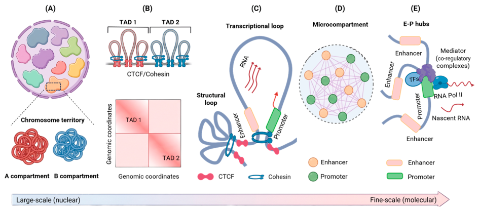
Figure 2.
Architectural, regulatory, and epigenetic mechanisms underlying chromatin loop formation and enhancer-promoter communication. (A) Structural chromatin loop formation and maintenance. Chromatin loops are established through cohesin-mediated loop extrusion and stabilized at convergent CTCF binding sites. NIPBL promotes cohesin loading onto chromatin, whereas WAPL facilitates cohesin release and loop re-modeling. ZNF143 associates with loop anchor regions and cooperates with architectural proteins to support chromatin looping and higher-order genome organization. (B) Cofactors and complexes at the enhancer-promoter interface. Active enhancers recruit coactivators such as p300/CBP and BRD4, which facilitate enhancer activation and transcriptional competence. At the enhancer-promoter interaction inter-face, Mediator, transcription factors (TFs), and RNA polymerase II (RNA Pol II) coordinate transcription initiation and gene activation. YY1 contributes to the stabilization of enhancer-promoter contacts through interactions with the transcriptional regulatory complex. (C) Chromatin states and histone modifications at regulatory elements. Active enhancers are characterized by enrichment of H3K27ac and H3K4me1, whereas active promoters are marked by H3K4me3 and the recruitment of transcriptional machinery. In contrast, poised or repressed regulatory elements are associated with repressive chromatin modifications, including H3K27me3 and H3K9me3. This figure was created with BioRender.com.
Figure 2.
Architectural, regulatory, and epigenetic mechanisms underlying chromatin loop formation and enhancer-promoter communication. (A) Structural chromatin loop formation and maintenance. Chromatin loops are established through cohesin-mediated loop extrusion and stabilized at convergent CTCF binding sites. NIPBL promotes cohesin loading onto chromatin, whereas WAPL facilitates cohesin release and loop re-modeling. ZNF143 associates with loop anchor regions and cooperates with architectural proteins to support chromatin looping and higher-order genome organization. (B) Cofactors and complexes at the enhancer-promoter interface. Active enhancers recruit coactivators such as p300/CBP and BRD4, which facilitate enhancer activation and transcriptional competence. At the enhancer-promoter interaction inter-face, Mediator, transcription factors (TFs), and RNA polymerase II (RNA Pol II) coordinate transcription initiation and gene activation. YY1 contributes to the stabilization of enhancer-promoter contacts through interactions with the transcriptional regulatory complex. (C) Chromatin states and histone modifications at regulatory elements. Active enhancers are characterized by enrichment of H3K27ac and H3K4me1, whereas active promoters are marked by H3K4me3 and the recruitment of transcriptional machinery. In contrast, poised or repressed regulatory elements are associated with repressive chromatin modifications, including H3K27me3 and H3K9me3. This figure was created with BioRender.com.
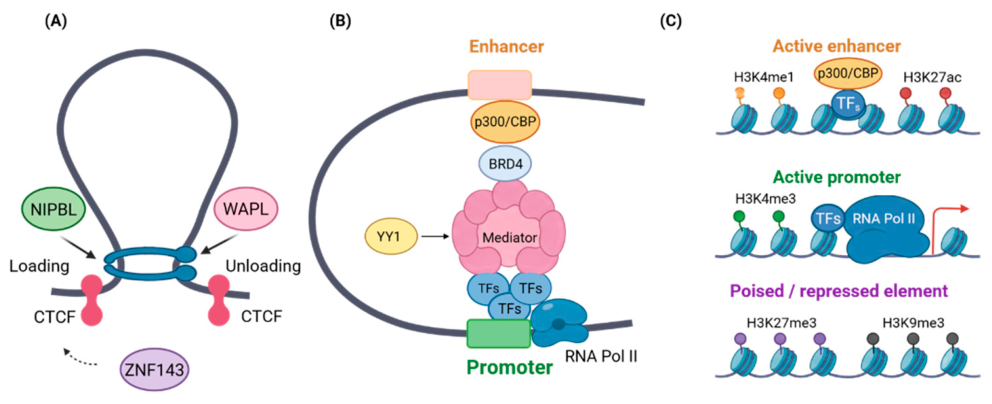
Figure 3.
Genomic structural variations (SVs) and architectural protein defects drive three-dimensional genome dysregulation in cancer. (A) SVs alter chromatin loops. SVs collapse native chromatin loops, this “loop rewriting” places distal enhancers to be placed in proximity to unintended oncogene promoters, -known as enhancer hijacking- while simultaneously silencing line-age-specific genes. (B) TAD boundary disruption. Under normal conditions, topologically associating domain (TAD) boundaries constrain enhancer-promoter interactions within insulated regulatory neighborhoods. Deletion of boundary elements can lead to TAD fusion, activating oncogenes that are normally insulated from these regulatory elements. (C) Compartment switching. Alterations in higher-order chromatin compartmentalization can promote transitions between transcriptionally active A compartments and inactive B compartments. B-to-A switching is frequently associated with activation of previously repressed genes, whereas A-to-B switching can contribute to transcriptional silence of genes, including tumor suppressor genes. (D) Loss or mutation of CTCF weakens chromatin insulation, resulting in TAD fusion and altered regulatory interactions. Dysfunction of the cohesin complex impairs loop extrusion and reduces chromatin loop formation, leading to widespread changes in chromatin architecture and long-range genomic interactions. This figure was created with BioRender.com.
Figure 3.
Genomic structural variations (SVs) and architectural protein defects drive three-dimensional genome dysregulation in cancer. (A) SVs alter chromatin loops. SVs collapse native chromatin loops, this “loop rewriting” places distal enhancers to be placed in proximity to unintended oncogene promoters, -known as enhancer hijacking- while simultaneously silencing line-age-specific genes. (B) TAD boundary disruption. Under normal conditions, topologically associating domain (TAD) boundaries constrain enhancer-promoter interactions within insulated regulatory neighborhoods. Deletion of boundary elements can lead to TAD fusion, activating oncogenes that are normally insulated from these regulatory elements. (C) Compartment switching. Alterations in higher-order chromatin compartmentalization can promote transitions between transcriptionally active A compartments and inactive B compartments. B-to-A switching is frequently associated with activation of previously repressed genes, whereas A-to-B switching can contribute to transcriptional silence of genes, including tumor suppressor genes. (D) Loss or mutation of CTCF weakens chromatin insulation, resulting in TAD fusion and altered regulatory interactions. Dysfunction of the cohesin complex impairs loop extrusion and reduces chromatin loop formation, leading to widespread changes in chromatin architecture and long-range genomic interactions. This figure was created with BioRender.com.
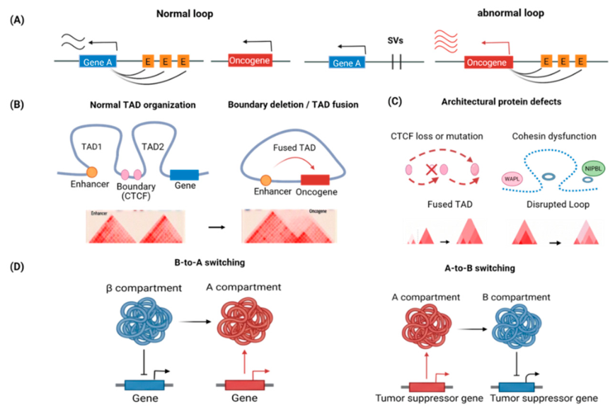

Table 1.
Major Chromosome Conformation Capture Technologies and Their Applications.
| Technology | Year Introduced | Interaction Scope | Main Application | Key Strength | Refs |
|---|---|---|---|---|---|
| Preselected Locus-Based Chromatin Interaction Analysis | |||||
| 3C | 2002 | One-versus-one | Validation of specific chromatin interactions | Quantitative analysis of predefined loci | 2 |
| 4C | 2006 | One-versus-all | Genome-wide contacts of a selected locus | Unbiased viewpoint-centered analysis | 71,72 |
| 5C | 2006 | Many-versus-many | Interaction mapping within selected genomic regions | Parallel analysis of regional interaction networks | 73 |
| Genome-Wide Approaches for 3D Genome Analysis | |||||
| Hi-C | 2009 | All-versus-all | Identification of chromatin compartments, TADs, and chromatin loops | Unbiased genome-wide chromatin architecture mapping | 3 |
| In situ Hi-C | 2014 | All-versus-all | High-resolution chromatin loop and enhancer-promoter mapping | Improved signal quality and kilobase-scale resolution | 4 |
| Micro-C | 2015 | All-versus-all | Fine-scale chromatin architecture and nucleosome-level interactions | Near-nucleosome resolution | 5,6,7 |
| Micro-C XL | 2016 | All-versus-all | Enhanced fine-scale chromatin interaction mapping | Improved interaction recovery and signal quality | 79 |
| scHi-C | 2013 | Single-cell, genome-wide | Cell-to-cell variability in chromosome architecture | Single-cell analysis of genome folding | 8 |
| scMicro-C | 2025 | Single-cell, genome-wide | High-resolution analysis of E-P hubs and chromatin architecture in individual cells | High-resolution single-cell analysis of genome folding | 10 |
| Genomic Locus/Regulatory Element Capture Approaches | |||||
| T2C | 2014 | Targeted regional | High-resolution interrogation of selected genomic loci | Deep coverage of predefined genomic regions | 82 |
| Capture-C | 2014 | Targeted,3C based | High resolution analysis of hundreds of cis-regulatory landscapes | Sensitive detection of interactions from selected viewpoints | 76 |
| 3C-MTS | 2015 | Targeted multi-locus | Simultaneous analysis of multiple regulatory regions | Multiplexed targeted chromatin interaction profiling | 83,84 |
| Promoter Capture Hi-C | 2015 | Promoter-centered | Genome-wide P-E interaction mapping | Enrichment of promoter-associated interactions | 43 |
| Enhancer Capture Hi-C | 2020 | Enhancer-centered | Mapping E-P networks | Enhanced sensitivity for enhancer-associated contacts | 77 |
| MCC | 2021 | Targeted, nucleosome-scale | High-resolution E-P interaction analysis | Combines sequence capture with Micro-C resolution | 44 |
| RCMC | 2023 | Targeted regional | Detection of E-P microcompartments and nested regulatory interactions | Ultra-high-resolution mapping within selected regions | 46 |
| Protein-Specific Enrichment Approaches | |||||
| ChIA-PET | 2010 | Protein-associated | Protein-mediated chromatin interactions | Direct identification of protein-anchored chromatin loops | 74 |
| PLAC-seq | 2016 | Protein-associated | Histone mark-associated chromatin interactions | Efficient and cost-effective enrichment | 75 |
| HiChIP | 2016 | Protein-associated | TF-, CTCF-, cohesin-, and enhancer-associated interactions | High sensitivity with reduced input requirements | 9 |
| Micro-C-ChIP | 2025 | Protein-associated | High-resolution mapping of protein-mediated chromatin interactions | Combines protein specificity with nucleosome-scale resolution | 78 |
Disclaimer/Publisher’s Note: The statements, opinions and data contained in all publications are solely those of the individual author(s) and contributor(s) and not of MDPI and/or the editor(s). MDPI and/or the editor(s) disclaim responsibility for any injury to people or property resulting from any ideas, methods, instructions or products referred to in the content. |
© 2026 by the authors. Licensee MDPI, Basel, Switzerland. This article is an open access article distributed under the terms and conditions of the Creative Commons Attribution (CC BY) license (http://creativecommons.org/licenses/by/4.0/).
Copyright: This open access article is published under a Creative Commons CC BY 4.0 license, which permit the free download, distribution, and reuse, provided that the author and preprint are cited in any reuse.