

Submitted:
01 October 2026
Posted:
06 October 2026
You are already at the latest version
Abstract
Fast, high-threshold motor units translate rapidly organized neural activity into explosive muscular force. Their deterioration may therefore have consequences that precede obvious muscle wasting or frailty. This paper develops the First to Fall hypothesis: the fast/high-threshold motor-unit system, particularly its Type IIx-associated elements, is an exceptionally vulnerable functional reserve whose early deterioration may initiate or amplify a broader cascade of human aging. The proposed process includes impaired activation and synaptic connectivity in surviving motor neurons, altered intrinsic excitability and discharge, neuromuscular-junction dysfunction, denervation, compensatory remodeling, and eventual structural loss. These stages need not occur in a fixed order in every muscle or person. Human longitudinal power data, rapid-force studies, disuse experiments, motor-unit recordings, and Masters-athlete observations are considered alongside anatomical and animal evidence. Together they support investigation of an early neurogenic contribution to declining explosive capacity, while leaving preferential lifelong loss of identified human Type IIx motor units and causation of systemic aging incompletely resolved. Persistent inward currents and synaptic-input changes deepen the proposed mechanism rather than replace the fast-motor-unit hypothesis. The paper distinguishes preservation of muscle fibers from preservation of their original motor-unit architecture, functional improvement from neuronal replacement, and general physical activity from exposure sufficient to maintain rapid neuromuscular demands. It further proposes that loss of fast neuromuscular reserve can reduce movement, loading, and muscle–organ signaling, creating feedback that accelerates downstream deterioration. Cellular aging mechanisms remain relevant within this account; the central challenge is whether they adequately explain the functional chronology without measuring neural control. Longitudinal and interventional tests are specified to determine whether early fast-system dysfunction predicts, mediates, and can modify subsequent systemic decline.
Keywords:
fast motor units
; Type II muscle fibers
; neuromuscular aging
; rate of force development
; persistent inward currents
; denervation
; reinnervation
; sarcosthenia
; physiological reserve
Introduction
Human aging is often recognized when strength, mobility, or independence has already declined. The First to Fall hypothesis asks whether an earlier disturbance has been missed: deterioration of the fast/high-threshold motor-unit system that supplies rapid force, explosive movement, and the ability to respond when little time is available. Its central proposition is that this system is not merely a late casualty of generalized aging. Its early dysfunction and subsequent loss may be an upstream contributor to the progression from diminished explosive capacity to reduced movement, declining physiological reserve, and broader systemic vulnerability. This is a causal hypothesis, not a claim that a single initiating lesion has been established in every human.
The neurogenic perspective has substantial precedent. Denervation, motor-unit remodeling, and altered neural activation have long been implicated in age-associated loss of muscle function. These processes can affect performance before tissue loss becomes clinically conspicuous. The central question—whether declining muscle function reflects intrinsic muscular degeneration or secondary consequences of impaired neuronal control—therefore remains scientifically important (Figueiredo et al., 2006; Lexell, 1997; Hepple & Rice, 2016). First to Fall advances that question by concentrating on the vulnerability and functional significance of the fast system, rather than treating every component of movement as interchangeable.
A motor unit comprises a motor neuron, its axon and terminal branches, and the muscle fibers it innervates. The output of this unit depends on the inputs reaching the neuron, its intrinsic response to those inputs, the reliability of neuromuscular transmission, and the properties of its muscle fibers. Consequently, a motor neuron can remain anatomically present while the unit delivers less useful rapid output. Conversely, a surviving neuron can reinnervate abandoned fibers without recreating the original population of independently controlled units (Piasecki et al., 2016; Mesquita et al., 2024). This distinction is essential to an account of early deterioration: the first measurable failure need not be neuronal death.
The model retains a strong temporal proposition. Fast/explosive capacity may begin to erode in early adulthood, while overall health, muscle appearance, and everyday movement remain satisfactory. However, power decline, fiber atrophy, motor-unit estimates, and actual motor-neuron counts are different measurements. Evidence for one cannot be silently relabeled as evidence for another. The early-adulthood hypothesis is therefore examined using converging observations rather than an assumed universal census showing Type IIx neurons dying at a fixed birthday (Lexell et al., 1988; Alcazar et al., 2023).
The paper preserves a second distinction: maintaining the amount of muscle is not necessarily maintaining the architecture that commands it. The nervous system may compensate for loss, and muscle can adapt to training, while the distribution of motor units and the time course of force production differ from those of young adulthood. The question is not simply whether an older person can become stronger. It is whether the fast system is preserved, how its deterioration affects later function, and whether preservation changes outcomes beyond the trained task (Power et al., 2016; Zange et al., 2026).
Scope and Approach to Evidence
This is a narrative review and hypothesis synthesis, not a systematic review or an original experiment. The literature considered includes sources underlying the original framework and subsequent research relevant to fast-system vulnerability, early functional changes, disuse, synaptic inputs, intrinsic excitability, remodeling, and systemic consequences. Primary human studies are used for specific empirical claims where available. Reviews provide physiological synthesis, and animal or tissue experiments are identified as such. No pooled effect size or new participant-level analysis is presented.
Four forms of statement are kept distinct throughout: observed findings, mechanistic interpretations, the proposed First to Fall model, and predictions requiring direct testing. A result obtained from slow submaximal contractions is not automatically evidence about explosive high-threshold recruitment. A histological fiber type is not an identified motor neuron. A change in a motor-unit estimate is not proof of neuronal death. These boundaries are used to sharpen the original hypothesis and its tests, rather than dilute its emphasis on fast/high-threshold vulnerability.
1. What Makes Fast Motor Units Unique—and Vulnerable
1.1. A Motor Unit Is an Integrated Functional Structure
The motor neuron is the final neural route through which multiple sources of command and feedback influence its muscle fibers. A useful representation must therefore include both the neural input and the peripheral output. Figure 1 depicts this architecture and distinguishes a motor unit from its participating muscle fibers. Preserving a fiber’s cross-sectional area does not establish preservation of the parent motor neuron, the original innervation pattern, or the rapid discharge needed for an explosive contraction (Hepple & Rice, 2016; Hunter et al., 2016).
Human muscle-fiber labels—Type I, IIa, and IIx—describe contractile phenotypes. Motor-unit classifications based on recruitment, force, speed, and fatigability are related to these phenotypes but are not interchangeable laboratory measurements. Here, “Type IIx-associated system” refers to the fast end of the neuromuscular continuum, including its neural recruitment and transmission requirements. It does not mean that ordinary surface EMG has identified the myosin phenotype of every recorded unit. Fiber phenotype can change with activity, and a reduction in the proportion of IIx fibers alone does not prove death of their motor neurons (Plotkin et al., 2021).
Recruitment and discharge jointly influence muscular force. Higher-threshold units can be engaged by sufficiently demanding contractions, including high-force tasks that are not visibly fast; a rapid intention can also produce a rapid recruitment burst. Thus, explosive movement is a major way to interrogate the fast system, but it is not the only condition in which higher-threshold units participate. Human recordings during explosive contractions link recruitment speed and initial discharge behavior to rapid force output (Del Vecchio, Negro, et al., 2019). This gives the model a measurable physiological basis beyond describing a person as simply “fit” or “unfit.”
1.2. Preferential Vulnerability Is a Hypothesis with Several Evidentiary Layers
The rationale for emphasizing fast elements combines age-associated Type II fiber atrophy, loss and remodeling of motor units, slowing of contractile behavior, and deterioration of rapid force production. These observations are compatible with preferential impairment of fast neuromuscular capacity, but their anatomical resolution varies. Whole-muscle histology, for example, can reveal fiber loss and selective changes in fiber size without identifying which original neurons disappeared (Lexell et al., 1988; Lexell, 1995; Hunter et al., 2016).
The distinction matters in interpreting classic evidence. Lexell and colleagues examined whole vastus lateralis cross-sections from 43 previously healthy men aged 15–83. Their analysis associated aging atrophy mainly with fiber loss without a predominant fiber-type effect, and secondarily with reduced fiber size, especially Type II. This supports early muscular change and differential fast-fiber atrophy; it is not a direct demonstration of selective IIx motor-neuron death (Lexell et al., 1988). The fast-system hypothesis remains the interpretation to be tested across these levels, rather than an identity assumed between them.
Motor-unit reviews likewise describe extensive remodeling while emphasizing that the precise selectivity of human motor-unit loss is difficult to establish from available EMG data. Sampling typically favors units accessible during particular contractions, and larger surviving units may reflect compensatory sprouting. This does not make fast-system vulnerability an empty proposition. It makes longitudinal physiological characterization, rather than fiber labeling alone, central to its verification (Piasecki et al., 2016).
The proposed preferential vulnerability is therefore functional as well as structural. A high-threshold unit might remain present yet be recruited too slowly, discharge less rapidly, receive less effective input, or transmit unreliably. Such changes could disproportionately reduce explosive output even when counts or maximal sustained force appear relatively preserved. This extension retains fast motor units as the focal system while allowing an earlier stage of failure before irreversible loss.
1.3. Vulnerability need not be uniform across the body
Different muscles have different habitual demands, motor-unit distributions, and opportunities for compensation. Evidence from one muscle cannot be converted into a whole-body percentage of surviving fast units. Nor should an endurance-trained tibialis anterior, an upper-limb muscle, and the diaphragm be treated as identical aging compartments (Piasecki et al., 2016; Fogarty et al., 2018). A useful First to Fall investigation must ask which fast functions are deteriorating, in which muscles, at what recruitment levels, and during what tasks.
This muscle-specific formulation also avoids making limb location a surrogate for fiber type. The argument does not require all lower-limb muscles to have a uniformly greater fast-fiber proportion than all upper-limb muscles. It requires the fast reserve relevant to a particular movement to be measured. The same person could retain strong habitual locomotion but have deficits in rapid balance recovery, upper-limb force, or another unpracticed demand. Whether those deficits predict later systemic deterioration is a further question, not a consequence established by their coexistence.
2. The Timeline of Decline—When Aging Really Begins
2.1. An Early Functional Change May Precede Overt Disability
First to Fall proposes an interval during which explosive reserve is deteriorating but ordinary tasks remain below the person’s available capacity. Functional decline can therefore begin before disability becomes visible. Compensation, spare capacity, and changes in movement strategy may postpone recognition. The relevant biological question is what changes during this interval, not simply when a clinical threshold is crossed (Atamna et al., 2018; Hepple & Rice, 2016).
The original emphasis on the twenties is retained as an early-adulthood hypothesis, supported by historical muscle morphology and longitudinal power research. It should not be expressed as proof that every person’s fast motor neurons begin dying between precisely 25 and 30. The age at which a modeled population trajectory turns downward is not an individual onset date, and structural loss may be preceded by a longer functional disturbance (Lexell et al., 1988; Alcazar et al., 2023).
Figure 2 makes this distinction explicit. The proposed sequence is functional deterioration, impaired activation or connectivity, denervation/remodeling, and eventual structural motor-unit loss, with overlapping pathways and feedback. Some peripheral disturbances can precede central changes, and neuronal death can initiate denervation directly. The model does not require every individual to traverse every stage in the same order. It predicts that measurable fast-system impairment will often be present before obvious frailty and will carry information about subsequent decline beyond muscle mass alone.
2.2. Ten-Year Longitudinal Power Evidence
Alcazar et al. followed 489 adults—178 women and 311 men, aged 19–68 at baseline—with a median follow-up of 9.6 years. Knee-extension maximum power was estimated from torque–velocity assessments. Power declined significantly in young, middle-aged, and older men and in middle-aged women; the young-women and small older-women subgroups did not show statistically significant absolute-power changes. Figure 3 reproduces reported group means and standard deviations, rather than constructing a hypothetical lifespan curve (Alcazar et al., 2023).
These within-person observations strengthen the case for studying deterioration before frailty. They do not identify the loss of IIx neurons as its cause. Selective follow-up, unequal subgroup sizes, and combined neural and muscular determinants limit causal interpretation. For First to Fall, the appropriate next step is to add repeated fast-unit physiology to the longitudinal power assessment, rather than substitute power for a neuronal count (Alcazar et al., 2023).
2.3. From Early Power Decline to Later Architectural Loss
In later life, reduced estimated motor-unit number and enlargement of surviving units provide a different layer of evidence. Their combination is consistent with a compensatory interval in which denervated fibers are rescued while independent neural control becomes less numerous. The size of the deficit depends on muscle, age, recording method, and participants; it is not appropriate to splice the smallest estimates from different studies into a single body-wide depletion curve (Piasecki et al., 2016).
This is why the proposed chronology distinguishes reserve from census. An individual may lose rapid capacity through changes in activation before losing an entire unit, and may lose units while retaining some muscle mass through reinnervation. These processes can coexist. A longitudinal account should measure rapid output, motor-unit discharge, transmission, and muscle morphology in the same people, rather than assume that their separate age associations describe one proven chain.
The model predicts a period of increasing effort or compensation before conventional weakness becomes apparent. It further predicts that a rapidly declining fast reserve will identify vulnerability that a single measurement of muscle mass misses. Neither prediction requires a universal age cutoff. The stronger proposition—that these changes initiate systemic aging—requires downstream outcomes and an intervention capable of separating the fast system from shared causes.
3. The First 50 milliseconds—A Window into Rapid Neuromuscular Failure
3.1. Force Available in Time Is Different from Eventual Maximum Force
Maximal strength describes the greatest force a person can achieve under specified conditions. Rate of force development describes how quickly force rises. During a short available interval, a person may never reach maximal force; the slope and early impulse then become especially relevant. The early approximately 50–75 ms after force onset is strongly influenced by rapid activation and motor-unit behavior, with muscular and mechanical properties also contributing (Maffiuletti et al., 2016). Figure 4 separates this mechanical window from reaction time.
Equation 1 defines average RFD over the first 50 ms after detected mechanical force onset; the denominator is 0.050 seconds. It is a measurement definition, not a biological aging equation. The onset algorithm, baseline force, filtering, sampling, contraction instruction, and joint configuration must be specified. A stimulus-to-EMG delay or stimulus-to-movement reaction time cannot be substituted for this interval (Maffiuletti et al., 2016).
The mechanistic relevance to First to Fall is that rapid output demands an appropriate neural burst as well as contractile tissue. Del Vecchio and colleagues recorded tibialis anterior motor units in 20 men during explosive contractions and linked recruitment speed and maximal discharge to explosive-force characteristics. That experiment provides a physiological bridge from motor-neuron behavior to rapid performance; it did not test aging or identify every unit as IIx (Del Vecchio, Negro, et al., 2019).
3.2. Aging Changes the Rapid Activation Pattern
Klass et al. reported 48% lower maximal rate of torque development during rapid dorsiflexor contractions in older than young adults. Initial motor-unit discharge frequencies were also lower, with reductions of 19%, 28%, and 34% across the first three interspike intervals. Electrically evoked responses indicated that contractile slowing also contributed. The study therefore supports a neural contribution to impaired rapid force without making every residual difference exclusively neural (Klass et al., 2008).
Very-old-adult work extends investigation beyond the age groups commonly used in laboratory comparisons. Varesco and colleagues assessed rapid knee-extensor activation and force development in very old men, emphasizing the need to examine early force production in advanced age rather than infer it from strength alone (Varesco et al., 2019). Neither isolated-muscle study establishes a universal hierarchy of whole-body aging. Their importance is that they expose a time-sensitive functional limitation that is central to the fast-system hypothesis.
The first 50 ms should consequently be treated as a measurement window, not a pure cortical signal or a magical boundary. Different windows emphasize different mixtures of neural activation, contractile behavior, and force transmission. A high-quality experiment should examine several prespecified intervals, report both absolute and normalized output, and avoid declaring restoration simply because one normalized measure improves (Maffiuletti et al., 2016).
3.3. Early Loss of Expression Can Precede Loss of the Unit
An electrically excitable muscle fiber and an anatomically surviving motor neuron do not guarantee a sufficiently rapid voluntary response. The system must recruit the relevant units quickly and coordinate their output within the time available. First to Fall therefore includes failure to express fast capacity as an early form of deterioration. This is a proposed extension of the original structural argument, not a claim that every low RFD value proves denervation.
The distinction permits discriminating tests. If voluntary RFD declines while evoked contractile capacity is relatively preserved, altered activation becomes a stronger candidate. If both deteriorate together, muscle properties must be investigated alongside neural drive. If a training intervention improves discharge and RFD without changing unit number, it has modified functional expression rather than demonstrated neuronal replacement. These comparisons can locate a limitation more precisely than muscle size or task completion alone (Klass et al., 2008; Del Vecchio, Casolo, et al., 2019).
4. The Fast Motor Unit Before Death—Excitability, Connectivity, and Command
4.1 Persistent Inward Currents as a Mechanism Within First to Fall
Persistent inward currents, or PICs, arise from sustained inward sodium and calcium currents in motor neurons. Their expression is modulated by descending monoaminergic input and inhibition, allowing them to amplify and prolong the effects of synaptic input. An equivalent external input can therefore yield different firing behavior depending on the neuron’s intrinsic and neuromodulatory state (Mesquita et al., 2024). Figure 5 places this amplification within the motor unit’s command system.
Hassan et al. found lower indirect PIC estimates and peak firing rates in older adults during submaximal elbow-flexor and elbow-extensor contractions. These results identify a plausible way for surviving neurons to deliver altered output (Hassan et al., 2021). They do not establish a direct measurement of IIx-unit failure during an explosive task. Human ΔF is inferred from paired motor-unit discharge and depends on the recording protocol and its assumptions; it is not a direct intracellular current measurement (Mesquita et al., 2024).
Within First to Fall, reduced amplification could make the fast system harder to recruit or sustain under demanding conditions. This is the proposed bridge between intrinsic excitability and explosive reserve. It must be tested using compatible motor-unit and rapid-force measurements in the same individuals. PIC changes are not designated the new universal first cause, nor is increasing amplification indefinitely assumed beneficial. Useful motor output requires appropriately timed excitation and inhibition.
Orssatto and colleagues studied 17 older adults through a nonexercise control period followed by six weeks of resistance training. Soleus PIC estimates, discharge behavior, and functional measures improved (Orssatto, Rodrigues, et al., 2023). This supports a modifiable stage in surviving neurons and therefore strengthens the rationale for investigating intervention before permanent loss. It does not show that the original number or architecture of fast motor units was restored. The broader aging-PIC literature provides mechanistic context but also underscores the need to establish how findings from controlled submaximal tasks relate to explosive behavior (Orssatto, Blazevich, & Trajano, 2023).
4.2. Synaptic Deterioration and VGLUT1 Findings
Castro et al. examined motor neurons and their inputs in mice, primates, and human tissue. Their work supports studying age-related synaptic changes even when sampled motor-neuron populations remain present. The extensive mouse observations included altered excitatory inputs, whereas the human comparison showed a trend toward reduced VGLUT1-positive inputs rather than a statistically established replication of every mouse result (Castro et al., 2023). Figure 6 preserves these species and evidence distinctions.
VGLUT1 labels a class of glutamatergic terminals; it is not a marker identifying an exclusively IIx command channel. Reduced labeling cannot, by itself, establish the timing or force of the affected movement. The relevance to First to Fall is the possibility that impaired connectivity undermines the functional fast unit before parent-neuron death. Establishing fast-unit selectivity will require experiments that connect synaptic alterations with recruitment, discharge, and contractile phenotype (Castro et al., 2023).
Mouse studies of aging microglia further implicate interactions between glial cells, motor neurons, and their synapses. Experimental exercise or CSF1R inhibition has also preserved functional motor-unit measures in an aging animal model (Castro et al., 2024; Giorgetti et al., 2019). These findings provide mechanistic candidates, not a demonstrated human sequence from inactivity to selective pruning to systemic aging. The model should use them to formulate experiments, while separating functional motor-unit estimates from direct neuron counts.
4.3. Firing Behavior Across Adulthood
Carbonaro et al. found age- and sex-related differences in motor-unit discharge behavior using high-density surface EMG, including reduced discharge modulation in older adults. The pattern depended on sex, contraction intensity, and recruitment characteristics (Carbonaro et al., 2026). Such observations support a functional stage of neuromuscular aging but caution against assuming that every motor pool follows the same discharge trajectory.
For the fast-system hypothesis, the decisive variable is not simply a lower average firing rate in any task. It is whether the pattern impairs the rapid recruitment and force expression required by the task under study. Higher-threshold units may be underrepresented in low-force protocols, and matching participants at a percentage of their own maximum does not equate absolute force. Both issues should be addressed when translating cross-sectional discharge differences into a proposed preferential fast-system mechanism (Piasecki et al., 2016; Hunter et al., 2016).
4.4. Descending Pathways and Predictive Control
Corticospinal and reticulospinal pathways contribute to the descending command reaching motor neurons, while sensory feedback and predictive processes influence its timing. They are included here because they can alter the expression of fast motor-unit capacity. They do not replace the motor-unit-centered hypothesis with an undifferentiated account of brain aging (Brownstone & Chopek, 2018; Rossignol et al., 2006).
In a two-week elbow-flexor training study, Siddique et al. compared 13 older and 12 younger adults. Strength increased in both groups, but the accompanying corticospinal and StartReact-related changes differed. The interpretation of greater reticulospinal contribution was indirect, based on physiological and behavioral measures rather than recordings from identified human reticulospinal axons (Siddique et al., 2026). This evidence supports distributed, task-dependent adaptation; it does not establish that one pathway can compensate for all aspects of fast-unit loss.
Cerebellar internal-model theory explains how predictive control can help organize movement before delayed sensory information is available (Wolpert et al., 1998). Age-related slowing of preparation provides a further candidate constraint on fast expression (Hardwick et al., 2022). However, aging does not impair every aspect of adaptation uniformly: Vandevoorde and Orban de Xivry found preserved internal-model recalibration despite age-related changes in adaptation (Vandevoorde & Orban de Xivry, 2019). Accordingly, cerebellar prediction failure is treated as a possible interacting limitation requiring task-specific evidence, not a universally demonstrated precursor of fast-unit deterioration.
The expanded mechanism in Figure 7 therefore centers on fast-system output while admitting several routes to its impairment. Connectivity, excitability, descending control, transmission, and contractile properties can interact. The original proposition is preserved: deterioration of this rapidly responsive reserve may precede and contribute to a broader decline. What remains open is which combination of mechanisms starts the process in a given person and whether targeting it changes downstream aging.
5. Why the Hallmarks of Aging Model Faces a Functional Timeline Test
5.1. The Original Challenge Concerns Explanatory Sufficiency
The First to Fall argument challenges whether a catalog of cellular aging mechanisms is sufficient to explain the chronology of declining explosive function. It does not require molecular damage to be absent in youth. The Hallmarks framework describes interacting cellular and systemic mechanisms, including processes that can affect neurons themselves. It is therefore inaccurate to call the framework exclusively myogenic or to place motor neurons outside cellular biology (López-Otín et al., 2013; López-Otín et al., 2023). The substantive challenge is instead whether the loss of effective fast neuromuscular command is an early functional transition that those mechanisms do not adequately characterize when studied without movement physiology.
Age-related molecular signatures can coexist with growth, maturation, and increasing performance. DNA-methylation age and epigenetic drift describe aspects of biological change, but neither by itself specifies how much rapid force is available during a time-critical movement (Horvath, 2013; Teschendorff et al., 2013). The model proposes that compensatory capacity and neuromuscular reserve can initially sustain function despite accumulating changes. Loss of that reserve may then expose or amplify their consequences. This is a threshold interpretation, not evidence that fast units suppress every hallmark through one known pathway.
The distinction preserves the original causal challenge. If rapid neuromuscular deterioration repeatedly precedes downstream metabolic, musculoskeletal, and functional changes, then an exclusively downstream assessment may miss an important initiating influence. However, temporal order alone does not establish primacy: a shared molecular cause could affect both fast units and other systems, or an unmeasured tissue process could begin earlier. The proposed upstream contribution must therefore be tested against common-cause and reciprocal models.
5.2. A Contribution to Systemic Aging Is Stronger than a Contribution to Weakness
Evidence that denervation impairs muscle function supports a local neurogenic mechanism. Demonstrating that early fast-unit deterioration contributes to aging elsewhere requires additional links. Reduced movement may alter mechanical loading, metabolic demand, and muscle-derived signaling; those changes could feed back on both the neuromuscular system and other organs. Muscle–organ communication provides biological plausibility for such interactions, but does not establish that loss of fast motor units initiates them in humans (Pedersen, 2019; Rutti et al., 2018; Szaroszyk et al., 2022).
The direction of influence can also run toward the motor system. Inflammation, vascular limitations, cellular stress, and changes within muscle may compromise neuromuscular function. A hypothesis in which fast-system failure becomes an upstream amplifier is compatible with such feedback. It does not need to deny cellular mechanisms; it needs to show that preserving fast function changes the later trajectory rather than merely marking a healthier organism.
The crucial prediction is therefore comparative: among people with similar age, baseline health, and relevant exposures, early changes in fast-system physiology should predict later deterioration beyond what cellular biomarkers and muscle mass explain. A stronger intervention test would selectively improve or preserve that physiology and demonstrate downstream benefit beyond nonspecific increases in exercise participation. Until such evidence exists, systemic primacy remains the manuscript’s proposed mechanism.
6. Why Modern Longevity Science May Misidentify the Starting Point
6.1. What Is Measured Can Shape What Is Considered Causal
Molecular biomarkers, imaging, and muscle-mass measurements each describe important parts of aging. Their accessibility does not make them complete descriptions of the system that produces movement. A person can have favorable values on selected biomarkers while the speed or reliability of motor output has changed. Conversely, a molecular abnormality may be detected before it produces functional impairment. These are different dimensions of assessment (Horvath, 2013; Hunter et al., 2016; Atamna et al., 2018).
The original concern about a measurement-driven account of aging is retained here as a methodological criticism. If investigators measure only late frailty and cellular signatures, the intervening loss of fast reserve may remain poorly characterized. First to Fall calls for repeated assessment of recruitment speed, discharge, early force, transmission, and motor-unit architecture alongside molecular and clinical measures. This would make the proposed ordering testable instead of assuming either neural or cellular primacy in advance.
The same caution applies to automated literature synthesis. A plausible narrative assembled from citations is not validation of the sequence it describes. A paper about neural adaptation, for example, cannot be used as evidence of permanent motor-neuron preservation unless it measured that outcome. This manuscript consequently treats source verification and the alignment between claim and method as part of the hypothesis’s development, not as a substitute for future experiments.
6.2. Compensation Can Delay Recognition
Collateral reinnervation can preserve fibers after their original innervation is lost. This provides a plausible reason why a neuromuscular disturbance could precede clinically obvious muscle wasting: the system can partly compensate before compensation becomes inadequate (Hepple & Rice, 2016; Piasecki et al., 2016). A large-looking muscle is therefore not a historical record of an intact original motor-unit population.
Skill can also maintain selected performance despite physiological change. Masters athletes are particularly informative because their continued training reduces the plausibility of explaining every deficit simply as inactivity. Nevertheless, athletes differ in genetics, training history, selection, and survival, so their cross-sectional advantages cannot establish the effect of a specific lifelong intervention (Power et al., 2016; Tanaka et al., 2020). The relevant question is which aspects are preserved, which are compensated, and which continue to decline.
In the First to Fall model, the fast system acts as an early reserve whose deterioration may remain hidden during routine tasks. Its loss becomes evident when task demands approach the remaining capacity. This does not imply that an ordinary task is biologically worthless or that every complex movement is diagnostic. It identifies a reason to measure time-sensitive capacity directly rather than infer youthful function from appearance or completion.
7. Movement as Food—The Forgotten Nutrient for the Nervous System
7.1. The Metaphor and Its Physiological Meaning
“Movement as food” expresses the proposal that the nervous system requires appropriate use, not only that it controls the use of muscle. It is a metaphor for activity-dependent loading, sensorimotor experience, and adaptation. Evidence for training-related changes in neural activation and motor-unit behavior supports this general principle (Aagaard et al., 2020; Kamen & Knight, 2004; Del Vecchio, Casolo, et al., 2019). It does not establish a universal maintenance dose that prevents neuronal death.
The core distinction is between doing substantial physical activity and repeatedly challenging the specific capacities of the fast system. Endurance fitness, maximal strength, explosive force, reactive coordination, and motor skill overlap but are not interchangeable outcomes. An intervention can be valuable in one domain without demonstrating preservation in every other domain (Fragala et al., 2019; Tanaka et al., 2020). The model therefore proposes that preservation studies should characterize recruitment demands and rapid-force requirements rather than classify participants only by total exercise time.
Novel or unpredictable tasks may demand ongoing sensorimotor adjustment, and practice can modify brain structure and network organization (Taubert et al., 2010; Voss et al., 2010). However, improved learning or a brain-imaging change does not establish reduced loss of high-threshold motor units. The more specific proposal—that varied, rapid, demanding movement better preserves fast neuromuscular reserve than an equally active but narrowly repetitive program—remains a comparative research question.
7.2. Twenty-One Days of Disuse Reveal Rapid Vulnerability
Sarto et al. studied nine healthy young men, aged 18–29, before and after 21 days of bed rest. The study combined muscle imaging, force testing, intramuscular and high-density surface EMG, and neuromuscular-junction assessments. Quadriceps cross-sectional area fell by 11.9%, while maximal voluntary force fell by 23.1%. Motor-unit discharge and transmission-related measures also changed (Sarto et al., 2025). Figure 8 displays the reported size and force changes as separate outcomes.
This experiment is important to First to Fall because substantial neuromuscular change occurred over weeks in young adults, rather than only in people with established frailty. The functional reduction exceeded the change in muscle size, directing attention toward activation, transmission, and muscle quality. Nevertheless, bed rest simultaneously alters loading and multiple physiological processes; the difference between the two percentages cannot be assigned entirely to neural impairment (Sarto et al., 2025).
The junction morphology requires particular care. The baseline and post-bed-rest junction samples were not paired within the same individuals, the post-bed-rest sample came from only three participants, and junction occupancy was not subjected to statistical analysis. Approximately 15% of the examined post-bed-rest junctions met the study’s morphological denervation criterion. This is an exploratory observation of junction status, not evidence that 15% of all motor neurons or high-threshold units died in 21 days (Sarto et al., 2025).
The physiological findings likewise should not be collapsed into a single claim of selective permanent IIx loss. Recruitment levels and sampling methods matter, and the experiment did not provide lifelong neuronal follow-up. Its contribution is more specific: disuse can rapidly disturb a functioning young neuromuscular system, supporting the hypothesis that loss of adequate demand could initiate or intensify a vulnerable functional stage.
The wider disuse literature describes reductions in explosive strength that can exceed those in maximal strength. Candidate mechanisms include altered recruitment and discharge, contractile slowing, excitation–contraction coupling, and tendon properties (Ruggiero & Gruber, 2026). These interacting mechanisms deepen the mechanistic account of declining explosive capacity without assuming that every acute deficit is permanent or that bed rest reproduces the whole biology of aging.
7.3. What Masters Athletes Do—and Do Not—demonstrate
Power et al. found more functioning tibialis anterior motor units and better transmission stability in world-class octogenarian athletes than age-matched controls (Power et al., 2016). Earlier Masters-runner work also supports examining long-term activity in relation to motor-unit preservation (Power et al., 2010). These findings must be retained even though they challenge a categorical claim that endurance activity provides no neural protection.
Piasecki et al. compared long-term endurance and power athletes with other groups and reported evidence of motor-unit size expansion accompanying reduced estimated numbers in older age (Piasecki et al., 2019). The useful distinction is that training may support compensatory remodeling without preserving every original unit. Results from different muscles and athletic populations should not be converted into a universal ranking in which one training modality necessarily prevents loss and another causes it.
First to Fall therefore retains the argument that general fitness is insufficient evidence of complete fast-system preservation, while recognizing genuine benefits of endurance and resistance activity. A person can receive cardiovascular, metabolic, or neural benefit without proving restoration of the complete young-adult architecture. Conversely, preserving an athlete’s performance in one muscle or event does not demonstrate equivalent preservation across the body.
7.4. Testing Use-Dependent Preservation
A direct study should compare interventions that differ in the relevant demands while accounting for overall dose, supervision, adherence, recovery, and baseline function. Outcomes should include rapid activation and early force, not only muscle mass and maximal strength. Motor-unit estimates and transmission measures would indicate whether a functional improvement coincides with preservation of peripheral organization. Long follow-up is necessary to distinguish a short-term adaptation from a change in the rate of loss.
The premise is that fast reserve must be challenged if it is to be preserved. The evidence currently justifies testing that premise, rather than prescribing one universal “chaotic” exercise program as proven prevention. High-force slow contractions, fast-intent resistance exercise, power training, balance challenges, and multidirectional tasks should be distinguished experimentally rather than grouped by appearance. Safety and feasibility depend on the participant’s musculoskeletal and clinical condition (Fragala et al., 2019).
8. Denervation, Collateral Reinnervation, and Sarcosthenia
8.1. Rescue of Fibers Is Not Restoration of the Original Architecture
When a motor neuron or its effective connection is lost, affected muscle fibers become denervated. Surviving axons can sprout and reinnervate some of those fibers. This can preserve tissue and useful force while producing fewer, larger motor units and fiber-type grouping. The process is compensatory, but it does not recreate the original set of independently controlled neurons (Hepple & Rice, 2016; Piasecki et al., 2016). Figure 9 depicts this distinction without assigning a fixed proportion of fast units to the loss.
Reinnervation can therefore conceal an earlier disruption if only muscle size is measured. It can also change the organization through which neural commands produce force. Whether a specific rescued fiber acquires a slower phenotype, how the enlarged unit behaves, and whether reinnervation succeeds depend on the biological context. The diagram must not be read as proof that every fast unit is invariably replaced by a slow unit.
This supports a central argument: neurogenic aging cannot be understood solely as loss of muscle mass. A muscle’s quantity, innervation architecture, activation, and contractile function can move in different directions. Training-induced hypertrophy may improve capacity, but the increase alone does not establish recovery of missing motor neurons. Likewise, a stable motor-unit estimate does not guarantee preserved synaptic input or explosive discharge (Piasecki et al., 2016; Castro et al., 2023).
8.2. Sarcosthenia Exposes the Limits of Tissue Quantity
Sarcosthenia describes deterioration of muscle functional quality beyond the amount of muscle available. In this manuscript it is used operationally for deficits in force or power relative to a relevant measure of muscle quantity; it is not treated as a unique diagnosis of neural degeneration. Lower output per muscle volume can reflect activation, intrinsic contractile properties, tissue composition, architecture, or task mechanics (Tanaka et al., 2020; Zange et al., 2026).
Zange et al. examined male Masters athletes and controls using muscle imaging and jumping measurements. Older athletes preserved more muscle and had less muscle fat than older controls, yet the older groups generated approximately half the peak jumping power per unit of the assessed muscle volume seen in the younger groups. Figure 10 presents reported volume and power values without inferring that their ratio isolates neural function (Zange et al., 2026).
These findings matter because an active, muscular body can still have substantially altered rapid output. They support the distinction between the visible tissue and the command-and-contraction system. They do not show that all unexplained power loss is caused by motor-unit death. The fast-system hypothesis predicts a contribution that should be measured alongside muscle-intrinsic mechanisms, rather than inferred from a residual statistical term.
Alvero-Cruz et al. studied 256 competitive Masters track-and-field athletes and found age-associated differences in body composition and jumping power. Age remained associated with peak power after adjustment for athletic specialization, percent fat-free mass, and phase angle (Alvero-Cruz et al., 2021). Continued training therefore does not make tissue composition or function age-invariant. Body composition contributes to the interpretation, but does not fully explain the observed relationship with age.
8.3. An Integrated Functional Consequence
Rapid movement depends on both force generation and the mass and geometry being moved. Muscle fat, altered contractile machinery, activation deficits, and tendon changes can affect performance through different mechanisms (Prochniewicz et al., 2007; Zange et al., 2026; Kwan et al., 2023). A meaningful account of fast-system decline should not make these disappear by relabeling every effect “neural.” It should investigate how impaired fast activation interacts with the quality and mechanical environment of the fibers it commands.
The proposed cascade is consequently an amplification model. Less effective fast output may reduce the movement demands an individual can comfortably sustain. Reduced demand may then reinforce deconditioning, while sarcosthenia makes the remaining neural output less productive. This feedback can link an early neuromuscular disturbance to later functional loss without requiring that every downstream tissue change originate in the same neuron or occur in one fixed order.
9. Why Fast Motor Units Cannot Simply Be Rebuilt When Other Tissues Are Repaired
9.1. Neuronal Loss Is a Different Restoration Problem
The restoration argument rests on a valid biological distinction: a mature motor neuron is a post-mitotic cell within a connected circuit, not a pool of freely dividing replacement tissue (Aranda-Anzaldo, 2012). Repairing muscle or increasing its size does not automatically supply the missing neuron, its correctly directed axon, its synapses, and its coordinated function. The problem is therefore one of functional integration as well as cell survival.
However, post-mitotic does not mean that every component of a motor unit is incapable of repair. Surviving neurons can sprout, peripheral axons can regenerate under suitable conditions, and neuromuscular synapses have regenerative mechanisms (Zelada et al., 2021; Madison & Robinson, 2014). A disconnected unit is not always equivalent to a dead neuron. Preserving this distinction makes the argument more precise: regeneration of a component is not evidence that the original integrated fast system has been restored.
Figure 11 separates survival or replacement of components from successful integration and coordinated use. No study reviewed here demonstrates reconstruction of the complete original fast-motor-unit architecture of an aging human with restoration of its young-adult integrated function. This is a statement about available demonstrations, not proof that every future regenerative strategy must fail.
9.2. Common Drive, Summation, and Learned Control
Common drive describes shared modulation of motor-unit activity contributing to force control (De Luca & Erim, 1994). Recruitment timing, discharge, and the temporal summation of muscular responses matter because force is produced by coordinated activity rather than the simple presence of cells. These concepts remain central to the emphasis on command and precision.
Their terminology must nevertheless be used accurately. “Muscular wisdom” is a physiological hypothesis concerning discharge adjustments during fatigue; it is not a scientific name for an entire lifetime’s learned motor skill (Garland & Gossen, 2002). Learned movement also involves central circuits: human research on motor-pattern replay during sleep provides evidence that learned activity is represented beyond the peripheral motor unit (Rubin et al., 2022). Loss of a unit therefore changes the available output architecture without proving that every associated skill memory was stored in that neuron and permanently erased.
The strong restoration question remains. Even if a new cell survives, can it receive appropriate inputs, recruit the correct fibers, generate suitable force, and operate within existing coordination? A successful isolated contraction would answer only part of that question. Full restoration would require task-relevant timing, sustained integration, and evidence that performance is not merely compensated through a different strategy. These are proposed criteria for evaluating restoration, not a claim that compensation is useless.
9.3. The Biomechanical Paradox
The biomechanical paradox presents an integration problem: restored neural or muscular output may encounter aged tendons, connective tissues, bones, or joints that have adapted to a different loading environment. Tendon aging research supports examining the mechanical context rather than assuming that greater output is automatically tolerable (Kwan et al., 2023).
The paradox does not establish that recovery must cause injury or that aged tissues cannot adapt. It predicts that restoration should be assessed across the load-bearing system. A neuron-centered intervention that improves rapid force but overloads another structure has not restored integrated youthful movement. Conversely, coordinated tissue and neural rehabilitation could address some of these constraints. The point is to specify what successful restoration would require, not declare it biologically impossible in advance.
9.4. Prevention and Functional Recovery Have Different Endpoints
Preventing loss of an original motor neuron, improving excitability in a survivor, reinnervating a fiber, increasing muscle size, and recovering a skilled task are different achievements. Each can be valuable, but none should be silently substituted for another. Evidence of increased discharge after training supports functional adaptation (Kamen & Knight, 2004; Orssatto, Rodrigues, et al., 2023). It does not by itself establish prevention of neuronal loss over decades.
This distinction sustains the priority given to preservation. If the fast architecture is difficult to reconstruct, detecting and addressing early dysfunction becomes especially important. Preservation should not be equated with a promise of complete arrest of aging; it should be defined by trajectories of specified neural and functional outcomes. Restoration claims should identify the recovered level and the appropriate young or pre-loss comparator.
10. From Fast-System Failure to Systemic Deterioration and Longevity
10.1. Plausible Downstream Routes
First to Fall proposes that loss of explosive neuromuscular reserve can change the organism’s daily interaction with its environment. A person who cannot generate or control force rapidly may reduce demanding movement, tolerate less perturbation, and gradually narrow activity. The resulting changes in loading and muscular activity could influence metabolic and musculoskeletal function. These are proposed causal routes, not consequences proven by a motor-unit count alone.
Muscle-derived signals provide additional plausibility. Experiments have identified Type II muscle-associated secreted factors with effects on pancreatic cells, while experimental musclin work demonstrates muscle–heart signaling in pathological overload (Rutti et al., 2018; Szaroszyk et al., 2022). These studies support possible connections between the fast muscular phenotype and other tissues. They do not demonstrate that loss of fast motor neurons in aging humans reduces those factors enough to initiate systemic deterioration.
The broader muscle–brain literature reinforces the relevance of movement to organ communication (Pedersen, 2019). The model predicts that preservation of fast function could have effects extending beyond the local muscle through maintained activity and signaling. To establish mediation, a study would need to show that the relevant neural change precedes the signaling change and that modifying the neural component changes the downstream outcome. Simply observing correlated decline would leave common causes unresolved.
10.2. Reserve and the First Indispensable Function to Become Limiting
The organism can fail when an indispensable function falls below its required capacity, even if other systems remain relatively preserved. This general reserve concept motivates the longevity extension of First to Fall (Atamna et al., 2018). Figure 12 illustrates it without fitting a lifespan curve or assigning a universal terminal organ.
Rk(t) = Ck(t) − Dk(t)
Equation 2 defines a conceptual functional reserve margin as available capacity minus task or physiological demand. Its terms must be measured in compatible units for each specified function. It is not a validated biomarker, a calibrated mortality model, or a claim that every system shares one numerical threshold.
For the fast-system hypothesis, the relevant prediction is that diminishing neuromuscular reserve can become a limiting contributor to resilience, independently of whether it is the immediate recorded cause of death. The stronger claim that it determines maximal human lifespan requires evidence from extreme survivors and appropriate competing-system measurements. Existing limb physiology cannot establish that all people exhaust their motor units at 110–120 years.
10.3. Respiratory Relevance Without a Universal Diaphragm Claim
Respiratory motor units make the reserve argument biologically concrete because ventilation and airway protection depend on neuromuscular output. Diaphragm physiology distinguishes lower-demand breathing from higher-force behaviors such as coughing; fast-unit deficits could therefore affect protective or high-demand functions before routine ventilation fails (Fogarty et al., 2018).
This possibility is consistent with the broader First to Fall emphasis on losing reserve before visible failure. It does not establish diaphragm motor-unit depletion as the universal terminal mechanism of extreme longevity. The required evidence would include respiratory function, neural or motor-unit measures where feasible, longitudinal clinical observations, and assessment of competing causes. Whole-body claims must not be inferred from the oldest participant’s limb-muscle estimate.
Preserving fast neuromuscular reserve may be necessary for maintaining a particular youthful function without being sufficient to maximize survival. Genetic influences, disease, environmental exposures, and other essential systems remain relevant. The model does not require a genetic termination clock, but rejecting such a clock would not by itself prove a neuromuscular lifespan ceiling. Its testable contribution is to identify a candidate limiting reserve and the measurements needed to evaluate it.
11. Discussion
11.1. Integration of the Evidence
The First to Fall hypothesis remains centered on fast/high-threshold, particularly Type IIx-associated, neuromuscular vulnerability as a possible early upstream event. The newer evidence expands the meaning of “fall” to include functional impairment before anatomical disappearance. PIC estimates, synaptic observations, rapid discharge, and force-time measurements offer candidate ways to detect this stage. Denervation and remodeling then provide a route through which compensation can preserve muscle while altering its original neural organization.
This formulation does not make the fast system synonymous with the entire nervous system. A descending or synaptic change belongs in the argument when it provides a plausible or measured route to impaired fast output. Likewise, sarcosthenia belongs as an interacting limitation and possible downstream expression, not as a substitute name for motor-unit loss. Table 1 summarizes the principal evidence and its relationship to the model.
The mechanistic strength lies in connecting time-sensitive performance with specific physiological processes. The main uncertainty lies in causal ordering across the lifespan and between organs. Evidence for early power decline, age-related neural changes, and muscle–organ communication cannot simply be joined and declared a demonstrated human aging cascade. The proposed sequence becomes stronger when its intermediate steps are measured together and challenged against alternatives.
11.2. Findings That Constrain the Hypothesis
Several observations prevent a simplistic interpretation. Human whole-muscle histology does not establish selective loss of Type II fibers in every context; motor-unit reviews acknowledge uncertain selectivity of loss; sampled motor neurons can remain present despite functional or synaptic alterations; and long-term endurance athletes can show neuromuscular advantages (Lexell et al., 1988; Piasecki et al., 2016; Castro et al., 2023; Power et al., 2016). These findings constrain categorical statements while leaving the proposed preferential vulnerability of rapid function open to direct testing.
Training-related gains also require precise interpretation. They demonstrate adaptive capacity in surviving systems and may be relevant to prevention. They do not, without structural evidence, show that missing original units have returned. Conversely, the fact that complete restoration has not been demonstrated does not justify calling every neural deficit permanent. The distinction between modifiable dysfunction and structural loss is a central element of the argument (Orssatto, Rodrigues, et al., 2023; Zelada et al., 2021).
Human studies sample a limited set of muscles and tasks. High-threshold recordings are technically challenging, cross-sectional differences are not within-person trajectories, and low-force EMG cannot resolve every unit’s phenotype. The hypothesis must therefore be tested with converging methods. It should not be protected from contradictory evidence by defining every unmeasured change as hidden fast-unit failure.
11.3. What Would Constitute Stronger Evidence
Evidence would become substantially stronger if early changes in identified fast-system physiology predicted later decline within individuals, persisted after accounting for major confounders, and mediated downstream outcomes. A selective preservation intervention would add causal leverage. The most informative result would combine maintained neural and transmission measures with maintained rapid force and a demonstrable change in later systemic function.
Evidence would challenge the model if early explosive decline were adequately explained by non-neural mechanisms despite preserved fast activation, if other systems consistently deteriorated first and explained the neural changes, or if a successful targeted preservation intervention failed to alter the downstream outcomes it was predicted to affect. Such outcomes would refine or limit the hypothesis. They should not be dismissed simply because the intervention improved some unrelated measure.
12. Future Directions
12.1. A Longitudinal Test Beginning Before Frailty
Recruit adults across early and middle adulthood and follow the same participants with repeated assessments of rapid force, maximal force, motor-unit behavior, transmission, muscle quantity and composition, activity exposure, and relevant health outcomes. The priority is not an arbitrary universal starting age but coverage of the period in which early changes are proposed to emerge. Sex, muscle group, training background, injury, disease, and measurement reliability should be prespecified sources of variation.
Power and RFD should be assessed separately. Motor-unit analysis should sample multiple contraction levels and include explosive tasks where technically feasible. Imaging and biopsy can characterize muscle but should not be presented as a complete neuronal census. Repeated molecular and clinical measures would allow the study to ask whether fast-system deterioration precedes downstream change rather than assume the order. Missingness, selective dropout, and survival must be addressed explicitly.
12.2. A Disuse and Recovery Test of Early Functional Deterioration
Supervised unloading studies offer a shorter experimental window but require clear boundaries on interpretation. Neural activation, transmission, rapid force, muscle size, and recovery should be followed together. Recovery of discharge or force without restoration of a changed structural measure would be particularly informative. A parallel comparison condition and adequate follow-up would improve separation of unloading effects from measurement variability and incomplete recovery.
The central question is whether repeated periods of inadequate demand leave lasting fast-system deficits, and whether an intervention aimed at rapid activation changes that trajectory. Sarto et al. provide a human perturbation study motivating this work, not a complete answer about cumulative loss or aging (Sarto et al., 2025). More demanding anatomical claims would require additional methods and cannot be inferred from the disappearance of a waveform alone.
12.3. Comparative Preservation Trials
Compare clearly specified training strategies with sufficient attention to total dose and task specificity. Endurance, high-force resistance, rapid-intent power, and variable multidirectional practice can provide different exposures. Outcomes must match the proposed mechanism rather than favor the practiced task. Independent testing and retention assessments would help distinguish learned test performance from broader preservation.
The important endpoints are maintenance of fast output and its neural support, not simply whether a group improves from baseline. A short-term benefit can justify further study without proving long-term motor-unit survival. A longitudinal trial should specify whether the target is excitability, transmission stability, reduced denervation, compensatory reinnervation, or maintained unit number. Those outcomes should be reported separately.
12.4. Testing Systemic Effects and the Longevity Extension
Downstream outcomes should be selected before analysis and measured independently of the trained task. Mediation analyses need repeated observations of the proposed sequence, and causal interpretation requires assumptions about confounding and intervention specificity. A null downstream result after confirmed target engagement would be important evidence, not a reason to replace the outcome retrospectively.
The maximal-lifespan extension requires a distinct program in very old adults and exceptional survivors. It should characterize competing reserve limitations, care context, respiratory and locomotor demands, and the timing of terminal decline. It cannot be established by extrapolating a percentage decline from middle age to a hypothetical zero. Table 2 separates tests of the local fast-system proposition from the stronger systemic and lifespan claims.
13. Conclusions
The First to Fall hypothesis proposes that early deterioration of the fast/high-threshold motor-unit system is a consequential upstream event in human aging. Its Type IIx-associated emphasis remains central: the loss of rapid neuromuscular reserve may begin before obvious frailty, change how the body moves and is loaded, and contribute to a broader deterioration that measurements of muscle mass or cellular state alone do not fully describe.
The expanded account recognizes that a unit can fail functionally before its neuron dies. Impaired input, altered excitability, slower recruitment or discharge, and unreliable transmission can reduce fast output; denervation and compensatory reinnervation can subsequently preserve fibers while changing the original architecture. Sarcosthenia and declining movement can interact with this process. These additions deepen the proposed mechanism without establishing one compulsory sequence for every person.
Preservation is therefore a distinct objective from rebuilding tissue after loss. Improved muscle size, better performance in a practiced task, or recovery of one neural measure does not by itself demonstrate restoration of the integrated youthful fast system. At the same time, retained plasticity makes early intervention a meaningful subject for investigation rather than a contradiction of the hypothesis.
The decisive test is whether early fast-system changes precede and explain subsequent decline, and whether specifically preserving that system modifies downstream outcomes. Until those tests are completed, fast motor units remain the proposed First to Fall—not a universally proven single cause, but a defined biological hypothesis with an explicit preservation target and falsifiable predictions.
Postscript: Reframing the Field
The central challenge is to include the command system in the chronology of aging. Cellular mechanisms explain important aspects of deterioration; the First to Fall proposal asks when their effects become functionally consequential and whether loss of fast neuromuscular reserve helps drive that transition. Preserving the capacity to generate and coordinate rapid force may prove essential to preserving youthful function. Its contribution to systemic aging and exceptional survival must be established through the same rigorous tests demanded of any competing mechanism.
Clarifications, Limitations, and Declarations
Clarification of Key Terms
Fast/high-threshold system refers to the neural and muscular elements supporting high-force and rapid output; it is not a claim that recruitment threshold and myosin phenotype have been directly matched in every cited study. Functional deterioration means impaired effective output and can occur in surviving units. Denervation means loss of effective innervation of a fiber and does not always imply parent-neuron death. Collateral reinnervation means rescue by branches of surviving axons. Structural motor-unit loss refers to loss of an original unit rather than an isolated decrease in one physiological estimate (Piasecki et al., 2016; Plotkin et al., 2021).
Common drive denotes shared neural modulation, and temporal summation denotes interaction of successive contractile responses. Neither is synonymous with all learned skill. Sarcosthenia denotes reduced functional quality beyond muscle quantity, with its measurement specified for each study. Neuromuscular reserve denotes capacity available above a defined demand; it should not be treated as a single established whole-body score (De Luca & Erim, 1994; Garland & Gossen, 2002; Tanaka et al., 2020; Atamna et al., 2018).
Limitations
This synthesis is selective and narrative. It combines human longitudinal and cross-sectional observations, short interventions, indirect electrophysiological estimates, histology, animal experiments, and physiological reviews. Those designs provide different kinds of evidence. They cannot collectively manufacture a direct lifelong identification of every fast motor unit or a demonstrated causal ordering of all aging processes.
Fast-system specificity is the central prediction requiring better resolution. A study of a slow-ramp contraction, whole-muscle power, or a general motor-neuron marker may inform the model without directly testing preferential IIx vulnerability. Early onset, irreversibility, and systemic effects must each be evaluated separately. The absence of a perfect lifespan experiment does not erase existing evidence, but it does limit the strength and precision of the conclusions drawn from it.
Author Contributions
Tony Ruggia conceived the First to Fall hypothesis, developed the original manuscript and its central arguments, and supplied material for their expansion. This manuscript reports no new laboratory experiments or participant-level data collection.
Funding
This work was conducted independently and received no external funding or institutional support.
Ethics Statement
This manuscript is a theoretical synthesis of published literature. No new human or animal experiments were conducted. Institutional ethics approval was not applicable.
Data Availability Statement
No new participant-level datasets were generated. Numerical values reproduced in the charts are identified by source, outcome, and units in the captions and accompanying figure-data file. Conceptual figures and equations are explicitly identified and do not contain participant data.
Acknowledgments and AI Assistance Disclosure
The author acknowledges the investigators whose published work informed this synthesis. OpenAI’s ChatGPT assisted with literature checking, drafting and editing, figure preparation, citation linking, and document formatting. The central hypothesis and original conceptual arguments were supplied by the author. AI assistance does not constitute authorship or independent scientific validation; responsibility for submission and interpretation remains with the author.
Conflicts of Interest
The author declares no conflict of interest.
About the Author
Tony Ruggia is an independent researcher and lifelong practitioner of physical training. His work examines the relationship between fast neuromuscular function, movement, and aging. The First to Fall framework proposes that early loss of fast-system reserve can have consequences extending beyond muscle quantity to integrated physical function and systemic vulnerability.
References
- Aagaard, P.; Bojsen-Møller, J.; Lundbye-Jensen, J. Assessment of neuroplasticity with strength training. Exercise and Sport Sciences Reviews 2020, 48(4), 151–162. [Google Scholar] [CrossRef]
- Alcazar, J.; Rodriguez-Lopez, C.; Delecluse, C.; Thomis, M.; Van Roie, E. Ten-year longitudinal changes in muscle power, force, and velocity in young, middle-aged, and older adults. Journal of Cachexia, Sarcopenia and Muscle 2023, 14(2), 1019–1032. [Google Scholar] [CrossRef]
- Alvero-Cruz, J. R.; Brikis, M.; Chilibeck, P.; Frings-Meuthen, P.; Vico Guzmán, J. F.; Mittag, U.; Michely, S.; Mulder, E.; Tanaka, H.; Tank, J.; Rittweger, J. Age-related decline in vertical jumping performance in masters track and field athletes: Concomitant influence of body composition. Frontiers in Physiology 12 2021, 643649. [Google Scholar] [CrossRef]
- Aranda-Anzaldo, A. The post-mitotic state in neurons correlates with a stable nuclear higher-order structure. Communicative & Integrative Biology 2012, 5(2), 134–139. [Google Scholar] [CrossRef]
- Atamna, H.; Tenore, A.; Lui, F.; Dhahbi, J. M. Organ reserve, excess metabolic capacity, and aging. Biogerontology 2018, 19(2), 171–184. [Google Scholar] [CrossRef]
- Brownstone, R. M.; Chopek, J. W. Reticulospinal systems for tuning motor commands. Frontiers in Neural Circuits 12 2018, 30. [Google Scholar] [CrossRef]
- Carbonaro, M.; Boccardo, M.; Brusco, C. M.; Pilotto, A. M.; Re, R.; Lauretani, F.; Porcelli, S.; Franchi, M. V.; Botter, A. Motor unit behavior adaptations across lifespan: Sex differences in young, middle-aged and old adults; Advance online publication; Npj Aging, 2026. [Google Scholar] [CrossRef]
- Castro, R. W.; Lopes, M. C.; De Biase, L. M.; Valdez, G. Aging spinal cord microglia become phenotypically heterogeneous and preferentially target motor neurons and their synapses. Glia 2024, 72(1), 206–221. [Google Scholar] [CrossRef]
- Castro, R. W.; Lopes, M. C.; Settlage, R. E.; Valdez, G. Aging alters mechanisms underlying voluntary movements in spinal motor neurons of mice, primates, and humans. JCI Insight 2023, 8(9), e168448. [Google Scholar] [CrossRef]
- De Luca, C. J.; Erim, Z. Common drive of motor units in regulation of muscle force. Trends in Neurosciences 1994, 17(7), 299–305. [Google Scholar] [CrossRef]
- Del Vecchio, A.; Casolo, A.; Negro, F.; Scorcelletti, M.; Bazzucchi, I.; Enoka, R.; Felici, F.; Farina, D. The increase in muscle force after 4 weeks of strength training is mediated by adaptations in motor unit recruitment and rate coding. The Journal of Physiology 2019, 597(7), 1873–1887. [Google Scholar] [CrossRef]
- Del Vecchio, A.; Negro, F.; Holobar, A.; Casolo, A.; Folland, J. P.; Felici, F.; Farina, D. You are as fast as your motor neurons: Speed of recruitment and maximal discharge of motor neurons determine the maximal rate of force development in humans. The Journal of Physiology 2019, 597(9), 2445–2456. [Google Scholar] [CrossRef]
- Figueiredo, P. A.; Mota, M. P.; Appell, H. J.; Duarte, J. A. Ceasing of muscle function with aging: Is it the consequence of intrinsic muscle degeneration or a secondary effect of neuronal impairments? European Review of Aging and Physical Activity 2006, 3(2), 75–83. [Google Scholar] [CrossRef]
- Fogarty, M. J.; Mantilla, C. B.; Sieck, G. C. Breathing: Motor control of diaphragm muscle. Physiology 2018, 33(2), 113–126. [Google Scholar] [CrossRef]
- Fragala, M. S.; Cadore, E. L.; Dorgo, S.; Izquierdo, M.; Kraemer, W. J.; Peterson, M. D.; Ryan, E. D. Resistance training for older adults: position statement from the national strength and conditioning association. Journal of Strength and Conditioning Research 2019, 33(8), 2019–2052. [Google Scholar] [CrossRef]
- Garland, S. J.; Gossen, E. R. The muscular wisdom hypothesis in human muscle fatigue. Exercise and Sport Sciences Reviews 2002, 30(1), 45–49. [Google Scholar] [CrossRef]
- Giorgetti, E.; Panesar, M.; Zhang, Y.; Joller, S.; Ronco, M.; Obrecht, M.; Lambert, C.; Accart, N.; Beckmann, N.; Doelemeyer, A.; Perrot, L.; Fruh, I.; Mueller, M.; Pierrel, E.; Summermatter, S.; Bidinosti, M.; Shimshek, D. R.; Brachat, S.; Nash, M. Modulation of microglia by voluntary exercise or CSF1R inhibition prevents age-related loss of functional motor units. Cell Reports 2019, 29(6), 1539–1554.e7. [Google Scholar] [CrossRef]
- Hardwick, R. M.; Forrence, A. D.; Costello, M. G.; Zackowski, K.; Haith, A. M. Age-related increases in reaction time result from slower preparation, not delayed initiation. Journal of Neurophysiology 2022, 128(3), 582–592. [Google Scholar] [CrossRef]
- Hassan, A. S.; Fajardo, M. E.; Cummings, M.; McPherson, L. M.; Negro, F.; Dewald, J. P. A.; Heckman, C. J.; Pearcey, G. E. P. Estimates of persistent inward currents are reduced in upper limb motor units of older adults. The Journal of Physiology 2021, 599(21), 4865–4882. [Google Scholar] [CrossRef]
- Hepple, R. T.; Rice, C. L. Innervation and neuromuscular control in ageing skeletal muscle. The Journal of Physiology 2016, 594(8), 1965–1978. [Google Scholar] [CrossRef]
- Horvath, S. DNA methylation age of human tissues and cell types. Genome Biology 2013, 14(10), R115. [Google Scholar] [CrossRef]
- Hunter, S. K.; Pereira, H. M.; Keenan, K. G. The aging neuromuscular system and motor performance. Journal of Applied Physiology 2016, 121(4), 982–995. [Google Scholar] [CrossRef]
- Kamen, G.; Knight, C. A. Training-related adaptations in motor unit discharge rate in young and older adults. The Journals of Gerontology: Series A 2004, 59(12), 1334–1338. [Google Scholar] [CrossRef]
- Klass, M.; Baudry, S.; Duchateau, J. Age-related decline in rate of torque development is accompanied by lower maximal motor unit discharge frequency during fast contractions. Journal of Applied Physiology 2008, 104(3), 739–746. [Google Scholar] [CrossRef]
- Kwan, K. Y. C.; Ng, K. W. K.; Rao, Y.; Zhu, C.; Qi, S.; Tuan, R. S.; Ker, D. F. E.; Wang, D. M. Effect of aging on tendon biology, biomechanics and implications for treatment approaches. International Journal of Molecular Sciences 2023, 24(20), 15183. [Google Scholar] [CrossRef]
- Lexell, J. Human aging, muscle mass, and fiber type composition. The Journals of Gerontology: Series A 1995, 50A(Special Issue), 11–16. [Google Scholar] [CrossRef]
- Lexell, J. Evidence for nervous system degeneration with advancing age. The Journal of Nutrition 1997, 127((5) Suppl, 1011S–1013S. [Google Scholar] [CrossRef]
- Lexell, J.; Taylor, C. C.; Sjöström, M. What is the cause of the ageing atrophy? Total number, size and proportion of different fiber types studied in whole vastus lateralis muscle from 15- to 83-year-old men. Journal of the Neurological Sciences 1988, 84(2-3), 275–294. [Google Scholar] [CrossRef]
- López-Otín, C.; Blasco, M. A.; Partridge, L.; Serrano, M.; Kroemer, G. The hallmarks of aging. Cell 2013, 153(6), 1194–1217. [Google Scholar] [CrossRef]
- López-Otín, C.; Blasco, M. A.; Partridge, L.; Serrano, M.; Kroemer, G. Hallmarks of aging: An expanding universe. Cell 2023, 186(2), 243–278. [Google Scholar] [CrossRef]
- Madison, R. D.; Robinson, G. A. Accuracy of regenerating motor neurons: influence of diffusion in denervated nerve. Neuroscience 273 2014, 128–140. [Google Scholar] [CrossRef]
- Maffiuletti, N. A.; Aagaard, P.; Blazevich, A. J.; Folland, J.; Tillin, N.; Duchateau, J. Rate of force development: Physiological and methodological considerations. European Journal of Applied Physiology 2016, 116(6), 1091–1116. [Google Scholar] [CrossRef]
- Mesquita, R. N. O.; Taylor, J. L.; Heckman, C. J.; Trajano, G. S.; Blazevich, A. J. Persistent inward currents in human motoneurons: Emerging evidence and future directions. Journal of Neurophysiology 2024, 132(4), 1278–1301. [Google Scholar] [CrossRef]
- Orssatto, L. B. R.; Blazevich, A. J.; Trajano, G. S. Ageing reduces persistent inward current contribution to motor neurone firing: Potential mechanisms and the role of exercise. The Journal of Physiology 2023, 601(17), 3705–3716. [Google Scholar] [CrossRef]
- Orssatto, L. B. R.; Rodrigues, P.; Mackay, K.; Blazevich, A. J.; Borg, D. N.; Souza, T. R.; Sakugawa, R. L.; Shield, A. J.; Trajano, G. S. Intrinsic motor neuron excitability is increased after resistance training in older adults. Journal of Neurophysiology 2023, 129(3), 635–650. [Google Scholar] [CrossRef]
- Pedersen, B. K. Physical activity and muscle-brain crosstalk. Nature Reviews Endocrinology 2019, 15(7), 383–392. [Google Scholar] [CrossRef]
- Piasecki, M.; Ireland, A.; Jones, D. A.; McPhee, J. S. Age-dependent motor unit remodelling in human limb muscles. Biogerontology 2016, 17(3), 485–496. [Google Scholar] [CrossRef]
- Piasecki, M.; Ireland, A.; Piasecki, J.; Degens, H.; Stashuk, D. W.; Swiecicka, A.; Rutter, M. K.; Jones, D. A.; McPhee, J. S. Long-term endurance and power training may facilitate motor unit size expansion to compensate for declining motor unit numbers in older age. Frontiers in Physiology 10 2019, 449. [Google Scholar] [CrossRef]
- Plotkin, D. L.; Roberts, M. D.; Haun, C. T.; Schoenfeld, B. J. Muscle fiber type transitions with exercise training: shifting perspectives. Sports 2021, 9(9), 127. [Google Scholar] [CrossRef]
- Power, G. A.; Allen, M. D.; Gilmore, K. J.; Stashuk, D. W.; Doherty, T. J.; Hepple, R. T.; Taivassalo, T.; Rice, C. L. Motor unit number and transmission stability in octogenarian world class athletes: Can age-related deficits be outrun? Journal of Applied Physiology 2016, 121(4), 1013–1020. [Google Scholar] [CrossRef]
- Power, G. A.; Dalton, B. H.; Behm, D. G.; Vandervoort, A. A.; Doherty, T. J.; Rice, C. L. Motor unit number estimates in masters runners: use it or lose it? Medicine & Science in Sports & Exercise 2010, 42(9), 1644–1650. [Google Scholar] [CrossRef]
- Prochniewicz, E.; Thompson, L. V.; Thomas, D. D. Age-related decline in actomyosin structure and function. Experimental Gerontology 2007, 42(10), 931–938. [Google Scholar] [CrossRef]
- Rossignol, S.; Dubuc, R.; Gossard, J. P. Dynamic sensorimotor interactions in locomotion. Physiological Reviews 2006, 86(1), 89–154. [Google Scholar] [CrossRef]
- Rubin, D. B.; Hosman, T.; Kelemen, J. N.; Kapitonava, A.; Willett, F. R.; Coughlin, B. F.; Halgren, E.; Kimchi, E. Y.; Williams, Z. M.; Simeral, J. D.; Hochberg, L. R.; Cash, S. S. Learned motor patterns are replayed in human motor cortex during sleep. The Journal of Neuroscience 2022, 42(25), 5007–5020. [Google Scholar] [CrossRef]
- Ruggiero, L.; Gruber, M. Neuromuscular mechanisms for the fast decline in rate of force development with muscle disuse—a narrative review. The Journal of Physiology 2026, 604(2), 735–760. [Google Scholar] [CrossRef]
- Rutti, S.; Dusaulcy, R.; Hansen, J. S.; Howald, C.; Dermitzakis, E. T.; Pedersen, B. K.; Pinget, M.; Plomgaard, P.; Bouzakri, K. Angiogenin and osteoprotegerin are type II muscle specific myokines protecting pancreatic beta-cells against proinflammatory cytokines. Scientific Reports 2018, 8(1), Article 10072. [Google Scholar] [CrossRef]
- Sarto, F.; Kalc, M.; Motanova, E. S.; Franchi, M. V.; Stashuk, D.; Murks, N.; Valli, G.; Negro, S.; Prašnikar, T.; Gasparini, M.; Martino, G.; De Vito, G.; Holobar, A.; Simunič, B.; Pišot, R.; Narici, M. V. Twenty-one days of bed rest alter motor unit properties and neuromuscular junction transmission in young adults. Journal of Applied Physiology 2025, 138(6), 1411–1424. [Google Scholar] [CrossRef]
- Siddique, U.; Frazer, A. K.; Tallent, J.; Hayman, O.; Akalu, Y.; Rostami, M.; Uribe, S.; Walker, S.; Kidgell, D. J. Age-related differences in corticospinal and reticulospinal adaptations to short-term strength training. European Journal of Applied Physiology 126 2026, 4095–4112. [Google Scholar] [CrossRef]
- Szaroszyk, M.; Kattih, B.; Martin-Garrido, A.; Trogisch, F. A.; Dittrich, G. M.; Grund, A.; Abouissa, A.; Derlin, K.; Meier, M.; Holler, T.; Korf-Klingebiel, M.; Völker, K.; Garfias Macedo, T.; Pablo Tortola, C.; Boschmann, M.; Huang, N.; Froese, N.; Zwadlo, C.; Malek Mohammadi, M.; …; Heineke, J. Skeletal muscle derived Musclin protects the heart during pathological overload. Nature Communications 2022, 13(1), 149. [Google Scholar] [CrossRef]
- Tanaka, H.; Tarumi, T.; Rittweger, J. Aging and physiological lessons from master athletes. Comprehensive Physiology 2020, 10(1), 261–296. [Google Scholar] [CrossRef]
- Taubert, M.; Draganski, B.; Anwander, A.; Müller, K.; Horstmann, A.; Villringer, A.; Ragert, P. Dynamic properties of human brain structure: learning-related changes in cortical areas and associated fiber connections. The Journal of Neuroscience 2010, 30(35), 11670–11677. [Google Scholar] [CrossRef]
- Teschendorff, A. E.; West, J.; Beck, S. Age-associated epigenetic drift: implications, and a case of epigenetic thrift? Human Molecular Genetics 2013, 22(R1), R7–R15. [Google Scholar] [CrossRef]
- Vandevoorde, K.; Orban de Xivry, J. J. Internal model recalibration does not deteriorate with age while motor adaptation does. Neurobiology of Aging 80 2019, 138–153. [Google Scholar] [CrossRef]
- Varesco, G.; Espeit, L.; Feasson, L.; Lapole, T.; Rozand, V. Rate of force development and rapid muscle activation characteristics of knee extensors in very old men. Experimental Gerontology 124 2019, 110640. [Google Scholar] [CrossRef]
- Voss, M. W.; Prakash, R. S.; Erickson, K. I.; Basak, C.; Chaddock, L.; Kim, J. S.; Alves, H.; Heo, S.; Szabo, A. N.; White, S. M.; Wójcicki, T. R.; Mailey, E. L.; Gothe, N.; Olson, E. A.; McAuley, E.; Kramer, A. F. Plasticity of brain networks in a randomized intervention trial of exercise training in older adults. Frontiers in Aging Neuroscience 2 2010, 32. [Google Scholar] [CrossRef]
- Wolpert, D. M.; Miall, R. C.; Kawato, M. Internal models in the cerebellum. Trends in Cognitive Sciences 1998, 2(9), 338–347. [Google Scholar] [CrossRef]
- Zange, J.; Endres, J.; Clemen, C. S.; Rittweger, J. Leg and hip muscles show muscle-specific effects of ageing and sport on muscle volume and fat fraction in male Masters athletes. The Journal of Physiology 2026, 604(2), 917–935. [Google Scholar] [CrossRef]
- Zelada, D.; Bermedo-García, F.; Collao, N.; Henríquez, J. P. Motor function recovery: deciphering a regenerative niche at the neuromuscular synapse. Biological Reviews 2021, 96(2), 752–766. [Google Scholar] [CrossRef]
Figure 1.
The fast motor unit as an integrated structure. Conceptual anatomy; no participant data. Neural input, a motor neuron, its axon and neuromuscular junctions (NMJs), and innervated fibers form distinct but connected levels. The drawing does not identify a myosin phenotype from recruitment threshold. Sources: (Hepple & Rice, 2016; Plotkin et al., 2021).
Figure 1.
The fast motor unit as an integrated structure. Conceptual anatomy; no participant data. Neural input, a motor neuron, its axon and neuromuscular junctions (NMJs), and innervated fibers form distinct but connected levels. The drawing does not identify a myosin phenotype from recruitment threshold. Sources: (Hepple & Rice, 2016; Plotkin et al., 2021).
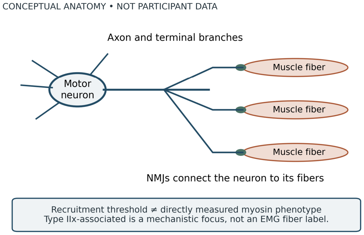
Figure 2.
Proposed chronology of fast-system deterioration. Conceptual model; no participant data. Dashed arrows represent the hypothesized progression and are not a demonstrated universal sequence. Functional impairment, connectivity changes, denervation, compensation, and structural loss may overlap or occur through alternative routes. No onset age or rate is assigned. Evidence informing the model: (Castro et al., 2023; Hassan et al., 2021; Piasecki et al., 2016).
Figure 2.
Proposed chronology of fast-system deterioration. Conceptual model; no participant data. Dashed arrows represent the hypothesized progression and are not a demonstrated universal sequence. Functional impairment, connectivity changes, denervation, compensation, and structural loss may overlap or occur through alternative routes. No onset age or rate is assigned. Evidence informing the model: (Castro et al., 2023; Hassan et al., 2021; Piasecki et al., 2016).
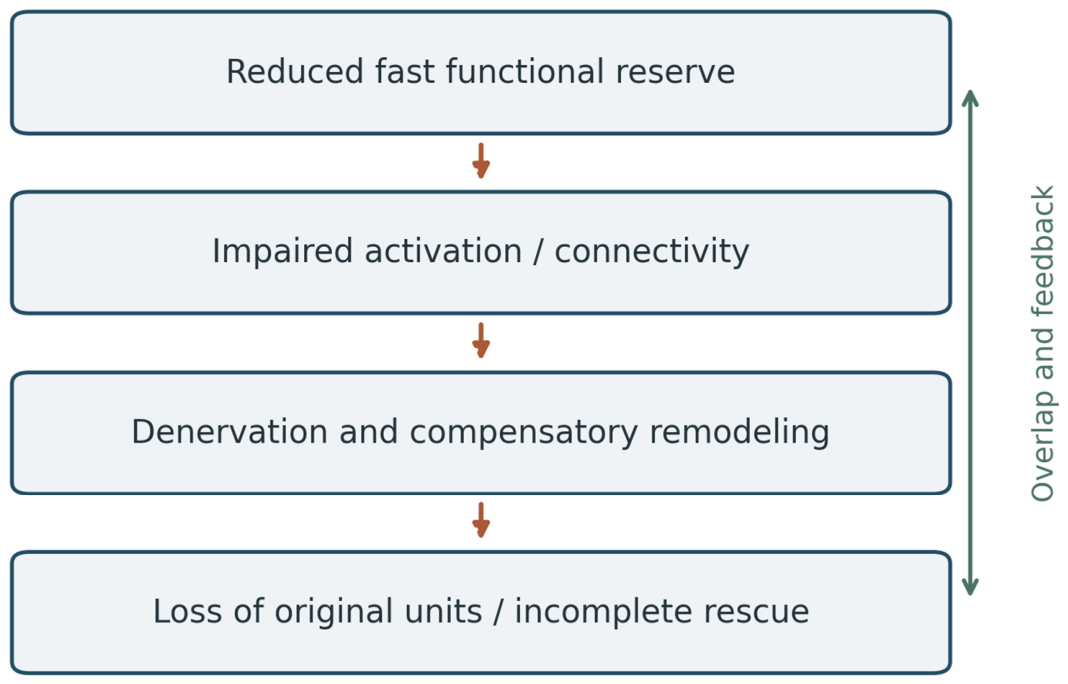
Figure 3.
Longitudinal knee-extension power across age groups. Measured summary data, redrawn from Table 3 of (Alcazar et al., 2023). Points show baseline and follow-up means; error bars are reported SDs, not uncertainty in the mean or paired change. Median follow-up was 9.6 years. Group labels follow the source (young, middle-aged, older); sample sizes appear beneath the axis. Between-group differences are not a lifespan trajectory. No values were digitized or extrapolated.
Figure 3.
Longitudinal knee-extension power across age groups. Measured summary data, redrawn from Table 3 of (Alcazar et al., 2023). Points show baseline and follow-up means; error bars are reported SDs, not uncertainty in the mean or paired change. Median follow-up was 9.6 years. Group labels follow the source (young, middle-aged, older); sample sizes appear beneath the axis. Between-group differences are not a lifespan trajectory. No values were digitized or extrapolated.
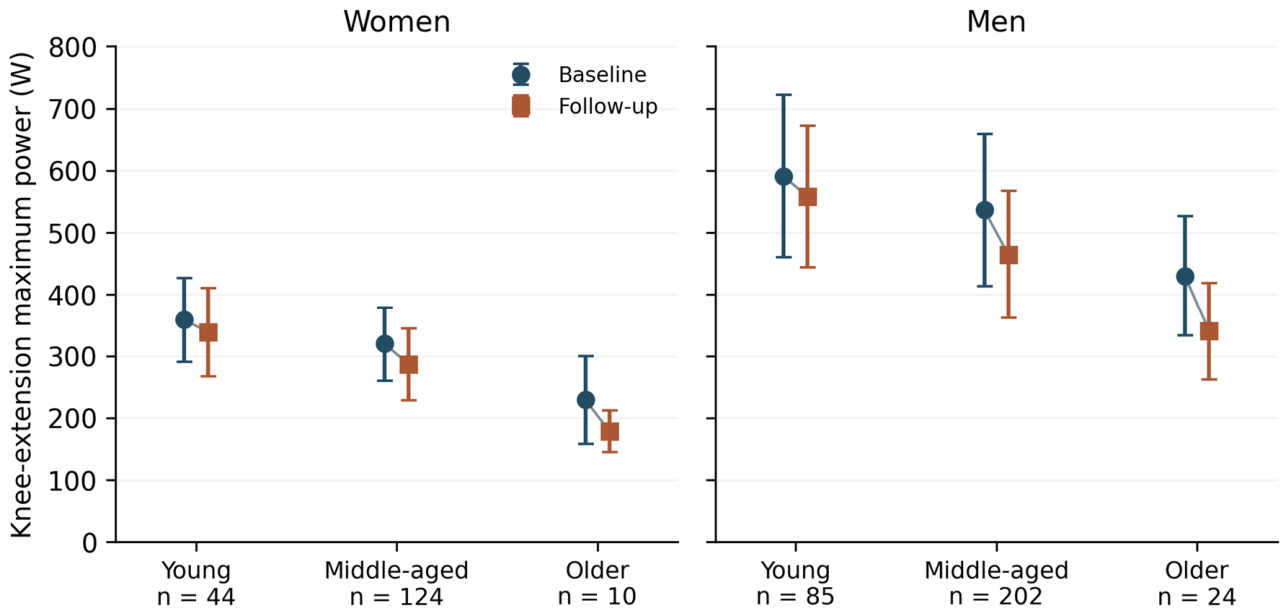
Figure 4.
The first 50 ms begins at force onset. Conceptual measurement schematic; no participant data. Horizontal distances are not proportional to measured latency. The stimulus-to-EMG interval, electromechanical delay, and early force-development interval are separate. Early RFD is not pure cortical drive. Source: (Maffiuletti et al., 2016).
Figure 4.
The first 50 ms begins at force onset. Conceptual measurement schematic; no participant data. Horizontal distances are not proportional to measured latency. The stimulus-to-EMG interval, electromechanical delay, and early force-development interval are separate. Early RFD is not pure cortical drive. Source: (Maffiuletti et al., 2016).
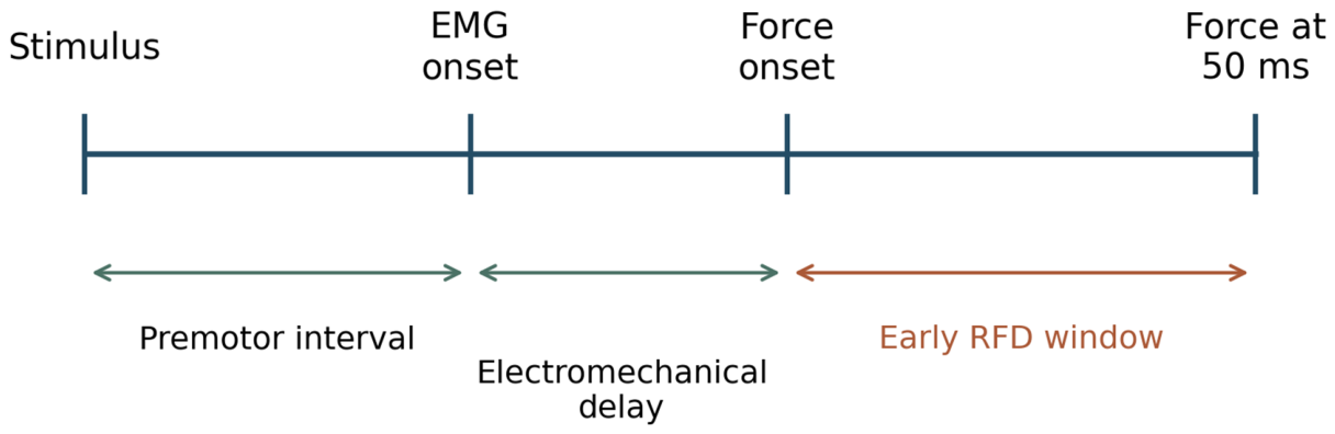
Figure 5.
Persistent inward currents within motor-neuron control. Conceptual physiological diagram; no participant data. PICs interact with synaptic inputs, inhibition, and neuromodulatory state to influence firing. Human ΔF estimates are indirect and task dependent. Sources: (Mesquita et al., 2024; Hassan et al., 2021; Orssatto, Rodrigues, et al., 2023).
Figure 5.
Persistent inward currents within motor-neuron control. Conceptual physiological diagram; no participant data. PICs interact with synaptic inputs, inhibition, and neuromodulatory state to influence firing. Human ΔF estimates are indirect and task dependent. Sources: (Mesquita et al., 2024; Hassan et al., 2021; Orssatto, Rodrigues, et al., 2023).
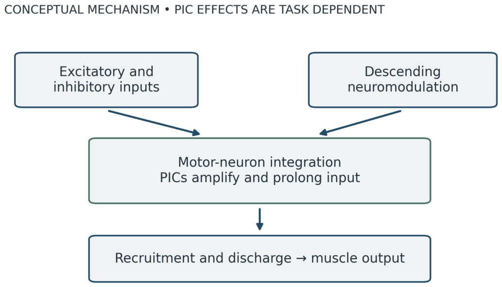
Figure 6.
Synaptic evidence and the proposed fast-system connection. Conceptual evidence map; no participant data plotted. Mouse synaptic findings and the human VGLUT1 trend are distinguished. The proposed link to preferential fast-unit dysfunction requires physiological confirmation; VGLUT1 is not an exclusive Type IIx marker. Source: (Castro et al., 2023).
Figure 6.
Synaptic evidence and the proposed fast-system connection. Conceptual evidence map; no participant data plotted. Mouse synaptic findings and the human VGLUT1 trend are distinguished. The proposed link to preferential fast-unit dysfunction requires physiological confirmation; VGLUT1 is not an exclusive Type IIx marker. Source: (Castro et al., 2023).
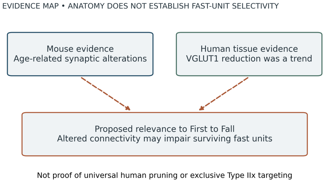
Figure 7.
The expanded First to Fall cascade. Proposed conceptual model; no participant data. Fast/high-threshold dysfunction remains central. Dashed links to remodeling, reduced movement, and systemic vulnerability are hypotheses, not established chronological causation. Feedback permits reciprocal influences. Physiological and mechanistic sources: (Hunter et al., 2016; Hepple & Rice, 2016; Mesquita et al., 2024; Pedersen, 2019).
Figure 7.
The expanded First to Fall cascade. Proposed conceptual model; no participant data. Fast/high-threshold dysfunction remains central. Dashed links to remodeling, reduced movement, and systemic vulnerability are hypotheses, not established chronological causation. Feedback permits reciprocal influences. Physiological and mechanistic sources: (Hunter et al., 2016; Hepple & Rice, 2016; Mesquita et al., 2024; Pedersen, 2019).
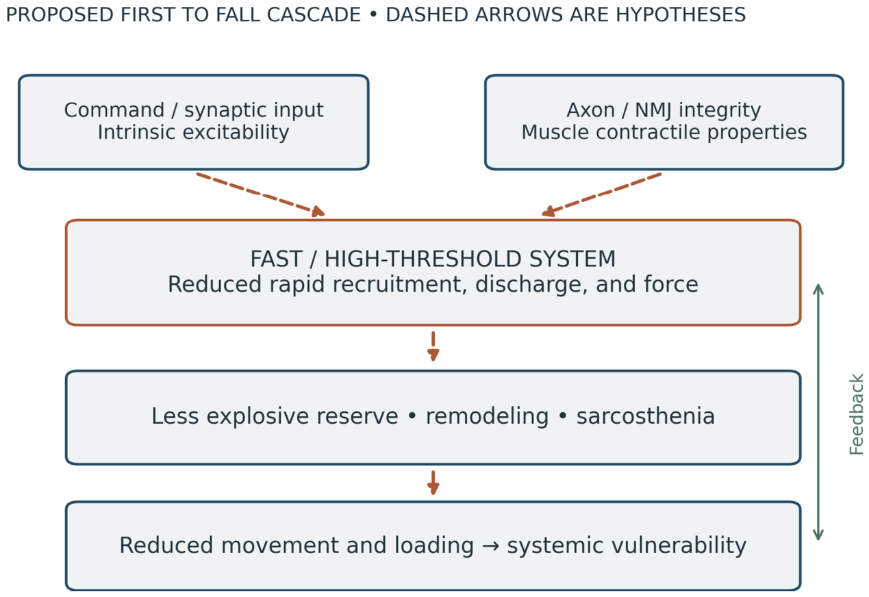
Figure 8.
Functional loss and muscle-size loss during bed rest. Reported measured percentage changes after 21 days in nine young men, reproduced from (Sarto et al., 2025). Cross-sectional area changed by −11.9% and maximal voluntary force by −23.1%. These are different outcomes; their difference does not isolate a neural contribution. Uncertainty intervals for these plotted summary percentages are not supplied here. No inference of motor-neuron death is made.
Figure 8.
Functional loss and muscle-size loss during bed rest. Reported measured percentage changes after 21 days in nine young men, reproduced from (Sarto et al., 2025). Cross-sectional area changed by −11.9% and maximal voluntary force by −23.1%. These are different outcomes; their difference does not isolate a neural contribution. Uncertainty intervals for these plotted summary percentages are not supplied here. No inference of motor-neuron death is made.
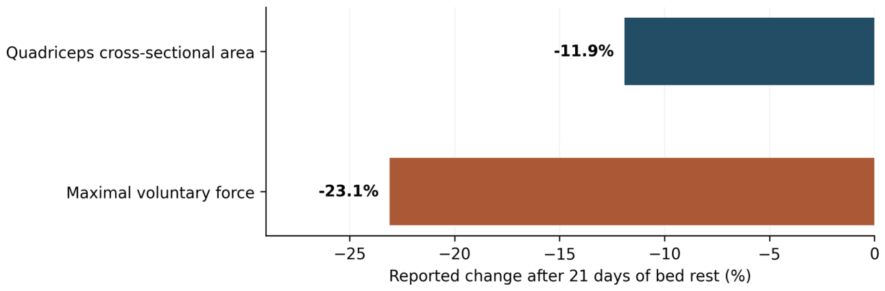
Figure 9.
Collateral reinnervation preserves fibers while changing architecture. Conceptual schematic; no participant data. Colors distinguish original units, not fast and slow fiber classes. A surviving axon can rescue fibers after loss of their original innervation, producing fewer independently controlled units. The diagram is not a quantitative account of age-related losses. Sources: (Piasecki et al., 2016; Hepple & Rice, 2016).
Figure 9.
Collateral reinnervation preserves fibers while changing architecture. Conceptual schematic; no participant data. Colors distinguish original units, not fast and slow fiber classes. A surviving axon can rescue fibers after loss of their original innervation, producing fewer independently controlled units. The diagram is not a quantitative account of age-related losses. Sources: (Piasecki et al., 2016; Hepple & Rice, 2016).
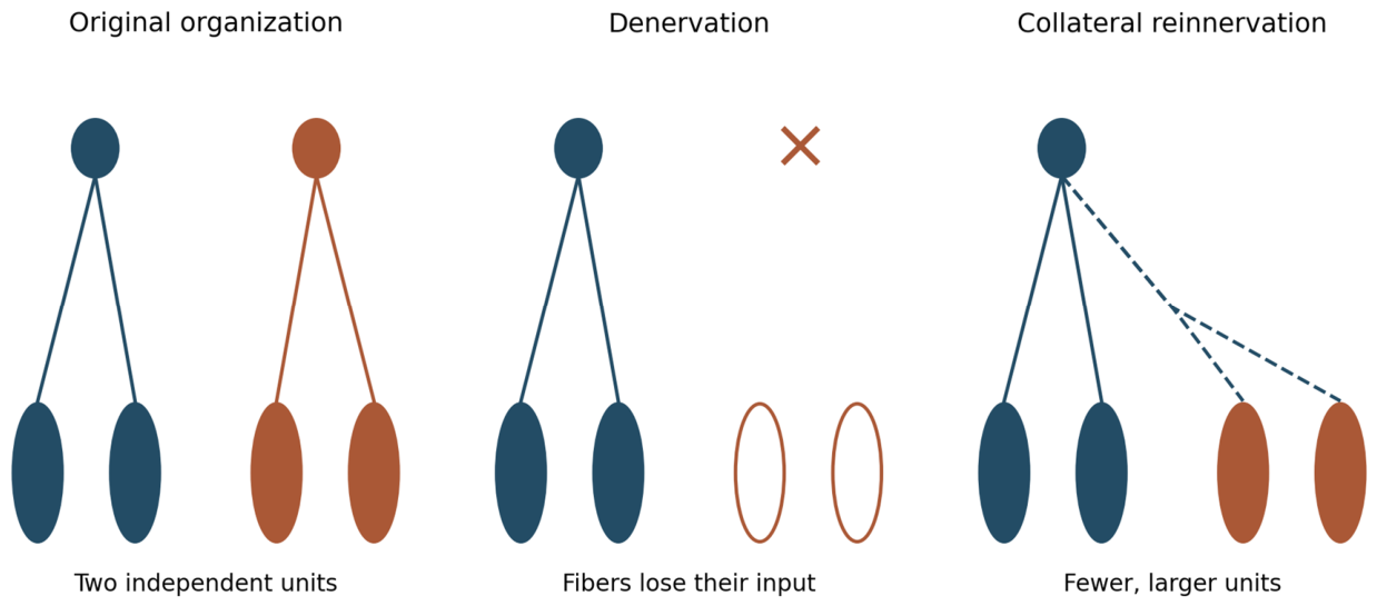
Figure 10.
Muscle volume and jumping power in male athletes and controls. Measured summary data redrawn from Table 7 of (Zange et al., 2026) (2026 issue; first published online in 2025). Bars are means ± SD. Young athletes n = 10, older athletes n = 10, young controls n = 12, older controls n = 11; age ranges were 20–35 and 60–75 years. Assessed volume combines gluteal, quadriceps, and triceps surae muscles. These cross-sectional measures do not isolate neural mechanisms. Numerical values are provided in the accompanying figure-data file.
Figure 10.
Muscle volume and jumping power in male athletes and controls. Measured summary data redrawn from Table 7 of (Zange et al., 2026) (2026 issue; first published online in 2025). Bars are means ± SD. Young athletes n = 10, older athletes n = 10, young controls n = 12, older controls n = 11; age ranges were 20–35 and 60–75 years. Assessed volume combines gluteal, quadriceps, and triceps surae muscles. These cross-sectional measures do not isolate neural mechanisms. Numerical values are provided in the accompanying figure-data file.
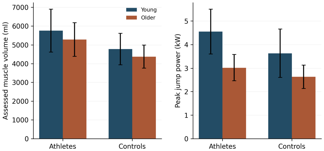
Figure 11.
Component repair versus integrated restoration. Conceptual criteria; no participant data. Success at one level does not establish success at all subsequent levels. The diagram preserves the original integration argument without claiming that future restoration is impossible. Sources on regeneration, coordination, and mechanical context: (Zelada et al., 2021; De Luca & Erim, 1994; Kwan et al., 2023).
Figure 11.
Component repair versus integrated restoration. Conceptual criteria; no participant data. Success at one level does not establish success at all subsequent levels. The diagram preserves the original integration argument without claiming that future restoration is impossible. Sources on regeneration, coordination, and mechanical context: (Zelada et al., 2021; De Luca & Erim, 1994; Kwan et al., 2023).
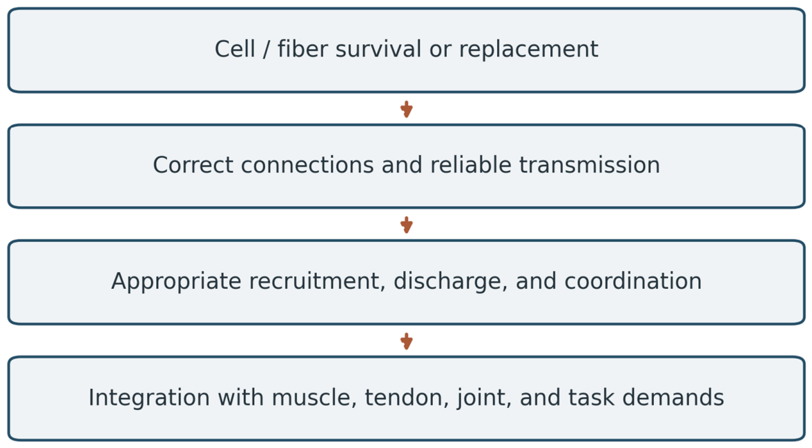
Figure 12.
One function can become limiting while other reserves remain. Conceptual reserve model; no participant data. Capacity and demand must be defined for a specific function in compatible units. No age axis, universal threshold, or maximum-lifespan estimate is assigned. Adapted conceptually from reserve principles in (Atamna et al., 2018); the fast-system application is the hypothesis developed here.
Figure 12.
One function can become limiting while other reserves remain. Conceptual reserve model; no participant data. Capacity and demand must be defined for a specific function in compatible units. No age axis, universal threshold, or maximum-lifespan estimate is assigned. Adapted conceptually from reserve principles in (Atamna et al., 2018); the fast-system application is the hypothesis developed here.
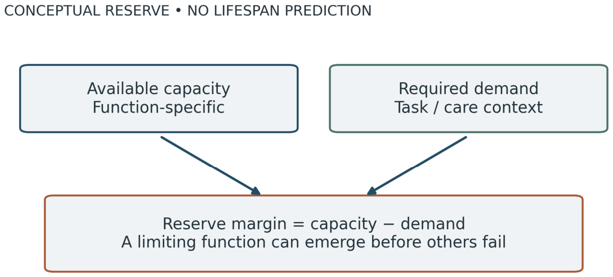

Table 1.
Evidence supporting and constraining First to Fall.
| Component | Evidence / population | Relevance and limitation |
|---|---|---|
| Early morphology | Whole vastus lateralis; 43 men, 15–83 years (Lexell et al., 1988) | Early atrophy and Type II size changes; not selective IIx-neuron counts. |
| Longitudinal power | 489 adults followed approximately ten years (Alcazar et al., 2023) | Within-person decline before frailty; cause not identified. |
| Rapid discharge | Young–older human dorsiflexor comparison (Klass et al., 2008) | Discharge and rapid torque differ; contractile slowing also contributes. |
| PIC estimates | Young–older upper-limb recordings (Hassan et al., 2021) | Altered excitability in surviving units; indirect, submaximal estimate. |
| PIC modification | 17 older adults; resistance training (Orssatto, Rodrigues, et al., 2023) | Functional plasticity; not evidence of neuronal replacement. |
| Synaptic inputs | Mouse, primate, and human tissue (Castro et al., 2023) | Connectivity can change with survival; human VGLUT1 finding was a trend. |
| Microglial mechanisms | Aging animal studies (Castro et al., 2024; Giorgetti et al., 2019) | Mechanistic candidates; human causal sequence remains untested. |
| Discharge modulation | Human age/sex comparisons (Carbonaro et al., 2026) | Task- and sex-dependent differences; not a universal fast-unit census. |
| Disuse vulnerability | Nine young men; 21-day bed rest (Sarto et al., 2025) | Rapid neural/muscular disturbance; small sample, no proof of neuron death. |
| Athletic preservation | Older athletes and controls (Power et al., 2016; Piasecki et al., 2019) | Preservation and compensation vary; selection and muscle specificity matter. |
| Sarcosthenia | Male athletes and controls; imaging and jump power (Zange et al., 2026) | Power loss beyond tissue quantity; not exclusively neural. |
| Systemic signaling | Cell and animal experimental work (Rutti et al., 2018; Szaroszyk et al., 2022) | Plausible muscle–organ links; fast-unit loss as human cause untested. |
Table 2.
Discriminating tests of the principal claims.
| Claim | Result that would strengthen it | Result that would challenge it |
|---|---|---|
| Preferential fast-system deterioration | Earlier within-person change in identified fast-unit function than appropriate comparator units. | Comparable or earlier deterioration in other units under matched measurement. |
| Function changes before structural loss | Impaired activation/connectivity with initially preserved anatomy, followed longitudinally. | Structural or muscle-intrinsic changes consistently explain the earliest deficit. |
| Specific movement preserves reserve | Targeted training slows fast-system deterioration beyond matched comparison exposure. | No specific preservation despite adherence and verified target engagement. |
| Fast-system change contributes to systemic decline | Neural preservation precedes and mediates independently measured downstream benefit. | Local preservation occurs without the predicted downstream effects. |
| Neuromuscular reserve can limit exceptional survival | Documented limiting neuromuscular function with competing capacities relatively preserved. | Different processes account for terminal limitation in the population studied. |
Disclaimer/Publisher’s Note: The statements, opinions and data contained in all publications are solely those of the individual author(s) and contributor(s) and not of MDPI and/or the editor(s). MDPI and/or the editor(s) disclaim responsibility for any injury to people or property resulting from any ideas, methods, instructions or products referred to in the content. |
© 2026 by the authors. Licensee MDPI, Basel, Switzerland. This article is an open access article distributed under the terms and conditions of the Creative Commons Attribution (CC BY) license (http://creativecommons.org/licenses/by/4.0/).
Copyright: This open access article is published under a Creative Commons CC BY 4.0 license, which permit the free download, distribution, and reuse, provided that the author and preprint are cited in any reuse.