

Submitted:
02 October 2026
Posted:
05 October 2026
You are already at the latest version
Abstract
Nanotheranostics integrates diagnostic and therapeutic functions within a single plat-form, representing a compelling strategy for personalized cancer therapy. Magnetic iron oxide nanoparticles are intrinsic suitable for imaging and hyperthermia. However, insuf-ficient colloidal stability in physiologic environment as well as a lack in surface function-ality often limit their application in biomedicine. In this work, we address these limitations by developing drug-loaded polymeric magnetic nanohybrids (pMNHs) stabilized by nu-cleobase-functionalized copolymers that serve as both stabilizing agents and drug carri-ers. Hydrophilic copolymers were synthesized via RAFT polymerization and functional-ized with cytosine and guanine. MNPs were prepared by continuous micromixer-assisted co-precipitation, followed by copolymer self-assembly to form stable pMNHs. Physico-chemical and magnetic characterization assessed their suitability for magnetic resonance imaging (MRI), magnetic particle imaging (MPI), and therapeutic magnetic hyperthermia. Polymer stabilization largely preserved magnetic performance, reducing the magnetic par-ticle spectroscopy (MPS)-derived A3* by 22% while changing A5/A3 by < 1%, and enhanced salt stability without further impairing the magnetic response. Following successful dox-orubicin (Dox) loading, the pMNHs showed evidence of cellular uptake while maintain-ing approximately 90% viability in HeLa cells across the tested Dox concentration range of 0.2–20 µM. Radiofrequency exposure induced a Dox-characteristic spectral change with-out substantially affecting the magnetic response, providing preliminary evidence for ex-ternally triggered Dox release. These findings establish nucleobase-functionalized pMNHs as effective platforms for cancer theranostics.
Keywords:
nanotheranostics
; magnetic nanohybrids
; iron oxide magnetic nanoparticles
; nucleobase polymers
; drug delivery
; magnetic hyperthermia
; magnetic particle imaging (MPI)
; doxorubicin (Dox)
1. Introduction
Nanotheranostics has emerged as an impactful field in modern medicine by combining diagnostic and therapeutic capabilities into a single integrated platform. This concept is particularly promising for personalized and targeted treatments, especially in oncological applications.[1,2,3] Iron oxide based magnetic nanoparticles (MNPs) have attracted significant attention as functional entities for nanotheranostic agents. They offer intrinsic advantages such as favorable biocompatibility, superparamagnetic behaviour, tunable magnetic properties, and the ability to be utilized in various biomedical applications.[4,5] These include magnetic imaging techniques such as magnetic resonance imaging (MRI) or magnetic particle imaging (MPI), and therapeutic approaches like magnetic hyperthermia.[6,7] However, bare MNPs often suffer from colloidal instability and aggregation in physiological environments. In addition, their unmodified surfaces generally provide limited opportunities for efficient drug loading, controlled release, or biological functionalization, which restricts their direct application in nanomedicine.[8]
To overcome these challenges, strategies involving the coating of MNPs with functional and stabilizing layers are frequently applied. Polymer coatings can improve colloidal stability, reduce nonspecific protein adsorption, and enhance the biocompatibility of magnetic nanomaterials.[9] Moreover, they can provide functional groups for covalent conjugation, electrostatic association, or encapsulation of therapeutic agents. Conventional polymers such as polyethylene glycol (PEG) and dextran are widely used because of their hydrophilicity, antifouling or stealth properties, and ability to reduce nanoparticle aggregation and opsonization.[10] However, additional characteristics—including drug-loading capacity, stimulus-responsive behaviour, release kinetics, and the possibility of introducing specific biological interactions—must also be considered for the rational design of polymer-coated magnetic nanocarriers as theranostic agents.
Recently, bioinspired polymers incorporating nucleobases have attracted increasing interest owing to their anticipated biocompatibility and the ability of nucleobases to mediate directional and reversible noncovalent interactions.[11] Complementary base pairing enables programmable self-assembly through hydrogen bonding, while nucleobases can additionally participate in π stacking interactions – weak attractive forces between aromatic ring systems that contribute to molecular stability and packing.[12]
Nucleobase-containing block copolymers, frequently prepared by controlled radical polymerization methods such as reversible addition–fragmentation chain-transfer (RAFT) polymerization, have been shown to form supramolecular assemblies with tuneable morphologies depending on polymer architecture, solvent, pH, and nucleobase content.[13,14] These systems can therefore combine the biomimetic character of nucleic-acid interactions with the synthetic flexibility of polymer chemistry. Polymeric materials, particularly in nanoparticulate form, are well-established carriers for drug molecules and active pharmaceutical ingredients (APIs), as they can encapsulate therapeutic compounds and enable their controlled release.[15,16] Exploiting these advantages, the integration of nucleobase-functionalized polymers with MNPs may yield multifunctional polymeric magnetic nanohybrids (pMNHs) for simultaneous drug delivery and magnetic imaging or hyperthermia. In contrast to conventional coated MNPs, which typically rely on passive surface layers merely for stabilization or basic conjugation, these nucleobase-driven systems introduce an active, dynamic interface. Here, complementary nucleobase interactions provide a molecular-recognition motif that can direct self-assembly,[17] mediate stimuli-responsive drug release,[18,19] and regulate interactions between nanostructures,[20] extending their functionality beyond that of a passive coating.[12,21]
This work presents the development and characterization of drug-loaded magnetic nanohybrids utilizing nucleobase-functionalized copolymers as stabilizing and drug-carrying moieties for iron oxide MNPs. To this end, we synthesized the hydrophilic diblock copolymers P(GMA)-b-P[AEMA-stat-(AEMA-C)] and P(GMA)-b-P[AEMA-stat-(AEMA-G)] via RAFT polymerization of 2,3-dihydroxypropyl methacrylate (glyceryl monomethacrylate, GMA) and 2-((tert-butoxycarbonyl)amino)ethyl methacrylate (Boc-AEMA), followed by post-polymerization modification with acetic acid functionalized cytosine and guanine. Single-core iron oxide MNPs were synthesized using a continuous micromixer-based co-precipitation approach via aqueous synthesis route.[22] Subsequently, the nucleobase-functionalized copolymers were self-assembled in the presence of the MNPs, resulting in the formation of stable pMNHs. The physicochemical and magnetic properties of the resulting pMNHs were comprehensively characterized, demonstrating their capability for diagnostic MRI and MPI applications as well as in therapeutic magnetic hyperthermia applications. In addition, the pMNHs were successfully loaded with the chemotherapeutic agent doxorubicin (Dox), and their cellular uptake and cytotoxicity were evaluated in HeLa cells.[23] Overall, these findings highlight the potential of nucleobase-functionalized polymer-based magnetic nanohybrids as versatile and effective platforms for advanced cancer theranostics.
2. Materials and Methods
2.1. Materials
Unless otherwise specified, all chemicals and solvents were used as received without further purification. N,N-dimethylformamide (DMF) and acetonitrile (ACN) of HPLC or LC–MS grade (CHROMASOLV™, ≥99.9%; Fisher Scientific GmbH, Schwerte, Germany) for analytical measurements and were supplemented with lithium bromide (anhydrous, 99.0+%; Thermo Fisher Scientific GmbH, Dreieich, Germany) or formic acid (LiChropur; Merck KGaA, Darmstadt, Germany). For polymerization reactions under inert atmosphere, anhydrous DMF (≥99.9%; Merck KGaA, Darmstadt, Germany) stored over 3 Å and 4 Å molecular sieves under nitrogen was used as reaction medium. Solvents used for purification and reactions under ambient conditions, including ACN (99.9%, Ph. Eur., HPLC grade), tetrahydrofuran (THF, 99.8%), diethyl ether (Et2O, ≥99.0%, extra pure), methylene chloride (DCM, Puriss. p.a., ACS reagent, ≥99.9% GC), methanol (MeOH, CHROMASOLV™, ≥99.9%), and ethyl acetate (EtOAc, CHROMASOLV™, ≥99.7%), were obtained from Fisher Scientific GmbH (Schwerte, Germany). Ethanol (99.8%, denatured with isopropanol) was purchased from AppliChem (Darmstadt, Germany). Ultrapure water was generated using a Milli-Q Plus purification system (Merck Millipore, Darmstadt, Germany) equipped with a QPAK® 2 cartridge.
The RAFT agent 4-cyano-4-[(phenylcarbonothioyl)thio]pentanoic acid (CPCTPA, 98.0%) and the monomers 2,3-dihydroxypropyl methacrylate (glyceryl monomethacrylate, GMA, 90.0%) and 2-((tert-butoxycarbonyl)amino)ethyl methacrylate (Boc-AEMA, 95.0%) were obtained from BLD Pharmtech Ltd. (Reinbek, Germany). Prior to use, monomers were purified by passage through a basic alumina column to remove polymerization inhibitors. 2,2′-Azobis(2-methylpropionitrile) (AIBN, 98.0%), methyl bromoacetate (97.0%), 2-amino-6-chloropurine (≥99.0%), cytosine (≥99.0%), doxorubicin hydrochloride (98.0–102.0%), ammonium sulfate (Reag. Ph. Eur.), and calcium acetate hydrate (extra pure) were purchased from Merck KGaA (Darmstadt, Germany). Cesium carbonate (98.0%), 1,4-diazabicyclo[2.2.2]octane (DABCO, 98.0%), di-tert-butyl dicarbonate (Boc2O, 98.0%), and N,N,N′,N′-tetramethyl-O-(7-azabenzotriazol-1-yl)uronium hexafluorophosphate (HATU, 99.0%) were obtained from Carbolution Chemicals GmbH (St. Ingbert, Germany). Trifluoroacetic acid (TFA, PEPTIPURE®, ≥99.9%) and N,N-diisopropylethylamine (DIPEA, PEPTIPURE®, ≥99.5%) were purchased from Carl Roth GmbH + Co. KG (Karlsruhe, Germany).
Reagents for magnetic nanoparticle synthesis and characterization, including iron(II) chloride tetrahydrate (≥99%), sodium nitrate (Ph. Eur.), sodium hydroxide, sucrose (Ph. Eur.), iron(II,III) oxide (98%), hydroxylamine hydrochloride (99%), and 1,10-phenanthroline monohydrate (reagent grade), were obtained from Sigma-Aldrich (Taufkirchen, Germany). Sodium chloride (Ph. Eur.) and acetic acid (Ph. Eur.) were supplied by Scharlau (Barcelona, Spain). Sodium acetate trihydrate (Riedel-de Haën) was purchased from Honeywell (Offenbach, Germany), while tannic acid was obtained from MP Biomedicals (Irvine, CA, USA).
2.2. Copolymer Synthesis
Nucleobase-functionalized diblock copolymers were synthesized via a multistep protocol as described in a related manuscript currently under review,[24] involving RAFT-mediated polymerization, post-polymerization functionalization, and deprotection steps.
Briefly, GMA (80 equiv.) was polymerized in anhydrous, degassed DMF using CPCTPA (1.0 equiv.) and AIBN (0.2 equiv.) under Schlenk conditions at 60 °C for 16 h to yield P(GMA) homopolymer (93%). This resulting macro-RAFT agent (1.0 equiv.) was chain-extended via copolymerization with Boc-AEMA (100.0 equiv.) in the presence of AIBN (0.2 equiv.) under inert conditions (DMF, 60 °C, 18 h), affording the diblock copolymer P(GMA)-b-P(Boc-AEMA) in 87% isolated yield.
Polymer purification was performed by precipitation (in ethyl acetate or diethyl ether) and dialysis against ultrapure water. Following Boc deprotection of AEMA amines, Boc-protected cytosine acetic acid (20.0 equiv.) or guanine acetic acid (20.0 equiv.) was conjugated to the polymer backbone (1.0 equiv.) via HATU-mediated (11c.0 equiv., 15.0 equiv. DIPEA) amidation (DMF, 2 h, room temperature).
RAFT end-group removal was achieved by radical-induced cleavage using excess (30 equiv.) AIBN under the mentioned polymerization conditions, eliminating the living character of polymer yields. Final purification (precipitation in diethyl ether, dialysis against DMSO and water) and Boc deprotection followed by lyophilization afforded the self-assembling diblock copolymers P(GMA)-b-P[AEMA-stat-(AEMA-C)] and P(GMA)-b-P[AEMA-stat-(AEMA-G)].
2.3. Nuclear Magnetic Resonance (NMR) Spectroscopy
NMR spectra were acquired at room temperature on Bruker Avance II 400 and Avance III 600 spectrometers (Bruker Corporation, Billerica, USA), both equipped with ATM and a 5.0 mm BBFO probe or 5.0 mm TCI CryoProbe respectively. 1H-NMR spectra were recorded at 400 or 600 MHz, while 13C-NMR spectra were obtained at 151 MHz using broadband 1H decoupling. Tetramethylsilane was employed as the calibration standard for both 1H and 13C measurements.
Spectral processing and analysis were performed with MestReNova v16.0.0-39276 (Mestrelab Research S.L., Santiago de Compostela, Spain). Chemical shifts (Δ) are reported in ppm and coupling constants (J) in Hz. Residual solvent signals of D2O and DMSO-d6 served as references for the respective spectra. Signal assignments were established based on COSY, HSQC, and HMBC experiments. Multiplicities are denoted as s (singlet), d (doublet), t (triplet), q (quartet), quin (quintet), m (multiplet), and br s (broad singlet).
2.4. Gel Permeation Chromatography (GPC)
The number-average molecular weight (Mₙ), weight-average molecular weight (Mw), and dispersity (Đ = Mw ∙ Mn‒1) of the homo- and copolymers were determined by gel permeation chromatography (GPC) using a modular 1260 Infinity II system (Agilent Technologies Deutschland GmbH, Mainz, Germany). The instrument configuration included a 1260 degasser, a 1260 vial sampler with an injection volume of 100 µL, a 1260 isocratic pump operating with DMF containing 2% LiBr at a flow rate of 1 mL ∙ min‒1, a 1260 GPC column oven maintained at 50 °C, an FC-AS fraction collector, and a 1260 refractive index detector. Separation was carried out using a GRAM precolumn (8 × 50 mm, 10 µm) connected in series to three GRAM analytical columns (1000 Å, 8 × 300 mm, 10 µm). Calibration was performed with 12 P(methyl methacrylate) (PMMA) standards covering a peak molar mass range from 600 to 2,000,000 Da. Molar mass and dispersity values were automatically calculated with the integrated WinGPC software.
2.5. Magnetic Nanoparticle (MNP) Synthesis
Iron oxide nanoparticles were prepared via continuous co-precipitation of iron salts under alkaline aqueous conditions using a micromixer-based approach, following a previously described protocol.[22] A caterpillar-type micromixer (Fraunhofer IMM, Mainz, Germany) enabled rapid mixing of iron(II) chloride, sodium nitrate, and sodium hydroxide solutions under controlled flow conditions. The resulting reaction stream was directed through a heated loop maintained at 40 °C, providing a residence time of 4 min at constant and symmetric flow rates. Tannic acid was introduced as a stabilizing agent during particle formation. Subsequent purification of the magnetic nanoparticles was achieved by magnetic separation to remove residual precursors and excess, non-adsorbed stabilizer.
2.6. Formation of Polymer-MNP Nanohybrids (pMNHs)
Self-assembly of polymeric MNP nanohybrids was carried out via a thermally assisted mixing protocol. Two separate stock dispersions were prepared by combining iron oxide nanoparticles (100 µL, 1.72 mM) with either P(GMA)-b-P[AEMA-stat-(AEMA-C)] or P(GMA)-b-P[AEMA-stat-(AEMA-G)] copolymer (100 µL, 410 µM each) in ultrapure water. Each dispersion, along with tris(hydroxymethyl)aminomethane (TRIS) buffer (50 µL, 50 mM) containing MgCl₂ (50 mM, pH 6), was independently heated to 95 °C for 10 min. The preheated MNP-polymer dispersions were subsequently combined and immediately supplemented with the preheated TRIS–MgCl₂ buffer. The resulting mixture was then allowed to cool gradually to room temperature at a controlled rate of 1 °C ∙ min⁻¹.
Formation of nanostructures was indicated by a distinct transition from an initially clear to an opaque dispersion.
2.7. Drug Loading
Drug loading was achieved by introducing 13 µL of a Dox stock solution (55 mM in ultrapure water) into 100 µL of the pMNHs dispersion and incubating the mixture at 60 °C for 1 h. The loading process was terminated by cooling to room temperature to yield Dox-loaded pMNHs (Dox-pMNHs). Non-encapsulated drug was subsequently removed by dialysis against ultrapure water using Xpress Micro Dialyzer MD300 cartridges (Scienova GmbH, Jena, Germany; MWCO 3.5 kDa).
2.8. Quantification of pMNH-Associated Dox
UV–vis absorption measurements were performed using a Tecan Spark® multimode microplate reader (Tecan Trading AG, Männedorf, Switzerland) to quantify Dox associated with the pMNHs. Aliquots of 100 µL were transferred to 96-well UV-STAR® microplates (Greiner Bio-One International GmbH, Kremsmünster, Austria), and absorbance was measured at 480 nm against appropriate controls and calibration standards.
2.9. Iron Concentration
The iron concentrations, c(Fe), of the MNP and pMNH samples were quantified by UV-Vis spectroscopy using a phenanthroline-based assay.[25] Briefly, 10 µL of the sample dispersion was dissolved in 45 µL of concentrated HCl (37%). After complete dissolution, 200 µL of ultrapure water was added. An aliquot of 25 µL of this solution was transferred to a 96-well plate and mixed with 50 µL hydroxylamine hydrochloride solution (10%) and 200 µL 1,10-phenanthroline hydrochloride solution (0.1%). Following an incubation period of 25 min, the absorbance of the resulting ferroin complex was recorded at 510 nm using a Tecan Spark® multimode microplate reader (Tecan Trading AG, Männedorf, Switzerland). Iron concentrations were determined from a calibration curve generated with iron(II,III) oxide standards over a concentration range of c(Fe) = 0–40 mmol ∙ L‒1. All samples were measured in triplicate.
2.10. DC Magnetization (DCM) Measurements
Room-temperature (T = 295 K) DC magnetization measurements of the basic MNP system were carried out using a SQUID magnetometer (MPMS-XL, Quantum Design, USA). The magnetic moment m(H) of 50 µL liquid samples was recorded as a function of an applied external magnetic field H up to 4 × 10⁶ A ∙ m‒1 (B = 5 T). The mass magnetization M(H), given in Am² ∙ kg(Fe)‒1 or emu ∙ g(Fe)‒1, was obtained by normalizing to the total iron content of the sample. The saturation magnetization Mₛ was taken as the value at B = 5 T after subtraction of the linear background contribution originating from sample container, water, and dia- and paramagnetic sample constituents. The overall uncertainty was estimated to be approximately 5%, including 2% from the instrument and 3% from sample preparation and iron quantification. The M(H) curves were not fitted using the Langevin model, as the underlying assumptions of isotropy and thermal equilibrium at room temperature are no longer valid for particles with core diameters exceeding 15 nm.
2.11. Magnetic Particle Spectroscopy (MPS)
The non-linear magnetic susceptibility was investigated by magnetic particle spectroscopy, where a sinusoidal excitation field is applied to the magnetic sample, and the nonlinear dynamic response of the magnetic moments for the MNPs and pMNHs is detected. From the resulting spectra, two characteristic parameters A₃ (amplitude of the 3rd harmonic) and A₅/A₃ (ratio of the 5th to the 3rd harmonic) were determined. These parameters depend on the MNP or pMNH size distribution,[26] magnetic anisotropy, binding state,[27] and interparticle interactions.[28] Since MPS is based on the same physical principle as MPI (MPS is often considered as 0D-MPI without spatial resolution) it is often used to obtain information on the suitability of the particles for MPI. Note that the MPS parameter A₅/A₃ is independent of concentration, thus uncertainties arising from sample preparation, such as pipetting uncertainties (normally up to 5%) are here negligible; accordingly, this parameter was determined with an uncertainty below 1%. Since the uncertainty bars were smaller than the symbol size, they are not shown in the corresponding figures.
Measurements were performed at 37 °C using a commercial MPS instrument (MPS-3, Bruker, Rheinstetten, Germany) operated with a sinusoidal excitation field amplitude of B = 25 mT at a fixed frequency f₀ = 25 kHz. Four sample aliquots of 10 µL volume were filled into PCR tubes and measured first in the fluid state. Subsequently, the first sample aliquot was kept in the original state, while to the second sample gypsum powder was added for fast immobilization. To the third aliquot, 10 µL of 2 M NaCl solution was added to test colloidal stability and the fourth sample was freeze-dried after adding 10 µL of a 10% mannitol solution. After 8 hours incubation all samples were remeasured by MPS to determine any changes.
2.12. AC-Magnetometry (Hyperthermia Measurements)
AC magnetometry (hysteresis) measurements were performed using a commercial AC Hyster™ magnetometer system (Nanotech Solutions S.L., Villacastín, Spain). The instrument is designed for the analysis of liquid nanoparticle samples with a volume of 40 µL exposed to RF-frequencies in the range 25 to 100 kHz. To minimize heat transfer from the excitation coil to the sample chamber the device is equipped with an integrated fluidic cooling system. According to the manufacturer’s specifications, the iron concentration c(Fe) of the samples was kept below 10 g ∙ L‒1 to avoid interference with the coil system.
The dynamic hysteresis loop of the sample to the applied drive field was detected by a pick-up coil. The enclosed loop area A is determined and after normalization to the iron mass of the sample reported as specific absorption rate (SAR) (in units of W ∙ g‒1). In hyperthermia applications, the SAR value is often called specific loss power (SLP). SAR or SLP depend on the excitation frequency and amplitude of the RF-field. To become (more) independent of the excitation field parameters the intrinsic loss power (ILP) is preferred, ILP = SLP ∙ (H² ∙ f)‒1. All measurements were conducted in triplicate, resulting in an estimated uncertainty of approximately 3%. In this study, magnetic hyperthermia behaviour was assessed at a frequency of 100 kHz and a field amplitude of 24 kA ∙ m‒1.
2.13. Relaxation Rate Measurements
For each sample, a dilution series in ultrapure water of five concentrations with iron concentrations between 0.001 and 0.5 mmol ∙ L‒1 was prepared. All stated iron concentrations refer to the final concentrations in the measured samples. Ultrapure water was measured as references to determine the relaxation rates of the undiluted matrix. 200 µL of each sample was filled into NMR tubes and equilibrated at 37 °C in a water bath for 10 min before measurement.
The longitudinal (T₁) and transverse (T₂) relaxation times of the water protons were measured at 37 °C and 1.41 T (proton resonance frequency 60 MHz) using a magnetic resonance relaxometer (minispec mq60, Bruker BioSpin, Germany). T₁ was determined with a saturation recovery pulse sequence using 24 geometrically spaced recovery delays. The delay range was adapted to the expected T₁ of each sample, from 3–1500 ms for the highest iron concentration to 100–30,000 ms for the lowest concentration and for the agar reference, and 400–40,000 ms for the water reference. T₂ was determined with a Carr–Purcell–Meiboom–Gill (CPMG) pulse sequence with a pulse separation of 0.04 ms, acquiring 32,000 echoes. Both relaxation times were obtained by mono-exponential fits using the instrument software. Identical settings were used for measurements in water and in agar. Each sample was measured once.
The relaxation rates R1 = 1 ∙ T1‒1 and R2 = 1 ∙ T2‒1 were plotted against the iron concentration c(Fe), and the relaxivities r₁ and r₂ were obtained as the slopes of linear fits. The stated uncertainties are the standard errors of the slopes.
2.14. Dynamic Light Scattering (DLS)
DLS measurements were performed on a Zetasizer Ultra Red (Malvern Panalytical, Malvern, United Kingdom) equipped with a He–Ne laser (λ = 633 nm) and a backscatter detector at 173°. Samples were equilibrated for 120 s and analyzed at 25 °C after dilution 1:100 (v/v) with ultrapure water in disposable Brand® UV micro cuvettes (Brand GmbH + Co KG, Wertheim, Germany). Autocorrelation data were evaluated using ZS Xplorer software with automatic position and attenuator settings and multiple runs to obtain cumulant particle size, intensity-weighted size distributions, Z-average of hydrodynamic diameter, and polydispersity index (PDI), which are reported as calculated. All Measurements were automatically performed in triplicates with a resulting uncertainty of less than 5%.
2.15. Differential Centrifugal Sedimentation (DCS)
DCS analyses were performed using a CPS Instruments system (CPS Instruments Inc. Measurements, Darmstadt, Germany) operated at 20,000 rpm (corresponding to 21,504 × g) and calibrated with a 245 nm silicon dioxide standard. Measurements were carried out in a sucrose density gradient ranging from 24% to 8%. Peak maxima and full widths at half maximum (FWHM) were extracted using Origin® software.
2.16. Transmission Electron Microscopy (TEM) and Cryogenic-TEM
MNPs were examined by TEM on a Zeiss Libra 120 microscope (Zeiss, Oberkochen, Germany) operated at 120 kV to determine the size and shape of the MNP core. Sample preparation was done by drop casting the sample on a carbon-coated copper grid. A magnetic field was applied for ~15 min to gather the MNP. The solvent evaporated at room temperature. Micrographs were acquired using a CCD camera.
Nanohybrid morphology was examined by cryo-TEM on a Zeiss Libra 120 microscope (Zeiss, Oberkochen, Germany) operated at 120 kV at 185 °C. TEM grids (Lacey Carbon, EMS, Morgantown, WV, USA) were treated with an argon/oxygen plasma for 60 s at room temperature (Tergeo EM Plasma Cleaner, PIE Scientific LLC, CA, USA). For sample preparation, 5 µL of the particle dispersion was deposited onto a TEM grid inside a Leica EM GP plunger (Leica Microsystems, Wetzlar, Germany) at 80% relative humidity, blotted with filter paper for 1.2 s, and vitrified in liquid propane at -180 °C. Samples were stored in liquid nitrogen until measurement. Micrographs were acquired under liquid-nitrogen cryogenic conditions (-20 °C) using a CCD camera.
Image analysis for TEM and cryo-TEM images was performed with ImageJ (National Institutes of Health, Bethesda, MD, USA) to determine mean particle diameter, size distribution, and standard deviation based on measurements of individual nanostructures (N ≥ 500).
2.17. Fluorescence Microscopic Imaging
Cellular uptake and intracellular localization of Dox-loaded pMNHs were evaluated by fluorescence microscopy 2 h after treatment. Prior to imaging, HeLa cells were washed and maintained in 50 µL of 1× PBS. Fluorescence images were acquired on a BZ-X800 microscope (Keyence, Frankfurt am Main, Germany) using a TRITC filter set (excitation: 545/25 nm; emission: 605/70 nm) at 10× magnification.
2.18. Cell Cultivation and Cytotoxicity Assay
Cytotoxicity evaluation was assessed in HeLa cells seeded at 1 × 10⁴ cells per well in 96-well plates, with all treatments performed in triplicate. After 24 h of cultivation in 100 µL Dulbecco′s Modified Eagle′s Medium (DMEM) supplemented with 10% (v/v) Fetal Bovine Serum (FBS) and 1% (v/v) penicillin/streptomycin at 37 °C and 5% CO2, the medium was replaced with 90 µL of fresh medium. Nanohybrid solutions (1–100 µM, prepared in ultrapure water) were then added in a volume of 10 µL per well; free Dox at corresponding concentrations served as a reference. Following 24 h incubation under standard culture conditions, cell viability was quantified using the CCK-8 assay (Merck KGaA, Darmstadt, Germany). To this end, the medium was exchanged with fresh medium containing 10% (v/v) CCK-8 reagent and cells were incubated for 2 h at 37 °C and 5% CO2. The absorbance was subsequently recorded at 450 nm using a Tecan Spark® multimode microplate reader (Tecan Trading AG, Männedorf, Switzerland), and viability was expressed relative to untreated controls.
2.19. Radiofrequency (RF) Drug Release Experiments
Radiofrequency (RF) drug release experiments were performed with a custom-built setup generating an alternating magnetic field (AMF) by an air-cooled litz-wire coil, as part of a resonant circuit connected in parallel to the amplifier. A sinusoidal signal provided by a function generator was amplified by a broadband power amplifier (1040L, E&I) driving the resonant circuit. Air cooling of the coil was provided by a fan mounted below the coil. The system was operated at a frequency of f = 320 kHz and a magnetic field amplitude of H0 = 12 kA m−1. Eppendorf sample containers of up to V = 500 µL can be placed within the homogeneous excitation field region (homogeneity better 97.5%) of the AMF. By a fiber-optic temperature probe tip inserted directly into the sample fluid, the temperature can be read-out every 0.1 s using a fiber-optic thermometer (FOTEMP4, Optocon). During warm-up of the system for twarm-up = 30 min, the resonance frequency was corrected by approximately Δf = 0.5 kHz, as the impedance of the circuit components slightly changed with increasing temperature. Steady state was reached once the resonance frequency remained constant for 5 min.
For the heating, an aliquot of 110 µL of a sample was heated for 20 min starting the temperature acquisition with inserting the sample into the RF coil. In parallel another aliquot of 110 µL was kept as an unheated sample. After the RF-treatment, treated and untreated aliquots of 100 µL of the pMNH and the Dox-pMNH systems were transferred into a 96-well plate for UV-Vis measurements in the wavelength range 190 to 900 nm (Δλ = 1 nm) (SpectraMax 384, Molecular Devices). The difference between treated and untreated samples was then calculated to identify changes in the spectra due to RF-treatment.
3. Results
3.1. Copolymer Synthesis and Characterization
The copolymers were prepared as detailed in section 2.2 using a multistep synthesic procedure described in a related manuscript currently under review.[24] The procedure comprises reversible addition–fragmentation chain transfer (RAFT) polymerization followed by post-polymerization functionalization (Scheme 1).
Glyceryl methacrylate was homopolymerized using AIBN as radical initiator and CPCTPA as chain transfer agent (CTA). The resulting P(GMA) homopolymer (I) was isolated as a red solid by precipitation into cold ethyl acetate (93% yield). The identity of the polymer was confirmed by 1H-NMR spectroscopy (n = 84 GMA repeating units, Mₙ = 13,734 g ∙ mol‒1) and gel permeation chromatography (GPC; Đ = 1.05).[24]
Exploiting the living chain-end character of the RAFT-synthesized macro-CTA, block extension was carried out with Boc-protected aminoethyl methacrylate (Boc-AEMA) using analogous reaction conditions as previously described for the homopolymerization, followed by precipitation in cold diethyl ether. ¹H-NMR analysis confirmed successful formation of the diblock copolymer P(GMA)-b-P(Boc-AEMA) (II) with m = 70 Boc-AEMA repeating units (Mₙ = 29,783 g ∙ mol‒1) and a narrow molecular weight distribution (Đ = 1.10) evaluated by GPC.
For subsequent post-polymerization functionalization, the copolymer was pre-treated to remove both the terminal RAFT end group and the Boc protecting groups from the AEMA amines. End group cleavage was achieved via radical-induced decomposition using a large excess of AIBN. Successful removal of the phenylcarbonothioylthio functionality was confirmed by UV–vis spectroscopy: the characteristic absorption peak at λmax = 300 nm was no longer detectable (Figure 1a). Consistently, ¹H-NMR spectra showed complete disappearance of the aromatic proton signals from the phenyl rings at δ = 7.81, 7.62, and 7.45 ppm (Figure 1b).
Boc deprotection of the AEMA side-chain amines was performed by dialysis against aqueous HCl and deionized water to get P(GMA)-b-P(AEMA) (III). The progress of deprotection was monitored by ¹H-NMR spectroscopy, tracking the disappearance of the tert-butyloxycarbonyl methyl protons at δ = 1.38 ppm (Figure 2a).
The primary amines obtained were subsequently engaged in a substoichiometric amidation reaction with the carboxylic acid-modified nucleobases guanine or cytosine. An amide-coupling strategy was employed, using DIPEA as non-nucleophilic base and HATU as coupling reagent for sterically demanding substrates. The 3:7 nucleobase-to-amine ratio afforded degrees of functionalization of 45% for both (IV-C) and (IV-G), corresponding to nine nucleobase moieties each. The crude products were purified by precipitation in cold diethyl ether, followed by stepwise dialysis against DMSO, 0.1 M HCl, and ultrapure water. The identities of the nucleobase-containing copolymers were confirmed by 1H-NMR spectroscopy and UV–vis absorbance measurements (Figure 2b).
3.2. MNP Synthesis and Characterization
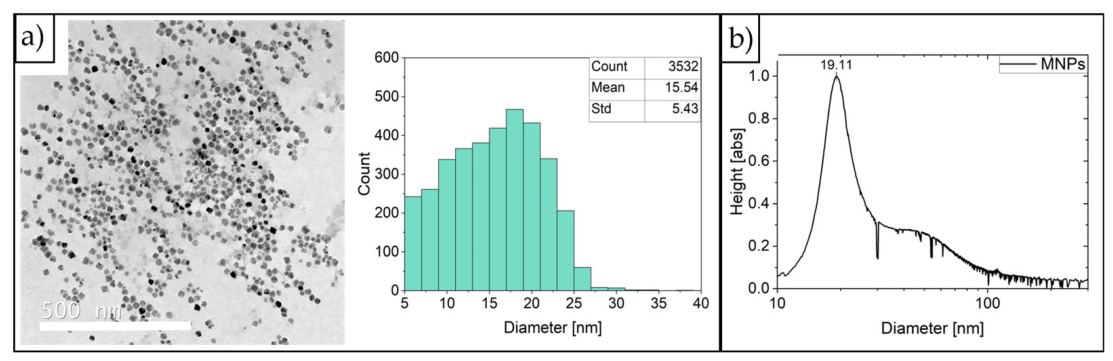
Magnetic single-core iron oxide nanoparticles were synthesized as described in section 2.5 via an aqueous route using a continuous micromixer-based platform.[22] TEM analysis revealed a mean core diameter of 15.5 ± 5.4 nm (mean ± standard deviation, n>500). DCS measurements yielded a main peak at an apparent particle diameter of 19.1 nm (FWHM = 7.6 nm). The DCS size distribution additionally showed a weak shoulder between 35 and 100 nm, indicating a minor population of larger particles or aggregates.
Figure 3.
a) TEM picture of MNPs (scale bar 500 nm) with the corresponding size distribution, analysed and evaluated with ImageJ. b) Size distribution of MNPs derived from DCS measurements. The distribution is dominated by a main particle population centred at 19.1 nm, with a minor high-size shoulder extending up to ~100 nm that that is consistent with a small fraction of di-/trimeric aggregates formed during magnetic purification.
Figure 3.
a) TEM picture of MNPs (scale bar 500 nm) with the corresponding size distribution, analysed and evaluated with ImageJ. b) Size distribution of MNPs derived from DCS measurements. The distribution is dominated by a main particle population centred at 19.1 nm, with a minor high-size shoulder extending up to ~100 nm that that is consistent with a small fraction of di-/trimeric aggregates formed during magnetic purification.
3.3. Formation and Characterization of Copolymer-MNP Nanohybrids (pMNHs)
Polymeric magnetic nanohybrids were assembled as described in 2.6 through a thermally assisted mixing protocol. Iron oxide MNPs were separately combined with either cytosine- (IV-C) or guanine-containing (IV-G) copolymer (Scheme 1) in ultrapure water and heated to 95 °C for 10 min. Following rapid mixing of the preheated MNP-polymer dispersions in the presence of preheated TRIS–MgCl₂ buffer (50 mM TRIS, 50 mM MgCl₂, pH 6), the samples were slowly cooled to room temperature. During controlled cooling, the initially transparent dispersion became distinctly opaque, providing a clear macroscopic indication of nanohybrid formation, as confirmed by DLS, DCS and cryo-TEM (Figure 4a-c). DLS measurements comparing polymer assembly and pMNHs clearly display a shift towards higher hydrodynamic sizes, indicating a size increase due to interaction between polymer assemblies and MNPs. DCS measurements comparing bare MNPs and pMNHs were performed based on the assumed density of magnetite (5.2 g ∙ cm‒3). Since the pMNHs have a reduced density compared to the assumed density of magnetite, the interaction between polymer and magnetite is displayed as an apparent decrease in size. Cryo-TEM image does not show micellar morphology, however MNPs and polymer seems to be in vicinity, indicating successful nano assembly of pMNHs.
3.4. Investigation of Magnetic Behaviour
The saturation magnetization MS of the base MNP system was determined using the DC-magnetization curves of the MNPs at 5 K and room temperature (295 K) as shown in Figure 5. MS-values of 119.9 Am² ∙ kg(Fe)‒1 at 5 K and 100.5 Am² ∙ kg(Fe)‒1 at 295 K are observed.
MPS measurements revealed that, relative to bare MNPs, pMNHs exhibited a moderate 22% reduction in A3*, whereas A5/A3 remained essentially unchanged. Immobilization in gypsum and freeze-drying markedly impaired the magnetic response of the MNPs, reducing A3* by at least 44% and A5/A3 by at least 13%. In contrast, the corresponding reductions for pMNHs were less pronounced: A3* decreased by around 30%, while A5/A3 decreased by approximately 10% under both conditions. Exposure to a final NaCl concentration of 1 M strongly attenuated the MNP response, with reductions of 83% in A3* and 92% in A5/A3, whereas both parameters remained unchanged for the pMNHs. Compared to the commercially available MRI contrast-agent Resotran, pMNHs exhibited a 50% higher A3*, despite a 16% lower A5/A3. Further, Resotran was strong affected by immobilization, freeze-drying, and exposure to 1 M NaCl, leading to strong decreases in A3* and A5/A3. The intrinsic loss power (ILP) of the pMNHs was 65% lower than that of the bare MNPs but 50% higher than that of Resotran.
Figure 6.
A3*, A5/A3, and ILP of the MNPs and pMNHs in fluid state (black), immobilized in gypsum (red), after freeze-drying (green) and under sodium salt exposure (blue) are shown. The commercially available contrast medium Resotran is shown as benchmark.
Figure 6.
A3*, A5/A3, and ILP of the MNPs and pMNHs in fluid state (black), immobilized in gypsum (red), after freeze-drying (green) and under sodium salt exposure (blue) are shown. The commercially available contrast medium Resotran is shown as benchmark.
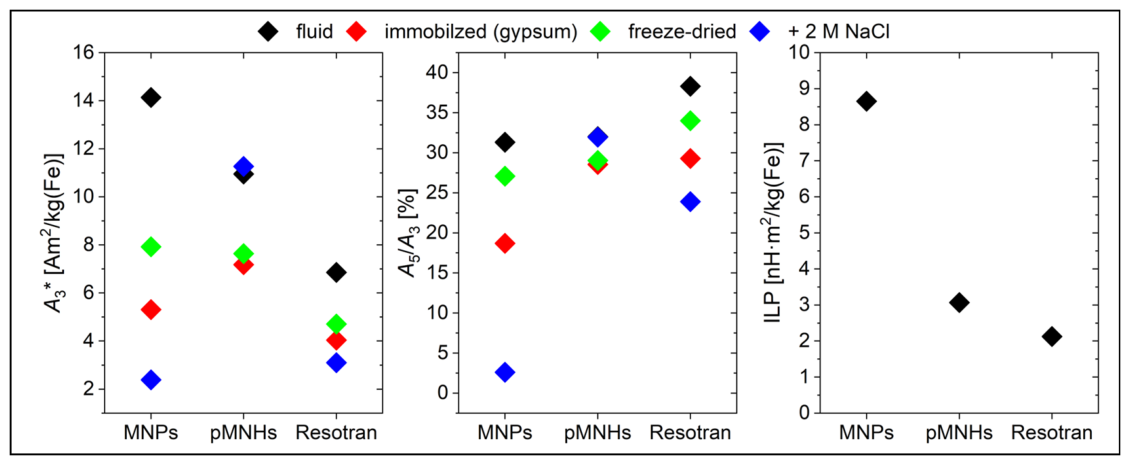
Figure 7 summarises the MRI performance of the pMNHs. The proton longitudinal and transverse relaxation rates, R1 and R2, determined by NMR relaxometry, are plotted as functions of the iron concentration c(Fe). The slopes of the corresponding linear fits yielded the longitudinal (r1) and transverse (r2) relaxivities. The pMNHs exhibited an r1 of 5.86(6) L ∙ mmol–1 ∙ s–1 and an r2 of 254(1) L ∙ mmol–1 ∙ s–1. In comparison, the bare MNPs showed higher relaxivities, with r1and r2 values of 30.14(4) and 583(4) L ∙ mmol–1 ∙ s–1, respectively.
3.5. Drug Loading with Dox
The Dox loading of the pMNHs was performed according to a literature-based protocol.[24] After removal of unbound Dox, the distinctly reddish Dox-pMNH suspension was analysed by UV–Vis spectroscopy to determine the amount of bound Dox, yielding a concentration of 20 µM.
For the toxicity assays, fluorescence microscopy was used to investigate whether the pMNHs were taken up by clinically relevant HeLa cells and could therefore be used for potential Dox treatment. After incubation, the pMNHs were observed to be internalised by the cells and displayed strong red fluorescence (Figure 8a). At an equivalent concentration, free Dox used as a positive control produced a comparable fluorescence signal (Figure 8c), whereas the negative control consisting of untreated cells showed no detectable emission (Figure 8b). Although signs of cellular damage were apparent in the bright-field images of the positive control, the Dox-pMNH-treated cells still appeared morphologically intact (Figure 8).
3.6. Cell Viability and Toxicity of Dox-Loaded pMNHs
The cytotoxicity of the Dox-loaded pMNHs (Dox-pMNHs) was evaluated in HeLa cells, an application-relevant model for assessing Dox-mediated cytotoxicity. Samples containing 0.1 – 10.0 µM polymer, corresponding to Dox concentrations of 0.2 – 20.0 µM, were analyzed. Irrespective of the polymer or Dox concentration, the Dox-pMNHs exhibited no significant cytotoxicity, with cell viabilities of approximately 90% after 24 h of incubation. In contrast, free Dox, included as a positive control under identical conditions, caused pronounced concentration-dependent cytotoxicity (Figure 9).
3.7. RF-Heating and Drug Release
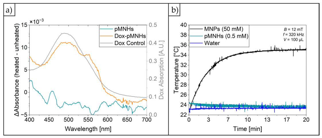
Heating performance and RF-triggered drug release were assessed for MNPs, pMNHs, and Dox-pMNHs. Figure 10a presents the differences between the UV–Vis spectra of pMNHs and Dox-pMNHs acquired before and after RF-heating, whereas Figure 10b shows the corresponding temperature profiles of MNPs, pMNHs, and water. No discernible spectral changes were observed for pMNHs following RF-heating. In contrast, the difference spectrum of Dox-pMNHs exhibited a pronounced absorption maximum at 490 nm. This feature is consistent with the characteristic absorption maximum of free Dox (25 µM), observed between 480 and 504 nm, indicating RF-induced Dox release.
Under RF exposure, MNPs displayed a rapid temperature increase of 11 °C within 7 min and reached a plateau temperature of 35 °C after 10 min. In contrast, neither pMNHs nor water showed a measurable temperature change over the 20 min period of RF-heating. To determine whether RF-heating affected the magnetic properties of the iron cores, MNPs, pMNHs, and Dox-pMNHs were analysed by MPS before and after RF exposure. No significant changes in A3* (<5%) or A5/A3 (<1%) were detected for all samples.
4. Discussion
4.1. Copolymer Synthesis
The RAFT polymerization protocol developed in related work currently under review[24] yielded a well-defined block copolymer with a narrow molar mass distribution. Subsequent post-polymerization treatment resulted in quantitative removal of the RAFT end group. This high degree of structural definition and purity constitutes a critical prerequisite for the reproducible application in the present study, as any variations in chain length or residual functionalities could directly influence the interactions with MNPs. The uniform nucleobase loading of nine units per chain offers an optimal compromise between functionality and biocompatibility. Notably, this degree of substitution provides sufficient C–G interactions to drive self-assembly while retaining a high density of functional groups for association with MNPs, and the polymer remains adequately soluble in aqueous solution for the targeted self-assembly investigations.
4.2. MNP Synthesis
The slight increase from the TEM-derived MNP core diameter (15.5 nm) to the DCS-derived hydrodynamic diameter (19.1 nm) can be attributed to the hydrated tannic acid shell, which is not visible in TEM images. The close agreement between both values indicates that the dispersion mainly consists of individually dispersed nanoparticles, with only minor aggregation. This confirms good colloidal stability and a narrow particle-size distribution. The assumed tannic acid shell thickness of approximately 1.8 nm is compatible with ultrathin polyphenol-derived surface layer, consistent with previous reports on tannic-acid-functionalized iron oxide nanoparticles and nanometre-scale polyphenol coating.[29,30] The weak shoulder between 35 and 100 nm likely reflects a small fraction of dimers, trimers, and occasional larger aggregates caused by a strong magnetic attraction during magnetic separation and purification.
4.3. pMNH Formation
Due to the given analytical data, the fabrication of pMNHs using nucleobase-functionalised copolymers and MNPs is interpreted as the formation of a hybrid nanoparticle architecture rather than a simple MNP coating with freely dispersed amphiphilic copolymer chains. Although DLS indicated an increase in the mean hydrodynamic diameter from 108.2 nm for the MNPs to 126.7 nm for the pMNHs, DCS yielded an apparent decrease of 3.7 nm in the pMNH radius relative to the unmodified MNPs. This apparent discrepancy does not necessarily reflect a genuine reduction in particle size. In contrast to DLS, DCS determines a sedimentation-equivalent size and is therefore highly sensitive to particle density, morphology and hydrodynamic drag. Considered alongside the DLS data, the delayed sedimentation of pMNHs is consistent with macromolecule association altering their effective density and sedimentation behaviour, thereby resulting in a smaller apparent DCS-derived size. Similar effects have previously been reported for casein-coated MNPs.[31]
Cryo-TEM provides qualitative support for this interpretation, as the high-contrast magnetic cores were consistently observed in close spatial proximity to lower-contrast, polymer-associated features. While these images do not demonstrate a uniform and continuous coating of individual MNPs, they support the formation of polymer-associated hybrid assemblies. Collectively, these findings suggest polymer-mediated surface functionalisation rather than a well-defined core–shell architecture.
In this context, the nucleobase-containing copolymers seem to provide both structural and functional contributions to the pMNHs. Complementary cytosine–guanine interactions can promote multivalent and directional cohesion, while aromatic association, hydration effects and interactions with MNP’s tannic acid and other components of the pMNH architecture are also likely to influence particle stability and molecular organisation. Accordingly, Watson–Crick base pairing should be regarded as an important stabilising contribution rather than the sole driving force of particle formation. This distinction is essential, as pMNHs cannot be equated with conventional micelles or purely self-assembled copolymer nanoparticles.
4.4. Investigation of Magnetic Behaviour
The magnetic behaviour of MNPs and pMNHs was investigated using DCM, MPS, AC-magnetometry, and NMR. The measured saturation magnetization (MS) approached the values reported for bulk magnetite and maghemite (111–127 Am² ∙ kg(Fe)‒1),[32,33] indicating a high magnetic order of the magnetic moments in the particles caused by a high iron oxide crystallinity of the synthesis method. Pronounced hysteresis is observed at Tmeas = 5 K, suggesting that the particle magnetic moments are blocked on the timescale of the measurement. In contrast, the magnetisation curve recorded at room temperature (295 K) is essentially free of hysteresis, demonstrating superparamagnetic behaviour under the applied measurement conditions. Accordingly, the blocking temperature, TB, lies between 5 and 295 K. Although MS alone does not determine tracer performance, high saturation magnetization is generally advantageous for MPI and magnetic hyperthermia, supporting the potential of these MNPs for both applications.
MPS measurements showed that the incorporation of MNPs into pMNHs reduced A3* by only 22%, whereas A5/A3 remained essentially unchanged. Thus, nanohybrid formation largely preserved the MPI-relevant magnetic response of the MNPs. The moderate attenuation of A3* may result from the increased hydrodynamic diameter and restricted rotational mobility of the MNPs within the polymeric assemblies, which may limit their ability to follow the oscillating magnetic field. Comparable size-dependent reductions in MPS parameters have previously been reported for casein- and BSA-coated MNPs.[31,34] Immobilization in gypsum and freeze-drying caused pronounced decreases in both A3* and A5/A3 for the bare MNPs, whereas the pMNHs were substantially less affected. Following either treatment, the A3* values of both formulations were approximately 7 Am² ∙ kg(Fe)‒1, demonstrating that the pMNHs retained a measurable nonlinear magnetic response under conditions of restricted particle mobility. This robustness supports their further evaluation as MPI tracers in cellular and tissue environments, in which nanoparticle mobility may be constrained.
Exposure to a final NaCl concentration of 1 M markedly reduced A3* and A5/A3 for the bare MNPs and Resotran but induced negligible changes in either parameter for the pMNHs. The polymeric assembly therefore conferred substantial resistance to salt-induced changes in magnetic behaviour, consistent with enhanced colloidal stability at high ionic strength. This finding is encouraging for biomedical applications, although stability in complex biological media should be verified separately.
AC-magnetometry yielded ILP values of 8.7 and 3.1 nH ∙ m2 kg(Fe)‒1 for the MNPs and pMNHs, respectively. The ILP of the pMNHs was approximately 50% higher than that of Resotran, while that of the bare MNPs was approximately 4-fold higher. This heating performance of MNPs and pMNHs, which is comparable to or exceeds that of other systems, should enable efficient hyperthermia treatment.[35] The lower ILP of the pMNHs relative to the bare MNPs may reflect restricted Brownian relaxation and altered interparticle magnetic interactions within the polymeric assemblies. Nevertheless, the pMNHs have retained appreciable heating efficiency, supporting their potential for magnetic hyperthermia. Under equivalent field conditions, a higher ILP may reduce the iron dose required to achieve therapeutically relevant heating.
Relaxometry further demonstrated that the transverse relaxivity of the pMNHs substantially exceeded their longitudinal relaxivity, supporting their suitability as T₂-weighted MRI contrast agents.[36] Although both relaxivities were lower than those of the bare MNPs, the pMNHs retained a pronounced transverse relaxation effect. This reduction may result from polymer-mediated changes in water accessibility, proton–core distance, and interparticle organisation, thereby attenuating the local magnetic-field inhomogeneities experienced by surrounding water protons. Collectively, their preserved MPI-relevant response, enhanced stability under high-salt and restricted-mobility conditions, pronounced transverse relaxivity, and measurable heating efficiency highlight the multifunctional potential of the pMNHs for MPI, T₂-weighted MRI, and magnetic hyperthermia.
4.5. Drug Loading
The thermally assisted loading procedure generated a Dox-associated pMNH formulation that retained 20 μM Dox after dialysis. This finding demonstrates that a fraction of Dox remained associated with the pMNHs under aqueous purification conditions. The planar anthracycline chromophore of Dox is well suited to non-covalent interactions with nucleobase-containing polymer domains, including π–π stacking, hydrophobic association and hydrogen bonding. Elevated temperature may further facilitate redistribution of Dox into accessible polymer domains during loading. However, the available data do not distinguish between Dox located within the pMNH structure and drug adsorbed at accessible particle interfaces. The formulation is therefore described as Dox-loaded or Dox-associated rather than as definitively core-encapsulated. Evidence from a previously published study of the same polymer system in the absence of MNPs provides a plausible explanation for this behaviour.[24] The remarkable retention of Dox upon changes in temperature and pH, together with its unexpectedly low cytotoxicity, suggests that the drug is effectively shielded within the polymer assemblies. Dox may be localised within the micellar interior, potentially intercalating between guanine–cytosine base pairs and thereby gaining additional stabilization. Such encapsulation would reduce the exposure of Dox to the surrounding environment and may account for its robust association with the polymer assemblies.
4.6. Cell Uptake and Cytotoxicity of pMNHs and Dox-pMNHs
Fluorescence microscopy provided initial evidence that Dox-associated pMNHs formulations interact with, and are likely internalised by, HeLa cells. The pronounced intracellular red fluorescence observed after 2 h incubation, together with the absence of detectable emission in untreated cells, confirms the delivery of Dox-associated fluorescent material to the cellular environment. The comparable signal obtained with free Dox validates the optical detectability of the drug under the applied imaging conditions. Importantly, the qualitatively preserved morphology of pMNH-treated cells relative to cells exposed to free Dox is consistent with reduced immediate intracellular availability of the cytotoxic payload. This observation may reflect retention of Dox within the pMNH structure and delayed release after cellular uptake.
Nevertheless, Dox fluorescence alone cannot establish whether intact Dox–pMNHs entered the cells or whether released Dox was subsequently internalised independently of the carrier. Likewise, the microscopy data does not demonstrate subcellular localisation, nuclear delivery or release of pharmacologically active Dox. Future studies should therefore combine dual-labelled pMNHs with confocal co-localisation analyses, endolysosomal and nuclear markers, and quantitative intracellular release measurements by HPLC or LC–MS.
Overall, the results support the use of nucleobase-functionalised pMNHs as stable, Dox-associating nanocarriers with detectable RF-induced release for cellular delivery. The key challenge for further development will be to balance sufficiently strong interactions for extracellular cargo retention with the externally RF-induced drug release mechanism that restores Dox bioavailability at the desired site of action. Therefore, further insights into the magnetically induced Dox release mechanism are mandatory.
4.7. RF-Heating and Drug Release
The UV-Vis spectra before and after RF-heating of Dox-pMNHs suggests that triggered drug release of Dox is possible. As the maximum position of Δabsorbance is the same as for the pure Dox absorbance, it can be assumed that in fact Dox was released. Further, no significant changes in the MPS values A3* and A5/A3 were detected for all samples, demonstrating that the magnetic behaviour remained largely preserved. Additional measurement of the heating curves showed no heating of the pMNHs and water, but for the MNPs. This can be explained by the concentration difference used for this experiment with almost a factor 100 higher c(Fe) of the MNPs compared to pMNHs. But since the same MNPs were used for the assembly and the Dox release were triggered it can be assumed, that the pMNHs were also heating, but because of their low concentration it was not enough to heat their surrounding medium.
These results show that triggered drug release is possible with nucleobase-functionalised and drug-loaded pMNHs.
5. Conclusions
This study demonstrates the successful development of nucleobase-functionalized polymeric magnetic nanohybrids combining drug-carrier properties with multiple magnetic functionalities. The association of the polymers with the iron oxide nanoparticles substantially improved their stability under high-salt conditions while largely preserving their magnetic response. Their high transverse relaxivity, detectable MPI signal, and intrinsic loss power highlight their potential for applications in T₂-weighted MRI, MPI, and magnetic hyperthermia.
Doxorubicin remained associated with the pMNHs after dialysis, and Dox-associated fluorescence was detected in HeLa cells. The consistently high cell viability observed across the investigated concentration range suggests effective retention of the drug under non-triggered conditions, which may help limit premature drug activity. Moreover, radiofrequency exposure induced a Dox-characteristic spectral change without compromising the magnetic properties of the nanohybrids or heating the surrounding environment, providing encouraging preliminary evidence for externally triggered drug release.
Overall, the developed pMNHs represent a promising and versatile platform for magnetically guided theranostic applications. Their combination of colloidal stability, multimodal imaging potential, magnetic responsiveness, and drug-retention capacity provides a strong foundation for further development. Future studies focusing on quantitative release kinetics, intracellular tracking, and optimized radiofrequency conditions may unlock their full potential for controlled and spatially targeted drug delivery.
Author Contributions
A.-N. E.-K. and R.B. conceived the study. A.-N. E.-K. synthesized and characterized the polymeric materials and performed the formulation. C.W. synthesized and characterized the magnetic nanoparticles. A.-N. E.-K. and C.W. carried out the physicochemical characterization. C.W. and F.W. conducted the magnetic characterization. L.D. designed and performed the RF-heating/DOX release experiments. J. N. performed fluorescence microscopy imaging and conducted cell viability and cytotoxicity assays and N.M. acquired the cryo-TEM images. A.-N. E.-K. and C. W. prepared the original draft of the manuscript, R.B., and F.W. contributed to its review and editing. R.B. supervised and administered the project and secured funding. All authors have read and agreed to the published version of the manuscript.
Funding
This work was supported by the Federal Ministry of Research, Technology and Space (BMFTR) through the BioTherNa junior research group (grant no. 13XP5113) and the German Research Foundation (DFG) through Collaborative Research Centres SFB 1552/1 (Project-ID 465145163, subproject C01) and SFB 1340/3 2026 (Project-ID 372486779, subproject A02).
Data Availability Statement
The original contributions presented in this study are included in the article/supplementary material. Further inquiries can be directed to the corresponding author(s).
Acknowledgments
The authors thank Christian Freese for providing access to laboratory facilities for the cell-based experiments and Carmen Schwind for her assistance with the fluorescence microscopy–based cellular uptake studies.
Conflicts of Interest
The authors declare no conflicts of interest
References
- Wang, L.-S.; Chuang, M.-C.; Ho, J.-A.A. Nanotheranostics--a review of recent publications. Int. J. Nanomed. 2012, 7, 4679–95. [Google Scholar] [CrossRef]
- Muthu, M.S.; Leong, D.T.; Mei, L.; Feng, S.-S. Nanotheranostics - application and further development of nanomedicine strategies for advanced theranostics. Theranostics 2014, 4(6), 660–77. [Google Scholar] [CrossRef]
- Kelkar, S.S.; Reineke, T.M. Theranostics: combining imaging and therapy. Bioconjug Chem. 2011, 22(10), 1879–903. [Google Scholar] [CrossRef]
- Arias, L.S.; Pessan, J.P.; Vieira, A.P.M.; de Lima, T.M.T.; Delbem, A.C.B.; Monteiro, D.R. Iron Oxide Nanoparticles for Biomedical Applications: A Perspective on Synthesis, Drugs, Antimicrobial Activity, and Toxicity. Antibiotics 2018, 7(2). [Google Scholar] [CrossRef]
- Wahajuddin, Arora S. Superparamagnetic iron oxide nanoparticles: magnetic nanoplatforms as drug carriers. Int. J. Nanomed. 2012, 7, 3445–71. [Google Scholar] [CrossRef]
- Billings, C.; Langley, M.; Warrington, G.; Mashali, F.; Johnson, J.A. Magnetic Particle Imaging: Current and Future Applications, Magnetic Nanoparticle Synthesis Methods and Safety Measures. Int. J. Mol. Sci. 2021, 22(14). [Google Scholar] [CrossRef]
- Pucci, C.; Degl'Innocenti, A.; Belenli Gümüş, M.; Ciofani, G. Superparamagnetic iron oxide nanoparticles for magnetic hypertherm mia: recent advancements, molecular effects, and future directions in the omics era. Biomater. Sci. 2022, 10(9), 2103–21. [Google Scholar] [CrossRef]
- Laurent, S.; Forge, D.; Port, M.; Roch, A.; Robic, C.; Vander Elst, L.; et al. Magnetic iron oxide nanoparticles: synthesis, stabilization, vectorization, physicochemical characterizations, and biological applications. Chem. Rev. 2008, 108(6), 2064–110. [Google Scholar] [CrossRef]
- Schubert, J.; Chanana, M. Coating Matters: Review on Colloidal Stability of Nanoparticles with Biocompatible Coatings in Biological Media, Living Cells and Organisms. Curr. Med. Chem. 2018, 25(35), 4553–86. [Google Scholar] [CrossRef]
- Mohammadi, M.R.; Malkovskiy, A.V.; Jothimuthu, P.; Kim, K.-M.; Parekh, M.; Inayathullah, M.; et al. PEG/Dextran Double Layer Influences Fe Ion Release and Colloidal Stability of Iron Oxide Nanoparticles. Sci. Rep. 2018, 8(1), 4286. [Google Scholar] [CrossRef]
- Arsenie, L.V.; Ladmiral, V.; Lacroix-Desmazes, P.; Catrouillet, S. Nucleobase-containing polymer architectures controlled by supramolecular interactions: the key to achieve biomimetic platforms with various morphologies. Polym. Chem. 2022, 13(41), 5798–810. [Google Scholar] [CrossRef]
- Sikder, A.; Esen, C.; O'Reilly, R.K. Nucleobase-Interaction-Directed Biomimetic Supramolecular Self-Assembly. Acc. Chem. Res. 2022, 55(12), 1609–19. [Google Scholar] [CrossRef]
- Kang, Y.; Pitto-Barry, A.; S Rolph, M.; Hua, Z.; Hands-Portman, I.; Kirby, N.; et al. Use of complementary nucleobase-containing synthetic polymers to prepare complex self-assembled morphologies in water. Polym. Chem. 2016, 7(16), 2836–46. [Google Scholar] [CrossRef]
- Arsenie, L.V.; Semsarilar, M.; Brendel, J.C.; Lacroix-Desmazes, P.; Ladmiral, V.; Catrouillet, S. Supramolecular co-assembly of water-soluble nucleobase-containing copolymers: bioinspired synthetic platforms towards new biomimetic materials. Polym. Chem. 2022, 13(39), 5604–15. [Google Scholar] [CrossRef]
- Kamaly, N.; Yameen, B.; Wu, J.; Farokhzad, O.C. Degradable Controlled-Release Polymers and Polymeric Nanoparticles: Mechanisms of Controlling Drug Release. Chem. Rev. 2016, 116(4), 2602–63. [Google Scholar] [CrossRef]
- Elsabahy, M.; Wooley, K.L. Design of polymeric nanoparticles for biomedical delivery applications. Chem. Soc. Rev. 2012, 41(7), 2545–61. [Google Scholar] [CrossRef]
- McHale, R.; O’Reilly, R.K. Nucleobase Containing Synthetic Polymers: Advancing Biomimicry via Controlled Synthesis and Self-Assembly. Macromolecules 2012, 45(19), 7665–75. [Google Scholar] [CrossRef]
- Lu, C.-H.; Willner, I. Stimuli-responsive DNA-functionalized nano-/microcontainers for switchable and controlled release. Angew. Chem. Int. Ed. 2015, 54(42), 12212–35. [Google Scholar] [CrossRef]
- Webber, M.J.; Appel, E.A.; Meijer, E.W.; Langer, R. Supramolecular biomaterials. Nat. Mater. 2016, 15(1), 13–26. [Google Scholar] [CrossRef]
- Jones, M.R.; Seeman, N.C.; Mirkin, C.A. Nanomaterials. Programmable materials and the nature of the DNA bond. Science 2015, 347(6224), 1260901. [Google Scholar] [CrossRef]
- Ilhami, F.B.; Bayle, E.A.; Cheng, C.-C. Complementary Nucleobase Interactions Drive Co-Assembly of Drugs and Nanocarriers for Selective Cancer Chemotherapy. Pharmaceutics 2021, 13(11). [Google Scholar] [CrossRef]
- Baki, A.; Löwa, N.; Remmo, A.; Wiekhorst, F.; Bleul, R. Micromixer Synthesis Platform for a Tuneable Production of Magnetic Single-Core Iron Oxide Nanoparticles. Nanomaterials 2020, 10(9). [Google Scholar] [CrossRef]
- Popescu, R.C.; Savu, D.; Dorobantu, I.; Vasile, B.S.; Hosser, H.; Boldeiu, A.; et al. Efficient uptake and retention of iron oxide-based nanoparticles in HeLa cells leads to an effective intracellular delivery of doxorubicin. Sci. Rep. 2020, 10(1), 10530. [Google Scholar] [CrossRef]
- Egler-Kemmerer, A.-N.; Nassauer, J.; Meier, N.; Thiermann, R.; Bleul, R. Synthesis and Self-Assembly of Nucleobase-Functionalized Hydrophilic Copolymers for Next-Generation Nanotherapeutics. Unpublished manuscript, under review. 2026. [Google Scholar]
- Pyenson, H.; Tracy, P.H. A 1,10—Phenanthroline Method for the Determination of Iron in Powdered Milk. J. Dairy Sci. 1945, 28(5), 401–12. [Google Scholar] [CrossRef]
- Ludwig, F.; Eberbeck, D.; Löwa, N.; Steinhoff, U.; Wawrzik, T.; Schilling, M.; et al. Characterization of magnetic nanoparticle systems with respect to their magnetic particle imaging performance. BioMed Tech (Berl) 2013, 58(6), 535–45. [Google Scholar] [CrossRef]
- Rauwerdink, A.M.; Weaver, J.B. Measurement of molecular binding using the Brownian motion of magnetic nanoparticle probes. Appl. Phys. Lett. 2010, 96(3), 33702. [Google Scholar] [CrossRef]
- Wu, K.; Su, D.; Saha, R.; Liu, J.; Wang, J.-P. Investigating the Effect of Magnetic Dipole-Dipole Interaction on Magnetic Particle Spectroscopy (MPS): Implications for Magnetic Nanoparticle-based Bioassays and Magnetic Particle Imaging (MPI). 2019. [Google Scholar] [CrossRef]
- Sileika, T.S.; Barrett, D.G.; Zhang, R.; Lau, K.H.A.; Messersmith, P.B. Colorless multifunctional coatings inspired by polyphenols found in tea, chocolate, and wine. Angew. Chem. Int. Ed. 2013, 52(41), 10766–70. [Google Scholar] [CrossRef]
- Ejima, H.; Richardson, J.J.; Liang, K.; Best, J.P.; van Koeverden, M.P.; Such, G.K.; et al. One-step assembly of coordination complexes for versatile film and particle engineering. Science 2013, 341(6142), 154–7. [Google Scholar] [CrossRef]
- Wenck, C.; Meier, N.; Heinrich, E.; Grützner, V.; Wiekhorst, F.; Bleul, R. Design and characterisation of casein coated and drug loaded magnetic nanoparticles for theranostic applications. RSC Adv. 2024, 14(36), 26388–99. [Google Scholar] [CrossRef]
- Hellwege, K.-H.; Madelung, O. (Eds.) Zahlenwerte und Funktionen aus Naturwissenschaften und Technik: Weiterführung und Ergänzung von Band 1; Springer: Berlin, 1982. [Google Scholar]
- Ahrens, T.J. (Ed.) Rock physics & phase relations: A handbook of physical constants; Washington, DC, 1995. [Google Scholar]
- Baki, A.; Remmo, A.; Löwa, N.; Wiekhorst, F.; Bleul, R. Albumin-Coated Single-Core Iron Oxide Nanoparticles for Enhanced Molecular Magnetic Imaging (MRI/MPI). Int. J. Mol. Sci. 2021, 22(12). [Google Scholar] [CrossRef]
- Cohen-Erner, M.; Khandadash, R.; Hof, R.; Shalev, O.; Antebi, A.; Cyjon, A.; et al. Fe 3 O 4 Nanoparticles and Paraffin Wax as Phase Change Materials Embedded in Polymer Matrixes for Temperature-Controlled Magnetic Hyperthermia. ACS Appl. Nano Mater. 2021, 4(10), 11187–98. [Google Scholar] [CrossRef]
- Estelrich, J.; Sánchez-Martín, M.J.; Busquets, M.A. Nanoparticles in magnetic resonance imaging: from simple to dual contrast agents. Int. J. Nanomed. 2015, 10, 1727–41. [Google Scholar] [CrossRef]
Scheme 1.
Multistep copolymer synthesis route based on RAFT polymerization technique under inert reaction conditions and post-polymerization functionalization. Nucleobase attachment was achieved via amide coupling with the modified nucleobase derivatives guanine acetic acid (G) and cytosine acetic acid (C).
Scheme 1.
Multistep copolymer synthesis route based on RAFT polymerization technique under inert reaction conditions and post-polymerization functionalization. Nucleobase attachment was achieved via amide coupling with the modified nucleobase derivatives guanine acetic acid (G) and cytosine acetic acid (C).
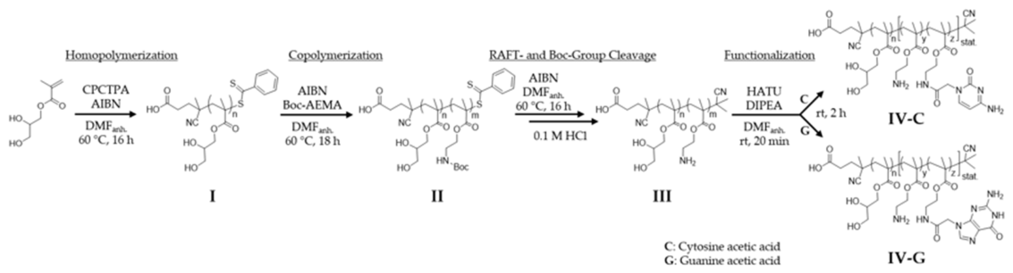
Figure 1.
a) UV–vis spectra of P(GMA)-b-P(Boc-AEMA) before and after RAFT end-group removal by radical-induced decomposition with AIBN, demonstrating successful cleavage by the disappearance of the characteristic RAFT-agent absorption band at λmax = 300 nm. b) Corresponding extract of the 1H-NMR spectrum showing the disappearance of aromatic proton signals in the downfield region.
Figure 1.
a) UV–vis spectra of P(GMA)-b-P(Boc-AEMA) before and after RAFT end-group removal by radical-induced decomposition with AIBN, demonstrating successful cleavage by the disappearance of the characteristic RAFT-agent absorption band at λmax = 300 nm. b) Corresponding extract of the 1H-NMR spectrum showing the disappearance of aromatic proton signals in the downfield region.
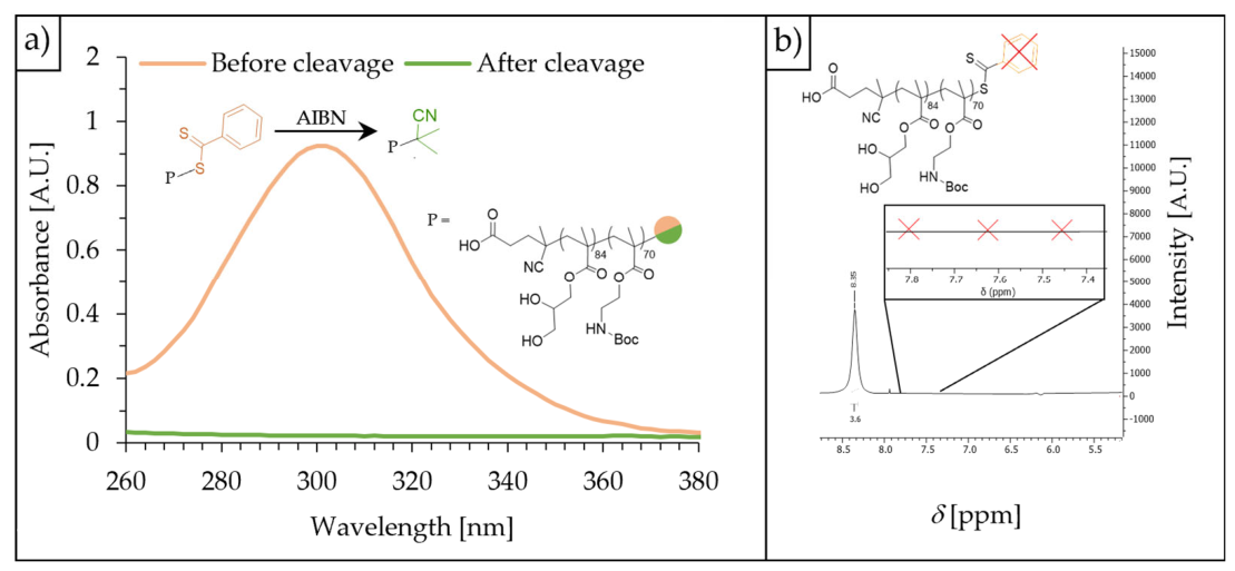
Figure 2.
a)1H-NMR spectrum of P(GMA)-b-P(AEMA) (III) prior to nucleobase functionalization, showing the disappearance of the characteristic Boc methyl proton signal, confirming successful deprotection. b) UV‒vis spectra of (IV-C) and (IV-G) displaying the characteristic absorbance of the nucleobase moieties conjugated to the polymer backbone at λ = 260 nm, recorded at a polymer concentration of 21.0 µM in ultrapure water.
Figure 2.
a)1H-NMR spectrum of P(GMA)-b-P(AEMA) (III) prior to nucleobase functionalization, showing the disappearance of the characteristic Boc methyl proton signal, confirming successful deprotection. b) UV‒vis spectra of (IV-C) and (IV-G) displaying the characteristic absorbance of the nucleobase moieties conjugated to the polymer backbone at λ = 260 nm, recorded at a polymer concentration of 21.0 µM in ultrapure water.
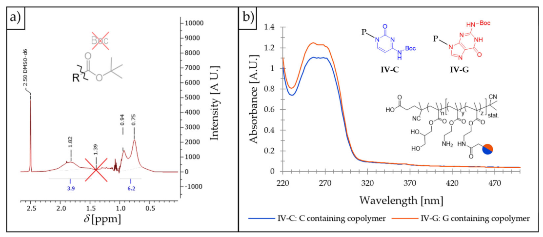
Figure 4.
a) Representative comparison of intensity-weighted size distributions (left) and corresponding autocorrelation functions (right) of pMNHs (turquoise, c = 5 μM) with MNP-free assemblies (red) obtained by DLS. The particle size, reported as the intensity-weighted harmonic mean of the hydrodynamic diameter (Z-average), and the polydispersity index (PDI) were calculated from three independent measurements. b) Representative comparison of size distributions of bare MNPs (19.1 nm, black) and pMNHs (apparent mean size 15.4 nm, turquoise) obtained by DCS. The observed shift in particle mean size is due to density change compared to the assumed density of magnetite (5.2 g ∙ cm‒3). c) Cryo-TEM image of the pMNH dispersion, illustrating nanohybrid formation, particle morphology and size. Scale bar = 200 nm.
Figure 4.
a) Representative comparison of intensity-weighted size distributions (left) and corresponding autocorrelation functions (right) of pMNHs (turquoise, c = 5 μM) with MNP-free assemblies (red) obtained by DLS. The particle size, reported as the intensity-weighted harmonic mean of the hydrodynamic diameter (Z-average), and the polydispersity index (PDI) were calculated from three independent measurements. b) Representative comparison of size distributions of bare MNPs (19.1 nm, black) and pMNHs (apparent mean size 15.4 nm, turquoise) obtained by DCS. The observed shift in particle mean size is due to density change compared to the assumed density of magnetite (5.2 g ∙ cm‒3). c) Cryo-TEM image of the pMNH dispersion, illustrating nanohybrid formation, particle morphology and size. Scale bar = 200 nm.
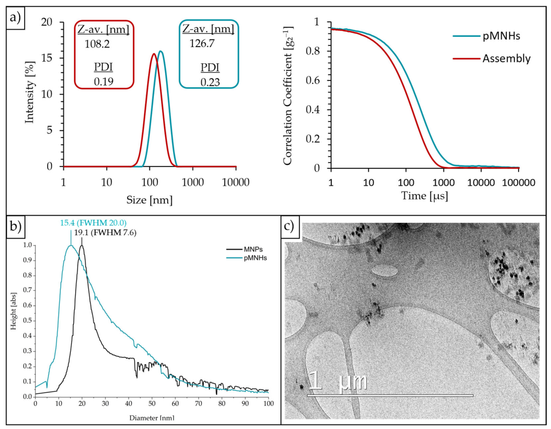
Figure 5.
DC-magnetization curves of the MNPs at room temperature (T = 295 K, red) and at 5 K (blue).
Figure 5.
DC-magnetization curves of the MNPs at room temperature (T = 295 K, red) and at 5 K (blue).
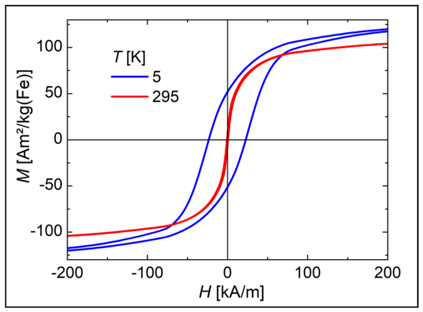
Figure 7.
The relaxation rates R1 and R2 of the MNPs and pMNHs against the iron concentration c(Fe) are shown. The relaxivities r₁ and r₂ were obtained as the slopes of linear fits.
Figure 7.
The relaxation rates R1 and R2 of the MNPs and pMNHs against the iron concentration c(Fe) are shown. The relaxivities r₁ and r₂ were obtained as the slopes of linear fits.
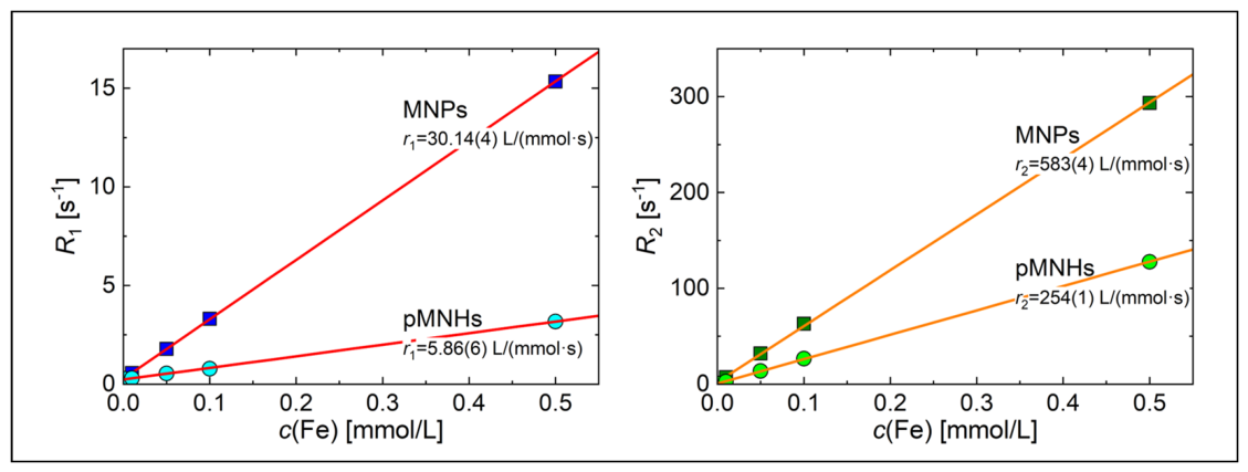
Figure 8.
Fluorescence microscopy images of HeLa cells in PBS (1×, pH 7.4) using a TRITC filter set (λex = 545/25 nm; λem = 605/70 nm). a) Cells treated with Dox-loaded pMNHs for 2 h showing a notable fluorescence signal. b) Untreated HeLa cells as negative control. c) Dox treated HeLa cells as positive control. Scale bar = 200 nm.
Figure 8.
Fluorescence microscopy images of HeLa cells in PBS (1×, pH 7.4) using a TRITC filter set (λex = 545/25 nm; λem = 605/70 nm). a) Cells treated with Dox-loaded pMNHs for 2 h showing a notable fluorescence signal. b) Untreated HeLa cells as negative control. c) Dox treated HeLa cells as positive control. Scale bar = 200 nm.
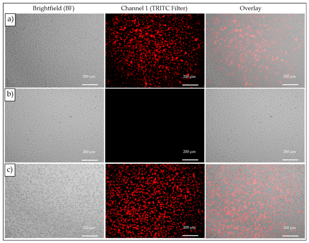
Figure 9.
Cell viability of HeLa cells following the treatment (24 h) with Dox-loaded nanohybrids (Dox-pMNHs), compared to the positive control (PC, unbound Dox) and negative control (NC, untreated HeLa cells) across a range of increasing correlated concentrations: 0.1, 0.4, 2.0 and 10.0 µM polymer concentration with 0.2, 0.8, 4.0 and 20.0 µM Dox The CCK-8 assay was performed with n = 3 independent experiments, each conducted in triplicate. Error bars represent the standard deviation of the mean.
Figure 9.
Cell viability of HeLa cells following the treatment (24 h) with Dox-loaded nanohybrids (Dox-pMNHs), compared to the positive control (PC, unbound Dox) and negative control (NC, untreated HeLa cells) across a range of increasing correlated concentrations: 0.1, 0.4, 2.0 and 10.0 µM polymer concentration with 0.2, 0.8, 4.0 and 20.0 µM Dox The CCK-8 assay was performed with n = 3 independent experiments, each conducted in triplicate. Error bars represent the standard deviation of the mean.
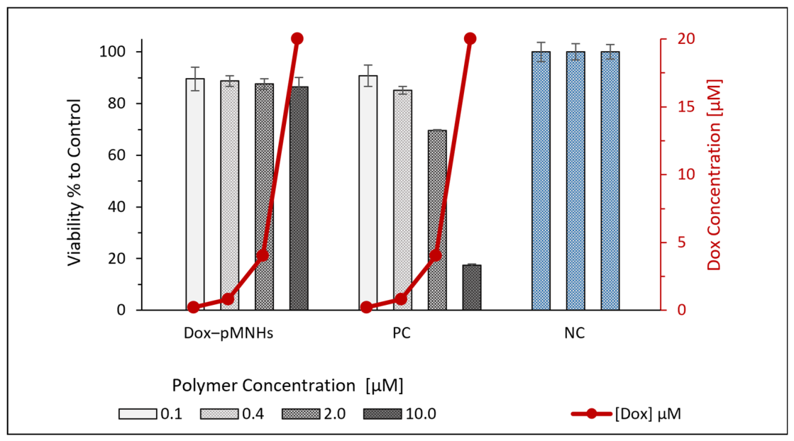
Figure 10.
a) ΔAbsorbance before and after magnetic hyperthermia treatment of pMNHs and Dox-pMNHs, and the absorbance of pure Dox (25 µM) as reference is shown. b) Heating curves of MNPs (50 mM), pMNHs (0.5 mM) and water against time.
Figure 10.
a) ΔAbsorbance before and after magnetic hyperthermia treatment of pMNHs and Dox-pMNHs, and the absorbance of pure Dox (25 µM) as reference is shown. b) Heating curves of MNPs (50 mM), pMNHs (0.5 mM) and water against time.
Disclaimer/Publisher’s Note: The statements, opinions and data contained in all publications are solely those of the individual author(s) and contributor(s) and not of MDPI and/or the editor(s). MDPI and/or the editor(s) disclaim responsibility for any injury to people or property resulting from any ideas, methods, instructions or products referred to in the content. |
© 2026 by the authors. Licensee MDPI, Basel, Switzerland. This article is an open access article distributed under the terms and conditions of the Creative Commons Attribution (CC BY) license.
Copyright: This open access article is published under a Creative Commons CC BY 4.0 license, which permit the free download, distribution, and reuse, provided that the author and preprint are cited in any reuse.