

Submitted:
01 October 2026
Posted:
05 October 2026
You are already at the latest version
Abstract
Magnetic tweezers (MT) have become an important single-molecule manipulation tool, with several advantages over alternate methods such as optical tweezers and atomic force microscopy. MT are easy to operate, cost effective, and can apply forces ranging from sub-piconewton (~ 0.1 pN) to hundreds of piconewtons. Furthermore, they offer exceptional spatial and temporal resolution. These features position MT as a versatile and powerful tool to study many biomolecular processes at the single-molecule level. MT have played an important role in advancing our understanding of DNA–protein interactions, making significant contributions across molecular biology, enzymology, and chromatin dynamics. In this review, we start by describing principal MT geometries along with force calibration methods and then pivot to a discussion of MT-based studies of DNA mechanics, chromatin organization, nucleosomes, nucleoid-associated proteins, topoisomerases, helicases, and polymerases. We aim to provide the research community with an integrated overview of MT-based approaches to studying DNA–protein interactions, highlighting both foundational work and recent innovations that continue to shape the field of single-molecule biophysics.
Keywords:
magnetic tweezers
; single-molecule biophysics
; DNA–protein interactions
; DNA mechanics
; chromatin dynamics
1. Introduction
DNA–protein interactions are fundamental to cellular processes such as transcription, replication, gene regulation, recombination, and DNA repair [1,2,3]. These interactions are governed by the sequence of the DNA binding region along with the amino-acid sequence of the protein. In addition, environmental conditions like ionic strength, temperature, and protein concentration can significantly modulate these interactions in cellular or in vitro settings. In living cells, these interactions are crucial for preserving genetic integrity and ensuring proper cellular functions [4].
In recent decades, single-molecule techniques have emerged as powerful tools that complement traditional bulk biochemical assays for studying macromolecules like proteins and DNA as well as DNA–protein interactions [5,6,7]. In contrast to bulk assays that perforce only sample population averages, single-molecule techniques enable researchers to observe real-time dynamics of individual molecules, revealing intrinsically stochastic behavior and transient intermediate states that are inaccessible to bulk experiments [8,9,10,11]. These methods also enable precise, molecular-level characterization of the mechanical properties of DNA and other biologically-relevant macromolecules—such as elasticity, twisting, and stretching moduli—as well as the kinetics of enzymatic interactions with DNA [5].
Among the various tools employed in single-molecule force spectroscopy, optical tweezers, atomic force microscopy, and magnetic tweezers are the most widely used, sometimes in combination with fluorescence microscopy. Each technique has its own set of advantages and limitations. In this study, we focus on magnetic tweezers, which offer a particularly robust and simple means of applying a constant force to single DNA tethers. Their design makes magnetic tweezers especially well-suited for extended-duration measurements lasting multiple hours [12]. The growing popularity of magnetic tweezers is largely due to their relatively simple design and low cost while providing excellent spatiotemporal resolution—achieving millisecond timing precision and sub-nanometer spatial accuracy—features that have been further enhanced by recent advances in hardware and software [13,14].
Our goal in this review is to document in easily accessible manner research into DNA–protein interactions using magnetic tweezers. After providing a brief introduction to MT, we describe in considerable detail the force calibration and quantitative force estimation methods that are crucial for precise biophysical measurements. We then focus on a variety of biological applications where MT have helped to clarify molecular mechanisms. We conclude by exploring the latest technological advancements and describing some of the new and exciting advances likely to emerge in the near future. The case studies in the use of MT illustrate their versatility and explain why they are well-suited for applications in biophysics, micro-robotics, and biomedical research.
2. Magnetic Tweezers
Magnetic tweezers originate in the classic work from 1996 of Strick and colleagues who used their tweezers apparatus to investigate the elastic properties of supercoiled DNA [15]. MTs are built around magnetic traps that use spatially inhomogeneous magnetic fields to manipulate micron-sized superparamagnetic beads in aqueous buffer at room temperature. Permanent magnets [16], electromagnets [17], and their combinations have been used in various geometries to produce magnetic fields with high spatial gradients capable of reliably generating forces that range from as low as femto-newtons (fN) to as high as nano-newtons, although typical micromanipulation experiments on DNA-based tethers apply forces in the pico-newtons (pN) to nano-newtons (nN) range [18,19]. This is also the range of forces that is suitable for mechanically probing genetic and biochemical processes at the single molecule level [20]. In a typical experiment, a magnetic bead is attached to one end of a biomolecule, typically a linear DNA tether a few to 10 microns long, while the other end is fixed to a functionalized glass surface. A magnetic field is then used to apply a force on the tethered microbead whose position is monitored in real time using video microscopy [21].
The position and movement of the tethered bead is recorded using a high-resolution camera. Image analysis of a recorded movie can be used to measure the changes in tethered extension [22], bead fluctuations [23], and rotational motions [24] of the tether. Such measures provide insights into the mechanical and conformational properties of the tethered biomolecules and their interactions with other proteins and DNA [25,26]. Based on the orientation of the applied force, magnetic tweezers can be classified as vertical and horizontal magnetic tweezers [6,15,27,28]. We will briefly introduce them in the following.
2.1. Vertical Magnetic Tweezers
Vertical magnetic tweezers (VMT) represent a powerful technique in force and torque spectroscopy, naturally functioning as a force clamp capable of applying a wide range of precisely controlled forces on macromolecular tethers, mainly DNA [29]. VMTs were introduced by Strick et al. [30] to investigate the entropic elasticity of individual supercoiled DNA molecules. In a typical VMT design, a spatially inhomogeneous magnetic field—generated by either permanent magnets or electromagnets—produces forces ranging from over 100 pN down to below 10 fN, with the force oriented vertically [31]. A standard implementation of a VMT have the magnets mounted above the sample cell, with the superparamagnetic beads tethered to one end of a biomolecule while the other end is anchored to a functionalized glass surface [32]. A reference or fiducial bead attached to the surface is used to correct for stage drift and ensure accurate extension measurements. An inverted microscope captures real-time images of the beads using CMOS or CCD imaging systems [7]. The resulting bright field video microscopy data allows for the three-dimensional tracking of bead motion down to (sub)nanometer resolution. Linear motors precisely control the vertical positioning of the magnets, thereby modulating the applied force [24]. Owing to its versatility and precision, the VMT have been widely employed to study DNA micro-mechanics—including extensional and torsional elasticity—as well as DNA–protein interactions and force-dependent conformational transitions in biomolecular systems [33]. A schematic of the setup is shown in Figure 1.
2.1. Horizontal Magnetic Tweezers
Horizontal or transverse magnetic tweezers (HMT) use magnetic traps to apply forces in the horizontal direction. Although less common compared to VMT, HMT, like their vertical counterparts, have been used to assay at the single molecule level the mechanical and dynamic properties of DNA, proteins, and molecular motors [34]. Smith et al. [6] first introduced HMT in 1992. Their setup used a permanent magnet positioned close to a superparamagnetic bead tethered to a linear DNA molecule. Their design allowed them to apply forces on the DNA large enough to extend the tether to near its contour length. The main advantage of the horizontal configuration, i.e., of applying the force in the horizontal or focal plane, is that it simplifies video-microscopy-based measurements of the DNA extension. As in VMT, video data are captured using CMOS/CCD camera.
Following the same working principle as a VMT, a HMT also involves tethering one end of a molecule, typically DNA, to a fixed surface and the other to a magnetic bead. A magnetic trap is then used to apply forces typically in the 0.1–100 pN range on the suspended bead. In some arrangements, a dual-bead tether construct is used with one bead fixed to a functionalized glass surface and connected via the DNA tether to other bead, which is suspended in the magnetic trap. In this case, the horizontal configuration enables simultaneous imaging of both beads for real-time tracking. The fixed bead then serves to both anchor the DNA as well as a fiducial bead whose centroid along with that of the suspended bead can be used to obtain a better estimate of the tether’s extension. Furthermore, drift correction is nearly automatic since the DNA’s extension is determined by subtracting the coordinates of two beads, which are equally affected by mechanical drift. Extension measurement based on bright field video-microscopy is considerably simpler than the more elaborate interferometric method used in VMT. The latter tends to be more difficult to implement and also more noise-prone [35]. The basic HMT setup is illustrated in Figure 2.
3. Force Calculation
In magnetic tweezers, the magnitude of the applied force depends upon the magnetic bead size and the magnetic field configuration. The magnetic field can be generated using either a single permanent magnet, a pair of permanent magnets, or an electromagnet. During an experiment, the force applied to the tether can be modulated by adjusting the distance between magnet and bead [36]. The external magnetic field (B ⃗) induces a (saturation) magnetic moment (m ⃗) within the bead and the resulting force (F ⃗) acting on the bead is proportional to the gradient of the field and is given by [37]:
Typical experimental conditions result in a harmonic trap.
Deriving the force from first principles is challenging due to a variety of reasons including the difficulties of measuring B ⃗ and μ ⃗ in-situ. To overcome this, the force is calibrated by analyzing the Brownian motion of the bead which results from the collisions with surrounding solvent molecules. According to the equipartition theorem, the restoring force acting on the tethered bead captured in a harmonic trap can be quantitatively derived from its transverse positional fluctuations and the mean extension of the molecule.
Here, KB is the Boltzmann constant, T is the absolute temperature, Lext is the average extension of the molecule, and 〈∆x2〉 is the mean-squared fluctuation of the bead in the transverse (to the force) direction. (The z-axis is taken to be oriented parallel to force; as the trap is harmonic, both directions transverse to the force, i.e., x and y, are stochastically independent and identically distributed.)
The variance in the lateral position of the bead, i.e., the variance of y, can be directly estimated from the time series of the magnetically-trapped bead’s centroid. Alternate force calibration methods also exist. Two important techniques are power spectral density (PSD) or Allan variance (AV)-based approaches—see Figure 3. The PSD and AV have been found to be very effective especially in the higher force range where the signal is dominated by the artifacts contributed from the various sources. Figure 3 presents typical positional fluctuation data for a magnetically trapped bead tethered to a single DNA molecule as well as force calculations using PSD and AV. Following this method, we can accurately calibrate the force for any magnet position.
4. Applications of Magnetic Tweezers in Biological Physics
4.1. DNA Mechanics
Magnetic tweezers have become a fundamental tool for investigating the mechanical properties, conformational transitions, and molecular interactions of DNA. Understanding DNA mechanics is essential for elucidating its physical behavior and biological roles [36]. The intrinsic elasticity of DNA enables it to undergo significant conformational changes, such as compacting into chromosomes or serving as a dynamic template during transcription and replication [39]. One key mechanical feature is the DNA’s linking number—the overwinding or underwinding of the double helix and the supercoiling of the molecule—which plays a crucial role in regulating various biological processes [40]. When external forces and torsional stresses are applied, DNA exhibits changes in elasticity [37], distinct stretching behaviors [41], and structural transitions, including the well-studied B-form to S-form transition [42]. Magnetic tweezers allow precise application of controlled force and torque to single DNA molecules, making them an indispensable technique for probing these behaviors at the molecular level. Figure 4 presents a study of DNA mechanics under applied force using transverse magnetic tweezers; refer to the caption for more details.
4.2. DNA–Proteins Interactions
Magnetic tweezers have yielded critical insights into the physical properties of DNA and its interactions with associated proteins and helped to elucidate the mechanism involved in DNA compaction, supercoiling, and enzymatic regulation. This section provides an overview of key studies employing magnetic tweezers to investigate DNA–protein interactions, emphasizing methodological advancements and novel findings. The discussion is organized chronologically to trace the evolution of magnetic tweezers and their expanding role in understanding DNA–protein interactions at the molecular level.
DNA-binding proteins, such as histones in eukaryotes and nucleoid-associated proteins in prokaryotes, play essential roles in genome organization and regulation, contributing to processes including transcription, replication, repair, and chromosomal compaction [43]. Magnetic tweezers provide a means to monitor DNA–protein interactions in real time by applying controlled forces and detecting corresponding changes in DNA extension, thereby enabling quantitative studies of enzymatic activity [44,45,46] and transcription factor dynamics. Moreover, this technique allows investigation of protein-induced DNA conformational changes—such as bending, looping, and crosslinking—that underlie higher-order chromatin architecture and gene regulation [47,48]. By measuring force-dependent kinetics and structural transitions at the single-molecule level, magnetic tweezers reveal detailed mechanistic insights into how proteins recognize, bind, and remodel DNA. Figure 5 illustrates a representative experiment from our laboratory examining DNA–histone interactions using an HMT configuration (see caption for details).
4.3. Nucleosome Reconstitution
In vitro reconstitution of nucleosomes is a foundational technique for studying chromatin structure and dynamics at the single-molecule level. Two widely used methods for nucleosome assembly are the salt-dialysis method and histone chaperone-assisted assembly [51,52].
The salt-dialysis approach involves mixing purified histone octamers and DNA at high salt concentrations, followed by gradual dialysis to physiological salt levels to promote nucleosome formation. This method provides tight control over stoichiometry and is often used with positioning sequences such as the Widom 601 sequence to ensure precise nucleosome placement [53,54].
Alternatively, histone chaperone-assisted assembly utilizes proteins such as nucleosome assembly protein (NAP1) to mediate histone deposition under milder conditions, which can be beneficial when working with long or fragile DNA templates [55].
For single-molecule applications, such as optical or magnetic tweezers and single-molecule FRET (smFRET), reconstituted nucleosomes must be adapted to experimental platforms. This typically involves end-labeling the nucleosomal DNA template with biotin and/or digoxigenin for tethering to beads and glass surface and incorporating fluorescent tags on DNA or specific histones residues for smFRET experiment [56]. These tailored preparations enable detailed investigations of nucleosome unwrapping, histone-DNA interactions, and chromatin remodeling activities at the single-molecule level [57].
5. Representative Magnetic-Tweezers Studies of DNA–Protein Interactions
5.1. Topoisomerases
Magnetic tweezers have been widely used to investigate DNA topology by applying controlled forces and torques on individual DNA molecules. Unlike conventional biochemical assays, magnetic tweezers uniquely enable the direct and simultaneous application of both tension and torsion, allowing precise manipulation of DNA topology in real time. Topoisomerases—enzymes controlling the topology of DNA—introduce or remove supercoils, knots, and catenations which are important for replication, transcription, repair, and reorganization of DNA. Topoisomerases are classified as type I and type II depending upon whether they relax DNA topology by cleaving one or both strands of the DNA respectively.
Type I topoisomerases cleave a single strand of DNA and relax topological stress by changing the DNA linking number in steps of one. In an early study investigating the details of the strand passage mechanism of type IA topoisomerases, which use strand passage to relax linking number stress, Dekker et al. [58] found that strand passage occurred at a pre-existing bubble in the DNA template and was strongly dependent on the tension applied to the DNA. Further, Mills and colleague [59] using a DNA template with an engineered hairpin showed that upon cleavage of the looped, i.e., single stranded section, of the hairpin by a type IA topoisomerase, the two newly-created ends were held by the enzyme in a hinge- or gate-like structure that could straighten up to 6.6 nm under forces in the range of 10 to 20 pN.
The relaxation kinetics of type IA bacterial topoisomerases Topo I and III were investigated by Terekhova et al. [60] who showed that Topo I’s overall relaxation rate was faster than that of Topo III. This was mainly due to shorter pauses between sequential strand passage events in Topo I compared to Topo III. Interestingly each individual strand passage event happened faster for Topo III compared to Topo I. In a follow-up study using braided DNA substrates, they demonstrated that Topo III exhibits a higher decatenation rate than Topo I, and that efficient decatenation requires bulged DNA substrates, while intact or nicked dsDNA shows minimal activity [61].
Some topoisomerases function in the presence of helper proteins. One such pair, human TopIIIα and Rmi1, was recently studied using magnetic tweezers. The goal was to investigate the role of Rmi1 in facilitating the action of TopIIIα. It was found that Rmi1 helps to trap ssDNA and stabilize the open cleaved complex to enhance the relaxation efficiency [62].
Koster et al. [63] investigated type IB topoisomerases which relax linking number using a swivel mechanism. They showed that these topoisomerases relax supercoils in successive steps with the relaxation rate increasing with applied torque. This suggests that DNA torsion drives rotation of the intact strand around the transiently nicked strand. Seol et al. [64] showed that in human nuclear Top1B (nTop1) the religation probability was nearly torque independent in contrast to mitochondrial Top1B. They proposed a kinetic clutch model to explain this behavior where during each rotation, the DNA enters a ligation-competent state that is insensitive to torque. In this state, DNA can either be religated or transition back to a rotation-competent state that is sensitive to torque.
Type II topoisomerases cleave both strands of DNA and changes the linking number in steps of two in the presence of ATP. The relaxation mechanism involves transient double-strand cleavage followed by passage of a different section of the DNA molecule through the break, after which the DNA is religated. In presence of ATP, Strick et al. [30] observed the relaxation of two supercoils per cycle by topoisomerase II. They observed the reaction rate has inverse relation to the force. Also, in absence of ATP, the enzyme does not relax supercoils but transiently clamped the DNA crossover complex. Topo IV preferentially relaxes positively supercoiled DNA, with rates ~20 times higher than for negative supercoils [65], due to differences in processivity, where positive supercoils are removed in highly processive bursts and negative supercoils in a more distributive manner [66]. Consistently, studies on braided DNA revealed that eukaryotic Topo II shows no chirality preference, while prokaryotic Topo IV exhibits strong chiral discrimination, efficiently relaxing left-handed braids but only partially resolving right-handed ones [67]. Seol et al. [68] further demonstrated that relaxation rates scale linearly with DNA collision frequency, contradicting the kinetic proofreading model (quadratic relation), while Topo III shows decatenation efficiency comparable to Topo IV. Topo VI displays strong geometry-dependent activity, relaxing positive supercoils ~2–3 fold faster than negative ones and unlinking DNA braids ~5-fold faster than supercoil relaxation, reflecting its preference for ~90° DNA crossings [69].
Gyrase is the only topoisomerase that introduces negative supercoils into DNA. Nöllmann et al. [70] demonstrated that gyrase activity is dynamically regulated by mechanical conditions such as force and torque. At low force, gyrase introduces negative supercoils, while at higher force and torque it switches to relaxing positive supercoils and shows selectivity for left-handed DNA braids. Zhiqiang et al. [71] revealed that gyrase–DNA interactions are weak and force-dependent, with binding disrupted at moderate to high forces but stabilized in the presence of inhibitors such as norfloxacin. Kinetic analyses by Ashley et al. [72] reported that gyrase removes positive supercoils more rapidly than it introduces negative supercoils, and prefers forming cleavage complexes on positively supercoiled DNA. Flow magnetic tweezers developed by Agarwal et al. [73] used an automated feature classification that enabled simultaneous measurements of tens of thousands of single molecules and the detection of rare enzymatic events. They showed force-dependent gyrase activity on supercoiling, i.e. negative supercoiling is more force sensitive than positive supercoiling. They revealed that gyrase-ciprofloxacin are stable under extreme torque, and the rate at which the DNA breaks is linked to enzymatic activity rather than drug concentration.
5.2. DNA-Architectural Proteins
DNA-architectural proteins are a class of proteins that organize chromatin by bending, wrapping, or bridging DNA, thereby facilitating DNA packaging and contributing to gene regulation. These processes help modulate chromatin compaction and accessibility, influencing transcription, replication, and DNA repair. MT studies have offered detailed insights into the mechanical properties and dynamic regulation of chromatin at a single molecule level.
5.2.1. Chromatin, Nucleosomes, and Histones
The nucleosome, the fundamental unit of chromatin, is formed when approximately 147 base pairs of DNA wrap around a histone octamer. This nucleosome assembly process enables efficient compaction and confinement of long DNA molecules within the limited space of the cell nucleus. In vivo, DNA-binding macromolecules exert pN-scale forces on nucleosomes, inducing changes in DNA conformation and promoting partial nucleosome unwrapping. These dynamic fluctuations enable enzyme access to DNA during key processes such as replication, transcription, and translation. MT enables direct observation of nucleosome assembly, stability, and unwrapping dynamics by monitoring real-time changes in DNA extension under applied tension.
The rate of chromatin assembly is highly influenced by the tension applied to DNA. For example, MT experiments on chromatin reconstituted using core histones and the histone chaperone NAP1 reported by Leuba et al. [74] showed that increasing force substantially reduced nucleosome formation with no nucleosome assembly for forces exceeding ~10 pN. The experiment also indicated a modest increase in DNA length during assembly, confirming reversible nucleosome formation and showed dynamic equilibrium between nucleosome assembly and disassembly. In experiments performed on reconstituted chromatin using a horizontal magnetic tweezers apparatus, McAndrew et al. [5] showed that histones assembled on DNA to form nucleosomal arrays, which, consistent with theoretical predictions, could be mechanically disrupted above a critical force of ~ 2 pN. Furthermore, they found that nucleosomes dissociated at a slow and steady rate at low forces (2.5-12.5 pN) whereas rapid dissociation occurred at higher forces (>12.5 pN). They observed stepwise DNA extension increments of ~50 nm (or multiples of 50 nm), corresponding to unbinding of one or more histones [75].
Nucleosome wrapping is dynamic and can exist in partially wrapped states due to thermal fluctuations and applied tension. To investigate this unwrapping behavior, Kruithof et al. [76] using a mononucleosome construct were able to resolve events smaller than Brownian fluctuations using Hidden Markov analysis of force-extension data. They determined the bending angles of nucleosomal DNA in solution for the first time and revealed fully wrapped, partially unwrapped, and fully unwrapped conformations of the nucleosome.
The effect of torsion on nucleosome assembly was investigated using constrained and nicked DNA in the presence of core histones and NAP1. Assembly occurred more rapidly on nicked DNA, whereas it was slower in constrained DNA. The study showed that nucleosome formation introduces positive supercoiling in constrained DNA, which limits further assembly, while relieving this torsional stress by magnet rotation allowed for additional assembly [77].
Histone post-translational modifications at specific positions play an important role in nucleosome dynamics. For example, experiments performed by Simon et al. [78] using an array of 17 consecutive Widom 601 sequences showed that histone release during repeated force cycles occurs more rapidly for acetylation near the dyad than at the DNA entry-exit. These results indicate that the dyad region regulates nucleosome disassembly, while the DNA entry-exit region controls DNA unwrapping. Using HMT, Gaire et al. [50] compared native and hyperacetylated histones and found that both exhibit the same critical dissociation force, but hyperacetylated nucleosomes dissociate more rapidly at lower forces (<6 pN), indicating weaker DNA–histone interactions.
Nucleosome assembly is a two-step process with each step mediated by NAP1. First, H3 and H4 bind to form tetrasomes followed by the addition of H2A and H2B to complete the octamer. The formation of each nucleosome changes the linking number by -1 and compacts DNA by ~56 nm. As shown by Vlijm et al. [79], the intermediate tetrasomes are highly dynamic, spontaneously switching between left and right-handed conformations under small torque. This dynamic behavior was stabilized upon addition of H2A and H2B by forming left-handed nucleosomes.
5.2.2. Structural Maintenance of Chromosomes (SMC) Proteins
SMC protein complexes are present in eukaryotes, prokaryotes and archaea and are essential for proper global chromosome organization. They function by linking distant segments of DNA to maintain higher-order chromosomal architecture. MT have proven to be a powerful tool for studying DNA condensation under tension and its effects on supercoiling mediated by SMC proteins.
In eukaryotic systems, SMC proteins showed distinct responses to applied force and DNA supercoiling. Strick et al. [80] were the first to report DNA compaction by the ATP-dependent condensin protein condensin I from Xenopus laevis. At an applied force of ~0.4 pN, DNA compaction occurred rapidly in discrete steps (1 kbp/s), with step sizes showing peaks at ~60 nm. Upon increasing the force to ~10 pN, they observed reversal of compaction, characterized by discrete stepwise extension events with peak step sizes of ~ 30 nm. Another study on budding yeast condensin showed that ATP is essential for DNA compaction and that condensin can work against forces up to ~2 pN. The observed step size is >50 nm, and compaction is reversible either by high force or increased ionic strength. This suggests that electrostatic binding initiates DNA association, followed by ATP-driven topological entrapment of DNA within the condensin ring, leading to two step compactions [81]. The SMC1-SMC3 cohesin complex compacts DNA in discrete steps of ~130 nm under an applied force of ~0.45 pN as shown by Sun et al. [82]. They also observed that positively supercoiled DNA was compacted more rapidly than negatively supercoiled or nicked DNA. Janissen et al. [83] conducted experiments in three eukaryotic SMC complexes. They found that those complexes introduced an identical negative twist of ΔLk ~ −0.6 into the extruded DNA loop. Each loop extrusion step was independent of step size and DNA tension.
In prokaryotic systems, the bacterial condensin MukB has shown a force-limited and topology-sensitive DNA condensation mechanism. Cui et al. [84] showed that DNA compaction did not occur at forces above ~0.5 pN, highlighting the strong force dependence of MukB-mediated condensation. Force–extension cycles showed hysteresis, indicating that the MukB–DNA complex forms stable, cooperative multi-protein assemblies that can withstand mechanical perturbations. In another study, two DNA molecules were formed into a braid. Upon rotation, there was a decrease in the braided tether’s extension which was fully restored when rotation was reversed. However, in the presence of MukB, the restoration of DNA extension was delayed, suggesting the formation of stable DNA bridges for right-handed DNA crossing but not for left-handed ones. This provided clear evidence for MukB’s preference for right-handed DNA crossings and that its bridging activity involves direct physical interaction between two DNA segments [85].
5.2.3. Nucleoid-Associated Proteins (NAPs)
NAPs are a group of bacterial proteins that shape the nucleoid and regulate genome architecture. They interact with DNA by bending, bridging, or wrapping the double helix, thereby modulating its conformation and topology. MT studies have provided valuable insights into DNA-NAP interactions and enhanced our understanding of the bacterial chromosome organization and dynamics.
Integration host factor (IHF) plays an important role in organizing DNA architecture through both specific and non-specific interactions. At nearly zero force, the DNA-IHF complex results in a random but more compact coiled DNA conformation as a consequence of binding to multiple sites with low sequence specificity [86]. Follow-on studies showed that non-specific binding resulted in DNA bending and was sensitive to environmental conditions such as KCl and IHF concentration, and applied force [87]. With increasing force, the intrinsic elasticity of bare DNA was recovered progressively. This highlights the reversible nature of IHF-induced structural changes [86]. IHF can further crosslink DNA into a highly compacted meshwork under specific ionic conditions: MgCl2 at low KCl concentration and high IHF-DNA stoichiometry [87]. Additionally, Lee et al. [88] studied the interplay between IHF and another NAP, Dps, by varying ionic conditions, pH values, and temperature. They found that IHF binding was favored at higher pH and MgCl2 concentrations and remained insensitive to temperature changes.
The heat-unstable nucleoid protein (HU) plays a key role in bacterial nucleoid organization. MT and AFM studies showed that HU had two concentration-dependent architectural effects on DNA. At low concentrations, those below 100 nM, HU binds as individual dimers and induces highly flexible DNA bends compacting DNA by up to 50%. At higher concentrations, HU binds cooperatively along DNA and forms a rigid nucleoprotein filament with a helical arrangement. In this state, HU increases DNA stiffness rather than promoting compaction. AFM images showed a regular filament structure with an approximately 16-nm periodicity [89]. Further studies using NaCl and K-glutamate showed that HU-DNA interactions are strongly salt-dependent. At physiological salt concentrations, such as 150–200 mM NaCl or K-glutamate, HU binding results in DNA compaction. In contrast, high salt concentrations of, for example, 300 mM NaCl inhibited HU binding to DNA. These studies also showed that increased DNA tension accelerates HU dissociation and decreases the number of HU dimers bound to DNA [88,89].
Factor for inversion stimulation (Fis) is a DNA-binding protein found abundantly in Escherichia coli. Early single-molecule magnetic tweezers studies by Skoko et al. [92] investigated the effects of force and protein concentration on Fis–DNA interactions. They observed that at forces above 1 pN, Fis-coated DNA had bends introduced into it that and was more compact as compared to naked DNA. Below a threshold force (< 1 pN), the Fis-coated DNA underwent rapid condensation by stabilizing thermally-formed DNA loops and DNA crossings. This threshold force was also dependent on Fis concentration. At higher forces (~10 pN), the condensed DNA reopened in discrete steps, showing that Fis stabilizes DNA crossings. Xiao et al. [93] later investigated the effect of force on Fis–DNA binding using a thermodynamic Maxwell relation. They showed that Fis binding is highly force dependent, with increasing tension destabilizing Fis binding and promoting Fis dissociation from DNA. At a Fis concentration of 20 nM, they estimated that approximately 220 and 110 Fis dimers were pulled off the DNA at forces of 12 pN and 6 pN respectively. Giuntoli et al. [94] further showed that Fis dissociation is accelerated by the presence of competitor double-stranded DNA in solution, with the dissociation rate increasing from about 3 × 10⁻⁴ s⁻¹ without competitor DNA to about 8 × 10⁻³ s⁻¹ at around 20 ng/μL competitor DNA. This suggested that partially dissociated Fis proteins were captured by competitor DNA segments.
Histone-like nucleoid structuring protein (H-NS) plays crucial roles in global silencing and structural organization of the bacterial nucleoid [95]. DNA supercoiling effects were examined by Lim et al. [96]. They found two DNA-binding modes depending on magnesium concentration. In the DNA-stiffening mode (low magnesium), H-NS forms a rigid nucleoprotein filament that stiffens DNA and suppresses plectonemes by shifting buckling transition to higher superhelical density. In contrast, DNA-bridging mode (high magnesium) promotes plectoneme formation, which begins immediately upon DNA winding without a buckling transition. They also showed that, under similar applied forces, H-NS in the stiffening mode maintains DNA in a more extended conformation, whereas H-NS in the bridging mode promotes DNA condensation by bridging distant DNA segments [97].
5.3. Helicases
Helicases are motor proteins that unwind double-stranded nucleic acids into single-strand templates required for replication, repair, and recombination. They use energy from ATP hydrolysis to separate the duplex and translocate along nucleic acids. MT experiments enable direct quantification of helicase dynamics, including unwinding rate, processivity, translocation rate, and protein dissociation in real time. Hairpin and fork assays are commonly used to study helicase activity. As a helicase unwinds the nucleic acid, the nucleic acid extension increases which makes it possible to quantitatively characterize the activity of the helicase.
An early MT-based study of helicase activity by Dessinges et al. [98] used E. coli UvrD helicase. Their work established that helicase activity could be studied using MT. Specifically, they directly monitored helicase unwinding and discovered strand switching behavior of UvrD which was not observed in bulk assays. Subsequent work by Sun et al. [99], using transverse MT, further characterized UvrD and identified three distinct unwinding profiles. In that study, the unwinding rate of hairpin was observed to reduce from ∼45 to ∼10 bp/s as the force increased from 2 to 12 pN indicating the important role of tension in modulating helicase activity.
Magnetic tweezers have also been used extensively to study ring-shaped replicative helicases. Lionnet et al. [100] used a DNA hairpin assay to examine the bacteriophage T4 gp41 helicase both during duplex unwinding and ssDNA translocation through the rezipping phase of the hairpin. The unwinding rate increased with applied force, whereas the rezipping rate was force independent and matched the ssDNA translocation rate. Although gp41 translocated rapidly on ssDNA, at approximately 400 bp/s, its extrapolated zero-force unwinding rate was much slower, approximately 30 bp/s. These results supported a passive unwinding model in which gp41 relies largely on spontaneous base-pair opening rather than strong active destabilization of the duplex junction. Related magnetic-tweezers work on the Simian Virus 40 large tumor antigen (LTag) showed force-dependent duplex unwinding detected clearly at approximately 35 pN, with an unwinding rate of about 5.2 bp/s and suggested that the observed extension increase resulted from unwinding by two separate LTag hexamers [101]. The first measurement on human XPD helicase revealed low processivity (~14 bp) and slow unwinding rate ~0.3 bp/s. The ssDNA translocation rate matched the unwinding rate, indicating that slow unwinding arose from intrinsically slow translocation [102].
Studies of DnaB further showed that helicase activity depends strongly on DNA geometry and the direction of applied tension. Ribeck et al. [103] reported that DnaB unwinding is modulated by both DNA substrate configuration and force, with activity differing between hairpin and fork-based geometries. In particular, DnaB was more efficient in fork geometries than in hairpin geometries, and unwinding was accompanied by frequent pauses whose frequency changed with applied tension [104]. Comparative work on T4 gp41 and DnaB showed that tension on the occluded strand hindered gp41 unwinding, whereas the same mechanical constraint did not produce an equivalent effect on DnaB, despite both enzymes belonging to superfamily IV hexameric helicases. These observations indicate that ring helicases can interact with replication-fork substrates in mechanistically distinct ways, leading to differences in force response, pausing behavior, and unwinding velocity [105].
More recent magnetic-tweezers studies have extended these approaches to examine helicase activity in the presence of accessory proteins. Altmannova et al. [106] investigated whether replication protein A (RPA) directly modulates Mer3 helicase activity using DNA hairpin substrates. Mer3 produced sawtooth-like trajectories consisting of repeated cycles of short unwinding, translocation, slippage, and eventual dissociation. Both Mer3-WT (WT=wild type,) and Mer3-8A mutant showed force-independent unwinding rates of approximately 21 bp/s, whereas processivity was strongly force dependent. The addition of RPA substantially enhanced Mer3-WT processivity at low forces of 10 pN or below, while Mer3-8A activity appeared unaffected, indicating that direct Mer3-RPA interaction is required for efficient processive unwinding under low-tension conditions.
5.4. Polymerases and Other Proteins
Polymerases are important enzymes responsible for the synthesis of DNA or RNA by the stepwise addition of nucleotides to the 3′-hydroxyl ends of a primer strand. These enzymes play a crucial role in nucleic acid processing such as replication, repair, recombination, and transcription. MT provides insight into how factors like DNA or RNA tension, template structure, and mechanical stress influence the activity of polymerases.
The first single-molecule force spectroscopy study of DNA polymerase (DNAP) was performed by Bustamante group using optical tweezers. They used T7 DNA polymerase to study the effect of tension on polymerase activity [107]. Subsequently, a group led by Maier [108] used MT to study the dynamics of Sequenase, a mutant of T7 DNAP, and the Klenow fragment of DNAP I from Escherichia coli. They found that the replication rate was highly force-dependent: replication rate increased at low forces, decreased above 4 pN, and processive activity ceased completely when the single-stranded DNA experienced forces greater than approximately 20 pN.
The research conducted by Monosas et al. investigated the strand displacement mechanism of T4 and T7 holoenzymes by using a DNA hairpin. Both holoenzymes showed rapid primer extension at applied forces of ~10 pN, but strand displacement synthesis was strongly inhibited and shifted to exonuclease activity below ~ 8 pN [109]. They further extended this work to study strand displacement synthesis when both helicase and polymerase are simultaneously present. In the coupled system, the helicase unwound the DNA at the fork which resulted in disruption of base pairing locally and the conversion of the duplex DNA into two single strands. This in turn lowered the kinetic barrier for polymerase activity, and as a result they observed rapid and continuous DNA synthesis. Therefore, helicase and polymerase acting together at DNA fork led to faster leading-strand synthesis compared to the polymerase acting alone [110].
RNA Polymerase (RNAP) is the enzyme that synthesizes RNA from a DNA template and is responsible for transcription of genetic information. Revyakin et al. [111,112] showed that RNAP-dependent promoter unwinding can be directly detected by monitoring bead-position changes in supercoiled DNA. RNAP unwinds about one turn of promoter DNA to form the open complex. Since the DNA was torsionally constrained in their experiment, this local unwinding was converted into a change in DNA supercoiling. On already positively supercoiled DNA, unwinding adds compensatory positive supercoils, causing DNA extension to decrease. On negatively supercoiled DNA, unwinding removes negative supercoils, causing DNA extension to increase. The authors used two promoters, lacCONS and rrnB P1, under negative and positive supercoiling conditions. They found that the stability of the RNAP open complex depends on both promoter sequence and DNA supercoiling state. Although lacCONS and rrnB P1 showed different open-complex stabilities, the DNA extension that results from promoter unwinding at rrnB P1 and lacCONS was the same, of about 50 ± 5 nm on negatively supercoiled DNA. They also showed that supercoiling affects promoter opening mainly through mechanical torque by studying the kinetics of open and rewinding of complexes.
RNAP stalled on DNA during transcription forms a highly stable complex. Using single-molecule magnetic trapping, Portman et al. [113] showed that stalled RNAP can remain bound to DNA for about 106 seconds. At low force, 0.3 pN, corresponding to a torque of about 12 pN·nm, on positively supercoiled DNA, RNAP unwinds the DNA, which is observed as a reduction in DNA extension. When the force was increased to 6 pN, corresponding to a torque of about 52 pN·nm, DNA extension increased after some time, indicating RNAP dissociation from DNA. Later, when the system returned to the low-force state, the DNA extension was the same as before RNAP binding, confirming RNAP removal. In contrast, no RNAP dissociation was observed from negatively supercoiled DNA under the tested conditions. The authors further studied the role of RapA in removing stalled RNAP. They found that the rate of stalled RNAP dissociation increased with increasing torque, but only on positively supercoiled DNA. They also observed that RapA-dependent RNAP dissociation was inhibited by GreB.
Bell et al. [114] used MT-based single-molecule methods to investigate Poly(ADP-ribose) polymerase 1 (PARP1) condenses both nicked and unnicked DNA at low force below ~ 1 pN. This condensation was reversed stepwise at higher force, suggesting that PARP1 does not wrap DNA tightly around itself like a nucleosome, but instead stabilizes DNA loops. DNA-braiding experiments further supported this model by showing that PARP1 can bridge two DNA double strands at their crossing point. In a second study, they compared PARP1 and PARP2 binding at DNA double-strand breaks. PARP2 forms a stable bridge between DNA ends, restoring torsion and enabling supercoiling. Also, PARP2 alternates between bridging and end-binding modes based on whether overhangs are 3′ or 5′. In contrast, PARP1 binds stably to both DNA ends but does not form a bridge when brought together [115]. Other DNA–protein and protein mechanics can be studied with magnetic tweezers. For example, titin, the largest muscle protein, has been studied for folding and unfolding transition of immunoglobulin domain under constant force [116], and, energy dissipation of interdomain linkers [117].
6. Future Direction
Our review of recent MT-based single molecule DNA–protein interaction studies make clear that magnetic tweezers have played a key role in advancing our knowledge of the mechanical behavior of DNA and its dynamic interactions with enzymes. These tools continue to offer exciting possibilities for uncovering the intricacies of biomolecular processes at the nanoscale, allowing researchers to apply precise forces and measure molecular responses with high spatial and temporal resolution. By employing various MT and tether designs, scientists can exert stable forces over extended periods, enabling studies of DNA extension, protein unfolding, and other molecular events under tension. Additionally, high-throughput implementations now permit simultaneous monitoring of hundreds of such events, offering deeper insights into how biomolecules behave under physiological stress conditions.
A promising direction for future research is the integration of magnetic tweezers with complementary techniques, such as fluorescence microscopy, to visualize molecular events in real time while applying mechanical force. This combination offers the potential to correlate structural changes with mechanical responses at the single-molecule level. Moreover, incorporating computational tools, particularly molecular dynamics simulations, will be crucial for interpreting complex datasets and generating predictive models of biomolecular behavior. As magnetic tweezers continue to evolve and are applied in live-cell and chromatin contexts, they offer a unique opportunity to quantitatively explore the role of mechanical forces in genome regulation and to illuminate fundamental biological processes with single-molecule resolution.
7. Conclusions
In conclusion, magnetic tweezers have established themselves as a powerful and versatile tool in the field of single-molecule biophysics. Their unique ability to apply precise and sustained forces and torques has enabled researchers to uncover the mechanical principles underlying a wide range of biological processes, from DNA elasticity and protein-DNA interactions to enzyme activity and chromatin dynamics. Unlike other single-molecule techniques, magnetic tweezers offer a non-invasive approach that minimizes photodamage, a concern in optical tweezers, and can support high-throughput analyses and long-duration observations, all of which are essential for studying slow or rare molecular events.
As technology continues to evolve and MT-based techniques are integrated with advanced single molecule imaging and computational methods, its potential to answer previously inaccessible biological questions is rapidly expanding. Emerging applications in live-cell environments and complex molecular assemblies will not only advance our fundamental understanding of genome organization and regulation but also open new avenues for biomedical research. For example, insights gained from magnetic tweezers studies are beginning to inform therapeutic strategies targeting DNA-processing enzymes and shed light on the role of mechanical stress in genome instability and disease. With its proven precision, adaptability, and growing set of capabilities, magnetic tweezers will remain at the forefront of molecular research, helping to bridge the gap between the physical forces at play on the nanoscale and their broader implications for cellular function and human health.
Author Contributions
Rajendra Bhandari and Santosh Gaire contributed equally to this work. Abhijit Sarkar and Ian Pegg provided supervision throughout the project. Rajendra Bhandari, Santosh Gaire, and Abhijit Sarkar were responsible for data collection and manuscript preparation. All authors have read and approved the final version of the manuscript.
Funding
A Sarkar and R Bhandari would like to acknowledge funding from the Catholic University of America and Vitreous State Laboratory. The authors have no other relevant affiliations or financial involvement with any organization or entity with a financial interest in or financial conflict with the subject matter or materials discussed in the manuscript apart from those disclosed. No writing assistance was utilized in the production of this manuscript.
Acknowledgments
We gratefully acknowledge the assistance of the following individuals: Briana Zakszeski, Anup Baral, and Narbada Upreti.
Institutional Review Board Statement
Not applicable.
Informed Consent Statement
Not applicable.
Conflicts of Interest
Declare conflicts of interest or state “The authors declare no conflicts of interest.” Authors must identify and declare any personal circumstances or interests that may be perceived as inappropriately influencing the representation or interpretation of reported research results. Any role of the funders in the design of the study; in the collection, analyses or interpretation of data; in the writing of the manuscript; or in the decision to publish the results must be declared in this section. If there is no role, please state “The funders had no role in the design of the study; in the collection, analyses, or interpretation of data; in the writing of the manuscript; or in the decision to publish the results”.
References
- R. A. C. Ferraz, A. L. G. Lopes, J. A. F. Da Silva, D. F. V. Moreira, M. J. N. Ferreira, and S. V. De Almeida Coimbra, “DNA–protein interaction studies: a historical and comparative analysis,” Plant Methods, vol. 17, no. 1, p. 82, Dec. 2021. [CrossRef]
- Z. Wang and J. Messing, “Modulation of gene expression by DNA–protein and protein–protein interactions in the promoter region of the zein multigene family”.
- H. P. Lu, “Single-Molecule Study of Protein-Protein and Protein-DNA Interaction Dynamics,” in Protein-Ligand Interactions, vol. 305, G. Ulrich Nienhaus, Ed., in Methods in Molecular Biology, vol. 305. , Totowa, NJ: Humana Press, 2005, pp. 385–413. [CrossRef]
- R. Fabian, C. Tyson, P. L. Tuma, I. Pegg, and A. Sarkar, “A Horizontal Magnetic Tweezers and Its Use for Studying Single DNA Molecules,” Micromachines, vol. 9, no. 4, p. 188, Apr. 2018. [CrossRef]
- C. P. McAndrew et al., “Simple Horizontal Magnetic Tweezers for Micromanipulation of Single DNA Molecules and DNA—Protein Complexes,” BioTechniques, vol. 60, no. 1, pp. 21–27, Jan. 2016. [CrossRef]
- S. B. Smith, L. Finzi, and C. Bustamante, “Direct Mechanical Measurements of the Elasticity of Single DNA Molecules by Using Magnetic Beads,” Sci. New Ser., vol. 258, no. 5085, pp. 1122–1126, 1992.
- C. Gosse and V. Croquette, “Magnetic Tweezers: Micromanipulation and Force Measurement at the Molecular Level,” Biophys. J., vol. 82, no. 6, pp. 3314–3329, Jun. 2002. [CrossRef]
- C. Bustamante, W. Cheng, and Y. X. Mejia, “Revisiting the Central Dogma One Molecule at a Time,” Cell, vol. 144, no. 4, pp. 480–497, Feb. 2011. [CrossRef]
- S. Kowalczykowski, “Watching Individual Molecules: Understanding Biology from Stochastic Behavior,” Biophys. J., vol. 114, no. 3, pp. 5a–6a, Feb. 2018. [CrossRef]
- A. D. Robison and I. J. Finkelstein, “High-throughput single-molecule studies of protein–DNA interactions,” FEBS Lett., vol. 588, no. 19, pp. 3539–3546, Oct. 2014. [CrossRef]
- I. Heller, T. P. Hoekstra, G. A. King, E. J. G. Peterman, and G. J. L. Wuite, “Optical Tweezers Analysis of DNA–Protein Complexes,” Chem. Rev., vol. 114, no. 6, pp. 3087–3119, Mar. 2014. [CrossRef]
- T. R. Strick, V. Croquette, and D. Bensimon, “Single-molecule analysis of DNA uncoiling by a type II topoisomerase,” Nature, vol. 404, no. 6780, pp. 901–904, Apr. 2000. [CrossRef]
- D. Dulin, “An Introduction to Magnetic Tweezers,” in Single Molecule Analysis, vol. 2694, I. Heller, D. Dulin, and E. J. G. Peterman, Eds., in Methods in Molecular Biology, vol. 2694. , New York, NY: Springer US, 2024, pp. 375–401. [CrossRef]
- D. Dulin, T. J. Cui, J. Cnossen, M. W. Docter, J. Lipfert, and N. H. Dekker, “High Spatiotemporal-Resolution Magnetic Tweezers: Calibration and Applications for DNA Dynamics,” Biophys. J., vol. 109, no. 10, pp. 2113–2125, Nov. 2015. [CrossRef]
- T. R. Strick, J.-F. Allemand, D. Bensimon, A. Bensimon, and V. Croquette, “The Elasticity of a Single Supercoiled DNA Molecule,” Sci. New Ser., vol. 271, no. 5257, pp. 1835–1837, 1996.
- F. Mosconi, J. F. Allemand, and V. Croquette, “Soft magnetic tweezers: A proof of principle,” Rev. Sci. Instrum., vol. 82, no. 3, p. 034302, Mar. 2011. [CrossRef]
- L. Chen, A. Offenhäusser, and H.-J. Krause, “Magnetic tweezers with high permeability electromagnets for fast actuation of magnetic beads,” Rev. Sci. Instrum., vol. 86, no. 4, p. 044701, Apr. 2015. [CrossRef]
- R. Sarkar and V. V. Rybenkov, “A Guide to Magnetic Tweezers and Their Applications,” Front. Phys., vol. 4, Dec. 2016. [CrossRef]
- R. Bhandari, S. Gaire, I. L. Pegg, and A. Sarkar, “BPS2025—Micromechanical analyses of methylated histones using transverse magnetic tweezers,” Biophys. J., vol. 124, no. 3, p. 252a, Feb. 2025. [CrossRef]
- Y. Zhou, Q. Tang, X. Zhao, X. Zeng, C. Chong, and J. Yan, “A novel design for magnetic tweezers with wide-range temperature control,” Biophys. J., vol. 122, no. 19, pp. 3860–3868, Oct. 2023. [CrossRef]
- D. Kilinc and G. U. Lee, “Advances in magnetic tweezers for single molecule and cell biophysics,” Integr. Biol., vol. 6, no. 1, pp. 27–34, Jan. 2014. [CrossRef]
- M. J. Shon, S.-H. Rah, and T.-Y. Yoon, “Submicrometer elasticity of double-stranded DNA revealed by precision force-extension measurements with magnetic tweezers,” Sci. Adv., vol. 5, no. 6, p. eaav1697, Jun. 2019. [CrossRef]
- A. Tempestini et al., “Magnetic tweezers measurements of the nanomechanical stability of DNA against denaturation at various conditions of pH and ionic strength,” Nucleic Acids Res., vol. 41, no. 3, pp. 2009–2019, Feb. 2013. [CrossRef]
- J. Lipfert, M. Wiggin, J. W. J. Kerssemakers, F. Pedaci, and N. H. Dekker, “Freely orbiting magnetic tweezers to directly monitor changes in the twist of nucleic acids,” Nat. Commun., vol. 2, no. 1, p. 439, Aug. 2011. Erratum in Nat Commun. 2015, 6, 7192. [CrossRef]
- F. Kriegel, N. Ermann, and J. Lipfert, “Probing the mechanical properties, conformational changes, and interactions of nucleic acids with magnetic tweezers,” J. Struct. Biol., vol. 197, no. 1, pp. 26–36, Jan. 2017. [CrossRef]
- D. Dulin, J. Lipfert, M. C. Moolman, and N. H. Dekker, “Studying genomic processes at the single-molecule level: introducing the tools and applications,” Nat. Rev. Genet., vol. 14, no. 1, pp. 9–22, Jan. 2013. [CrossRef]
- V. F. Geyer and S. Diez, “Horizontal Magnetic Tweezers to Directly Measure the Force–Velocity Relationship for Multiple Kinesin Motors,” Small, vol. 19, no. 30, p. 2300558, Jul. 2023. [CrossRef]
- G. Vargas, T. P. Corrales, J. T. Inman, and M. D. Wang, “Transverse Magnetic Tweezers for Direct DNA Extension Measurements,” Biophys. J., vol. 108, no. 2, p. 167a, Jan. 2015. [CrossRef]
- I. De Vlaminck and C. Dekker, “Recent Advances in Magnetic Tweezers,” Annu. Rev. Biophys., vol. 41, no. 1, pp. 453–472, Jun. 2012. [CrossRef]
- T. R. Strick, V. Croquette, and D. Bensimon, “Single-molecule analysis of DNA uncoiling by a type II topoisomerase,” Nature, vol. 404, no. 6780, pp. 901–904, Apr. 2000. [CrossRef]
- Z. Yu et al., “A force calibration standard for magnetic tweezers,” Rev. Sci. Instrum., vol. 85, no. 12, p. 123114, Dec. 2014. [CrossRef]
- J. Nowitzke, S. Bista, S. Raman, N. Dahal, G. Stirnemann, and I. Popa, “Mechanical Unfolding of Network Nodes Drives the Stress Response of Protein-Based Materials,” ACS Nano, vol. 18, no. 45, pp. 31031–31043, Nov. 2024. [CrossRef]
- E. Ostrofet, F. S. Papini, and D. Dulin, “Correction-free force calibration for magnetic tweezers experiments,” Sci. Rep., vol. 8, no. 1, p. 15920, Oct. 2018. Erratum in Sci Rep. 2018, 8, 17811. [CrossRef]
- V. F. Geyer and S. Diez, “Horizontal Magnetic Tweezers to Directly Measure the Force–Velocity Relationship for Multiple Kinesin Motors,” Small, vol. 19, no. 30, p. 2300558, Jul. 2023. [CrossRef]
- “Horizontal magnetic tweezers and its applications in single molecule micromanipulation experiments,” in Methods in Enzymology, vol. 694, Elsevier, 2024, pp. 191–207. [CrossRef]
- M. Mondal, L. Yang, Z. Cai, P. Patra, and Y. Q. Gao, “A perspective on the molecular simulation of DNA from structural and functional aspects,” Chem. Sci., vol. 12, no. 15, pp. 5390–5409, 2021. [CrossRef]
- Y. Seol, J. Li, P. C. Nelson, T. T. Perkins, and M. D. Betterton, “Elasticity of Short DNA Molecules: Theory and Experiment for Contour Lengths of 0.6–7μm,” Biophys. J., vol. 93, no. 12, pp. 4360–4373, Dec. 2007. [CrossRef]
- D. R. Jacobson and O. A. Saleh, “Magnetic tweezers force calibration for molecules that exhibit conformational switching,” Rev. Sci. Instrum., vol. 87, no. 9, p. 094302, Sep. 2016. [CrossRef]
- C. Zhe, W. Ping, and Z. Ying-Hong, “Ashkin–Teller Formalism for Elastic Response of DNA Molecule to External Force and Torque,” Commun. Theor. Phys., vol. 49, no. 2, pp. 525–528, Feb. 2008. [CrossRef]
- A. Moorthy, A. Sarvestani, W. Massock, and C. De Silva, “Twisting and extension: Application of magnetic tweezers to DNA studies,” AIMS Biophys., vol. 10, no. 3, pp. 317–346, 2023. [CrossRef]
- C. Haber and D. Wirtz, “Magnetic tweezers for DNA micromanipulation,” Rev. Sci. Instrum., vol. 71, no. 12, pp. 4561–4570, Dec. 2000. [CrossRef]
- M. C. Williams, I. Rouzina, and M. J. McCauley, “Peeling back the mystery of DNA overstretching,” Proc. Natl. Acad. Sci., vol. 106, no. 43, pp. 18047–18048, Oct. 2009. [CrossRef]
- J. F. Marko, “Biophysics of protein–DNA interactions and chromosome organization,” Phys. Stat. Mech. Its Appl., vol. 418, pp. 126–153, Jan. 2015. [CrossRef]
- S. Hodeib et al., “Single molecule studies of helicases with magnetic tweezers,” Methods, vol. 105, pp. 3–15, Aug. 2016. [CrossRef]
- Y. Seol and K. C. Neuman, “Magnetic Tweezers for Single-Molecule Manipulation,” in Single Molecule Analysis, vol. 783, E. J. G. Peterman and G. J. L. Wuite, Eds., in Methods in Molecular Biology, vol. 783. , Totowa, NJ: Humana Press, 2011, pp. 265–293. [CrossRef]
- Q. Xin et al., “Magnetic tweezers for the mechanical research of DNA at the single molecule level,” Anal Methods, vol. 9, no. 39, pp. 5720–5730, 2017. [CrossRef]
- J. Lipfert, D. A. Koster, I. D. Vilfan, S. Hage, and N. H. Dekker, “Single-Molecule Magnetic Tweezers Studies of Type IB Topoisomerases,” in DNA Topoisomerases, vol. 582, D. J. Clarke, Ed., in Methods in Molecular Biology, vol. 582. , Totowa, NJ: Humana Press, 2009, pp. 71–89. [CrossRef]
- N. Laurens, D. A. Rusling, C. Pernstich, I. Brouwer, S. E. Halford, and G. J. L. Wuite, “DNA looping by FokI: the impact of twisting and bending rigidity on protein-induced looping dynamics,” Nucleic Acids Res., vol. 40, no. 11, pp. 4988–4997, Jun. 2012. [CrossRef]
- R. Fabian, S. Gaire, C. Tyson, R. Adhikari, I. Pegg, and A. Sarkar, “A Horizontal Magnetic Tweezers for Studying Single DNA Molecules and DNA-Binding Proteins,” Molecules, vol. 26, no. 16, p. 4781, Aug. 2021. [CrossRef]
- S. Gaire, R. L. Fabian, R. Adhikari, P. L. Tuma, I. L. Pegg, and A. Sarkar, “Micromechanical Study of Hyperacetylated Nucleosomes Using Single Molecule Transverse Magnetic Tweezers,” Int. J. Mol. Sci., vol. 24, no. 7, p. 6188, Mar. 2023. [CrossRef]
- L. N. Changolkar and J. R. Pehrson, “Reconstitution of Nucleosomes with Histone MacroH2A1.2,” Biochemistry, vol. 41, no. 1, pp. 179–184, Jan. 2002. [CrossRef]
- A. J. Andrews, X. Chen, A. Zevin, L. A. Stargell, and K. Luger, “The Histone Chaperone Nap1 Promotes Nucleosome Assembly by Eliminating Nonnucleosomal Histone DNA Interactions,” Mol. Cell, vol. 37, no. 6, pp. 834–842, Mar. 2010. [CrossRef]
- T. T. M. Ngo, Q. Zhang, R. Zhou, J. G. Yodh, and T. Ha, “Asymmetric Unwrapping of Nucleosomes under Tension Directed by DNA Local Flexibility,” Cell, vol. 160, no. 6, pp. 1135–1144, Mar. 2015. [CrossRef]
- P. N. Dyer et al., “Reconstitution of Nucleosome Core Particles from Recombinant Histones and DNA,” in Methods in Enzymology, vol. 375, Elsevier, 2003, pp. 23–44. [CrossRef]
- J. Mazurkiewicz, J. F. Kepert, and K. Rippe, “On the Mechanism of Nucleosome Assembly by Histone Chaperone NAP1,” J. Biol. Chem., vol. 281, no. 24, pp. 16462–16472, Jun. 2006. [CrossRef]
- J. Lee, J. B. Crickard, J. C. Reese, and T.-H. Lee, “Single-molecule FRET method to investigate the dynamics of transcription elongation through the nucleosome by RNA polymerase II,” Methods, vol. 159–160, pp. 51–58, Apr. 2019. [CrossRef]
- S. K. Das, M. T. Huynh, J. Gao, B. Sengupta, S. P. Yadav, and T.-H. Lee, “Methods to investigate nucleosome structure and dynamics with single-molecule FRET,” Methods, vol. 215, pp. 17–27, Jul. 2023. [CrossRef]
- N. H. Dekker et al., “The mechanism of type IA topoisomerases,” Proc. Natl. Acad. Sci., vol. 99, no. 19, pp. 12126–12131, Sep. 2002. [CrossRef]
- M. Mills, Y.-C. Tse-Dinh, and K. C. Neuman, “Direct observation of topoisomerase IA gate dynamics,” Nat. Struct. Mol. Biol., vol. 25, no. 12, pp. 1111–1118, Dec. 2018. [CrossRef]
- K. Terekhova, K. H. Gunn, J. F. Marko, and A. Mondragón, “Bacterial topoisomerase I and topoisomerase III relax supercoiled DNA via distinct pathways,” Nucleic Acids Res., vol. 40, no. 20, pp. 10432–10440, Nov. 2012. [CrossRef]
- K. Terekhova, J. F. Marko, and A. Mondragón, “Single-molecule analysis uncovers the difference between the kinetics of DNA decatenation by bacterial topoisomerases I and III,” Nucleic Acids Res., vol. 42, no. 18, pp. 11657–11667, Oct. 2014. [CrossRef]
- L. Yun, F. Garnier, T. R. Strick, and M. Nadal, “Deciphering the human TopIIIα activity modulated by Rmi1 using magnetic tweezers,” Nucleic Acids Res., vol. 53, no. 8, p. gkaf308, Apr. 2025. [CrossRef]
- D. A. Koster, V. Croquette, C. Dekker, S. Shuman, and N. H. Dekker, “Friction and torque govern the relaxation of DNA supercoils by eukaryotic topoisomerase IB,” Nature, vol. 434, no. 7033, pp. 671–674, Mar. 2005. [CrossRef]
- Y. Seol, H. Zhang, Y. Pommier, and K. C. Neuman, “A kinetic clutch governs religation by type IB topoisomerases and determines camptothecin sensitivity,” Proc. Natl. Acad. Sci., vol. 109, no. 40, pp. 16125–16130, Oct. 2012. [CrossRef]
- N. J. Crisona, T. R. Strick, D. Bensimon, V. Croquette, and N. R. Cozzarelli, “Preferential relaxation of positively supercoiled DNA by E. coli topoisomerase IV in single-molecule and ensemble measurements,” Genes Dev., vol. 14, no. 22, pp. 2881–2892, Nov. 2000. [CrossRef]
- K. C. Neuman, G. Charvin, D. Bensimon, and V. Croquette, “Mechanisms of chiral discrimination by topoisomerase IV,” Proc. Natl. Acad. Sci., vol. 106, no. 17, pp. 6986–6991, Apr. 2009. [CrossRef]
- G. Charvin, D. Bensimon, and V. Croquette, “Single-molecule study of DNA unlinking by eukaryotic and prokaryotic type-II topoisomerases,” Proc. Natl. Acad. Sci., vol. 100, no. 17, pp. 9820–9825, Aug. 2003. [CrossRef]
- Y. Seol, A. H. Hardin, M.-P. Strub, G. Charvin, and K. C. Neuman, “Comparison of DNA decatenation by Escherichia coli topoisomerase IV and topoisomerase III: implications for non-equilibrium topology simplification,” Nucleic Acids Res., vol. 41, no. 8, pp. 4640–4649, Apr. 2013. [CrossRef]
- S. J. McKie, P. R. Desai, Y. Seol, A. M. Allen, A. Maxwell, and K. C. Neuman, “Topoisomerase VI is a chirally-selective, preferential DNA decatenase,” eLife, vol. 11, p. e67021, Jan. 2022. [CrossRef]
- M. Nöllmann et al., “Multiple modes of Escherichia coli DNA gyrase activity revealed by force and torque,” Nat. Struct. Mol. Biol., vol. 14, no. 4, pp. 264–271, Apr. 2007. [CrossRef]
- Z. Zhang, Y. Liu, P. Xie, W. Li, S. Dou, and P. Wang, “Studying the interaction between gyrase and DNA using magnetic tweezers,” Chin. Sci. Bull., vol. 57, no. 27, pp. 3560–3566, Sep. 2012. [CrossRef]
- R. E. Ashley, A. Dittmore, S. A. McPherson, C. L. Turnbough, K. C. Neuman, and N. Osheroff, “Activities of gyrase and topoisomerase IV on positively supercoiled DNA,” Nucleic Acids Res., vol. 45, no. 16, pp. 9611–9624, Sep. 2017. [CrossRef]
- R. Agarwal and K. E. Duderstadt, “Multiplex flow magnetic tweezers reveal rare enzymatic events with single molecule precision,” Nat. Commun., vol. 11, no. 1, p. 4714, Sep. 2020. [CrossRef]
- S. H. Leuba, M. A. Karymov, M. Tomschik, R. Ramjit, P. Smith, and J. Zlatanova, “Assembly of single chromatin fibers depends on the tension in the DNA molecule: Magnetic tweezers study,” Proc. Natl. Acad. Sci., vol. 100, no. 2, pp. 495–500, Jan. 2003. [CrossRef]
- R. Fabian, S. Gaire, C. Tyson, R. Adhikari, I. Pegg, and A. Sarkar, “A Horizontal Magnetic Tweezers for Studying Single DNA Molecules and DNA-Binding Proteins,” Molecules, vol. 26, no. 16, p. 4781, Aug. 2021. [CrossRef]
- M. Kruithof and J. Van Noort, “Hidden Markov Analysis of Nucleosome Unwrapping Under Force,” Biophys. J., vol. 96, no. 9, pp. 3708–3715, May 2009. [CrossRef]
- P. Gupta, J. Zlatanova, and M. Tomschik, “Nucleosome Assembly Depends on the Torsion in the DNA Molecule: A Magnetic Tweezers Study,” Biophys. J., vol. 97, no. 12, pp. 3150–3157, Dec. 2009. [CrossRef]
- M. Simon et al., “Histone fold modifications control nucleosome unwrapping and disassembly,” Proc. Natl. Acad. Sci., vol. 108, no. 31, pp. 12711–12716, Aug. 2011. [CrossRef]
- R. Vlijm, J. S. J. Smitshuijzen, A. Lusser, and C. Dekker, “NAP1-Assisted Nucleosome Assembly on DNA Measured in Real Time by Single-Molecule Magnetic Tweezers,” PLoS ONE, vol. 7, no. 9, p. e46306, Sep. 2012. [CrossRef]
- T. R. Strick, T. Kawaguchi, and T. Hirano, “Real-Time Detection of Single-Molecule DNA Compaction by Condensin I,” Curr. Biol., vol. 14, no. 10, pp. 874–880, May 2004. [CrossRef]
- J. M. Eeftens, S. Bisht, J. Kerssemakers, M. Kschonsak, C. H. Haering, and C. Dekker, “Real-time detection of condensin-driven DNA compaction reveals a multistep binding mechanism,” EMBO J., vol. 36, no. 23, pp. 3448–3457, Dec. 2017. [CrossRef]
- M. Sun, T. Nishino, and J. F. Marko, “The SMC1-SMC3 cohesin heterodimer structures DNA through supercoiling-dependent loop formation,” Nucleic Acids Res., vol. 41, no. 12, pp. 6149–6160, Jul. 2013. [CrossRef]
- R. Janissen, R. Barth, I. F. Davidson, J.-M. Peters, and C. Dekker, “All eukaryotic SMC proteins induce a twist of −0.6 at each DNA loop extrusion step,” Sci. Adv., vol. 10, no. 50, p. eadt1832, Dec. 2024. [CrossRef]
- Y. Cui, Z. M. Petrushenko, and V. V. Rybenkov, “MukB acts as a macromolecular clamp in DNA condensation,” Nat. Struct. Mol. Biol., vol. 15, no. 4, pp. 411–418, Apr. 2008. [CrossRef]
- Z. M. Petrushenko, Y. Cui, W. She, and V. V. Rybenkov, “Mechanics of DNA bridging by bacterial condensin MukBEF in vitro and in singulo,” EMBO J., vol. 29, no. 6, pp. 1126–1135, Mar. 2010. [CrossRef]
- B. M. J. Ali, R. Amit, I. Braslavsky, A. B. Oppenheim, O. Gileadi, and J. Stavans, “Compaction of single DNA molecules induced by binding of integration host factor (IHF),” Proc. Natl. Acad. Sci., vol. 98, no. 19, pp. 10658–10663, Sep. 2001. [CrossRef]
- J. Lin, H. Chen, P. Dröge, and J. Yan, “Physical Organization of DNA by Multiple Non-Specific DNA-Binding Modes of Integration Host Factor (IHF),” PLoS ONE, vol. 7, no. 11, p. e49885, Nov. 2012. [CrossRef]
- S. Y. Lee, C. J. Lim, P. Dröge, and J. Yan, “Regulation of Bacterial DNA Packaging in Early Stationary Phase by Competitive DNA Binding of Dps and IHF,” Sci. Rep., vol. 5, no. 1, p. 18146, Dec. 2015. [CrossRef]
- J. van Noort, S. Verbrugge, N. Goosen, C. Dekker, and R. T. Dame, “Dual architectural roles of HU: Formation of flexible hinges and rigid filaments,” Proc. Natl. Acad. Sci. U. S. A., vol. 101, no. 18, pp. 6969–6974, May 2004. [CrossRef]
- B. Xiao, H. Zhang, R. C. Johnson, and J. F. Marko, “Force-driven unbinding of proteins HU and Fis from DNA quantified using a thermodynamic Maxwell relation,” Nucleic Acids Res., vol. 39, no. 13, pp. 5568–5577, Jul. 2011. [CrossRef]
- B. Xiao, R. C. Johnson, and J. F. Marko, “Modulation of HU–DNA interactions by salt concentration and applied force,” Nucleic Acids Res., vol. 38, no. 18, pp. 6176–6185, Oct. 2010. [CrossRef]
- D. Skoko, J. Yan, R. C. Johnson, and J. F. Marko, “Low-Force DNA Condensation and Discontinuous High-Force Decondensation Reveal a Loop-Stabilizing Function of the Protein Fis,” Phys. Rev. Lett., vol. 95, no. 20, p. 208101, Nov. 2005. [CrossRef]
- B. Xiao, H. Zhang, R. C. Johnson, and J. F. Marko, “Force-driven unbinding of proteins HU and Fis from DNA quantified using a thermodynamic Maxwell relation,” Nucleic Acids Res., vol. 39, no. 13, pp. 5568–5577, Jul. 2011. [CrossRef]
- R. D. Giuntoli et al., “DNA-Segment-Facilitated Dissociation of Fis and NHP6A from DNA Detected via Single-Molecule Mechanical Response,” J. Mol. Biol., vol. 427, no. 19, pp. 3123–3136, Sep. 2015. [CrossRef]
- V. McGovern, N. P. Higgins, R. S. Chiz, and A. Jaworski, “H-NS over-expression induces an artificial stationary phase by silencing global transcription,” Biochimie, vol. 76, no. 10–11, pp. 1019–1029, 1994. [CrossRef]
- C. J. Lim, L. J. Kenney, and J. Yan, “Single-molecule studies on the mechanical interplay between DNA supercoiling and H-NS DNA architectural properties,” Nucleic Acids Res., vol. 42, no. 13, pp. 8369–8378, Jul. 2014. [CrossRef]
- R. S. Winardhi and J. Yan, “Applications of Magnetic Tweezers to Studies of NAPs,” Methods Mol. Biol., vol. 1624, pp. 173–191, 2017. [CrossRef]
- M.-N. Dessinges, T. Lionnet, X. G. Xi, D. Bensimon, and V. Croquette, “Single-molecule assay reveals strand switching and enhanced processivity of UvrD,” Proc. Natl. Acad. Sci. U. S. A., vol. 101, no. 17, pp. 6439–6444, Apr. 2004. [CrossRef]
- B. Sun et al., “Impediment of E. coli UvrD by DNA-destabilizing force reveals a strained-inchworm mechanism of DNA unwinding,” EMBO J., vol. 27, no. 24, pp. 3279–3287, Dec. 2008. [CrossRef]
- T. Lionnet, M. M. Spiering, S. J. Benkovic, D. Bensimon, and V. Croquette, “Real-time observation of bacteriophage T4 gp41 helicase reveals an unwinding mechanism,” Proc. Natl. Acad. Sci., vol. 104, no. 50, pp. 19790–19795, Dec. 2007. [CrossRef]
- B. A. Berghuis, M. Köber, T. Van Laar, and N. H. Dekker, “High-throughput, high-force probing of DNA-protein interactions with magnetic tweezers,” Methods, vol. 105, pp. 90–98, Aug. 2016. [CrossRef]
- C. Mao and M. Mills, “Characterization of human XPD helicase activity with single-molecule magnetic tweezers,” Biophys. J., vol. 123, no. 2, pp. 260–271, Jan. 2024. [CrossRef]
- N. Ribeck, D. L. Kaplan, I. Bruck, and O. A. Saleh, “DnaB Helicase Activity Is Modulated by DNA Geometry and Force,” Biophys. J., vol. 99, no. 7, pp. 2170–2179, Oct. 2010. Erratum in Biophys J. 2011, 100, 3056. [CrossRef]
- D. L. Kaplan, O. A. Saleh, and N. Ribeck, “Single-molecule and bulk approaches to the DnaB replication fork helicase,” Front. Biosci., vol. 18, no. 1, pp. 224–240, Jan. 2013. [CrossRef]
- N. Ribeck and O. A. Saleh, “DNA unwinding by ring-shaped T4 helicase gp41 is hindered by tension on the occluded strand,” PloS One, vol. 8, no. 11, p. e79237, 2013. [CrossRef]
- V. Altmannova et al., “RPA directly stimulates Mer3 helicase processivity to ensure normal crossover formation in meiosis,” Nat. Commun., vol. 17, no. 1, p. 2621, Mar. 2026. [CrossRef]
- G. J. L. Wuite, S. B. Smith, M. Young, D. Keller, and C. Bustamante, “Single-molecule studies of the effect of template tension on T7 DNA polymerase activity,” Nature, vol. 404, no. 6773, pp. 103–106, Mar. 2000. [CrossRef]
- B. Maier, D. Bensimon, and V. Croquette, “Replication by a single DNA polymerase of a stretched single-stranded DNA,” Proc. Natl. Acad. Sci., vol. 97, no. 22, pp. 12002–12007, Oct. 2000. [CrossRef]
- M. Manosas, M. M. Spiering, F. Ding, V. Croquette, and S. J. Benkovic, “Collaborative coupling between polymerase and helicase for leading-strand synthesis,” Nucleic Acids Res., vol. 40, no. 13, pp. 6187–6198, Jul. 2012. [CrossRef]
- M. Manosas et al., “Mechanism of strand displacement synthesis by DNA replicative polymerases,” Nucleic Acids Res., vol. 40, no. 13, pp. 6174–6186, Jul. 2012. [CrossRef]
- A. Revyakin, C. Liu, R. H. Ebright, and T. R. Strick, “ABORTIVE INITIATION AND PRODUCTIVE INITIATION BY RNA POLYMERASE INVOLVE DNA SCRUNCHING,” Science, vol. 314, no. 5802, pp. 1139–1143, Nov. 2006. [CrossRef]
- A. Revyakin, R. H. Ebright, and T. R. Strick, “Promoter unwinding and promoter clearance by RNA polymerase: Detection by single-molecule DNA nanomanipulation,” Proc. Natl. Acad. Sci., vol. 101, no. 14, pp. 4776–4780, Apr. 2004. [CrossRef]
- J. R. Portman, M. Z. Qayyum, K. S. Murakami, and T. R. Strick, “On the stability of stalled RNA polymerase and its removal by RapA,” Nucleic Acids Res., vol. 50, no. 13, pp. 7396–7405, Jul. 2022. [CrossRef]
- N. A. W. Bell et al., “Single-molecule measurements reveal that PARP1 condenses DNA by loop stabilization,” Sci. Adv., vol. 7, no. 33, p. eabf3641, Aug. 2021. [CrossRef]
- N. A. W. Bell and J. E. Molloy, “Single-molecule force spectroscopy reveals binding and bridging dynamics of PARP1 and PARP2 at DNA double-strand breaks,” Proc. Natl. Acad. Sci., vol. 120, no. 22, p. e2214209120, May 2023. [CrossRef]
- H. Chen et al., “Dynamics of Equilibrium Folding and Unfolding Transitions of Titin Immunoglobulin Domain under Constant Forces,” J. Am. Chem. Soc., vol. 137, no. 10, pp. 3540–3546, Mar. 2015. [CrossRef]
- P. Saha, T. Joshi, G. K. Bhati, A. Adarsh, D. Bisht, and S. Rakshit, “Interdomain linkers tune the energy dissipation of immunoglobulin domains in titin,” Protein Sci., vol. 35, no. 7, p. e70663, 2026. [CrossRef]
Figure 1.
Schematic of a vertical magnetic tweezers setup. A collimated LED beam positioned between two permanent magnets is directed through the condenser to illuminate the flow cell. These magnets generate a controllable magnetic field that exerts both force and torque on the tethered molecule. One end of the molecular tether is anchored to the floor of the flow cell, a nearby reference bead also fixed to the floor of the flow cell serving to correct stage drift during measurements. The flow cell is equipped with inlet and outlet tubing, allowing controlled exchange of buffer solutions or introduction of specific proteins throughout the experiment. Light emerging from the sample is collected by a high numerical aperture and magnification objective lens, and the resulting images are captured by a camera equipped with a CMOS or CCD sensor. Subsequent computer-aided video analysis reveals the mechanical behavior of the molecule, providing quantitative insights into biomolecular mechanics under well-defined experimental conditions.
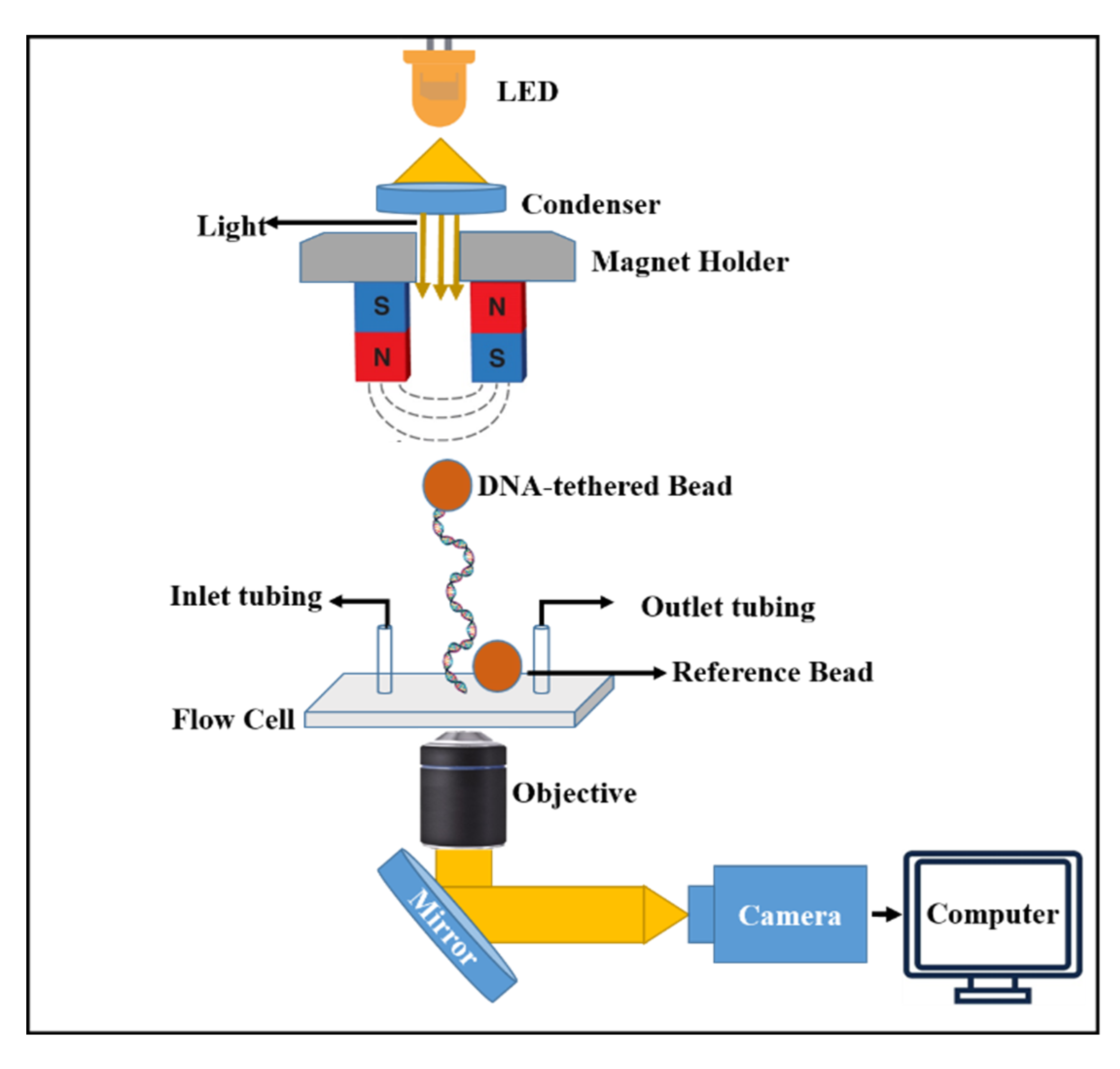
Figure 2.
In the horizontal magnetic tweezers setup, illumination from an LED passes through the condenser to brightly light the flow cell, where the sample is aligned horizontally with respect to the optical axis. One end of the tethered biomolecule is attached—either directly or via a fixed bead to which the tether is connected to the surface of a rigid glass micropipette, and the opposite end is bound to a magnetic bead. The inlet and outlet ports of the flow cell enable continuous buffer exchange and controlled delivery of proteins during the experiment. Light transmitted from the specimen is collected by a high-magnification objective lens and imaged onto a camera to record the bead’s motion. The recorded data are then analyzed computationally to extract the molecule’s mechanical properties, offering insight into its structural dynamics under controlled environmental conditions.
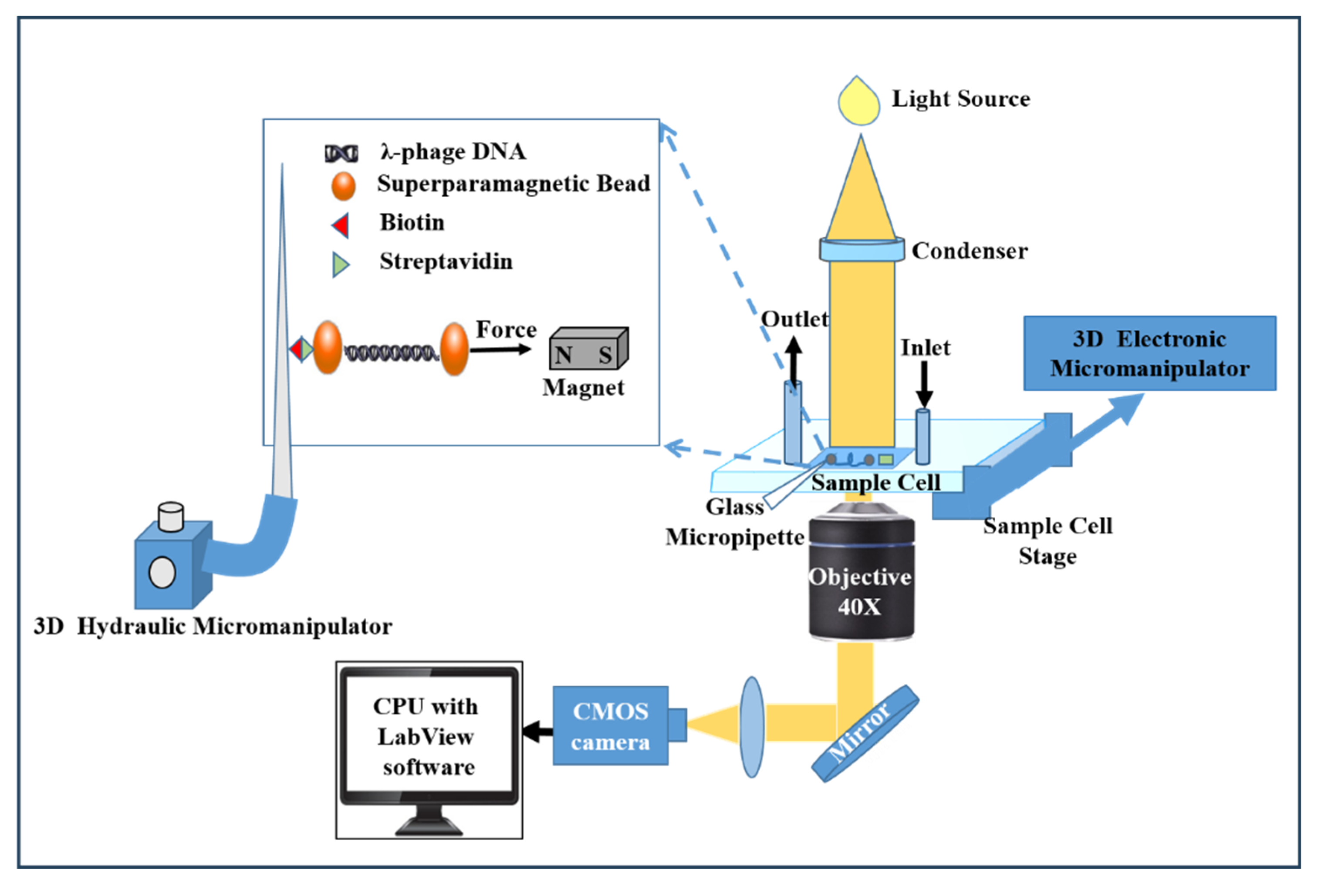
Figure 3.
Adapted quantitative analysis of bead fluctuations [31,38]. (A) The time trace shows the bead’s thermal (Brownian) motion fluctuating around its mean position, reflecting the intrinsic dynamics of the trapped bead. (B) The corresponding power spectral density (PSD) of the bead motion (blue curve) is fitted with a maximum likelihood estimation (MLE) model (red dashed line). The corner frequency obtained from this fit is used to determine the trap stiffness and viscous drag coefficient—two parameters fundamental to accurate force calibration. (C) The Allan variance (AV) of bead fluctuations is plotted as a function of the averaging time (τ); the parameter τ provides a measure of the system’s temporal noise characteristics. The AV data (blue) are fitted with the same MLE model (red dashed line), yielding consistent estimates of the trap stiffness and drag coefficient, which together enable precise determination of the applied force.
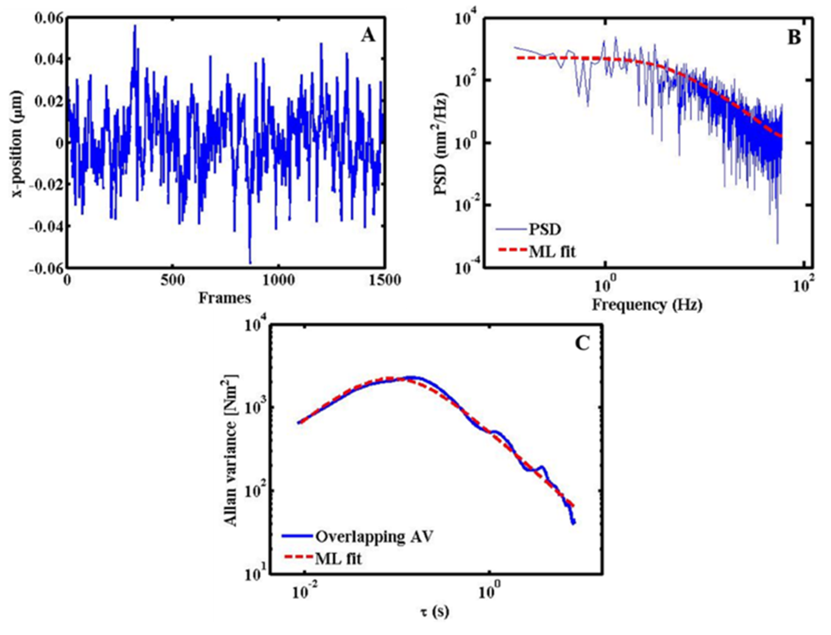
Figure 4.
DNA under applied force. (A) Schematic of the experimental configuration, where a single DNA molecule is tethered between two streptavidin-coated magnetic beads. One bead is immobilized on a biotin-functionalized glass surface, while the other experiences a controlled magnetic force generated by a nearby permanent magnet. (B) The DNA extension trace shows discrete, stepwise increases as the magnet is moved incrementally closer to the sample, thereby increasing the applied force. Each plateau (~30 s) represents a steady state in which the magnet is stationary, allowing the molecule to equilibrate under constant load. As the force approaches ~65 pN, the DNA undergoes a sudden nonlinear elongation—known as the overstretching transition—where the contour length increases by approximately 70% relative to its native B-form (~16.4 µm) due to structural reorganization under high tension. (C) The extension–force relationship with blue data points representing experimental measurements and the cyan curve showing the theoretical prediction based on the worm-like chain (WLC) model. The WLC model is valid for forces where the DNA’s response is dominated by entropic elasticity and in this regime the experimental data and the theoretical prediction show a good fit.
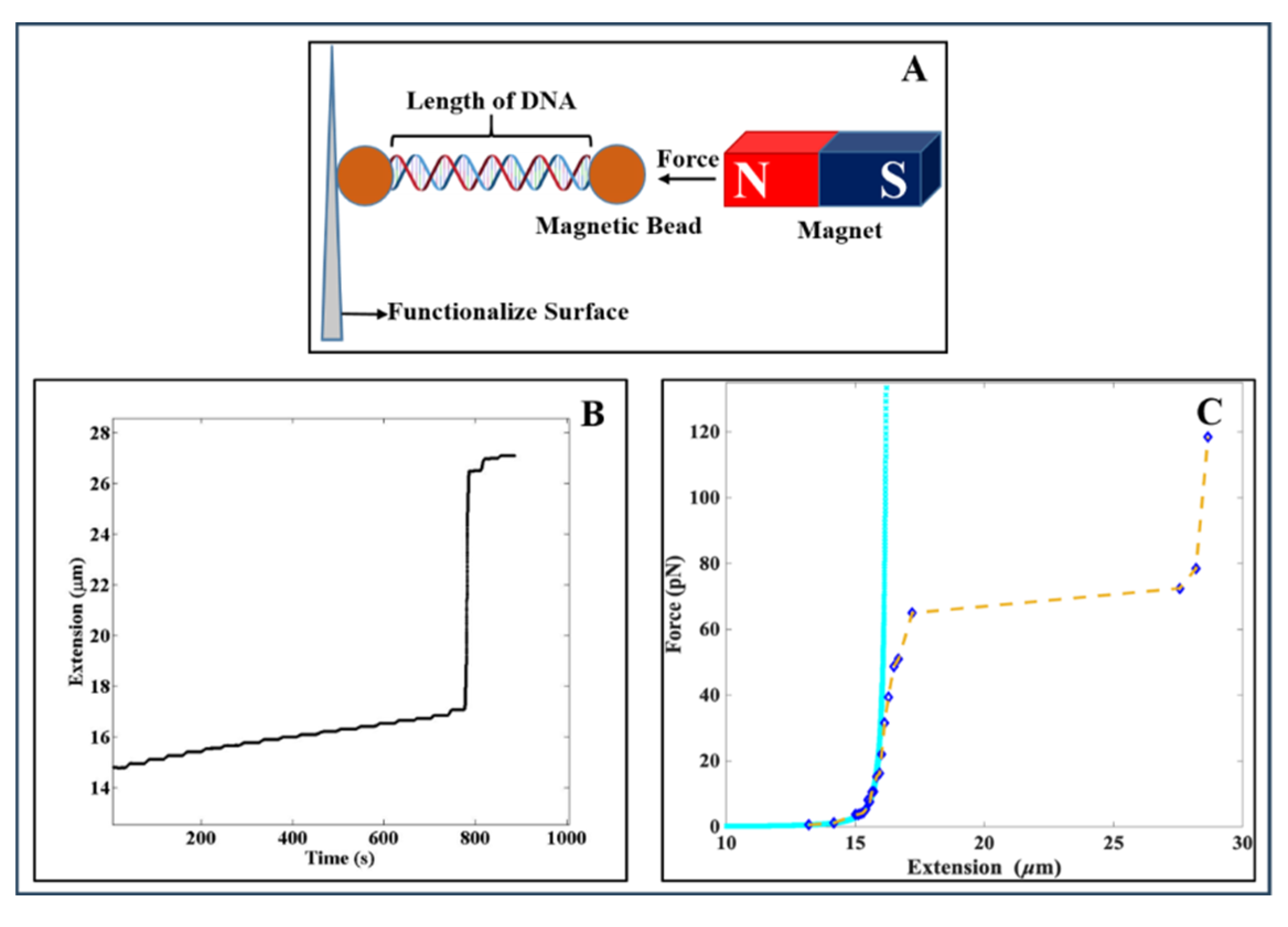
Figure 5.
DNA–protein interactions experiment using horizontal magnetic tweezers (for experimental details see Refs [48,49].) (a) Schematic of the custom-built flow-cell. The protein solution is introduced through the inlet tubing near the magnet, where a single DNA molecule with a magnetic bead at both ends is tethered between a functionalized rigid glass micropipette and a suspended magnetic bead. Excess buffer is withdrawn through an outlet tube to prevent overflow from the open end of the flow cell. (b) Bright-field images showing bead positions before and after protein introduction. The red dotted line serves as a reference; following protein addition, the distance between the two beads decreases, indicating DNA compaction likely caused by wrapping or condensation around the protein. (c) Adapted force–extension curve of the DNA molecule. Experimental data (black) demonstrate a shortening of DNA extension after protein interaction. Upon gradual increase in force, the extension recovers, suggesting progressive unwrapping or dissociation of the protein from DNA. The experimental results are compared with previous work (green) and the theoretical worm-like chain (WLC) model (blue) [49].
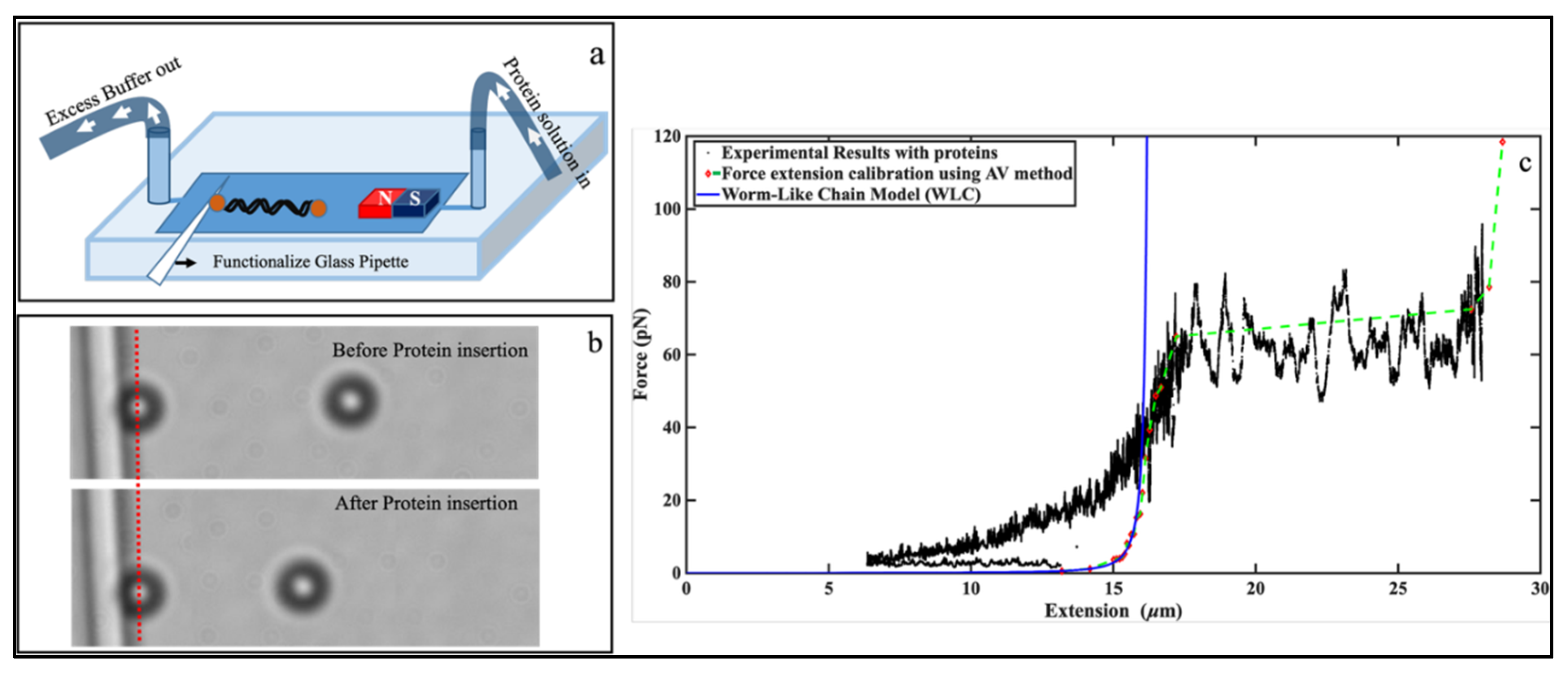
Disclaimer/Publisher’s Note: The statements, opinions and data contained in all publications are solely those of the individual author(s) and contributor(s) and not of MDPI and/or the editor(s). MDPI and/or the editor(s) disclaim responsibility for any injury to people or property resulting from any ideas, methods, instructions or products referred to in the content. |
© 2026 by the authors. Licensee MDPI, Basel, Switzerland. This article is an open access article distributed under the terms and conditions of the Creative Commons Attribution (CC BY) license (http://creativecommons.org/licenses/by/4.0/).
Copyright: This open access article is published under a Creative Commons CC BY 4.0 license, which permit the free download, distribution, and reuse, provided that the author and preprint are cited in any reuse.