Preprint
Article

This version is not peer-reviewed.

In Vitro Evaluation of Polypropylene Microplastics Biodegradation Potential by the Probiotic Bifidobacterium Species

Submitted:

01 October 2026

Posted:

05 October 2026

You are already at the latest version

Abstract

Polypropylene (PP) is a widely used plastic that can fragment into microplastics, raising concerns about environmental accumulation and human health, particularly through ingestion and medical exposure. These microplastics may disrupt microbiota and intestinal function, highlighting the need to explore microbial interactions and biodegradation potential. Although probiotics are increasingly recognised for their benefits to gut health, their potential to degrade microplastics, particularly PP, remains largely unexplored. To address this gap, the present study evaluates the ability of Bifidobacterium bifidum BCMC® 12130, Bifidobacterium longum BCMC® 02120, and Bifidobacterium longum subsp. infantis BCMC® 02129 to biodegrade PP powder and commercially available PP film under controlled laboratory conditions. Bacterial growth was screened in Bushnell-Haas broth (BHB) containing varying PP concentrations, followed by incubation at 25℃ with agitation. The results identified 0.10% (w/v) PP as the optimal concentration, and B. longum BCMC® 02120 demonstrated significant degradation, achieving an 86.7% reduction in PP mass after 30 days (P < 0.05). Scanning electron microscopy revealed dense biofilm formation alongside surface roughening, voids, and erosion, consistent with microbial colonisation and deterioration of the polymer surface. Energy-dispersive X-ray spectroscopy showed a decrease in surface carbon content from 92.62% to 90.00% and an increase in oxygen content from 7.38% to 10.00%, suggesting possible surface oxidation. Together, these preliminary findings suggest that the probiotic strain B. longum BCMC® 02120 has potential for PP microplastic biodegradation. Further studies are needed to confirm polymer breakdown and determine whether this activity could help mitigate microplastic-related effects on gut health.

Keywords: 
;  ;  ;  

1. Introduction

Polypropylene microplastics (PP-MPs) are a major type of synthetic particles measuring less than 5 mm, consisting of a mixture of chemical components. Rochman et al. described these particles as belonging to two main categories based on their source [1]. Primary PP-MPs are deliberately produced for specific applications, such as resin pellets, cosmetic additives, or drug delivery systems [2]. In contrast, secondary PP-MPs originate from the breakdown of larger plastic products, which can occur through environmental processes including photodegradation, mechanical stress, or biodegradation [3]. Among the different classes of microplastics (MPs), polypropylene (PP), polyethylene (PE), and polystyrene (PS) are the most common [4]. Once released, these small particles can move up the food chain and eventually accumulate in humans. Ingestion has been identified as a major pathway of exposure. For instance, PP-MPs have been identified as a major pathway of exposure. For instance, PP-MPs have been detected in fecal samples from both infants and adults [5], and even within human placental tissue [6]. Strikingly, PP is the most prevalent type of microplastic found in adults, accounting for roughly 61% of the total mass detected in feces [7]. Infants consuming formula from PP bottles are particularly vulnerable, with estimated daily exposures ranging between 14,600 and 4,550,000 particles per individual depending on geographic and lifestyle factors [8]. These findings emphasize the urgent need to evaluate the health consequences of PP-MPs, especially given their widespread use in the food industry, from takeaway containers and packaging films to other common wrapping materials [9,10]. As these plastics deteriorate, they release particles that may contaminate food. Indeed, MPs have been reported in table salt, teabags, drinking water and seafood [11,12], with dietary intake alone estimated to contribute between 39,000 and 52,000 MPs per year [13]. Food packaging, particularly single-use takeout materials, has been highlighted as a major contributor [14,15].
Due to their small size and chemical persistence, MPs can move easily across trophic levels and bioaccumulate in organisms [16]. They are also widely distributed in air and water, making them difficult to avoid [17,18,19,20,21]. Consequently, MPs enter human systems through multiple routes, including inhalation. MPs have been identified not only in indoor dust but also in lung tissue samples [22,23,24]. The gastrointestinal tract represents the first line of contact for ingested MPs, and the intestine plays a central role in both nutrient absorption and defense against foreign particles [25]. The intestinal barrier helps reduce the toxicity of MPs, particularly PP-MPs, by preventing their passage into circulation. However, MPs can penetrate this barrier, accumulate in tissues, and disrupt both microbial communities and metabolic processes. Such disruptions may weaken intestinal integrity and increase susceptibility to MP-related toxicity. Despite growing evidence, the specific effects of orally ingested PP-MPs on intestinal health are poorly understood. Human exposure to MPs is almost unavoidable. Studies show that individuals with liver cirrhosis have significantly higher MP accumulation compared with healthy controls (P<0.05) [26]. Similarly, Zhao et al. suggested a possible role of MPs in triggering or worsening inflammatory bowel disease (IBD) [27]. While clinical studies on the protective role of probiotics in humans are still limited, animal models provide some promising findings. In mice, the administration of Lactobacillus strains has been shown to significantly enhance the fecal excretion of PS particles, effectively reducing plastic retention within the gut and surrounding tissues [28,29]. Moreover, probiotic treatment reduced MP retention in the ileum and cecum by up to 67%. These results point to the potential of probiotics as a strategy to reduce MP accumulation and mitigate their long-term health impacts.
The persistence of PP-MPs poses serious environmental challenges, as they undergo natural degradation and remain in ecosystems and food webs for extended periods. Innovative solutions are urgently needed to address this problem. Recent studies have turned to beneficial microbes, such as Bifidobacterium spp., which show promise in degrading PP films and contributing to MP breakdown. By identifying exposure pathways, evaluating risks, and developing microbial solutions, researchers can better understand the broader impact of MPs and design effective mitigation strategies [30]. Despite progress, the health risks posed by MPs remain only partially understood [31].
In this context, the present study investigates the degradation potential of Bifidobacterium bifidum BCMC® 12130, Bifidobacterium longum BCMC® 02120, and Bifidobacterium longum subsp. infantis BCMC® 02129. Specifically, we assess their ability to break down PP films and contribute to the disintegration of MPs. By examining the underlying microbial mechanisms, this work seeks to fill a key knowledge gap and provide insights into sustainable strategies for reducing the health and environmental risks associated with MPs.

2. Materials and Methods

2.1. Preparation of Inoculum for Bifidobacterium sp. Strains

The isolates were recovered from a 10% milk culture by cultivation on de Man–Rogosa–Sharpe (MRS) agar (Oxoid, United Kingdom). 50 mg of each Bifidobacterium strain was then inoculated into a separate tube containing 9 mL of modified Bifidobacterium broth. Briefly, the broth was prepared by dissolving 7.865 g of Bifidobacterium broth powder and 1 g of Yeast Extract Peptone Dextrose (YEPD; HiMedia, India) in 100 mL of distilled water. The inoculum was cultured overnight at 37 °C under anaerobic conditions, reaching a cell density of 10⁵–10⁶ CFU/mL. Following pellet formation, the bacterial cells were stored at 8 °C for subsequent use, and the medium was replaced every 3–4 days (Rossi et al., 2011). The strains used in this study are listed in Table 1 [32].

2.2. Preparation of PP Powder and Screening of PP Concentrations for Bifidobacterium Growth

PP pellets (Sigma-Aldrich, Germany) were ground into a fine powder using a food-grade grinder and then sieved through a 750 μm mesh. The resulting powder was sterilized by autoclaving at 121°C for 20 minutes, followed by oven drying. The sterilized PP powder was stored at room temperature until further use.
Subsequently, 3.27 g of Bushnell-Haas Broth (BHB) powder (HiMedia, India) was dissolved in 1000 mL of distilled water and sterilized by autoclaving to prepare a stock solution. PP powder was then incorporated into 15 mL aliquots of the BHB solution at varying concentrations, as detailed in Table 2. All experiments were conducted in triplicate.
Each PP concentration (including the 0% control) was inoculated individually with one of the Bifidobacterium strains (S01, S02, or S03). A control set (BHB without PP) was maintained to compare bacterial growth in the absence of the polymer. The optical density (OD) at 600 nm was monitored daily for 7 days to assess bacterial growth. Additionally, the viability of all three strains over the 7 days was evaluated using the pour plate method on MRS agar to determine the optimal PP concentration for a subsequent 30-day biodegradation experiment.

2.3. Biodegradation Experiment

2.3.1. Experimental Design-Shake Flask Setup

Based on the preliminary screening results, a concentration of 0.01% (w/v) PP was selected for the 30-day biodegradation study. Each of the three Bifidobacterium strains, at a concentration of 4 × 10⁹ CFU/mL, was individually inoculated into 15 mL of sterilized BHB containing 0.015 g of autoclaved PP powder and PP films, respectively, which served as the sole carbon source.
A control group was established using PP powder and PP films in BHB without microbial inoculation to assess abiotic degradation. All experimental setups were performed in triplicate [33].

2.3.2. Preparation of PP Samples

The first form of PP was commercial PP sheets, which were cut into 2 × 2 cm² squares. Each film was pre-weighed and sterilized by immersing in 70% ethanol for 10 seconds, followed by two rinses with sterile distilled water. The films were then air-dried in a laminar flow hood before being aseptically transferred into Erlenmeyer flasks containing 15 mL of BHB inoculated with the respective Bifidobacterium strains.
Followed by, PP pellets were ground using a food-grade grinder and sieved through a 750 μm mesh. The powder was sterilized by autoclaving at 121°C for 20 minutes and oven-dried. An amount of 0.0015 g of dried PP powder was added to 100 mL flasks containing 15 mL of BHB inoculated with each strain (S01, S02, S03) [33].

2.3.3. Bacterial Viability

Bacterial viability was assessed by colony-forming unit (CFU) counts. Sampling was performed on days 0, 1, 2, 4, 7, 10, 20, and 30 using the pour plate method on MRS agar incubated anaerobically [34].

2.3.4. Post-Incubation Recovery and Biodegradation Determination

After 30 days, the PP films and powder were carefully removed from the flasks and subjected to two washing steps: first with Phosphate Buffer Solution (PBS) and then with sterile distilled water. The samples were then air-dried in a laminar flow hood [33]. For PP films, the final dry mass were recorded, and biodegradation (%) was calculated using the standard gravimetric mass loss formula (Equation 1):
Biodegradation   ( % )   = W i − W f W i × 100
Where:
  • Wi is the initial dry mass of the PP sample before degradation (g)
  • Wf is the final dry mass of the recovered PP sample after incubation (g)

2.3.5. Storage of Samples for Further Analysis

Post-incubation, the retrieved PP films and powders were stored in airtight containers containing desiccant to prevent moisture accumulation. Samples were kept at room temperature until further physicochemical and morphological analyses.

2.4. SEM-EDX Analysis

PP powder was extracted from BHB that had been inoculated with Bifidobacterium sp. The extracted PP powder underwent analysis through the EDX Oxford Instrument INCA system; software version 5.02 (Oxford Instruments, UK) was used to evaluate alterations in the PP material. The surface morphology and elemental composition of the PP powder were examined using Scanning Electron Microscopy (SEM), while EDX facilitated the identification and quantification of elements present on the surfaces of the powder, with particular attention to indications of bacterial biofilm formation or other modifications resulting from microbial activity [35].

2.5. Statistical Analysis

All experiments were conducted with a minimum of three replicates, unless stated otherwise. Results were expressed as mean ± standard deviation. Statistical significance between group means was evaluated using One-way Analysis of Variance (ANOVA), followed by Tukey’s post-hoc test to assess pairwise differences. Analyses were performed using SPSS version 22.0 (IBM Corp., Armonk, NY, USA), and a p-value of less than 0.05 was considered statistically significant.

3. Results

3.1. Optimization of Polypropylene (PP) Concentration

Preliminary screening was conducted to determine the optimal PP concentration for the 30-day incubation study. Figure 1 shows the growth curve of S02 across different PP concentrations over 7 days. Among the tested concentrations (0. 0.01, 0.10, and 1.00 % w/v), 0.10% PP demonstrated a markedly higher optical density (OD600), particularly from Day 3 onwards, indicating enhanced bacterial growth of Bifidobacterium longum BCMC® 02120 (S02). In comparison, 0.01% PP showed moderate but relatively stable growth, while 1.00% PP exhibited suppressed growth, suggesting a potential inhibitory effect at higher concentrations. The control (0% PP) maintained consistently low OD600 values throughout the incubation period.

2.2. Enumeration of Lactic Acid Bacteria in BHB with PP

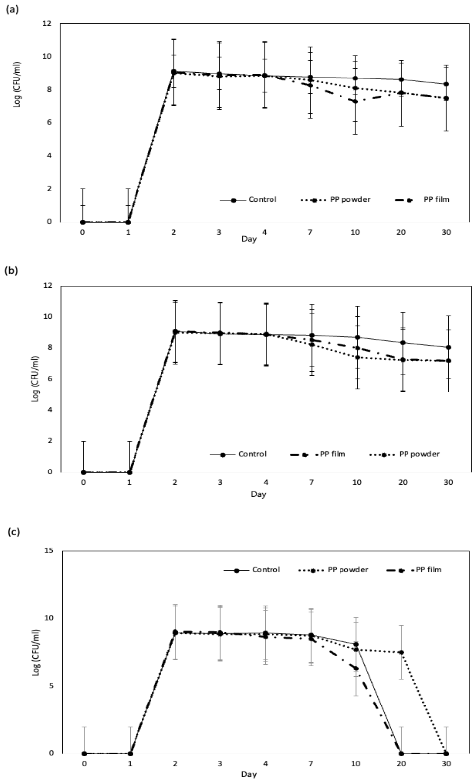
Colony-forming unit (CFU) enumeration over 30 days showed an initial increase in viable cell counts across all treatments, including the BHB control without PP. After Day 5, a gradual decline in planktonic CFU counts was observed in PP-containing systems. The reduction was more pronounced in PP film compared to PP powder (Figure 2).

3.3. Post-Incubation Recovery and Mass Loss Analysis

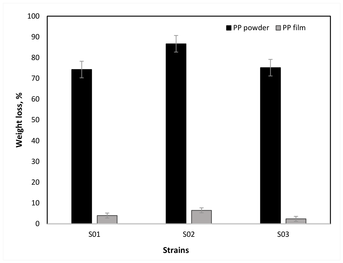
Mass loss analysis after 30 days showed varying degradation levels among strains S01, S02, and S03 (Figure 3). Strain S02 exhibited the highest degradation, with PP microplastic powder showing up to 86.7% mass loss. PP film samples showed comparatively lower mass reduction. All strains demonstrated measurable degradation activity at ambient temperature (25°C ± 2).

3.4. SEM-EDX Analysis

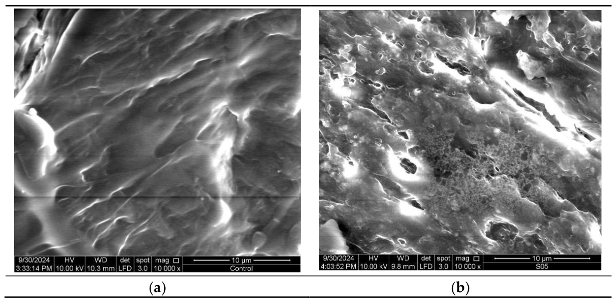
SEM analysis revealed distinct differences between control and treated PP samples. The control PP surface appeared smooth, whereas the S02-treated sample exhibited visible surface roughness, voids, pits, and irregularities after 30 days (Figure 4).
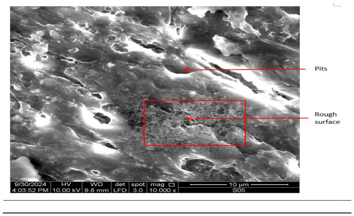
EDX analysis showed a decrease in carbon content from 92.62% (control) to 90.00% (S02-treated), accompanied by an increase in oxygen content from 7.38% to 10.00% (Table 3). However, these differences were not statistically significant (p > 0.05). SEM micrographs (Figure 5) further revealed microbial colonization and formation of dense clusters on the PP surface.

4. Discussion

The use of BHB, a carbon-deficient medium, provides a stringent platform for evaluating microbial interactions with PP, thereby minimizing confounding contributions from external carbon sources [36]. Within this system, the enhanced growth of Bifidobacterium longum BCMC® 02120 (S02) at 0.01% (w/v) PP suggests that this concentration offers an optimal balance between particle dispersion and accessible surface area for microbial colonization. At lower PP loading, limited surface availability may constrain bacterial attachment, whereas higher loading may promote particle aggregation, reducing effective microbial access [37]. Nevertheless, increased optical density should not be interpreted as direct evidence of polymer utilization, as microbial proliferation may still be supported by residual nutrients or trace organic components [38,39].
Temporal dynamics of viable cell counts further support this interpretation. The initial increase in CFU across all treatments, including the control, likely reflects nutrient carryover from inoculum preparation or background organic residues. The subsequent decline in planktonic populations in PP-containing systems after Day 5 suggests a shift from free-living to surface-associated growth, consistent with microbial attachment and biofilm formation. Such surface colonization is widely recognized as a prerequisite for polymer degradation, as it facilitates localized enzymatic activity and sustained interaction with the substrate [38,39,40]. The comparatively greater stability observed in PP powder relative to film further underscores the importance of surface area, with finer particles providing enhanced sites for microbial adhesion and retention [37,41].
Notably, the substantial mass reduction observed, particularly the 86.7% loss in PP powder mediated by strain S02, indicates a strong interaction between the microbial system and the polymer matrix. While this value exceeds typical degradation rates reported for PP by pure cultures (10–12%), it aligns with emerging evidence that certain microbial systems, under optimized conditions, can achieve markedly enhanced degradation efficiencies, including up to 90% degradation of recalcitrant polymers such as LDPE [41,42]. The superior performance observed in PP powder relative to film further reinforces the role of physical morphology in governing biodegradation outcomes, likely through its influence on biofilm development and enzyme–substrate proximity [37].
Microscopic and elemental analyses provide additional mechanistic insight. SEM imaging revealed pronounced surface deterioration in S02-treated samples, characterized by increased roughness, pits, and fissures, indicative of progressive polymer disruption [43]. These structural alterations are consistent with surface erosion driven by microbial colonization and metabolic activity [42]. Concurrently, EDX analysis showed a reduction in carbon content alongside an increase in oxygen, suggesting oxidative modification of the polymer surface. The introduction of oxygen-containing functional groups, such as carbonyl, hydroxyl, and carboxyl groups, is a critical step in converting the chemically inert polyolefin backbone into a more degradable form [41,44,45].
These transformations are likely driven by a combination of microbial metabolism and extracellular enzymatic processes. Organic acids such as lactic and acetic acid produced by Bifidobacterium spp. may lower local pH and contribute to chemical weakening of the polymer matrix [38,46]. Acidification has been shown to facilitate hydrolytic processes, particularly in the presence of susceptible additives or oxidized sites [47]. In parallel, extracellular enzymes such as esterases, oxidoreductases, and depolymerases may contribute to oxidative chain scission, even under limited oxygen availability, through interactions with dissolved or residual oxygen [48,49,50]. Enzymatic oxidation mediated by laccases and peroxidases has also been implicated in polyolefin degradation pathways [51,52].
The presence of dense microbial aggregates on PP surfaces further supports the formation of biofilm-like structures, which are central to effective polymer degradation. Biofilms enhance microbial retention and create localized microenvironments that concentrate enzymes and metabolic byproducts, thereby accelerating degradation processes [41,53]. The porous architecture of PP microplastic powder likely further facilitates microbial adhesion and colonization [54]. While such systems may potentially support secondary microbial populations capable of further polymer transformation, this remains beyond the scope of the present study [55].
Despite these findings, interpretation should be made with appropriate caution. Although the combined evidence of mass loss, surface modification, and elemental changes strongly indicates significant polymer–microbe interaction, these parameters alone do not confirm complete biodegradation or mineralization of PP. Definitive validation would require additional analyses such as molecular mass distribution profiling, identification of degradation intermediates, and carbon mineralization assays.

5. Conclusions

This study shows that strain S02 (Bifidobacterium longum BCMC® 02120) is capable of breaking down PP-MPs through processes such as oxidative modification, surface erosion, and biofilm formation. The strongest bacterial activity was observed at a PP concentration of 0.10%, where S02 achieved its highest degradation efficiency, leading to an 86.7% reduction in PP powder mass. SEM revealed clear surface damage, including pits and voids, which are consistent with microbial colonization and enzymatic attack on the polymer. Complementary SEM-EDX analysis further supported these observations, showing a decrease in carbon content (from 92.62% to 90.00%) together with an increase in oxygen content (from 7.38% to 10.00%), suggesting that oxidative metabolism contributed significantly to the degradation process. The formation of S02 biofilms highlights the importance of microbial adhesion in polymer breakdown, supporting the idea that surface characteristics influence both colonization and enzymatic activity. Taken together, these findings identify S02 as a promising strain for the biodegradation of PP microplastics and point to its potential use in bioremediation strategies aimed at reducing plastic pollution. Further research will be needed to unravel the detailed metabolic pathways involved and to test its effectiveness under real environmental conditions.

Author Contributions

Conceptualization, S.F.O.; methodology, S.P., H.F.A, and A.A.A.; software, S.P.; validation, S.P.; formal analysis, S.P.; investigation, S.P.; resources, S.P.; data curation, S.P. and A.A.A.; writing—original draft preparation, S.P. and H.F.A,; writing—review and editing, A.A.A., N.M.T., A.A.M.N., S.F.O. and H.F.A,; visualization, S.P.; supervision, A.A.A. and H.F.A,; project administration, S.P.; funding acquisition, S.P., A.A.A. and S.F.O. All authors have read and agreed to the published version of the manuscript.

Funding

This research was supported by internal laboratory funding from B-Crobes Laboratory Sdn. Bhd. No grant number is applicable.

Institutional Review Board Statement

Not applicable.

Data Availability Statement

The original contributions presented in this study are included in the article. Further inquiries can be directed to the corresponding authors.

Acknowledgments

The author, Soopna Puspanadan (Dr), wishes to express sincere gratitude to B-Crobes Laboratory Sdn Bhd for the generous financial support that made this research possible. The author also extends heartfelt appreciation to the dedicated Research and Development team members at B-Crobes Laboratory Sdn. Bhd. for their invaluable assistance and technical support throughout the research.

Conflicts of Interest

The authors declare no conflicts of interest.:.

Abbreviations

PP Polypropylene
BHB Bushnell-Hass broth
SEM Scanning Electron Microscopy
MPs Microplastics
PE Polyethylene
PS Polystyrene
CFU Colony-forming unit
EDX Energy Dispersive X-ray

References

  1. Rochman, C.M.; Browne, M.A.; Underwood, A.J.; van Franeker, J.A.; Thompson, R.C.; Amaral-Zettler, L.A. The ecological impacts of marine debris: Unraveling the demonstrated evidence from what is perceived. Ecology 2016, 97, 302–312. [Google Scholar] [CrossRef]
  2. Shim, W.J.; Hong, S.H.; Eo, S. Marine Microplastics: Abundance, Distribution, and Composition. In Microplastic Contamination in Aquatic Environments; Zeng, E.Y., Ed.; Elsevier: Amsterdam, The Netherlands, 2018; pp. 1–26. [Google Scholar] [CrossRef]
  3. Xu, S.; Ma, J.; Ji, R.; Pan, K.; Miao, A.J. Microplastics in aquatic environments: Occurrence, accumulation, and biological effects. Sci. Total Environ. 2020, 703, 134699. [Google Scholar] [CrossRef]
  4. Zolotova, N.; Kosyreva, A.; Dzhalilova, D.; Fokichev, N.; Makarova, O. Harmful effects of the microplastic pollution on animal health: a literature review. PeerJ 2022, 10, e13503. [Google Scholar] [CrossRef]
  5. Schwabl, P.; Köppel, S.; Königshofer, P.; Bucsics, T.; Trauner, M.; Reiberger, T.; Liebmann, B. Detection of various microplastics in human stool: A prospective case series. Ann. Intern. Med. 2019, 171, 453–457. [Google Scholar] [CrossRef]
  6. Ragusa, A.; Svelato, A.; Santacroce, C.; Catalano, P.; Notarstefano, V.; Carnevali, O.; Papa, F.; Rongioletti, M.C.A.; Baiocco, F.; Draghi, S.; D’Amore, E.; Rinaldo, D.; Matta, M.; Giorgini, E. Plasticenta: First evidence of microplastics in human placenta. Environ. Int. 2021, 146, 106274. [Google Scholar] [CrossRef]
  7. Zhang, N.; Li, Y.B.; He, H.R.; Zhang, J.F.; Ma, G.S. You are what you eat: Microplastics in the feces of young men living in Beijing. Sci. Total Environ. 2021, 767, 144345. [Google Scholar] [CrossRef]
  8. Li, D.; Shi, Y.; Yang, L.; Xiao, L.; Kehoe, D.K.; Gun’ko, Y.K.; Boland, J.J.; Wang, J.J. Microplastic release from the degradation of polypropylene feeding bottles during infant formula preparation. Nat. Food 2020, 1, 746–754. [Google Scholar] [CrossRef]
  9. Du, F.; Cai, H.; Zhang, Q.; Chen, Q.; Shi, H. Microplastics in take-out food containers. J. Hazard Mater. 2020, 399, 122969. [Google Scholar] [CrossRef]
  10. Iñiguez, M.E.; Conesa, J.A.; Fullana, A. Microplastics in Spanish table salt. Sci. Rep. 2017, 7, 8620. [Google Scholar] [CrossRef]
  11. Wang, Q.; Zhu, X.; Hou, C.; Wu, Y.; Teng, J.; Zhang, C.; Tan, H.; Shan, E.; Zhang, W.; Zhao, J. Microplastic uptake in commercial fishes from the Bohai Sea, China. Chemosphere 2021, 263, 127962. [Google Scholar] [CrossRef]
  12. Hernandez, L.M.; Xu, E.G.; Larsson, H.C.E.; Tahara, R.; Maisuria, V.B.; Tufenkji, N. Plastic teabags release billions of microparticles and nanoparticles into tea. Environ. Sci. Technol. 2019, 53, 12300–12310. [Google Scholar] [CrossRef]
  13. Cox, K.D.; Covernton, G.A.; Davies, H.L.; Dower, J.F.; Juanes, F.; Dudas, S.E. Human consumption of microplastics. Environ. Sci. Technol. 2019, 53, 7068–7074. [Google Scholar] [CrossRef]
  14. Jin, M.; Wang, X.; Ren, T.; Wang, J.; Shan, J. Microplastics contamination in food and beverages: Direct exposure to humans. J. Food Sci. 2021, 86, 2816–2837. [Google Scholar] [CrossRef]
  15. Trachana, V.; Ntoumou, E.; Anastasopoulou, L.; Tsezou, A. Studying microRNAs in osteoarthritis: Critical overview of different analytical approaches. Mech. Ageing Dev. 2018, 171, 15–23. [Google Scholar] [CrossRef]
  16. Vethaak, A.D.; Legler, J. Microplastics and human health. Science 2021, 371, 672–674. [Google Scholar] [CrossRef]
  17. Andrady, A.L. Microplastics in the marine environment. Mar. Pollut. Bull. 2011, 62, 1596–1605. [Google Scholar] [CrossRef]
  18. Waldschläger, K.; Lechthaler, S.; Stauch, G.; Schüttrumpf, H. The way of microplastic through the environment - Application of the source-pathway-receptor model (review). Sci. Total Environ. 2020, 713, 136584. [Google Scholar] [CrossRef]
  19. Fox, S.; Stefánsson, H.; Peternell, M.; Zlotskiy, E.; Ásbjörnsson, E.J.; Sturkell, E.; Wanner, P.; Konrad-Schmolke, M. Physical characteristics of microplastic particles and potential for global atmospheric transport: A meta-analysis. Environ. Pollut. 2024, 342, 122938. [Google Scholar] [CrossRef]
  20. Michels, J.; Stippkugel, A.; Lenz, M.; Wirtz, K.; Engel, A. Rapid aggregation of biofilm-covered microplastics with marine biogenic particles. Proc. Biol. Sci. 2018, 285, 20181203. [Google Scholar] [CrossRef]
  21. Zettler, E.R.; Mincer, T.J.; Amaral-Zettler, L.A. Life in the "plastisphere": microbial communities on plastic marine debris. Environ. Sci. Technol. 2013, 47, 7137–7146. [Google Scholar] [CrossRef]
  22. Amato-Lourenço, L.F.; Carvalho-Oliveira, R.; Júnior, G.R.; Dos Santos Galvão, L.; Ando, R.A.; Mauad, T. Presence of airborne microplastics in human lung tissue. J. Hazard. Mater. 2021, 416, 126124. [Google Scholar] [CrossRef]
  23. Yan, Z.; Liu, Y.; Zhang, T.; Zhang, F.; Ren, H.; Zhang, Y. Analysis of Microplastics in Human Feces Reveals a Correlation between Fecal Microplastics and Inflammatory Bowel Disease Status. Environ. Sci. Technol. 2022, 56, 414–421. [Google Scholar] [CrossRef]
  24. Zhu, J.; Zhang, X.; Liao, K.; Wu, P.; Jin, H. Microplastics in dust from different indoor environments. Sci. Total Environ. 2022, 833, 155256. [Google Scholar] [CrossRef]
  25. Wu, C.; Tanaka, K.; Tani, Y.; Bi, X.; Liu, J.; Yu, Q. Effect of particle size on the colonization of biofilms and the potential of biofilm-covered microplastics as metal carriers. Sci. Total Environ. 2022, 821, 153265. [Google Scholar] [CrossRef]
  26. Horvatits, T.; Tamminga, M.; Liu, B.; Sebode, M.; Carambia, A.; Fischer, L.; Püschel, K.; Huber, S.; Fischer, E.K. Microplastics detected in cirrhotic liver tissue. EBioMedicine 2022, 82, 104147. [Google Scholar] [CrossRef]
  27. Zhao, Y.; Liu, S.; Xu, H. Effects of microplastic and engineered nanomaterials on inflammatory bowel disease: A review. Chemosphere 2023, 326, 138486. [Google Scholar] [CrossRef]
  28. Teng, X.; Zhang, T.; Rao, C. Novel probiotics adsorbing and excreting microplastics in vivo show potential gut health benefits. Front. Microbiol. 2025, 15, 1522794. [Google Scholar] [CrossRef]
  29. Zhao, L.; Wei, Z.; Wang, Y.; Chen, W.; Zhang, W.; Xie, M.; Chen, H.; Zhang, Y.; Gao, H.; Jiang, X. Lactiplantibacillus plantarum ZP-6 mitigates polystyrene nanoplastics-induced liver damage in colitis mice via the gut-liver axis. Front. Microbiol. 2025, 16, 1626941. [Google Scholar] [CrossRef]
  30. Zhang, Q.; Xu, E.G.; Li, J.; Chen, Q.; Ma, L.; Zeng, E.Y.; Shi, H. A Review of Microplastics in Table Salt, Drinking Water, and Air: Direct Human Exposure. Environ. Sci. Technol. 2020, 54, 3740–3751. [Google Scholar] [CrossRef]
  31. Lee, Y.; Cho, J.; Sohn, J.; Kim, C. Health Effects of Microplastic Exposures: Current Issues and Perspectives in South Korea. Yonsei Med. J. 2023, 64, 301–308. [Google Scholar] [CrossRef]
  32. Rossi, M.; Amaretti, A.; Raimondi, S. Folate production by probiotic bacteria. Nutrients 2011, 3, 118–134. [Google Scholar] [CrossRef]
  33. Auta, H.S.; Emenike, C.U.; Fauziah, S.H. Screening of Bacillus strains isolated from mangrove ecosystems in Peninsular Malaysia for microplastic degradation. Environ. Pollut. 2017, 231, 1552–1559. [Google Scholar] [CrossRef]
  34. Soukoulis, C.; Behboudi-Jobbehdar, S.; Yonekura, L.; Parmenter, C.; Fisk, I.D. Stability of Lactobacillus rhamnosus GG in prebiotic edible films. Food Chem. 2014, 159, 302–308. [Google Scholar] [CrossRef]
  35. Bonhomme, S.; Cuer, A.; Delort, A.-M.; Lemaire, J.; Sancelme, M.; Scott, G. Environmental biodegradation of polyethylene. Polym. Degrad. Stabil. 2003, 81, 441–452. [Google Scholar] [CrossRef]
  36. Bushnell, L.D.; Haas, H.F. The Utilization of Certain Hydrocarbons by Microorganisms. J. Bacteriol. 1941, 41, 653–673. [Google Scholar] [CrossRef]
  37. Skariyachan, S.; Setlur, A.S.; Naik, S.Y.; Naik, A.A.; Usharani, M.; Vasist, K.S. Enhanced biodegradation of low and high-density polyethylene by novel bacterial consortia formulated from plastic-contaminated cow dung under thermophilic conditions. Environ. Sci. Pollut. Res. Int. 2017, 24, 8443–8457. [Google Scholar] [CrossRef]
  38. Tokiwa, Y.; Calabia, B.P.; Ugwu, C.U.; Aiba, S. Biodegradability of plastics. Int. J. Mol. Sci. 2009, 10, 3722–3742. [Google Scholar] [CrossRef]
  39. Singh, G.; Singh, A.K.; Bhatt, K. Biodegradation of polythenes by bacteria isolated from soil. Int. J. Res. Dev. Pharm. Life Sci. 2016, 5, 2056–2062. Available online: https://www.researchgate.net/publication/295186158_BIODEGRADATION_OF_POLYTHENES_BY_BACTERIA_ISOLATED_FROM_SOIL. [CrossRef]
  40. Speranza, B.; Liso, A.; Russo, V.; Corbo, M.R. Evaluation of the potential of biofilm formation of Bifidobacterium longum subsp. infantis and Lactobacillus reuteri as competitive biocontrol agents against pathogenic and food spoilage bacteria. Microorganisms 2020, 8, 177. [Google Scholar] [CrossRef]
  41. Kale, S.K.; Deshmukh, A.G.; Dudhare, M.S.; Patil, V.B. Microbial degradation of plastic: A review. J. Biochem. Tech. 2015, 6, 952–961. Available online: https://jbiochemtech.com/storage/models/article/RLVOybv0ejQcKcpQrrJRX6AaXOgj4YN8AcbTDS2FzqLBjMD84buciQL8J332/microbial-degradation-of-plastic-a-review.pdf. [CrossRef]
  42. Lv, S.; Li, Y.; Zhao, S.; Shao, Z. Biodegradation of Typical Plastics: From Microbial Diversity to Metabolic Mechanisms. Int. J. Mol. Sci. 2024, 25, 593. [Google Scholar] [CrossRef]
  43. Restrepo-Flórez, J.M.; Bassi, A.; Thompson, M.R. Microbial degradation and biodegradation of plastics: A review. Int. Biodeterior. Biodegrad. 2014, 88, 83–90. [Google Scholar] [CrossRef]
  44. Arutchelvi, P.; Sudhakar, M.; Arkatkar, A.; Doble, M.; Bhaduri, S.; Palaniswamy, B. Biodegradation of polyethylene and polypropylene. Indian J. Biotechnol. 2008, 7, 9–22. Available online: https://www.researchgate.net/publication/228090462_Biodegradation_of_polyethylene_and_polypropylene.
  45. Oluwole, O.A.; Oluyege, J.O.; Olowomofe, T.O. Biodegradation of polyethylene based films used in water packaging by dumpsite bacteria. Bioremediat. J. 2024, 28, 1–13. [Google Scholar] [CrossRef]
  46. Rivière, A.; Selak, M.; Lantin, D.; Leroy, F.; De Vuyst, L. Bifidobacteria and butyrate-producing colon bacteria: Importance and strategies for their stimulation in the human gut. Front. Microbiol. 2016, 7, 979. [Google Scholar] [CrossRef]
  47. Gewert, B.; Plassmann, M.M.; MacLeod, M. Pathways for degradation of plastic polymers floating in the marine environment. Environ. Sci. Process. Impacts 2015, 17, 1513–1521. [Google Scholar] [CrossRef]
  48. Shah, A.A.; Hasan, F.; Hameed, A.; Ahmed, S. Biological degradation of plastics: a comprehensive review. Biotechnol. Adv. 2008, 26, 246–265. [Google Scholar] [CrossRef]
  49. Yoon, M.G.; Jeon, H.J.; Kim, M.N. Biodegradation of polyethylene by a soil bacterium and AlkB cloned recombinant cell. J. Bioremed. Biodeg. 2012, 3, 145. [Google Scholar] [CrossRef]
  50. Ghosh, S.K.; Pal, S.; Ray, S. Study of microbes having potentiality for biodegradation of plastics. Environ. Sci. Pollut. Res. Int. 2013, 20, 4339–4355. [Google Scholar] [CrossRef]
  51. Wei, R.; Zimmermann, W. Microbial enzymes for the recycling of recalcitrant petroleum-based plastics: how far are we? Microb. Biotechnol. 2017, 10, 1308–1322. [Google Scholar] [CrossRef]
  52. Montazer, Z.; Habibi Najafi, M.B.; Levin, D.B. Challenges with verifying microbial degradation of polyethylene. Polymers 2020, 12, 123. [Google Scholar] [CrossRef]
  53. Flemming, H.C.; Wingender, J. The biofilm matrix. Nat. Rev. Microbiol. 2010, 8, 623–633. [Google Scholar] [CrossRef]
  54. Dubey, A.P.; Thalla, A.K. Bioprospecting indigenous bacteria from landfill leachate for enhanced polypropylene microplastics degradation. J. Hazard. Mater. 2025, 487, 137139. [Google Scholar] [CrossRef]
  55. Urbanek, A.K.; Rymowicz, W.; Mirończuk, A.M. Degradation of plastics and plastic-degrading bacteria in cold marine habitats. Appl. Microbiol. Biotechnol. 2018, 102, 7669–7678. [Google Scholar] [CrossRef]
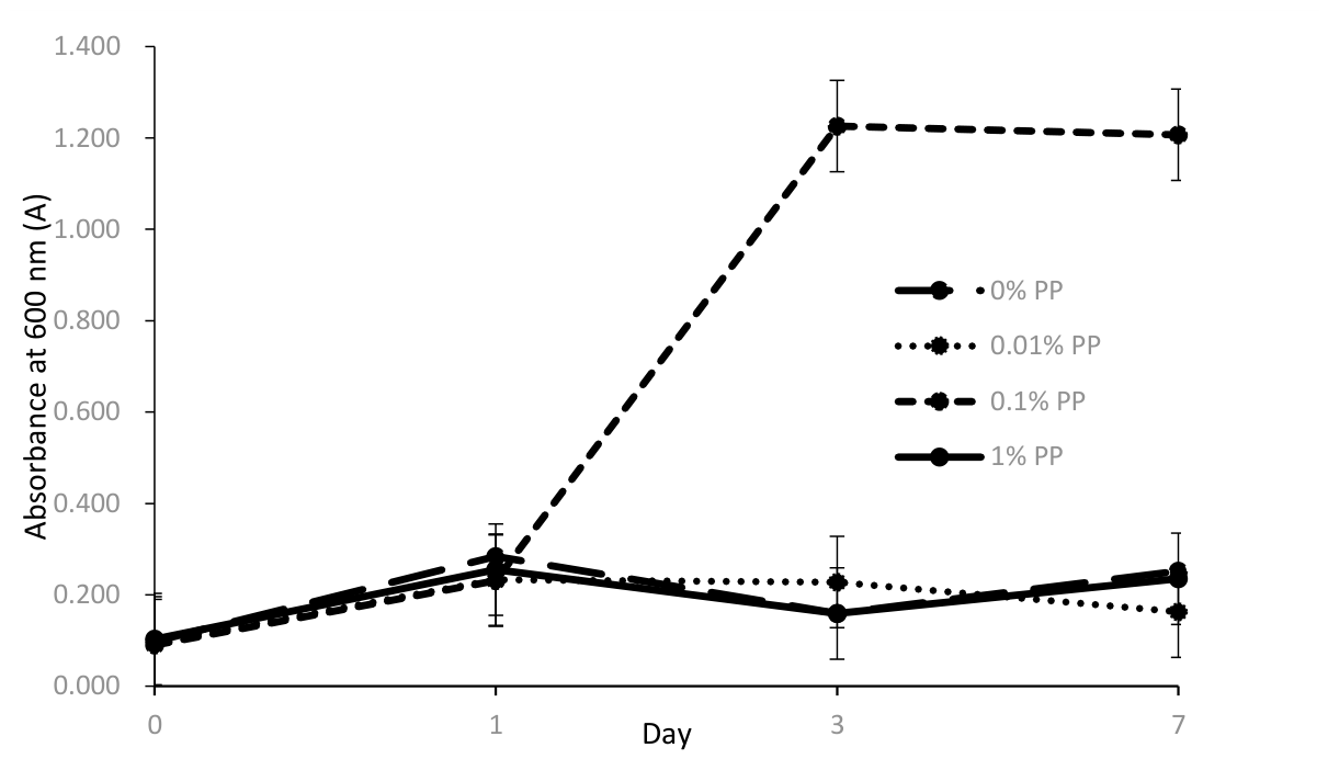
Figure 1. Growth curve of S02 across different PP concentrations over 7 days. Error bars represent mean ± standard deviation of triplicates.
Preprints 236198 g001
Figure 2. Microbial growth trend for: (a) Bifidobacterium bifidum BCMC® 12130, (b) Bifidobacterium longum BCMC® 02120, (c) Bifidobacterium longum subsp. infantis BCMC® 02129. CFU values below 10 CFU/mL were considered below the detection limit and recorded as 0 Log10 (CFU/mL). Error bars represent mean ± standard deviation.
Preprints 236198 g002
Figure 3. Comparative mass loss of PP powder and film across different strains. Different superscript letters indicate statistically significant differences (P < 0.05). Error bars represent mean ± standard deviation.
Preprints 236198 g003
Figure 4. SEM images at 10,000x magnification in BHB after 30 days incubation period (a) PP powder only (Control) (b) PP powder with S02.
Preprints 236198 g004
Figure 5. SEM micrograph at 10,000× magnification showing colonization of strain S02 on the surface of PP powder after 30 days of incubation. The image reveals the presence of microbial clusters and biofilm-like roughened areas (highlighted in red box), as well as surface defects such as pits and erosion features indicative of biodegradation activity.
Preprints 236198 g005
Table 1. List of strains and code used for the current experiment.
List of strains Code
Bifidobacterium bifidum BCMC® 12130 S01
Bifidobacterium longum BCMC® 02120 S02
Bifidobacterium longum subsp. infantis BCMC® 02129 S03
Table 2. Preparation of BHB with Different Concentrations of PP.
PP powder (g) Volume of BHB
(mL)
PP concentration
(%, w/v)
0.0000 15 0
0.0015 15 0.01
0.0150 15 0.1
0.1500 15 1.0
BHB= Bushnell-Haas Broth, PP=polypropylene.
Table 3. The Carbon and Oxygen Mass Percentages of PP powder, determined by SEM-EDX analysis.
Carbon Mass, % Oxygen Mass, %

Control
92.62 ± 2.00a 7.38 ± 2.00a

S02 (Bifidobacterium longum BCMC® 02120)

90.00 ± 2.60a

10.00 ± 2.60a
Values with different letters are significant at p < 0.05 between groups.
Disclaimer/Publisher’s Note: The statements, opinions and data contained in all publications are solely those of the individual author(s) and contributor(s) and not of MDPI and/or the editor(s). MDPI and/or the editor(s) disclaim responsibility for any injury to people or property resulting from any ideas, methods, instructions or products referred to in the content.
Copyright: This open access article is published under a Creative Commons CC BY 4.0 license, which permit the free download, distribution, and reuse, provided that the author and preprint are cited in any reuse.