

Submitted:
30 September 2026
Posted:
02 October 2026
You are already at the latest version
Abstract
In the frame of our ongoing research on multitarget alkaloid-derived anticancer agents, relying on a modular synthetic strategy terminated by well-established sequential Sonogashira and CuAAC protocols, we synthesized a systematic selection of chalcone-vindoline hybrids incorporating 1,4-disubstituted 1,2,3-triazole as a linker. The use of the same synthetic approach led to the construction of a zidovudine-derived vindoline hybrid, which served as a negative control highlighting the essential contribution of the chalcone moiety to the antiproliferative activity. Two analogous aryl-substituted chalcone hybrids 14a,b were also prepared to assess the contribution of the vindoline fragment to the desired effect. The targeted hybrids and the references were evaluated for their antiproliferative activity against a panel of human adherent gynecological cancer cell lines (HeLa, SiHa, MCF-7, MDA-MB-231 and A2780) as well as on non-malignant fibroblasts (NIH/3T3). Vindoline hybrids 10a and 10i comprising 3,4,5- and 1,4,6-trimethoxyphenyl-substituted chalcone fragments, respectively, displayed substantial activity, featuring IC50 values lower than 0.4 µM on all the investigated cancer cell lines. On the other hand, hybrids 10c–e containing monomethoxy-substituted chalcones, also featuring submicromolar IC50 values on malignant cells, showed moderately lower activities against non-malignant fibroblasts (NIH/3T3), indicating a certain degree of cancer selectivity. The related ferrocene-derived hybrid (10i) and vindoline-free chalcones 14a,b were identified as antiproliferative agents with significantly decreased efficiency, while the ferrocene hybrid comprising an alternative enone sequence in the chalcone residue (11) as well as the zidovudine-derived vindoline hybrid 12 were inactive. Modeling results indicate that the vindoline hybrids bind to the inter-dimer vinca crevice of tubulin oligomers and confirm that the two segments exert a mutually stabilizing effect on the binding of the other, providing a framework for future structure optimization.
Keywords:
vindoline
; chalcone
; hybrid molecules
; Sonogashira reaction
; CuAAC
; antiproliferative effect
; tubuline inhibitor
; docking studies
1. Introduction
According to the latest statistical data, 20 million cancer cases were reported worldwide in 2022, resulting in 10 million deaths. Among women, breast and cervical cancers are the second most common types, together accounting for 6.9% of all cancer-related deaths. Among men, prostate cancer ranks as the second most common type, with 1.5 million new cases and represents the fifth leading cause of cancer-related mortality. According to the forecasts, the number of cases could reach 35 million by 2050 [1]. Chemotherapy is the most common method for cancer treatment [2,3,4]. The discovery of the first effective chemotherapeutic agents, namely nitrogen mustard derivatives, dates back to World War II (1940s) [5,6]. Although chemotherapy has continuously improved over the past 80 years, resistance, in addition to lack of selectivity, remains a major challenge. Consequently, a rational approach to address these limitations is the implementation of combination strategies, wherein two or more agents act simultaneously on distinct molecular targets [7]. Molecular hybridization is an emerging strategy in modern drug discovery. In addition to providing multitarget effects, its advantages include synergism and reduced side effects, since the therapeutic effect is achieved with a single molecule, thereby minimizing the number of potential interactions compared to conventional combination therapy. Several indole-based hybrids have been reported in the literature [8,9], including recently published compounds and FDA-approved drugs currently undergoing clinical trials (e.g., Dacinostat, Cediranib, Quisinostat) [10,11,12,13,14,15].
The indole moiety is also present in vinblastine and vindoline, members of the Vinca alkaloid family discovered in the early 1950s (Figure 1). Vinblastine represents one of the most important plant-derived chemotherapeutic agents and is the second most commonly used. Its anticancer activity was initially revealed during investigations into its antidiabetic effects. This discovery is attributed to Clark and Noble, as well as Beer [16,17]. The mechanism of action of these alkaloids involves the inhibition of tubulin polymerization [18,19]. Microtubules play a crucial role in multiple cellular functions, including mitosis, cell signaling, and organelle trafficking, which makes them a druggable target for cancer therapy. However, upon treatment with Vinca-based agents, resistance primarily arises through increased drug efflux by P-glycoprotein (MDR1) [20,21,22] and gene-mutation-induced structural alterations in β-tubulin that reduce their binding affinity for tubulin [23]. To overcome resistance, vinca alkaloids were often used as part of combination therapy with agents that have different mechanisms of action [24,25]. Besides resistance, neutropenia is another main side effect, with severe toxic reactions limiting the dosage of vinca alkaloids [21]. Although vindoline itself does not exhibit significant antiproliferative activity, several vindoline-based hybrids with promising biological activities have been reported, highlighting the utility of this scaffold in molecular hybridization approaches [26].
In the context of the mechanism of the basic anticancer effect exerted by Vinca-derived agents, a variety of chalcones are also known as tubulin polymerization inhibitors capable of selectively targeting the colchicine binding site of tubulin to prevent its polymerization, eventually leading to mitotic arrest [27]. Notably, methoxy-substituted chalcones have been identified as highly efficient agents that typically inhibit the growth of leukemia cells even at nanomolar concentrations [28,29,30]. Underlying their therapeutic potential, besides targeting tubulin polymerization, chalcones exhibit cytotoxic activity through the modulation of further intracellular signaling pathways [31,32,33,34,35,36], including inhibition of protein deacetylation [37] and even multidrug-resistance channels, such as P-glycoprotein [38] and transporter protein ABCG2 implicated in the development of resistance in breast cancer [39,40]. Some chalcones have been reported to inhibit the MDM2-p53 pathway, preventing the degradation of the p53 protein implicated in cell cycle regulation as a tumor suppressor [41,42,43]. It has also been demonstrated that certain bromine-containing chalcones induce cell death by inhibiting the JAK/STAT pathways, which often drive uncontrolled cancer progression [44]. In an attempt to overcome the problems associated with resistance and gene-mutation-induced structural alterations in β-tubulin, in our previous work, we synthesized and evaluated alkyne-tethered hybrids of vindoline, including a trimethoxy-substituted chalcone which was identified as the most potent model displaying significant cytotoxic effects with remarkable selectivity indices at low micromolar concentrations on several human malignant cell lines, especially on the A2780 ovarian cancer cells [45].
As a continuation of this research, we intended to develop such a class of novel antiproliferative hybrid agents comprising vindoline and chalcone fragments that display increased activity and water-solubility. Accordingly, we envisaged replacing the alkyne linker with a triazole in molecular hybridization. It must also be noted here that C-17-linked triazole-containing vindoline derivatives, in which the triazole moiety was intended to function as a pharmacophoric unit rather than a linker, have been previously described, although they proved to be devoid of antiproliferative activity [26]. Besides its easy accessibility by CuAAC click chemistry [46], the amide bioisostere 1,4-disubstituted 1,2,3-triazole ring with improved pharmacokinetic properties is capable of forming hydrogen bonds with various receptors, enzymes, and proteins [47]. Consequently, incorporating a 1,2,3-triazole linker into vindoline-chalcone hybrids might be a beneficial strategy within the framework of fragment-based drug design. This view is further supported by the fact that this heterocyclic unit can be found in a variety of small molecule bioactive agents with anticancer [48,49,50], antimicrobial [51], anti-inflammatory [52], anti-HIV [53] and antiviral [54] effects. In this context, it must also be highlighted that in antiproliferative assays and colony-formation experiments on head and neck cancer cells, triazole-linked chalcone-erlotinib hybrids markedly outperformed their alkyne-containing counterparts [55]. Thus, we performed the synthesis and antiproliferative evaluation of a set of novel hybrid molecules in which the vindoline and the chalcone warheads are tethered with a 1,4-disubstituted 1,2,3-triazole linker. In our experiments, the ultrapotent tubulin-targeting vinblastine was used as a positive control, and a triazole-tethered vindoline-zidovudine hybrid – incorporating a fragment of a well-known FDA-approved drug [56] with a different mechanism of action associated with DNA-termination – served as a negative reference. Finally, in silico conformational analysis was also carried out to obtain information about the binding mode of vindoline-chalcone hybrids to the appropriate tubulin binding sites.
2. Results and Discussion
2.1. Synthesis of the Targeted Model Compounds
The targeted hybrids were synthesized via connected modular reaction pathways as outlined in Schemes 1A and 1B. Along the first pathway, azidochalcones 4a–i and 6 with alternative enone sequences were obtained in moderate to good yields through an optimized base-catalyzed Claisen-Schmidt condensation of 4-azidobenzaldehyde 1 or 4-azidoacetophenone 2 with acetophenones 3a–i and formylferrocene 5, respectively (Scheme 1A), as we reported earlier for the synthesis of 4a,b,d,g [57], 4i [58] and 6 [55].
The ethynyl group required for the subsequent Cu(I)-catalyzed azide-alkyne cycloaddition (CuAAC) was introduced into the alkaloid skeleton by Sonogashira cross-coupling of 10-iodovindoline 7 with ethynyltrimethylsilane, affording the protected alkyne 8, which was subjected to TBAF-mediated desilylation in situ generating 10-ethynylvindoline 9 [45]. Without isolation, this unprotected intermediate was coupled to the appropriate azido components (4a–i, 6 or zidovudine) under standard CuAAC conditions to afford the desired hybrids 10a–i, 11 and 12 with a 1,4-disubstituted 1,2,3-triazole linker in the central position of the molecular architecture (Scheme 1B).
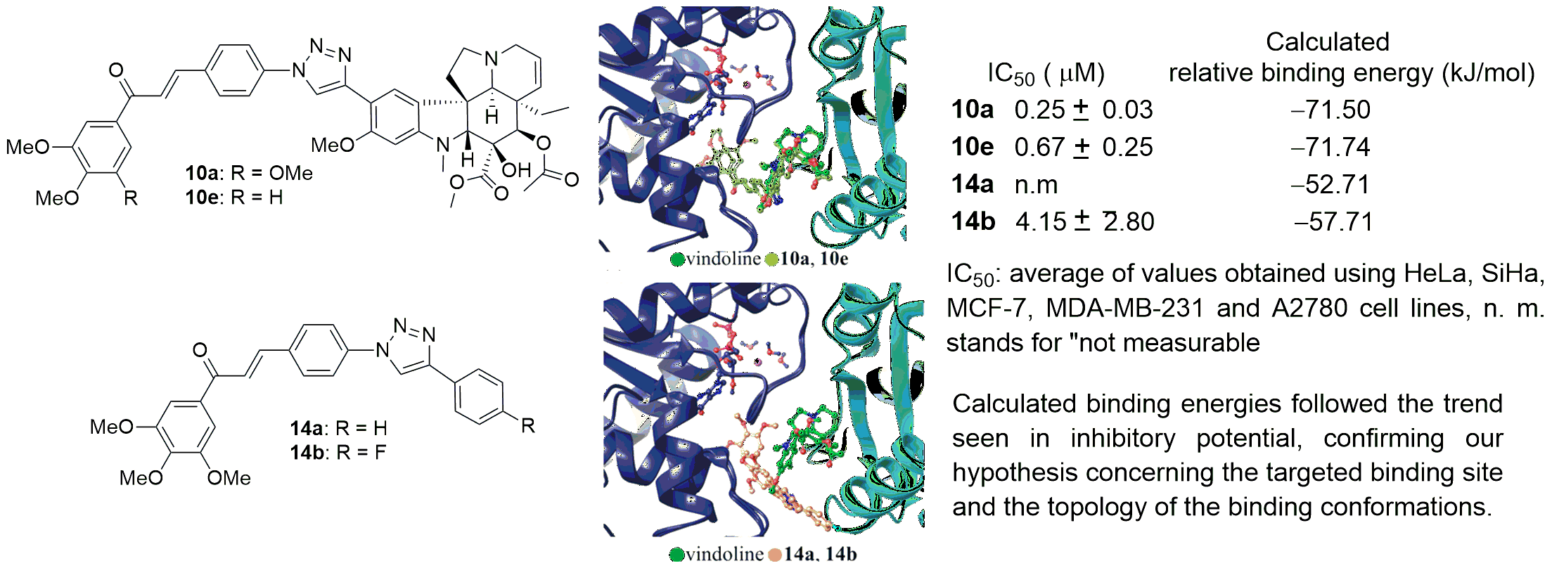
Since the antiproliferative assays (discussed in Section 2.2.) identified hybrid 10a as the most potent model among our novel vindoline hybrids, by means of CuAAC using azidochalcone 4a and phenylacetylenes 13a,b, we prepared triazolyl chalcones 14a,b to serve as further reference models (Scheme 2), the investigation of which was expected to allow estimation of the contribution of the vindoline moiety to the antiproliferative effect of the novel hybrids.
2.2. Antiproliferative Evaluation of the Model Compounds
The antiproliferative properties of the prepared hybrids (10a–i, 11, 12) and their chalcone building blocks (14a,b) were evaluated against a panel of human adherent gynecological cancer cell lines. Generally, the vindoline-chalcone hybrids exhibited pronounced activity, with calculated IC50 values in the low micromolar or nanomolar range (Table 1). Compounds 10a and 10f proved to be the most effective analogs, with IC50 values against cancer cells below 0.4 μM, evidencing the advantage of three methoxy groups on the chalcone moiety. Hybrids 10c–e, on the other hand, showed moderately lower activities against non-malignant fibroblasts (NIH/3T3), indicating a certain degree of cancer selectivity. One of the ferrocene-vindoline hybrids (10i) exhibited substantially lower antiproliferative activity, while its isomer with an alternative enone sequence (11) and the zidovudine-derived vindoline hybrid (12) were inactive.
The triazole-coupled chalcone building block (14a) exhibited modest cell-growth inhibitory activity, with some degree of cancer selectivity. The fluorine-substituted chalcone (14b) showed similar activity against most malignant cells, but was inactive against fibroblasts, indicating greater selectivity. Finally, the reference agent vinblastine proved superior in both antiproliferative activity and cancer selectivity; it elicited nanomolar IC50 values against the malignant cells but 2.14 µM against the fibroblasts.
2.3. Conformational Analysis of the Tubulin-Inhibitor Complexes
Microtubules are noncovalent polymeric filaments composed of heterodimers of two rather similar GTP-ases, the α- and β-tubulins. Microtubules are stabilized at their growing ends by a region enriched in GTP-tubulin, the GTP-cap, where the attaching heterodimers transform from curved to straight geometry. Within the growing microtubule, the nucleotides at the more exposed β-site of the heterodimeric units gradually hydrolyze to GDP. However, the polymeric state of the GDP-bound form is unstable – as it becomes exposed by the degradation of the GTP-loaded protective cap, an explosive depolymerization event, the “catastrophe” takes place, leading to rapid dissociation. The opposing process, the “rescue”, is triggered by the re-establishment of the GTP-cap. The dynamic equilibrium of growth and shrinkage allows for maintenance and quick remodeling of the cytoskeleton. Both colchicine and vinblastine disrupt the first step of the growth process – the straightening of the assembling oligomers and thus inhibit microtubule formation [59,60,61].
The curved conformation of tubulin oligomers can be stabilized by complex formation with chaperone proteins such as stathmin, DARPins, or the TOG domain of microtubule polymerases [62], with bend angles ranging between approximately 10-40°, with the intra-dimer angles typically lower than those between the heterodimeric units [63,64]. Vinblastine and colchicine both bind to the cavities created by the curvature, to sites that are not accessible in the straight conformation [65]. The colchicine site is located at the intra-dimer interface between the α- and β-subunits, while the vinca site is located at the more spacious inter-unit opening between the outer surfaces of two neighboring α/β heterodimers. A further difference in their binding mode is that while colchicine primarily interacts with β-tubulin, the vinca site is a true inter-domain pocket, where both α- and β-tubulin contribute to the stabilization of vinblastine and its homologs (see Figure 2). Since the vindoline segment of vinblastine that was used in the present work as the core scaffold of the designed inhibitors does not fit into the more restricted intra-dimer binding site without inducing considerable rearrangement, here we only considered the inter-dimer location as the targeted binding cavity.
The binding mode of vinblastine can be broken into the binding of its two distinct moieties: vindoline is docked via purely steric interactions into a pocket that complements its shape nearly perfectly, while catharanthine forms a few crucial H-bonds with the side-chain amide of Asn329α and the backbone carbonyl of Pro222β (see Figure 2c). Interestingly, it is the former that contributes more to the total binding energy. The binding energy of the entire vinblastine molecule (calculated using the MM-GBSA protocol described in the Methods section) is –80.4 kJ/mol, while, keeping their vinblastine-like conformation, the binding energies of the vindoline and catharanthine moieties by themselves were found to be –52.4 kJ/mol and –40.0 kJ/mol, respectively. We also allowed relaxation of the isolated moieties in the binding cavity by applying an MCMM conformational search (using the same protocol that was later applied to investigate the binding mode of the designed inhibitors). While the catharanthine moiety practically did not shift from its position in vinblastine (RMSD of non-H atoms with respect to their position in the crystal structure of the complex: 0.60 Å), the rearrangement of the vindoline moiety was more substantial (RMSD: 5.00 Å), involving its rigid-body rotation which allowed the formation of a single H-bond between the ligand and the protein, and the relaxation of the key Asn329α side-chain to its most favorable rotameric state. The shift of the molecule’s center-of-mass is not significant (1.85 Å), but the rearrangement causes a marked decrease in its binding energy to –32.4 kJ/mol. Thus, it can be concluded that the presence of the catharanthine moiety fine-tunes the binding conformation of the vindoline segment of vinblastine, while catharanthine seems to be an autonomous binder.
Next, MCMM conformational searches were carried out to model the binding geometries of the developed vindoline-hybrid inhibitors (except for the ferrocene variant (11), because this moiety is not expected to be well described by a classical forcefield) within the inter-dimer crevice. The starting geometry in each case was built based on the vinblastine complex of α-1B/β-2B tubulin heterodimers in the curved conformation (PDB: 5j2t) [67]. During the simulations of 10a–h, the vindoline moiety remained stably bound in a conformation nearly identical to that seen in the case of the vinblastine complex, with the RMSD of vindoline non-hydrogen atoms (with respect to their position in the crystal structure of the complex) ranging between 0.69–0.79 Å.
The chalcone moiety of the inhibitors docked into the pocket formed by the N-terminal end of H1β (the first helix of β-tubulin outlined in Figure 3), and the C-terminal tip of H7β, in close proximity to the guanosine of the GDP-bound to the β subunit, in a similar conformation for inhibitors 10a–h (Figure 4). The binding site of the 2,3,4-trimethoxyphenyl moiety of colchicine is located at the opposite side of β-tubulin, between the Kβ-loop, H8β-helix and S11β-strand at the intra-heterodimeric interface. The corresponding pocket of α-tubulin that could be reached from the inter-unit binding site is closed-off by the tilting of the H8α in the curved conformation (and is a bit too far for our constructs); thus, the chalcone moiety of our vindoline hybrids is found in a novel location.
Removing most of the vindoline core of 10a, as in case of 14a and 14b, resulted in the shift of the inhibitors toward the solvent, the 3,4,5-trimethoxyphenyl moiety shifted by 2.1 Å and 5.4 Å (measured at their centers-of-mass), respectively. In case of the negative reference molecule, where the vindoline core was coupled to the zidovudine fragment (12), in the most favorable arrangement of the complex, the inhibitor was bound outside the inter-unit crevice, on the surface of the β-tubulin unit. At this binding site, the inhibitor practically does not interfere with the straightening of the tubulin heterodimers – thus it most likely does not hinder formation of a functional microtubule either (Figure 5).
Using the most favorable binding geometries, binding energies were calculated for all considered complexes (Table 2). Calculated binding energies followed the trend seen in inhibitory potential, confirming our hypothesis concerning the targeted binding site and the topology of the binding conformations.
As reflected from the data listed in Table 2 the qualitative agreement of the ordering supports the hypothesis that inhibitors bind to the vinca site. It is important to note that inhibitory potential depends not only on the binding energies, but on the extent of target protein conformational changes (which might be under-represented in our semi-rigid model system) and also on how severely the presence of the inhibitor in a specific location interferes with the straightening of the oligomeric microtubule. The results also demonstrate that the two segments of our vindoline-hybrids exert a mutually stabilizing effect on the binding of the other. Vindoline coordination at the inter-heterodimeric site of the curved conformation is strong enough to anchor the methoxy-chalcones into the pocket (see the binding conformations of 10a–h), while in its absence, the chalcone derivatives drift out from the binding site (14a and 14b). Conversely, in the absence of the methoxy-chalcone or the catharanthine moiety, or in the presence of a suboptimal partner (12), the vindoline core is dislodged from its binding site.
3. Materials and Methods
All chemicals were obtained from commercially available sources (Merck, Budapest, Hungary; Fluorochem, Headfield, UK; Molar Chemicals, Halásztelek, Árpád street 1., 2314 Hungary; VWR, Debrecen, Hungary) and used without further purification. Merck Kieselgel (230–400 mesh, 60 Å) was used for flash column chromatography. Melting points (uncorrected) were determined with a Büchi M-560. The 1H and 13C NMR spectra were recorded in CDCl3 or DMSO-d6 solution in 5 mm tubes at room temperature on a Bruker DRX-500 spectrometer (Bruker Biospin, Karlsruhe, Baden-Württemberg, Germany) at 500 (1H) and 125 (13C) MHz, with the deuterium signal of the solvent as the lock and TMS as an internal standard (1H and 13C). The 2D-COSY-, HSQC- and HMBC spectra, which support the exact assignments of 1H and 13C NMR signals, were recorded using the standard Bruker pulse programs. HRMS measurements were carried out on a Q-TOF Premier mass spectrometer (Waters Corporation, Milford, MA, USA) in positive electrospray ionization mode.
3.1. General Procedure for the Synthesis of Azidochalcone Derivatives
The appropriate methyl ketone (2 or 3a–i, 1.0 mmol, 1 eq.) was dissolved in EtOH (3 mL), and then 10% NaOH solution (1.0 mmol, 1 eq.) was added. The solution of the aldehyde component (1 or 5, 1.0 mmol, 1 eq.) in EtOH (2 mL) was added dropwise at room temperature. The mixture was stirred for 12 h at room temperature in darkness. The product was collected by filtration, then washed with EtOH and diethyl ether. In our earlier works we prepared and characterized azidochalcones 4a,b,d,g [57], 4i [58] and 6 [55].
3.2. Characterization of the Novel Azidochalcones
3.2.1. (E)-3-(4-Azidophenyl)-1-(3-methoxyphenyl)prop-2-en-1-one (4c)
White solid. 104-107 ºC (decomp.). Yield: 118 mg (39%). 1H NMR (500 MHz, CDCl3): 7.76 (d, J = 15.5 Hz, 1H, H-7), 7.62 (d, J = 8.5 Hz, 2H, H-3,5), 7.59 (br d, J ~ 8 Hz, 1H, H-15), 7.53 (br s, 1H, H-11), 7.49 (t, J = 8.0 Hz, 1H, H-14), 7.45 (d, J = 15.5 Hz, 1H, H-8), 7.13 (dd, J = 8.0 Hz and 1.9 Hz, 1H, H-13), 7.06 (d, J = 8.5 Hz, 2H, H-2,6), 3.88 (s, 3H, H-16) ppm. 13C NMR (125 MHZ, CDCl3): 190.0 (C-9); 160.0 (C-12), 143.7 (C-7), 142.2 (C-1), 139.6 (C-10), 131.7 (C-4), 130.0 (C-3,5), 129.6 (C-14), 121.6 (C-8), 121.0 (C-15), 119.6 (C-2,6), 119.3 (C-13), 112.9 (C-11), 55.5 (C-16) ppm. HRMS (ESI+) m/z calculated for C16H13N3O2 [M+Na]+ 302.0905, found 302.0903.

3.2.2. (E)-3-(4-Azidophenyl)-1-(3,4-dimethoxyphenyl)prop-2-en-1-one (4e)
White solid. 134-137 °C (decomp.). Yield: 272 mg (82%). 1H NMR (500 MHz, DMSO-d6): 7.94, 7.92 and 7.90 (overlapping d’s, J = 8.5 Hz and 15.5 Hz and dd, J = 8.5 Hz and 1.9 Hz, 4H, H-3,5, H-8 and H-15), 7.70 (d, J = 15.5 Hz, 1H, H-7), 7.61 (d, J = 1.9 Hz, 1H, H-11), 7.19 (d, J = 8.5 Hz, 2H, H-2,6), 7.10 (d, J = 8.5 Hz, 1H, H-14), 3.88 (s, 3H, H-16), 3.86 (s, 3H, H-17) ppm. 13C NMR (125 MHz, DMSO-d6): 187.7 (C-9), 153.7 (C-13), 149.3 (C-12), 142.5 (C-7), 141.7 (C-1), 132.3 (C-4), 131.1 (C-3,5), 131.0 (C-10), 123.9 (C-15), 121.9 (C-8), 120.0 (C-2,6), 111.4 (C-14), 111.2 (C-11), 56.3 (C-16), 56.1 (C-17) ppm. HRMS (ESI+) m/z calculated for C17H15N3O3 [M+Na]+ 332.1011, found 332.1011.

3.2.3. (E)-3-(4-Azidophenyl)-1-(2,4,6-trimethoxyphenyl)prop-2-en-1-one (4f)
White solid. 122-125 °C (decomp.). Yield: 286 mg (79%). 1H NMR (500 MHz, DMSO-d6): 7.72 (d, J = 8.3 Hz, 2H, H-3,5), 7.20 (d, J = 15.5 Hz, 1H, H-7), 7.13 (d, J = 8.3 Hz, 2H, H-2,6), 6.94 (d, J = 15.5 Hz, 1H, H-8), 6.31 (s 2H, H-12,14), 3.88 (s, 6H, H-16,18), 3.86 (s, 3H, H-17) ppm. 13C NMR (125 MHz, DMSO-d6): 193.6 (C-9), 162.4 (C-13), 158.4 (C-11,15), 143.0 (C-7), 141.8 (C-1), 131.8 (C-4), 130.7 (C-3,5), 129.1 (C-8), 120.2 (C-2,6), 111.5 (C-10), 91.6 (C-12,14), 56.3 (C-16,18), 55.9 (C-17) ppm. HRMS (ESI+) m/z calculated for C18H17N3O4 [M+Na]+ 362.1117, found 362.1113.

3.2.4. (E)-3-(4-Azidophenyl)-1-(benzo[d][1,3]dioxol-5-yl)prop-2-en-1-one (4h)
White solid. 167-171 °C (decomp.). Yield: 269 mg (85%). 1H NMR (500 MHz, DMSO-d6): 7.93 (d, J = 8.3 Hz, 2H, H-3,5), 7.88 (d, J = 15.5 Hz, 1H, H-8), 7.86 (br d, J = 8.5 Hz, 1H, H-15), 7.68 (d, J = 15.5 Hz, 1H, H-7), 7.65 (br s, 1H, H-11), 7.19 (d, J = 8.5 Hz, 2H, H-2,6), 7.08 (d, J = 8.5 Hz, 1H, H-14), 6.16 (s, 2H, H-16). 13C NMR (125 MHz, DMSO-d6): 187.3 (C-9), 152.0 (C-13), 148.5 (C-12), 142.9 (C-7), 141.8 (C-1), 132.7 (C-10), 132.2 (C-4), 131.1 (C-3,5), 125.5 (C-15), 121.9 (C-8), 119.2 (C-2,6), 108.6 (C-14), 108.4 (C-11), 102.6 (C-16). HRMS (ESI+) m/z calculated for C16H11N3O3 [M+Na]+ 316.0698; Found 316.0700.

3.3. General Procedure for the Deprotection and CuAAC Reactions of 8 Producing the Targeted Vindoline Hybrids 10a–i, 11 and 12.
The mixture of 8 (1.0 mmol, 1 eq.), TBAF (2.0 mmol, 2 eq.), the appropriate azidochalcone 4a–i and 6 or zidovudine (1.0 mmol, 1 eq.), CuSO4.5 H2O (0.5 mmol, 0.5 eq.), NaOH (1.0 mmol, 1 eq.), and L-ascorbic acid (1.0 mmol, 1 eq.) was dissolved in 1-butanol:water (1:1.5 mL). The mixture was stirred for 24 hours at room temperature under argon atmosphere and then poured into water, extracted with DCM (3x10 mL). The organic phase was washed with water (1x15 mL) and brine, then dried over Na2SO4. After evaporation of the solvent, the crude product was purified by preparative TLC using a solvent mixture of DCM:MeOH (70:1) as eluent and crystallized from Et2O to obtain the pure product. The preparation of TMS-protected alkyne 8 was adapted from our earlier publication [45].
3.4. Characterization of the Targeted Vindoline Hybrids
3.4.1. Methyl (3aR,3a1R,4R,5S,5aR,10bR)-4-acetoxy-3a-ethyl-5-hydroxy-8-methoxy-6-methyl-9-(1-(4-((E)-3-oxo-3-(3,4,5-trimethoxyphenyl)prop-1-en-1-yl)phenyl)-1H-1,2,3-triazol-4-yl)-3a,3a1,4,5,5a,6,11,12-octahydro-1H-indolizino [8,1-cd]carbazole-5-carboxylate (10a)
Yellowish-brown solid. Mp: 187-189 °C Yield: 303 mg (37%). 1H NMR (500 MHz, DMSO-d6): 8.91 (s, 1H, OH on C16); 8.85 (s, 1H, H-29); 8.16 (d, J = 8.7 Hz, 2H, H-35,37); 8.10 (d, J = 8.7 Hz, 2H, H-34,38); 8.05 (d, J = 15.7 Hz, 1H, H-40); 7.97 (s, 1H, H-9); 7.83 (d, J = 15.7 Hz, 1H, H-39); 7.46 (s, 2H, H-43,47); 6.46 (s, 1H, H-12); 5.86 (ddd, J = 10.3 Hz, 4.6 Hz and 1.5 Hz, 1H, H-14); 5.22 (s, 1H, H-17); 5.12 (br d, J = 10.3 Hz, 1H, H-15); 3.98 (s, 3H, H-27); 3.93 (s, 6H, H-48,50); 3.79 (s, 3H, H-49); 3.69 (s, 3H, H-25); 3.44 (dd, J = 16.4 Hz and 4.6 Hz, 1H, H-3/A); 3.35 (overlapped by the HDO signal of the solvent as detected by 2D-HSQC, H-5/A); 2.90 (br ~d, J ~ 16 Hz, 1H, H-3/B); 2.81 (s, 1H, H-21); 2.70 and 2.69 (overlapping s and m, 4H, H-5/B and H-26); 2.29 (m, 2H, H-6); 1.96 (s, 3H, H-24); 1.51 (m, 1H, H-19/A); 1.00 (m, 1H, H-19/B); 0.45 (t, J = 7.2 Hz, 3H, H-18). 13C NMR (125 MHz DMSO-d6): 188.3 (C-41); 172.1 (C-22); 170.6 (C-23); 157.8 (C-11); 154.1 (C-13); 153.5 (C-44,46); 144.5 (C-28); 143.0 (C-39); 142.6 (C-45); 138.3 (C-33); 135.1 (C-36); 133.4 (C-42); 130.9 (C-35,37); 130.3 (C-15); 125.6 (C-8); 125.0 (C-14); 123.3 (C-40); 121.5 (C-9); 120.7 (C-34,38); 120.0 (C-29); 109.7 (C-10); 106.8 (C-43,47); 93.7 (C-12);83.3 (C-2); 79.2 (C-16); 76.4 (C-17); 66.7 (C-21); 60.7 (C-49); 52.7 (C-7); 51.5 (C-5); 56.7 (C-48,50); 56.2 (C-27); 52.3 (C-25); 50.8 (C-3); 44.2 (C-6); 43.0 (C-20); 38.3 (C-26); 31.0 (C-19); 21.2 (C-24); 8.0 (C-18). HRMS (ESI+) m/z calculated for C45H49N5O10 [M+H]+ 820.3558, found 820.3566.

3.4.2. Methyl (3aR,3a1R,4R,5S,5aR,10bR)-4-acetoxy-3a-ethyl-5-hydroxy-8-methoxy-6-methyl-9-(1-(4-((E)-3-oxo-3-(2-methoxyphenyl)prop-1-en-1-yl)phenyl)-1H-1,2,3-triazol-4-yl)-3a,3a1,4,5,5a,6,11,12-octahydro-1H-indolizino [8,1-cd]carbazole-5-carboxylate (10b)
Yellowish-brown solid. Mp: 174-176 °C. Yield: 167 mg (22%). 1H NMR (500 MHz, DMSO-d6): 8.91 (s, 1H, OH on C16); 8.83 (s, 1H, H-29); 8.06 (d, J = 8.5 Hz, 2H, H-34,38); 7.98 and 7.97 (partly overlapping (d, J = 8.5 Hz,) and s, 3H, H-35,37 and H-9); 7.60 and 7.57 (overlapping d (J = 15.6 Hz) and t (J = 7.6 Hz), 2H, H-39 and H-45); 7.54 and 7.52 (overlapping t (J = 7.6 Hz) and d (J = 15.6 Hz), 2H, H-47 and H-40); 7.22 (d, J = 7.8 Hz, 1H, H-44); 7.09 (t, J = 7.6 Hz, 1H, H-46); 6.46 (s, 1H, H-12); 5.86 (ddd, J = 10.3 Hz, 4.6 Hz and 1.5 Hz, 1H, H-14); 5.23 (s, 1H, H-17); 5.12 (br d, J = 10.3 Hz, 1H, H-15); 3.97 (s, 3H, H-27); 3.89 (s, 3H, H-48); 3.69 and 3.68 (partly overlapping s’s, 4H, H-25 and H-2); 3.44 (dd, J = 16.4 Hz and 4.6 Hz, 1H, H-3/A); 3.35 (overlapped by the HDO signal of the solvent as detected by 2D-HSQC, H-5/A); 2.91 (br ~d, J ~ 16 Hz, 1H, H-3/B); 2.81 (s, 1H, H-21); 2.70 and 2.69 (overlapping s and m, 4H, H-5/B and H-26); 2.35-2.25 (m, 2H, H-6); 1.96 (s, 3H, H-24); 1.51 (m, 1H, H-19/A); 1.01 (m, 1H, H-19/B); 0.44 (t, J = 7.2 Hz, 3H, H-18). 13C NMR (125 MHz, DMSO-d6): 192.5 (C-41); 172.1 (C-22); 170.6 (C-23); 158.3 (C-43); 157.8 (C-11); 154.1 (C-13); 144.5 (C-28); 141.5 (C-39); 138.2 (C-33); 135.0 (C-36); 133.7 (C-45); 130.5 (C-35,37); 130.3 (C-15); 130.0 (C-47); 129.2 (C-42); 128.2 (C-40); 125.6 (C-8); 125.3 (C-14); 121.4 (C-9); 120.7 (C-34,38); 121.1 (C-46); 119.9 (C-29); 112.9 (C-44); 109.8 (C-10); 93.7 (C-12); 83.7 (C-2); 79.2 (C-16); 76.4 (C-17); 66.7 (C-21); 56.4 (C-48); 56.1 (C-27); 52.7 (C-7); 52.3 (C-25); 51.6 (C-5); 50.9 (C-3); 44.2 (C-6); 43.0 (C-20); 38.3 (C-26); 31.1 (C-19); 21.1 (C-24); 8.0 (C-18). HRMS (ESI+) m/z calculated for C43H45N5O8 [M+H]+ 760.3346, found 760.3356.

3.4.3. Methyl (3aR,3a1R,4R,5S,5aR,10bR)-4-acetoxy-3a-ethyl-5-hydroxy-8-methoxy-6-methyl-9-(1-(4-((E)-3-oxo-3-(3-methoxyphenyl)prop-1-en-1-yl)phenyl)-1H-1,2,3-triazol-4-yl)-3a,3a1,4,5,5a,6,11,12-octahydro-1H-indolizino [8,1-cd]carbazole-5-carboxylate (10c)
Yellowish-brown solid. Mp: 160-162 °C Yield: 182 mg (24%). 1H NMR (500 MHz, DMSO-d6): 8.91 (s, 1H, OH on C16); 8.85 (s, 1H, H-29); 8.15 (d, J = 8.7 Hz, 2H, H-35,37); 8.09 (d, J = 8.7 Hz, 2H, H-34,38); 8.03 (d, J = 15.7 Hz, 1H, H-40); 7.98 (s, 1H, H-9); 7.83 (d, J = 15.7 Hz, 1H, H-39); 7.81 (br d J ~ 8 Hz, 1H, H-43); 7.65 (br s, 1H, H-47); 7.52 (t, J = 8.0 Hz, 1H, H-44); 7.27 (dd, J = 8.0 Hz and 2.0 Hz, 1H, H-45); 6.46 (s, 1H, H-12); 5.86 (ddd, J = 10.3 Hz, 4.6 Hz and 1.5 Hz, 1H, H-14); 5.23 (s, 1H, H-17); 5.12 (br d, J = 10.3 Hz, 1H, H-15); 3.98 (s, 3H, H-27); 3.87 (s, 3H, H-48); 3.69 and 3.68 ( partly overlapping s’s, 4H, H-25 and H-2); 3.44 (dd, J = 16.2 Hz and 4.3 Hz, 1H, H-3/A); 3.35 (overlapped by the HDO signal of the solvent as detected by 2D-HSQC, H-5/A); 2.90 (br ~d, J ~ 16 Hz, 1H, H-3/B); 2.81 (s, 1H, H-21); 2.70 and 2.69 (overlapping s and m, 4H, H-5/B and H-26); 2.31 (m, 2H, H-6); 1.96 (s, 3H, H-24); 1.51 (m, 1H, H-19/A); 1.00 (m, 1H, H-19/B); 0.45 (t, J = 7.2 Hz, 3H, H-18). 13C NMR (125 MHz, DMSO-d6): 189.3 (C-41); 172.1 (C-22); 170.6 (C-23); 160.1 (C-46) 157.8 (C-11); 154.1 (C-13); 144.5 (C-28); 143.2 (C-39); 139.4 (C-42); 138.3 (C-33); 135.0 (C-36); 130.9 (C-35,37); 130.5 (C-44); 130.3 (C-15); 125.6 (C-8); 125.1 (C-14); 123.2 (C-40); 121.5 (C-9); 120.7 (C-34,38); 120.0 (C-29); 119.9 (C-43); 119.7 (C-45); 113.7 (C-47); 109.8 (C-10); 93.7 (C-12); 83.3 (C-2); 79.2 (C-16); 76.4 (C-17); 66.7 (C-21); 56.2 (C-27); 55.9 (C-48); 52.7 (C-7); 52.3 (C-25); 51.6 (C-5); 50.9 (C-3); 44.3 (C-21), 44.2 (C-6); 43.0 (C-20); 38.3 (C-26); 31.0 (C-19); 21.2 (C-24); 8.0 (C-18). HRMS (ESI+) m/z: calculated for C43H45N5O8 [M+H]+ 760.3346, found 760.3358.

3.4.4. Methyl (3aR,3a1R,4R,5S,5aR,10bR)-4-acetoxy-3a-ethyl-5-hydroxy-8-methoxy-6-methyl-9-(1-(4-((E)-3-oxo-3-(4-methoxyphenyl)prop-1-en-1-yl)phenyl)-1H-1,2,3-triazol-4-yl)-3a,3a1,4,5,5a,6,11,12-octahydro-1H-indolizino [8,1-cd]carbazole-5-carboxylate (10d)
Yellowish-brown solid. Mp: 192-195 °C Yield: 236 mg (31%). 1H NMR (500 MHz, DMSO-d6): 8.92 (s, 1H, OH on C16); 8.84 (s, 1H, H-29); 8.21 (d, J = 8.6 Hz, 2H, H-43,47); 8.13 (d, J = 8.5 Hz, 2H, H-35,37); 8.13 (d, J = 8.5 Hz, 2H, H-34,38); 8.05 (d, J = 15.8 Hz, 1H, H-40); 7.97 (s, 1H, H-9); 7.11 (d, J = 8.6 Hz, 2H, H-44,46); 7.79 (d, J = 15.8 Hz, 1H, H-39); 6.46 (s, 1H, H-12); 5.86 (dd, J = 9.8 Hz and 4.5 Hz, 1H, H-14); 5.22 (s, 1H, H-17); 5.12 (br ~d, J ~ 10 Hz, H-15); 3.98 (s, 3H, H-27); 3.89 (s, 3H, H-48); 3.69 (s, 3H, H-25); 3.68 (s, 1H, H-2); 3.44 (dd, J = 16.5 Hz and 4.5 Hz, 1H, H-3/A); 3.35 (overlapped by the HDO signal of the solvent as detected by 2D-HSQC, H-5/A); 2.91 (br ~d, J ~ 17 Hz, 1H, H-3/B); 2.82 (s, 1H, H-21); 2.70 and 2.69 (overlapping s and m, 4H, H-5/B and H-26); 2.38-2.27 (m, 2H, H-6); 1.96 (s, 3H, H-24); 1.50 (m, 1H, H-19/A); 1.00 (m, 1H, H-19/B); 0.45 (t, J = 7.2 Hz, 3H, H-18). 13C NMR (125 MHz, DMSO-d6): 187.8 (C-41); 172.1 (C-22); 170.6 (C-23); 163.8 (C-45); 157.8 (C-11); 154.1 (C-13); 144.5 (C-28); 142.3 (C-39); 138.2 (C-33); 135.2 (C-36); 131.5 (C-43,47); 131.3 (C-42); 130.8 (C-35,37); 130.3 (C-15); 125.6 (C-8); 125.2 (C-14); 123.4 (C-40); 121.5 (C-9); 120.7 (C-34,38); 120.0 (C-29); 109.8 (C-10); 93.7 (C-12); 83.3 (C-2); 79.2 (C-16); 76.4 (C-17); 66.8 (C-21); 56.17 (C-27); 56.09 (C-48); 52.7 (C-7); 52.3 (C-25); 51.6 (C-5); 50.9 (C-3); 44.2 (C-6); 43.0 (C-20); 38.4 (C-26); 31.1 (C-19); 21.2 (C-24); 8.0 (C-18). HRMS (ESI+) m/z calculated for C43H45N5O8 [M+H]+ 760.3346, found 760.3357.

3.4.5. Methyl (3aR,3a1R,4R,5S,5aR,10bR)-4-acetoxy-3a-ethyl-5-hydroxy-8-methoxy-6-methyl-9-(1-(4-((E)-3-oxo-3-(3,4-dimethoxyphenyl)prop-1-en-1-yl)phenyl)-1H-1,2,3-triazol-4-yl)-3a,3a1,4,5,5a,6,11,12-octahydro-1H-indolizino [8,1-cd]carbazole-5-carboxylate (10e)
Yellowish-brown solid. Mp: 186-188 °C Yield: 221 mg (28%). 1H NMR (500 MHz, DMSO-d6): 8.91 (s, 1H, OH on C-16); 8.84 (s, 1H, H-29); 8.14 (d, J = 8.7 Hz, 2H, H-35,37); 8.09 (d, J = 8.7 Hz, 2H, H-34,38); 8.06 (d, J = 15.7 Hz, 1H, H-40); 7.97 and 7.96, (partly overlapping s and dd, J = 8.5 Hz and 1.9 Hz, 2H, H-9 and H-47); 7.80 (d, J = 15.7 Hz, 1H, H-39); 7.64 (d, J = 1.9 Hz, 1H, H-43); 7.14 (d, J = 8.5 Hz, 1H, H-46); 6.46 (s, 1H, H-12); 5.86 (dd, J = 9.8 Hz and 4.5 Hz, 1H, H-14); 5.23 (s, 1H, H-17); 5.12 (br ~d, J ~ 10 Hz, H-15); 3.98 (s, 3H, H-27); 3.90 (s, 3H, H-48); 3.69 (s, 3H, H-25); 3.68 (s, 1H, H-2); 3.44 (dd, J = 16.5 Hz and 4.5 Hz, 1H, H-3/A); 3.35 (overlapped by the HDO signal of the solvent as detected by 2D-HSQC, H-5/A); 2.91 (br ~d, J ~ 17 Hz, 1H, H-3/B); 2.81 (s, 1H, H-21); 2.70 and 2.69 (overlapping s and m, 4H, H-5/B and H-26); 2.26 (m, 2H, H-6); 1.96 (s, 3H, H-24); 1.50 (m, 1H, H-19/A); 0.99 (m, 1H, H-19/B); 0.45 (t, J = 7.2 Hz, 3H, H-18). 13C NMR (125 MHz, DMSO-d6): 187.8 (C-41); 172.1 (C-22); 170.6 (C-23); 157.8 (C-11); 154.1 (C-13); 153.8 (C-45); 149.3 (C-44); 144.5 (C-28); 142.2 (C-39); 138.2 (C-33); 135.3 (C-36); 130.9 (C-42); 130.8 (C-35,37); 130.3 (C-15); 125.4 (C-8); 125.2 (C-14); 124.1 (C-47); 123.3 (C-40); 121.5 (C-9); 120.7 (C-34,38); 120.1 (C-29); 111.4 (C-46); 111.3 (C-43); 109.7 (C-10); 93.7 (C-12); 83.4 (C-2); 78.9 (C-16); 76.3 (C-17); 66.7 (C-21); 56.3 (C-48); 56.15 (C-27); 56.1 (C-49); 52.6 (C-7); 52.3 (C-25); 51.5 (C-5); 50.8 (C-3); 44.2 (C-6); 42.9 (C-20); 38.4 (C-26); 31.1 (C-19); 21.2 (C-24); 8.0 (C-18). HRMS (ESI+) m/z calculated for C44H47N5O9 [M+H]+ 790.3452, found: 790.3460.

3.4.6. Methyl (3aR,3a1R,4R,5S,5aR,10bR)-4-acetoxy-3a-ethyl-5-hydroxy-8-methoxy-6-methyl-9-(1-(4-((E)-3-oxo-3-(2,4,6-trimethoxyphenyl)prop-1-en-1-yl)phenyl)-1H-1,2,3-triazol-4-yl)-3a,3a1,4,5,5a,6,11,12-octahydro-1H-indolizino [8,1-cd]carbazole-5-carboxylate (10f).
Yellowish-brown solid. Mp: 181-183 °C. Yield: 205 mg (25%). 1H NMR (500 MHz, DMSO-d6): 8.91 (s, 1H, OH on C-16); 8.83 (s, 1H, H-29); 8.04 (d, J = 8.7 Hz, 2H, H-34,38); 7.92 (d, J = 8.7 Hz, 2H, H-35,37); 7.97 (s, 1H, H-9); 7.31 (d, J = 15.7 Hz, 1H, H-39); 7.07 (d, J = 15.7 Hz, 1H, H-40); 6.45 (s, 1H, H-12); 6.34 (s, 2H, H-44,46); 5.85 (dd, J = 10.0 Hz and 4.4 Hz, 1H, H-14); 5.22 (s, 1H, H-17); 5.12 (br d, J ~ 10 Hz, 1H, H-15); 3.96 (s, 3H, H-27); 3.85 (s, 3H, H-25); 3.74 (s, 6H, H-48,49); 3.68 (s, 3H, H-50); 3.67 (s, 1H, H-2); 3.44 (dd, J = 16.4 Hz and 4.0 Hz, 1H, H-3/A); 3.34 (overlapped by the HDO signal of the solvent as detected by 2D-HSQC, H-5/A); 2.90 (br ~d, J ~ 16 Hz, 1H, H-3/B); 2.80 (s, 1H, H-21); 2.69 (overlapping s and m, 4H, H-26 and H-5/B); 1.96 (s, 3H, H-24); 1.50 (m, 1H, H-19/A); 1.00 (m, 1H, H-19/B); 0.43 (t, J = 7.3 Hz, 3H, H-18). 13C NMR (125 MHz, DMSO-d6): 193.6 (C-41); 172.1 (C-22); 170.6 (C-23); 162.5 (C-45); 158.6 (C-43,47); 157.8 (C-11); 154.1 (C-13); 144.5 (C-29); 142.6 (C-39);138.2 (C-33); 134.8 (C-36); 130.4 (C-35,37); 130.3 (C-15); 130.2 (C-40); 125.6 (C-8); 125.2 (C-14); 121.5 (C-9); 120.6 (C-34,38); 119.9 (C-29); 111.4 (C-42); 109.8 (C-10); 93.6 (C-12); 91.6 (C-44,46); 83.3 (C-2); 79.2 (C-16); 76.4 (C-17); 66.7 (C-21); 56.4 (C-48,49); 56.1 (C-27); 56.0 (C-25); 52.6 (C-7); 51.6 (C-5); 50.9 (C-3); 44.2 (C-6); 43.0 (C-20); 38.3 (C-26); 31.1 (C-19); 21.2 (C-24); 8.0 (C-18). HRMS (ESI+) m/z calculated for C45H49N5O10 [M+H] 820.3558 found: 820.3564.

3.4.7. Methyl (3aR,3a1R,4R,5S,5aR,10bR)-4-acetoxy-3a-ethyl-5-hydroxy-9-(1-(4-((E)-3-(4-hydroxy-3,5-dimethylphenyl)-3-oxoprop-1-en-1-yl)phenyl)-1H-1,2,3-triazol-4-yl)-8-methoxy-6-methyl-3a,3a1,4,5,5a,6,11,12-octahydro-1H-indolizino [8,1-cd]carbazole-5-carboxylate (10g).
Light brown solid. Mp: 207-209 °C (dec.) Yield: 186 mg (24%). 1H NMR (500 MHz, DMSO-d6): 8.91 (s, 1H, OH on C16); 8.83 (s, 1H, H-29); 8.11 (d, J = 8.5 Hz, 2H, H-35,37); 8.07 (d, J = 8.5 Hz, 2H, H-34,38); 8.02 (d, J = 15.7 Hz, 1H, H-40); 7.96 (s, 1H, H-9); 7.87 (s, 2H, H-43,47); 7.75 (d, J = 15.7 Hz, 1H, H-39); 6.46 (s, 1H, H-12); 5.86 (br dd, J = 10.3 Hz and 4.6 Hz 1H, H-14); 5.23 (s, 1H, H-17); 5.12 (br d, J = 10.3 Hz, 1H, H-15); 3.97 (s, 3H, H-27); 3.69 (s, 3H, H-25); ~3.4 (br m, 1H, H-3/A); 3.35 (overlapped by the HDO signal of the solvent as detected by 2D-HSQC, H-5/A); 2.90 (br ~d, J ~ 16 Hz, 1H, H-3/B); 2.81 (s, 1H, H-21); 2.70 and 2.68 (partly overlapping s and m, 4H, H-5/B and H-26); 2.34-2.25 and 2.28 (overlapping m and s, 8H, H-6 and H-48,49); 1.96 (s, 3H, H-24); 1.51 (m, 1H, H-19/A); 1.01 (m, 1H, H-19/B); 0.45 (t, J = 7.2 Hz, 3H, H-18). 13C NMR (125 MHz, DMSO-d6): 187.7 (C-41); 172.1 (C-22); 170.6 (C-23); 158.9 (C-44,46); 157.8 (C-11); 154.0 (C-13); 144.4 (C-28); 141.6 (C-39); 138.1 (C-33); 135.4 (C-36); 130.6 (C-35,37); 130.2 (C-44,46); 130.3 (C-15); 129.3 (C-42); 125.6 (C-8); 125.1 (C-14); 124.7 (C-43,47); 123.6 (C-40); 121.5 (C-9); 120.7 (C-34,38); 120.0 (C-29); 109.9 (C-10); 93.7 (C-12); 83.3 (C-2); 79.2 (C-16); 76.4 (C-17); 66.7 (C-21); 56.2 (C-27); 52.7 (C-7); 51.6 (C-5); 52.3 (C-25); 50.9 (C-3); 44.6 (C-6); 43.0 (C-20); 38.3 (C-26); 31.1 (C-19); 21.2 (C-24); 8.0 (C-18). HRMS (ESI+) m/z calculated for C44H47N5O8 [M+H]+ 774.3503, found: 774.3500.

3.4.8. Methyl (3aR,3a1R,4R,5S,5aR,10bR)-4-acetoxy-9-(1-(4-((E)-3-(benzo[d][1,3]dioxol-5-yl)-3-oxoprop-1-en-1-yl)phenyl)-1H-1,2,3-triazol-4-yl)-3a-ethyl-5-hydroxy-8-methoxy-6-methyl-3a,3a1,4,5,5a,6,11,12-octahydro-1H-indolizino [8,1-cd]carbazole-5-carboxylate (10h).
Yellow solid. Mp: 178-180 °C (dec.). Yield: 170 mg (22%). 1H NMR (500 MHz, DMSO-d6): 8.91 (s, 1H, OH on C-16); 8.84 (s, 1H, H-29); 8.13 (d, J = 8.4 Hz, 2H, H-35,37); 8.08 (d, J = 8.4 Hz, 2H, H-34,38); 8.03 (d, J = 15.7 Hz, 1H, H-40); 7.98 (s, 1H, H-9); 7.91 (dd, J = 8.1 Hz and 1.6 Hz, 1H, H-47); 7.79 (d, J = 15.7 Hz, 1H, H-39); 7.70 (br s, 1H, H-43); 7.12 (d, J = 8.1 Hz, 1H, H-46); 6.45 (s, 1H, H-12); 6.18 (s, 2H, H-48); 5.86 (dd, J = 10.1 Hz and 4.5 Hz, 1H, H-14); 5.23 (s, 1H, H-17); 5.12 (br ~d, J ~ 10 Hz, H-15); 3.98 (s, 3H, H-27); 3.69 (s, 3H, H-25); 3.68 (s, 1H, H-2); 3.44 (dd, J = 16.2 Hz and 4.4 Hz, 1H, H-3/A); 3.35 (overlapped by the HDO signal of the solvent as detected by 2D-HSQC, H-5/A); 2.91 (br ~d, J ~ 16 Hz, 1H, H-3/B); 2.81 (s, 1H, H-21); 2.70 (overlapping s and m, 4H, H-5/B and H-26); 2.38-2.27 (m, 2H, H-6); 1.96 (s, 3H, H-24); 1.50 (m, 1H, H-19/A); 0.99 (m, 1H, H-19/B); 0.45 (t, J = 7.2 Hz, 3H, H-18). 13C NMR (125 MHz, DMSO-d6): 187.3 (C-41); 172.2 (C-22); 170.6 (C-23); 157.8 (C-11); 154.1 (C-13); 152.2 (C-45); 148.6 (C-44); 144.5 (C-28); 142.5 (C-39); 138.2 (C-33); 135.2 (C-36); 132.7 (C-42); 130.8 (C-35,37); 130.3 (C-15); 125.7 (C-47); 125.6 (C-8); 125.2 (C-14); 123.2 (C-40); 121.5 (C-9); 120.7 (C-34,38); 120.0 (C-29); 108.7 (C-46); 108.4 (C-43); 110.0 (C-10); 102.6 (C-48); 93.7 (C-12); 83.3 (C-2); 79.2 (C-16); 76.4 (C-17); 66.7 (C-21); 56.2 (C-27); 52.7 (C-7); 52.3 (C-25); 51.6 (C-5); 50.9 (C-3); 44.2 (C-6); 43.0 (C-20); 38.3 (C-26); 31.1 (C-19); 21.2 (C-24); 8.0 (C-18). HRMS (ESI+) m/z calculated for C43H43N5O9 [M+H]+ 774.3139, found: 774.3152.

3.4.9. Methyl (3aR,3a1R,4R,5S,5aR,10bR)-4-acetoxy-3a-ethyl-5-hydroxy-8-methoxy-6-methyl-9-(1-(4-((E)-3-oxo-3-phenylprop-1-en-1-yl)ferrocenyl)-1H-1,2,3-triazol-4-yl)-3a,3a1,4,5,5a,6,11,12-octahydro-1H-indolizino [8,1-cd]carbazole-5-carboxylate (10i);
Red solid. Mp: 191-193 °C (dec.) Yield: 134 mg (16%). 1H NMR (500 MHz, DMSO-d6): 8.91 (s, 1H, OH on C-16); 8.84 (s, 1H, H-29); 8.12 (d, J = 8.7 Hz, 2H, H-35,37); 8.08 (d, J = 8.7 Hz, 2H, H-34,38); 7.98 (s, 1H, H-9); 7.72 (d, J = 15.9 Hz, 1H, H-40); 7.54 (d, J = 15.9 Hz, 1H, H-39); 6.48 (s, 1H, H-12); 5.87 (ddd, J = 10.3 Hz, 4.6 Hz and 1.5 Hz, 1H, H-14); 5.23 (s, 1H, H-17); 5.12 (br d, J = 10.3 Hz, 1H, H-15); 5.10 (br s, 2H, H-43,46); 4.71 (br s, 2H, H-44,45); 4.26 (s, 5H, H-47,48,49,50,51); 3.98 (s, 3H, C-27); 3.69 (overlapping s’s, 4H, H-2 and H-25); 3.44 (dd, J = 16.2 Hz and 4.3 Hz, 1H, H-3/A); 3.35 (overlapped by the HDO signal of the solvent as detected by 2D-HSQC, H-5/A); 2.91 (br ~d, J ~ 16 Hz, 1H, H-3/B); 2.82 (s, 1H, H-21); 2.70 and 2.69 (overlapping s and m, 4H, H-5/B and H-26); 2.30 (m, 2H, H-6); 1.96 (s, 3H, H-24); 1.52 (m, 1H, H-19/A); 1.02 (m, 1H, H-19/B); 0.45 (t, J = 7.2 Hz, 3H, H-18). 13C NMR (125 MHz, DMSO-d6): 192.4 (C-41); 172.2 (C-22); 170.6 (C-23); 157.8 (C-11); 154.3 (C-13); 144.5 (C-28), 138.9 (C-39), 135.4 (C-40); 135.3 (C-33); 130.5 (C-35,37); 130.3 (C-36); 130.2 (C-15); 125.4 (C-8); 125.2 (C-14); 120.8 (C-34,38); 120.1 (C-29); 109.8 (C-11); 109.7 (C-10); 93.7 (C-12); 83.4 (C-2); 81.2 (C-42); 79.1 (C-16); 76.4 (C-17), 73.3 (C-44,45); 70.4 (C-47,48,49,50,51); 70.1 (C-43,46); 66.7 (C-21); 56.2 (C-27); 52.5 (C-7), 52.3 (C-25); 51.6 (C-5); 50.8 (C-3);, 44.3 (C-6); 43.2 (C-20); 38.3 (C-26); 31.1 (C-19); 21.2 (C-24); 8.0 (C-18). HRMS (ESI+) m/z: calculated for C46H47FeN5O7 [M+H]+ 838.2903, found 838.2914.

3.4.10. Methyl (3aR,3a1R,4R,5S,5aR,10bR)-4-acetoxy-3a-ethyl-5-hydroxy-8-methoxy-6-methyl-9-(1-(4-((E)-3-(ferrocenyl)acryloyl)phenyl)-1H-1,2,3-triazol-4-yl)-3a,3a1,4,5,5a,6,11,12-octahydro-1H-indolizino [8,1-cd]carbazole-5-carboxylate (11)
Dark purple solid. Mp: 194-196 °C (dec.) Yield: 151 mg (18%). 1H-NMR (500 MHz, DMSO-d6): 8.91 (s, 1H, OH on C-16); 8.90 (s, 1H, H-29); 8.29 (d, J = 8.7 Hz, 2H, H-35,37); 8.19 (d, J = 8.7 Hz, 2H, H-34,38); 7.99 (s, 1H, H-9); 7.75 (d, J = 15.5 Hz, 1H, H-41); 7.51 (d, J = 15.5 Hz, 1H, H-40); 6.47 (s, 1H, H-12); 5.86 (ddd, J = 10.3 Hz, 4.6 Hz and 1.5 Hz, 1H, H-14); 5.23 (s, 1H, H-17); 5.12 (br d, J = 10.3 Hz, 1H, H-15); 4.90 (br s, 2H, H-43,46); 4.59 (br s, 2H, H-44,45); 4.22 (s, 5H, H-47,48,49,50,51); 3.98 (s, 3H, C-27); 3.70 and 3.69 (partly overlapping s’s, 4H, H-2 and H-25); 3.44 (dd, J = 16.2 Hz and 4.3 Hz, 1H, H-3/A); 3.35 (overlapped by the HDO signal of the solvent as detected by 2D-HSQC, H-5/A); 2.91 (br ~d, J ~ 16 Hz, 1H, H-3/B); 2.82 (s, 1H, H-21); 2.70 (overlapping s and m, 4H, H-5/B and H-26); 2.30 (m, 2H, H-6); 1.96 (s, 3H, H-24); 1.52 (m, 1H, H-19/A); 1.02 (m, 1H, H-19/B); 0.45 (t, J = 7.2 Hz, 3H, H-18). 13C NMR (125 MHz, DMSO-d6): 187.5 (C-39); 172.1 (C-22); 170.6 (C-23); 157.9 (C-11); 154.1 (C-13); 147.6 (C-41); 144.6 (C-28), 139.9 (C-33), 130.5 (C-35,37); 130.3 (C-15); 125.6 (C-8); 125.1 (C-14); 121.5 (two coalesced lines, C-9 and C-36); 120.3 (C-34,38); 120.1 (C-29); 119.2 (C-40); 109.8 (C-10); 93.7 (C-12); 83.4 (C-2); 79.5 (C-42); 79.2 (C-16); 76.5 (C-17), 72.0 (C-44,45); 70.1 (C-47,48,49,50,51); 69.9 (C-43,46); 66.7 (C-21); 56.2 (C-27); 52.5 (C-7), 52.3 (C-25); 51.6 (C-5); 50.9 (C-3);, 44.3 (C-6); 43.0 (C-20); 38.3 (C-26); 31.2 (C-19); 21.2 (C-24); 8.0 (C-18). HRMS (ESI+) m/z calculated for C46H47FeN5O7 [M+H]+ 838.2903, found 838.2917.

3.4.11. Methyl (3aR,3a1R,4R,5S,5aR,10bR)-4-acetoxy-3a-ethyl-5-hydroxy-9-(1-((2R,3R,5S)-2-(hydroxymethyl)-5-(5-methyl-2,4-dioxo-3,4-dihydropyrimidin-1(2H)-yl)tetrahydrofuran-3-yl)-1H-1,2,3-triazol-4-yl)-8-methoxy-6-methyl-3a,3a1,4,5,5a,6,11,12-octahydro-1H-indolizino [8,1-cd]carbazole-5-carboxylate (12)
Light yellow solid. Mp: 159-161 °C. Yield: 112 mg (15%). 1H NMR (500 MHz, DMSO-d6): 11.36 (s, H-41); 8.87 (s, 1H, OH on C-16); 8.37 (s, 1H, H-29); 7.91 (s, 1H, H-9); 7.84 (s, 1H, H-44); 6.46 (t, J = 6.4 Hz, 1H, H-33); 6.43 (s, 1H, H-12); 5.85 (dd, J = 9.8 Hz and 4.5 Hz, 1H, H-14); 5.42 (m, 1H, H-33); 5.21 (s, 1H, H-17); 5.11 (br ~d, J ~ 10 Hz, H-15); 4.31 (m, 1H, H-34), 3.92 (s, 3H, H-27); 3.79 (m, 1H, H-37/A); 3.71 (m, 1H, H-38/A); 3.68 (s, 3H, H-25); 3.65 (s, 1H, H-2); 3.64 (m, 1H, H-38/B); 3.44 (dd, J = 7 16.5 Hz and 4.6 Hz, 1H, H-3/A); 3.35 (overlapped by the HDO signal of the solvent, as detected by 2D-HSQC, H-5/A); 2.87 (br ~d, J ~ 17 Hz, 1H, H-3/B); 2.80 (s, 1H, H-21); 2.67 (overlapping s and m, 4H, H-26 and H-5/B); 2.67 (overlapping s and 2xm, 5H, H-26, H-5/B and H-37/B); 2.28 (m, 2H, 6-H); 1.96 (s, 3H, H-24); 1.83 (s, 3H, H-45); 1.49 (m, 1H, H-19/A); 0.99 (m, 1H, H-19/B); 0.42 (t, J = 7.2 Hz, 3H, H-18). 13C NMR (125 MHz, DMSO-d6): 172.1 (C-22); 170.6 (C-23); 164.4 (C-42); 157.5 (C-11); 153.7 (C-13); 151.2 (C-40); 143.4 (C-28); 136.9 (C-44); 130.3 C-15); 125.6 (C-8); 125.2 (C-14); 122.1 (C-29); 121.2 (C-9); 110.5 (C-7); 110.2 (C-43); 109.8 (C-10); 93.8 (C-12); 84.7 (C-34); 84.3 (C-36); 83.4 (C-2); 79.2 (C.16); 76.4 (C-17); 66.7 (C-21); 61.3 (C-38); 59.5 (C-36); 56.1 (C-27); 52.6 (C-7); 52.3 (C-25); 51.6 (C-5); 50.9 (C-3); 44.3 (C-6); 43.0 (C-20); 38.4 (C-26); 37.8 (C-37); 31.1 (C-19); 21.2 (C-24); 12.7 C-45); 8.0 (C-18). HRMS (ESI+) m/z calculated for C37H45N7O10 [M+H]+ 748.3306, found: 748.3311.

3.5. General Procedure for the Reference Models (14a,b)
The mixture of phenylacetylene derivatives (13a or 13b) (1.0 mmol, 1 eq.), azidochalcone (4a) (1.0 mmol, 1 eq.), L-ascorbic acid (1.0 mmol, 1.0 eq.), NaOH (1.0 mmol, 1.0 eq.) and CuSO4 5 H2O (0.1 mmol, 0.1 eq.) was dissolved in 5 mL of 1-butanol:water (1:1). The mixture was stirred for 24 h at room temperature under argon atmosphere, then poured into water, extracted with DCM (3x10 mL). The organic phase was washed with water (1x15 mL) and brine, then dried over Na2SO4. After evaporation of the solvent, the crude product was purified by column chromatography on silica using solvent mixture of DCM:MeOH (50:1) as eluent and crystallized from Et2O to obtain the pure product.
3.6. Characterization of Triazole-Linked Vindoline-Free Hybrids
3.6.1. (E)-3-(4-(4-phenyl-1H-1,2,3-triazol-1-yl)phenyl)-1-(3,4,5-trimethoxyphenyl)prop-2-en-1-one (14a)
Yellow solid. Mp: 178-180 °C. Yield: 225 mg (51%). 1H-NMR (500 MHz, DMSO-d6): 9.39 (s, 1H, H-8); 8.20 (d, J = 8.4 Hz, 2H, H-11,13); 8.07 and 8.06 (overlapping d’s, J = 8.4 Hz and 15.5 Hz, 3H, H-10,14 and H-16); 7.97 (br ~d, J ~ 8 Hz, 2H, H-3,5); 7.83 (d, J = 15.5 Hz, 1H, H-15); 7.52 (t, J = 7.6 Hz, 2H, H-2,6); 7.47 (s, 2H, H-19,23); 7.40 (t, J = 7.6 Hz, 1H, H-1); 3.92 (s, 6H, H-24,26); 3.79 (s, 3H, H-25). 13C-NMR (125 MHz, DMSO-d6): 188.3 (C-17); 153.4 (C-20,22); 148.0 (C-7); 142.8 (C-15); 142.6 (C-21); 138.2 C-9); 135.3 (C-12); 133.4 (C-18); 131.0 (C-11,13); 130.6 (C-4); 129.6 (C-2,6); 126.9 (C-1); 125.9 (C-3,5); 123.4 (C-16); 120.5 (C-10,14); 120.0 (C-8); 106.8 (C-19,23); 60.7 (C-25); 56.7 (C-24,26). HRMS (ESI+) m/z calculated for C26H23N3O4 [M+H]+ 442.1767, found: 442.1768.

3.6.2. (E)-3-(4-(4-(4-fluorophenyl)-1H-1,2,3-triazol-1-yl)phenyl)-1-(3,4,5-trimethoxyphenyl)prop-2-en-1-one (14b)
Yellow solid. 194-196 °C. Yield: 202 mg (44%). 1H-NMR (500 MHz, DMSO-d6): .37 (s, 1H, H-8); 8.18 (d, J = 8.4 Hz, 2H, H-11,13); 8.07 and 8.05 (overlapping d’s, J = 8.4 Hz and 15.5 Hz, 3H, H-10,14 and H-16); 8.00 (dd, J = 8.9 Hz and 5.6 Hz, 2H, H-3,5); 7.82 (d, J = 15.5 Hz, 1H, H-15); 7.47 (s, 2H, H-19,23); 7.36 (t, J = 9.0 Hz, 2H, H-2,6); 3.92 (s, 6H, H-24,26); 3.79 (s, 3H, H-25). 13C-NMR (125 MHz, DMSO-d6): 188.2 (C-17); 162.6 (d, J =247.1 Hz, C-1); 153.4 (C-20,22); 147.1 (C-7); 142.8 (C-15); 142.6 (C-21); 138.0 C-9); 135.5 (C-12); 133.3 (C-18); 131.0 (C-11,13); 127.9 (d, J = 8.4 Hz, C-3,5); 127.2 (C-4); 123.4 (C-16); 120.5 (C-10,14); 119.9 (C-8); 116.5 (d, J = 21.7 Hz, C-2,6); 106.8 (C-19,23); 60.7 (C-25); 56.7 (C-24,26). HRMS (ESI+) m/z calculated for C26H22FN3O4 [M+H]+ 460.1673, found: 460.1671.

3.7. Determination of the Antiproliferative Properties
The cell growth-inhibitory effects of the prepared hybrids were determined using a standard MTT (3-(4,5-dimethylthiazol-2-yl)-2,5-diphenyltetrazolium bromide) assay against a panel of human gynecological cancer cell lines, as reported earlier [45]. The panel contained cell lines representing cervical (HeLa, SiHa), breast (MDA-MB-231, MCF-7), and ovarian (A2780) cancers. Murine NIH/3T3 fibroblasts were also used to obtain preliminary results on the cancer selectivity. All cell lines were procured from the European Collection of Cell Cultures (Salisbury, UK) except for SiHa (American Type Culture Collection (ATCC), Manassas, VA, USA). The cells were cultivated in a minimal essential medium containing 10% fetal bovine serum, 1% non-essential amino acids, and 1% penicillin–streptomycin at 37 °C in a humidified atmosphere with 5% CO2. All media and supplements were obtained from Lonza Group Ltd. (Basel, Switzerland). The cells were seeded into 96-well microplates (5000 cells/well) and incubated with the tested hybrids applied at two concentrations (10 µM and 30 µM) for 72 hours. After the treatment period, cell viability was determined by adding 20 μL of MTT solution (5 mg/mL), and the precipitated formazan crystals were dissolved in 100 µL dimethylsulfoxide at 37 °C. The absorbance of the wells was measured at 545 nm using a microplate reader (SPECTROstar Nano, BMG Labtech GmbH, Ortenberg, Germany). Two independent experiments were performed with five wells per condition in each. For the most promising agents, the assays were repeated using a set of dilutions, and the IC50 values were calculated using GraphPad Prism 10.0 (GraphPad Software, Boston, MA, USA).
3.8. Monte Carlo Multiple Minimum (MCMM) Conformation Search of the Tubulin-Hyrbrid Complexes and Binding Energy Estimation
Starting models for the calculations were built from the crystal structure of the tubulin-vinblastine complex (PDB: 5j2t) [68] The published structure contains two α/β heterodimeric units. For modeling the inter-dimeric site, two tubulin subunits were included: β tubulin of the first heterodimer and the α unit of the second. To complete the coordination sphere of the Mg2+ ion bound to GDP, 3 water molecules were also included in the model. In the starting structures, vindoline and catharanthine moieties (where present) were built in as seen in the crystal structure. For the hybrid inhibitors, a common starting structure was generated by MCMM conformational search (as in the following) carried out for inhibitor 10g in the uncomplexed state - the lowest-energy conformation of which was fitted to the vindoline moiety of structure 5j2t. Starting models for all other inhibitor-complexes were generated from this structure, with the least possible modification. In the case of the complexes, a 1500- 3000-step MCMM conformational search [69] as implemented in the Schrödinger Modeling Suite (Schrödinger Release 2025-4: MacroModel, Schrödinger, LLC, New York, NY, 2025), was carried out to obtain the optimal binding geometry. Each step of the search involved the random rotation (between 0-180°) of a randomly selected subset of all torsion angles of the inhibitors and those of the 245-250 segment of α and 15, 19, 217–222, 228, 173–181 residues of β-tubulin. Each step also included a random extent of relative translation (between 0-5Å) and rotation (between 0–180°) of the two components (the tubulins and the inhibitors), followed by energy minimization of the inhibitors and the 243–252, 329 residues of α, and 13–21, 215–230, 171–183 residues of β tubulin, while the rest of the structure was kept frozen. To estimate the binding energy, the global minimum energy complex structures of the MCMM protocol were subjected to MM-GBSA calculation using the Prime module of the Schrödinger Suite, allowing free movement of the inhibitors and tubulin regions described above (Schrödinger Release 2025–4: Prime, Schrödinger, LLC, New York, NY, 2025) with the OPLS4 forcefield [70].
4. Conclusions
Employing a modular synthetic strategy terminated by well-established sequential Sonogashira and CuAAC protocols, we synthesized a systematic selection of novel chalcone-vindoline hybrids with 1,4-disubstituted 1,2,3-triazole linker as potential anticancer agents. Their antiproliferative evaluation on a panel of human adherent gynecological cancer cell lines (HeLa, SiHa, MCF-7, MDA-MB-231 and A2780) as well as on non-malignant fibroblasts (NIH/3T3) identified two hybrids carrying three methoxy groups on the chalcone residue displaying substantial activity characterized by IC50 values lower than 0.4 μM on all the investigated cancer cell lines and three isomeric hybrids containing monomethoxy-substituted chalcones also featuring submicromolar IC50 values on malignant cells and a moderately increased cancer selectivity. To assess the contribution of the molecular fragments to the antiproliferative activity, we also prepared and tested two isomeric ferrocene-containing vindoline hybrids with alternative enone sequences in the chalcone residue, a zidovudine-derived vindoline hybrid, and two vindoline-free aryltriazolyl-substituted chalcones. The results of their antiproliferative tests unambiguously indicate that the presence of both a methoxy-substituted chalcone and the vindoline fragment are essential for producing a substantial activity against the malignant cells. These structure-activity relationships rationalized by comparative modeling studies indicate that the vindoline hybrids bind to the inter-dimer vinca crevice of tubulin oligomers and suggest that the two segments exert a mutually stabilizing effect on the binding of the other. Collectively, the reported biological results and well-defined, modeling-supported novel structure-activity relationships along with the disclosed details of the signal-triggering tubulin-binding may contribute to rational design of structurally related fine-tuned anticancer compounds that display enhanced activity and selectivity with widened therapeutic windows and improved safety profile.
Supplementary Materials
The following supporting information can be downloaded at the website of this paper posted on Preprints.org. Copies of the NMR spectra of the novel chalcones (S1-S8) and the 1,2,3-triazole-tethered hybrid molecules (S9-S34).
Author Contributions
“Conceptualization, A.C.; D.K.M. and L.H.; methodology, E.F. and P.K.; validation, I.Z.; formal analysis, A.C.; investigation, E.F.; A.K.; A.G. and L.D.; resources, L.H.; I.Z. and A.C.; data curation, E.F.; writing—original draft preparation, E.F.; D.K.M and I.Z.; writing—review and editing, A.C. and I.Z; visualization, D.K.M.; I.Z. and A.C.; supervision, P.K. and I.Z.; project administration, E.F.; funding acquisition, D.K.M and A.C.
Funding
This work was funded by the ELTE Thematic Excellence Programme Synth+, supported by the Hungarian Ministry for Innovation and Technology. ADV_25 grant (number 153035) of the National Research, Development and Innovation Office of Hungary (to DKM) is also gratefully acknowledged.
Institutional Review Board Statement
Not applicable.
Informed Consent Statement
Not applicable.:
Data Availability Statement
The data generated and analyzed during our research are not available in any public database or repository but will be shared by the corresponding author upon reasonable request.
Conflicts of Interest
The authors declare no conflicts of interest.
References
- Bray, F.; Laversanne, M.; Sung, H.; Ferlay, J.; Siegel, R.L.; Soerjomataram, I.; Jemal, A. Global Cancer Statistics 2022: GLOBOCAN Estimates of Incidence and Mortality Worldwide for 36 Cancers in 185 Countries. CA Cancer J Clin 2024, 74, 229–263. [CrossRef]
- Nygren, P. What Is Cancer Chemotherapy? Acta Oncol (Madr) 2001, 40, 166–174.
- Ansell, S.M. Hodgkin Lymphoma: Diagnosis and Treatment. Mayo Clin Proc 2015, 90, 1574–1583. [CrossRef]
- Brianna; Lee, S.H. Chemotherapy: How to Reduce Its Adverse Effects While Maintaining the Potency? Medical Oncology 2023, 40, 88. [CrossRef]
- Krumbhaar, E.B.; Krumbhaar, H.D. The Blood and Bone Marrow in Yelloe Cross Gas (Mustard Gas) Poisoning: Changes Produced in the Bone Marrow of Fatal Cases. J Med Res 1919, 40, 497-508.
- Spurr, C.L.; Smith, T.R.; Block, M.; Jacobson, L.O. The Role of Nitrogen Mustard Therapy in the Treatment of Lymphomas and Leukemias. Am J Med 1950, 8, 710–723. [CrossRef]
- Yardley, D.A. Drug Resistance and the Role of Combination Chemotherapy in Improving Patient Outcomes. Int J Breast Cancer 2013, 2013, 137414. [CrossRef]
- Wang, R.; Chen, H.; Yan, W.; Zheng, M.; Zhang, T.; Zhang, Y. Ferrocene-Containing Hybrids as Potential Anticancer Agents: Current Developments, Mechanisms of Action and Structure-Activity Relationships. Eur J Med Chem 2020, 190, 112109. [CrossRef]
- Mirzaei, H.; Shokrzadeh, M.; Modanloo, M.; Ziar, A.; Riazi, G.H.; Emami, S. New Indole-Based Chalconoids as Tubulin-Targeting Antiproliferative Agents. Bioorg Chem 2017, 75, 86–98. [CrossRef]
- Dadashpour, S.; Emami, S. Indole in the Target-Based Design of Anticancer Agents: A Versatile Scaffold with Diverse Mechanisms. Eur J Med Chem 2018, 150, 9–29. [CrossRef]
- Singh, A.K.; Kumar, A.; Singh, H.; Sonawane, P.; Paliwal, H.; Thareja, S.; Pathak, P.; Grishina, M.; Jaremko, M.; Emwas, A.-H.; et al. Concept of Hybrid Drugs and Recent Advancements in Anticancer Hybrids. Pharmaceuticals 2022, 15, 1071. [CrossRef]
- Yusenko, M. V.; Klempnauer, K.-H. Characterization of the MYB-Inhibitory Potential of the Pan-HDAC Inhibitor LAQ824. BBA Advances 2022, 2, 100034. [CrossRef]
- Kato, Y.; Salumbides, B.C.; Wang, X.-F.; Qian, D.Z.; Williams, S.; Wei, Y.; Sanni, T.B.; Atadja, P.; Pili, R. Antitumor Effect of the Histone Deacetylase Inhibitor LAQ824 in Combination with 13- Cis -Retinoic Acid in Human Malignant Melanoma. Mol Cancer Ther 2007, 6, 70–81. [CrossRef]
- Lindsay, C.R.; MacPherson, I.R.; Cassidy, J. Current Status of Cediranib: The Rapid Development of a Novel Anti-Angiogenic Therapy. Future Oncology 2009, 5, 421–432. [CrossRef]
- Morales Torres, C.; Wu, M.Y.; Hobor, S.; Wainwright, E.N.; Martin, M.J.; Patel, H.; Grey, W.; Grönroos, E.; Howell, S.; Carvalho, J.; et al. Selective Inhibition of Cancer Cell Self-Renewal through a Quisinostat-Histone H1.0 Axis. Nat Commun 2020, 11, 1792. [CrossRef]
- Noble RL, Beer CT, Cutts JH. Role of chance observations in chemotherapy: Vinca rosea. Ann N Y Acad Sci. 1958, 76, 882–894. [CrossRef]
- Moudi M, Go R, Yien CY, Nazre M. Vinca alkaloids. Int J Prev Med. 2013, 4, 1231–1235.
- Himes, R.H. Interactions of the Catharanthus (Vinca) Alkaloids with Tubulin and Microtubules. Pharmacol Ther 1991, 51, 257–267. [CrossRef]
- Wood, K. Past and Future of the Mitotic Spindle as an Oncology Target. Curr Opin Pharmacol 2001, 1, 370–377. [CrossRef]
- Bukowski, K.; Kciuk, M.; Kontek, R. Mechanisms of Multidrug Resistance in Cancer Chemotherapy. Int J Mol Sci 2020, 21, 3233. [CrossRef]
- Martino, E.; Casamassima, G.; Castiglione, S.; Cellupica, E.; Pantalone, S.; Papagni, F.; Rui, M.; Siciliano, A.M.; Collina, S. Vinca Alkaloids and Analogues as Anti-Cancer Agents: Looking Back, Peering Ahead. Bioorg Med Chem Lett 2018, 28, 2816–2826. [CrossRef]
- Ueda, K.; Cardarelli, C.; Gottesman, M.M.; Pastan, I. Expression of a Full-Length CDNA for the Human “MDR1” Gene Confers Resistance to Colchicine, Doxorubicin, and Vinblastine. Proceedings of the National Academy of Sciences 1987, 84, 3004–3008. [CrossRef]
- Kavallaris, M. Microtubules and resistance to tubulin-binding agents. Nature Reviews Cancer, 2010, 10, 194–204. [CrossRef]
- Rodriguez, M.A.; Pytlik, R.; Kozak, T.; Chhanabhai, M.; Gascoyne, R.; Lu, B.; Deitcher, S.R.; Winter, J.N. Vincristine Sulfate Liposomes Injection (Marqibo) in Heavily Pretreated Patients with Refractory Aggressive Non-Hodgkin Lymphoma. Cancer 2009, 115, 3475–3482. [CrossRef]
- Lee, C.-T.; Huang, Y.-W.; Yang, C.-H.; Huang, K.-S. Drug Delivery Systems and Combination Therapy by Using Vinca Alkaloids. Curr Top Med Chem 2015, 15, 1491–1500. [CrossRef]
- Keglevich, A.; Dányi, L.; Rieder, A.; Horváth, D.; Szigetvári, Á.; Dékány, M.; Szántay, C., Jr.; Latif, A.D.; Hunyadi, A.; Zupkó, I.; et al. Synthesis and Cytotoxic Activity of New Vindoline Derivatives Coupled to Natural and Synthetic Pharmacophores. Molecules 2020, 25, 1010. [CrossRef]
- Ducki, S.; Rennison, D.; Woo, M.; Kendall, A.; Chabert, J.F.D.; McGown, A.T.; Lawrence, N.J. Combretastatin-like Chalcones as Inhibitors of Microtubule Polymerization. Part 1: Synthesis and Biological Evaluation of Antivascular Activity. Bioorg Med Chem 2009, 17, 7698–7710. [CrossRef]
- Salum, L.B.; Altei, W.F.; Chiaradia, L.D.; Cordeiro, M.N.S.; Canevarolo, R.R.; Melo, C.P.S.; Winter, E.; Mattei, B.; Daghestani, H.N.; Santos-Silva, M.C.; et al. Cytotoxic 3,4,5-Trimethoxychalcones as Mitotic Arresters and Cell Migration Inhibitors. Eur J Med Chem 2013, 63, 501–510. [CrossRef]
- Vitorović-Todorović, M.D.; Erić-Nikolić, A.; Kolundžija, B.; Hamel, E.; Ristić, S.; Juranić, I.O.; Drakulić, B.J. (E)-4-Aryl-4-Oxo-2-Butenoic Acid Amides, Chalcone–Aroylacrylic Acid Chimeras: Design, Antiproliferative Activity and Inhibition of Tubulin Polymerization. Eur J Med Chem 2013, 62, 40–50. [CrossRef]
- Kerr, D.J.; Hamel, E.; Jung, M.K.; Flynn, B.L. The Concise Synthesis of Chalcone, Indanone and Indenone Analogues of Combretastatin A4. Bioorg Med Chem 2007, 15, 3290–3298. [CrossRef]
- Kumar, D.; Maruthi Kumar, N.; Tantak, M.P.; Ogura, M.; Kusaka, E.; Ito, T. Synthesis and Identification of α-Cyano Bis(Indolyl)Chalcones as Novel Anticancer Agents. Bioorg Med Chem Lett 2014, 24, 5170–5174. [CrossRef]
- Syam, S.; Abdelwahab, S.I.; Al-Mamary, M.A.; Mohan, S. Synthesis of Chalcones with Anticancer Activities. Molecules 2012, 17, 6179–6195. [CrossRef]
- Constantinescu, T.; Lungu, C.N. Anticancer Activity of Natural and Synthetic Chalcones. Int J Mol Sci 2021, 22, 11306. [CrossRef]
- Panaka, S.; Trivedi, R.; Jaipal, K.; Giribabu, L.; Sujitha, P.; Kumar, C.G.; Sridhar, B. Ferrocenyl Chalcogeno (Sugar) Triazole Conjugates: Synthesis, Characterization and Anticancer Properties. J Organomet Chem 2016, 813, 125–130. [CrossRef]
- Smit, F.J.; Bezuidenhout, J.J.; Bezuidenhout, C.C.; N’Da, D.D. Synthesis and in Vitro Biological Activities of Ferrocenyl-Chalcone Amides. Medicinal Chemistry Research 2016, 25, 568–584. [CrossRef]
- Rajendran, G.; Bhanu, D.; Aruchamy, B.; Ramani, P.; Pandurangan, N.; Bobba, K.N.; Oh, E.J.; Chung, H.Y.; Gangadaran, P.; Ahn, B.-C. Chalcone: A Promising Bioactive Scaffold in Medicinal Chemistry. Pharmaceuticals 2022, 15, 1250. [CrossRef]
- Seidel, C.; Schnekenburger, M.; Zwergel, C.; Gaascht, F.; Mai, A.; Dicato, M.; Kirsch, G.; Valente, S.; Diederich, M. Novel Inhibitors of Human Histone Deacetylases: Design, Synthesis and Bioactivity of 3-Alkenoylcoumarines. Bioorg Med Chem Lett 2014, 24, 3797–3801. [CrossRef]
- Liu, X.-L.; Tee, H.-W.; Go, M.-L. Functionalized Chalcones as Selective Inhibitors of P-Glycoprotein and Breast Cancer Resistance Protein. Bioorg Med Chem 2008, 16, 171–180. [CrossRef]
- Winter, E.; Devantier Neuenfeldt, P.; Chiaradia-Delatorre, L.D.; Gauthier, C.; Yunes, R.A.; Nunes, R.J.; Creczynski-Pasa, T.B.; Di Pietro, A. Symmetric Bis-Chalcones as a New Type of Breast Cancer Resistance Protein Inhibitors with a Mechanism Different from That of Chromones. J Med Chem 2014, 57, 2930–2941. [CrossRef]
- Juvale, K.; Pape, V.F.S.; Wiese, M. Investigation of Chalcones and Benzochalcones as Inhibitors of Breast Cancer Resistance Protein. Bioorg Med Chem 2012, 20, 346–355. [CrossRef]
- Stoll, R.; Renner, C.; Hansen, S.; Palme, S.; Klein, C.; Belling, A.; Zeslawski, W.; Kamionka, M.; Rehm, T.; Mühlhahn, P.; et al. Chalcone Derivatives Antagonize Interactions between the Human Oncoprotein MDM2 and P53. Biochemistry 2001, 40, 336–344. [CrossRef]
- Issaenko, O.A.; Amerik, A.Y. Chalcone-Based Small-Molecule Inhibitors Attenuate Malignant Phenotype via Targeting Deubiquitinating Enzymes. Cell Cycle 2012, 11, 1804–1817. [CrossRef]
- Boumendjel, A.; McLeer-Florin, A.; Champelovier, P.; Allegro, D.; Muhammad, D.; Souard, F.; Derouazi, M.; Peyrot, V.; Toussaint, B.; Boutonnat, J. A Novel Chalcone Derivative Which Acts as a Microtubule Depolymerising Agent and an Inhibitor of P-Gp and BCRP in in-Vitro and in-Vivoglioblastoma Models. BMC Cancer 2009, 9, 242. [CrossRef]
- Pinz, S.; Unser, S.; Brueggemann, S.; Besl, E.; Al-Rifai, N.; Petkes, H.; Amslinger, S.; Rascle, A. The Synthetic α-Bromo-2′,3,4,4′-Tetramethoxychalcone (α-Br-TMC) Inhibits the JAK/STAT Signaling Pathway. PLoS One 2014, 9, e90275. [CrossRef]
- Ferenczi, E.; Keglevich, P.; Tayeb, B.A.; Minorics, R.; Papp, D.; Schlosser, G.; Zupkó, I.; Hazai, L.; Csámpai, A. Synthesis and Antiproliferative Effect of New Alkyne-Tethered Vindoline Hybrids Containing Pharmacophoric Fragments. Int J Mol Sci 2024, 25, 7428. [CrossRef]
- Kolb, H.C.; Sharpless, K.B. The Growing Impact of Click Chemistry on Drug Discovery. Drug Discov Today 2003, 8, 1128–1137. [CrossRef]
- Bonandi, E.; Christodoulou, M.S.; Fumagalli, G.; Perdicchia, D.; Rastelli, G.; Passarella, D. The 1,2,3-Triazole Ring as a Bioisostere in Medicinal Chemistry. Drug Discov Today 2017, 22, 1572–1581. [CrossRef]
- Allam, M.M.; Bhavani, A.K.D.; Mudiraj, A.; Ranjan, N.; Thippana, M.; Babu, P.P. Synthesis of Pyrazolo [3,4-d]Pyrimidin-4(5H)-Ones Tethered to 1,2,3-Triazoles and Their Evaluation as Potential Anticancer Agents. Eur J Med Chem 2018, 156, 43–52. [CrossRef]
- Liang, T.; Sun, X.; Li, W.; Hou, G.; Gao, F. 1,2,3-Triazole-Containing Compounds as Anti–Lung Cancer Agents: Current Developments, Mechanisms of Action, and Structure–Activity Relationship. Front Pharmacol 2021, 12. [CrossRef]
- Alam, M.M. 1,2,3-Triazole Hybrids as Anticancer Agents: A Review. Arch Pharm (Weinheim) 2022, 355. [CrossRef]
- Yadav, P.; Lal, K.; Kumar, L.; Kumar, A.; Kumar, A.; Paul, A.K.; Kumar, R. Synthesis, Crystal Structure and Antimicrobial Potential of Some Fluorinated Chalcone-1,2,3-Triazole Conjugates. Eur J Med Chem 2018, 155, 263–274. [CrossRef]
- Teixeira, R.R.; Gazolla, P.A.R.; da Silva, A.M.; Borsodi, M.P.G.; Bergmann, B.R.; Ferreira, R.S.; Vaz, B.G.; Vasconcelos, G.A.; Lima, W.P. Synthesis and Leishmanicidal Activity of Eugenol Derivatives Bearing 1,2,3-Triazole Functionalities. Eur J Med Chem 2018, 146, 274–286. [CrossRef]
- Tian, Y.; Liu, Z.; Liu, J.; Huang, B.; Kang, D.; Zhang, H.; De Clercq, E.; Daelemans, D.; Pannecouque, C.; Lee, K.-H.; et al. Targeting the Entrance Channel of NNIBP: Discovery of Diarylnicotinamide 1,4-Disubstituted 1,2,3-Triazoles as Novel HIV-1 NNRTIs with High Potency against Wild-Type and E138K Mutant Virus. Eur J Med Chem 2018, 151, 339–350. [CrossRef]
- Kaoukabi, H.; Kabri, Y.; Curti, C.; Taourirte, M.; Rodriguez-Ubis, J.C.; Snoeck, R.; Andrei, G.; Vanelle, P.; Lazrek, H.B. Dihydropyrimidinone/1,2,3-Triazole Hybrid Molecules: Synthesis and Anti-Varicella-Zoster Virus (VZV) Evaluation. Eur J Med Chem 2018, 155, 772–781. [CrossRef]
- Murányi, J.; Duró, C.; Gurbi, B.; Móra, I.; Varga, A.; Németh, K.; Simon, J.; Csala, M.; Csámpai, A. Novel Erlotinib–Chalcone Hybrids Diminish Resistance in Head and Neck Cancer by Inducing Multiple Cell Death Mechanisms. Int. J. Mol. Sci. 2023, 24, 3456. [CrossRef]
- McLeod, G.X.; Hammer, S.M. Zidovudine: Five Years Later. Ann Intern Med 1992, 117, 487–501. [CrossRef]
- Jernei, T.; Duró, C.; Dembo, A.; Lajkó, E.; Takács, A.; Kőhidai, L.; Schlosser, G.; Csámpai, A. Synthesis, Structure and In Vitro Cytotoxic Activity of Novel Cinchona—Chalcone Hybrids with 1,4-Disubstituted- and 1,5-Disubstituted 1,2,3-Triazole Linkers. Molecules 2019, 24, 4077. [CrossRef]
- Kocsis, L.; Szabó, I.; Bősze, Sz.; Jernei, T.; Hudecz, F.; Csámpai, A. Synthesis, structure and in vitro cytostatic activity of ferrocene—Cinchona hybrids. Bioorg. Med. Chem. Lett. 2016, 26, 946–949. [CrossRef]
- Zehr, E.A.; Sun, S.; Sarbanes, S.L. et al. Microtubules in the axon are GDP bound but adopt a stable GTP-like expanded state. Nat Struct Mol Biol 2026, 33, 631–640. [CrossRef]
- Estévez-Gallego, J., Blum, T.B., Ruhnow, F. et al. Hydrolysis-deficient mosaic microtubules as faithful mimics of the GTP cap. Nat Commun 2025, 16, 2396. [CrossRef]
- Steinmetz, M.O.; Prota, A.E. Structure-based discovery and rational design of microtubule-targeting agents. Curr Opin Struct Biol. 2024, 87, 102845. [CrossRef]
- Brouhard, G.J.; Rice, L.M.; The contribution of αβ-tubulin curvature to microtubule dynamics. J. Cell Biol. 2014, 207, 323–334. [CrossRef]
- Knostow, M.; Campanacei, V.; Khodja, L.A.; Gigant, B. The Mechanism of Tubulin Assembly into Microtubules: Insights from Structural Studies. iScience, 2020, 23, 101511. [CrossRef]
- Carman, P.J.; Barrie, K.R.; Dominguez, R. Novel human cell expression method reveals the role and prevalence of posttranslational modification in nonmuscle tropomyosins. J. Biol. Chem. 2021, 297, Article 101154. https://doi: 10.1016/j.jbc.2021.101138.
- Wranik, M.; Kepa, M.W.; Beale, E.V. et al. A multi-reservoir extruder for time-resolved serial protein crystallography and compound screening at X-ray free-electron lasers. Nature Commun., 2023, 14, 1-12. [CrossRef]
- Gigant, B.; Wang, C.; Ravelli, R. et al. Structural basis for the regulation of tubulin by vinblastine. Nature 2005, 435, 519–522. [CrossRef]
- Waight, A.B.; Bargsten, K; Doronina, S.; Steinmetz, M.O, Sussman, D.; Prota, A.E. Structural Basis of Microtubule Destabilization by Potent Auristatin Anti-Mitotics. PLoS ONE, 2016, 11, e0160890. [CrossRef]
- Debs, G.E.; Cha, M.; Liu, X.; Huehn, A.R.; C.V. Sindelar, C.V. Dynamic and asymmetric fluctuations in the microtubule wall captured by high-resolution cryoelectron microscopy, PNAS, 2020, 117, 16976-16984. [CrossRef]
- Kolossváry, I.; Guida, W.C. Low Mode Search. An Efficient, Automated Computational Method for Conformational Analysis: Application to Cyclic and Acyclic Alkanes and Cyclic Peptides. J. Am. Chem. Soc. 1996, 118, 5011–5019. [CrossRef]
- Lu, C.; Wu, C.; Ghoreishi, D.; Chen, W.; Wang, L.; Damm, W.; Ross, G.A.; Dahlgren, M.K.; Russell, E.; Von Bargen, C.D.; Abel, R.; Friesner, R.A.; Harder, E.D. OPLS4: Improving Force Field Accuracy on Challenging Regimes of Chemical Space. J. Chem. Theory Compout. 2021, 17, 4291–4300. [CrossRef]
Figure 1.
Structures of vinblastine (I) and its vindoline subunit.

Scheme 1.
(A,B) Modular synthetic pathways to the desired drug candidates and a zidovudine-derived reference hybrid. A: Synthesis of the azido-substituted chalcones for the CuAAC reactions. B: Two-step synthesis of triazole-tethered vindoline-chalcone hybrids as drug candidates (10a–i and 11) and a zidovudine-derived analog (12) serving as a negative control in the antiproliferative assays. Reaction conditions i): 1 or 2 (1.0 eq.), 3a–i or 5 (1.0 eq.), 10% NaOH solution (in water, 1.0 eq.) EtOH, 12h, rt., darkness; ii) Ethynyltrimethylsilane (1.0 eq.), CuI (0.2 eq.), PdCl2(PPh3)2 (0.1 eq.), DIPEA (3.0 eq.), DMF, 24 h, rt. (cf. [44]); iii) TBAF (2.5 eq.), azide component, (1.0 eq.), CuSO4.5 H2O (0.5 eq.), L-ascorbic acid (1.0 eq.), NaOH (1.0 eq.), BuOH:water (1:1), 24 h, rt.

Scheme 2.
Synthesis of reference triazolyl chalcones with simple aryl groups replacing the vindoline moiety. Reaction conditions: v):4a (1.0 eq.), phenylacetylene derivatives 13a,b (1.0 eq.), CuSO4.5 H2O (0.5 eq.), L-ascorbic acid (1.0 eq.), NaOH (1.0 eq.), BuOH:water (1:1), 24 h, rt.
Scheme 2.
Synthesis of reference triazolyl chalcones with simple aryl groups replacing the vindoline moiety. Reaction conditions: v):4a (1.0 eq.), phenylacetylene derivatives 13a,b (1.0 eq.), CuSO4.5 H2O (0.5 eq.), L-ascorbic acid (1.0 eq.), NaOH (1.0 eq.), BuOH:water (1:1), 24 h, rt.

Figure 2.
The curved structure of tubulin heterodimers. (a) Two α/β heterodimers stabilized by a stathmin-like domain peptide (PDB code: 1z2b) [66] with vinblastine (in green), colchicine (orange) and the GTP, GDP nucleotides shown explicitly. The binding sites of (b) colchicine at the intra-, and (c) vinblastine at the inter-heterodimeric site.

Figure 3.
The topological buildup of tubulin monomers, based on the α-1B chain of PDB structure 8clc [65]. The GTP nucleotide with the coordinated Mg2+-ion is shown explicitly.
Figure 3.
The topological buildup of tubulin monomers, based on the α-1B chain of PDB structure 8clc [65]. The GTP nucleotide with the coordinated Mg2+-ion is shown explicitly.

Figure 4.
Calculated biding modes of inhibitors 10a and 10e (a), 10b, 10d and 10g (b), 10c, 10f, 10h, and 14a,14b (d). The position of the vindoline moiety as seen in the vinblastine complex is shown in pine green, while inhibitors are grouped and colored according to Table 2.
Figure 4.
Calculated biding modes of inhibitors 10a and 10e (a), 10b, 10d and 10g (b), 10c, 10f, 10h, and 14a,14b (d). The position of the vindoline moiety as seen in the vinblastine complex is shown in pine green, while inhibitors are grouped and colored according to Table 2.

Figure 5.
The binding conformation of vinblastine (a.) and the zidovudine-vindoline chimera inhibitor 12 (b.) the curved conformation of tubulin heterodimers (light blue, dark blue) compared to straight conformation of the tubulin array (gray) - at the inter-dimeric site. While the presence of vinblastine obscures the straightening, the presence of inhibitor 12 creates only a modest clash with the straight conformation of the microtubule and could easily be accommodated by a slight shift of the zidovudine segment. The vinblastine complex is shown as seen in crystal structure 5j2t [67], with the blue colors indicating the tubulin units of the curved conformation, and gray the Taxol stabilized straight conformation 6wvr [68]. The complex form of the zidovudine-chimera shown is the lowest energy conformer obtained in our MCMM calculation using the curved conformation of the tubulin array (5j2t) [67].


Table 1.
Antiproliferative properties of the prepared hybrids against cancer cells and NIH/3T3 fibroblasts. Inhibition and calculated IC50 values with 95% confidence intervals (95% CI) are from two independent experiments. n.d.: not determined.
Table 1.
Antiproliferative properties of the prepared hybrids against cancer cells and NIH/3T3 fibroblasts. Inhibition and calculated IC50 values with 95% confidence intervals (95% CI) are from two independent experiments. n.d.: not determined.

Table 2.
Calculated binding energies of the hybrids 10a–e compared with the measured IC50 values (average of values obtained using HeLa, SiHa, MCF-7, MDA-MB-231 and A2780 cell lines, n. m. stands for “not measurable”). Cells are colored according to the values and ordered by the IC50 data.
Table 2.
Calculated binding energies of the hybrids 10a–e compared with the measured IC50 values (average of values obtained using HeLa, SiHa, MCF-7, MDA-MB-231 and A2780 cell lines, n. m. stands for “not measurable”). Cells are colored according to the values and ordered by the IC50 data.
| IC50 (μM) | relative binding energy (kJ/mol) | |
| vinbalastine | 0.02 ± 0.02 | –80.35 |
| 10f | 0.23 ± 0.08 | –69.51 |
| 10a | 0.25 ± 0.03 | –71.50 |
| 10g | 0.45 ± 0.14 | –70.22 |
| 10c | 0.65 ± 0.13 | –67.63 |
| 10e | 0.67 ± 0.25 | –71.74 |
| 10h | 0.82 ± 0.22 | –67.75 |
| 10d | 0.87 ± 0.11 | –69.09 |
| 10b | 1.39 ± 0.40 | –69.38 |
| 14b | 4.15 ± 2.80 | –57.71 |
| 14a | n. m. | –52.71 |
| 12 | n. m. | –51.66 |
Disclaimer/Publisher’s Note: The statements, opinions and data contained in all publications are solely those of the individual author(s) and contributor(s) and not of MDPI and/or the editor(s). MDPI and/or the editor(s) disclaim responsibility for any injury to people or property resulting from any ideas, methods, instructions or products referred to in the content. |
© 2026 by the authors. Licensee MDPI, Basel, Switzerland. This article is an open access article distributed under the terms and conditions of the Creative Commons Attribution (CC BY) license (http://creativecommons.org/licenses/by/4.0/).
Copyright: This open access article is published under a Creative Commons CC BY 4.0 license, which permit the free download, distribution, and reuse, provided that the author and preprint are cited in any reuse.