

Submitted:
30 September 2026
Posted:
01 October 2026
You are already at the latest version
Abstract
Background: Percutaneous endoscopic gastrostomy (PEG) is widely used to provide long-term enteral nutrition in neurologically impaired children. Although generally safe, inadvertent visceral organ damage during placement may result in rare, catastrophic events like gastrocolic perforation. Subsequent laparoscopic repair introduces further subtle risks, including the retention of broken surgical sharps due to the physical limitations of minimally invasive environments. Finding and extracting these small ferromagnetic components poses a severe technical dilemma, often prolonging anesthesia or forcing conversion to open laparotomy. Magnet-assisted techniques offer a rapid, low-impact rescue alternative. Case Presentation: A 14-year-old boy with severe neurodevelopmental deficits (Dandy–Walker malformation, cerebral palsy, refractory generalized epilepsy) presented with severe peritonitis two weeks following a PEG tube placement. Urgent diagnostic laparoscopy identified multi-site visceral injuries: a through-and-through perforation involving both the anterior and posterior walls of the transverse colon alongside an anterior gastric perforation, secondary to severe colon interposition. While laparoscopically repairing these visceral defects, a 26-mm curved suturing needle fractured inside a 10-mm working trocar, and the sharp tip disappeared into the peritoneal environment. Initial thorough inspection failed to localize the sharp. Postoperative abdominal radiography verified the fragment’s location within the left upper quadrant. During a secondary re-exploration, an improvised magnetic delivery system (comprising small sterile neodymium spheres sealed within a surgical glove finger) was passed through a trocar to rapidly target, capture, and extract the fragment. The subsequent course was complicated by standard adhesive mechanical ileus, requiring further laparoscopic adhesiolysis before successful stabilization. Conclusion: Concurrent gastrocolic organ perforation and intra-operative instrument fracturing represent a compounding crisis in fragile pediatric cohorts. This case highlights the biomechanical vulnerabilities of minimal-access closures and confirms that an improvised, glove-contained magnetic capture array provides a safe, highly reproducible means to locate and remove retained sharps without open surgery.
Keywords:
percutaneous endoscopic gastrostomy (PEG)
; gastrocolic perforation
; retained surgical needle
; magnet-assisted technique
; case report
Introduction
Percutaneous endoscopic gastrostomy (PEG) has long served as the gold standard for maintaining nutritional stability in pediatric cohorts suffering from severe neurodevelopmental disorders, bulbar palsies, and structural dysphagia [1,2]. While minor complications such as exit-site breakdown, localized peristomal infection, and minor hypergranulation are frequently documented, major structural complications remain exceedingly rare, traditionally occurring in fewer than 1% of cases [3,5]. Among these, gastrocolic interposition and subsequent organ perforation represent severe, life-threatening sequelae. This occurs primarily because the transabdominal needle tract is established blindly; if the transverse colon is hypermobile or displaced superiorly due to underlying skeletal deformation, it can become pinned between the anterior abdominal wall and the stomach [15].
When such structural perforations occur, emergency laparoscopic intervention is mandatory to achieve direct visceral closure and mitigate diffuse fecal peritonitis. However, minimal-access re-interventions inherently amplify secondary operative risks. Unintentionally retained surgical items (RSIs)—particularly metallic sharps like suture needles—are classic surgical "never events" that carry profound patient morbidity and strict legal liabilities [9]. In the laparoscopic domain, the incidence of needle loss hovers around 1 in 9,000 cases. This risk is driven by reduced haptic feedback, mechanical stress inside narrow rigid trocars, and the visual limitations of a fixed camera field [8].
A fractured or lost needle within the vast peritoneal folds can rapidly migrate, causing secondary vascular laceration, delayed bowel wall erosion, or chronic inflammatory granulomas [1]. Standard recovery pathways require tedious mechanical sweeping of the bowel loops or repeated exposure to ionizing radiation via fluoroscopy. To address this technical challenge, we present an adapted, highly efficient, and low-cost magnet-assisted retrieval strategy that avoids the morbidity of converting to an open laparotomy.
Case Presentation
A 14-year-old boy with a complex neurodevelopmental profile - comprising a Dandy–Walker malformation, secondary obstructive hydrocephalus, spastic quadriplegic cerebral palsy, and refractory generalized epilepsy - was admitted for severe neurogenic dysphagia and progressive failure to thrive. To establish long-term enteral access, an uneventful PEG placement was carried out using standard endoscopic transillumination and a pull-through configuration.
Two weeks following discharge, the patient returned to the emergency department presenting with acute abdominal distension, high-grade pyrexia (38.9°C), marked tachycardia (134 bpm), and guarded abdominal rigidity. Initial laboratory investigations demonstrated an elevated white blood cell count (18.4*10^9/L with left shift) and a C-reactive protein (CRP) level of 142 mg/L. Plain upright abdominal radiography and emergency contrast-enhanced computed tomography (CT) demonstrated a displaced gastrostomy tube flange, dense free intraperitoneal air, and fluid collections tracking along the paracolic gutters, confirming a hollow-viscus perforation.
The patient was taken immediately to the operating theater for an emergency diagnostic laparoscopy. Upon establishing a 10-mm infraumbilical pneumoperitoneum, macroscopically evident fecaloid fluid was encountered and suctioned. Systematic inspection of the upper abdomen revealed that the transverse colon had been dragged superiorly and interposed directly over the anterior gastric wall. The PEG tract had inadvertently transfixed the colon. Gentle blunt dissection separated the organs, revealing three distinct structural defects: a clean perforation on the anterior wall of the stomach, a corresponding through-and-through perforation on the posterior wall of the transverse colon, and an exit perforation on the anterior colonic wall. All three visceral defects were methodically debrided and closed laparoscopically using 3-0 polyglactin single-layer interrupted sutures.
During the final extraction of the needle holder through a 10-mm working trocar, mechanical friction against the inner valve caused the curved 26-mm needle to snap. The proximal suture tail remained within the grasp of the instrument, but the distal sharp metallic tip fractured and fell backward into the paracolic gutter. An immediate, highly structured 20-minute visual sweep of the quadrants, retrohepatic recesses, and paracolic spaces was performed, but the tiny fragment could not be identified due to dense pelvic workspace constraints and early fibrinous exudates. The primary operation was concluded with thorough peritoneal irrigation and drain placement.
Figure 1.
Postoperative abdominal radiograph demonstrating retained metallic needle.
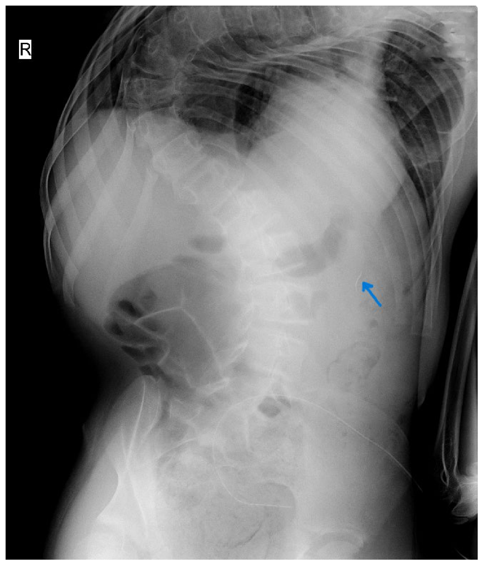
To prevent long-term migration injuries, a secondary laparoscopic exploration was scheduled for the subsequent morning. Initial conventional camera-guided visualization of the left upper quadrant was unrevealing due to early localized omental tracking over the repair site. Rather than pursuing an extensive, traumatic dissection of the newly sutured bowel loops, an improvised magnetic retrieval apparatus was assembled at the sterile back table.
A sterile latex-free glove finger (from a standard surgical glove) was sharply excised. Four high-grade, sterile, ferromagnetic neodymium spheres (each measuring 5 mm in diameter) were placed into the apex of the glove finger (Figure 2 and Figure 3). The open cuff of the glove finger was then tightly gathered and hermetically sealed using a heavy 2-0 silk ligature, leaving a 5-cm long trailing suture anchor.
A standard laparoscopic grasper was introduced through a right-sided 5-mm port to securely clamp the long silk trailing anchor. The entire glove-enclosed magnetic assembly was then pushed directly down through the lumen of a left-sided 10-mm trocar into the peritoneal workspace (Figure 4).
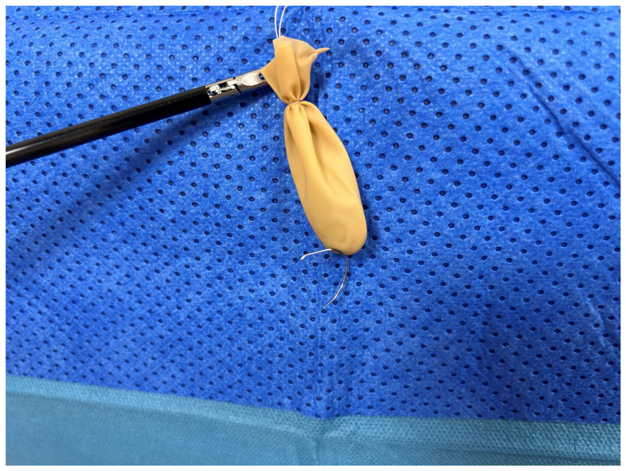
Guided by the preoperative radiograph coordinates, the magnetic assembly was lowered into the left paracolic gutter and slowly swept over the surface of the descending colon and the gastrosplenic ligament. Within 90 seconds of entry, a distinct tactile "click" was transmitted through the shaft of the grasper. The magnetic assembly was gently lifted, demonstrating that the lost needle tip had been successfully attracted and held flush against the external latex wall of the glove finger (Figure 5). The entire apparatus—along with the captured needle tip—was safely withdrawn through the 10-mm port under direct vision, ensuring the sharp tip did not shear off against the trocar valve.
Figure 5.
Successful attraction and retrieval of the lost laparoscopic needle.
The early postoperative course was stable, with CRP levels down-trending to 22 mg/L by day three. However, on postoperative day six, the patient developed bilious emesis and localized abdominal asymmetric distension. Contrast abdominal imaging confirmed a mechanical small bowel obstruction secondary to early fibrinous band adhesions. The patient underwent two subsequent targeted laparoscopic adhesiolysis procedures to clear bands obstructing the mid-jejunum. Enteral nutrition was successfully re-established using a nasojejunal feeding tube. Following a 20-day intensive care unit stay for metabolic optimization, the patient was successfully stabilized and transitioned to specialized pediatric palliative care for long-term support.
Discussion
While PEG placement remains an essential tool for pediatric nutritional support, it is critical to contextualize its associated anatomical risks [1,9]. Gastrocolic fistula or perforation occurs in fewer than 1% of standard series, arising almost exclusively from an unrecognized interposition of the transverse colon between the anterior stomach wall and the abdominal parietal peritoneum [3,5]. Children with severe spastic quadriplegic cerebral palsy and pronounced scoliosis, like our patient, exhibit altered intra-abdominal volumetric dynamics and fixed diaphragmatic shifts. This structural distortion places the transverse colon at a higher risk of superior displacement, predisposing them to blind transfixion injuries [15].
A lost surgical sharp inside a minimal-access space creates an immediate clinical dilemma. As detailed in the standard management algorithm proposed by Gulack et al., initial attempts at visual localization are frequently frustrated by the narrow focal depth of laparoscopic optics and the tendency of tiny sharps to slide beneath heavy solid organs or within dense mesenteric fat blocks [14]. If initial camera sweeps are unsuccessful, many surgical teams rely heavily on intraoperative C-arm fluoroscopy or plain radiographs. However, locating a fractured fragment with rigid graspers can cause the sharp to migrate deeper or puncture underlying mesenteric vessels [9].
The mechanism of needle loss highlighted in this report—structural failure and fracturing during extraction through a tight trocar valve—matches the recent observations of Aykanat et al., who identified that standard valved trocars create severe frictional choke points [8]. When high-tensile curved needles are pulled backward without perfect axial alignment, they undergo acute bending stresses, resulting in metal fatigue and fracturing.
To circumvent these issues, several teams have explored the physics of magnetic attraction. Early data from pioneering animal models confirmed that magnetic sweeps could reduce intra-abdominal search times by up to 60% compared to manual blind grasping. Dedicated articulating magnetic instruments have been prototyped, but their commercial availability remains limited in everyday emergency pediatric surgical arrays.
Our case demonstrates that an improvised magnetic delivery system - utilizing standard neodymium spheres safely sealed inside a sterile glove finger - provides a highly accessible alternative. Enclosing the raw magnets within an impermeable latex/polyisoprene barrier is a critical safety step; it completely eliminates the catastrophic risk of a loose magnet escaping into the abdomen while providing a perfectly smooth, bio-inert profile that glides over bowel loops without causing serosal tearing.
As noted in Table 1, while techniques like those utilized by Brahmbhatt et al. achieve successful retrieval under strict fluoroscopic guidance, they expose the pediatric patient to significant fields of ionizing radiation and add considerable cost. Our approach minimizes tissue manipulation and shortens operative time. The subsequent mechanical ileus seen in this patient was clearly related to dense fibrin deposition from the initial multi-perforation fecal peritonitis, rather than any structural damage from the brief magnetic sweeping procedure.
Conclusion
This complex case emphasizes that while PEG-related visceral perforations require immediate surgical management, the technical execution of minimal-access closures carries inherent mechanical risks. When a laparoscopic needle fractures or drops, surgeons should resist the urge to perform aggressive, blind manual tissue dissections. Adhering to structured surgical checklists and implementing an improvised, glove-contained magnetic capture array offers a safe, efficient and accessible solution that protects vulnerable pediatric patients from the morbidity of an open conversion.
Author Contributions
Conceptualisation: J. Povilavičius, K. Bagdonaitė, G. Pikturnaitė, R. Bernatavičienė, A. Strumila. Methodology and data curation: J. Povilavičius, K. Bagdonaitė. Surgery and clinical supervision: J. Povilavičius. Writing – original draft: J. Povilavičius, K. Bagdonaitė. Writing – review and editing: A. Strumila, J. Povilavičius, K. Bagdonaitė, R. Bernatavičienė. All authors read and approved the final manuscript.
Funding
This research received no external grant funding, public corporate sponsorships, or institutional financial subsidies.
Ethics approval and consent to participate
Ethical review and approval were waived for this study by the Institutional Review Board of Vilnius University hospital Santaros Clinics in Lithuania due to signed written informed consent, obtained directly from the patient and patient’s legal guardian [Approval No.: SR-4919; 16th of July 2026].
Consent for publication
Written informed consent for the publication of this comprehensive clinical case report, including all associated de-identified imaging and bench-top device illustrations, was obtained directly from the patient and patient’s legal guardian. A signed copy of the consent documentation is retained securely by the corresponding author [Consent retrieval: 2nd of December 2025].
Availability of data and materials
Data sharing is not applicable to this article as no independent, large-scale datasets were generated during this single-patient clinical study.
Competing Interests
The authors declare that they have no competing financial or professional interests that could have influenced the presentation of this work.
Abbreviations
| PEG: | Percutaneous Endoscopic Gastrostomy; |
| CT: | Computed Tomography; |
| ICU: | Intensive Care Unit; |
| WHO: | World Health Organization; |
| RFID: | Radio-Frequency Identification; |
| CRP: | C-Reactive Protein; |
| RSI: | Retained Surgical Item. |
Learning Points
• Inadvertent gastrocolic interposition during blind PEG placement is a rare but critical complication in children with severe neuromuscular scoliosis, requiring timely laparoscopic closure of all gastric and colonic defects.
• Frictional stress inside narrow trocar valves during extraction are a major cause of suture needle fracturing and subsequent intra-abdominal sharp loss.
• An improvised, glove-enclosed magnetic retrieval assembly serves as a safe, cost-effective, and rapid non-operative rescue method that avoids extensive tissue dissection and open laparotomy.
Statement of Novelty/Importance
This expanded case report documents the first clinical instance of concurrent multi-site gastrocolic organ perforation and intra-operative needle fracturing managed entirely through minimally invasive techniques in a pediatric patient. It validates a highly practical, low-cost bench-to-bedside magnetic capture solution using standard surgical supplies. This work adds clear clinical evidence to current pediatric safety protocols and fills a key gap in the literature regarding the management of retained sharps in low-resource emergency settings.
References
- Homan, M.; et al. Percutaneous Endoscopic Gastrostomy in Children: An Update to the ESPGHAN Position Paper. J. Pediatr. Gastroenterol. Nutr. 2021, 73(3), 415–426. [Google Scholar]
- Takalo, M.; et al. Complications after pediatric percutaneous endoscopic gastrostomy: comparison of the push and pull technique. World J. Pediatr. Surg. 2024, 7(1), e000687. [Google Scholar] [CrossRef]
- Tazi, K.; et al. Complications of Percutaneous and Surgical Gastrostomy Placements in Children: A Single-centre Series. JPGN Rep. 2023, 4(2), e316. [Google Scholar] [CrossRef]
- Bell, N.; Rabe, E.; Kufeji, D. Gastrocolocutaneous fistula: a rare complication of percutaneous endoscopic gastrostomy—case report. J. Transl. Gastroenterol. 2024, 2(1), e00007. [Google Scholar]
- Non-operative management of a rare late gastrocolic fistula after percutaneous endoscopic gastrostomy tube placement in a pediatric patient. SciDirect J. Ped Surg. Case Rep. 2025, 110, 102710.
- World Health Organization. WHO Surgical Safety Checklist. 2008; updated 2024. Available online: https://www.who.int/patientsafety/safesurgery.
- Hirose, R.; et al. Intraoperative breakage of a laparoscopic needle holder confirmed by routine postoperative abdominal X-ray: case report and literature review. J. Case Rep. Images Obstet. Gynecol. 2023, 9(1), 17–20. [Google Scholar] [CrossRef]
- Aykanat, I.C.; et al. Entrapped surgical needle in the valveless AirSeal trocar: a case report. Int. J. Surg. Case Rep. 2024, 114, 109035. [Google Scholar]
- Weprin, S.; et al. Risk factors and preventive strategies for unintentionally retained surgical sharps: a systematic review. Patient Saf. Surg. 2021, 15(1), 37. [Google Scholar] [CrossRef]
- Bawazir, O.A. Percutaneous endoscopic gastrostomy in children <10 kg: safety and outcomes. Saudi J. Gastroenterol. 2020, 26(2), 78–83. [Google Scholar]
- Ergin, A.; et al. Management of Needle Loss in Laparoscopic Operations. Eurasian J. Med. Invest. 2019, 3(1), 78–82. [Google Scholar]
- Brahmbhatt, S.; et al. Intra-abdominal Breakage of Laparoscopic Needle Holder Tip and Its Retrieval Under Fluoroscopic Guidance. J. Minim. Access Surg. 2021, 17(4), 572–575. [Google Scholar]
- Articulating magnetic instrumentation arrays for target object sweeping in minimally invasive workspaces. J. Laparoendosc. Adv. Surg. Tech 2023, 33(8), 741–746.
- Gulack, B.C.; et al. The Lost Suture Needle: An Algorithm to Standardize Management. J. Pediatr. Surg. 2021, 56(12), 2456–2461. [Google Scholar] [CrossRef]
- Complications and long-term surveillance of percutaneous gastrostomy configurations in pediatric spastic cerebral palsy cohorts. Eur. J. Pediatr. 2024, 183(4), 1611–1619.
Figure 2.
Construction of improvised magnetic retrieval device.
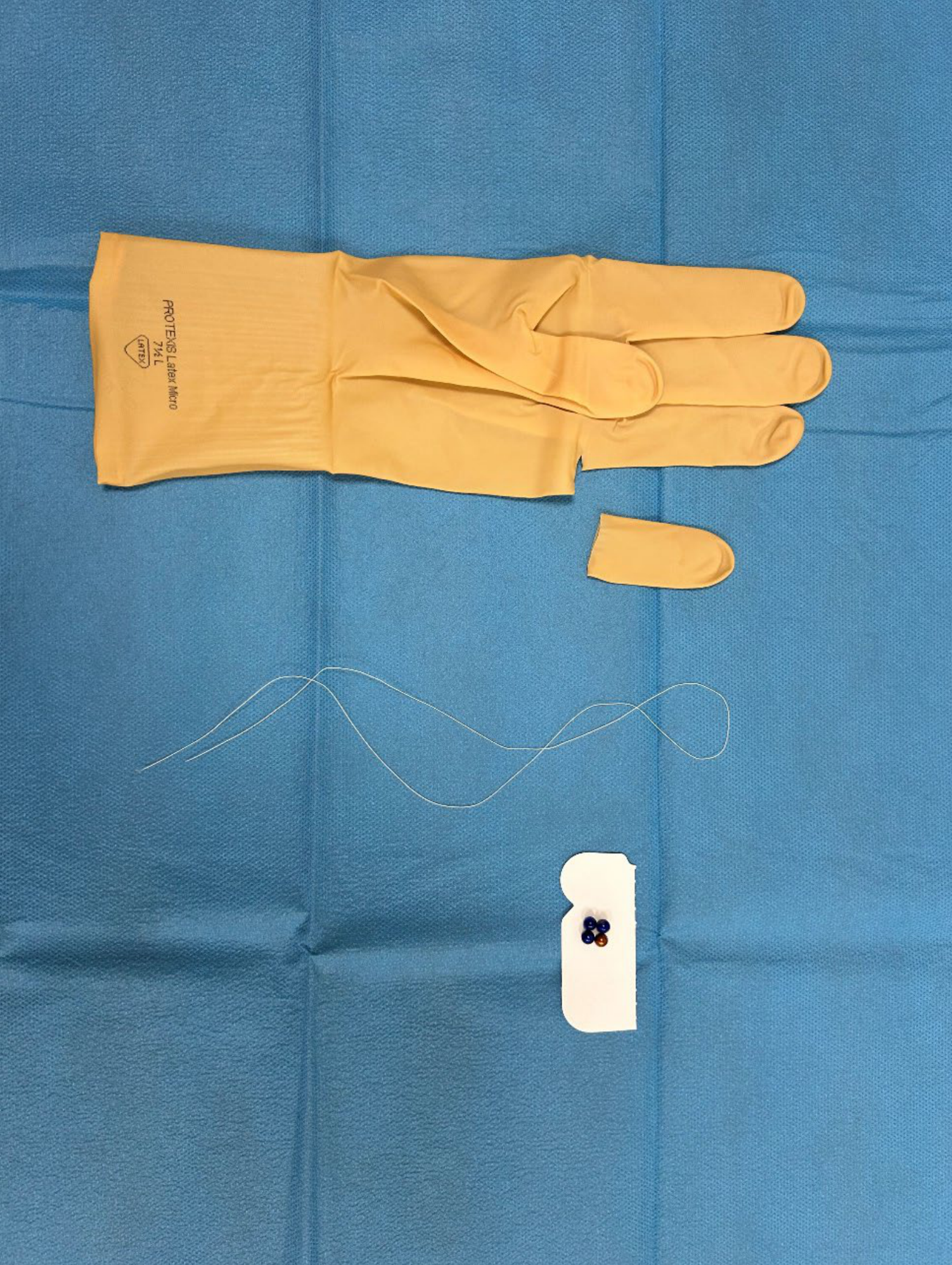
Figure 3.
Magnets enclosed within a glove finger and secured with ligature suture.
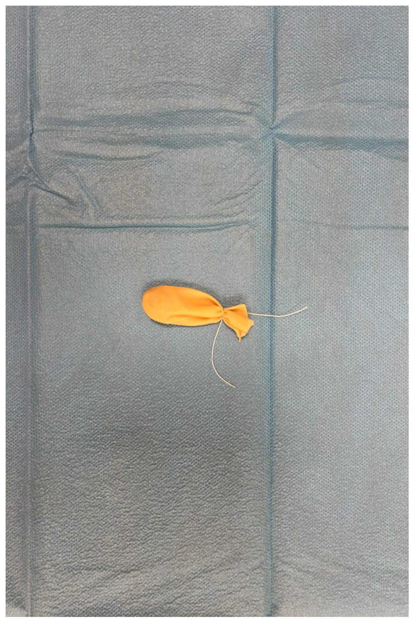
Figure 4.
Insertion of magnetic device through 10-mm trocar.
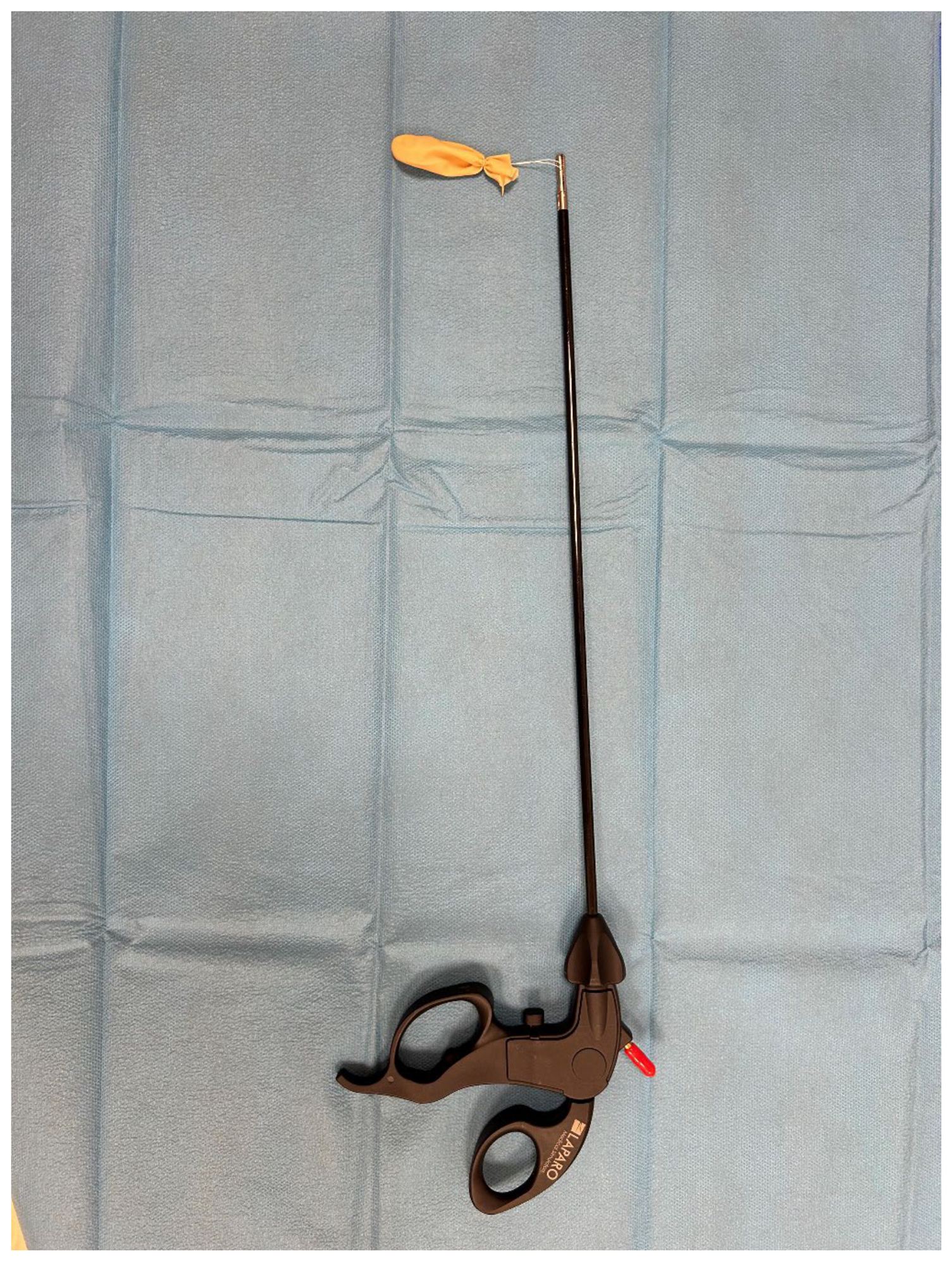

Table 1.
Literature comparison of lost needle management strategies in laparoscopies.
| Author / study | Patient cohort | Location of lost item | Localization strategy | Retrieval method | Primary outcome / success |
|---|---|---|---|---|---|
| Ergin et al. (2019) | Adult Laparoscopic | Variable peritoneal | Visual exploration & intraoperative C-arm | Standard laparoscopic graspers | With prolonged search times & tissue handling. |
| Gulack et al. (2021) | Pediatric Laparoscopic | Upper/lower quadrants | Quadrant algorithm & plain films | Laparoscopic instruments / laparotomy | Standardized search, small needles need fluoroscopy. |
| Brahmbhatt et al. (2021) | Pediatric Minimal-access | Retroperitoneal / pelvic | Continuous fluoroscopic guidance | Fluoroscopic-guided grasping | Successful, but elevated radiation exposure. |
| Aykanat et al. (2024) | Adult Laparoscopic | Trocar valve assembly | Port disassembly / visual search | Direct extraction | Highlights trocars as primary site of damage. |
| Present case (2026) | Pediatric Laparoscopic | Left paracolic gutter | Preoperative plain X-ray | Improvised glove magnetic sweeper | Rapid capture (<90 s); zero radiation; no laparotomy. |
Disclaimer/Publisher’s Note: The statements, opinions and data contained in all publications are solely those of the individual author(s) and contributor(s) and not of MDPI and/or the editor(s). MDPI and/or the editor(s) disclaim responsibility for any injury to people or property resulting from any ideas, methods, instructions or products referred to in the content. |
© 2026 by the authors. Licensee MDPI, Basel, Switzerland. This article is an open access article distributed under the terms and conditions of the Creative Commons Attribution (CC BY) license (http://creativecommons.org/licenses/by/4.0/).
Copyright: This open access article is published under a Creative Commons CC BY 4.0 license, which permit the free download, distribution, and reuse, provided that the author and preprint are cited in any reuse.