
Submitted:
30 September 2026
Posted:
01 October 2026
You are already at the latest version
Abstract
Paget’s seed-and-soil theory explains why cancer spreads to some organs and not others. It describes a travelling tumour cell that meets a receptive organ. It does not say how the organ becomes receptive. Extracellular vesicles (EVs) carry proteins and RNA between cells, and in experimental models tumour EVs can prepare a distant organ before tumour cells arrive [1–6]. We propose EVs as a third signalling component of Paget’s scheme: a signal that can travel ahead of the cell. It does not replace the seed, and preparing a tissue is not metastasis. We also place two further findings in this frame: metastases can differ genetically from their primary tumours [7,8], and metastatic progression is often linked to new blood vessels [9,10].The framework separates four levels of inference: an effect of an EV-containing sample, an effect shown to depend on EVs, a state that outlasts EV exposure, and a named mechanism for that state. Among the claims proposed here, EV-mediated tissue preparation has the strongest support. Yet most of that support sits at the first level, because few studies tested EV dependence with depletion and add-back controls. Two optional branches ask whether EVs can leave a lasting state in the receiving cell, through a change in protein shape (Route A) or through RNA reaching the genome (Route B). Dropping either branch leaves the core intact. Each claim is graded by its evidence and paired with the result that would prove it wrong.
Keywords:
extracellular vesicles
; metastasis
; seed and soil
; pre‐metastatic niche
; p53
; LINE‐1
; falsifiability
1. Introduction
In 1889 Stephen Paget studied 735 fatal breast-cancer cases and saw that metastases were not scattered at random. He proposed that a tumour cell, the seed, grows only where the local environment, the soil, suits it [11]. The idea has lasted because organs really do differ in how open they are to tumour growth. But the model names no molecular mechanism by which a distant organ becomes receptive.
Later work shows that distant tissue can be prepared before tumour cells settle in it. In 2005, bone-marrow progenitor cells were shown to set up a pre-metastatic niche [12], and this is now reviewed as a general feature of organ-specific spread [13]. Vesicles shed by tumours are one of the signals: cells in organs that later host metastases take them up and change [3,4,5,6]. This is a small extension of Paget’s logic. It does not compete with clonal evolution. It adds a parallel signalling layer.
Preparation of distant organs by EVs is well documented, and we do not claim it as new. We add two things. First, we place it inside Paget’s scheme as a time dimension: preparation can come before colonisation. Second, we separate a brief preparing signal, which has support in models, from a lasting state in the receiving cell, which has not been shown.
Earlier work reviews niche biology [13] and lists what EVs do at the niche [5,22,23]. A 2026 review of 120 manuscripts on in vivo studies shows how those claims were tested and names tests of necessity as the main gap [38]. We add a different tool: a staged framework for judging what such tests can and cannot show. It separates the activity of an EV-containing sample from EV dependence, from persistence after exposure, and from a named mechanism. For each step it states a result that would count against the claim. To our knowledge, this combination has not been set out before.
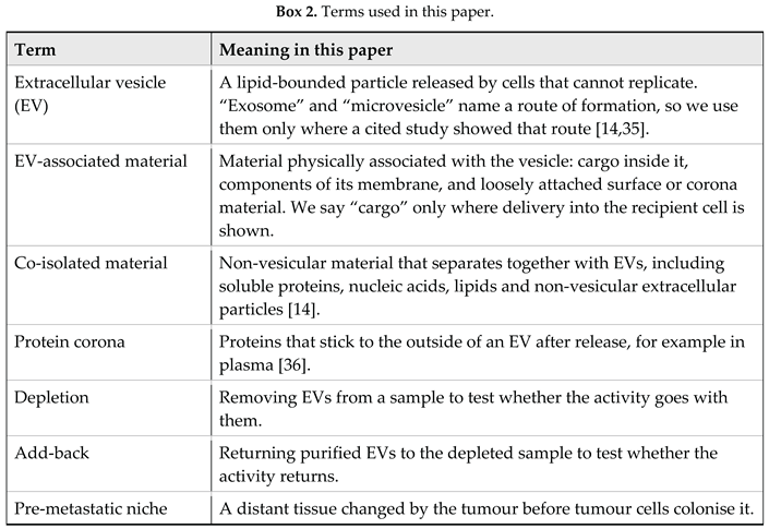
We report no new data. This is a narrative conceptual review, not a systematic one. Sources were chosen to represent founding studies, current EV guidance, primary experimental work and known limits. No risk-of-bias tool or pooled analysis was used, and contrary studies may have been missed. Every claim is a proposal. The grades are our reading of the literature, not a verdict. Box 1 states the scope, and Box 2 defines the terms.
Box 1. What this paper claims, and what it does not.
It claims that a
staged, falsifiable structure is a useful way to organise the literature on
EVs and the pre-metastatic niche. It claims that
EV-containing samples from tumours can prepare distant tissue in experimental
models. It does not claim that
EVs are necessary for metastasis, that they cause genomic divergence, or that
any lasting state in a recipient cell exists. It does not claim that
Route A or Route B happens in patients.
2. What Is Already Known
We grade evidence in three colours, used in the text, the tables and Figure 1. [Established] (blue) means backed by several independent studies or broad agreement in the field, not resting on one model. [Supported] (green) means shown in at least one experimental system, such as cell culture or animals, but not in patients, and often tied to that model. [Hypothesised] (yellow) means proposed here, with no direct test yet. A grade describes the evidence, not how plausible we find a claim. Each label appears with its colour, so the scheme survives black-and-white printing.
One caution covers every green grade. Most studies in Section 2.2 predate MISEV2023, and a 2024 commentary notes that most EV papers do not cite the MISEV guidelines or follow their reporting requirements [35]. So [Supported] here means consistent results in models. It does not mean that EV dependence is proven.
2.1. Vesicles Carry Working Cargo Between Cells
Small EVs from mast cells (called exosomes in the original study) carry mRNA and microRNA, and the mRNA can be made into protein in a new cell [1]. Glioblastoma EVs carry RNA and vessel-building proteins that healthy cells take up and respond to [2]. So cargo can work after transfer. These studies do not show stable genome change or lasting reprogramming.
Two rules from MISEV2023 shape how we read this literature [14]. First, we say “extracellular vesicle” unless a study showed how the vesicle formed [14,35]. Second, an effect of an EV-containing sample is not automatically an effect of the EV. No separation method removes all other material, so a preparation can carry co-isolated proteins, nucleic acids and lipids. Depletion and add-back experiments are needed to test whether activity tracks with the EV-associated fraction rather than with co-isolated material [14,34,35].
2.2. Tumour EVs Help Prepare a Distant Organ
Melanoma EVs push bone-marrow progenitor cells toward a pro-metastatic type through MET signalling, and make vessels leakier at future metastatic sites [4]. One study linked integrins on small EVs (called exosomes in that paper) to where the vesicles are taken up: α6β4 and α6β1 with the lung, αvβ5 with the liver. The authors reported that blocking them reduced uptake and spread in those models [3]. In August 2026 Nature added an Editorial Expression of Concern to that paper because of apparent image overlap in several figures [15]. This is not a retraction, but the affected figures need careful reading, so we do not rest organ-specific targeting on it.
Liver preparation has been shown another way. Small EVs from pancreatic cancer, taken up by Kupffer cells, create a scarred, macrophage-rich liver environment [16]. Small EVs from lung-cancer cells with mutant p53 pushed fibroblasts to lay down more matrix [17]. This fits a wider finding that mutant p53 changes what tumour cells secrete and how they signal to stroma [18]. It shows that the p53 status of the source cell can shape what its EVs do. It does not show that p53 protein itself was handed over.
Two points about integrins should be kept apart. That EVs can carry working integrins is well shown [19,20,21]. That integrins act as an address code for specific organs depends on the model and, after the notice above, is uncertain. Two 2025 reviews still describe integrin addresses for lung and liver, but they draw on the 2015 study and predate the notice, so they are not independent support. One says the role of EV integrins in cancer is still under study [22]. The other says the full set of address codes is poorly understood [23].
Broader reviews describe a network of inflammation, immune suppression, new and leaky vessels, stromal change and altered metabolism, with EVs as one part [5]. In one study, cells in the pre-metastatic niche captured EVs released by xenografted tumours and reacted with inflammation [6]. Two limits apply. EVs have been isolated, described and tracked in many different ways. And most studies give cells or animals isolated EVs instead of relying on those a tumour releases itself. The same 2026 review of 120 manuscripts found this pattern: 115 used experimentally prepared EVs given to the animals, only five tracked EVs released by implanted tumours, and doses ranged from 5 to 200 µg per mouse [38]. Its authors name loss-of-function tests of necessity as the main unmet challenge [38].
Some experiments point to necessity as well as sufficiency. Blocking EV release by Rab27a knockdown reduced spread in melanoma and mammary-carcinoma models [4,24]. Antibodies that clear circulating cancer EVs reduced lung and lymph-node metastases in a breast-cancer model without changing primary tumour growth [25]. But these tools are not EV-specific. Rab27a also affects other secretions [23,24], and MISEV2023 warns that any change to EV release can act on other pathways [14,35]. No universally validated marker currently separates tumour-derived EVs from host EVs without ambiguity in vivo [5]. So blocking release alone cannot prove that EVs were the cause.
EVs work alongside soluble tumour factors, immune-cell recruitment, vessel change and local stromal responses [5]. We therefore write “contribute to”, not “cause”, unless EV dependence has been shown directly. Most of the work cited here has not made that test.
2.3. Metastases Are Not Genetic Copies of the Primary Tumour
Metastatic subclones can branch off early or late in a tumour’s life [7]. A 2026 study followed 24 patients with non-small-cell lung cancer through 501 samples from the TRACERx study and the PEACE autopsy programme. Metastasis genomes diverged sharply from the primary tumour, with new driver alterations and genome doublings after spread, and more than half of the sampled metastases were seeded by other metastases [8]. This fits the idea that a distant environment shapes later evolution. But the study does not name the cause, and ordinary clonal selection is an established alternative explanation.
2.4. New Blood Vessels Go with Metastasis
Many tumours depend on new blood vessels for steady growth, although some use vessels already there [10]. In invasive breast cancer, every extra 10 microvessels per 200× field, counted in the most vascular area of the tumour, went with a 1.17-fold higher risk of distant metastasis [9]. That is a link with risk, not proof that new vessels alone drive spread. EVs offer one way for the soil to gain vessels before colonisation. Glioblastoma EVs carry vessel-building proteins and trigger tube formation in endothelial cells [2], and melanoma EVs push progenitor cells toward a vessel-building type [4].
2.5. Building Blocks
Table 1 lists the pieces we draw on and grades each one. They are not proof of our proposal. They are building blocks that we relate to each other. Routes A and B are the two independent, optional routes described in Section 3.1.
3. The Proposal
“Third signalling component” is shorthand for a proposed temporal signalling layer: a transferable molecular signal that can prepare the soil. It is not a third member of the seed-and-soil pair, and we do not claim that EVs are a third biological entity equal to seed and soil. The classic model has two parts acting together: a seed that arrives and a soil that accepts or rejects it. Section 2 adds time: a cell-free signal that can act on the soil before colonisation. Figure 1 shows the framework. One part has support in models. The rest is hypothesised, and we keep the two apart on purpose.
We keep four levels of inference apart. Level 1 is an effect of an EV-containing sample. Level 2 is an effect shown to depend on EVs. Level 3 is a state that outlasts EV exposure. Level 4 is a named mechanism for that state. A result at one level does not count as a result at the next. Most of the evidence in Section 2 sits at Level 1. Claims 1 and 2 ask for Level 2, in tissue and in cells. Claim 3 asks for Level 3, first as persistence (3a) and then as inheritance through cell division (3b). Routes A and B are proposals at Level 4.
Claim 1 [Supported] (at Level 1; Level 2 is largely untested). Tumour EVs help prepare a distant organ before tumour cells colonise it [3,4,5,6]. EVs are one of several mediators, and they do not replace the seed.
Claim 2 [Hypothesised] During exposure, EV-associated activity contributes to a recipient-cell response that soluble co-factors alone do not reproduce. “Contributes” means a gain over matched non-EV fractions that depletion reduces and add-back restores.
Claim 3 [Hypothesised] That response outlasts EV exposure. Claim 3a: it persists after EV material has fallen below a detection limit set in advance. Claim 3b: it passes to daughter cells through cell division. Route A could support 3a without any change to DNA. Route B could support 3b, if the insertion sits in a cell that divides and is inherited by its daughters.
We say “lasting state”, not “transformation”. A cell that changes shape, releases other cytokines or moves faster is not necessarily transformed. A state counts as lasting only if it survives EV washout for a period fixed in advance, if contamination and cell fusion are excluded, and if it arises in the recipient-derived lineage. Passing through cell division is a stronger test still (Claim 3b). Each claim can be tested and rejected alone. Losing Claim 2 or 3 does not touch Claim 1.

Table 2.
The three claims at a glance. Colours as in Figure 1.
Table 2.
The three claims at a glance. Colours as in Figure 1.
| Claim | What it says | Evidence status | What must be shown next |
|---|---|---|---|
| Claim 1 (core; Level 1 evidence, Level 2 target) | Tumour EVs help prepare a distant organ before tumour cells colonise it. | [Supported] in experimental models, mostly at Level 1 [3,4,5,6]. Organ-specific integrin targeting depends on the model [3,15]. | EV dependence shown by depletion and add-back, not by an EV-enriched sample alone (Section 4.2). |
| Claim 2 (optional, Level 2) | During exposure, EV-associated activity contributes to a recipient-cell response that soluble co-factors alone do not reproduce. | [Hypothesised] No direct evidence yet for an EV-specific state in recipient cells. Reviews note that EV cargo transfer, such as miRNA, is hard to confirm in living animals [23]. That is a gap in method, not support. | A recipient-cell response that follows the EV fraction and is not reproduced by matched soluble or non-EV fractions. |
| Claim 3 (optional, Level 3) | 3a: that response persists after EV exposure ends. 3b: it passes to daughter cells. | [Hypothesised] Each route is supported only in dish or engineered-reporter systems (Section 3.1). | 3a: a state that survives washout and is verified in the recipient-derived lineage. 3b: the same state in clonally derived daughter cells. |
3.1. Two Optional Routes to a Lasting State
Once a vesicle is taken up, two mechanisms could let its signal outlast the vesicle. Both are optional examples, and neither is part of Paget’s theory.
Route A: a change in protein state. Mutant p53 delivered by an EV may change p53 signalling in the receiving cell. A bolder idea is that this misfolded p53 could push the cell’s own p53 into the same misfolded shape, as happens in prion diseases. There is a precedent for one form of p53 disabling another: some mutants bind wild-type p53 and inactivate it [18]. But that happens by direct binding inside one cell, not by conversion. Amyloid-like clumping of mutant p53 should not be read as templated conversion of the recipient’s own wild-type p53. The dish evidence also differs by mutation, so a result for one hotspot mutant does not carry over to another [26,27,28]. Six things must be shown: donor mutant p53 enters the recipient; the recipient’s own p53 can be told apart from the donor’s; that own p53 changes shape; simple binding of the two is ruled out; p53 function is lost; and the change lasts after washout. None of this has been shown in a living body.
A question of numbers comes first: how much mutant p53 does an EV deliver, compared with the recipient cell’s own p53 pool? Templating needs enough seed protein to matter, and protein that never leaves the vesicle compartment cannot template anything. The ratio can be measured as a chain: EVs taken up per cell, mutant p53 per EV, the share that reaches the cytosol, and assay recovery. The product is then compared with the recipient’s own p53, measured in the same cells. We give no numbers because none has been measured for this system. Until then, Route A stays a proposal.
Route B: an RNA-to-DNA change. EV-borne LINE-1 RNA could in principle be copied into DNA by LINE-1’s own enzyme and inserted into the receiving cell’s genome. This has been shown with an engineered, working LINE-1 reporter carried by EVs in cultured cells [32]. Naturally shed tumour microvesicles were also reported to carry retrotransposon-derived sequences [33], so the cargo is not only an artefact of engineering. That shows the idea is technically possible. It does not show that natural LINE-1 does this in a living body. A new insertion could also arise without any EV cargo, because loss of p53 can release the recipient’s own LINE-1 copies in human cells [37]. Five steps would each need direct proof: uptake of the RNA, copying into DNA, insertion into the recipient’s genome, proof that the DNA belongs to the recipient and not the source, and inheritance by clonally derived daughter cells.
The routes are independent. Evidence against one says nothing about the other. We keep them apart because merging them would make the argument look smoother, and more proven, than the evidence allows. Neither route needs protein to write into DNA or RNA: Route A changes protein shape only, and Route B copies RNA into DNA, which retrotransposon enzymes can do, but it is a chain of several independent steps. Driver mutations remain a main cause of most tumours. We propose only an extra, parallel route that might work in some cases. Whether such cases exist is a question for experiment (Section 4).
3.2. Genetic Divergence and New Vessels
EV-mediated tissue preparation could, in principle, change the selective environment in which disseminated tumour cells go on to evolve. That is a statement about selection. It does not mean that EVs cause the genomic divergence itself. Only Route B, if it exists, would let EVs alter the genome directly, and it stays a proposal. The genomic studies cited here [7,8] test neither idea, and ordinary selection is an established alternative explanation.
4. How to Test It
A useful hypothesis must say what would prove it wrong. Table 3 lists our predictions with the result that would support each one and the result that would count against it. P1 tests the core claim. P2 asks whether EV-associated activity contributes to a recipient-cell response during exposure. P3 and P4 ask whether that response persists, and by what route. P2, P3 and P4 are optional: nothing in P1 depends on them.
These failure conditions have limits of their own. If depletion does not reduce the preparing effect in P1, the EV-dependent reading of Claim 1 is weakened, but only if depletion efficiency and the survival of non-EV activity were validated independently. A failed add-back weakens Claim 1 only if EV integrity, dose and purification losses were checked, since damaged or lost EVs would also fail to restore activity. If a matched non-EV fraction reproduces the full prespecified effect in P2, with no added EV contribution, Claim 2 falls, and with it the case for both routes. A partial non-EV effect does not do this, because EVs and soluble factors may act together. If neither insertion nor templating can be shown with proper lineage attribution, the matching route should be dropped on its own. The plan is staged so that losing a later stage never takes down an earlier one.
The clearest test of Claim 3 is a washout experiment. Recipient cells are exposed to EVs, washed, and cultured for a long time. If EV material stays below the detection limit, contamination and cell fusion are excluded, and the state still fades before the endpoint fixed in advance, Claim 3 fails. The observation period should be long enough that any carried-over EV material must have been diluted below detection by cell division. The primary endpoint, the detection limit, the depletion efficiency, the smallest effect that counts and the way EV dose is normalised should all be fixed before the experiment is run (Q5).
Three further rules keep a negative result honest. First, a negative result counts only if a positive control shows the assay could have detected the effect. Second, a result is scored inconclusive, not negative, when a required control is missing or an assay’s sensitivity is unconfirmed. Third, EV doses are fixed in advance and reported with cell viability, EV source, dose per cell, and duration and frequency of exposure, and a negative result is not later explained as “too little EV”. If an effect appears only at doses far above what a tumour could deliver, it shows that the step can happen (feasibility), not that it happens in the body (plausibility).
4.1. What Would Count as Evidence of Transfer
Any study claiming a lasting change after EV exposure must show that the change arose in the recipient-derived lineage, not in carried-over material from the source. In practice this means:
- showing that the preparation is free of cells and cell fragments;
- using source and recipient lines that can be told apart by genotype or lineage label;
- checking for cell fusion with dual-labelled lines;
For Route A, conformation-sensitive antibodies such as PAb1620 and PAb240 can flag a shape change. Staining alone does not prove templating, so seeding assays and structural methods are needed. For Route B, sequencing of insertion sites against the recipient’s own genotype can show a new insertion and separate it from source-cell DNA. The lack of a validated marker for cancer-derived EVs [5] makes attribution harder.
4.2. An Effect of EVs, or an Effect That Involves Them?
MISEV2023 stresses the difference between activity seen in an EV-containing preparation and activity that can be tied to EVs specifically, which needs depletion, add-back and non-EV controls [14]. The guidelines recommend dose-response and time-course tests, both EV and non-EV negative controls, and a stated level of confidence that an activity belongs to EVs and not to other components [14,34,35]. They also warn that tools which block EV release can affect other pathways [14,35]. This matters because proteins on the EV surface, including corona-associated material, may contribute to an observed activity [35,36]. Every claim here stays on the “linked” side of that line unless such controls say otherwise.
This consensus concerns how EV studies should be run and reported. Following it makes a result more credible, but it does not make a claim true.
4.3. What Else Could Explain the Same Finding
Table 4 lists the other explanations a study must rule out before blaming EV-associated material.
5. Limits
Several links have no direct evidence yet. EV-borne LINE-1 insertion has been shown only with a reporter in cultured cells [32]. Whether it happens in the body, how often, and whether it matters for cancer are open questions. Prion-like p53 templating is mostly a dish finding. EV transfer of mutant p53 to other cells is well documented [29,30], but a stable, self-sustaining conversion of the recipient’s own p53 in a living body is a proposal, not a result.
Organ-specific targeting once leaned on a paper that now carries an Expression of Concern (Section 2.2). Many studies give EVs as a set bolus, and how this relates to a tumour’s own steady output is uncertain. Reviews add that EV cargo transfer is hard to confirm in vivo and that tools used to block EVs are not EV-specific [5,23]. Our grades rest on experimental models, and we do not grade human evidence. We give no estimates of EV numbers, cargo frequency or event rates. The data are not yet available in a form we can cite responsibly, and an invented figure would be worse than an open question.
Even if every step is real, all of them must happen in the same cell. The chance of the whole chain cannot exceed the chance of its weakest step. We do not know whether the steps are independent, so multiplying separate estimates could mislead in either direction. This is why we ask for each step to be measured directly, and why we give no combined estimate.
The framework itself can fail. If no depletion method can be validated for a system, because EV and non-EV activity cannot be separated there, Level 2 cannot be reached and the ladder stalls. That would be informative, but it would not settle the biology. The grades are also our own judgement, and another reader could weigh the same studies differently. They would change if a well-controlled depletion and add-back study reached a different result, or if a key source were corrected or withdrawn. Finally, this article offers predictions, not results. If the extension is right, clonal evolution is not wrong, only incomplete.
6. Conclusions
The core proposal is small. In experimental models, tumour EVs can act as signals that travel ahead of the cell. This adds a signalling layer to seed and soil without replacing either. Whether EV exposure can also leave a lasting state in a receiving cell, through Route A or Route B, is a separate and more speculative question.
This article offers a method, not a new finding: it turns a body of model results into a ladder of claims, each with a stated way to fail. That tumours shed EVs, and that EVs can prepare distant tissue in models, is documented. What is open is whether the preparing effect holds once non-EV explanations are ruled out, and whether any of it becomes a lasting state in the receiving cell. Because losing a later stage never takes down an earlier one, the framework can be falsified rather than merely argued.
Open questions
Q1. Can an EV-dependent effect on distant tissue survive proper depletion and add-back controls, rather than only tracking an EV-enriched sample (P1)?
Q2. Does EV pre-treatment change the outcome of a later tumour-cell challenge in a way that non-EV factors do not reproduce (P1, P2)?
Q3. Can a state induced by EVs be shown to last after washout, with EV material below a set detection limit, and can Route A or Route B be shown with verified lineage in vivo (P3, P4)?
Q4. What is the ratio of EV-delivered mutant p53 to the recipient’s own p53, and is it high enough for templating to be plausible?
Q5. Can the failure conditions in Section 4 be written down, and ideally registered, before studies begin, so that a negative result cannot be explained away afterwards?
Q1 and Q2 can be tackled now with standard methods. Q3 and Q4 need new tools. Q5 can be settled before any experiment by writing the failure criteria down. No clinical or transfusion-practice recommendation follows from this framework. The next step is experiment, starting with the prepare-then-challenge design and the depletion and add-back controls in Table 3.
Declarations
Author Contributions
Said Ganihodjaev: conceptualisation, writing (original draft), writing (review and editing). Sole author.
Funding
None. No external funding was received for this work.
Data Availability Statement
This is a conceptual review. No original data were generated or analysed.
Conflicts of Interest
The author declares no competing interests.
References
- Valadi, H.; Ekström, K.; Bossios, A.; Sjöstrand, M.; Lee, J.J.; Lötvall, J.O. Exosome-mediated transfer of mRNAs and microRNAs is a novel mechanism of genetic exchange between cells. Nat. Cell Biol. 2007, 9(6), 654–659. [Google Scholar] [CrossRef]
- Skog, J.; Würdinger, T.; van Rijn, S.; et al. Glioblastoma microvesicles transport RNA and proteins that promote tumour growth and provide diagnostic biomarkers. Nat. Cell Biol. 2008, 10(12), 1470–1476. [Google Scholar] [CrossRef]
- Hoshino, A.; Costa-Silva, B.; Shen, T.L.; et al. Tumour exosome integrins determine organotropic metastasis. Nature 2015, 527(7578), 329–335. [Google Scholar] [CrossRef]
- Peinado, H.; Alecković, M.; Lavotshkin, S.; et al. Melanoma exosomes educate bone marrow progenitor cells toward a pro-metastatic phenotype through MET. Nat. Med. 2012, 18(6), 883–891. [Google Scholar] [CrossRef]
- Li, Y.; Zheng, Y.; Tan, X.; Du, Y.; Wei, Y.; Liu, S. Extracellular vesicle-mediated pre-metastatic niche formation via altering host microenvironments. Front Immunol. 2024, 15, 1367373. [Google Scholar] [CrossRef]
- Blavier, L.; Nakata, R.; Neviani, P.; Sharma, K.; Shimada, H.; Benedicto, A.; Matei, I.; Lyden, D.; DeClerck, Y.A. The capture of extracellular vesicles endogenously released by xenotransplanted tumours induces an inflammatory reaction in the premetastatic niche. J. Extracell. Vesicles 2023, 12(5), e12326. [Google Scholar] [CrossRef]
- Turajlic, S.; Swanton, C. Metastasis as an evolutionary process. Science 2016, 352(6282), 169–175. [Google Scholar] [CrossRef]
- Hessey, S.; Bunkum, A.; Huebner, A.; et al. Evolutionary characterization of lung cancer metastasis. Nature 2026, 653(8115), 911–922. [Google Scholar] [CrossRef]
- Weidner, N.; Semple, J.P.; Welch, W.R.; Folkman, J. Tumor angiogenesis and metastasis: correlation in invasive breast carcinoma. N Engl. J. Med. 1991, 324(1), 1–8. [Google Scholar] [CrossRef]
- Folkman, J. Tumor angiogenesis: therapeutic implications. N Engl. J. Med. 1971, 285(21), 1182–1186. [Google Scholar] [CrossRef]
- Paget, S. The distribution of secondary growths in cancer of the breast. Lancet 1889, 133(3421), 571–573. [Google Scholar] [CrossRef]
- Kaplan, R.N.; Riba, R.D.; Zacharoulis, S.; et al. VEGFR1-positive haematopoietic bone marrow progenitors initiate the pre-metastatic niche. Nature 2005, 438, 820–827. [Google Scholar] [CrossRef]
- Peinado, H.; Zhang, H.; Matei, I.R.; et al. Pre-metastatic niches: organ-specific homes for metastases. Nat. Rev. Cancer 2017, 17, 302–317. [Google Scholar] [CrossRef]
- Welsh, J.A.; Goberdhan, D.C.I.; O’Driscoll, L.; et al. Minimal information for studies of extracellular vesicles (MISEV2023): from basic to advanced approaches. J. Extracell. Vesicles 2024, 13(2), e12404. [Google Scholar] [CrossRef]
- Editorial Expression of Concern: Tumour exosome integrins determine organotropic metastasis. Nature Published online. 2026, 656, E44. [CrossRef]
- Costa-Silva, B.; Aiello, N.M.; Ocean, A.J.; et al. Pancreatic cancer exosomes initiate pre-metastatic niche formation in the liver. Nat. Cell Biol. 2015, 17(6), 816–826. [Google Scholar] [CrossRef]
- Novo, D.; Heath, N.; Mitchell, L.; et al. Mutant p53s generate pro-invasive niches by influencing exosome podocalyxin levels. Nat. Commun. 2018, 9, 5069. [Google Scholar] [CrossRef]
- Qiu, L.; Ma, Z.; Wu, X. Mutant p53-mediated tumor secretome: bridging tumor cells and stromal cells. Genes 2024, 15(12), 1615. [Google Scholar] [CrossRef]
- Singh, A.; Fedele, C.; Lu, H.; Nevalainen, M.T.; Keen, J.H.; Languino, L.R. Exosome-mediated transfer of αvβ3 integrin from tumorigenic to nontumorigenic cells promotes a migratory phenotype. Mol. Cancer Res. 2016, 14(11), 1136–1146. [Google Scholar] [CrossRef]
- Fedele, C.; Singh, A.; Zerlanko, B.J.; Iozzo, R.V.; Languino, L.R. The αvβ6 integrin is transferred intercellularly via exosomes. J. Biol. Chem. 2015, 290(8), 4545–4551. [Google Scholar] [CrossRef]
- Fuentes, P.; Sesé, M.; Guijarro, P.J.; et al. ITGB3-mediated uptake of small extracellular vesicles facilitates intercellular communication in breast cancer cells. Nat. Commun. 2020, 11, 4261. [Google Scholar] [CrossRef]
- Ciobanasu, C.; Le Clainche, C. Integrins from extracellular vesicles as players in tumor microenvironment and metastasis. Cancer Metastasis Rev. 2025, 44(3), 68. [Google Scholar] [CrossRef]
- Nidhi, G.; Yadav, V.; Singh, T.; Sharma, D.; Bohot, M.; Satapathy, S.R. Beyond boundaries: exploring the role of extracellular vesicles in organ-specific metastasis in solid tumors. Front Immunol. 2025, 16, 1593834. [Google Scholar] [CrossRef]
- Bobrie, A.; Krumeich, S.; Reyal, F.; et al. Rab27a supports exosome-dependent and -independent mechanisms that modify the tumor microenvironment and can promote tumor progression. Cancer Res. 2012, 72, 4920–4930. [Google Scholar] [CrossRef]
- Nishida-Aoki, N.; Tominaga, N.; Takeshita, F.; Sonoda, H.; Yoshioka, Y.; Ochiya, T. Disruption of circulating extracellular vesicles as a novel therapeutic strategy against cancer metastasis. Mol. Ther. 2017, 25, 181–191. [Google Scholar] [CrossRef]
- Silva, J.L.; De Moura Gallo, C.V.; Costa, D.C.; Rangel, L.P. Prion-like aggregation of mutant p53 in cancer. Trends Biochem Sci. 2014, 39(6), 260–267. [Google Scholar] [CrossRef]
- Ano Bom, A.P.D.; Rangel, L.P.; Costa, D.C.F.; et al. Mutant p53 aggregates into prion-like amyloid oligomers and fibrils: implications for cancer. J. Biol. Chem. 2012, 287(33), 28152–28162. [Google Scholar] [CrossRef]
- Rangel, L.P.; Costa, D.C.F.; Vieira, T.C.R.G.; Silva, J.L. The aggregation of mutant p53 produces prion-like properties in cancer. Prion 2014, 8(1), 75–84. [Google Scholar] [CrossRef]
- Bhatta, B.; Luz, I.; Krueger, C.; Teo, F.X.; Lane, D.P.; Sabapathy, K.; Cooks, T. Cancer cells shuttle extracellular vesicles containing oncogenic mutant p53 proteins to the tumor microenvironment. Cancers 2021, 13(12), 2985. [Google Scholar] [CrossRef]
- Ma, S.; McGuire, M.H.; Mangala, L.S.; et al. Gain-of-function p53 protein transferred via small extracellular vesicles promotes conversion of fibroblasts to a cancer-associated phenotype. Cell Rep. 2021, 34(6), 108726. [Google Scholar] [CrossRef]
- Baltimore, D. RNA-dependent DNA polymerase in virions of RNA tumour viruses. Nature 1970, 226(5252), 1209–1211. [Google Scholar] [CrossRef]
- Kawamura, Y.; Sanchez Calle, A.; Yamamoto, Y.; Sato, T.A.; Ochiya, T. Extracellular vesicles mediate the horizontal transfer of an active LINE-1 retrotransposon. J. Extracell. Vesicles 2019, 8(1), 1643214. [Google Scholar] [CrossRef]
- Balaj, L.; Lessard, R.; Dai, L.; et al. Tumour microvesicles contain retrotransposon elements and amplified oncogene sequences. Nat. Commun. 2011, 2, 180. [Google Scholar] [CrossRef]
- Witwer, K.W.; Goberdhan, D.C.; O’Driscoll, L.; et al. Updating MISEV: evolving the minimal requirements for studies of extracellular vesicles. J. Extracell. Vesicles 2021, 10, e12182. [Google Scholar] [CrossRef]
- Samuels, M.; Giamas, G. MISEV2023: shaping the future of EV research by enhancing rigour, reproducibility and transparency. Cancer Gene Ther. 2024. [Google Scholar] [CrossRef]
- Toth, E.A.; Turiak, L.; Visnovitz, T.; et al. Formation of a protein corona on the surface of extracellular vesicles in blood plasma. J. Extracell. Vesicles 2021, 10, e12140. [Google Scholar] [CrossRef]
- Tiwari, B.; Jones, A.E.; Caillet, C.J.; Das, S.; Royer, S.K.; Abrams, J.M. p53 directly represses human LINE1 transposons. Genes Dev. 2020, 34(21-22), 1439–1451. [Google Scholar] [CrossRef]
- Blavier, L.; Crnjac, A.; DeClerck, Y.A. Contribution of tumor-derived extracellular vesicles in the establishment of the pre-metastatic niche: lessons learned from past experimentations and future directions. Clin. Exp. Metastasis 2026, 43(2), 16. [Google Scholar] [CrossRef]
Figure 1.
Overview of the framework. EV, extracellular vesicle. Blue: established (the classic Paget model). Green: supported in experimental models, here the core, EV-mediated tissue preparation [3,4,5,6]; EV dependence under depletion and add-back controls is largely untested, and organ-specific integrin targeting depends on the model [3,15]. Yellow with dashed borders: hypothesised, namely two optional routes to a lasting state in the recipient cell, Route A (mutant p53 templating) and Route B (LINE-1 RNA reverse transcription and insertion). Bottom: predictions P1–P4 with their evidence status and the result that would count against each. Losing P2, P3 or P4 does not invalidate P1.
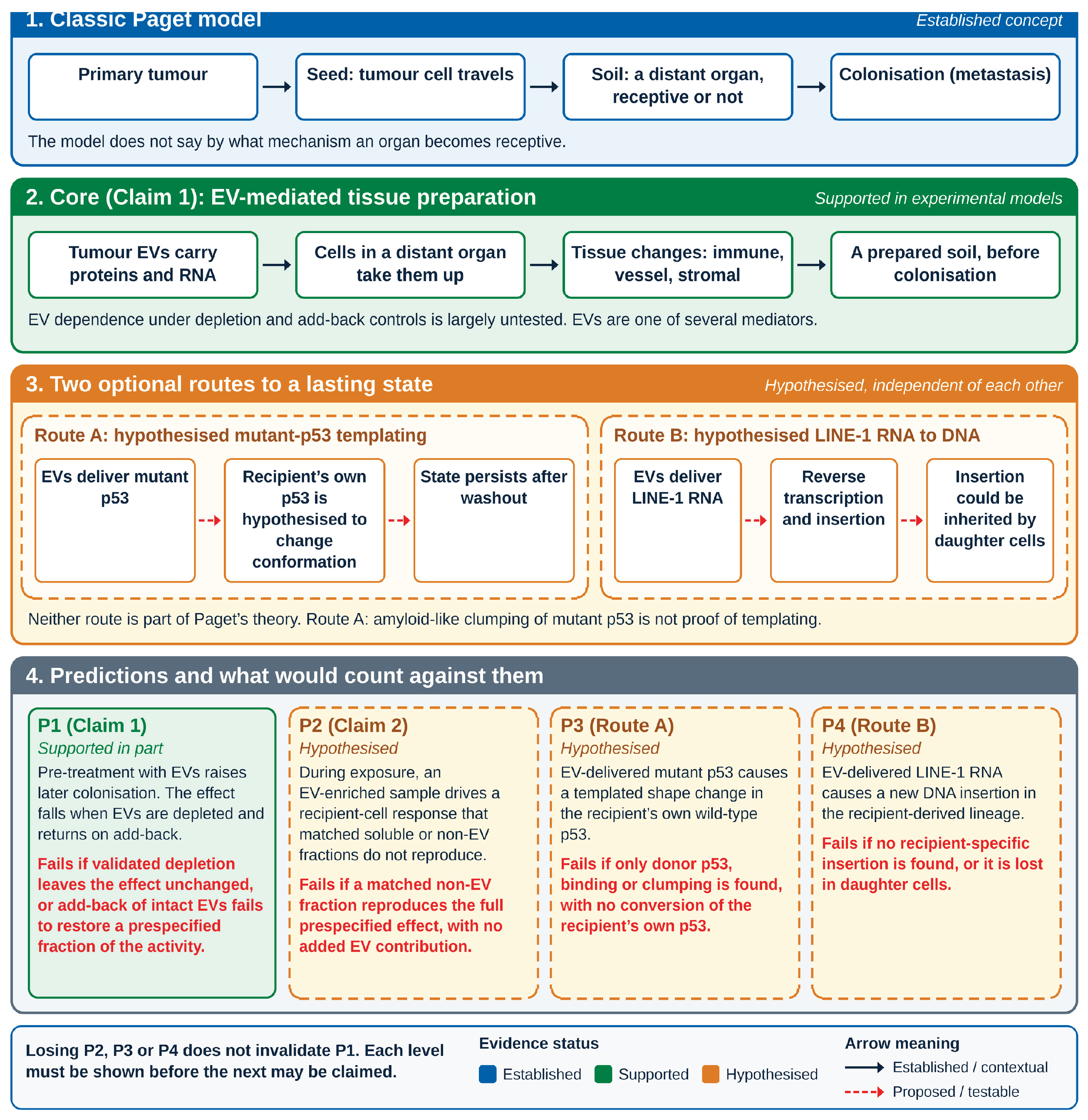
Table 1.
Building blocks and their status. Colours as in Figure 1: blue established, green supported, yellow hypothesised. Tumour metabolism and telomere maintenance are left out because no direct evidence links them to this framework.
Table 1.
Building blocks and their status. Colours as in Figure 1: blue established, green supported, yellow hypothesised. Tumour metabolism and telomere maintenance are left out because no direct evidence links them to this framework.
| Module | Established finding | Proposed role | Status |
|---|---|---|---|
| Prion-like templating (Route A, optional) | A misfolded protein can template misfolding of its normal counterpart. | Spread of a mutant protein state, notably p53. |
[Supported] Mutant p53 forms amyloid-like clumps that seed wild-type p53 in the dish [26,27,28]. Separately, mutant p53 is packed into EVs and carried to nearby and stromal cells [29,30]. These are two findings, not one mechanism. Clumping of mutant p53 is not evidence that the recipient’s own p53 is converted. Some mutants bind and disable wild-type p53 inside the same cell [18], but by direct binding, not conversion. [Hypothesised] A stable, self-spreading conversion of the recipient’s own p53 in a living body. |
| Information-flow boundary | RNA can be copied into DNA by reverse transcription. Protein is not used as a template to make nucleic acid. | Marks the boundary both routes must respect. | [Established] This is a general principle of molecular biology, not evidence for the EV mechanism. Reverse transcription fits the framework. Copying protein into nucleic acid is not proposed. |
| Reverse transcription (Route B, optional) | RNA can be copied back into DNA [31]. | A way for transferred RNA to reach the genome. |
[Supported] in an engineered cell model: EV-borne LINE-1 RNA led to insertion of a working reporter into DNA in culture [32]. Naturally shed tumour microvesicles were also reported to carry retrotransposon-derived sequences [33]. [Hypothesised] How often this happens naturally, and whether it matters in a living body. |
Table 3.
Predictions, failure conditions and evidence today. Colours as in Figure 1.
Table 3.
Predictions, failure conditions and evidence today. Colours as in Figure 1.
| # | Prediction | Finding that would support it | Fails if | Evidence today |
|---|---|---|---|---|
| P1 (Claim 1; Level 2 test) | Pre-treatment with EVs from a defined tumour line increases later colonisation by tumour cells. The effect falls when EVs are depleted and returns on add-back. | Prepare first, challenge later. Controls: intact preparation, EV-depleted fraction, EV add-back, matched soluble fraction, vehicle. A second, independent depletion method. EV counts after depletion. Cell viability. Dose-response and time course. Depletion must remove EV material without abolishing unrelated soluble activity. | Validated depletion leaves the effect unchanged, or add-back of intact EVs fails to restore a prespecified fraction of the activity. | [Supported] in part [3,4,5,6]. EV dependence under depletion and add-back controls is largely untested. |
| P2 (Claim 2) | During exposure, an EV-enriched sample from a defined tumour line drives a recipient-cell response that matched soluble or non-EV fractions do not reproduce. | Split the sample and add parts back: EV-depleted fraction, EV add-back, protein-only and non-EV controls, matched soluble-factor controls. Dose-response and time course, with EV-to-cell ratio reported. | A matched non-EV fraction reproduces the full prespecified effect, with no added EV contribution. | [Hypothesised] Indirect support from documented EV cargo transfer [1,2]. |
| P3 (Route A) | EV-delivered mutant p53 causes a templated shape change in the recipient’s own wild-type p53. | The mutant tested is named and justified. Delivery to the cytosol is shown, not surface binding alone. Donor p53 and the recipient’s own p53 tracked separately. A shape change in the recipient’s own p53 that simple binding or clumping does not explain. Independent structural and seeding assays, at a measured ratio of donor to endogenous p53. Controls: EVs from an isogenic TP53-knockout donor line, and free (non-vesicular) mutant p53 at a matched amount. Loss of a p53-dependent response fixed before the experiment. Persistence after EV removal. | Only donor p53, binding or clumping is found, with no conversion of the recipient’s own p53. | [Hypothesised] Dish seeding data support only the protein-transfer step [26,27,28,29,30]. |
| P4 (Route B) | EV-delivered LINE-1 RNA causes a new DNA insertion in the recipient-derived lineage. | An insertion junction shown to lie in the recipient’s own genome, told apart from source-cell DNA, and kept in clonally derived daughter cells. Scored against the recipient’s insertion burden before exposure, so that release of its own LINE-1 copies is not mistaken for EV cargo. Transfer as RNA, not DNA, and dependence on reverse transcription are shown (RNase, DNase and reverse-transcriptase controls). | No recipient-specific insertion is found, or it is lost in daughter cells. | [Hypothesised] Reporter-based support only [32]. |
Table 4.
Alternative explanations. Colours as in Figure 1.
Table 4.
Alternative explanations. Colours as in Figure 1.
| Finding | EV-based explanation | Other explanations to rule out | Support for the alternative |
|---|---|---|---|
| More colonisation of a pre-treated organ | EV preparation of the niche | Soluble tumour factors, recruited bone-marrow or immune cells, non-EV material in the preparation | [Established] as parallel routes of niche formation [5,12,13]. |
| Vessel changes at the future metastatic site | EV-borne vessel-building cargo | Systemic VEGF-type signals, inflammation, progenitor-cell recruitment | [Supported] in the same models [4,12]. |
| Genetic difference between primary tumour and metastasis | Post-dissemination divergence possibly influenced by EV-mediated tissue preparation | Late clonal divergence, seeding from other metastases, ordinary selection | [Established] by genomic studies [7,8]. |
| Changed state of recipient cells in a dish | EV-associated effect | Free cytokines, protein clumps, co-isolated proteins, lipids or RNA, corona-associated material, doses far above physiological levels | A standing concern in most EV work [14,23,35,36]. |
| A response that persists after EV removal | A lasting recipient-cell state | Cell fusion, carried-over donor cells or EV material, slow clearance of cargo, selection of pre-existing clones | Standard concerns in cell-transfer work (Section 4.1). |
| Organ-specific uptake of EVs | Integrin address code | Blood flow, vessel structure, organ-specific stromal features, differences in how EVs are distributed in the body | Integrin evidence depends on the model and carries an Expression of Concern [3,15]. |
Disclaimer/Publisher’s Note: The statements, opinions and data contained in all publications are solely those of the individual author(s) and contributor(s) and not of MDPI and/or the editor(s). MDPI and/or the editor(s) disclaim responsibility for any injury to people or property resulting from any ideas, methods, instructions or products referred to in the content. |
© 2026 by the authors. Licensee MDPI, Basel, Switzerland. This article is an open access article distributed under the terms and conditions of the Creative Commons Attribution (CC BY) license (http://creativecommons.org/licenses/by/4.0/).
Copyright: This open access article is published under a Creative Commons CC BY 4.0 license, which permit the free download, distribution, and reuse, provided that the author and preprint are cited in any reuse.