

Submitted:
30 September 2026
Posted:
01 October 2026
You are already at the latest version
Abstract
Spatial organization in the nucleus is an active layer of RNA regulation in skeletal muscle, yet localization is often interpreted more strongly than the evidence permits. This critical review compares physiological nuclear RNA compartments with pathological repeat-RNA assemblies using a graded evidence framework. NEAT1-associated paraspeckles provide the clearest muscle-linked example of an RNA-marked nuclear body, although isoform-specific architectural causality remains untested in muscle. MALAT1 regulates speckle-associated splicing-factor and chromatin states but is dispensable for gross speckle formation, and muscle studies report opposing differentiation phenotypes. Nucleolar FRG2 transcripts in facioscapulohumeral muscular dystrophy connect spatial RNA organization to rDNA transcription and translational capacity. Expanded DMPK CUG and CNBP CCUG RNAs in myotonic dystrophy sequester processing factors and drive spliceopathy, although many endogenous DM1 puncta are individual retained transcripts instead of bulk condensates. We separate spatial association, molecular participation, functional necessity, in vivo relevance, and therapeutic target engagement. The resulting framework maps intervention strategies to toxic-RNA abundance, RNA-protein binding, repeat-locus transcription, downstream splicing, or compartment state. Progress will require isoform-resolved perturbations, endogenous molecule counting, matched measurements of assembly and RNA processing, and rescue in muscle-relevant models.
Keywords:
noncoding RNA
; skeletal muscle
; nuclear compartment
; RNA processing
; myotonic dystrophy
; RNA foci
1. Introduction
Skeletal muscle differentiation requires coordinated post-transcriptional regulation [1], while nuclear architecture spatially organizes gene-regulatory and RNA-processing activities [2]. A proliferating myoblast and a post-mitotic myotube therefore differ not only in the abundance of individual RNAs and proteins, but also in where these molecules are concentrated and which interactions become possible. Nuclear bodies can retain selected transcripts, partition RNA-binding proteins, and support RNA processing [2]. These spatial effects are especially relevant to muscle because alternative splicing, transcriptional competence, and ribosome production must be remodeled over a short developmental interval and then maintained in a large, multinucleated cell.
Long noncoding RNAs can participate in this organization in several distinct ways. An RNA may be necessary for assembly of a nuclear body, regulate the activity of proteins within an existing compartment, transiently enter a compartment without determining its structure, or accumulate because a pathological transcript cannot be exported or degraded. These roles are not interchangeable. The distinction is visible in three muscle-relevant systems. NEAT1 marks paraspeckles and has a defined architectural long isoform [3,4]. MALAT1 is concentrated at nuclear speckles but is dispensable for their gross structural integrity [5,6]. Expanded DMPK or CNBP repeat RNAs form nuclear foci in myotonic dystrophy and reduce the available pool of splicing regulators [7,8]. The similarities are spatial; the drivers, dynamics, and biological consequences differ.
The term biomolecular condensate adds a further evidentiary requirement. Microscopic concentration, round morphology, or RNA fluorescence in situ hybridization (FISH) puncta do not by themselves establish phase separation. A condensate claim is stronger when assembly depends on defined multivalent interactions and when the structure shows dynamic exchange, concentration buffering, reversibility, fusion, or another material-property measurement [9]. Paraspeckles satisfy several of these criteria in non-muscle experimental systems, including domain-resolved assembly and phase-separation assays [10,11,12]. Whether the same properties determine a particular muscle phenotype must still be tested in the relevant muscle state.
Muscle also poses a spatial problem that ordinary mononuclear cell culture does not capture. A mature myofiber contains many nuclei distributed across a shared cytoplasm, and individual myonuclei can specialize near the neuromuscular junction, myotendinous junction, or sites of repair [13,14]. Nuclear RNA abundance and compartment state may therefore vary along one fiber. Averaging across nuclei can conceal a restricted but functionally important population. The most informative studies will preserve nuclear position, cell identity, and differentiation state while measuring the proposed RNA assembly.
This review asks four connected questions. Which nuclear noncoding RNAs are linked to defined compartments or processing reactions in skeletal muscle? Which experiments distinguish architectural causality from expression change or colocalization? How do physiological compartments differ from repeat-RNA assemblies that drive spliceopathy? Finally, which therapeutic interventions have demonstrated target engagement at the relevant causal layer? Throughout this review, compartment, nuclear body, or RNA focus is used whenever condensate behavior has not been measured.
The scope is intentionally selective. Cytoplasmic competing-endogenous-RNA networks, extracellular-vesicle RNAs, and expression-only biomarker studies are outside the central argument. Chromatin-associated lncRNAs are included only when physical localization or interaction is connected to a defined nuclear process. Non-muscle studies establish experimental principles but are labeled as extrapolations. This boundary keeps the review focused on spatial nuclear mechanisms and prevents a broad catalog of muscle lncRNAs from obscuring the evidence needed to assign causality.
2. Evidence Review Approach
2.1. Literature Identification, Scope, and Source Verification
This article presents a structured critical narrative review. PubMed was searched through 29 September 2026 using combinations of terms related to skeletal muscle and myogenesis, nuclear bodies and RNA assemblies, noncoding and repeat RNAs, and RNA processing. Citation chaining was used to identify additional primary studies. For ongoing clinical programs, trial registries and dated sponsor disclosures were checked through the same date. Studies using myoblasts, myotubes, animal muscle, or patient muscle were prioritized. Foundational non-muscle studies were included only when they defined an architectural mechanism or material property not yet examined in skeletal muscle. Statements about evidence gaps are therefore limited to the literature identified by this search.
Central and disputed claims were evaluated according to species, model, perturbation, comparator, measured outcome, and relevant negative findings. Publication status was checked in PubMed and publisher records before the reference list was finalized.
2.2. Terminology Tied to Measurements and Evidence
A nuclear compartment is a spatially organized domain demonstrated by imaging or proximity-based measurements. A nuclear body is a recognized structure with accepted components, such as a paraspeckle, nuclear speckle, or nucleolus. An RNA focus is an operational imaging term: RNA signal is concentrated in a discrete region, without a claim about material state. A biomolecular condensate is a mechanistic interpretation that requires evidence for assembly through multivalent molecular interactions and for relevant material behavior. An architectural RNA is more restrictive still. Its depletion should impair assembly or maintenance, and an isoform- or domain-specific rescue should restore the structure when feasible.
These definitions separate the measured observable from the inferred biological quantity. RNA FISH measures spatial signal and, depending on calibration, molecule number. It does not establish direct binding or phase behavior. Biochemical fractionation measures recovery in an operational fraction, not immobility in the intact nucleus. Co-immunoprecipitation supports membership in the same biochemical complex, but not necessarily direct contact. Changes in exon inclusion after RNA depletion establish a functional relationship; they do not identify whether altered speckle organization, transcription kinetics, splicing-factor phosphorylation, or an indirect stress response is causal. Each inference therefore requires its own control.
Paraspeckles illustrate why isoform identity matters. Alternative 3′-end processing produces short NEAT1_1 and long NEAT1_2 transcripts, and HNRNPK-dependent production of NEAT1_2 initiates paraspeckle construction [3]. Genome editing showed that a substantial fraction of NEAT1_1 can localize outside paraspeckles, while NEAT1_1-specific loss produced limited phenotypes in the systems examined [15,16]. Semi-extractability is another useful property of several architectural nuclear RNAs, but it is a biochemical signature and is not sufficient proof of an architectural role [17]. A study reporting “NEAT1” without resolving isoform or assembly state may therefore establish a NEAT1-dependent phenotype without establishing a paraspeckle-dependent mechanism.
Nuclear speckles impose a different interpretive boundary. Super-resolution imaging supports a multilayer organization of speckle proteins and RNA [18]. MALAT1 is enriched around these domains and can alter the distribution and phosphorylation of serine/arginine-rich splicing factors in cultured human cells [5]. Yet three independent mouse loss-of-function studies found normal viability and no requirement for MALAT1 in gross speckle formation, with context-dependent effects on neighboring transcription or molecular phenotypes [6,19,20]. The evidence supports a regulatory, concentration-dependent role more strongly than an obligatory architectural role.
The evidence ladder used here assigns the permitted interpretation and does not rank a paper globally. E0 denotes expression association. E1 adds nuclear enrichment or colocalization. E2 establishes physical interaction or maps components. E3 shows that perturbation changes assembly or RNA processing. E4 adds rescue, domain mapping, epistasis, or an orthogonal perturbation. E5 links the mechanism to an in vivo or human phenotype. E6 requires target engagement together with molecular and phenotypic rescue. A single study can occupy different tiers for different claims. For example, a study may provide E5 evidence that an RNA affects muscle regeneration but only E1 evidence that the effect occurs through a named nuclear body.
The modules that meet the inclusion threshold and the interpretation supported by each evidence tier are summarized in Table 1.
The ladder also constrains language. E1 supports “localizes to” or “is associated with,” not “scaffolds.” E2 supports participation in a molecular complex, not necessity. E3 supports a functional contribution within the tested perturbation and model. E4 strengthens causal assignment by showing that the phenotype tracks with a mapped RNA element, partner, or rescue construct. E5 is required before a cellular mechanism is presented as muscle physiology or disease biology. E6 is reserved for interventions that reach the intended target and correct a downstream molecular defect together with a relevant phenotype.
2.3. Physiological Compartments and Pathological Assemblies are not Equivalent
Physiological nuclear bodies are maintained by regulated synthesis, turnover, and exchange of their components. Their assembly can be coupled to differentiation or stress without being pathogenic. Repeat-expansion RNAs are produced from mutant alleles, retained for abnormal durations, and present repeated binding motifs that distort the availability of RNA-binding proteins. Both categories can concentrate RNA and protein, but the selective pressures and consequences are different. The comparison is useful only when it preserves these differences.
Table 2 compares the assembly evidence and processing consequences of the principal physiological and pathological systems.
This framework avoids two common errors. First, loss of a visible focus after treatment does not necessarily prove that dissolution of a condensate caused the biological rescue; the treatment may simply reduce RNA abundance. Second, a phenotype after depletion of a compartment-associated lncRNA does not prove that changing compartment architecture caused the phenotype. The decisive experiment measures RNA abundance, assembly state, molecular processing, and muscle output in the same perturbation series and includes a rescue that separates these variables.
Assembly and function should therefore be represented as separable causal nodes. RNA abundance can affect assembly, but it can also alter transcription, chromatin occupancy, or protein binding without changing a visible body. Assembly can affect processing, yet processing can also change because the same RNA or protein acts in the surrounding nucleoplasm. A muscle phenotype may arise from either route or from a change in cell-state composition. Factorial perturbations are more informative than a single knockdown: one intervention changes RNA abundance, a second changes assembly competence while preserving abundance, and a third restores the proposed downstream processing event. Concordant rescue across these levels supports the complete path. Divergent rescue identifies where the proposed mechanism fails.
OpenAI ChatGPT was used to generate conceptual artwork for Figure 1, Figure 2 and Figure 3. The authors subsequently reviewed and modified each figure in Microsoft PowerPoint and verified all scientific labels, molecular relationships, and interpretations against the cited evidence.
Figure 1 integrates the compartment-centered modules across the myoblast-to-myotube transition while marking direct muscle evidence and disease-specific inference separately.
3. NEAT1 and Paraspeckles in Myogenic State Transitions
Among the systems reviewed here, NEAT1 is the strongest muscle-linked candidate for an architectural nuclear RNA, but direct muscle studies currently separate into evidence for paraspeckle association and evidence for NEAT1-dependent myogenic regulation; few experiments connect both in the same system.
3.1. Differentiation-Dependent Paraspeckle Association in C2C12 Cells
The foundational muscle study identified the then-named Men ε/β transcripts as differentiation-regulated, nuclear-retained RNAs and later recognized them as NEAT1 isoforms [4]. During C2C12 differentiation, transcript abundance increased approximately 3.2- to 4.9-fold, and RNA FISH showed larger and more numerous nuclear foci in myotubes. The RNA signal colocalized with PSPC1, and biochemical experiments linked the transcripts to NONO and other DBHS-family paraspeckle proteins. These observations provide direct muscle evidence for differentiation-dependent spatial association and molecular participation, corresponding to E2 for the C2C12 paraspeckle claim.
The same paper also provided strong architectural evidence, but the model boundary is important. Antisense-mediated depletion disrupted paraspeckles in most depleted cells, and transcriptional reactivation experiments placed the RNA early in paraspeckle reassembly. Those perturbation experiments were performed in HeLa cells, not in C2C12 myoblasts or myotubes. They therefore support the general architectural function of NEAT1, not direct proof that paraspeckle assembly is necessary for myogenic differentiation. A later study in human embryonic stem-cell differentiation similarly linked NEAT1 expression, paraspeckle formation, and nuclear retention of selected inverted-repeat-containing transcripts, but again outside skeletal muscle [39].
Domain-resolved work strengthened the non-muscle architectural model. NEAT1_2 contains separable regions that recruit NONO and SFPQ, promote assembly, and generate a core–shell organization; perturbations of these regions and in vitro assays support phase separation [10]. Block-copolymer and triblock-micelle models further explain how an RNA scaffold with distinct interaction domains can impose paraspeckle geometry [11,12]. These studies justify condensate language for paraspeckles as a general structure. They do not establish that a change in condensate material state mediates a specific muscle phenotype.
3.2. NEAT1-Dependent Myogenesis is not Yet a Paraspeckle Mechanism
Neat1 perturbation in C2C12 cells and mouse muscle provides direct evidence for myogenic function [21]. Knockdown reduced proliferation and promoted differentiation and fusion, whereas gain-of-function produced the opposite cellular tendency. In vivo depletion increased myofiber cross-sectional area but delayed cardiotoxin-induced regeneration, consistent with reduced expansion of PAX7-positive progenitors before differentiation. Mechanistically, Neat1 interacted with EZH2, and perturbation altered EZH2 or H3K27me3 occupancy at Cdkn1a, Myog, Myh4, and Tnni2. A mapped RNA segment and epistasis with EZH2 or P21 strengthen the chromatin-regulatory claim to E4, while the regeneration result reaches E5 for a Neat1-dependent muscle phenotype.
The study did not demonstrate that paraspeckle assembly mediated these effects. It did not quantify NEAT1_2 separately, measure paraspeckle number or dynamics after perturbation, or rescue the phenotype with isoform-specific constructs. The mapped 1001–1540 nucleotide region lies within sequence shared by the shorter and longer transcripts. Consequently, the supported conclusion is that Neat1 can regulate myogenic proliferation and differentiation through an EZH2-linked mechanism. Calling this a paraspeckle mechanism would silently add an untested causal step.
The apparent tension between larger fibers and delayed regeneration also illustrates the need to define the observation window. Accelerating cell-cycle exit can increase differentiation in a short in vitro assay while depleting the proliferative pool needed for efficient repair in vivo. Fiber cross-sectional area, progenitor expansion, and regeneration kinetics are distinct outputs. A time-resolved experiment that quantifies satellite-cell activation, clonal expansion, fusion, and mature force is needed before these outcomes are collapsed into a single “pro-” or “anti-myogenic” label.
The physiological interpretation must also account for myonuclear heterogeneity. Adult skeletal muscle contains nuclei from myofibers, satellite cells, endothelial cells, fibro-adipogenic progenitors, immune cells, and other populations. A change measured in whole muscle may reflect altered cellular composition and not a within-nucleus change in NEAT1 or paraspeckles. Single-nucleus RNA sequencing can localize expression, but it does not by itself measure nuclear-body assembly and may under-recover long, semi-extractable RNAs. Combining isoform-resolved in situ hybridization with cell-type markers and quantitative three-dimensional imaging would distinguish cell-composition effects from remodeling within a defined lineage.
Stress is an additional confounder. NEAT1_2 and paraspeckles respond to several stresses in non-muscle systems, while muscle injury simultaneously changes inflammation, oxygenation, mechanical load, and progenitor state. Increased paraspeckles after injury could represent an adaptive retention program, a consequence of stress signaling, or a prerequisite for regeneration. These alternatives predict different timing. An adaptive response should follow the initiating stress and may be dispensable for lineage progression; an architectural requirement should precede or coincide with the processing transition and should be rescued by assembly-competent NEAT1_2.
3.3. PABPN1, MATR3, and Paraspeckle-Linked RNA Processing in Muscle Disease
Work on poly(A)-binding protein nuclear 1 (PABPN1) and MATR3 provides a more direct bridge from muscle disease proteins to NEAT1-centered RNA processing [22]. In primary mouse myoblasts, depletion of PABPN1 or MATR3 increased Neat1 stability and nuclear foci, redistributed SFPQ, and altered A-to-I editing of the paraspeckle-retained Ctn transcript. Co-depletion of Neat1 moved the editing phenotype toward control, supporting functional placement of Neat1 downstream of these proteins. The study also examined altered SFPQ distribution in an oculopharyngeal muscular dystrophy mouse model and nuclear–cytoplasmic redistribution of selected transcripts in PABPN1-deficient human myoblasts.
This module is stronger than colocalization because it combines muscle cells, perturbation, RNA-processing output, and genetic interaction. It nevertheless does not prove that altered material properties of a paraspeckle are causal. Increased foci could reflect greater transcript abundance, greater stability, altered protein recruitment, or a change in assembly kinetics. Quantitative molecule counting, isoform-specific depletion, and rescue with assembly-competent versus assembly-deficient NEAT1_2 would distinguish these possibilities.
Muscle studies establish differentiation-regulated NEAT1 localization, interaction with paraspeckle proteins, Neat1-dependent chromatin regulation, and a PABPN1/MATR3-linked processing phenotype. Architectural necessity, domain-resolved rescue, and material behavior have been established mainly in non-muscle systems. The central unresolved question is whether NEAT1_2-dependent paraspeckle assembly is itself required for a defined myogenic transition or disease phenotype.
4. MALAT1, Nuclear Speckles, and Context-Dependent RNA Processing
MALAT1 is best treated as a speckle-associated regulator whose effect depends on cell state and perturbation history, not as an obligatory speckle scaffold or a uniformly pro- or anti-myogenic RNA.
4.1. A Regulatory Role at Speckles
In human cultured cells, MALAT1 associates with serine/arginine-rich splicing factors, changes their distribution between nucleoplasm and speckles, alters phosphorylation-associated pools, and affects a subset of alternative-splicing events [5]. These experiments provide a mechanistic route from a nuclear-retained lncRNA to splice-site choice. The route is not equivalent to structural maintenance of the speckle. Mouse knockout studies found preserved speckle morphology and generally normal development, although local transcriptional effects and context-specific phenotypes remained possible [6,19,20].
Recent biophysical studies refine this distinction. Speckle proteins can form compositionally distinct microphases, and MALAT1 can preferentially associate with particular serine/arginine-rich protein phases [36]. Under hypoxia in cancer cells, MALAT1 was reported to promote SRSF1 condensation near speckles and to alter pre-mRNA processing [40]. These findings make assembly state a plausible regulatory variable. They are non-muscle results obtained under specific conditions and should guide tests in myoblasts and myotubes.
4.2. Opposing Myogenesis Phenotypes
Three frequently cited muscle studies do not support a single directional model. In C2C12 and primary human myoblasts, MALAT1 increased during differentiation, and myostatin reduced its abundance; acute silencing impaired proliferation and modestly altered a differentiation endpoint [23]. A second C2C12 study reported that MALAT1 promoted differentiation through a proposed miR-133–SRF competing-endogenous-RNA pathway [24]. These data support a pro-myogenic interpretation under the tested schedules, although the stoichiometric requirements of the ceRNA mechanism were not established.
By contrast, an independent study observed a pronounced early decline in Malat1 after differentiation induction and found that two loss-of-function approaches increased myogenic markers, Myogenin- or myosin-heavy-chain-positive cells, and MyoD-responsive reporters [25]. Malat1 recruited Suv39h1, HP1β, and HDAC1 to MyoD target genes, while miR-181a/Ago2 contributed to nuclear Malat1 degradation. Constitutive Malat1 loss did not disrupt baseline mouse muscle but enhanced regeneration after cardiotoxin injury and in the mdx background. These data support an inhibitory chromatin mechanism during an early differentiation window and E5 evidence for a regeneration phenotype.
The divergent results remain a genuine contradiction. They differ in confluence, differentiation timing, acute versus constitutive loss, knockdown magnitude, proliferation state, and chosen endpoint. A late increase in MALAT1 does not exclude an early decline, and an effect on proliferation can alter the number of cells available to differentiate. None of these differences was isolated experimentally across studies. They are candidate moderators, not an established reconciliation.
The proposed miRNA-sponge mechanism requires an additional quantitative test. A competing endogenous RNA can measurably alter a miRNA target only when binding-site abundance and occupancy are sufficient relative to the miRNA and competing targets. Reporter rescue and reciprocal expression are supportive, but they do not establish this stoichiometry. For a predominantly nuclear transcript such as MALAT1, the subcellular availability of the relevant miRNA and Argonaute complex is also critical. Absolute molecule counting, site mutation in endogenous MALAT1, and rescue at physiological abundance would determine whether miR-133 or miR-181a interactions explain the muscle phenotype or accompany another nuclear mechanism.
Constitutive knockout adds a different uncertainty. The absence of an overt baseline muscle phenotype can mean that MALAT1 is dispensable under homeostasis, that related pathways compensate during development, or that the chosen endpoints are insensitive. Enhanced regeneration after injury identifies a conditional phenotype but does not distinguish these alternatives. An inducible adult knockout, paired with acute RNA degradation in isolated satellite cells, would reveal whether the same effect appears without developmental adaptation.
A decisive experiment would use the same myoblast preparation at matched density and quantify MALAT1 every few hours during cell-cycle exit and fusion. It would compare rapid degradation, CRISPR interference, and constitutive loss, then rescue with expression calibrated to the endogenous range. Readouts would include cell-cycle state, MyoD occupancy, chromatin marks, SR-protein localization and phosphorylation, transcriptome-wide splicing, fusion, and force after maturation. If the direction of the phenotype changes with timing while the perturbation and endpoint are held constant, the temporal-state model would be supported. If it does not, differences in reagent specificity or genetic adaptation would become more likely.
4.3. The Unclosed Link Between MALAT1 and Muscle Splicing
The strongest MALAT1–splicing mechanism was established outside muscle, whereas the direct muscle studies emphasized proliferation, miRNA-linked regulation, chromatin occupancy, and differentiation. Among the primary muscle studies evaluated here, none combined MALAT1 perturbation with transcriptome-wide alternative-splicing analysis and quantitative speckle or SR-protein measurements. This is a bounded statement about the source set searched through 29 September 2026, not proof that no relevant study exists.
That gap matters because changes in myogenic markers can arise through several routes. MALAT1 may influence chromatin at MyoD-responsive genes, local concentrations of splicing factors, transcription elongation, RNA stability, or cell-cycle composition. A speckle-mediated mechanism should predict that an assembly-selective perturbation changes a defined splicing program even when total MALAT1 abundance and proliferation are controlled. A chromatin-mediated mechanism should instead track with local histone marks and promoter occupancy. Both mechanisms could operate at different stages.
MALAT1 has direct muscle phenotypes, including an in vivo regeneration phenotype, but its structural role at speckles is not supported. The direction of its myogenic effect remains context-dependent, and a direct causal chain from MALAT1 through speckle state to muscle alternative splicing has not yet been demonstrated in the source set examined.
5. Other Nuclear Muscle ncRNAs that Meet a Mechanistic Threshold
Additional muscle lncRNAs should enter this framework only when spatial localization is connected to a defined nuclear process; most chromatin-associated myogenic lncRNAs are regulatory candidates, not established nuclear-body scaffolds.
5.1. FRG2A/B Transcripts Define a Nucleolar Disease Module
Nucleolar FRG2 transcripts provide the strongest additional compartment-centered module. In primary human myoblasts from individuals with facioscapulohumeral muscular dystrophy (FSHD), FRG2A/B transcripts were elevated and localized to a defined nucleolar region [26]. Chromatin isolation by RNA purification enriched rDNA and centromeric sequences, and FSHD cells showed increased associations between centromeres and nucleoli. These measurements support spatial participation in nucleolar and perinucleolar organization, while the high sequence identity of FRG2A and FRG2B limits paralog-specific assignment.
The functional chain extends beyond localization. FSHD myoblasts had reduced 45S pre-rRNA abundance, lower nascent nucleolar RNA synthesis, altered nucleolar number and area, and reduced nascent protein synthesis. Depletion of FRG2A/B transcripts increased 45S pre-rRNA and nascent RNA synthesis and reversed the protein-synthesis defect. Proteomics identified incomplete compensation during myotube differentiation, with persistent underrepresentation of several muscle-related proteins. These results support E4 evidence for a nucleolar rDNA-transcription mechanism in patient-derived cells.
The study does not establish that FRG2 transcripts create a separate condensate or that this mechanism explains the full clinical phenotype of FSHD. rDNA transcription, nucleolar morphology, translation, and disease severity were not all linked by an in vivo rescue. The supported model is narrower: D4Z4-associated derepression increases nucleolar FRG2 RNA, which contributes to impaired rRNA synthesis and translational capacity in the tested FSHD cells.
The measured quantities further constrain the model. Lower 45S pre-rRNA and reduced nucleolar 5-ethynyl uridine incorporation support impaired nascent rRNA production, whereas unchanged processing intermediates argue against a primary maturation defect in the assays used. Reduced homopropargylglycine incorporation reports lower nascent protein synthesis, not selective degradation of mature proteins. Proteomic underrepresentation of contractile proteins after differentiation is consistent with an insufficient translational response, but it could also reflect transcriptional or differentiation differences. Ribosome profiling, pulse–chase proteomics, and rescue of FRG2 at calibrated abundance would separate initiation, elongation, protein stability, and cell-state effects.
5.2. Chromatin-Associated Regulators Belong at the Boundary
Ppp1r1b-lncRNA illustrates a chromatin-centered mechanism that reaches the inclusion threshold without defining a nuclear body. In mouse and human muscle models, the RNA interacted with polycomb repressive complex 2 (PRC2) and promoted myogenic gene expression; genetic studies linked it to cardiac and skeletal muscle development [27]. More recent work showed that hnRNPA1 stabilizes a Ppp1r1b-lncRNA–PRC2 complex that helps maintain muscle-progenitor identity [28]. The evidence supports a nuclear RNP and chromatin-regulatory mechanism, but not condensate or compartment terminology.
Myoparr arises near the Myog locus and supports myogenic differentiation through promoter-associated transcriptional regulation [29]. Linc-RAM interacts with MyoD and promotes assembly of a transcriptional complex at myogenic genes [30]. Dum recruits DNA methyltransferases to repress Dppa2 during differentiation and regeneration [31]. MYH1G-AS provides an avian example of a chromatin-associated lncRNA that regulates skeletal muscle development, but its species context and absence of a named nuclear-body mechanism limit extrapolation to mammalian muscle [41]. These RNAs are most useful as comparators that demonstrate the range of nuclear lncRNA mechanisms, not as evidence that every chromatin-associated RNA forms a compartment.
FRG2A/B transcripts connect nucleolar localization to rDNA transcription and translation in patient-derived muscle cells. Other selected lncRNAs connect physical interaction or chromatin occupancy to myogenic transcription. None yet has the domain-resolved assembly and material-property evidence required to define a new muscle nuclear condensate.
6. Pathological Repeat-RNA Assemblies in DM1 and DM2
Expanded CUG and CCUG RNAs cause disease through abnormal nuclear retention and loss of RNA-binding-protein function, but the visible structures range from single retained transcripts to multimeric assemblies; “RNA focus” is therefore the correct default term.
6.1. Genetic Lesions and Sequestration of Processing Factors
Myotonic dystrophy type 1 (DM1) is caused by a CTG expansion in the 3′ untranslated region of DMPK. The mutant transcript contains expanded CUG repeats. Early work showed repeat-length-dependent binding of muscleblind proteins and their accumulation with expanded RNA in nuclear foci from affected cells [7]. DM2 is caused by a CCTG expansion in intron 1 of CNBP (formerly ZNF9), producing expanded CCUG RNA and establishing a parallel toxic-RNA mechanism [34]. In both disorders, the mutation changes RNA behavior without requiring loss of the encoded protein to explain the core spliceopathy.
The most reproducible molecular consequence is reduced functional availability of muscleblind-like (MBNL) proteins. MBNL and CELF families normally regulate developmentally programmed RNA processing, often in opposing directions [42,43]. Expanded repeats recruit MBNL, while CELF1 activity can increase through separate signaling and post-translational mechanisms. Increased nuclear CELF1 activity is sufficient to produce muscle wasting in mice, demonstrating that redistribution or activation of a processing factor can drive tissue pathology without being a simple readout of an RNA focus [44].
Patient and model studies place the expanded RNAs near processing domains but do not make them physiological speckles. DM1 RNA accumulated at the periphery of nuclear speckles in cultured cells [45]. DM2 patient myoblasts and tissues contain CCUG RNA/MBNL1-positive foci [8,35]. These observations support spatial proximity to RNA-processing machinery and sequestration. They do not show that the foci share the composition, topology, or reversibility of a normal nuclear speckle.
6.2. From Phase Transition to Single Retained RNA
Purified repeat RNAs can undergo length- and concentration-dependent sol–gel transitions, and repeat-RNA foci in transfected cells were sensitive to interventions that disrupted intermolecular base pairing [37]. This work established biophysical plausibility for RNA-driven phase transitions and identified repeat length as a determinant of assembly. It is appropriate evidence for condensate-like behavior under the tested in vitro and cellular conditions.
Quantitative imaging in patient-derived DM1 myotubes produced a critical refinement. Most apparent foci corresponded to individual nuclear-retained expanded transcripts, while larger multimeric foci were less common, arose from transcriptional bursts, and dissipated over time [32]. Individual transcripts were sufficient to bind MBNL1, whereas multimeric focus formation depended on MBNL. Thus, sequestration and toxicity need not require a bulk RNA condensate. An image-limited punctum can represent one unusually large RNA–protein complex.
The two results are compatible only at a defined level. Repeat RNA has an intrinsic capacity for intermolecular assembly, but endogenous molecule number, transcriptional bursting, protein occupancy, and nuclear clearance determine whether that capacity is realized in a patient cell. The material state of one experimental construct cannot be assigned to every endogenous focus. Future studies should combine calibrated molecule counting, single-particle tracking, fluorescence recovery, perturbation of repeat–repeat pairing, and measures of MBNL availability in the same patient-derived muscle model.
This distinction changes how focus burden should be interpreted. Counting puncta can underestimate dispersed individual expanded transcripts, while integrated fluorescence can overestimate molecule number when probe accessibility varies with repeat length or protein occupancy. A treatment that converts a multimeric focus into several individual RNAs may reduce focus size without restoring MBNL availability. Conversely, degradation of individual transcripts can improve splicing without a dramatic change in the number of large foci if a transcription site remains active. Molecular imaging should therefore be calibrated against transcript abundance and paired with a functional sequestration readout.
6.3. Spliceopathy, Developmental State, and Tissue Specificity
Transcriptome studies of DM skeletal muscle and heart show extensive but tissue-dependent changes in alternative splicing and gene expression [46]. Congenital DM1 disrupts prenatal RNA processing and myogenesis, placing the mechanism within a developmental window, not only in degenerating adult muscle [47]. DM1 and DM2 are not molecular duplicates: competition between rbFOX1 and MBNL1 for CCUG RNA and comparisons of human cellular models identify subtype-specific processing signatures [38,48].
Specific splicing events connect loss of processing-factor activity to physiological output. Mis-splicing of CACNA1S changes CaV1.1 channel gating and contributes to weakness [49]. Combined loss of MBNL1 and MBNL2 in mice produces severe cardiac electrical and processing phenotypes, while zebrafish mbnl mutants reproduce molecular and physical features across tissues [50,51]. These models support MBNL loss of function as a causal layer, although no single model reproduces the repeat mutation, multisystem distribution, and temporal progression of human disease.
Human data increasingly connect molecular biomarkers to function. Splicing changes in children with DM were associated with physical performance, and hybrid myotube systems combining patient and unaffected nuclei modified several cellular hallmarks [52,53]. Differentiation can itself aggravate the RNA phenotype in DM1 models, showing that the myogenic state changes the relationship among transcript abundance, nuclear retention, and processing-factor demand [54]. Such results caution against comparing proliferating patient myoblasts with mature control muscle as though cell state were a nuisance variable.
Splicing signatures also require causal discipline. An exon that correlates with disease severity can be a sensitive biomarker without mediating weakness. Conversely, a physiologically important event may be difficult to measure reproducibly in a small biopsy. Mechanistic prioritization should combine direct MBNL or CELF binding, perturbation of the candidate exon, protein-level confirmation, and a relevant physiological readout. This standard is met most closely for selected ion-channel events; it remains incomplete for many transcriptome-wide changes.
Regeneration is another causal layer. Experimental reduction of MBNL activity impairs aspects of skeletal muscle regeneration, while a repeat-RNA mouse model showed loss of PAX7-positive satellite cells, delayed maturation, fibrosis, and partial correction by a DMPK-targeting oligonucleotide [55,56]. Congenital DM models also show altered myokine signaling [57]. These studies extend spliceopathy beyond contraction toward progenitor maintenance and repair, but they do not imply that every regenerative defect results from the same exon-level event.
Spatial distribution varies within muscle. Ribonuclear foci occur at neuromuscular junctions, and sense and antisense DMPK foci show developmental and tissue-specific patterns [58,59]. Abnormal nuclear aggregation has been linked to myotube degeneration in DM1 models [60]. In DM2, multi-level profiling points to mitochondrial dysfunction [61]. Separate CCUG-repeat studies implicate chloride-channel splicing [62], reduced global protein synthesis [63], repeat-associated non-AUG translation [64], and apoptosis [65]. These findings support a shared toxic-RNA core with disease- and tissue-specific downstream branches.
DM2 requires particular restraint because its average expansion can be much longer than the DM1 repeat, yet clinical anticipation and tissue patterns differ. The CCUG repeat is excised from an intron, and the fate of the host transcript, excised repeat RNA, and associated proteins need not match that of the DMPK 3′ untranslated region. Evidence for MBNL sequestration is strong, but contributions from CNBP dosage, RAN translation, and other RNA-binding proteins remain incompletely separated. A therapy effective against CUG RNA cannot be assumed to bind, traffic, or degrade CCUG RNA with the same selectivity.
Human genetics, patient tissue, and multiple animal models establish repeat-RNA toxicity and MBNL-dependent spliceopathy. The number, composition, and material state of individual RNA foci remain variable. Condensate terminology is justified for experiments that directly measure phase or gel behavior, not as a default description of all DM1 or DM2 puncta.
Figure 2 separates physiological RNA compartments from pathological repeat-RNA assemblies and indicates where evidence supports architectural, regulatory, or pathogenic roles.
7. Matching Intervention to Causal Layer
Therapeutic evidence is strongest when an intervention reaches the intended molecular layer, restores a processing readout, and improves a muscle phenotype; disappearance of foci or change in RNA abundance alone is insufficient.
7.1. Reducing Toxic RNA Abundance
Systemic antisense oligonucleotides (ASOs) targeting nuclear-retained CUG-expanded RNA produced a rapid and durable reduction of toxic RNA in the human skeletal actin long-repeat (HSA^LR) mouse model, correcting physiological, histopathological, and transcriptomic abnormalities [33]. This study is an E6 preclinical anchor because it measured target reduction, processing rescue, and tissue phenotypes together. It also demonstrated efficient knockdown of the nuclear-retained lncRNA Malat1, supporting nuclear residence as a determinant of ASO susceptibility.
Clinical translation exposed delivery as the limiting variable. In a phase 1/2a trial, subcutaneous baliforsen was generally tolerated and accumulated in muscle, but concentrations were below those predicted to produce substantial DMPK reduction [66]. A transferrin-receptor-1-targeted antibody–siRNA conjugate improved muscle delivery in a subsequent phase 1/2 trial. Delpacibart etedesiran reduced DMPK RNA in muscle biopsies and improved a composite missplicing score in some dose groups, while serious adverse events occurred in two treated participants [67]. Mechanistic analyses linked DMPK reduction to increased estimated functional MBNL and dose-dependent splicing correction [68]. However, a sponsor-reported topline analysis of the phase III HARBOR trial announced on 8 September 2026 did not show a statistically significant improvement over placebo in the primary endpoint, video hand opening time, although activity was reported for secondary and exploratory endpoints [69]. Detailed peer-reviewed phase III results were not available at the review cutoff. These findings establish muscle delivery and molecular activity, but not clinical efficacy.
The contrast between these trials is informative but not a head-to-head comparison. The molecules differ in chemistry, delivery route, dosing, tissue exposure, sampling time, and pharmacodynamic assays. Higher muscle concentration does not guarantee uniform delivery across fiber types or to the heart and other affected organs. Biopsy measurements are also local and can be influenced by fat replacement, fibrosis, and the fraction of nuclei carrying high repeat-RNA burden. Future trials should prespecify how tissue exposure, DMPK reduction, splicing correction, and functional outcomes will be linked within participants.
Clinical endpoints operate on different time scales. Splicing can respond within days, myotonia may improve after downstream protein turnover, and strength or endurance may require structural remodeling and rehabilitation. Irreversible fibrosis, motor-unit loss, or central nervous system involvement may limit the response even when muscle target engagement is adequate. Trial duration and enrollment stage should therefore match the predicted reversibility of the selected endpoint. A molecularly successful early study can justify continued development without proving clinical efficacy, whereas a clinical claim requires a controlled and sufficiently powered functional outcome.
Splicing can serve as a pharmacodynamic bridge if it is measured rigorously. Targeted sequencing of a panel of MBNL-responsive exons showed graded responses to CUG-expanded RNA, MBNL dosage, and ASO treatment in mouse models, with molecular correction preceding loss of myotonia [70]. Such composite biomarkers reduce dependence on one exon and can compare interventions that act at different upstream layers. They do not substitute for functional outcomes, especially when fatigue, regeneration, cognition, or cardiac disease may follow different kinetics.
7.2. Editing or Blocking Production of Repeat RNA
Programmable nucleases can act on RNA or DNA. RNA-targeting Cas9 eliminated repeat RNA in patient cells and reversed foci, protein relocalization, and DM1 splicing defects [71]. Catalytically inactive Cas9 targeted to expanded DNA impeded transcription in a repeat-length-, strand-, and PAM-dependent manner and improved molecular and physiological outcomes in models [72]. Genome editing that excises or contracts the CTG tract offers a more durable strategy, and time-controlled, muscle-specific approaches have improved molecular phenotypes in mice [73,74]. Each approach must be evaluated for delivery to distributed skeletal muscle, allele selectivity, DNA or RNA off-targets, immune responses, and durability.
7.3. Small Molecules and Engineered Binders
Repeat structure creates binding sites for multivalent small molecules and targeted degraders. A transcriptome-wide study developed a photoaffinity-informed degrader that bound a limited set of cellular RNAs and selectively reduced r(CUG)^exp, improving broad RNA-sequencing defects in DM1 myotubes [75]. Cugamycin and a related structure-selective dimer cleaved expanded CUG RNA in patient-derived myotubes or mouse models while sparing shorter repeats more effectively than sequence-only recognition [76,77]. These experiments demonstrate that structure can provide selectivity, but metabolism, exposure, and off-target binding remain separate translational questions.
Repurposed or optimized ligands have reached varying levels of evidence. Erythromycin disrupted CUG–MBNL interactions and improved splicing and myotonia in models [78]. Furamidine acted through several mechanisms, including repeat binding and increased MBNL abundance, complicating assignment of a single causal target [79]. Quercetin and enzymatically modified isoquercitrin reduced repeat-RNA toxicity in preclinical systems, and newer polycyclic compounds rescued missplicing in disease models [80,81]. These compounds should be compared using free drug concentration, repeat-length range, transcriptome-wide specificity, and exposure in affected muscle.
Engineered proteins and endogenous RNA-remodeling factors provide additional routes. A designed pentatricopeptide-repeat protein targeted CUG RNA and improved toxicity in a DM1 mouse model [82]. Increasing DDX5 reduced toxic RNAs in DM1 and DM2 models [83]. These strategies can alter RNA occupancy or turnover without necessarily dissolving a condensate. Their mechanism should be judged by changes in RNA number, MBNL availability, processing, and phenotype, not by focus morphology alone.
7.4. Oligonucleotide Architectures and Restoration of Processing Capacity
Gapmer ASOs [84], splice-modulating oligonucleotides [85], repeat-directed RNA interference [86], and AAV-delivered U7 small nuclear RNAs [87] represent distinct pharmacological architectures. A gapmer recruits RNase H to reduce RNA abundance; a steric blocker can displace proteins or alter processing without degradation; RNA interference depends on access to a compatible compartment; and an AAV cassette provides persistent expression but introduces vector-specific risk. These modalities should not be grouped simply as “antisense therapy.”
Increasing functional MBNL is an orthogonal strategy. Initial antagomiR-23b and antagomiR-218 studies increased MBNL and corrected molecular phenotypes in preclinical models [88,89]. Peptide-conjugated and primary-patient-cell studies extended the approach to splicing, myogenic defects, and, in some systems, reduced DMPK RNA or foci [90,91]. Exercise combined with ASO treatment improved fatigue more completely than molecular therapy alone in aged DM1 mice, indicating that correction of the initiating RNA lesion may not immediately reverse every systems-level phenotype [92]. Histone-deacetylase inhibition and modulation of the GSK3β–CELF1 axis target downstream regulatory states, not the expanded RNA itself [93,94].
Direct restoration of RNA-binding-protein function is also possible. Engineered RNA-binding proteins improved splicing regulation in experimental systems, and MBNL1 overexpression showed therapeutic effects in skeletal muscle or cardiac models [95,96,97]. The safety concern is dosage: MBNL proteins regulate broad developmental programs, and excessive or ectopic activity can create new processing defects. A successful strategy must restore an adult processing range without maximizing one factor.
Representative interventions are mapped to their causal targets and evidence limits in Table 3.
Figure 3 places current therapeutic approaches along the causal path from repeat-locus transcription to muscle dysfunction and separates molecular from clinical validation.
Therapeutic claims should therefore be stated at the level directly demonstrated. Target engagement is RNA reduction, repeat occupancy, protein release, or editing at the intended locus. Molecular rescue is restoration of MBNL availability or a validated splicing signature. Tissue rescue includes myotonia, force, histology, regeneration, or another relevant outcome. Clinical benefit requires a controlled patient endpoint. Conflating these levels obscures why an intervention can be molecularly active yet clinically limited.
Normal transcript function is another design constraint. DMPK reduction has been tolerated in several preclinical and early clinical settings, but an intervention aimed at the expanded allele should still measure wild-type transcript and protein, local genomic effects, and longer-term cardiac or metabolic consequences. Repeat-selective strategies exploit structure or length, whereas non-allele-selective strategies rely on a therapeutic window that reduces both alleles. The appropriate balance may differ across organs and disease stages.
Finally, compartment-directed therapy is less mature than toxic-repeat targeting. NEAT1 or MALAT1 depletion may simultaneously alter progenitor proliferation, stress adaptation, chromatin regulation, and RNA processing. A compound that changes the size or number of paraspeckles or speckles is not therapeutic evidence unless it also shows the intended processing correction and a favorable muscle phenotype without disrupting normal nuclear function. Physiological compartments should therefore be modulated only after identifying the causal assembly feature.
8. Unresolved Questions and Experimental Standards
8.1. What Would Prove an Architectural RNA Role in Muscle?
Box 1 summarizes the experimental combination needed to assign an architectural RNA role in muscle.
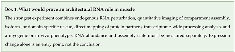
The key experimental principle is separation of RNA abundance from assembly state. For NEAT1, an ideal experiment would deplete endogenous Neat1, rescue NEAT1_1 or NEAT1_2 at matched abundance, and compare wild-type NEAT1_2 with a domain mutant that retains stability but cannot organize paraspeckles. Quantitative imaging would measure molecule number, paraspeckle number, size, and exchange. RNA sequencing and retention assays would define processing outputs, while differentiation, regeneration, and force would provide muscle endpoints. A phenotype rescued only by assembly-competent NEAT1_2 would support architectural causality.
For MALAT1, the decisive design must be temporal. Rapid degradation and reversible rescue would minimize developmental compensation. Density-matched myoblasts would be sampled before cell-cycle exit, during commitment, and during fusion. Speckle morphology, SR-protein microphase partitioning, phosphorylation, nascent transcription, chromatin occupancy, and splicing would be measured in the same cells or matched aliquots. If a processing phenotype follows altered protein partitioning while chromatin occupancy remains stable, the speckle route gains support; the converse result favors a chromatin mechanism.
For repeat RNA, counting endogenous molecules is essential. RNA FISH protocols and expanded-repeat reverse transcription require calibration because probe accessibility and reverse-transcriptase processivity can bias the inferred abundance [98,99,100]. Live-cell imaging should distinguish individual RNAs, transcription-site bursts, and multimeric foci. Material-property perturbations must be separated from global RNA depletion or stress. The most informative endpoint is functional MBNL availability linked to a processing signature and muscle phenotype.
Model selection should follow the claim. C2C12 cells are useful for controlled differentiation and imaging but do not reproduce the multinucleated architecture, loading, innervation, or aging of adult human muscle. Primary patient myoblasts preserve disease alleles but vary in repeat length, passage, epigenetic history, and differentiation capacity. Mouse models enable regeneration and force measurements but can differ in repeat context, transcript level, and tissue distribution. Human biopsy establishes clinical relevance yet offers limited perturbation and spatial sampling. Strong conclusions use these models as complementary tests without treating one as a universal standard.
Negative results should be equally explicit. Failure to detect a compartment change is informative only if the assay could resolve the expected magnitude, measured the relevant isoform, and included the proposed transition in the observation window. Failure to rescue can reflect incorrect mechanism, inadequate expression, or a rescue construct that lacks proper processing or localization. Predefined positive controls, blinded imaging, and reporting of detection limits are necessary before absence is interpreted as biological dispensability.
8.2. Contested Claims and Decisive Experiments
Table 4 links each contested claim to a discriminating experiment and a result that would weaken the proposed mechanism.
These experiments are falsifiable. If NEAT1_2-specific assembly rescue does not restore the muscle phenotype, paraspeckle architecture is unlikely to be the required causal layer. If MALAT1 timing does not alter the sign of the differentiation phenotype under matched conditions, temporal state cannot explain the published contradiction. If endogenous DM1 RNAs remain predominantly single molecules across repeat lengths and transcription rates, a bulk condensation model would have limited relevance to sequestration. If molecular correction consistently fails to improve a mature muscle endpoint despite adequate exposure and duration, researchers must address downstream tissue damage or parallel mechanisms.
The broader repeat-expansion literature identifies common principles involving repeat structure, RNA-binding-protein redistribution, and multiple downstream processing defects, while emphasizing the disease-specific effects of sequence, genomic locus, tissue, and developmental context [101,102]. Therapeutic reviews similarly document a wide portfolio of modalities without resolving which combination will provide durable multisystem benefit [103]. DM1 and DM2 remain clinically and molecularly heterogeneous diseases and cannot be reduced to a single focus-formation disorder.
9. Conclusions
Nuclear noncoding RNAs influence skeletal muscle through several spatial mechanisms that should not be reduced to a single condensate model. NEAT1 links differentiation to paraspeckle association, chromatin regulation, and PABPN1/MATR3-dependent RNA processing, but isoform-specific architectural causality in muscle remains to be shown. MALAT1 regulates nuclear processes and muscle state without being required for gross speckle formation, and its opposing myogenesis phenotypes require a controlled temporal comparison. FRG2 transcripts connect a nucleolar RNA to rDNA transcription and translational capacity in FSHD cells. Expanded DMPK and CNBP RNAs produce a well-supported toxic-RNA and spliceopathy axis, yet individual retained transcripts can be pathogenic without forming bulk condensates.
The most useful unifying principle is therefore causal layering. Spatial localization identifies where a mechanism may occur. Perturbation and rescue determine whether an RNA or assembly is required. In vivo and patient evidence establish physiological relevance. Target engagement plus molecular and tissue rescue define translational progress. Applying these layers consistently will clarify which nuclear structures organize normal muscle RNA processing, which assemblies reflect pathology, and which intervention points are sufficiently supported for therapy.
Author Contributions
Conceptualization, W.L.; investigation, T.H.P.N. and W.L.; writing—original draft preparation, W.L.; writing—review and editing, W.L.; visualization, T.H.P.N. and W.L.; funding acquisition, W.L. All authors have read and agreed to the published version of the manuscript.
Funding
This study was supported by the National Research Foundation of Korea (NRF), which is funded by the Korean government (grant no. RS-2026-25480711).
Institutional Review Board Statement
Not applicable.
Informed Consent Statement
Not applicable.
Data Availability Statement
No new datasets were generated in this study. All information evaluated in this review is available in the cited publications and public clinical trial registries.
Acknowledgments
During the preparation of this manuscript, the authors used OpenAI ChatGPT (5.6 Sol, OpenAI, San Francisco, CA, USA; accessed 1 September 2026) to generate conceptual artwork for Figures 1–3 and to assist with language and consistency checks. The authors subsequently reviewed and modified each figure in Microsoft PowerPoint, verified all scientific content against the cited evidence, and take full responsibility for the content of this publication.
Conflicts of Interest
The authors declare no conflict of interest. The funder had no role in the design of the review; in the collection, analysis, or interpretation of the literature; in the writing of the manuscript; or in the decision to publish the results.
Abbreviations
The following abbreviations are used in this manuscript:
| AAV | adeno-associated virus |
| ASO | antisense oligonucleotide |
| CELF | CUGBP Elav-like family |
| ChIRP | chromatin isolation by RNA purification |
| CNBP | CCHC-type zinc finger nucleic acid-binding protein |
| CRISPR | clustered regularly interspaced short palindromic repeats |
| DM1 | myotonic dystrophy type 1 |
| DM2 | myotonic dystrophy type 2 |
| DMPK | dystrophia myotonica protein kinase |
| FISH | fluorescence in situ hybridization |
| FSHD | facioscapulohumeral muscular dystrophy |
| HDAC | histone deacetylase |
| lncRNA | long noncoding RNA |
| MBNL | muscleblind-like |
| NEAT1 | nuclear paraspeckle assembly transcript 1 |
| NONO | non-POU domain-containing octamer-binding protein |
| PABPN1 | poly(A)-binding protein nuclear 1 |
| PRC2 | polycomb repressive complex 2 |
| PSPC1 | paraspeckle component 1 |
| RBP | RNA-binding protein |
| rDNA | ribosomal DNA |
| RNP | ribonucleoprotein |
| SFPQ | splicing factor proline- and glutamine-rich |
| SR | serine/arginine-rich |
| TfR1 | transferrin receptor 1 |
References
- Shi, D.L.; Grifone, R. RNA-Binding Proteins in the Post-transcriptional Control of Skeletal Muscle Development, Regeneration and Disease. Front Cell Dev. Biol. 2021, 9, 738978. [Google Scholar] [CrossRef]
- Mao, Y.S.; Zhang, B.; Spector, D.L. Biogenesis and function of nuclear bodies. Trends Genet 2011, 27, 295–306. [Google Scholar] [CrossRef]
- Naganuma, T.; Nakagawa, S.; Tanigawa, A.; Sasaki, Y.F.; Goshima, N.; Hirose, T. Alternative 3'-end processing of long noncoding RNA initiates construction of nuclear paraspeckles. EMBO J. 2012, 31, 4020–34. [Google Scholar] [CrossRef]
- Sunwoo, H.; Dinger, M.E.; Wilusz, J.E.; Amaral, P.P.; Mattick, J.S.; Spector, D.L. MEN epsilon/beta nuclear-retained non-coding RNAs are up-regulated upon muscle differentiation and are essential components of paraspeckles. Genome Res. 2009, 19, 347–59. [Google Scholar] [CrossRef]
- Tripathi, V.; Ellis, J.D.; Shen, Z.; Song, D.Y.; Pan, Q.; Watt, A.T.; Freier, S.M.; Bennett, C.F.; Sharma, A.; Bubulya, P.A.; et al. The nuclear-retained noncoding RNA MALAT1 regulates alternative splicing by modulating SR splicing factor phosphorylation. Mol. Cell 2010, 39, 925–38. [Google Scholar] [CrossRef]
- Nakagawa, S.; Ip, J.Y.; Shioi, G.; Tripathi, V.; Zong, X.; Hirose, T.; Prasanth, K.V. Malat1 is not an essential component of nuclear speckles in mice. RNA 2012, 18, 1487–99. [Google Scholar] [CrossRef]
- Miller, J.W.; Urbinati, C.R.; Teng-Umnuay, P.; Stenberg, M.G.; Byrne, B.J.; Thornton, C.A.; Swanson, M.S. Recruitment of human muscleblind proteins to (CUG)(n) expansions associated with myotonic dystrophy. EMBO J. 2000, 19, 4439–48. [Google Scholar] [CrossRef]
- Perdoni, F.; Malatesta, M.; Cardani, R.; Giagnacovo, M.; Mancinelli, E.; Meola, G.; Pellicciari, C. RNA/MBNL1-containing foci in myoblast nuclei from patients affected by myotonic dystrophy type 2: an immunocytochemical study. Eur. J. Histochem 2009, 53, e18. [Google Scholar] [CrossRef]
- Alberti, S.; Gladfelter, A.; Mittag, T. Considerations and Challenges in Studying Liquid-Liquid Phase Separation and Biomolecular Condensates. Cell 2019, 176, 419–434. [Google Scholar] [CrossRef]
- Yamazaki, T.; Souquere, S.; Chujo, T.; Kobelke, S.; Chong, Y.S.; Fox, A.H.; Bond, C.S.; Nakagawa, S.; Pierron, G.; Hirose, T. Functional Domains of NEAT1 Architectural lncRNA Induce Paraspeckle Assembly through Phase Separation. Mol. Cell 2018, 70, 1038–1053.e7. [Google Scholar] [CrossRef]
- Yamazaki, T.; Yamamoto, T.; Yoshino, H.; Souquere, S.; Nakagawa, S.; Pierron, G.; Hirose, T. Paraspeckles are constructed as block copolymer micelles. EMBO J. 2021, 40, e107270. [Google Scholar] [CrossRef]
- Yamamoto, T.; Yamazaki, T.; Hirose, T. Triblock copolymer micelle model of spherical paraspeckles. Front Mol. Biosci. 2022, 9, 925058. [Google Scholar] [CrossRef]
- Petrany, M.J.; Swoboda, C.O.; Sun, C.; Chetal, K.; Chen, X.; Weirauch, M.T.; Salomonis, N.; Millay, D.P. Single-nucleus RNA-seq identifies transcriptional heterogeneity in multinucleated skeletal myofibers. Nat. Commun. 2020, 11, 6374. [Google Scholar] [CrossRef]
- Kim, M.; Franke, V.; Brandt, B.; Lowenstein, E.D.; Schöwel, V.; Spuler, S.; Akalin, A.; Birchmeier, C. Single-nucleus transcriptomics reveals functional compartmentalization in syncytial skeletal muscle cells. Nat. Commun. 2020, 11, 6375. [Google Scholar] [CrossRef]
- Li, R.; Harvey, A.R.; Hodgetts, S.I.; Fox, A.H. Functional dissection of NEAT1 using genome editing reveals substantial localization of the NEAT1_1 isoform outside paraspeckles. RNA 2017, 23, 872–881. [Google Scholar] [CrossRef]
- Adriaens, C.; Rambow, F.; Bervoets, G.; Silla, T.; Mito, M.; Chiba, T.; Asahara, H.; Hirose, T.; Nakagawa, S.; Jensen, T.H.; et al. The long noncoding RNA NEAT1_1 is seemingly dispensable for normal tissue homeostasis and cancer cell growth. RNA 2019, 25, 1681–1695. [Google Scholar] [CrossRef]
- Chujo, T.; Yamazaki, T.; Kawaguchi, T.; Kurosaka, S.; Takumi, T.; Nakagawa, S.; Hirose, T. Unusual semi-extractability as a hallmark of nuclear body-associated architectural noncoding RNAs. EMBO J. 2017, 36, 1447–1462. [Google Scholar] [CrossRef]
- Fei, J.; Jadaliha, M.; Harmon, T.S.; Li, I.T.S.; Hua, B.; Hao, Q.; Holehouse, A.S.; Reyer, M.; Sun, Q.; Freier, S.M.; et al. Quantitative analysis of multilayer organization of proteins and RNA in nuclear speckles at super resolution. J. Cell Sci. 2017, 130, 4180–4192. [Google Scholar] [CrossRef]
- Zhang, B.; Arun, G.; Mao, Y.S.; Lazar, Z.; Hung, G.; Bhattacharjee, G.; Xiao, X.; Booth, C.J.; Wu, J.; Zhang, C.; et al. The lncRNA Malat1 is dispensable for mouse development but its transcription plays a cis-regulatory role in the adult. Cell Rep. 2012, 2, 111–23. [Google Scholar] [CrossRef]
- Eißmann, M.; Gutschner, T.; Hämmerle, M.; Günther, S.; Caudron-Herger, M.; Groß, M.; Schirmacher, P.; Rippe, K.; Braun, T.; Zörnig, M.; et al. Loss of the abundant nuclear non-coding RNA MALAT1 is compatible with life and development. RNA Biol. 2012, 9, 1076–87. [Google Scholar] [CrossRef]
- Wang, S.; Zuo, H.; Jin, J.; Lv, W.; Xu, Z.; Fan, Y.; Zhang, J.; Zuo, B. Long noncoding RNA Neat1 modulates myogenesis by recruiting Ezh2. Cell Death Dis. 2019, 10, 505. [Google Scholar] [CrossRef]
- Banerjee, A.; Vest, K.E.; Pavlath, G.K.; Corbett, A.H. Nuclear poly(A) binding protein 1 (PABPN1) and Matrin3 interact in muscle cells and regulate RNA processing. Nucleic Acids Res. 2017, 45, 10706–10725. [Google Scholar] [CrossRef]
- Watts, R.; Johnsen, V.L.; Shearer, J.; Hittel, D.S. Myostatin-induced inhibition of the long noncoding RNA Malat1 is associated with decreased myogenesis. Am. J. Physiol. Cell Physiol. 2013, 304, C995–1001. [Google Scholar] [CrossRef]
- Han, X.; Yang, F.; Cao, H.; Liang, Z. Malat1 regulates serum response factor through miR-133 as a competing endogenous RNA in myogenesis. FASEB J. 2015, 29, 3054–64. [Google Scholar] [CrossRef]
- Chen, X.; He, L.; Zhao, Y.; Li, Y.; Zhang, S.; Sun, K.; So, K.; Chen, F.; Zhou, L.; Lu, L.; et al. Malat1 regulates myogenic differentiation and muscle regeneration through modulating MyoD transcriptional activity. Cell Discov. 2017, 3, 17002. [Google Scholar] [CrossRef]
- Salsi, V.; Losi, F.; Fosso, B.; Ferrarini, M.; Pini, S.; Manfredi, M.; Vattemi, G.; Mongini, T.; Maggi, L.; Pesole, G.; et al. Nucleolar FRG2 lncRNAs inhibit rRNA transcription and cytoplasmic translation, linking FSHD to dysregulation of muscle-specific protein synthesis. Nucleic Acids Res. Correction: Nucleic Acids Res. 2026, 54, gkag798. https://doi.org/10.1093/nar/gkag798 (funding statement only).. 2025, 53, gkaf643. [Google Scholar] [CrossRef]
- Kang, X.; Zhao, Y.; Van Arsdell, G.; Nelson, S.F.; Touma, M. Ppp1r1b-lncRNA inhibits PRC2 at myogenic regulatory genes to promote cardiac and skeletal muscle development in mouse and human. RNA 2020, 26, 481–491. [Google Scholar] [CrossRef]
- Kang, X.; Zhao, Y.; Nelson, S.F.; Pyle, A.; Lusis, A.J.; Touma, M. Heterogeneous nuclear ribonucleoprotein A1 (hnRNPA1) maintains muscle progenitor identity by stabilizing the Ppp1r1b-lncRNA-PRC2 complex. Nucleic Acids Res. Correction: Nucleic Acids Res. 2026, 54, gkag912. https://doi.org/10.1093/nar/gkag912 (graphical abstract promoter labels). 2026, 54, gkag497. [Google Scholar] [CrossRef]
- Hitachi, K.; Nakatani, M.; Takasaki, A.; Ouchi, Y.; Uezumi, A.; Ageta, H.; Inagaki, H.; Kurahashi, H.; Tsuchida, K. Myogenin promoter-associated lncRNA Myoparr is essential for myogenic differentiation. EMBO Rep. 2019, 20, e47468. [Google Scholar] [CrossRef]
- Yu, X.; Zhang, Y.; Li, T.; Ma, Z.; Jia, H.; Chen, Q.; Zhao, Y.; Zhai, L.; Zhong, R.; Li, C.; et al. Long non-coding RNA Linc-RAM enhances myogenic differentiation by interacting with MyoD. Nat. Commun. Author correction: Nat Commun 2025, 16, 11151. https://doi.org/10.1038/s41467-025-64348-4 (Figure 2e image replacement; conclusions unchanged). 2017, 8, 14016. [Google Scholar] [CrossRef]
- Wang, L.; Zhao, Y.; Bao, X.; Zhu, X.; Kwok, Y.K.; Sun, K.; Chen, X.; Huang, Y.; Jauch, R.; Esteban, M.A.; et al. LncRNA Dum interacts with Dnmts to regulate Dppa2 expression during myogenic differentiation and muscle regeneration. Cell Res. 2015, 25, 335–50. [Google Scholar] [CrossRef]
- Ouyang, J.P.T.; Shukla, S.; Bensalah, M.; Parker, R. DM1 repeat-expanded RNAs confer RNA toxicity as individual nuclear-retained RNAs. Cell Rep. 2025, 44, 115582. [Google Scholar] [CrossRef]
- Wheeler, T.M.; Leger, A.J.; Pandey, S.K.; MacLeod, A.R.; Nakamori, M.; Cheng, S.H.; Wentworth, B.M.; Bennett, C.F.; Thornton, C.A. Targeting nuclear RNA for in vivo correction of myotonic dystrophy. Nature 2012, 488, 111–5. [Google Scholar] [CrossRef]
- Liquori, C.L.; Ricker, K.; Moseley, M.L.; Jacobsen, J.F.; Kress, W.; Naylor, S.L.; Day, J.W.; Ranum, L.P. Myotonic dystrophy type 2 caused by a CCTG expansion in intron 1 of ZNF9. Science 2001, 293, 864–7. [Google Scholar] [CrossRef]
- Lukáš, Z.; Falk, M.; Feit, J.; Souček, O.; Falková, I.; Štefančíková, L.; Janoušová, E.; Fajkusová, L.; Zaorálková, J.; Hrabálková, R. Sequestration of MBNL1 in tissues of patients with myotonic dystrophy type 2. Neuromuscul. Disord. 2012, 22, 604–16. [Google Scholar] [CrossRef]
- Shinn, M.K.; Tomares, D.T.; Liu, V.; Pant, A.; Qiu, Y.; Vitalis, A.; Song, Y.J.; Ayala, Y.; Ruff, K.M.; Strout, G.W.; et al. Nuclear speckle proteins form intrinsic and MALAT1-dependent microphases. Cell 2026, 189, 832–852.e24. [Google Scholar] [CrossRef]
- Jain, A.; Vale, R.D. RNA phase transitions in repeat expansion disorders. Nature 2017, 546, 243–247. [Google Scholar] [CrossRef]
- Sellier, C.; Cerro-Herreros, E.; Blatter, M.; Freyermuth, F.; Gaucherot, A.; Ruffenach, F.; Sarkar, P.; Puymirat, J.; Udd, B.; Day, J.W.; et al. rbFOX1/MBNL1 competition for CCUG RNA repeats binding contributes to myotonic dystrophy type 1/type 2 differences. Nat. Commun. 2018, 9, 2009. [Google Scholar] [CrossRef]
- Chen, L.L.; Carmichael, G.G. Altered nuclear retention of mRNAs containing inverted repeats in human embryonic stem cells: functional role of a nuclear noncoding RNA. Mol. Cell 2009, 35, 467–78. [Google Scholar] [CrossRef]
- Song, Y.J.; Shinn, M.K.; Bangru, S.; Wang, Y.; Sun, Q.; Hao, Q.; Chaturvedi, P.; Freier, S.M.; Perez-Pinera, P.; Nelson, E.R.; et al. LncRNA-splicing factor condensates regulate hypoxia-responsive pre-mRNA processing near nuclear speckles. Mol. Cell 2026, 86, 1061–1080.e10. [Google Scholar] [CrossRef]
- Cai, B.; Ma, M.; Yuan, R.; Zhou, Z.; Zhang, J.; Kong, S.; Lin, D.; Lian, L.; Li, J.; Zhang, X.; et al. MYH1G-AS is a chromatin-associated lncRNA that regulates skeletal muscle development in chicken. Cell Mol. Biol. Lett. 2024, 29, 9. [Google Scholar] [CrossRef]
- Ishiura, S.; Kino, Y.; Nezu, Y.; Onishi, H.; Ohno, E.; Sasagawa, N. Regulation of splicing by MBNL and CELF family of RNA-binding protein. Acta Myol. 2005, 24, 74–7. [Google Scholar]
- Konieczny, P.; Stepniak-Konieczna, E.; Sobczak, K. MBNL proteins and their target RNAs, interaction and splicing regulation. Nucleic Acids Res. 2014, 42, 10873–87. [Google Scholar] [CrossRef]
- Cox, D.C.; Guan, X.; Xia, Z.; Cooper, T.A. Increased nuclear but not cytoplasmic activities of CELF1 protein leads to muscle wasting. Hum. Mol. Genet 2020, 29, 1729–1744. [Google Scholar] [CrossRef]
- Holt, I.; Mittal, S.; Furling, D.; Butler-Browne, G.S.; Brook, J.D.; Morris, G.E. Defective mRNA in myotonic dystrophy accumulates at the periphery of nuclear splicing speckles. Genes Cells 2007, 12, 1035–48. [Google Scholar] [CrossRef]
- Wang, E.T.; Treacy, D.; Eichinger, K.; Struck, A.; Estabrook, J.; Olafson, H.; Wang, T.T.; Bhatt, K.; Westbrook, T.; Sedehizadeh, S.; et al. Transcriptome alterations in myotonic dystrophy skeletal muscle and heart. Hum. Mol. Genet 2019, 28, 1312–1321. [Google Scholar] [CrossRef]
- Thomas, J.D.; Sznajder, Ł.J.; Bardhi, O.; Aslam, F.N.; Anastasiadis, Z.P.; Scotti, M.M.; Nishino, I.; Nakamori, M.; Wang, E.T.; Swanson, M.S. Disrupted prenatal RNA processing and myogenesis in congenital myotonic dystrophy. Genes Dev. 2017, 31, 1122–1133. [Google Scholar] [CrossRef]
- Kim, E.Y.; Barefield, D.Y.; Vo, A.H.; Gacita, A.M.; Schuster, E.J.; Wyatt, E.J.; Davis, J.L.; Dong, B.; Sun, C.; Page, P.; et al. Distinct pathological signatures in human cellular models of myotonic dystrophy subtypes. JCI Insight 2019, 4, e122686. [Google Scholar] [CrossRef]
- Tang, Z.Z.; Yarotskyy, V.; Wei, L.; Sobczak, K.; Nakamori, M.; Eichinger, K.; Moxley, R.T.; Dirksen, R.T.; Thornton, C.A. Muscle weakness in myotonic dystrophy associated with misregulated splicing and altered gating of Ca(V)1.1 calcium channel. Hum. Mol. Genet 2012, 21, 1312–24. [Google Scholar] [CrossRef]
- Lee, K.Y.; Seah, C.; Li, C.; Chen, Y.F.; Chen, C.Y.; Wu, C.I.; Liao, P.C.; Shyu, Y.C.; Olafson, H.R.; McKee, K.K.; et al. Mice lacking MBNL1 and MBNL2 exhibit sudden cardiac death and molecular signatures recapitulating myotonic dystrophy. Hum. Mol. Genet 2022, 31, 3144–3160. [Google Scholar] [CrossRef]
- Hinman, M.N.; Richardson, J.I.; Sockol, R.A.; Aronson, E.D.; Stednitz, S.J.; Murray, K.N.; Berglund, J.A.; Guillemin, K. Zebrafish mbnl mutants model physical and molecular phenotypes of myotonic dystrophy. Dis. Model Mech. 2021, 14, dmm045773. [Google Scholar] [CrossRef]
- Hartman, J.M.; Ikegami, K.; Provenzano, M.; Bates, K.; Butler, A.; Jones, A.S.; Berggren, K.N.; Dekdebrun, J.; McKay, M.J.; Baldwin, J.N.; et al. RNA mis-splicing in children with congenital myotonic dystrophy is associated with physical function. Ann. Clin. Transl. Neurol. 2024, 11, 3175–3191. [Google Scholar] [CrossRef]
- Raaijmakers, R.H.L.; Ausems, C.R.M.; Willemse, M.; Cumming, S.A.; van Engelen, B.G.M.; Monckton, D.G.; van Bokhoven, H.; Wansink, D.G. Ameliorated cellular hallmarks of myotonic dystrophy in hybrid myotubes from patient and unaffected donor cells. Stem Cell Res. Ther. 2024, 15, 302. [Google Scholar] [CrossRef]
- Ripken, L.; van den Broek, W.J.A.A.; van Cruchten, R.T.P.; Smits, J.G.A.; Riepe, T.V.; 't Hoen, P.A.C.; Wansink, D.G. Commitment to Myogenic Differentiation Significantly Aggravates the RNA Phenotype in Myotonic Dystrophy Type 1. Neuropathol. Appl. Neurobiol. 2026, 52, e70069. [Google Scholar] [CrossRef]
- Yadava, R.S.; Mandal, M.; Mahadevan, M.S. Studying the Effect of MBNL1 and MBNL2 Loss in Skeletal Muscle Regeneration. Int. J. Mol. Sci. 2024, 25, 2687. [Google Scholar] [CrossRef]
- Yadava, R.S.; Mandal, M.; Giese, J.M.; Rigo, F.; Bennett, C.F.; Mahadevan, M.S. Modeling muscle regeneration in RNA toxicity mice. Hum. Mol. Genet 2021, 30, 1111–1130. [Google Scholar] [CrossRef]
- Nakamori, M.; Hamanaka, K.; Thomas, J.D.; Wang, E.T.; Hayashi, Y.K.; Takahashi, M.P.; Swanson, M.S.; Nishino, I.; Mochizuki, H. Aberrant Myokine Signaling in Congenital Myotonic Dystrophy. Cell Rep. 2017, 21, 1240–1252. [Google Scholar] [CrossRef]
- Wheeler, T.M.; Krym, M.C.; Thornton, C.A. Ribonuclear foci at the neuromuscular junction in myotonic dystrophy type 1. Neuromuscul. Disord. 2007, 17, 242–7. [Google Scholar] [CrossRef]
- Michel, L.; Huguet-Lachon, A.; Gourdon, G. Sense and Antisense DMPK RNA Foci Accumulate in DM1 Tissues during Development. PLoS ONE 2015, 10, e0137620. [Google Scholar] [CrossRef]
- Wang, Y.; Hao, L.; Li, H.; Cleary, J.D.; Tomac, M.P.; Thapa, A.; Guo, X.; Zeng, D.; Wang, H.; McRae, M.; et al. Abnormal nuclear aggregation and myotube degeneration in myotonic dystrophy type 1. Neurol. Sci. 2019, 40, 1255–1265. [Google Scholar] [CrossRef]
- Kleefeld, F.; Horvath, R.; Pinal-Fernandez, I.; Mammen, A.L.; Casal-Dominguez, M.; Hathazi, D.; Melchert, S.; Hahn, K.; Sickmann, A.; Muselmann-Genschow, C.; et al. Multi-level profiling unravels mitochondrial dysfunction in myotonic dystrophy type 2. Acta Neuropathol. 2024, 147, 19. [Google Scholar] [CrossRef]
- Ursu, S.F.; Alekov, A.; Mao, N.H.; Jurkat-Rott, K. ClC1 chloride channel in myotonic dystrophy type 2 and ClC1 splicing in vitro. Acta Myol. 2012, 31, 144–53. [Google Scholar]
- Schneider-Gold, C.; Timchenko, L.T. CCUG repeats reduce the rate of global protein synthesis in myotonic dystrophy type 2. Rev. Neurosci. 2010, 21, 19–28. [Google Scholar] [CrossRef]
- Zu, T.; Cleary, J.D.; Liu, Y.; Bañez-Coronel, M.; Bubenik, J.L.; Ayhan, F.; Ashizawa, T.; Xia, G.; Clark, H.B.; Yachnis, A.T.; et al. RAN Translation Regulated by Muscleblind Proteins in Myotonic Dystrophy Type 2. Neuron 2017, 95, 1292–1305.e5. [Google Scholar] [CrossRef]
- Yenigun, V.B.; Sirito, M.; Amcheslavky, A.; Czernuszewicz, T.; Colonques-Bellmunt, J.; García-Alcover, I.; Wojciechowska, M.; Bolduc, C.; Chen, Z.; López Castel, A.; et al. (CCUG)n RNA toxicity in a Drosophila model of myotonic dystrophy type 2 (DM2) activates apoptosis. Dis. Model Mech. 2017, 10, 993–1003. [Google Scholar] [CrossRef]
- Thornton, C.A.; Moxley, R.T.; Eichinger, K.; Heatwole, C.; Mignon, L.; Arnold, W.D.; Ashizawa, T.; Day, J.W.; Dent, G.; Tanner, M.K.; et al. Antisense oligonucleotide targeting DMPK in patients with myotonic dystrophy type 1: a multicentre, randomised, dose-escalation, placebo-controlled, phase 1/2a trial. Lancet Neurol. 2023, 22, 218–228. [Google Scholar] [CrossRef]
- Johnson, N.E.; Tai, L.J.; Hamel, J.I.; Day, J.W.; Statland, J.M.; Soltanzadeh, P.; Subramony, S.H.; Thornton, C.A.; Arnold, W.D.; Wicklund, M.; et al. An Antibody-Oligonucleotide Conjugate for Myotonic Dystrophy Type 1. N Engl. J. Med. 2026, 394, 763–772. [Google Scholar] [CrossRef]
- Kwan, T.T.; Meng, Q.; Delos Santos, N.; Johnson, N.E.; Thornton, C.A.; Malecova, B.; Burke, R.; Tai, L.J.; Levin, A.A.; Younis, H.S.; et al. Delpacibart etedesiran improves the molecular pathology of myotonic dystrophy type 1 in the phase 1/2 MARINA study. Mol. Ther. 2026, 34, 3872–3884. [Google Scholar] [CrossRef]
- Novartis. Novartis Provides Update on Delpacibart Etedesiran (Del-desiran) Phase III HARBOR Study for the Treatment of Myotonic Dystrophy Type 1 (DM1). 2026. Available online: https://www.novartis.com/news/media-releases/novartis-provides-update-delpacibart-etedesiran-del-desiran-phase-iii-harbor-study-treatment-myotonic-dystrophy-type-1-dm1 (accessed on 30 September 2026).
- Tanner, M.K.; Tang, Z.; Thornton, C.A. Targeted splice sequencing reveals RNA toxicity and therapeutic response in myotonic dystrophy. Nucleic Acids Res. 2021, 49, 2240–2254. [Google Scholar] [CrossRef]
- Batra, R.; Nelles, D.A.; Pirie, E.; Blue, S.M.; Marina, R.J.; Wang, H.; Chaim, I.A.; Thomas, J.D.; Zhang, N.; Nguyen, V.; et al. Elimination of Toxic Microsatellite Repeat Expansion RNA by RNA-Targeting Cas9. Cell 2017, 170, 899–912.e10. [Google Scholar] [CrossRef]
- Pinto, B.S.; Saxena, T.; Oliveira, R.; Méndez-Gómez, H.R.; Cleary, J.D.; Denes, L.T.; McConnell, O.; Arboleda, J.; Xia, G.; Swanson, M.S.; et al. Impeding Transcription of Expanded Microsatellite Repeats by Deactivated Cas9. Mol. Cell 2017, 68, 479–490.e5. [Google Scholar] [CrossRef]
- Cardinali, B.; Provenzano, C.; Izzo, M.; Voellenkle, C.; Battistini, J.; Strimpakos, G.; Golini, E.; Mandillo, S.; Scavizzi, F.; Raspa, M.; et al. Time-controlled and muscle-specific CRISPR/Cas9-mediated deletion of CTG-repeat expansion in the DMPK gene. Mol. Ther. Nucleic Acids 2022, 27, 184–199. [Google Scholar] [CrossRef]
- Wang, Y.; Hao, L.; Wang, H.; Santostefano, K.; Thapa, A.; Cleary, J.; Li, H.; Guo, X.; Terada, N.; Ashizawa, T.; et al. Therapeutic Genome Editing for Myotonic Dystrophy Type 1 Using CRISPR/Cas9. Mol. Ther. 2018, 26, 2617–2630. [Google Scholar] [CrossRef]
- Gibaut, Q.M.R.; Bush, J.A.; Tong, Y.; Baisden, J.T.; Taghavi, A.; Olafson, H.; Yao, X.; Childs-Disney, J.L.; Wang, E.T.; Disney, M.D. Transcriptome-Wide Studies of RNA-Targeted Small Molecules Provide a Simple and Selective r(CUG)exp Degrader in Myotonic Dystrophy. ACS Cent. Sci. 2023, 9, 1342–1353. [Google Scholar] [CrossRef]
- Angelbello, A.J.; Rzuczek, S.G.; Mckee, K.K.; Chen, J.L.; Olafson, H.; Cameron, M.D.; Moss, W.N.; Wang, E.T.; Disney, M.D. Precise small-molecule cleavage of an r(CUG) repeat expansion in a myotonic dystrophy mouse model. Proc. Natl. Acad. Sci. U S A 2019, 116, 7799–7804. [Google Scholar] [CrossRef]
- Benhamou, R.I.; Angelbello, A.J.; Andrews, R.J.; Wang, E.T.; Moss, W.N.; Disney, M.D. Structure-Specific Cleavage of an RNA Repeat Expansion with a Dimeric Small Molecule Is Advantageous over Sequence-Specific Recognition by an Oligonucleotide. ACS Chem. Biol. 2020, 15, 485–493. [Google Scholar] [CrossRef]
- Nakamori, M.; Taylor, K.; Mochizuki, H.; Sobczak, K.; Takahashi, M.P. Oral administration of erythromycin decreases RNA toxicity in myotonic dystrophy. Ann. Clin. Transl. Neurol. 2016, 3, 42–54. [Google Scholar] [CrossRef]
- Jenquin, J.R.; Coonrod, L.A.; Silverglate, Q.A.; Pellitier, N.A.; Hale, M.A.; Xia, G.; Nakamori, M.; Berglund, J.A. Furamidine Rescues Myotonic Dystrophy Type I Associated Mis-Splicing through Multiple Mechanisms. ACS Chem. Biol. 2018, 13, 2708–2718. [Google Scholar] [CrossRef]
- Mishra, S.K.; Hicks, S.M.; Frias, J.A.; Zhang, J.; Handley, M.T.; Vangaveti, S.; Reid, T.S.; Pandey, A.; McMichael, O.; Crumbaugh, C.; et al. Identification of enzymatically modified isoquercitrin as a therapeutic lead for myotonic dystrophy type 1. NAR Mol. Med. 2026, 3, ugag007. [Google Scholar] [CrossRef]
- Frias, J.A.; Hicks, S.M.; Mazdiyasni, H.; Mishra, S.K.; Sarkar, K.; Yeboah, C.; LeFever, N.M.; Scotti, M.M.; Zeghal, H.; Brandt, N.; et al. Modified Polycyclic Compounds Rescue Mis-splicing in Myotonic Dystrophy Type 1 Disease Models. ACS Chem. Biol. 2026, 21, 502–518. [Google Scholar] [CrossRef]
- Imai, T.; Miyai, M.; Nemoto, J.; Tamai, T.; Ohta, M.; Yagi, Y.; Nakanishi, O.; Mochizuki, H.; Nakamori, M. Pentatricopeptide repeat protein targeting CUG repeat RNA ameliorates RNA toxicity in a myotonic dystrophy type 1 mouse model. Sci. Transl. Med. 2025, 17, eadq2005. [Google Scholar] [CrossRef]
- Jones, K.; Wei, C.; Schoser, B.; Meola, G.; Timchenko, N.; Timchenko, L. Reduction of toxic RNAs in myotonic dystrophies type 1 and type 2 by the RNA helicase p68/DDX5. Proc. Natl. Acad. Sci. U S A 2015, 112, 8041–5. [Google Scholar] [CrossRef]
- Nguyen, Q.; Yokota, T. Degradation of Toxic RNA in Myotonic Dystrophy Using Gapmer Antisense Oligonucleotides. Methods Mol. Biol. 2020, 2176, 99–109. [Google Scholar] [CrossRef]
- Stepniak-Konieczna, E.; Konieczny, P.; Cywoniuk, P.; Dluzewska, J.; Sobczak, K. AON-induced splice-switching and DMPK pre-mRNA degradation as potential therapeutic approaches for Myotonic Dystrophy type 1. Nucleic Acids Res. 2020, 48, 2531–2543. [Google Scholar] [CrossRef]
- Sobczak, K.; Wheeler, T.M.; Wang, W.; Thornton, C.A. RNA interference targeting CUG repeats in a mouse model of myotonic dystrophy. Mol. Ther. 2013, 21, 380–7. [Google Scholar] [CrossRef]
- Almeida, C.F.; Robriquet, F.; Vetter, T.A.; Huang, N.; Neinast, R.; Hernandez-Rosario, L.; Rajakumar, D.; Arnold, W.D.; McBride, K.L.; Flanigan, K.M.; et al. Promising AAV.U7snRNAs vectors targeting DMPK improve DM1 hallmarks in patient-derived cell lines. Front Cell Dev. Biol. 2023, 11, 1181040. [Google Scholar] [CrossRef]
- Cerro-Herreros, E.; González-Martínez, I.; Moreno-Cervera, N.; Overby, S.; Pérez-Alonso, M.; Llamusí, B.; Artero, R. Therapeutic Potential of AntagomiR-23b for Treating Myotonic Dystrophy. Mol. Ther. Nucleic Acids 2020, 21, 837–849. [Google Scholar] [CrossRef]
- Cerro-Herreros, E.; González-Martínez, I.; Moreno, N.; Espinosa-Espinosa, J.; Fernández-Costa, J.M.; Colom-Rodrigo, A.; Overby, S.J.; Seoane-Miraz, D.; Poyatos-García, J.; Vilchez, J.J.; et al. Preclinical characterization of antagomiR-218 as a potential treatment for myotonic dystrophy. Mol. Ther. Nucleic Acids 2021, 26, 174–191. [Google Scholar] [CrossRef]
- González-Martínez, I.; Cerro-Herreros, E.; Moreno, N.; García-Rey, A.; Espinosa-Espinosa, J.; Carrascosa-Sàez, M.; Piqueras-Losilla, D.; Arzumanov, A.; Seoane-Miraz, D.; Jad, Y.; et al. Peptide-conjugated antimiRs improve myotonic dystrophy type 1 phenotypes by promoting endogenous MBNL1 expression. Mol. Ther. Nucleic Acids 2023, 34, 102024. [Google Scholar] [CrossRef]
- Cerro-Herreros, E.; Núñez-Manchón, J.; Naldaiz-Gastesi, N.; Carrascosa-Sàez, M.; García-Rey, A.; Losilla, D.P.; González-Martínez, I.; Espinosa-Espinosa, J.; Moreno, K.; Poyatos-García, J.; et al. AntimiR treatment corrects myotonic dystrophy primary cell defects across several CTG repeat expansions with a dual mechanism of action. Sci. Adv. 2024, 10, eadn6525. [Google Scholar] [CrossRef]
- Hu, N.; Kim, E.; Antoury, L.; Li, J.; González-Pérez, P.; Rutkove, S.B.; Wheeler, T.M. Antisense oligonucleotide and adjuvant exercise therapy reverse fatigue in old mice with myotonic dystrophy. Mol. Ther. Nucleic Acids 2021, 23, 393–405. [Google Scholar] [CrossRef]
- Neault, N.; Ravel-Chapuis, A.; Baird, S.D.; Lunde, J.A.; Poirier, M.; Staykov, E.; Plaza-Diaz, J.; Medina, G.; Abadía-Molina, F.; Jasmin, B.J.; et al. Vorinostat Improves Myotonic Dystrophy Type 1 Splicing Abnormalities in DM1 Muscle Cell Lines and Skeletal Muscle from a DM1 Mouse Model. Int. J. Mol. Sci. 2023, 24, 3794. [Google Scholar] [CrossRef]
- Lutz, M.; Levanti, M.; Karns, R.; Gourdon, G.; Lindquist, D.; Timchenko, N.A.; Timchenko, L. Therapeutic Targeting of the GSK3β-CUGBP1 Pathway in Myotonic Dystrophy. Int. J. Mol. Sci. 2023, 24, 10650. [Google Scholar] [CrossRef]
- Hale, M.A.; Richardson, J.I.; Day, R.C.; McConnell, O.L.; Arboleda, J.; Wang, E.T.; Berglund, J.A. An engineered RNA binding protein with improved splicing regulation. Nucleic Acids Res. 2018, 46, 3152–3168. [Google Scholar] [CrossRef]
- Chamberlain, C.M.; Ranum, L.P. Mouse model of muscleblind-like 1 overexpression: skeletal muscle effects and therapeutic promise. Hum. Mol. Genet 2012, 21, 4645–54. [Google Scholar] [CrossRef]
- Hu, R.C.; Zhang, Y.; Nitschke, L.; Johnson, S.J.; Hurley, A.E.; Lagor, W.R.; Xia, Z.; Cooper, T.A. MBNL overexpression rescues cardiac phenotypes in a myotonic dystrophy type 1 heart mouse model. J. Clin. Invest 2025, 135, e186416. [Google Scholar] [CrossRef]
- Wojciechowska, M.; Sobczak, K.; Kozlowski, P.; Sedehizadeh, S.; Wojtkowiak-Szlachcic, A.; Czubak, K.; Markus, R.; Lusakowska, A.; Kaminska, A.; Brook, J.D. Quantitative Methods to Monitor RNA Biomarkers in Myotonic Dystrophy. Sci. Rep. 2018, 8, 5885. [Google Scholar] [CrossRef]
- Urbanek, M.O.; Krzyzosiak, W.J. RNA FISH for detecting expanded repeats in human diseases. Methods 2016, 98, 115–123. [Google Scholar] [CrossRef]
- Carrell, S.T.; Tang, Z.; Mohr, S.; Lambowitz, A.M.; Thornton, C.A. Detection of expanded RNA repeats using thermostable group II intron reverse transcriptase. Nucleic Acids Res. 2018, 46, e1. [Google Scholar] [CrossRef]
- Sznajder, Ł.J.; Swanson, M.S. Short Tandem Repeat Expansions and RNA-Mediated Pathogenesis in Myotonic Dystrophy. Int. J. Mol. Sci. 2019, 20, 3365. [Google Scholar] [CrossRef]
- Fujino, Y.; Nagai, Y. The molecular pathogenesis of repeat expansion diseases. Biochem Soc. Trans. 2022, 50, 119–134. [Google Scholar] [CrossRef]
- Timchenko, L. Development of Therapeutic Approaches for Myotonic Dystrophies Type 1 and Type 2. Int. J. Mol. Sci. 2022, 23, 10491. [Google Scholar] [CrossRef]
Figure 1.
Nuclear RNA organization across myogenic state transitions. The upper sequence depicts a proliferating myoblast, a differentiating myocyte, and a multinucleated myotube. Teal meshes indicate NEAT1-associated paraspeckles containing NONO and SFPQ. Their increase during differentiation reflects direct C2C12 localization evidence but does not establish assembly causality for myogenesis. Magenta domains denote MALAT1-enriched nuclear speckles containing SR proteins. Their equivalent representation across stages avoids assigning a uniform direction to the context-dependent MALAT1 phenotype. Navy strands denote chromatin-linked lncRNAs and do not represent a named nuclear body. Variation among myotube nuclei is putative and remains unresolved. The lower inset summarizes the FRG2A/B nucleolar module in FSHD patient-derived muscle cells. FRG2A/B depletion corrected reduced nascent rRNA production and nascent protein synthesis, whereas in vivo rescue remains untested. Symbols indicate localization, not material state. The FSHD module is not part of the normal myogenic trajectory.
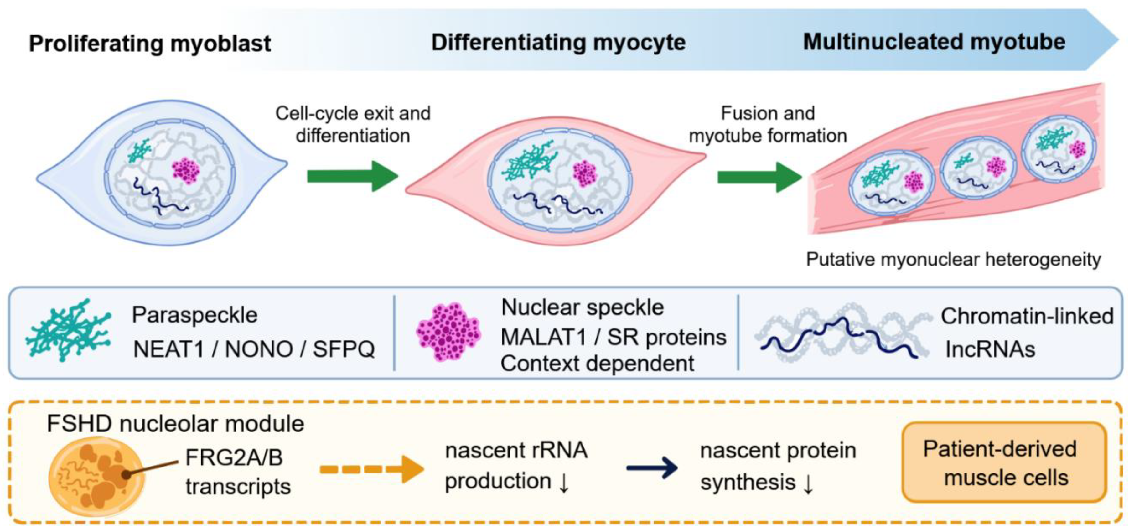
Figure 2.
Functional nuclear compartments and pathological repeat-RNA assemblies. The three panels compare distinct spatial mechanisms. In paraspeckles, NEAT1_2 provides an architectural scaffold that recruits NONO and SFPQ, although direct assembly causality remains incompletely established in skeletal muscle. In nuclear speckles, MALAT1 is enriched with SR proteins and may influence their regulatory partitioning, but gross speckles persist after MALAT1 loss. Expanded DMPK CUG and CNBP CCUG RNAs form retained
ribonucleoproteins and RNA foci that reduce functional MBNL availability and drive spliceopathy. Calibrated imaging in DM1 indicates that most apparent foci represent individual retained transcripts, whereas multimeric foci are less frequent. The drawing is schematic and does not imply a shared assembly mechanism or material state.
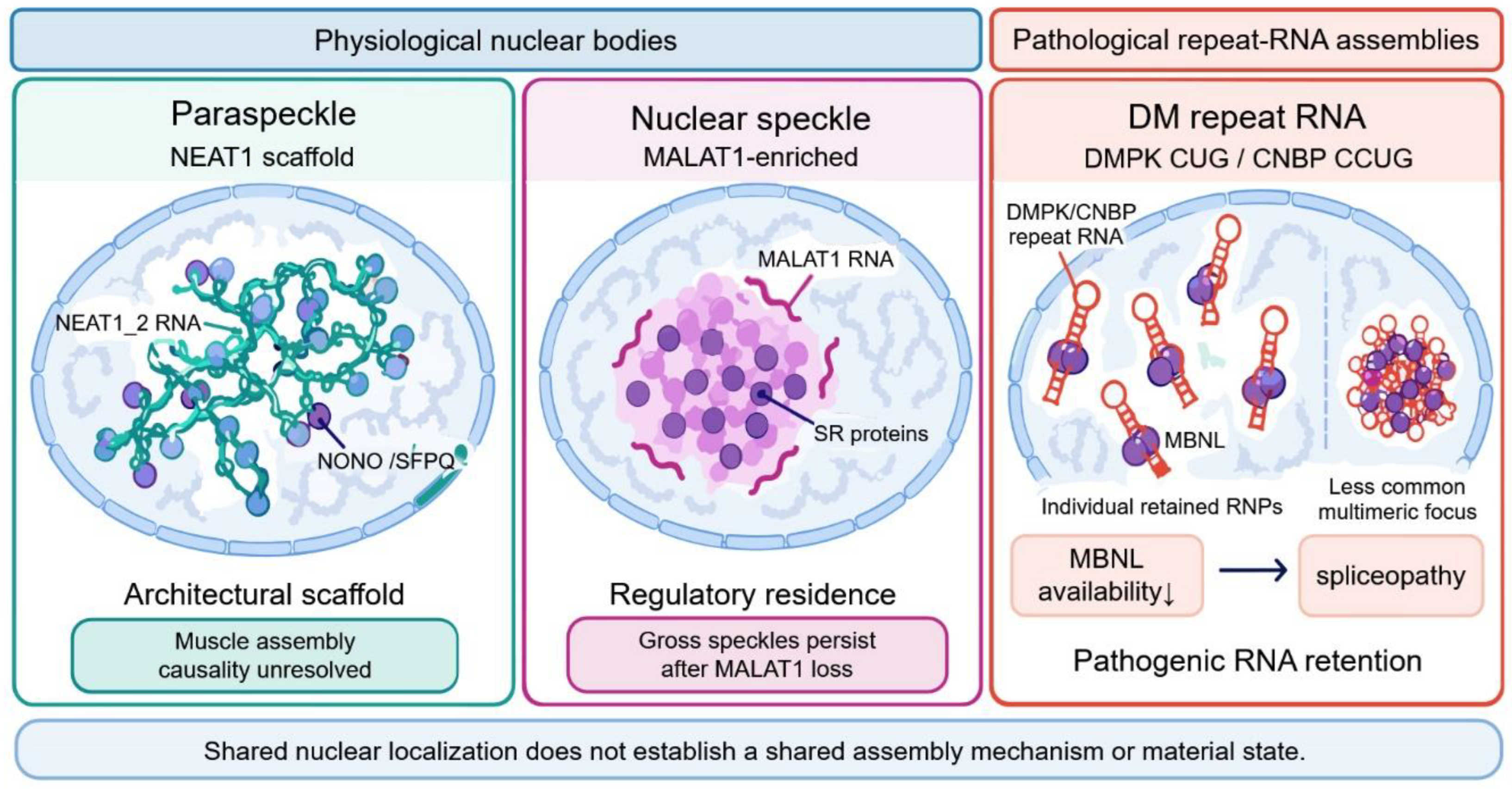
Figure 3.
Therapeutic intervention map for repeat-RNA disease. The upper row depicts the shared toxic-RNA core of myotonic dystrophy, progressing from expanded CTG or CCTG DNA to expanded CUG or CCUG RNA, RNA-binding protein sequestration, missplicing, and muscle dysfunction. This schematic does not exclude parallel disease- or tissue-specific mechanisms. The green dashed connectors identify intervention points and
do not indicate causal activation or established efficacy. Genome editing or transcriptional blockade acts at the repeat locus. ASOs, siRNAs, RNA-cleaving approaches, and degraders reduce expanded RNA. Repeat binders or MBNL restoration address RNA-binding-protein sequestration. Splice-directed or downstream approaches target missplicing, whereas rehabilitation and organ-level care address tissue consequences. The lower row separates target engagement, molecular rescue, tissue function, and clinical benefit. Success at one gate does not establish success at subsequent gates. Modulation of physiological NEAT1- or MALAT1-associated compartments remains exploratory and requires evidence of the intended processing correction and a favorable muscle phenotype. ASO denotes antisense oligonucleotide, MBNL denotes muscleblind-like protein, RBP denotes RNA-binding protein, and siRNA denotes small interfering RNA.
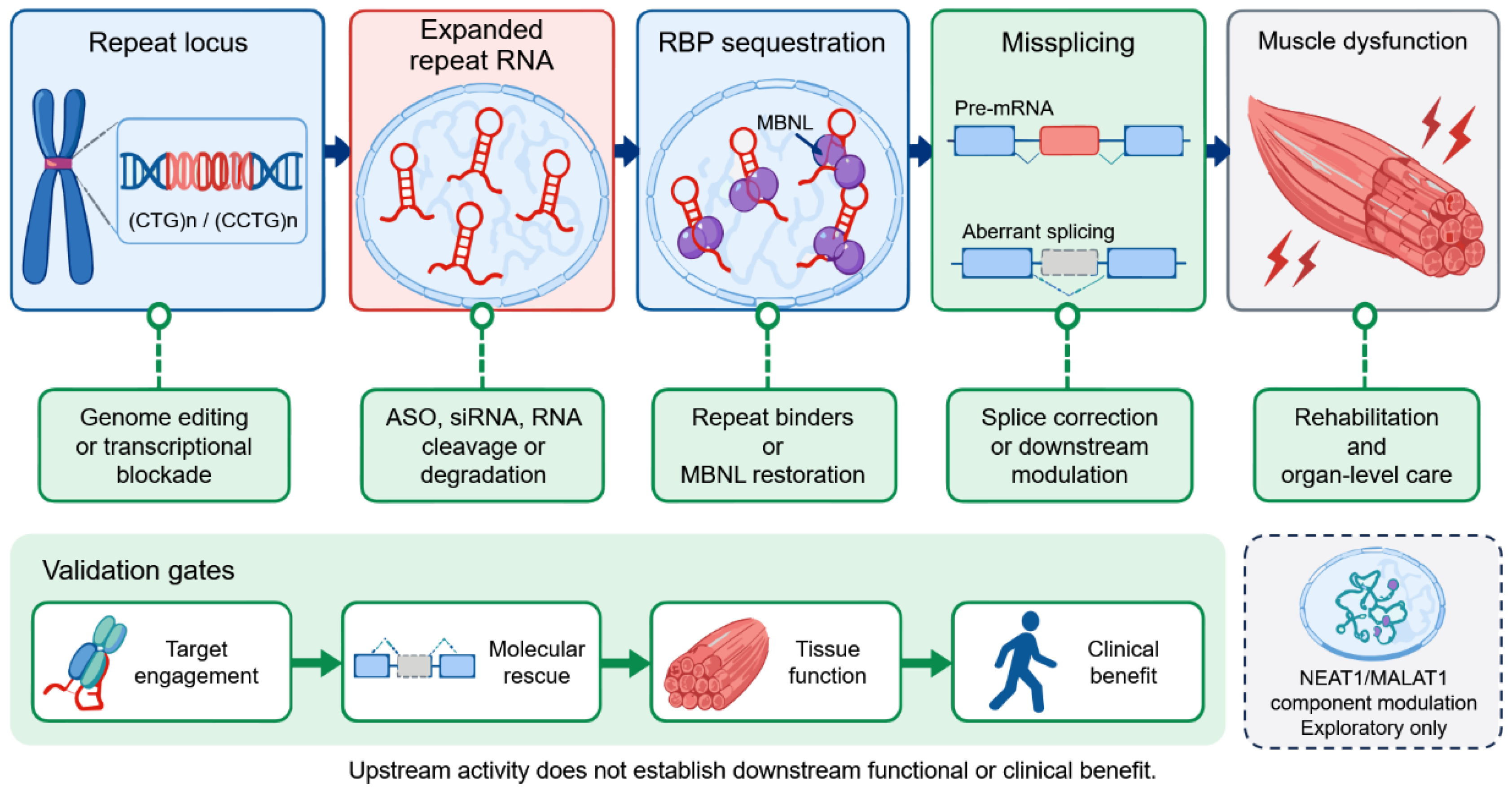

Table 1.
Evidence-graded nuclear ncRNA modules in skeletal muscle.
| RNA / module | Nuclear context | Direct muscle model | Perturbation and principal output | Tier | Supported interpretation |
|---|---|---|---|---|---|
| NEAT1 | Paraspeckle-associated | C2C12 myoblasts and myotubes | Differentiation-linked induction; RNA FISH; PSPC1/NONO association [4] | E2 | Spatial and molecular participation in muscle |
| Neat1 | Chromatin / EZH2 | C2C12; mouse injury | Loss and gain of function; mapped EZH2 interaction; regeneration phenotype [21] | E4–E5 | Neat1-dependent myogenesis; paraspeckle mediation untested |
| Neat1 | PABPN1/MATR3-linked paraspeckle processing | Primary mouse and human myoblasts; OPMD model | PABPN1 or MATR3 depletion; SFPQ redistribution; editing and transport readouts [22] | E4–E5 | Functional NEAT1-centered processing module |
| MALAT1 | Nuclear speckle-associated | Non-muscle mechanism; mouse genetics | SR-factor partitioning/splicing; preserved speckles after knockout [5,6,19,20] | E3–E5 | Regulatory role; not an obligatory architectural RNA |
| Malat1 | Chromatin and myogenic state | C2C12; mouse regeneration | Opposing acute phenotypes; constitutive loss enhances regeneration [23,24,25] | E3–E5 | Context-dependent muscle regulator |
| FRG2A/B | Nucleolus / rDNA | FSHD patient myoblasts and myotubes | RNA localization, ChIRP, depletion, nascent rRNA and translation rescue [26] | E4 | Nucleolar regulator of rDNA transcription in patient cells |
| Ppp1r1b-lncRNA | Chromatin / PRC2 RNP | Mouse and human progenitors | Genetic perturbation; PRC2 and hnRNPA1 interaction [27,28] | E4–E5 | Chromatin RNP; no named body established |
| Myoparr; Linc-RAM; Dum | Promoter- or chromatin-associated | Myoblasts; selected mouse models | RNA loss, partner mapping, and myogenic output [29,30,31] | E3–E5 | Nuclear transcriptional regulators |
| DMPK CUG RNA | Single retained RNPs and RNA foci | Patient muscle cells; mouse models | MBNL recruitment, splicing, physiology, and therapeutic rescue [7,32,33] | E5–E6 | Pathogenic RNA assembly; material state varies |
| CNBP CCUG RNA | RNA/MBNL foci | DM2 patient cells and tissue | Genetic lesion, MBNL sequestration, subtype-specific processing [8,34,35] | E5 | Pathogenic RNA focus; not equivalent to DM1 in every downstream effect |
Abbreviations: E, evidence tier; EZH2, enhancer of zeste homolog 2; FISH, fluorescence in situ hybridization; ncRNA, noncoding RNA; OPMD, oculopharyngeal muscular dystrophy; PRC2, polycomb repressive complex 2; RBP, RNA-binding protein. Evidence-tier ranges indicate that different claims within a row reach different tiers. They do not represent a single global score for a study.
Table 2.
Physiological nuclear compartments and pathological RNA assemblies.
| System | Principal RNA driver or resident | Proteins | Material / assembly evidence | Processing output | Muscle evidence boundary |
|---|---|---|---|---|---|
| Paraspeckle | NEAT1_2 scaffold; NEAT1_1 partly outside | NONO, SFPQ, PSPC1, HNRNPK | Domain mapping, reassembly, phase-separation and micelle models [3,10,11,15] | Retention, editing, protein availability | Direct association and processing in muscle; material tests mainly non-muscle |
| Nuclear speckle | MALAT1-enriched resident, not obligatory scaffold | SR proteins, SON, SRRM2 and others | Multilayer architecture; protein microphases; context-dependent MALAT1 effects [18,36] | Splicing-factor state and transcript processing | Direct myogenic phenotypes; muscle speckle-to-splicing causality unresolved |
| Nucleolus in FSHD | FRG2A/B transcripts | rDNA/chromatin-associated factors | Spatial domain and chromatin association; no material-property test [26] | 45S pre-rRNA synthesis and translational capacity | Direct patient-derived muscle-cell evidence; in vivo rescue absent |
| DM1 expanded RNA | DMPK CUG-expanded transcript | MBNL proteins; CELF dysregulation | In vitro sol–gel transition; endogenous population dominated by individual retained RNAs [32,37] | Alternative splicing, retention, transcriptional and regenerative effects | Human and in vivo causality strong; focus material state heterogeneous |
| DM2 expanded RNA | Excised CNBP CCUG-expanded RNA | MBNL, rbFOX1 and additional RBPs | Discrete RNA/MBNL foci; no universal material-property assignment [8,38] | Overlapping and subtype-specific spliceopathy | Human tissue evidence; fewer intervention studies than DM1 |
Abbreviations: DM1, myotonic dystrophy type 1; DM2, myotonic dystrophy type 2; RBP, RNA-binding protein; rDNA, ribosomal DNA; SR, serine/arginine rich.
Table 3.
Therapeutic strategies mapped to causal layer and evidence.
| Target layer | Representative modality | Model / stage | Target engagement | Molecular and tissue rescue | Readiness / principal limitation |
|---|---|---|---|---|---|
| Toxic-RNA abundance | Systemic ASO | HSA^LR mouse | CUG-expanded RNA knockdown [33] | Splicing, myotonia, histology, transcriptome | E6 preclinical; delivery determines translation |
| Toxic-RNA abundance | Baliforsen ASO | Phase 1/2a | Muscle exposure below predicted effective range [66] | No substantial target reduction established | Human safety/delivery lesson |
| Muscle RNA delivery | TfR1 antibody–siRNA conjugate | Phase 1/2 and phase III topline | DMPK reduction in biopsy [67,68] | Composite missplicing improvement in phase 1/2; phase III primary endpoint not met [69] | Human target engagement and molecular rescue; clinical efficacy unconfirmed |
| Repeat transcription / locus | dCas9 blockade or genome editing | Patient cells; mouse models | Repeat-directed transcriptional block or excision [72,73,74] | Foci, splicing, ClC-1, and myotonia rescue | Delivery, off-targets, durability |
| Repeat RNA | RNA-targeting Cas9 | Patient myotubes and cellular models | Repeat-RNA elimination [71] | Protein relocalization and splicing rescue | Programmable; vector and immune constraints |
| RNA structure | Small-molecule degrader / cleaver | Patient myotubes; mice | Selective binding and RNA degradation [75,76,77] | Broad molecular and selected phenotypic rescue | Exposure and transcriptome-wide specificity |
| RNA–protein occupancy | Erythromycin, furamidine, flavonoid leads | Cells and mice | Repeat binding or reduced foci [78,79] | Splicing and myotonia in models | Often multimodal; clinical exposure uncertain |
| Persistent antisense | AAV–U7snRNA | Patient-derived cells | DMPK/repeat targeting [87] | Foci, MBNL localization, global splicing | Durability versus vector risk |
| Functional MBNL | AntimiR-23b/218 or MBNL expression | Patient cells and mice | Higher MBNL abundance [88,89,90,91] | Splicing, fusion, histology, function | Dose control and broad MBNL targets |
| Downstream regulatory state | HDAC or GSK3β–CELF1 modulation | Cells and mice | Pathway engagement [93,94] | Selected splicing and muscle phenotypes | Does not remove mutant RNA |
| Physiological compartment | NEAT1/MALAT1 modulation | Exploratory | RNA or assembly change | Must show intended processing and favorable muscle output | No mature therapeutic evidence; pleiotropy |
Abbreviations: AAV, adeno-associated virus; ASO, antisense oligonucleotide; dCas9, catalytically inactive Cas9; DMPK, dystrophia myotonica protein kinase; HDAC, histone deacetylase; HSA^LR, human skeletal actin long-repeat mouse model; MBNL, muscleblind-like; TfR1, transferrin receptor 1.
Table 4.
Contested claims and decisive experiments.
| Contested claim | Evidence supporting it | Evidence limiting it | Likely moderator | Decisive experiment and interpretation |
|---|---|---|---|---|
| NEAT1-dependent myogenesis is a paraspeckle mechanism | C2C12 paraspeckle association; NEAT1 perturbation changes myogenesis [4,21] | Assembly loss and muscle phenotype were not tested together; isoform unresolved | NEAT1_1 versus NEAT1_2; cell state; stress | Matched isoform and assembly-deficient rescue. Only assembly-competent NEAT1_2 rescue supports architectural causality. |
| MALAT1 uniformly promotes or inhibits differentiation | Each direction has direct perturbation evidence [23,24,25] | Protocols differ in timing, density, perturbation duration, and endpoint | Early versus late state; proliferation; adaptation | Time-resolved, density-matched acute and constitutive perturbations with physiological rescue. |
| MALAT1 is a speckle scaffold in muscle | Speckle enrichment and SR-factor regulation [5] | Speckles persist in knockout mice [6,19,20] | Stress- and protein-state-dependent microphases | Assembly-selective perturbation at fixed MALAT1 abundance paired with muscle splicing readouts. |
| Every DM RNA focus is a condensate | Repeat RNA undergoes phase transition in vitro and in transfected cells [37] | Most endogenous puncta in patient myotubes are individual retained RNAs [32] | Repeat length, transcriptional burst, protein occupancy | Endogenous molecule counting plus dynamics and material perturbation across repeat lengths. |
| FRG2 nucleolar RNA is a major driver of FSHD | Patient-cell depletion rescues rRNA and protein synthesis [26] | No in vivo rescue or direct clinical linkage; paralog resolution limited | D4Z4 context, differentiation, rDNA dosage | Paralog-specific perturbation and rescue in human muscle tissue or an appropriate in vivo model. |
| Loss of foci demonstrates therapeutic mechanism | Several treatments reduce foci with molecular rescue | Focus number can change independently of RNA number or MBNL availability | Probe access, RNA fragmentation, assembly state | Calibrated RNA counting, MBNL availability, splicing, and function in the same dose series. |
Abbreviations: DM, myotonic dystrophy; MALAT1, metastasis-associated lung adenocarcinoma transcript 1; NEAT1, nuclear paraspeckle assembly transcript 1; RNA, ribonucleic acid.
Disclaimer/Publisher’s Note: The statements, opinions and data contained in all publications are solely those of the individual author(s) and contributor(s) and not of MDPI and/or the editor(s). MDPI and/or the editor(s) disclaim responsibility for any injury to people or property resulting from any ideas, methods, instructions or products referred to in the content. |
© 2026 by the authors. Licensee MDPI, Basel, Switzerland. This article is an open access article distributed under the terms and conditions of the Creative Commons Attribution (CC BY) license (http://creativecommons.org/licenses/by/4.0/).
Copyright: This open access article is published under a Creative Commons CC BY 4.0 license, which permit the free download, distribution, and reuse, provided that the author and preprint are cited in any reuse.