

Submitted:
27 September 2026
Posted:
01 October 2026
You are already at the latest version
Abstract
Post-translational modifications are rarely studied as an integrated system. Most studies have focused on acetylation or ubiquitination in isolation, despite both frequently targeting the same lysine residues on the same proteins. This study investigated whether the interplay between these modifications, rather than their individual effects, constitutes a meaningful regulatory layer in cervical cancer cells. Quantitative stoichiometric proteomics was applied to HPV-positive and HPV-negative cell models, with a non-cancerous keratinocyte serving as control. The results demonstrate that acetylation and ubiquitination form coordinated, context-dependent patterns rather than acting independently. In HPV-positive cells acetylation predominated and was enriched in transcriptional regulators. Conversely, the HPV-negative C33a line exhibited a substantially larger ubiquitinome, indicative of increased proteostatic surveillance. Site-specific stoichiometry analysis revealed that the relative occupancy of each modification at shared lysine residues determines regulatory outcomes, rather than their mere presence. This competition is proposed to constitute a "lysine fate code" that integrates transcriptional regulation, metabolic adaptation, and immune signaling into a unified proteostatic logic. Beyond their implications for cervical cancer biology, these findings suggest that the acetylation-ubiquitination balance may represent a key regulatory principle. Targeting enzymes that modulate this balance could provide novel therapeutic strategies that surpass single-pathway inhibition.
Keywords:
lysine acetylation
; ubiquitination
; post-translational modifications
; cervical cancer
; stoichiometry
; DIA mass spectrometry
; HPV
; proteostasis
; PTM crosstalk
; proteomics
1. Introduction
Cervical cancer is the fourth most prevalent cancer among women worldwide. In 2018 there were, over 570,000 new cases and 311,000 deaths. The disease has a higher impact on women in low- and middle-income countries. Limited access to screening and treatment leads to higher incidence and mortality [1,2]. Persistent infection with high-risk human papillomavirus (HPV) especially HPV16 and HPV18 is the leading cause of cervical cancer [3,4]. These viruses drive malignant transformation through the viral oncoproteins E6 and E7 [5].
Despite the availability of preventive strategies and treatments such as platinum-based chemotherapy [6], disease progression and recurrence remain significant challenges. This ongoing difficulty underscores the necessity for a deeper molecular understanding.
Post-translational modifications (PTMs) regulate protein function and stability in cancer [7]. Among these, lysine acetylation and ubiquitination are highly dynamic, interconnected, and often share target residues; however, they are usually studied separately, leaving their joint regulation unclear. This gap raises the question of how their interplay shapes proteome regulation in cancer, a relationship that remains poorly understood. Both modifications are central to protein stability, DNA repair, and apoptosis processes notably affected by HPV. Specifically, acetylation via KATs and KDACs, modulates chromatin dynamics and transcription [8,9], while ubiquitination, through E1–E2–E3 enzyme cascades [10], targets proteins for proteasomal degradation. Importantly, up to 30% of lysine sites may overlap for these modifications, creating potential for competition or cooperation [11].
HPV exploits this molecular interplay: The E6 oncoprotein hijacks the ubiquitin ligase E6AP to induce p53 degradation [12], while E7 drives ubiquitin-mediated degradation of IFI16 to help the virus evade immune recognition [13]. Acetylation stabilizes p53 and protects it from ubiquitination; however, this regulatory balance is disrupted during infection [14].
High-resolution mass spectrometry now enables precise mapping and quantification of PTM occupancy [15], allows the study of regulatory networks. Evidence shows that acetylation and ubiquitination interact, affecting protein stability, localization, and activity. However, their interplay at proteome-wide and site-specific levels especially in cervical cancer remains largely uncharacterized, leaving the roles and interactions of these PTMs in cervical cancer underexplored.
In this context an integrative, proteome-wide approach was used to map and quantify lysine acetylation and ubiquitination in human cervical cancer cells. The data show that these two modifications do not act independently; rather, they converge on shared lysine residues and interact to influence protein stability and function. These interaction patterns are particularly prominent in proteins involved in cell cycle control, chromatin organization, and metabolic regulation. Collectively, these findings show that cross-talk between acetylation and ubiquitination constitutes an underappreciated regulatory layer in cervical cancer, with significant implications for identifying epigenetic and proteostatic vulnerabilities as therapeutic targets.
Beyond describing acetylation and ubiquitination in isolation, this study frames the ubiquitin-proteasome system (UPS) as a pleiotropic regulator of cellular homeostasis. Using a site-specific, stoichiometric map of ubiquitination across four cervical cell models with distinct HPV/p53 backgrounds (SiHa, HeLa, C33a, HaCaT), we show that UPS activity is co-regulated with lysine acetylation at shared residues, is hijacked by HPV oncoproteins (E6/E6AP-mediated degradation of p53), and is quantitatively remodeled according to oncogenic status, with the HPV-negative C33a line showing a markedly expanded ubiquitinome consistent with compensatory proteostatic surveillance in the absence of viral E6/E7 activity. This positions UPS regulation as extending beyond simple protein turnover to intersect transcriptional regulation, genome maintenance, antiviral signaling and metabolic control, in line with the pleiotropic homeostatic role.
2. Results and Discussion
2.1. Comprehensive PTM Profiling Reveals Cell Line-Specific Acetylation and Ubiquitination Landscapes
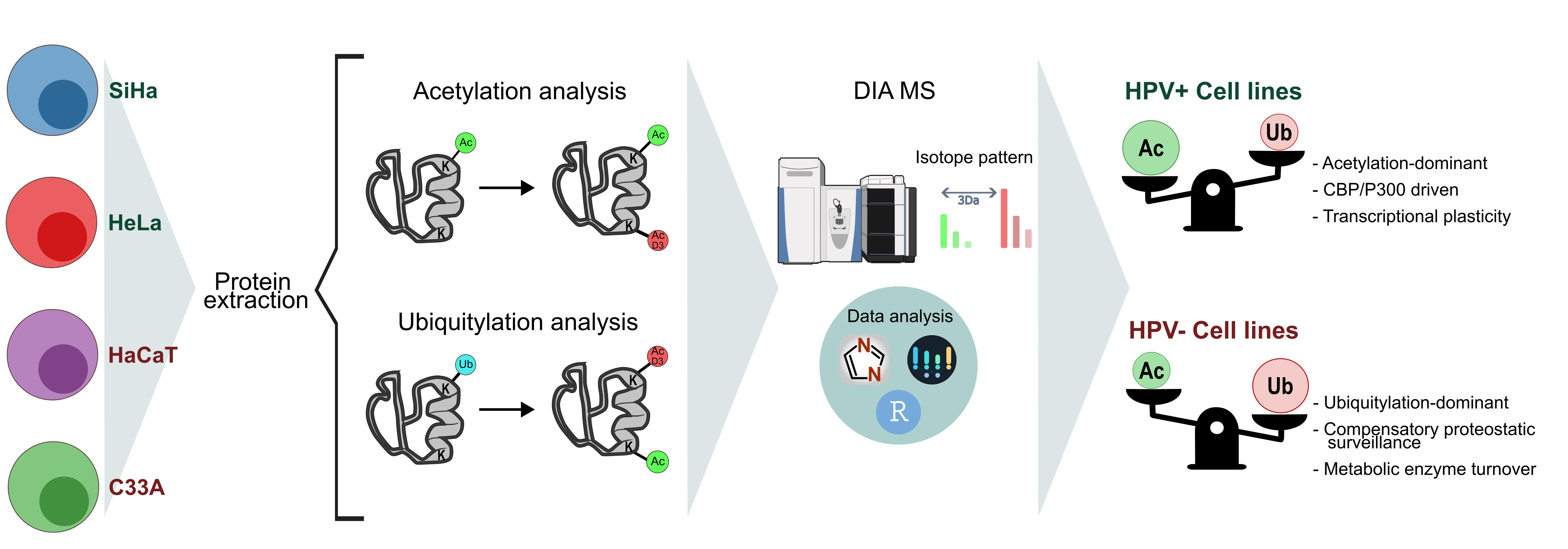
A DIA-MS-based stoichiometry quantification approach using chemical labeling to tag lysine residues was employed to characterize the PTM landscape in cervical cancer (Figure 1). Four cell lines with distinct molecular backgrounds were analyzed: SiHa (HPV16-positive), HeLa (HPV18-positive), C33a (HPV-negative harboring p53 R273C mutation), and HaCaT (immortalized keratinocyte control). This experimental model enabled the dissection of the individual contributions of viral oncoproteins and p53 mutation to PTM regulation during cervical carcinogenesis.
Comparative analysis demonstrated extensive remodeling of lysine-centered PTM landscapes among cervical cell models, with distinct regulatory architectures linked to HPV status (Table 1) (Figure 2A-B). Notably, overall proteome coverage was highly consistent across all four cell lines, with total peptide identifications in the acetylome analysis ranging from 33,581 to 35,918 and total protein identifications ranging from 6,014 to 6,135, reflecting less than 7% and 2% variation, respectively. In the ubiquitinome analysis, total peptide identifications ranged from 27,786 to 31,546, and total protein identifications ranged from 5,497 to 5,816, with variations of 6% and 13%, respectively. This comparable depth of proteome sampling ensured that differences observed at the PTM level were not attributable to systematic disparities in protein detection. In contrast, PTM-specific landscapes showed marked divergence. The HPV-negative C33a cell line showed a pronounced expansion of the ubiquitinome, identifying 5,570 peptides across 2,062 proteins, representing a 1.9-2.3-fold increase compared to HPV-positive lines. This expanded ubiquitination profile could reflect compensatory adaptation of proteostasis networks in the absence of viral E6/E7-mediated degradation pathways. In HPV-positive contexts, these viral proteins redirect the ubiquitin–proteasome system toward selective degradation of key tumor suppressors, including, p53 and other cellular proteins for proteasomal degradation [12,16,17]. Conversely, HeLa cells exhibited the most extensive acetylation landscape with 858 acetylated peptides across 725 proteins, consistent with increased CBP/p300 acetyltransferase activity in HPV-positive cervical cancer [18,19,20,21]. Supporting this, proteomic profiling confirmed the exclusive detection of p300 protein in SiHa and HeLa cells, reinforcing its role as an HPV-associated regulatory factor absent in HPV-negative and non-tumorigenic contexts. The observed divergence between acetylation and ubiquitination profiles across cell lines indicates that HPV status fundamentally reshapes the competitive dynamics governing lysine modification states.
Venn diagram analyses of the acetylated and ubiquitylated peptides (Figure 2 C-D), revealed asymmetric distributions of shared and unique PTMs, supporting the existence of differential lysine modification hierarchies. HeLa presents the highest number of unique acetylated peptides with 373, followed by C33a (269), SiHa (178), and HaCaT (160). For ubiquitination, C33a exhibited the greatest number of unique ubiquitylated peptides with 1946, followed by HeLa (483), SiHa (439), and HaCaT (355).
The strong asymmetry between acetylation and ubiquitination implies potential competition for overlapping lysine residues, which may influence protein fate decisions through mutually exclusive or sequential modification states. However, it is important to acknowledge that technical and stochastic factors may also contribute to the observed asymmetry. These include differences in the number of proteins identified per condition, the inherently low stoichiometry of PTM sites, stochastic peptide sampling during data acquisition, and suboptimal quantification of low-abundance modified species. Therefore, the biological interpretation of lysine crosstalk should be considered in the context of these methodological limitations. A mechanistic model is proposed in which HPV-positive cervical cancer cells employ viral E6/E7 oncoproteins to bias lysine modification equilibria toward selective ubiquitin-mediated degradation, while concurrently enhancing acetylation-dependent transcriptional plasticity [22,23]. In contrast, HPV-negative C33a cells may compensate for the absence of viral regulatory mechanisms by expanding endogenous ubiquitin-dependent quality control pathways, potentially mitigating proteotoxic stress caused by accumulated mutant p53. In this framework, lysine residues function as regulatory hubs where acetylation and ubiquitination dynamically compete or cooperate, forming a tunable molecular switch that governs protein stability, functional state, and signaling output.
2.2. Stoichiometry Distributions Indicate Distinct Regulatory Architectures for Lysine Acetylation and Ubiquitination
A central objective of this study was to quantify site-specific PTM stoichiometry using a DIA-MS workflow, thereby enabling direct comparison of regulatory occupancy across acetylation and ubiquitination landscapes. Stoichiometry distributions revealed fundamentally distinct modification architectures across all examined cellular models (Figure 2E-F), suggesting divergent regulatory principles governing lysine modification states.
Acetylation stoichiometry displayed a broad distribution, with substantial populations of sites occupying intermediate to high occupancy ranges 10–50% range (Figure 2E). This distribution contrasts sharply with previous reports of extremely low global acetylation stoichiometry of only 0.02% in HeLa cells measured by SILAC-based approaches [24]. The stoichiometry values reported here are notably higher than those in the broader literature, and the discrepancy warrants careful consideration. While enrichment protocols can preferentially recover high-occupancy enzyme-catalyzed acetylation events, the magnitude of the difference observed cannot be fully explained by biological enrichment alone; technical factors, such as enrichment efficiency variability, peptide recovery biases, and quantification accuracy at low-abundance sites, likely contribute to the apparent elevation. As such, absolute stoichiometry values should be interpreted with caution and primarily considered in relative, within-experiment comparisons rather than as direct measures of global acetylation occupancy. Nonetheless, high stoichiometry acetylation sites (>The elevated stoichiometry values observed here likely reflect enrichment for functionally relevant, enzyme-catalyzed acetylation events rather than stochastic or nonenzymatic modifications. Notably, high stoichiometry acetylation sites (1%) were predominantly localized to nuclear proteins involved in transcriptional regulation and chromatin remodeling, consistent with models in which acetylation acts as a stable regulatory signal modulating protein function and transcriptional output, and aligning with prior work demonstrating that high-occupancy acetylation preferentially occurs on transcriptional regulators, acetyltransferases, and histone substrates [24].
In contrast, ubiquitylation exhibited a markedly different stoichiometric profile, characterized by low site occupancy, with the majority of quantified sites below 10% stoichiometry (Figure 2F). This pattern is consistent with recent global analyses showing that ubiquitylation occupancy spans several orders of magnitude and is typically substantially lower than phosphorylation (median 0.0081% for ubiquitination vs. 2.5% for phosphorylation) [25]. The low ubiquitin site occupancy reflects its rapid turnover kinetics inherent to ubiquitin-mediated signaling, in which approximately 67% of ubiquitylation sites exhibit half-lives below thirty minutes under basal conditions [26,27]. Such low steady state occupancy enables highly dynamic signaling while conserving the finite cellular ubiquitin pool (approximately 4.5 × 10⁷ molecules per HeLa cell) [25], suggesting that ubiquitination functions as a transient regulatory mark optimized for rapid proteostatic decision-making.
Together, these findings support a model in which lysine acetylation and ubiquitination occupy distinct regions of regulatory space: acetylation primarily operates as a stable regulatory modification that alters protein activity, protein-protein interactions, and subcellular localization [8,28], as a higher occupancy observed for acetylation sites (10–50% range) is consistent with modifications that must be present at enough levels to exert direct effects on protein function in the cell. In comparison, ubiquitination acts as a low-occupancy, high-flux signal enabling cells to maintain regulatory flexibility while conserving ubiquitin pools [25]. Within this framework, lysine residues emerge as competitive regulatory nodes where differential stoichiometry may determine whether proteins adopt stable functional states or enter degradation and quality-control pathways.
To examine whether regulatory enzyme abundance predicts acetylation stoichiometry, we performed Spearman rank correlations between the protein levels of 16 KATs and KDACs and the median site-specific acetylation stoichiometry across cell lines (Figure S1A). Among enzymes with sufficient quantitative coverage (n ≥ 9 sites), SIRT3 showed a significant negative correlation (ρ = −0.64, p = 0.024, n = 12), as did HDAC1 (ρ = −0.59, p = 0.045, n = 12). Both results are consistent with the known deacetylase activity of these enzymes: SIRT3 reduces acetylation occupancy at its mitochondrial targets [29,30], while HDAC1 operates within nuclear corepressor complexes to remove acetyl groups from histone and non-histone substrates [31]. A precedent for this type of enzyme-level vs. stoichiometry correlation exists in human cervical cancer cell lines: Gil et al. (2017) reported that higher SIRT1 abundance correlates with lower acetylation occupancy across HeLa, SiHa and CaLo cells [9]. In contrast, none of the tested KATs, including KAT7, CREBBP, ELP3, NCOA3 and KAT8, reached statistical significance (all p > 0.35). This pattern aligns with the broader observation that most lysine acetylation occurs at very low stoichiometry and that class I deacetylases preferentially target the subset of high-occupancy sites, while writer abundance has comparatively little predictive power over site-level acetyl occupancy [24,32].
A parallel analysis for ubiquitination included 48 E3 ligases and DUBs (Figure S1B). No enzyme with n ≥ 9 quantified sites reached statistical significance (p < 0.05). The strongest trends were observed for UCHL5 (ρ = +0.48, p = 0.112) and OTUB1 (ρ = +0.47, p = 0.124) both DUBs with positive, non-significant associations. UCHL5/UCH37 is a proteasome-resident DUB that debranches K48-linked ubiquitin chains; crucially, its activity is gated by binding partners (RPN13 vs. NFRKB) rather than by expression levels [33,34]. OTUB1 regulates substrates such as p53, RhoA and SMAD2/3 through both catalytic chain cleavage and non-catalytic E2 sequestration, and its involvement in tumor biology is well documented [35,36]. The overall absence of significant correlations between enzyme abundance and ubiquitination stoichiometry across the four cell lines is consistent with the known architecture of the ubiquitin system: humans encode >600 E3 ligases and ~100 DUBs [37], and substrate modification depends far more on enzyme activity than abundance, a point reinforced by the finding that USP7 inhibition reshapes the ubiquitinome within minutes without changes in DUB protein levels [38], and by direct proteome-wide evidence showing no correlation between protein abundance changes and ubiquitination-site dynamics [39]. Beyond substrate redundancy, E3 ligase activity is regulated post-translationally by autoinhibition, cullin neddylation, and substrate clustering [40], all of which decouple enzyme concentration from modification output.
2.3. The Global PTM Landscape Organization Distinguishes HPV-Positive and HPV-Negative Regulatory States
To evaluate higher-order organization of PTM landscapes, we performed hierarchical clustering and principal component analysis (PCA) of quantified acetylated and ubiquitinated proteins (Figure 3).
Heatmap analysis of the 150 most variable acetylated proteins revealed distinct clustering patterns that separated HPV-positive cell lines from HPV-negative C33a cells and HaCaT control (Figure 3A) (Table S1). Remarkably, proteins involved in chromatin remodeling complexes and transcriptional regulation contributed substantially to this separation, including components of the CBP/p300 acetyltransferase machinery and their downstream targets. This finding aligns with established mechanisms in which HPV E6 and E7 oncoproteins modulate CBP/p300 activity, thereby reshaping host transcriptional programs through altered acetylation patterns and gene expression dynamics [41,42]. C33a's separation was driven in part by a distinct set of epigenetic regulators absent from HPV-positive line, such as UHRF1, a multidomain reader that coordinates DNA methylation with histone modification status, and KDM1A (LSD1), whose acetylation state directly modulates its H3K4 demethylase activity, pointing to a p53 mutation-driven epigenetic reprogramming axis that operates independently of viral oncoproteins [43,44].
PCA analysis of 1,036 acetylated further demonstrated that HPV-positive cell lines clustered closely along the primary principal component, whereas C33a and HaCaT occupied distinct regions of PTM space (Figure 3B). Loading analysis identified proteins involved in RNA processing, ribosomal biogenesis, and mRNA splicing as major contributors to separation along PC1, in accordance with the known roles of HPV oncoproteins (E6/E7) in reprogramming transcriptional and RNA-processing pathways [45,46]. Sub-clustering within the HPV-positive group further distinguished SiHa from HeLa: SiHa carried an acetylation signature enriched for DNA repair factors, XRCC1, RAD50, and ARID2, while HeLa showed a profile centered on nuclear transport machinery and the DEK oncogene, consistent with HPV18 having a higher integrated viral copy number and broader disruption of host chromatin architecture [47,48]. These analyses suggest that viral oncogene expression represents a dominant organizing principle of the acetylome.
In contrast, ubiquitination landscapes exhibited a distinct clustering architecture (Figure 3C) (Table S2). The 150 most variable ubiquitinated proteins segregated C33a into a unique cluster distinct from those of all other cell lines, reflecting the expanded ubiquitinome observed in C33a cells and suggesting that p53 mutation-associated alterations fundamentally reshape ubiquitin-dependent proteostatic networks. Within this cluster, SIRT6, a NAD⁺-dependent deacetylase that promotes NHEJ-mediated DSB repair and base excision repair, and regulates telomere integrity through TRF2 deacetylation, acts as a central genome maintenance factor, occupying the regulatory niche that E6/E6AP complexes fill in HPV-positive lines [49]. Its co-occurrence with KRT5 and KRT14, basal-layer keratins absent from all HPV-positive lines, indicates that C33a retains squamous differentiation identity while relying on intrinsic chromatin guardians rather than viral cooperative mechanisms to sustain transformation. Ribonucleotide reductase RRM1 and nucleoside monophosphate kinase CMPK1 were also C33a-specific contributors, suggesting that dNTP pool regulation, rather than replication fork protection or homologous recombination, is C33a's primary replication stress management strategy [50].
PCA analysis of 2,240 ubiquitylated proteins reinforced this observation, with C33a occupying a clearly separated position within PTM space (Figure 3D). Loading analysis identified proteasomal components, ubiquitin-conjugating enzymes, and metabolic enzymes as major contributors to cell line discrimination, suggesting large-scale remodeling of proteostasis networks in the absence of viral regulatory inputs. The E3 ligase repertoires contributing to HPV-positive cell line positioning were serotype-specific: HeLa carried UBE3C, HERC4, and RNF213, while SiHa relied on UBE4B, NSMCE1, and CDC2, distinct substrate-targeting programs shaped by HPV18 versus HPV16 oncoproteins, respectively [47,51].
Interestingly, despite clear differences in acetylation profiles HeLa and SiHa exhibited relatively similar ubiquitination landscapes, suggesting convergence of HPV16- and HPV18-driven ubiquitin signaling toward shared substrate networks. That convergence likely operates primarily through E6/E6AP-mediated p53 destruction, a substrate common to both HPV16 and HPV18 E6 proteins, while the broader immune evasion programs diverged: HeLa specifically deployed HERC4 to degrade STING and UBE3C to target IRF3/7, an innate immune suppression axis with no equivalent in SiHa [51,52]. These observations imply that acetylation and ubiquitination provide complementary, partially orthogonal layers of molecular classification: whereas the acetylome captures transcriptional reprogramming associated with viral oncogenesis, the ubiquitinome reflects proteostatic adaptation and quality-control dynamics.
2.4. Differential Stoichiometry Identifies PTM-Dependent Regulatory Nodes
To identify proteins with significant cell line–specific-differences in modification levels, we performed one-way ANOVA analysis with Benjamini-Hochberg FDR correction (p < 0.05) to the acetylated and the ubiquitylated peptides, identifying 152 differentially modified proteins, including: 97 acetylated and 55 ubiquitinated targets (Figure 4A–B).
Although the absolute expansion of the ubiquitinome was more pronounced, the greater number of significantly differential acetylated proteins suggests that acetylation changes may be more consistently distributed across the proteome. In contrast, ubiquitination exhibits greater variance across cell lines, as reflected by a markedly higher mean F-statistic (179.6 vs. 69.5 for acetylation). This observation is consistent with the biochemical properties of ubiquitination as a low-occupancy, high-turnover modification, where small changes in E3 ligase or deubiquitinase activity can produce large shifts in steady-state modification levels.
Collectively, these results support a model in which acetylation provides a relatively stable regulatory framework that defines cell state, whereas ubiquitination functions as a dynamic amplifier of proteostatic stress responses. Within lysine-centered regulatory networks, differential stoichiometry may determine whether acetylation stabilizes protein function or ubiquitination directs rapid turnover, reinforcing the concept of competitive PTM switching as a key determinant of protein fate in cervical cancer cells.
2.5. Important Differentially Ubiquitinated Proteins Define Regulatory Axes Associated with Cervical Cancer Cell Identity
Analysis of proteins exhibiting extreme ubiquitination variance identified a subset of candidates that may function as molecular signatures distinguishing cervical cancer subtypes:
Among these, the cysteine protease inhibitor (CST1) showed the highest F-statistic across the entire dataset F = 4,542, indicating near-complete cell line specificity, with HeLa cells exhibiting the highest ubiquitination stoichiometry (100%). As a member of the cystatin family, which regulates cathepsin-mediated proteolysis, CST1 has been implicated in cancer progression and metastasis [53]. The dramatic differences observed here suggest that ubiquitin-dependent modulation of protease inhibitors may represent a mechanism for tuning proteolytic cascades in a context-dependent manner, potentially influencing extracellular matrix remodeling and invasive capacity. Hexosaminidase subunit alpha (HEXA; F = 300.6), a lysosomal enzyme responsible for glycosphingolipid degradation, exhibited markedly elevated ubiquitination in HeLa cells. Given the established links between glycosphingolipid metabolism and cancer cell survival, stress adaptation, and drug resistance [54], differential ubiquitination of HEXA may reflect altered lysosomal flux or metabolic rewiring associated with HPV-driven oncogenesis. Similarly, glutamine -fructose-6-phosphate transaminase 1 (GFPT1; F = 275.2) (Figure 4C) the rate-limiting enzyme of the hexosamine biosynthetic pathway, displayed cell line–specific ubiquitination patterns, with the highest stoichiometry observed in SiHa cells. Because GFPT1 controls flux into the O-GlcNAcylation pathway, which intersects with phosphorylation and transcriptional regulation in cancer signaling networks [55,56], these findings raise the possibility that ubiquitination may indirectly modulate signaling plasticity by regulating metabolic entry points into PTM cross-talk networks. Fatty acid binding protein 5 (FABP5, F=378.95) a lipid chaperone that regulates nuclear receptor signaling and PPARγ activity, exhibited elevated ubiquitination in SiHa cells, suggesting potential remodeling of lipid metabolism and transcriptional signaling—pathways that contribute to oncogenic adaptation [57]. Likewise, aquaporin-3 (AQP3; F= 544.63) (Figure 4D), a membrane channel involved in osmotic regulation and cellular trafficking, showed high ubiquitination across cell lines. Given reports linking AQP3 regulation to PDPK1–AKT–mTOR signaling and autophagic responses in cancer contexts⁴⁰, differential ubiquitination may influence membrane localization dynamics or turnover in response to distinct oncogenic drivers [58].
Additional highly significant ubiquitinated proteins included MORF4L2, a chromatin remodeling factor; CLPTM1, a transmembrane protein implicated in stress responses; HBS1L, a translational GTPase involved in ribosome-associated quality control; MEAF6, a component of the histone acetyltransferase complex; and CCNC, a mediator complex subunit central to transcriptional regulation. The convergence of ubiquitination changes on chromatin regulators and transcription-associated machinery further reinforces the concept that ubiquitination participates not only in protein degradation but also in fine-tuning transcriptional and epigenetic networks.
2.6. Proteostasis–Epigenetics Coupling Emerges Through Ubiquitination Hotspots
Beyond protein-level differences, differentially ubiquitinated proteins in our dataset were not uniformly distributed across functional categories but instead clustered within modules connecting proteostasis with chromatin and transcriptional regulation. Among the most significantly ubiquitinated targets were MORF4L2, MEAF6, and CCNC—proteins with well-established roles in chromatin remodeling and transcriptional control—indicating that ubiquitin-mediated regulation extends into epigenetic machinery not traditionally considered a primary target of ubiquitination. This convergence suggests that, at least in cervical cancer cells, ubiquitination does not exclusively antagonize acetylation but may cooperate with or compete against it at shared lysine residues to modulate chromatin accessibility and transcriptional output [59].
A related pattern developed for metabolic and lipid-associated proteins. Differential ubiquitination of GFPT1, the rate-limiting enzyme of the hexosamine biosynthetic pathway, and FABP5, a regulator of nuclear receptor signaling, raises the possibility that ubiquitin-dependent enzyme turnover contributes to metabolic rewiring in a cell-line-specific manner. Given that Acetyl-CoA availability—directly tied to metabolic state—is a primary determinant of acetylation capacity [60], ubiquitination-mediated control of metabolic enzymes could indirectly reshape the acetylation landscape [61,62]. This feedback loop appears to be differentially engaged depending on HPV status: HPV-positive cells may redirect ubiquitin signaling toward chromatin-associated substrates via oncoprotein activity, while HPV-negative cells compensate by expanding endogenous quality-control networks. Together, these observations suggest that ubiquitination hotspots function not as isolated degradation signals but as regulatory hubs integrating protein turnover, metabolic flux, and epigenetic state.
2.7. Stoichiometry Defines Functional Dominance in Lysine PTM Competition
A central and often underappreciated dimension of PTM crosstalk is not simply which modifications occur, but at what occupancy. Our quantitative data show that acetylation and ubiquitination occupy fundamentally different regions of stoichiometric space: acetylation exhibited intermediate to high site occupancy—consistent with its role toward stabilizing protein conformation and maintaining transcriptionally active chromatin states—while ubiquitination operated at predominantly low occupancy with high variability across cell lines, indicating the rapid turnover kinetics inherent to proteostatic signaling.
This stoichiometric asymmetry has direct functional consequences. At shared or proximal lysine residues, the relative balance between modifications—rather than their mere presence—appears to determine regulatory outcome. Mechanistically, sustained acetylation may limit ubiquitin conjugation through steric occlusion or competition for the same lysine, while even low-level ubiquitination can direct proteins toward degradation when acetylation is reduced or absent. In our dataset, this competition appears to be tuned by HPV status: HPV-positive cell lines displayed acetylation-dominant stoichiometric profiles consistent with transcriptional plasticity and sustained signaling, whereas the HPV-negative C33a line showed elevated ubiquitination occupancy aligned with enhanced proteostatic surveillance. These differences indicate that viral oncogenesis does not simply alter which proteins are modified, but actively reshapes the quantitative balance between competing marks—effectively recalibrating the regulatory logic governing protein fate at the proteome level.
2.8. Differential Acetylation Targets Converge on Metabolic, Immune, and Transcriptional Regulatory Axes
Among the most striking examples, sulfide:quinone oxidoreductase (SQOR, F = 1,155) showed exceptionally high differential acetylation, particularly in SiHa cells. Given SQOR’s central role in mitochondrial sulfide metabolism and redox control, acetylation may stabilize mitochondrial proteins, which could modulate cellular redox state [63]. Interferon-induced protein with tetratricopeptide repeats 3 (IFIT3, F= 117.85) (Figure 4E) provides a complementary perspective on how acetylation dynamics relate to immune modulation. Elevated acetylation in HPV-negative or less oncogenically driven contexts suggests that acetylation may preserve antiviral signaling components by limiting ubiquitin-dependent turnover [64]. Conversely, reduced acetylation in HPV-positive models may facilitate ubiquitination-driven suppression of innate immunity, with differential acetylation potentially regulating antiviral activities. HPV18 may exert stronger suppressive effects on interferon signaling than HPV16. Similarly, acetylation of Target of Myb1 membrane trafficking protein (TOM1, F= 418.97) (Figure 4F): An adaptor involved in endosomal sorting of ubiquitinated cargo, highlights a regulatory intersection between trafficking and proteostasis [65]. We propose that acetylation of TOM1 may influence its ability to bind ubiquitinated substrates or membrane compartments, thereby modulating vesicular sorting pathways that are essential for viral entry, intracellular trafficking, and receptor recycling. Such regulation suggests that acetylation does not simply oppose ubiquitination but can reprogram ubiquitin signaling outcomes.
Acetylation changes observed in mediator complex subunit 11(MED11, F= 88.40) and Poly(ADP-ribose) polymerase 9 (PARP9, F= 48.71) additionally indicate that the lysine modification switch extends to transcriptional regulation and immune signaling pathways. Increased acetylation of MED11 in HPV-positive lines may stabilize Mediator complex assembly [66]. reinforcing transcriptional rewiring driven by viral oncogenes [67], while differential acetylation of PARP9 (HeLa and Siha show higher acetylation, while C33a shows lower stoichiometry) suggests PTM-dependent modulation of interferon responses. Together, this evidence supports a model in which acetylation selectively stabilizes regulatory hubs that define subtype-specific transcriptional and immune states.
Additional highly significant acetylated proteins included WDR43 (ribosome biogenesis), SCCPDH (lysine catabolism), UFD1 (ubiquitin recognition factor in ERAD), NLN (metallopeptidase), and ALG3 (N-glycosylation enzyme). Notably, the identification of acetylation on proteins directly involved in ubiquitin recognition and degradation, such as UFD1, suggests the existence of feedback loops in which acetylation regulates components of the ubiquitin machinery.
2.9. A Lysine Fate Code Governing Proteostatic Decisions in Cervical Cancer
The patterns emerging from our multi-layered analysis point toward a coherent regulatory logic in which lysine residues serve as integration points for competing post-translational signals. Across all four cell lines examined, acetylation and ubiquitination did not behave as independent systems—their distributions, stoichiometries, and functional associations were consistently interrelated, with the balance between them shifting in ways that tracked both HPV status and p53 mutational background. We propose that this balance constitutes a "lysine fate code": a modification-level logic that determines whether a given protein is stabilized, functionally reprogrammed, or directed toward degradation. This concept aligns with emerging evidence that competition among PTMs at shared lysine residues encodes proteome-scale regulatory outcomes [68].
HPV status emerged as the dominant factor shaping this code in our dataset. HPV-positive cell lines displayed acetylation-dominant modification landscapes enriched in transcriptional regulators and chromatin-associated proteins, consistent with E6/E7-mediated engagement of host acetyltransferase networks such as CBP/p300. Notably, HPV-driven acetylation of conserved lysine residues has been shown to directly regulate viral and host transcriptional programs [69], suggesting that viral oncoproteins actively exploit lysine modification dynamics rather than simply disrupting them. In contrast, the HPV-negative C33a line exhibited a markedly expanded ubiquitinome enriched in proteostasis and ERAD pathways, likely reflecting compensatory activation of endogenous quality-control mechanisms in the absence of viral degradation machinery—a pattern consistent with E6AP-independent ubiquitin-proteasome activity previously described in HPV-negative cervical cancer contexts [70]. This difference corresponds to distinct functional architectures: acetylation-dominant states are associated with transcriptional plasticity, while ubiquitination-dominant states are associated with proteostatic surveillance.
What makes this system biologically meaningful is that it connects several apparently distinct processes through a common regulatory axis. Differentially modified proteins in our dataset span mitochondrial metabolism, where acetylation of enzymes like SQOR may stabilize metabolic configurations amid oncogenic pressure, a role consistent with the established link between lysine acetylation and metabolic sensing [71], innate immune signaling, where acetylation of IFIT3 appears linked to antiviral pathway maintenance, and vesicular trafficking, where TOM1 acetylation may influence ubiquitinated cargo sorting. That these functional domains converge on shared lysine regulatory hubs suggests that PTM competition operates not as a protein-by-protein mechanism but as a proteome-scale regulatory system. Targeting the enzymatic machinery that governs this regulation: acetyltransferases, deacetylases, E3 ligases, and deubiquitinases may therefore offer therapeutic leverage that goes beyond the inhibition of individual pathways.
2.10. Functionally Coherent Protein Networks and Lysine Fate Regulation
Functional enrichment analysis of uniquely modified proteins revealed that acetylation and ubiquitination define coherent regulatory modules rather than isolated signaling events, suggesting that lysine modification patterns encode system-level regulatory states determined by oncogenic context. Across cervical cancer cell lines, distinct PTM-enriched pathways emerged that reflect divergent strategies for balancing transcriptional plasticity, proteostasis, metabolism, and immune regulation (Figure 5A-B).
In HPV16-positive SiHa cells, acetylated proteins were strongly enriched in RNA processing and mRNA splicing pathways (adjusted p < 0.001), while ubiquitinated proteins preferentially mapped to DNA repair and nucleotide excision repair processes. These findings align with the established mechanism by which HPV16 E6 drives E6AP-mediated degradation of p53 [72] , which in turn reduces acetylation at key C-terminal regulatory lysines (K370, K372, K373, K381, K382, K386). The resulting shift in lysine modification balance may promote genomic adaptability by simultaneously dampening p53-dependent transcriptional programs while enhancing ubiquitin-dependent surveillance of DNA damage response factors. This pattern suggests that HPV16 oncogenesis reprograms lysine modification networks to favor adaptive genome maintenance under persistent replicative stress.
HeLa cells exhibited a distinct regulatory architecture characterized by coordinated acetylation and ubiquitination within cell cycle and mitotic checkpoint pathways. Enrichment of chromatin-associated acetylation targets supports a model in which HPV18 E7-driven recruitment of CBP/p300 reshapes enhancer landscapes and transcriptional output, as H3K27 acetylation [73].
Notably, elevated acetylation of ribosomal proteins and translation initiation factors (IFIT3, EIF4B, EIF4G1, RPS19, RPS3, RPL30, RPL8), indicates that acetylation may regulate translational capacity at the systems level, extending PTM control more than individual proteins toward coordinated regulation of functional complexes. This observation supports a model in which HPV18 transformation relies on acetylation-mediated stabilization of the biosynthetic machinery to sustain rapid proliferation.
In contrast, the HPV-negative C33a cell line demonstrated dramatically expanded ubiquitinome enriched for protein quality control, endoplasmic reticulum-associated degradation (ERAD), and proteostasis pathways (adjusted p < 0.001). The enrichment of ubiquitinated metabolic enzymes (GFPT1, FABP5, HEXA, ACAA1, ECHS1, GATM), suggests metabolic reprogramming through ubiquitin-mediated enzyme turnover. The enrichment of metabolic enzymes reflects the Warburg effect and altered lipid metabolism characteristic of cancer cells, with PTM-dependent regulation providing a mechanism for rapid metabolic adaptation [74].
Acetylated proteins were enriched in metabolic pathways and mitochondrial functions, with proteins like SQOR, SCCPDH, BCKDHA, and ACO1 showing highly significant differences. The absence of viral E6-driven p53 degradation, combined with the R273C DNA contact mutation, creates a unique regulatory environment in which mutant p53 accumulates but lacks transcriptional activity, demanding alternative mechanisms to maintain proliferative capacity [75,76].
Several non-mutually exclusive hypotheses may explain the expanded ubiquitinome in C33a cells: A) Compensation for absent E6-mediated degradation: In HPV-positive cells, E6/E6AP efficiently degrades p53 and other substrates, may reduce the pool of proteins requiring ubiquitination through endogenous E3 ligases [77]. The absence of this viral degradation pathway in C33a may necessitate expanded endogenous ubiquitin-proteasome system activity. B) p53 R273C-associated proteostatic stress: Mutant p53 accumulation, combined with gain-of-function activities including enhanced cell proliferation and metabolic reprogramming, may impose proteostatic burdens requiring expanded ubiquitin-dependent quality control [78,79]. C) Altered E3 ligase expression: The transcriptional programs controlled by wild-type p53, including regulation of MDM2 and other E3 ligases [14], are disrupted in C33a cells, potentially altering global ubiquitination machinery expression. This evidence supports a compensatory model in which ubiquitination becomes a dominant regulatory strategy when viral modulation of acetylation networks is absent.The HaCaT keratinocyte control exhibited enrichment patterns associated with epithelial differentiation and cytoskeletal organization, establishing a baseline regulatory architecture from which oncogenic rewiring can be inferred. Compared with transformed lines, HaCaT cells maintained PTM distributions consistent with their differentiated identities: keratins (KRT7, KRT10), epithelial markers (FERMT2, CALD1), and membrane trafficking proteins (LAMP1, DAB2), underscoring how cancer-associated signaling shifts lysine modification equilibria toward proliferative and adaptive states.
Comparative analysis of HPV16-positive (SiHa) and HPV18-positive (HeLa) cell lines revealed subtype-specific modulation of CBP/p300-associated acetylation networks. Although both E6 variants target the CBP/p300 acetyltransferase complex through interactions with the CH3 domain, inhibiting p53 acetylation at C-terminal regulatory lysines [20,77] and promoting ubiquitin-mediated degradation, quantitative differences in acetylation patterns suggest distinct mechanisms of acetylome remodeling that emerged between SiHa and HeLa cells.
HeLa and SiHa cells exhibited higher global acetylation levels across CBP/p300 substrates, with the ANOVA analysis revealing that several known CBP/p300 targets showed significant differences, including MED11 (Mediator complex component), PARP9 (poly(ADP-ribose) polymerase family member), and chromatin-associated proteins. This asymmetry suggests that HPV E6/E7 oncoproteins have profound effects on the acetylome.
Structural studies stablish that HPV16 E6 binds E6AP with higher affinity (Kd= 144 pM) than HPV18 E6 (Kd = 980 pM), suggesting that HPV type-specific differences in E6-E6AP complex stability may influence downstream PTM landscape organization [80]. Paradoxically, HeLa cells show elevated acetylation despite lower E6-E6AP affinity. This observation suggests that HPV18 E7 may play a more dominant role in acetylome remodeling by enhancing p300/CBP recruitment to enhancer regions and altering H3K27ac deposition patterns.
The p53 protein showed reduced acetylation at canonical CBP/p300 target sites in both HPV-positive lines compared to the C33a and HaCaT controls. In SiHa and HeLa cells, this reduction indicated E6-mediated inhibition of p300/CBP activity rather than p53 protein loss alone. The reciprocal relationship between p53 acetylation and ubiquitination—where acetylation at C-terminal lysines blocks MDM2-mediated ubiquitination—is disrupted by E6, which simultaneously enhances E6AP-mediated ubiquitination and suppresses CBP/p300-mediated acetylation [14].
2.11. Acetylation-Ubiquitination Cross-Talk Governs Lysine Fate
The coordinated patterns across cell lines point to one conclusion: acetylation and ubiquitination compete for the same lysine residues, and that competition determines protein fate [81,82].
At shared lysine residues, the logic is binary. Acetylation holds proteins in stable, active states; ubiquitination routes them toward degradation or signaling turnover. GFPT1, FABP5, HEXA, and SQOR all carry dual modification sites, and the opposing PTM occupancies measured on those residues confirm that a single lysine can determine whether a protein remains functional at its target compartment or is cleared. There is no graded middle ground there—it is one modification or the other.
What complicates the picture is that the two machineries are not independent. Acetylation of UFD1, a ubiquitin recognition factor, limits how efficiently the cell processes misfolded or surplus proteins. At the same time, SIAH1/SIAH2 E3 ligases ubiquitinate CBP/p300, TIP60, and PCAF, placing a ceiling on global acetylation output [83]. Each layer constrains the other. The cross-cell-line data capture this feedback under distinct oncogenic conditions, HPV-positive lines, HPV-negative C33a, and non-cancerous cell line HaCaT, which is where the comparison earns its keep.
The most consequential finding sits in the third pattern. HPV E6 collapses the p53 methylation–acetylation–ubiquitination cascade by suppressing stabilizing acetylation while driving E6AP-dependent degradation simultaneously. That mechanism is well documented for p53. What the data add is that the same hierarchical rewiring extends across a much broader set of substrates: proteins with divergent acetylation and ubiquitination abundances across cell lines show that viral oncoproteins reset lysine fate decisions at proteome scale, a scope that a single point mutation in p53 alone cannot replicate.
2.12. PTM Signatures and Systems-Level Regulatory States
Proteins with extreme F-statistics mark where PTM stoichiometry diverges from protein abundance, and that divergence is where the biology gets interesting. CST1 (F = 4,542; ubiquitinated specifically in HeLa), SQOR (F = 1,155; acetylated in SiHa), and TSG101 (F = 1,450; ubiquitinated in SiHa) show near-cell-line-exclusive modification patterns that expression-level proteomics would miss entirely. Lysine occupancy, not protein quantity, appears to encode oncogenic identity here.
Our findings extend previous proteomic characterizations of cervical cancer cell lines and integrate with recent proteogenomic analyses of patient tumors [84,85]. A recent study identified EP300-driven acetylation of FOSL2-K222 as a cervical cancer proliferation driver [86]. FOSL2 did not surface among our top ANOVA hits, but MED11, ARID2, and GTF3C5—all part of the chromatin remodeling and general transcription machinery—did, each with significant cell-line-dependent acetylation differences. EP300 likely acetylates a much wider set of transcriptional substrates than FOSL2 alone, and which targets dominate depends on the oncogenic context.
IFIT3 and PARP9 stand out for a different reason. Both are interferon-stimulated genes, and both appear among the most differentially acetylated proteins across cell lines. The acetylation state of these innate immune proteins differs between HPV16 and HPV18 contexts—a finding that implicates PTM-dependent mechanisms in genotype-specific immune modulation and may help account for the well-documented differences in immune clearance and oncogenic potential between the two viral subtypes [87].
One hundred and fifty-two proteins with significant cell-line-dependent PTM differences constitute a candidate pool for subtype classification. AQP3, CST1, TOM1, and IFIT3 sit at the high end of that distribution. Whether they hold up in patient tumor specimens is the obvious next question.
The therapeutic angle is more concrete. C33a's expanded ubiquitinome reflects greater proteasome dependence, which translates into a testable vulnerability to proteasome inhibitors. HeLa's elevated acetylation suggests the opposite therapeutic handle: HDAC inhibitors operating in a context where the acetylome is already broadly amplified. These are not hypothetical; they follow directly from the stoichiometric differences documented here.
What the data collectively describe is a lysine-centered regulatory network in which HPV oncoproteins shift the balance between stabilizing and degradative modifications to coordinate transcriptional and proteostatic programs simultaneously. The stoichiometry of that shift is what separates one cellular state from another. This framework reframes PTM biology from isolated modification events toward a systems-level regulatory language in which modification stoichiometry determines protein fate and cellular phenotype.
2.13. Methodological Considerations and Future Directions
This study demonstrates the utility of data-independent acquisition mass spectrometry (DIA-MS) for comprehensive quantification of acetylation and ubiquitination stoichiometry. Compared with traditional data-dependent acquisition (DDA) approaches, DIA provides greater reproducibility, fewer missing values, and broader dynamic range coverage [88,89]. The stoichiometry distributions observed are consistent with the biochemical constraints of these PTMs, supporting the robustness of our approach.
No individual MS spectra are displayed in this manuscript. This study relies on large-scale, MS1-based quantification and site-level stoichiometry calculations rather than manual spectral interpretation; representative spectra were therefore not included as figures, which we note as a limitation of the presentation (though not of the underlying quantification, which is precursor/MS1-based by design).
However, several limitations should be considered. Cell line models may not fully capture the heterogeneity of PTMs in patient tumors, Primary cervical tumor specimens and patient-derived organoids will help validate findings in clinically relevant contexts. Second, steady-state stoichiometry analysis does not reveal the temporal dynamics of modification and turnover. Time-course experiments, including pulse-chase labeling or synchronized cell-cycle analysis with DIA-MS, would monitor PTM flux and turnover. Third, the total cellular analysis lysate lacks insight into subcellular PTM localization. Subcellular fractionation and organelle-specific enrichment are needed to study compartmentalized acetylation and ubiquitination. Fourth, not assessing ubiquitin chain topology (K48 vs. K63 linkages), limits our understanding of degradation versus non-degradative outcomes [90].
Future work should also integrate additional PTMs, including phosphorylation, SUMOylation, and methylation, to better define PTM cross-talk networks. Functional validation, including CRISPR-based mutagenesis or modulation of CBP/p300 acetyltransferases and E3 ubiquitin ligases, will determine effects on stability, localization, and function. Key assays, such as protein turnover, transcriptional reporting, and analyses of proliferation or apoptosis, will directly connect lysine PTM changes to phenotypes. Employ single-cell proteomics and targeted perturbation of acetyltransferases, deacetylases, ubiquitin ligases, and deubiquitinases to reveal if lysine fate decisions are governed by shared regulatory rules in cancer. Targeting enzymes that regulate the acetylation–ubiquitination balance may reprogram cancer states by shifting the balance of stability–degradation at lysine residues, opening opportunities in precision oncology
3. Materials and Methods
3.1. Cell Culture
The cell lines for this study were SiHa (derived from HPV 16-positive cervical squamous cell carcinoma), HeLa (derived from HPV 18-positive cervical squamous cell carcinoma), C33A (derived from HPV-negative cervical squamous cell carcinoma), and HaCaT (a non-cancerous, immortalized cell line derived from human keratinocytes). The cell lines used in this study were obtained from Dr. Berenice Illades-Aguiar’s research group, which has previously employed these models in studies of cervical carcinogenesis and human papillomavirus-associated molecular alterations [91,92,93].
HaCaT cells (RRID:CVCL_0038) were obtained from Dr. Berenice Illades-Aguiar’s research group. The original commercial supplier could not be confirmed from the available records. Cellosaurus notes that CLS is the only HaCaT supplier officially approved by the German Cancer Research Center, although the line is distributed through several sources. HaCaT was included as an immortalized, HPV-negative, non-tumor-derived keratinocyte comparator and not as an equivalent of primary normal cervical keratinocytes.
All cell lines were cultured in RPMI-1640 (Gibco, cat. no. 11875093) medium with 10% fetal bovine serum (FBS), 1% L-glutamine, 1% penicillin, and 1% streptomycin(Gibco, cat. no. 5240-060). The culture environment was maintained at 37°C with saturated humidity and a constant 5% CO2 atmosphere.
3.2. Protein Extraction
Cell pellets were lysed in RIPA buffer containing (10 mM Tris Base (pH 7.7), 1 mM ethylenediaminetetraacetic acid (EDTA), 1% IGEPAL (a nonionic detergent), 0.1% sodium deoxycholate (SDC; Sigma-Aldrich, cat. no. 30970), and 140 mM sodium chloride (NaCl)). Each cell pellet was resuspended in 1 mL RIPA buffer (Tris50mM, NaCl 150mM, Deoxycholate .5%, NP401% SDS.1%), 10 µL protease inhibitor (Pierce protease inhibitor mini tablets, 1X), 10 µL DNase I (Thermo Fisher Scientific, cat. no. EN0521), 20 mM N-ethylmaleimide (a deubiquitinase inhibitor; Sigma-Aldrich, cat. no. E3876-5G), and 0.1 M dithiothreitol (DTT; Bio-Rad, cat. no. 1610611). Samples were incubated overnight at room temperature to lyse the cells.
After incubation, the tubes were centrifuged at 12,000 × g for 15 min. The supernatant was collected into a 15 mL conical tube. The pellet was resuspended in 100 µL of the previously described RIPA buffer to recover membrane proteins. The suspension was centrifuged again at 12,000 × g for 15 min, and the supernatant was pooled with the previous extract.
The sample were then incubated at 40°C for 30 min to reduce disulfide bonds in the proteins. Free cysteine residues were modified by adding iodoacetamide (IAA; Sigma-Aldrich, cat. no. I1149) to a final concentration of 0.2 M, incubating at room temperature in the dark for 30 min.
To precipitate proteins, nine volumes of absolute ethanol (J.T.Baker, cat. no. 9000-03) were added to the tube. The mixture was incubated at −20°C overnight. The tubes were centrifuged at 10,000 × g for 30 sec to pellet proteins. Excess ethanol was removed. The pellet was washed three times with 90% ethanol.
3.3. Chemical Acetylation for Acetylation Analysis
The pellet was resuspended in deubiquitinase buffer (10 mM MES (Sigma-Aldrich, cat. no. M2933), 0.5 mM DTT (Bio-Rad, cat. no. 1610611), 50 mM Tris-HCl, pH 7.5), and the catalytic domain of USP2 (USP2cc; Enzo Life Sciences, cat. no. BML-UW9850) was added at a 1:50 (enzyme to protein) ratio to remove ubiquitin tags from lysine residues. The reaction was incubated at 37°C overnight. SDS was added to stop the reaction. Proteins were then precipitated with ethanol, as previously mentioned.
After deubiquitylation, exposed amino groups were acetylated with heavy-isotope N-acetoxysuccinimide (NAS-d3) as alkylating reagent. A 100-fold molar excess of NAS-d3 (dissolved in DMSO; Sigma-Aldrich, cat. no. D4540) relative to free amino groups was added. The reaction was incubated for 1 hour at room temperature and the steps were repeated to ensure complete lysine modification. To revert O-acetylation side reactions (on tyrosine, serine, or threonine), the samples were incubated with 5% hydroxylamine for 20 minutes. Excess reagents and detergents were removed by ethanol precipitation as described before (Gil, et al [9]).
3.4. Chemical Acetylation for Ubiquitination Analysis
Proteins were solubilized in a solution of 0.5% SDS, 0.5% SDC, and 0.1 M triethylammonium bicarbonate (TEAB Sigma-Aldrich, cat. no. T7408; pH 8.0). Free amino groups on lysine residues were acetylated with N-acetoxysuccinimide (NAS-d0). A 100-fold molar excess of NAS-d0 (dissolved in dimethyl sulfoxide (DMSO; Sigma-Aldrich, cat. no. D4540)) relative to free amino groups was added. The reaction was performed at room temperature for 1 hour and repeated to ensure all free lysine residues were modified. O-acetylation side reactions were reverted as described previously. Excess reagents and detergents were removed by ethanol precipitation as described previously.
O-acetylation side reactions were reverted as described above. Excess reagents and detergents were removed by ethanol precipitation as described earlier.
The pellet was resuspended in deubiquitinase buffer, and USP2cc (USP2cc; Enzo Life Sciences, cat. no. BML-UW9850) (1:50) was added. This deubiquitylation process was performed as described previously. After deubiquitylation, newly exposed amino groups were acetylated with NAS-d3 using the same two-step protocol as for NAS-d0. Hydroxylamine treatment (5%, 20 min) was repeated to eliminate O-acetylation artifacts. Excess reagents were removed by ethanol precipitation.
NAS-d0 and NAS-d3 were synthesized in-house according to (Gil, et al [9]) and therefore have no commercial catalogue numbers. Their synthesis used N-hydroxysuccinimide (Sigma-Aldrich, cat. no. 130672) and acetic anhydride-d0; (Sigma-Aldrich, cat. no. 110043) or acetic anhydride-d6 (Sigma-Aldrich, cat. no. 175641), respectively. NAS-d0 and NAS-d3 were dissolved in dimethyl sulfoxide (DMSO; Sigma-Aldrich, cat. no. D4540). O-acetylation side reactions were reversed by incubation with 5% hydroxylamine (Sigma-Aldrich, cat. no. 467804) for 20 min.
3.5. Protein Digestion and LC-MS/MS Analysis
Modified proteins were solubilized in 0.5% SDS and 50 mM ammonium bicarbonate (ABC; Sigma-Aldrich cat. No 40867.), then digested with trypsin (Promega, cat. no. V511) (1:50 enzyme:substrate ratio) at 37°C for 16 h. After digestion, detergents were removed by ethyl acetate (J.T. Baker Cat. no. 9003-03) extraction and acidification with 0.5% trifluoroacetic acid (TFA; Sigma Aldrich, cat. no. 3022031). The organic phase was discarded after centrifugation, and the process was repeated once (without TFA).
Finally, peptides were desalted using reversed-phase (RP) chromatography, dried, and stored at −80°C until LC-MS/MS analysis.
LC-MS analysis Peptide samples were analyzed using a high-resolution LC-MS system (Dionex Ultimate 3000 RSLCnano UPLC coupled to a Q-Exactive HF-X mass spectrometer) (Thermo Fischer Scientific, Waltham, MA, USA). Data was acquired using a high-resolution variable window DIA method. Briefly, 1 µg of peptides was loaded and desalated on an Acclaim PepMap100 C18 trap column (3 µm, 100 100 Å, 75 µm i.d. × 2 cm) with aqueous 0.1% TFA at a flow rate of 5 µL/min for 6 min. The separation of peptides was performed in an EASY-spray RSLC C18 analytical column (2 µm, 100 Å, 75 µm i.d. × 50 cm) at a flow rate of 300 nL/min. The temperatures of the trap and analytical columns were set at 35 °C and 60 °C, respectively.
Peptides were separated using a non-linear gradient over 60 min, starting from 2% to 27% buffer B in 55 min, then from 27% to 35% and from 35% to 55% over 2 min each, and finally reaching 90% buffer B in 1 min. The acquisition method was consisted of a full acquisition cycle comprising 54 variable-width MS2 scans and 3 MS1 scans over the mass range of 385–1460 m/z. The MS2 window width was determined based on the empirical distribution of signals from DDA runs with similar samples, with an overlap of 1 Da between adjacent windows.
The parameters for MS1 were as follows: resolution of 120,000 (@200 m/z), AGC target of 3 × 10⁶, and maximum injection time of 50 ms. For MS2, the resolution was set to 30,000, with an AGC target of 1 × 10⁶, an automatic maximum injection time, and an NCE of 28.
3.6. Raw Data Analysis with DIA-NN
DIA-NN version 2.0 [94] was used to search DIA raw files for precursor and fragment identifications based on two-dimensional peak positions (retention time and m/z precursor)(DIA-NN (version 2.0) is freely available academic software (Demichev laboratory; https://github.com/vdemichev/DiaNN).) using AlphaPeptDeep-predicted spectral libraries (acetyl labeled tryptic H sapiens)( AlphaPeptDeep is open-source software developed by Mann Labs, Max Planck Institute of Biochemistry (https://github.com/MannLabs/alphapeptdeep)).
The DIA-NN search parameters included: Proteotypicity = 'Genes', Quantification strategy = 'QuantumUMS (high precision)', Cross-run normalization = 'RT-dependent', Library Generation = 'IDs, RT and IM Profiling', and Speed and RAM usage = 'Optimal results'. Additionally, 'MBR' and 'Protein inference' settings were enabled. Trypsin was specified as the protease, and cysteine carbamidomethylation was set as a fixed modification. The maximum number of variable modifications was set to 2, with methionine oxidation and N-terminal acetylation considered as variable modifications. The peptide length range was set to 7–30 amino acids, precursor charge range to 1–4, precursor m/z to 300–1800, and fragment ion m/z range to 200–1800. For acetyl labeled samples, the following additional commands were entered into the DIA-NN command line GUI: (1) {--fixed-mod Acetyl, 42.010565, nK}, (2) {--channels Acetyl,0,K,0:0; Acetyl,3,K,3.018:3.018}, (3) {--original-mods}, (4) {-- channel-spec-norm}, (5) {-- peak-translation}, (6) {-- site-ms1-quant}, and (7) {-- report-lib-info}. The UniProt database of reviewed sequences for Homo sapiens (UP000005640, May 2025, https://www.uniprot.org/proteomes/UP000005640)
3.7. AlphaPeptDeep Library Prediction
Libraries were predicted by AlphaPeptDeep [95] using the same fasta file as described above. Predefined standard models of MS2, RT and CCS were refined by transfer learning using either MaxQuant, AlphaPept or DIA-NN output files. Peptide labeling was set to ‘Acetyl@K’ and ‘Acetyl@Any N-term’ with a mass of 42.0106 (∆0). Please note that the library only needs to be predicted with one channel (in our case ∆0, as used for the reference channel), since DIA-NN creates the library for the other channels on its own with the added commands. All other settings were used as standard. In short, ‘None’ was used as decoy method and trypsin was selected as the protease. A maximum missed cleavage of 1 was allowed. ‘Carbamidomethyl@C’ was set as fixed modification and ‘Oxidation@M’ as variable modification, with a maximum of one variable modification. The peptide length was set from 7 to 30, the precursor charge from 1 to 4 and the precursor m/z from 200 to 1800. b- and y-ions were used as fragment types with a maximum fragment charge of 2. The top 12 fragments per precursor were kept from 200 to 2000 m/z. Predicted RT was converted to iRT and a tsv file was exported for DIA-NN search.
3.8. Data Analysis
The DIA-NN parquet report was analyzed using R version 4.5 (R Foundation for Statistical Computing, Vienna, Austria; https://www.r-project.org). To calculate MaxLFQ abundance for protein groups, we used the DIA-NN R package. For MS2-centric methods, MaxLFQ abundance was based on the 'Precursor.Normalised' column, whereas for MS1-centric methods, it was based on the 'Ms1.Area' column. Results were filtered for Global.PG.Q.Value < 0.01 and PG.Q.Value < 0.05, with additional filtering for Channel.Q.Value < 0.01 applied to labeled datasets to ensure high-confidence identifications.
After obtaining filtered data from DIA-NN, we selected Channel.0 and Channel.3, corresponding to endogenous acetylation (d₀) and heavy acetylation (d₃) respectively, representing MS1 isotope intensities, from the Channel column. Data were further filtered to retain only entries with non-zero values in Channel.0 or Channel.3, indicating endogenous acetylation in acetylation samples and ubiquitinome samples, respectively. We also selected peptides present in the three replicates. Using the formula established by Baeza et al. [96], acetylation stoichiometry for each peptide was calculated:
Stoichiometry (%) = [endogenous / (endogenous + heavy)] × 100
Whereas ubiquitinome stoichiometry for each peptide was calculated using the following formula:
Stoichiometry (%) = [heavy / (endogenous + heavy)] × 100
Three biological replicates were performed for each cell line. Stoichiometry data are presented as mean ± standard deviation. To identify statistically significant differences in stoichiometry across experimental groups, we performed a one-way analysis of variance (ANOVA) in R (version 4.5; R Foundation for Statistical Computing, Vienna, Austria), with multiple testing correction applied using the Benjamini–Hochberg method to control the false discovery rate (FDR). For each modified peptide, one-way ANOVA compared peptide-level stoichiometry values across the four cell lines (C33a, HaCaT, HeLa and SiHa), independently for the acetylome and the ubiquitinome datasets; no additional post hoc pairwise test was applied. Peptides with an adjusted p-value < 0.05 were considered statistically significant. To visualize abundance patterns among differentially regulated peptides, a hierarchical clustering heatmap was generated using z-score normalized stoichiometry values, with rows clustered using consensus clustering. Additionally, principal component analysis (PCA) was performed on the normalized dataset to assess the overall structure of the data, evaluate reproducibility across replicates, and identify major sources of variation between experimental conditions.
3.9. Functional Enrichment Analysis
Functional enrichment analysis of uniquely modified proteins identified per cell line was performed in Metascape (https://metascape.org), querying Gene Ontology (GO), KEGG (https://www.genome.jp/kegg/) and Reactome (https://reactome.org) pathway annotations; terms with an adjusted p-value < 0.05 were considered significantly enriched. Protein–protein interaction networks were retrieved from the STRING (https://string-db.org) database and visualized and analyzed in Cytoscape (version 3.10.3; Cytoscape Consortium; https://cytoscape.org).
3.10. Availability of Data and Materials
The mass-spectrometry proteomics data generated in this study have been deposited in the ProteomeXchange Consortium through the PRIDE partner repository under the dataset identifier PXD076171 and are available at https://www.ebi.ac.uk/pride/archive/projects/ PXD076171. The dataset is currently accessible to the Editors and reviewers through confidential reviewer credentials and will be made fully and freely available upon acceptance/publication of the article. The deposited files include the raw mass-spectrometry data, sample metadata, sequence database and spectral-library inputs, DIA-NN parameters and output reports, and the processed protein-, peptide-, modification-site-, and stoichiometry-level quantitative data.
3.11. Declaration of Generative AI Use
During the preparation of this work, the authors, AI tools, as Claude Sonnet 4.5 (Anthropic, San Francisco, CA, USA), were used to improve the readability as and language of the manuscript or to generate images, and subsequently, the authors revised and edited the content produced by the AI tools as necessary, taking full responsibility for the ultimate content of the present manuscript.
4. Conclusions
Collectively, this study shows that protein acetylation and ubiquitination are deeply interconnected regulatory systems. Their patterns in cervical cancer cell lines are fundamentally altered by HPV status and p53 mutational background. Using a DIA-MS-based stoichiometric quantification approach, we identified distinct cell line–specific PTM architectures. HPV-positive cells displayed acetylation-dominant profiles driven by CBP/p300 activity, transcriptional plasticity, and chromatin remodeling. In contrast, the HPV-negative C33a line exhibited an expanded ubiquitinome linked to proteostasis, ERAD, and proteotoxic stress surveillance. This marked asymmetry in the stoichiometry of acetylation and ubiquitination highlights lysine residues as competitive regulatory nodes. These nodes determine whether proteins remain functionally active or are directed toward degradation. The identification of 152 proteins with significant cell line–dependent PTM differences advances the understanding of cervical cancer heterogeneity and provides a valuable resource for biomarker discovery. Moreover, differential PTM modifications of key proteins, including IFIT3, TOM1, GFPT1, SQOR, and CST1 reveal that PTM competition regulates not only protein stability but also metabolism, antiviral immunity, vesicular trafficking, and epigenetic programs linked to oncogenesis. These findings support the idea of a “lysine fate code,” where acetylation and ubiquitination occupancy govern proteome-wide decisions (Figure 6).
Taken together, this work advances the field in three ways. Methodologically, it introduces a multiplexed DIA-MS chemical-labeling workflow that quantifies acetylation and ubiquitination stoichiometry side by side [15,26,89,94,96]. Conceptually, it shows that the relative occupancy of acetylation versus ubiquitination at shared lysine residues, rather than the mere presence of either mark, determines protein fate, the 'lysine fate code' proposed here [68,81,82]. Biologically, it provides the first integrated acetylome/ubiquitinome stoichiometric dataset across HPV-positive, HPV-negative and non-tumorigenic cervical models, linking HPV status and p53 mutational background to systems-level PTM remodeling and nominating testable therapeutic angles, proteasome inhibition for the ubiquitination-dominant C33a state, and HDAC inhibition for the acetylation-dominant HeLa state.
Overall, this work provides new insights into cervical cancer’s molecular basis. It also suggests PTM-regulating enzymes as therapeutic vulnerabilities and establishes a conceptual framework for precision oncology strategies aimed at reprogramming the balance between protein stability and degradation.
Supplementary Materials
The following supporting information can be downloaded at Preprints.org, Figure S1: Correlation between enzyme abundance and global modification stoichiometry. Each point represents one enzyme, plotted as its cell-line-averaged protein abundance against median site-specific modification stoichiometry; Spearman's ρ and p-value are annotated on each panel. (A) Spearman rank correlations between the abundance of 16 KATs/KDACs and median acetylation stoichiometry across cell lines. (B) Spearman rank correlations between the abundance of 48 E3 ligases/DUBs and median ubiquitination stoichiometry across cell lines.;.
Author Contributions
RAB conceived the study, performed the laboratory experiments, conducted the data analysis, and was a major contributor in writing the manuscript. MAR contributed to experimental work and data interpretation. GMV and JG contributed to mass spectrometry analysis. SEG conceived the study, supervised the project, contributed to study design, and critically revised the manuscript. RAB and SEG confirm the authenticity of all the raw data. All authors read and approved the final manuscript.
Funding
R.A-B is a doctoral student of the Programa de Doctorado en Ciencias Bioquímicas, Universidad Nacional Autónoma de México (UNAM) and has received a fellowship (No. 855155) from the Secretaría de Ciencia, Humanidades, Tecnología e Innovación (SECIHTI, formerly CONAHCYT). Additionally, the project was supported by the "Programa de Apoyo a Proyectos de Investigación e Innovación Tecnológica (PAPIIT-UNAM)", grants IN213522 and IN216626, awarded to S.E-G.
Institutional Review Board Statement
Not applicable. This study used established, commercially available cell lines and did not involve human subjects, human tissue, or animal experiments.
Informed Consent Statement
Not applicable.
Data Availability Statement
The mass spectrometry proteomics data are available via the ProteomeXchange Consortium through the PRIDE partner repository, dataset identifier PXD076171 (https://www.ebi.ac.uk/pride/archive/projects/ PXD076171). Any additional requests can be directed to the corresponding author.
Acknowledgments
We would like to express our gratitude to B.Sc. Angel Gabriel Martínez-Batallar (Centro de Ciencias Genómicas, Universidad Nacional Autónoma de México) for his technical sport during the protein extraction and protein digestion, and Dr. María del Carmen Vargas Lagunas (Centro de Ciencias Genómicas, Universidad Nacional Autónoma de México) for her technical support.
Conflicts of Interest
The authors declare that they have no competing interests.
Abbreviations
The following abbreviations are used in this manuscript:
| PTM | post-translational modification |
| DIA-MS | data-independent acquisition mass spectrometry |
| HPV | human papillomavirus |
| KAT | lysine acetyltransferase |
| KDAC | lysine deacetylase |
| DUB | Deubiquitinase |
| UPS | ubiquitin-proteasome system |
| ANOVA | analysis of variance |
| FDR | false discovery rate |
| PCA | principal component analysis |
| ERAD | endoplasmic reticulum-associated degradation |
| NAS | N-acetoxysuccinimide |
| USP2cc | catalytic domain of ubiquitin-specific protease 2 |
References
- Arbyn, M.; Weiderpass, E.; Bruni, L.; de Sanjosé, S.; Saraiya, M.; Ferlay, J.; Bray, F. Estimates of incidence and mortality of cervical cancer in 2018: a worldwide analysis. Lancet Glob. Health 2020, 8, e191–e203. [Google Scholar] [CrossRef] [PubMed]
- Singh, D.; Vignat, J.; Lorenzoni, V.; Eslahi, M.; Ginsburg, O.; Lauby-Secretan, B.; Arbyn, M.; Basu, P.; Bray, F.; Vaccarella, S. Global estimates of incidence and mortality of cervical cancer in 2020: a baseline analysis of the WHO Global Cervical Cancer Elimination Initiative. Lancet Glob. Health 2023, 11, e197–e206. [Google Scholar] [CrossRef] [PubMed]
- de Sanjose, S.; Quint, W.G.V.; Alemany, L.; Geraets, D.T.; Klaustermeier, J.E.; Lloveras, B.; Tous, S.; Felix, A.; Bravo, L.E.; Shin, H.R.; et al. Human papillomavirus genotype attribution in invasive cervical cancer: a retrospective cross-sectional worldwide study. Lancet Oncol. 2010, 11, 1048–1056. [Google Scholar] [CrossRef] [PubMed]
- Walboomers, J.M.M.; Jacobs, M.V.; Manos, M.M.; Bosch, F.X.; Kummer, J.A.; Shah, K.V.; Snijders, P.J.F.; Peto, J.; Meijer, C.J.L.M.; Muñoz, N. Human papillomavirus is a necessary cause of invasive cervical cancer worldwide. J. Pathol. 1999, 189, 12–19. [Google Scholar]
- Moody, C.A.; Laimins, L.A. Human papillomavirus oncoproteins: pathways to transformation. Nat. Rev. Cancer 2010, 10, 550–560. [Google Scholar] [CrossRef] [PubMed]
- Rose, P.G.; Bundy, B.N.; Watkins, E.B.; Thigpen, J.T.; Deppe, G.; Maiman, M.A.; Clarke-Pearson, D.L.; Insalaco, S. Concurrent cisplatin-based radiotherapy and chemotherapy for locally advanced cervical cancer. N Engl. J. Med. 1999, 340, 1144–1153. [Google Scholar] [CrossRef] [PubMed]
- Geffen, Y.; Anand, S.; Akiyama, Y.; Yaron, T.M.; Song, Y.; Johnson, J.L.; Govindan, A.; Babur, Ö.; Li, Y.; Huntsman, E.; et al. Pan-cancer analysis of post-translational modifications reveals shared patterns of protein regulation. Cell 2023, 186, 3945–3967.e26. [Google Scholar] [CrossRef] [PubMed]
- Narita, T.; Weinert, B.T.; Choudhary, C. Functions and mechanisms of non-histone protein acetylation. Nat. Rev. Mol. Cell Biol. 2019, 20, 156–174. [Google Scholar] [CrossRef] [PubMed]
- Gil, J.; Ramírez-Torres, A.; Chiappe, D.; Luna-Penãloza, J.; Fernandez-Reyes, F.C.; Arcos-Encarnación, B.; Contreras, S.; Encarnación-Guevara, S. Lysine acetylation stoichiometry and proteomics analyses reveal pathways regulated by Sirtuin 1 in human cells. J. Biol. Chem. 2017, 292, 18129–18144. [Google Scholar] [CrossRef] [PubMed]
- Nandi, D.; Tahiliani, P.; Kumar, A.; Chandu, D. The ubiquitin-proteasome system. J. Biosci. 2006, 31, 137–155. [Google Scholar] [CrossRef] [PubMed]
- Danielsen, J.M.R.; Sylvestersen, K.B.; Bekker-Jensen, S.; Szklarczyk, D.; Poulsen, J.W.; Horn, H.; Jensen, L.J.; Mailand, N.; Nielsen, M.L. Mass spectrometric analysis of lysine ubiquitylation reveals promiscuity at site level. Mol. Cell Proteom. 2011, 10, M110.003590. [Google Scholar] [CrossRef] [PubMed]
- Scheffner, M.; Werness, B.A.; Huibregtse, J.M.; Levine, A.J.; Howley, P.M. The E6 oncoprotein encoded by human papillomavirus types 16 and 18 promotes the degradation of p53. Cell 1990, 63, 1129–1136. [Google Scholar] [CrossRef] [PubMed]
- Song, Y.; Wu, X.; Xu, Y.; Zhu, J.; Li, J.; Zou, Z.; Chen, L.; Zhang, B.; Hua, C.; Rui, H.; et al. HPV E7 inhibits cell pyroptosis by promoting TRIM21-mediated degradation and ubiquitination of the IFI16 inflammasome. Int. J. Biol. Sci. 2020, 16, 2924–2937. [Google Scholar] [CrossRef] [PubMed]
- Li, M.; Luo, J.; Brooks, C.L.; Gu, W. Acetylation of p53 inhibits its ubiquitination by Mdm2. J. Biol. Chem. 2002, 277, 50607–50611. [Google Scholar] [CrossRef] [PubMed]
- Aebersold, R.; Mann, M. Mass-spectrometric exploration of proteome structure and function. Nature 2016, 537, 347–355. [Google Scholar] [CrossRef] [PubMed]
- Munger, K.; Phelps, W.C.; Bubb, V.; Howley, P.M.; Schlegel, R. The E6 and E7 genes of the human papillomavirus type 16 together are necessary and sufficient for transformation of primary human keratinocytes. J. Virol. 1989, 63, 4417–4421. [Google Scholar] [CrossRef] [PubMed]
- Wang, J.C.K.; Baddock, H.T.; Mafi, A.; Foe, I.T.; Bratkowski, M.; Lin, T.Y.; Jensvold, Z.D.; Preciado López, M.; Stokoe, D.; Eaton, D.; et al. Structure of the p53 degradation complex from HPV16. Nat. Commun. 2024, 15, 1842. [Google Scholar] [CrossRef] [PubMed]
- Zimmermann, H.; Degenkolbe, R.; Bernard, H.U.; O'Connor, M.J. The human papillomavirus type 16 E6 oncoprotein can down-regulate p53 activity by targeting the transcriptional coactivator CBP/p300. J. Virol. 1999, 73, 6209–6219. [Google Scholar] [CrossRef] [PubMed]
- Patel, D.; Huang, S.M.; Baglia, L.A.; McCance, D.J. The E6 protein of human papillomavirus type 16 binds to and inhibits co-activation by CBP and p300. EMBO J. 1999, 18, 5061–5072. [Google Scholar] [CrossRef] [PubMed]
- Thomas, M.C.; Chiang, C.M. E6 oncoprotein represses p53-dependent gene activation via inhibition of protein acetylation independently of inducing p53 degradation. Mol. Cell 2005, 17, 251–264. [Google Scholar] [CrossRef] [PubMed]
- Xie, X.; Piao, L.; Bullock, B.N.; Smith, A.; Su, T.; Zhang, M.; Teknos, T.N.; Arora, P.S.; Pan, Q. Targeting HPV16 E6-p300 interaction reactivates p53 and inhibits the tumorigenicity of HPV-positive head and neck squamous cell carcinoma. Oncogene 2014, 33, 1037–1046. [Google Scholar] [CrossRef] [PubMed]
- Huh, K.; Zhou, X.; Hayakawa, H.; Cho, J.Y.; Libermann, T.A.; Jin, J.; Wade Harper, J.; Munger, K. Human papillomavirus type 16 E7 oncoprotein associates with the Cullin 2 ubiquitin ligase complex, which contributes to degradation of the retinoblastoma tumor suppressor. J. Virol. 2007, 81, 9737–9747. [Google Scholar] [CrossRef] [PubMed]
- McLaughlin-Drubin, M.E.; Munger, K. Viruses associated with human cancer. Biochim Biophys. Acta 2008, 1782, 127–150. [Google Scholar] [CrossRef] [PubMed]
- Hansen, B.K.; Gupta, R.; Baldus, L.; Lyon, D.; Narita, T.; Lammers, M.; Choudhary, C.; Weinert, B.T. Analysis of human acetylation stoichiometry defines mechanistic constraints on protein regulation. Nat. Commun. 2019, 10, 1055. [Google Scholar] [CrossRef] [PubMed]
- Prus, G.; Satpathy, S.; Weinert, B.T.; Narita, T.; Choudhary, C. Global, site-resolved analysis of ubiquitylation occupancy and turnover rate reveals systems properties. Cell 2024, 187, 2875–2892.e21. [Google Scholar] [CrossRef] [PubMed]
- Li, Y.; Evers, J.; Luo, A.; Erber, L.; Postler, Z.; Chen, Y. A quantitative chemical proteomics approach for site-specific stoichiometry analysis of ubiquitination. Angew. Chem. Int. Ed. Engl. 2019, 58, 537–541. [Google Scholar] [CrossRef] [PubMed]
- Steger, M.; Karayel, Ö.; Demichev, V. Ubiquitinomics: history, methods, and applications in basic research and drug discovery. Proteomics 2022, 22, e2200074. [Google Scholar] [CrossRef] [PubMed]
- Choudhary, C.; Kumar, C.; Gnad, F.; Nielsen, M.L.; Rehman, M.; Walther, T.C.; Olsen, J.V.; Mann, M. Lysine acetylation targets protein complexes and co-regulates major cellular functions. Science 2009, 325, 834–840. [Google Scholar] [CrossRef] [PubMed]
- Weinert, B.T.; Moustafa, T.; Iesmantavicius, V.; Zechner, R.; Choudhary, C. Analysis of acetylation stoichiometry suggests that SIRT3 repairs nonenzymatic acetylation lesions. EMBO J. 2015, 34, 2620–2632. [Google Scholar] [CrossRef] [PubMed]
- Ali, H.R.; Assiri, M.A.; Harris, P.S.; Michel, C.R.; Yun, Y.; Marentette, J.O.; Huynh, F.K.; Orlicky, D.J.; Shearn, C.T.; Saba, L.M.; et al. Quantifying competition among mitochondrial protein acylation events induced by ethanol metabolism. J. Proteome Res. 2019, 18, 1513–1531. [Google Scholar] [CrossRef] [PubMed]
- Seto, E.; Yoshida, M. Erasers of histone acetylation: the histone deacetylase enzymes. Cold Spring Harb. Perspect. Biol. 2014, 6, a018713. [Google Scholar] [CrossRef] [PubMed]
- Choudhary, C.; Weinert, B.T.; Nishida, Y.; Verdin, E.; Mann, M. The growing landscape of lysine acetylation links metabolism and cell signalling. Nat. Rev. Mol. Cell Biol. 2014, 15, 536–550. [Google Scholar] [CrossRef] [PubMed]
- Song, A.; Hazlett, Z.; Abeykoon, D.; Dortch, J.; Dillon, A.; Curtiss, J.; Martinez, S.B.; Hill, C.P.; Yu, C.; Huang, L.; et al. Branched ubiquitin chain binding and deubiquitination by Uch37 facilitate proteasome clearance of stress-induced inclusions. Elife 2021, 10, e72798. [Google Scholar] [CrossRef] [PubMed]
- Deol, K.K.; Crowe, S.O.; Du, J.; Bisbee, H.A.; Guenette, R.G.; Strieter, E.R. Proteasome-bound UCH37/UCHL5 debranches ubiquitin chains to promote degradation. Mol. Cell 2020, 80, 796–809.e9. [Google Scholar] [CrossRef] [PubMed]
- Sun, X.X.; Challagundla, K.B.; Dai, M.S. Positive regulation of p53 stability and activity by the deubiquitinating enzyme Otubain 1. EMBO J. 2012, 31, 576–592. [Google Scholar] [CrossRef] [PubMed]
- Saldana, M.; VanderVorst, K.; Berg, A.L.; Lee, H.; Carraway, K.L. Otubain 1: a non-canonical deubiquitinase with an emerging role in cancer. Endocr. Relat. Cancer 2019, 26, R1–R14. [Google Scholar] [CrossRef] [PubMed]
- Clague, M.J.; Heride, C.; Urbé, S. The demographics of the ubiquitin system. Trends Cell Biol. 2015, 25, 417–426. [Google Scholar] [CrossRef] [PubMed]
- Steger, M.; Demichev, V.; Backman, M.; Ohmayer, U.; Ihmor, P.; Müller, S.; Ralser, M.; Daub, H. Time-resolved in vivo ubiquitinome profiling by DIA-MS reveals USP7 targets on a proteome-wide scale. Nat. Commun. 2021, 12, 5399. [Google Scholar] [CrossRef] [PubMed]
- Akimov, V.; Barrio-Hernandez, I.; Hansen, S.V.F.; Hallenborg, P.; Pedersen, A.K.; Bekker-Jensen, D.B.; Puglia, M.; Christensen, S.D.K.; Vanselow, J.T.; Nielsen, M.M.; et al. UbiSite approach for comprehensive mapping of lysine and N-terminal ubiquitination sites. Nat. Struct. Mol. Biol. 2018, 25, 631–640. [Google Scholar] [CrossRef] [PubMed]
- Mund, T.; Pelham, H.R. Substrate clustering potently regulates the activity of WW-HECT domain-containing ubiquitin ligases. J. Biol. Chem. 2018, 293, 5200–5209. [Google Scholar] [CrossRef] [PubMed]
- Kumar, A.; Zhao, Y.; Meng, G.; Zeng, M.; Srinivasan, S.; Delmolino, L.M.; Gao, Q.; Dimri, G.; Weber, G.F.; Wazer, D.E.; et al. Human papillomavirus oncoprotein E6 inactivates the transcriptional coactivator human ADA3. Mol. Cell Biol. 2002, 22, 5801–5812. [Google Scholar] [CrossRef] [PubMed]
- Avvakumov, N.; Torchia, J.; Mymryk, J.S. Interaction of the HPV E7 proteins with the PCAF acetyltransferase. Oncogene 2003, 22, 3833–3841. [Google Scholar] [CrossRef] [PubMed]
- Metzger, E.; Wissmann, M.; Yin, N.; Müller, J.M.; Schneider, R.; Peters, A.H.F.M.; Günther, T.; Buettner, R.; Schüle, R. LSD1 demethylates repressive histone marks to promote androgen-receptor-dependent transcription. Nature 2005, 437, 436–439. [Google Scholar] [CrossRef] [PubMed]
- Ashraf, W.; Ibrahim, A.; Alhosin, M.; Zaayter, L.; Ouararhni, K.; Papin, C.; Ahmad, T.; Hamiche, A.; Mély, Y.; Bronner, C.; Mousli, M. The epigenetic integrator UHRF1: on the road to become a universal biomarker for cancer. Oncotarget 2017, 8, 51946–51962. [Google Scholar] [CrossRef] [PubMed]
- Spangle, J.M.; Münger, K. The human papillomavirus type 16 E6 oncoprotein activates mTORC1 signaling and increases protein synthesis. J. Virol. 2010, 84, 9398–9407. [Google Scholar] [CrossRef] [PubMed]
- Moody, C.A.; Laimins, L.A. Human papillomavirus oncoproteins: pathways to transformation. Nat. Rev. Cancer 2010, 10, 550–560. [Google Scholar] [CrossRef] [PubMed]
- Cosper, P.F.; Hrycyniak, L.C.F.; Paracha, M.; Lee, D.L.; Wan, J.; Jones, K.; Bice, S.A.; Nickel, K.; Mallick, S.; Taylor, A.M.; et al. HPV16 E6 induces chromosomal instability due to polar chromosomes caused by E6AP-dependent degradation of the mitotic kinesin CENP-E. Proc. Natl. Acad. Sci. USA 2023, 120, e2216700120. [Google Scholar] [CrossRef] [PubMed]
- Privette Vinnedge, L.M.; McClaine, R.; Wagh, P.K.; Wikenheiser-Brokamp, K.A.; Waltz, S.E.; Wells, S.I. The human DEK oncogene stimulates beta catenin signaling, invasion and mammosphere formation in breast cancer. Oncogene 2011, 30, 2741–2752. [Google Scholar] [CrossRef] [PubMed]
- Mao, Z.; Hine, C.; Tian, X.; Van Meter, M.; Au, M.; Vaidya, A.; Seluanov, A.; Gorbunova, V. SIRT6 promotes DNA repair under stress by activating PARP1. Science 2011, 332, 1443–1446. [Google Scholar] [CrossRef] [PubMed]
- Van Rompay, A.R.; Johansson, M.; Karlsson, A. Substrate specificity and phosphorylation of antiviral and anticancer nucleoside analogues by human deoxyribonucleoside kinases and ribonucleoside kinases. Pharmacol. Ther. 2003, 100, 119–139. [Google Scholar] [CrossRef] [PubMed]
- Wootton, L.M.; Morgan, E.L. Ubiquitin and ubiquitin-like proteins in HPV-driven carcinogenesis. Oncogene 2025, 44, 713–723. [Google Scholar] [CrossRef] [PubMed]
- Mutlu, M.; Schmidt, I.; Morrison, A.I.; Goretzki, B.; Freuler, F.; Begue, D.; Simic, O.; Pythoud, N.; Ahrne, E.; Kapps, S.; et al. Small molecule induced STING degradation facilitated by the HECT ligase HERC4. Nat. Commun. 2024, 15, 4584. [Google Scholar] [CrossRef] [PubMed]
- Magister, Š.; Kos, J. Cystatins in immune system. J. Cancer 2012, 4, 45–56. [Google Scholar] [CrossRef] [PubMed]
- Ledeen, R.W.; Wu, G. The multi-tasked life of GM1 ganglioside, a true factotum of nature. Trends Biochem Sci. 2015, 40, 407–418. [Google Scholar] [CrossRef] [PubMed]
- Li, D.; Guan, M.; Cao, X.; Zha, Z.Q.; Zhang, P.; Xiang, H.; Zhou, Y.; Peng, Q.; Xu, Z.; Lu, L.; Liu, G. GFPT1 promotes the proliferation of cervical cancer via regulating the ubiquitination and degradation of PTEN. Carcinogenesis 2022, 43, 969–979. [Google Scholar] [CrossRef] [PubMed]
- Hart, G.W.; Housley, M.P.; Slawson, C. Cycling of O-linked β-N-acetylglucosamine on nucleocytoplasmic proteins. Nature 2007, 446, 1017–1022. [Google Scholar] [CrossRef] [PubMed]
- Furuhashi, M.; Hotamisligil, G.S. Fatty acid-binding proteins: role in metabolic diseases and potential as drug targets. Nat. Rev. Drug Discov. 2008, 7, 489–503. [Google Scholar] [CrossRef] [PubMed]
- Liang, Y.; Chen, P.; Wang, S.; Cai, L.; Zhu, F.; Jiang, Y.; Li, L.; Zhu, L.; Heng, Y.; Zhang, W.; et al. SCFFBXW5-mediated degradation of AQP3 suppresses autophagic cell death through the PDPK1-AKT-mTOR axis in hepatocellular carcinoma cells. Autophagy 2024, 20, 1984–1999. [Google Scholar] [CrossRef] [PubMed]
- Bailey, L.T.; Northall, S.J.; Schalch, T. Breakers and amplifiers in chromatin circuitry: acetylation and ubiquitination control the heterochromatin machinery. Curr. Opin. Struct. Biol. 2021, 71, 156–163. [Google Scholar] [CrossRef] [PubMed]
- Shi, L.; Tu, B.P. Acetyl-CoA and the regulation of metabolism: mechanisms and consequences. Curr. Opin. Cell Biol. 2015, 33, 125–131. [Google Scholar] [CrossRef] [PubMed]
- Lacoursiere, R.E.; Hadi, D.; Shaw, G.S. Acetylation, phosphorylation, ubiquitination (oh my!): following post-translational modifications on the ubiquitin road. Biomolecules 2022, 12, 467. [Google Scholar] [CrossRef] [PubMed]
- Wojcik, F.; Dann, G.P.; Beh, L.Y.; Debelouchina, G.T.; Hofmann, R.; Muir, T.W. Functional crosstalk between histone H2B ubiquitylation and H2A modifications and variants. Nat. Commun. 2018, 9, 1394. [Google Scholar] [CrossRef] [PubMed]
- Olson, K.R.; Straub, K.D. The role of hydrogen sulfide in evolution and the evolution of hydrogen sulfide in metabolism and signaling. Physiology 2016, 31, 60–72. [Google Scholar] [CrossRef] [PubMed]
- Wu, R.; Yang, H.; Liu, C. IFIT3: a crucial mediator in innate immunity and tumor progression with therapeutic implications. Front Immunol. 2025, 16, 1515718. [Google Scholar] [CrossRef] [PubMed]
- Yamakami, M.; Yoshimori, T.; Yokosawa, H. Tom1, a VHS domain-containing protein, interacts with Tollip, ubiquitin, and clathrin. J. Biol. Chem. 2003, 278, 52865–52872. [Google Scholar] [CrossRef] [PubMed]
- Seizl, M.; Larivière, L.; Pfaffeneder, T.; Wenzeck, L.; Cramer, P. Mediator head subcomplex Med11/22 contains a common helix bundle building block with a specific function in transcription initiation complex stabilization. Nucleic Acids Res. 2011, 39, 6291–6304. [Google Scholar] [CrossRef] [PubMed]
- Skelin, J.; Luk, H.Y.; Butorac, D.; Boon, S.S.; Tomaić, V. The effects of HPV oncoproteins on host communication networks: therapeutic connotations. J. Med. Virol. 2023, 95, e29315. [Google Scholar] [CrossRef] [PubMed]
- Shukri, A.H.; Lukinović, V.; Charih, F.; Biggar, K.K. Unraveling the battle for lysine: a review of the competition among post-translational modifications. Biochim Biophys. Acta Gene Regul. Mech. 2023, 1866, 194990. [Google Scholar] [CrossRef] [PubMed]
- Thomas, Y.; Androphy, E.J. Human papillomavirus replication regulation by acetylation of a conserved lysine in the E2 protein. J. Virol. 2018, 92, e01912-17. [Google Scholar] [CrossRef] [PubMed]
- Wang, Y.; Xie, Y.; Sun, B.; Guo, Y.; Song, L.; Mohammednur, D.E.; Zhao, C. The degradation of Rap1GAP via E6AP-mediated ubiquitin-proteasome pathway is associated with HPV16/18-infection in cervical cancer cells. Infect. Agent Cancer 2021, 16, 35. [Google Scholar] [CrossRef] [PubMed]
- Eftekhari, A.; Sabir, U.; Kasumov, T. The role of lysine acetylation in metabolic sensing and proteostasis. Pharmacol. Ther. 2025, 274, 108908. [Google Scholar] [CrossRef] [PubMed]
- Huibregtse, J.M.; Scheffner, M.; Howley, P.M. A cellular protein mediates association of p53 with the E6 oncoprotein of human papillomavirus types 16 or 18. EMBO J. 1991, 10, 4129–4135. [Google Scholar] [CrossRef] [PubMed]
- Brehm, A.; Nielsen, S.J.; Miska, E.A.; McCance, D.J.; Reid, J.L.; Bannister, A.J.; Kouzarides, T. The E7 oncoprotein associates with Mi2 and histone deacetylase activity to promote cell growth. EMBO J. 1999, 18, 2449–2458. [Google Scholar] [CrossRef] [PubMed]
- Heiden, M.G.V.; Cantley, L.C.; Thompson, C.B. Understanding the Warburg effect: the metabolic requirements of cell proliferation. Science 2009, 324, 1029–1033. [Google Scholar] [CrossRef] [PubMed]
- Freed-Pastor, W.A.; Prives, C. Mutant p53: one name, many proteins. Genes Dev. 2012, 26, 1268–1286. [Google Scholar] [CrossRef] [PubMed]
- Yue, X.; Zhao, Y.; Xu, Y.; Zheng, M.; Feng, Z.; Hu, W. Mutant p53 in cancer: accumulation, gain-of-function, and therapy. J. Mol. Biol. 2017, 429, 1595–1606. [Google Scholar] [CrossRef] [PubMed]
- Narisawa-Saito, M.; Kiyono, T. Basic mechanisms of high-risk human papillomavirus-induced carcinogenesis: roles of E6 and E7 proteins. Cancer Sci. 2007, 98, 1505–1511. [Google Scholar] [CrossRef] [PubMed]
- Mantovani, F.; Collavin, L.; Del Sal, G. Mutant p53 as a guardian of the cancer cell. Cell Death Differ. 2019, 26, 199–212. [Google Scholar] [CrossRef] [PubMed]
- Chen, X.; Zhang, T.; Su, W.; Dou, Z.; Zhao, D.; Jin, X.; Lei, H.; Wang, J.; Xie, X.; Cheng, B.; et al. Mutant p53 in cancer: from molecular mechanism to therapeutic modulation. Cell Death Dis. 2022, 13, 974. [Google Scholar] [CrossRef] [PubMed]
- Zanier, K.; Nominé, Y.; Charbonnier, S.; Ruhlmann, C.; Schultz, P.; Schweizer, J.; Travé, G. Formation of well-defined soluble aggregates upon fusion to MBP is a generic property of E6 proteins from various human papillomavirus species. Protein Expr. Purif. 2007, 51, 59–70. [Google Scholar] [CrossRef] [PubMed]
- Yang, X.J.; Seto, E. Lysine acetylation: codified crosstalk with other posttranslational modifications. Mol. Cell 2008, 31, 449–461. [Google Scholar] [CrossRef] [PubMed]
- Caron, C.; Boyault, C.; Khochbin, S. Regulatory cross-talk between lysine acetylation and ubiquitination: role in the control of protein stability. Bioessays 2005, 27, 408–415. [Google Scholar] [CrossRef] [PubMed]
- Grishina, I.; Debus, K.; García-Limones, C.; Schneider, C.; Shresta, A.; García, C.; Calzado, M.A.; Schmitz, M.L. SIAH-mediated ubiquitination and degradation of acetyl-transferases regulate the p53 response and protein acetylation. Biochim Biophys. Acta 2012, 1823, 2287–2296. [Google Scholar] [CrossRef] [PubMed]
- Burk, R.D.; Chen, Z.; Saller, C.; Tarvin, K.; Carvalho, A.L.; Scapulatempo-Neto, C.; Silveira, H.C.; Fregnani, J.H.; Creighton, C.J.; Anderson, M.L.; et al. Integrated genomic and molecular characterization of cervical cancer. Nature 2017, 543, 378–384. [Google Scholar] [CrossRef] [PubMed]
- Chakravarthy, A.; Reddin, I.; Henderson, S.; Dong, C.; Kirkwood, N.; Jeyakumar, M.; Rodriguez, D.R.; Martinez, N.G.; McDermott, J.; Su, X.; et al. Integrated analysis of cervical squamous cell carcinoma cohorts from three continents reveals conserved subtypes of prognostic significance. Nat. Commun. 2022, 13, 5818. [Google Scholar] [CrossRef] [PubMed]
- Yu, J.; Gui, X.; Zou, Y.; Liu, Q.; Yang, Z.; An, J.; Guo, X.; Wang, K.; Guo, J.; Huang, M.; et al. A proteogenomic analysis of cervical cancer reveals therapeutic and biological insights. Nat. Commun. 2024, 15, 10114. [Google Scholar] [CrossRef] [PubMed]
- Kiamba, E.W.; Goodier, M.R.; Clarke, E. Immune responses to human papillomavirus infection and vaccination. Front Immunol. 2025, 16, 1591297. [Google Scholar] [CrossRef] [PubMed]
- Ludwig, C.; Gillet, L.; Rosenberger, G.; Amon, S.; Collins, B.C.; Aebersold, R. Data-independent acquisition-based SWATH-MS for quantitative proteomics: a tutorial. Mol. Syst. Biol. 2018, 14, e8126. [Google Scholar] [CrossRef] [PubMed]
- Yang, Y.; Qiao, L. Data-independent acquisition proteomics methods for analyzing post-translational modifications. Proteomics 2023, 23, e2200046. [Google Scholar] [CrossRef] [PubMed]
- Komander, D.; Rape, M. The ubiquitin code. Annu Rev. Biochem 2012, 81, 203–229. [Google Scholar] [CrossRef] [PubMed]
- Catalán-Castorena, O.; Garibay-Cerdenares, O.L.; Illades-Aguiar, B.; Castillo-Sánchez, R.; Zubillaga-Guerrero, M.I.; Leyva-Vazquez, M.A.; Encarnacion-Guevara, S.; Flores-Alfaro, E.; Ramirez-Ruano, M.; Del Carmen Alarcón-Romero, L. Bioinformatics analysis of human papillomavirus 16 integration in cervical cancer: changes in MAGI-1 expression in premalignant lesions and invasive carcinoma. Cancers 2024, 16, 2225. [Google Scholar] [CrossRef] [PubMed]
- Arizmendi-Izazaga, A.; Navarro-Tito, N.; Campos-Viguri, G.E.; Jiménez-Wences, H.; Acevedo-Quiroz, M.E.; Salmerón-Bárcenas, E.G.; Illades-Aguiar, B.; Leyva-Vázquez, M.A.; Ortiz-Ortiz, J. Exometabolome and molecular signatures associated with HPV 16 in cervical cancer: integrative metabolomic and transcriptomic analysis for biomarker discovery. Molecules 2025, 30, 3909. [Google Scholar] [CrossRef] [PubMed]
- Priego-Hernández, V.D.; Arizmendi-Izazaga, A.; Soto-Flores, D.G.; Santiago-Ramón, N.; Feria-Valadez, M.D.; Navarro-Tito, N.; Jiménez-Wences, H.; Martínez-Carrillo, D.N.; Salmerón-Bárcenas, E.G.; Leyva-Vázquez, M.A.; Illades-Aguiar, B.; Alarcón-Romero, L.D.C.; Ortiz-Ortiz, J. Expression of HIF-1alpha and genes involved in glucose metabolism is increased in cervical cancer and HPV-16-positive cell lines. Pathogens 2022, 12, 33. [Google Scholar] [CrossRef] [PubMed]
- Demichev, V.; Messner, C.B.; Vernardis, S.I.; Lilley, K.S.; Ralser, M. DIA-NN: neural networks and interference correction enable deep proteome coverage in high throughput. Nat. Methods 2020, 17, 41–44. [Google Scholar] [CrossRef] [PubMed]
- Zeng, W.F.; Zhou, X.X.; Willems, S.; Ammar, C.; Wahle, M.; Bludau, I.; Voytik, E.; Strauss, M.T.; Mann, M. AlphaPeptDeep: a modular deep learning framework to predict peptide properties for proteomics. Nat. Commun. 2022, 13, 7238. [Google Scholar] [CrossRef] [PubMed]
- Baeza, J.; Dowell, J.A.; Smallegan, M.J.; Fan, J.; Amador-Noguez, D.; Khan, Z.; Denu, J.M. Stoichiometry of site-specific lysine acetylation in an entire proteome. J. Biol. Chem. 2014, 289, 21326–21338. [Google Scholar] [CrossRef] [PubMed]
Figure 1.
Graphical representation of the methodology used for the identification and quantification of acetylation and ubiquitylation in proteins through chemical acetylation and mass spectrometry to perform bioinformatics analysis. Created with Inkscape 1.4 (https://inkscape.org) using illustrations from NIH NIAID BioArt (https://bioart.niaid.nih.gov).
Figure 1.
Graphical representation of the methodology used for the identification and quantification of acetylation and ubiquitylation in proteins through chemical acetylation and mass spectrometry to perform bioinformatics analysis. Created with Inkscape 1.4 (https://inkscape.org) using illustrations from NIH NIAID BioArt (https://bioart.niaid.nih.gov).
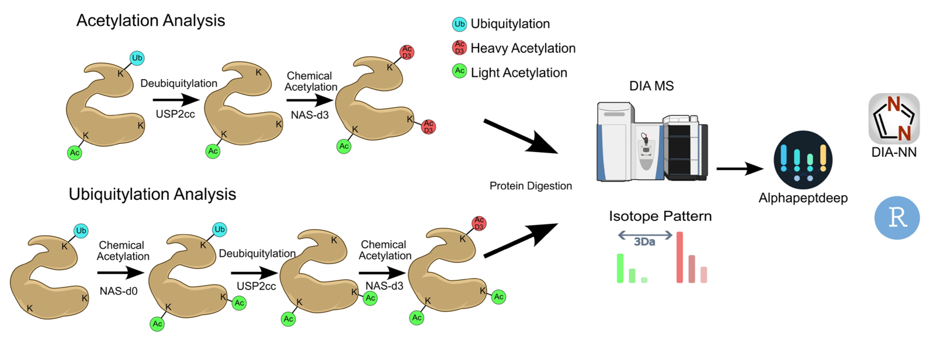
Figure 2.
Identification of peptides, proteins and stoichiometry of the different cell lines. A) Number of identified peptides acetylated and ubiquitylated across C33a, HaCaT, HeLa and SiHa cell lines. B) Number of identified proteins acetylated and ubiquitylated across C33a, HaCaT, HeLa and SiHa cell lines. C) Venn diagram of the acetylated peptides across the cell lines. D) Venn diagram of the ubiquitylated peptides across the cell lines. E) Ridgeline plot of acetylation stoichiometry distribution of C33a, HaCaT, HeLa and SiHa cell lines. F) Ridgeline plot of ubiquitylation stoichiometry distribution of C33a, HaCaT, HeLa and SiHa cell lines.
Figure 2.
Identification of peptides, proteins and stoichiometry of the different cell lines. A) Number of identified peptides acetylated and ubiquitylated across C33a, HaCaT, HeLa and SiHa cell lines. B) Number of identified proteins acetylated and ubiquitylated across C33a, HaCaT, HeLa and SiHa cell lines. C) Venn diagram of the acetylated peptides across the cell lines. D) Venn diagram of the ubiquitylated peptides across the cell lines. E) Ridgeline plot of acetylation stoichiometry distribution of C33a, HaCaT, HeLa and SiHa cell lines. F) Ridgeline plot of ubiquitylation stoichiometry distribution of C33a, HaCaT, HeLa and SiHa cell lines.
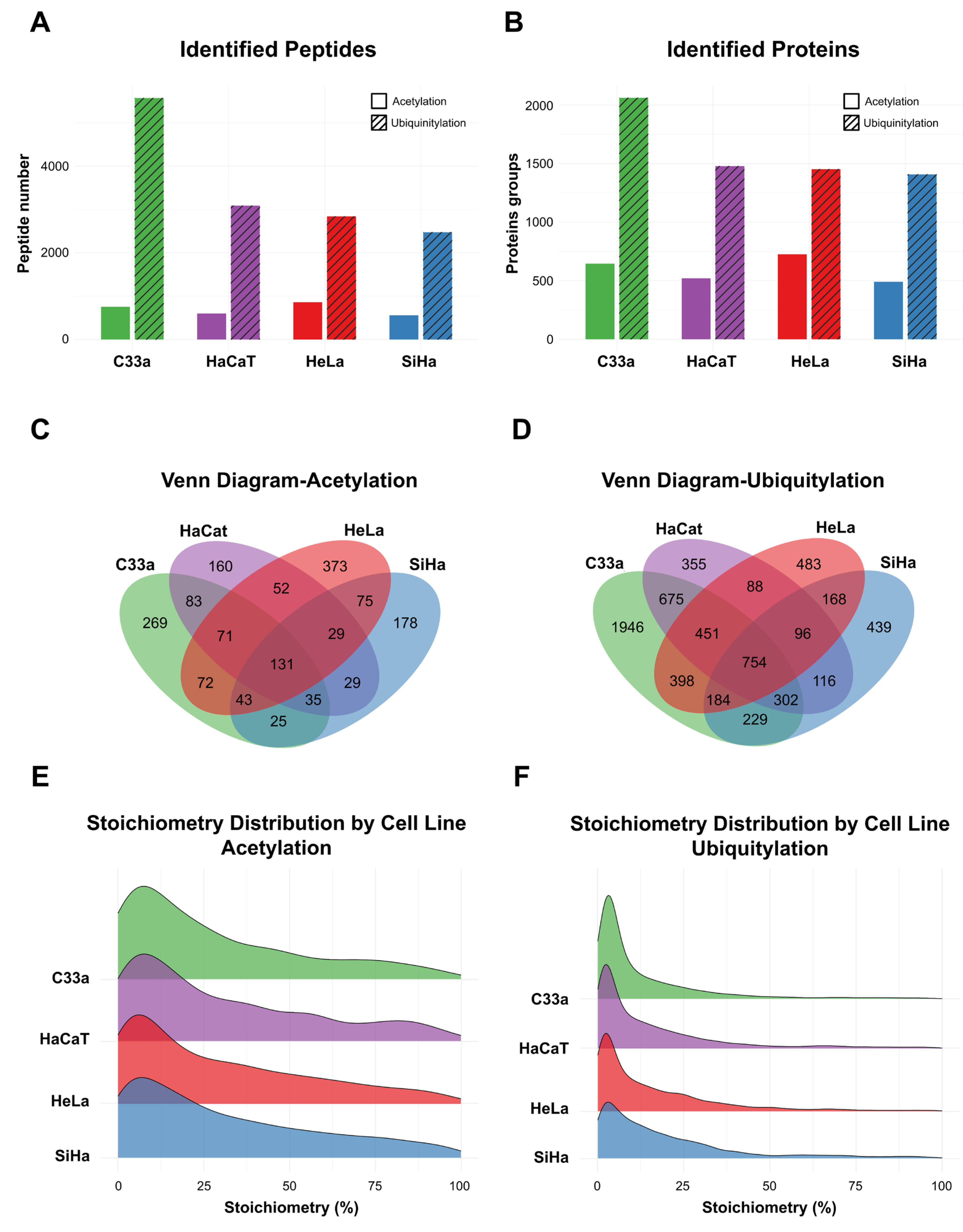
Figure 3.
Hierarchical clustering heatmaps and PCA of the acetylated and ubiquitylated proteins. A) Hierarchical clustering heatmap of the 150 most variable acetylated proteins of C33a, HaCaT, HeLa and SiHa cell lines. B) PCA analysis of 1,036 acetylated proteins of C33a, HaCaT, HeLa and SiHa cell lines. C) Hierarchical clustering heatmap of the 150 most variable ubiquitylated proteins of C33a, HaCaT, HeLa and SiHa cell lines. D) PCA analysis of 2,240 ubiquitylated proteins of C33a, HaCaT, HeLa and SiHa cell lines.
Figure 3.
Hierarchical clustering heatmaps and PCA of the acetylated and ubiquitylated proteins. A) Hierarchical clustering heatmap of the 150 most variable acetylated proteins of C33a, HaCaT, HeLa and SiHa cell lines. B) PCA analysis of 1,036 acetylated proteins of C33a, HaCaT, HeLa and SiHa cell lines. C) Hierarchical clustering heatmap of the 150 most variable ubiquitylated proteins of C33a, HaCaT, HeLa and SiHa cell lines. D) PCA analysis of 2,240 ubiquitylated proteins of C33a, HaCaT, HeLa and SiHa cell lines.
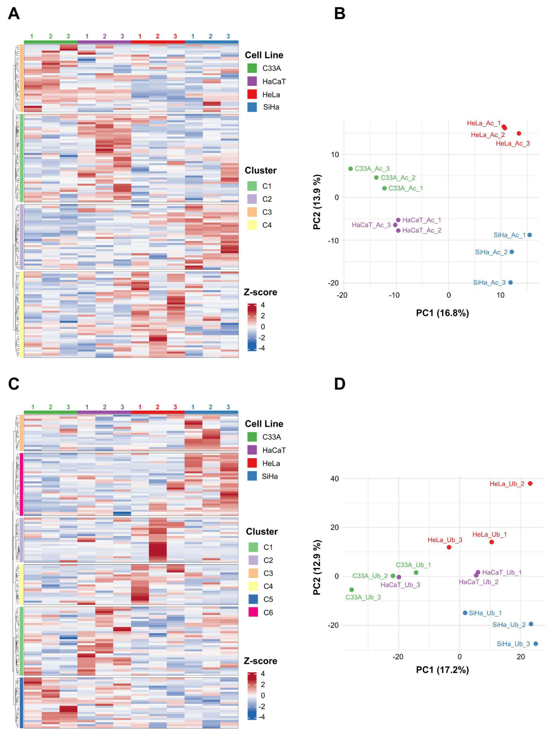
Figure 4.
One-way ANOVA (across the four cell lines: C33a, HaCaT, HeLa and SiHa) with Benjamini-Hochberg FDR correction (adjusted p < 0.05) applied to peptide-level stoichiometry values, identifying differentially modified acetylated and ubiquitinated proteins. A) Volcano plot of ANOVA results for acetylated peptides. B) Volcano plot of ANOVA results for ubiquitinated peptides. C) Ubiquitylation stoichiometry boxplot of GFPT1 across the cell lines (ANOVA/BH-significant). D) Ubiquitylation stoichiometry boxplot of AQP3 across the cell lines (ANOVA/BH-significant). E) Acetylation stoichiometry boxplot of IFIT3 across the cell lines (ANOVA/BH-significant). F) Acetylation stoichiometry boxplot of TOM1 across the cell lines (ANOVA/BH-significant).
Figure 4.
One-way ANOVA (across the four cell lines: C33a, HaCaT, HeLa and SiHa) with Benjamini-Hochberg FDR correction (adjusted p < 0.05) applied to peptide-level stoichiometry values, identifying differentially modified acetylated and ubiquitinated proteins. A) Volcano plot of ANOVA results for acetylated peptides. B) Volcano plot of ANOVA results for ubiquitinated peptides. C) Ubiquitylation stoichiometry boxplot of GFPT1 across the cell lines (ANOVA/BH-significant). D) Ubiquitylation stoichiometry boxplot of AQP3 across the cell lines (ANOVA/BH-significant). E) Acetylation stoichiometry boxplot of IFIT3 across the cell lines (ANOVA/BH-significant). F) Acetylation stoichiometry boxplot of TOM1 across the cell lines (ANOVA/BH-significant).
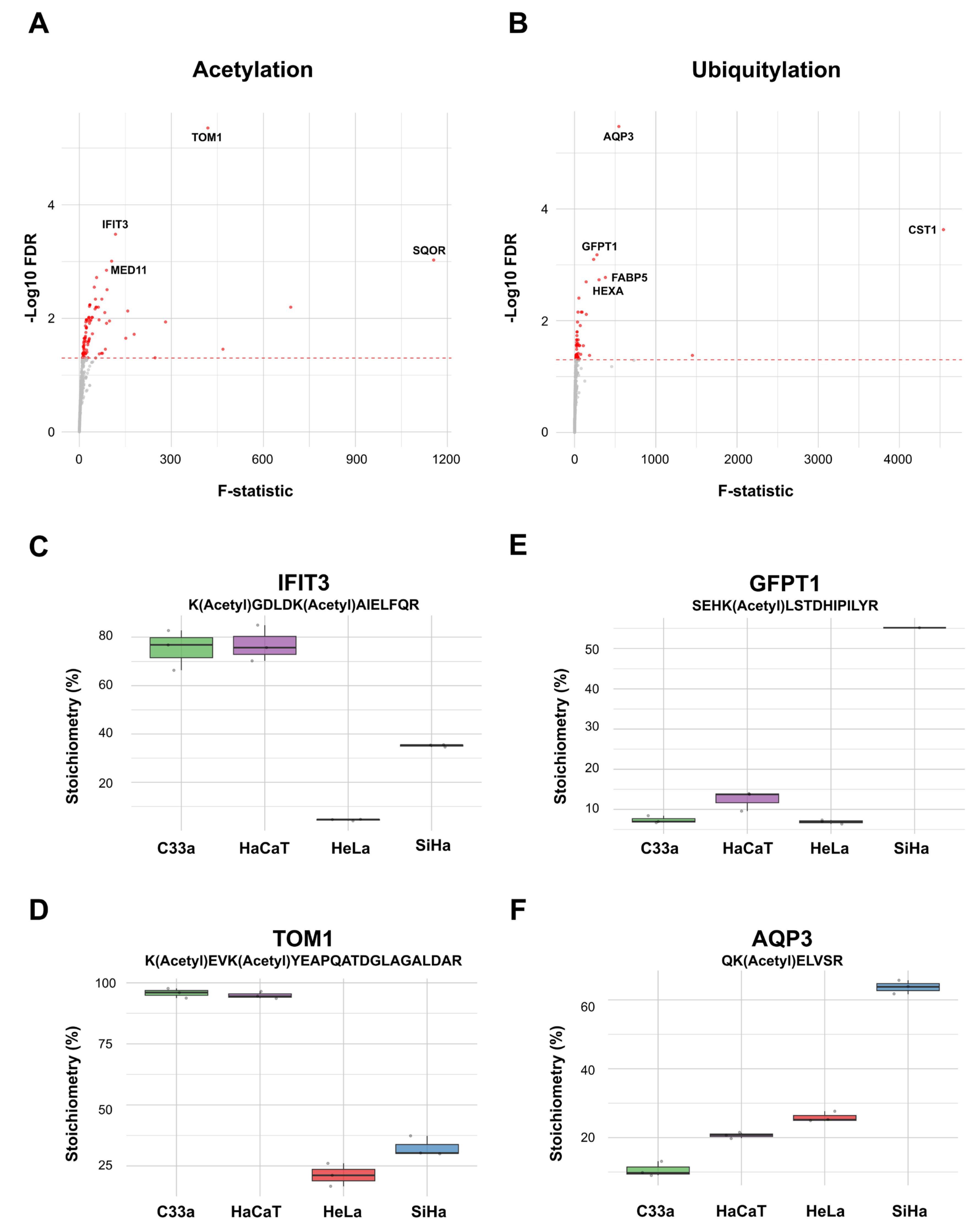
Figure 5.
Pathway enrichment analysis showing cell line-specific functional signatures. A) Pathway enrichment of unique identified acetylated proteins for each cell line. B) Pathway enrichment of unique identified ubiquitylated proteins for each cell line. Green for C33a, purple for HaCaT, red for HeLa, and blue for SiHa. The percentage of filled circle represents the amount of protein presence per cell line.
Figure 5.
Pathway enrichment analysis showing cell line-specific functional signatures. A) Pathway enrichment of unique identified acetylated proteins for each cell line. B) Pathway enrichment of unique identified ubiquitylated proteins for each cell line. Green for C33a, purple for HaCaT, red for HeLa, and blue for SiHa. The percentage of filled circle represents the amount of protein presence per cell line.
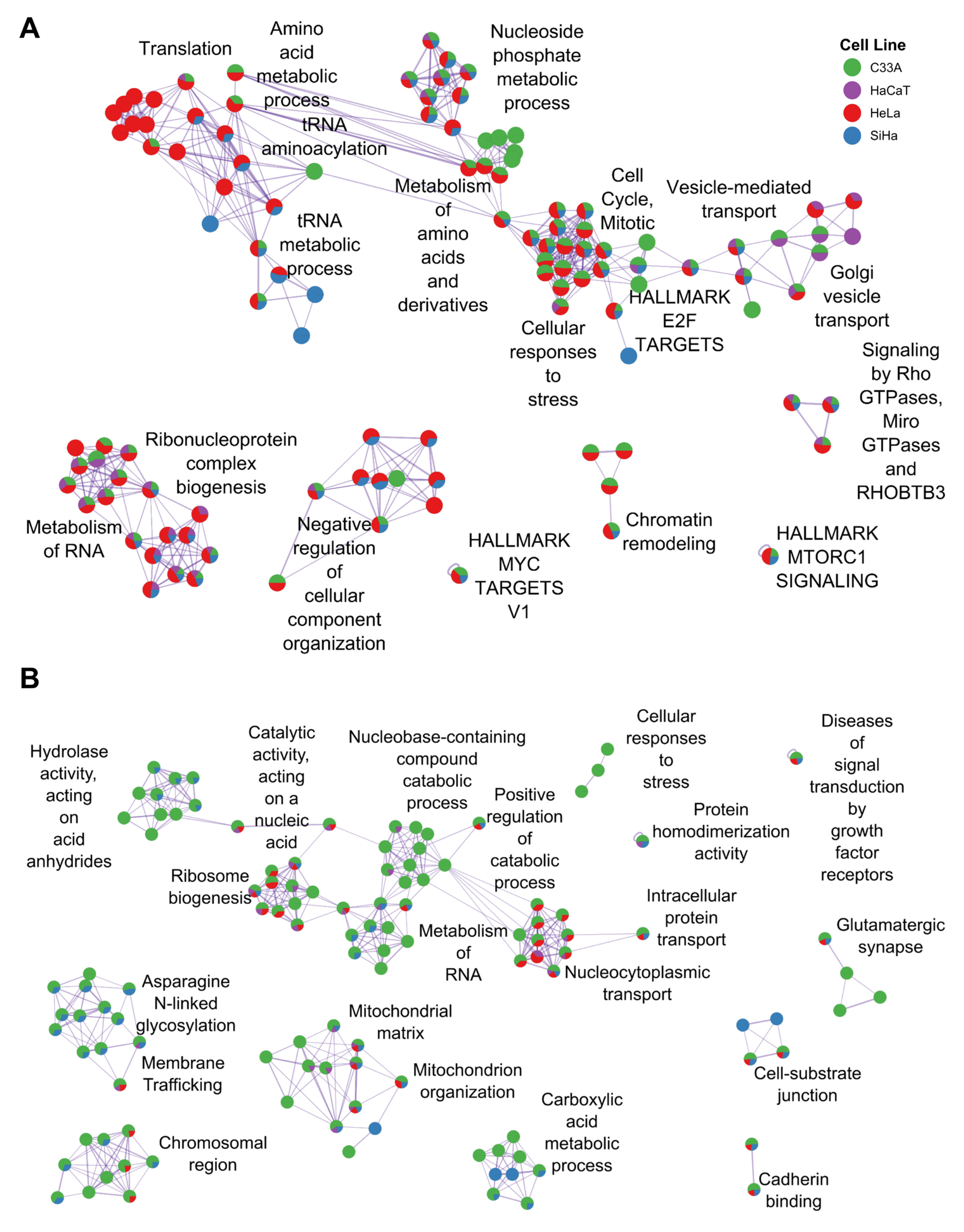
Figure 6.
Schematic model of the lysine fate code in cervical cancer. A shared lysine residue is subject to competing modification by lysine acetyltransferases/deacetylases (KATs/KDACs) and E3 ubiquitin ligases/deubiquitinases (DUBs). High-occupancy acetylation favors a stable, functionally active protein state (transcriptional activity, chromatin remodeling, metabolic stability), whereas ubiquitination favors degradation or turnover (p53 clearance, innate immune suppression, ERAD/quality control). The balance between these outcomes is set by cellular context: HPV-positive lines (SiHa, HeLa) show acetylation-dominant, CBP/p300-driven signaling with selective p53 clearance; the HPV-negative C33a line shows an expanded, ubiquitination-dominant proteostatic surveillance state; and the non-tumorigenic HaCaT control shows a more balanced, baseline regulatory architecture reflecting differentiation programs. Created with Inkscape 1.4 (https://inkscape.org).
Figure 6.
Schematic model of the lysine fate code in cervical cancer. A shared lysine residue is subject to competing modification by lysine acetyltransferases/deacetylases (KATs/KDACs) and E3 ubiquitin ligases/deubiquitinases (DUBs). High-occupancy acetylation favors a stable, functionally active protein state (transcriptional activity, chromatin remodeling, metabolic stability), whereas ubiquitination favors degradation or turnover (p53 clearance, innate immune suppression, ERAD/quality control). The balance between these outcomes is set by cellular context: HPV-positive lines (SiHa, HeLa) show acetylation-dominant, CBP/p300-driven signaling with selective p53 clearance; the HPV-negative C33a line shows an expanded, ubiquitination-dominant proteostatic surveillance state; and the non-tumorigenic HaCaT control shows a more balanced, baseline regulatory architecture reflecting differentiation programs. Created with Inkscape 1.4 (https://inkscape.org).
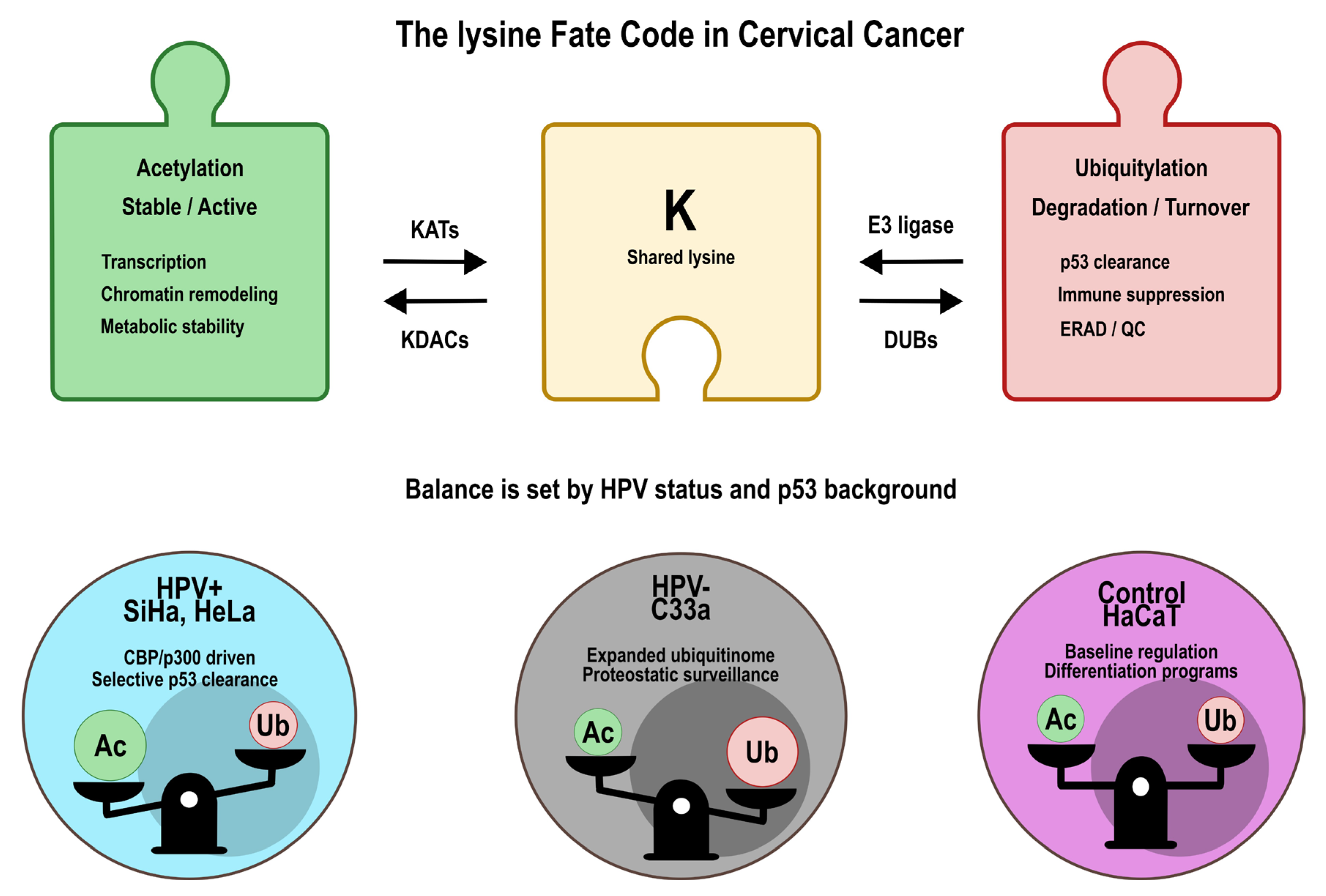

Table 1.
Summary of acetylated and ubiquitinated peptide and protein identifications across cell lines.
Table 1.
Summary of acetylated and ubiquitinated peptide and protein identifications across cell lines.
| Cell Line | HPV Status | Acetylation | Ubiquitylation | ||||||
|---|---|---|---|---|---|---|---|---|---|
| Total Peptides | Total Proteins | Modified Peptides | Modified Proteins | Total Peptides | Total Proteins | Modified Peptides | Modified Proteins | ||
| SiHa | HPV16+ | 33581 | 6014 | 557 | 490 | 28713 | 5602 | 2,473 | 1,407 |
| HeLa | HPV18+ | 35401 | 6072 | 858 | 725 | 30141 | 5624 | 2,839 | 1,452 |
| C33a | HPV− | 35918 | 6135 | 753 | 645 | 31546 | 5816 | 5,570 | 2,062 |
| HaCaT | Control | 33893 | 6066 | 601 | 520 | 27786 | 5497 | 3,089 | 1,478 |
Values represent raw identification counts per cell line; no statistical comparison was performed on these data. Statistical analysis (one-way ANOVA, FDR-corrected) was applied to peptide-level stoichiometry values only, as described in the Data analysis subsection and shown in Figure 4.
Disclaimer/Publisher’s Note: The statements, opinions and data contained in all publications are solely those of the individual author(s) and contributor(s) and not of MDPI and/or the editor(s). MDPI and/or the editor(s) disclaim responsibility for any injury to people or property resulting from any ideas, methods, instructions or products referred to in the content. |
© 2026 by the authors. Licensee MDPI, Basel, Switzerland. This article is an open access article distributed under the terms and conditions of the Creative Commons Attribution (CC BY) license (http://creativecommons.org/licenses/by/4.0/).
Copyright: This open access article is published under a Creative Commons CC BY 4.0 license, which permit the free download, distribution, and reuse, provided that the author and preprint are cited in any reuse.