
Submitted:
30 September 2026
Posted:
30 September 2026
You are already at the latest version
Abstract
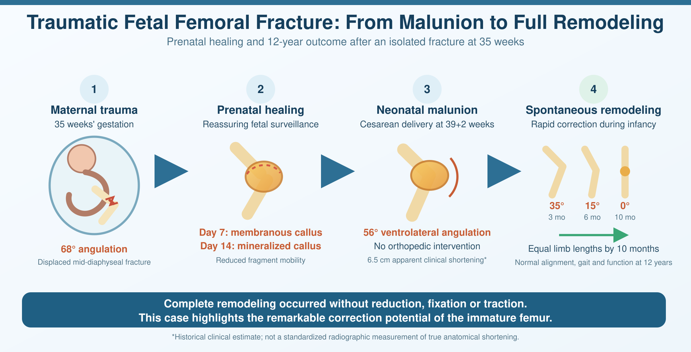
Background: Fetal long-bone fractures are rare and may result from maternal trauma, skeletal dysplasia, or, less commonly, apparently spontaneous events. Prenatal diagnosis is primarily based on ultrasonography, while the subsequent course of fetal fracture healing and long-term remodeling remains poorly documented. Case Presentation: A 35-week fetus sustained a displaced mid-diaphyseal fracture of the right femur following a maternal frontal motor vehicle collision. Initial ultrasonography demonstrated approximately 68° of interfragmentary angulation and surrounding soft-tissue edema. Maternal and fetal surveillance over the following 7 days showed no evidence of fetal compromise. Serial ultrasound examinations demonstrated progressive callus formation, with a globular membranous callus at day 7 and a prominent hyperechoic callus with peripheral vascularization and reduced fragment mobility at day 14. Delivery occurred by cesarean section at 39+2 weeks for an obstetric indication. The newborn weighed 3150 g and had Apgar scores of 9 and 10. The fracture was clinically consolidated but remained in severe malunion, with approximately 56° of ventrolateral angulation and a clinically estimated 6.5 cm apparent clinical shortening. No orthopedic intervention was undertaken. Progressive spontaneous correction was documented, with residual angulation of 35° at 3 months and 15° at 6 months. By 10 months, complete angular correction and limb-length equalization were documented. Independent walking was achieved at 12 months. At 12 years of age, the child had normal limb alignment, equal limb lengths, normal gait, and no functional sequelae. Conclusions: This case demonstrates an unusual combination of prenatal fracture healing, severe neonatal malunion, and complete long-term spontaneous correction without orthopedic intervention. The reported 6.5-cm shortening should be interpreted as apparent clinical shortening rather than definitive anatomical shortening because it was not established by standardized radiographic limb-length measurement. Geometric analysis indicates that angular deformity alone could account for only approximately 0.8–1.0 cm of shortening at term. The subsequent course is consistent with substantial growth-related remodeling and provides a rare longitudinal example of the remodeling potential of the immature femur.
Keywords:
fetal femoral fracture
; fetal trauma
; prenatal diagnosis
; ultrasonography
; callus formation
; malunion
; spontaneous remodeling
; femoral shaft fracture
; physeal growth
; long-term follow-up
1. Introduction
Trauma during pregnancy is an important cause of maternal and fetal morbidity and requires coordinated maternal and fetal assessment. Motor vehicle collisions are among the most common causes of blunt trauma during pregnancy. The evaluation of the pregnant trauma patient prioritizes maternal stabilization, assessment for placental and uterine injury, and appropriate fetal surveillance. A multidisciplinary approach is recommended to optimize maternal and fetal outcomes.[1,2]
Fetal skeletal injury following maternal trauma is uncommon. When a fetal long-bone fracture is identified, the differential diagnosis includes direct traumatic injury, birth-related trauma when the diagnosis is made postnatally, and underlying skeletal fragility or skeletal dysplasia. Isolated intrauterine femoral fractures have been reported in otherwise normal fetuses, including cases in which no maternal trauma was identified.[3]
Ultrasonography is the principal imaging modality for prenatal skeletal assessment. Standard fetal femur-length measurement requires visualization of the ossified diaphysis and measurement along its longest axis, with appropriate image-plane selection and caliper placement. These technical requirements become particularly important when the femur is acutely fractured and severely angulated, because the proximal and distal fragments may no longer lie in a single imaging plane.[4]
Under such circumstances, the geometric assumptions of routine femur-length measurement may no longer be applicable. The resulting measurement may reflect a combination of true fragment length, angular configuration, fragment overlap, projectional effects, and limb position rather than the anatomical length of a straight femoral shaft. Technical factors such as femoral orientation relative to the ultrasound beam, depth, and gain have been shown to influence fetal femur measurements.[5]
We present a rare case of a traumatic fetal femoral shaft fracture diagnosed at 35 weeks’ gestation, followed by documented prenatal callus formation, delivery at term with severe residual malunion, conservative postnatal management, and complete spontaneous correction within the first year of life, with normal limb alignment and function documented at 12 years.
2. Case Presentation
2.1. Maternal Trauma and Initial Fetal Assessment
A pregnant woman at 35 weeks’ gestation was involved in a frontal motor vehicle collision. She was restrained with a three-point seat belt. Following maternal assessment, fetal ultrasonography demonstrated an acute displaced fracture of the right femoral mid-diaphysis. The fracture was associated with approximately 68° of interfragmentary angulation and moderate surrounding soft-tissue edema. The remainder of the fetal skeletal examination did not reveal multiple fractures, generalized long-bone bowing, abnormal thoracic configuration, or other findings suggestive of skeletal dysplasia. The temporal relationship between the motor vehicle collision and the newly identified isolated femoral fracture strongly supported an acquired traumatic etiology. The absence of additional fractures or generalized skeletal abnormalities further argued against an underlying skeletal dysplasia. The current nosology of genetic skeletal disorders provides a framework for the differential diagnosis.[6]
2.2. Maternal-Fetal Surveillance
The pregnant patient underwent continued maternal and fetal surveillance for 7 days. Fetal heart-rate monitoring remained reassuring. Doppler examinations and biophysical assessment did not demonstrate evidence of fetal compromise. The decision to continue the pregnancy was based on the absence of fetal distress or other obstetric indications requiring immediate delivery. Contemporary trauma guidance emphasizes maternal stabilization and appropriate fetal surveillance; an isolated fetal skeletal injury, in the absence of fetal compromise, placental injury, maternal deterioration, or another obstetric indication, does not by itself establish a universal indication for preterm delivery.[1,2]
2.3. Serial Prenatal Ultrasonographic Evolution
Prenatal ultrasound examinations were performed using equipment commonly available in Romania during 2012–2016, including a Voluson 730 Expert/Voluson E8 system with a 3.5–5 MHz convex transabdominal probe and, when needed, a high-frequency linear probe for detailed assessment of the fracture site. Seven days after the initial injury, ultrasonography demonstrated a globular membranous callus at the fracture site. At 14 days after trauma, corresponding to approximately 37 weeks’ gestation, the callus had become markedly hyperechoic and showed peripheral vascularization. Mobility between the fracture fragments was reduced, although fetal movement of the affected limb remained preserved. These serial changes were interpreted as progressive fracture consolidation, with increasing organization and mineralization of the callus and decreasing interfragmentary mobility.
The prenatal ultrasound examination was performed with the transducer positioned over the region of maximal femoral angulation, with the imaging field oriented to include both fracture fragments. Because of the severe angulation, the proximal and distal fragments were not necessarily located in the same imaging plane. This technical detail is important because standard fetal femur-length measurement assumes visualization of the ossified diaphysis along its longest axis. In the present case, the severe angular displacement altered this geometry, potentially increasing uncertainty in any linear measurement of the fractured femur.[4]
The proximal fragment appeared relatively hypoechoic compared with the more clearly visualized distal fragment. In the context of the marked angular deformity, this appearance was considered potentially related to altered insonation geometry and out-of-plane orientation rather than evidence of abnormal bone mineralization. Changes in depth, gain, and the angle of the femur relative to the ultrasound beam have previously been shown to influence fetal femur-length measurements.[5]
Figure 1.
Routine prenatal ultrasound following maternal road traffic accident demonstrating fetal femoral shaft fracture at 35 weeks’ gestation. Legend: Three representations of the same ultrasound image are shown. (A) Original unannotated high-resolution image demonstrating a displaced mid-diaphyseal fracture of the right femur with marked angulation. (B) The same image with anatomical identification: asterisks (*) clearly marking the proximal and distal femoral fragments and the fracture site. The marked angulation is evident.(C) The same image with longitudinal axes drawn along the proximal fragment (green arrow) and distal fragment (blue arrow). The white arrow indicates the fracture site with surrounding soft-tissue edema.
Figure 1.
Routine prenatal ultrasound following maternal road traffic accident demonstrating fetal femoral shaft fracture at 35 weeks’ gestation. Legend: Three representations of the same ultrasound image are shown. (A) Original unannotated high-resolution image demonstrating a displaced mid-diaphyseal fracture of the right femur with marked angulation. (B) The same image with anatomical identification: asterisks (*) clearly marking the proximal and distal femoral fragments and the fracture site. The marked angulation is evident.(C) The same image with longitudinal axes drawn along the proximal fragment (green arrow) and distal fragment (blue arrow). The white arrow indicates the fracture site with surrounding soft-tissue edema.
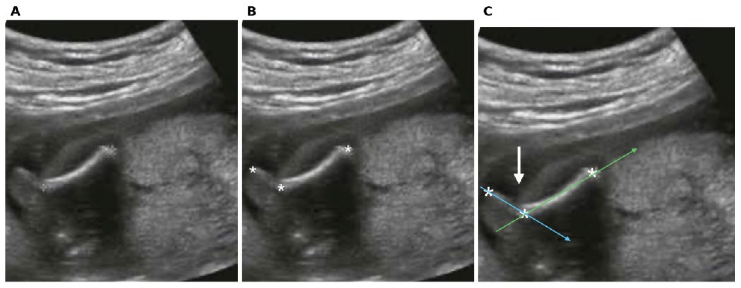
Interfragmentary angulation was estimated at approximately 68° at presentation, with approximately 56° ventrolateral angulation documented postnatally at term in the consolidated malunion. As a result of the severe angulation, the proximal femoral fragment lies in a different imaging plane than the distal fragment and appears relatively hypoechoic. This appearance is interpreted as related to altered insonation geometry and out-of-plane orientation rather than evidence of abnormal bone mineralization, as noted in the original observation: the proximal fragment lies in a different plane than the distal fragment and appears hypoechoic. The associated apparent clinical shortening through the arched configuration was recorded as 6.5 cm apparent clinical shortening at birth. Geometric analysis indicates that angular deformity alone accounts for only approximately 0.8–1.0 cm of projected shortening for an expected term femur length of 7.2–7.8 cm; the historical value is therefore retained as apparent clinical shortening and not as definitive anatomical shortening.[4,5,7]
2.4. Delivery and Neonatal Examination
At 39 weeks of gestation, the patient underwent an elective cesarean delivery. The mode of delivery was individualized in consideration of the prenatally diagnosed fetal femoral fracture, which was undergoing osseous consolidation, as well as the mother’s preference for cesarean delivery, following appropriate counseling and shared decision-making. The newborn weighed 3150 g and had Apgar scores of 9 and 10. At birth, the right femur was clinically consolidated in severe malunion, with approximately 56° of ventrolateral angular deformity. A hard, seemingly painless callus was palpable. Hip and knee motion was preserved. A clinical estimate of 6.5 cm apparent clinical shortening was recorded. This historical value requires an important methodological qualification. It was a clinical estimate rather than a standardized radiographic measurement of true femoral or limb length. The examination was performed without forced extension because of concern about disturbing the recently consolidated, exuberant callus; the hip and knee therefore remained in physiological flexion. In addition, the marked angular deformity and possible longitudinal overlap of the fracture fragments made conventional linear assessment difficult. No immediate postnatal radiograph or standardized radiographic limb-length measurement was performed at birth because the fracture was already clinically consolidated with a hard, painless callus, the infant had preserved hip and knee motion and neurovascular status, and forced extension was avoided to protect the recently consolidated exuberant callus. No orthopedic reduction, fixation, traction, or other operative treatment was undertaken at that time.
2.5. Postnatal Evolution
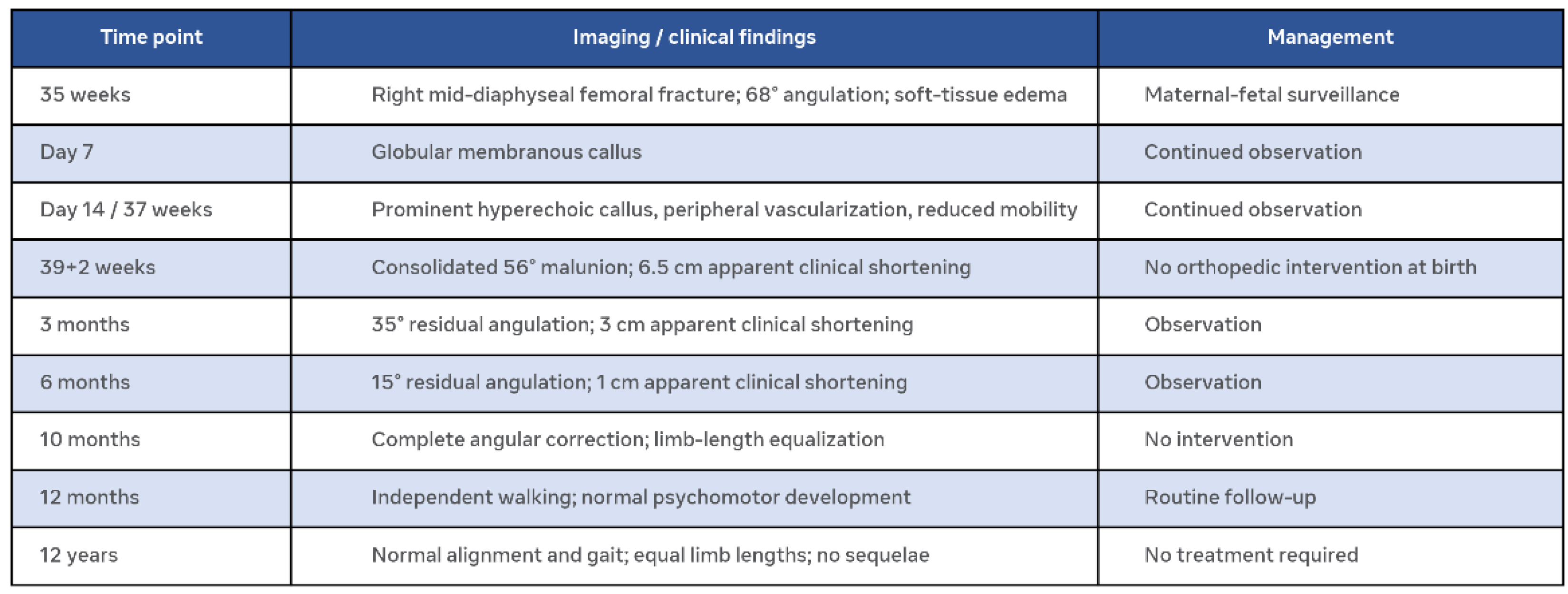
The infant was followed clinically with serial assessment of femoral alignment and limb length. Although the parents received an express recommendation to attend an orthopedic consultation immediately after birth, they were informed by ward nursing staff that other newborns with birth-related fractures had healed without treatment. Consequently, they presented for orthopedic evaluation at 3, 6, 10 months and at 1 year, despite the initial recommendation. At 3 months, residual angulation had decreased to approximately 35°, with approximately 3 cm apparent clinical shortening. At 6 months, the residual angulation was approximately 15°, with approximately 1 cm apparent clinical shortening. At 10 months, complete angular correction and equalization of limb length were documented. At 12 months, the child was independently walking with normal psychomotor development. At 12 years of age, clinical examination demonstrated normal lower-limb alignment, equal limb lengths, normal gait, and no functional limitation or other orthopedic sequelae.

Table 1.
Clinical and imaging evolution.
 |
3. Quantitative Consideration of the Reported Shortening
The reported 6.5-cm apparent clinical shortening is one of the most striking features of this case but is also the measurement that requires the greatest methodological caution. We therefore distinguish between: (1) geometric shortening attributable to angular deformity, (2) true shortening resulting from longitudinal overlap of fracture fragments, and (3) apparent shortening related to limb position and clinical measurement technique.
For a symmetric angular deformity, the projected end-to-end length can be approximated geometrically as: Leffective = L cos(alpha/2) and therefore: S = L [1 - cos(alpha/2)] where L represents the original straight femoral length, alpha the angular deformity, and S the component of shortening attributable solely to the angular configuration.
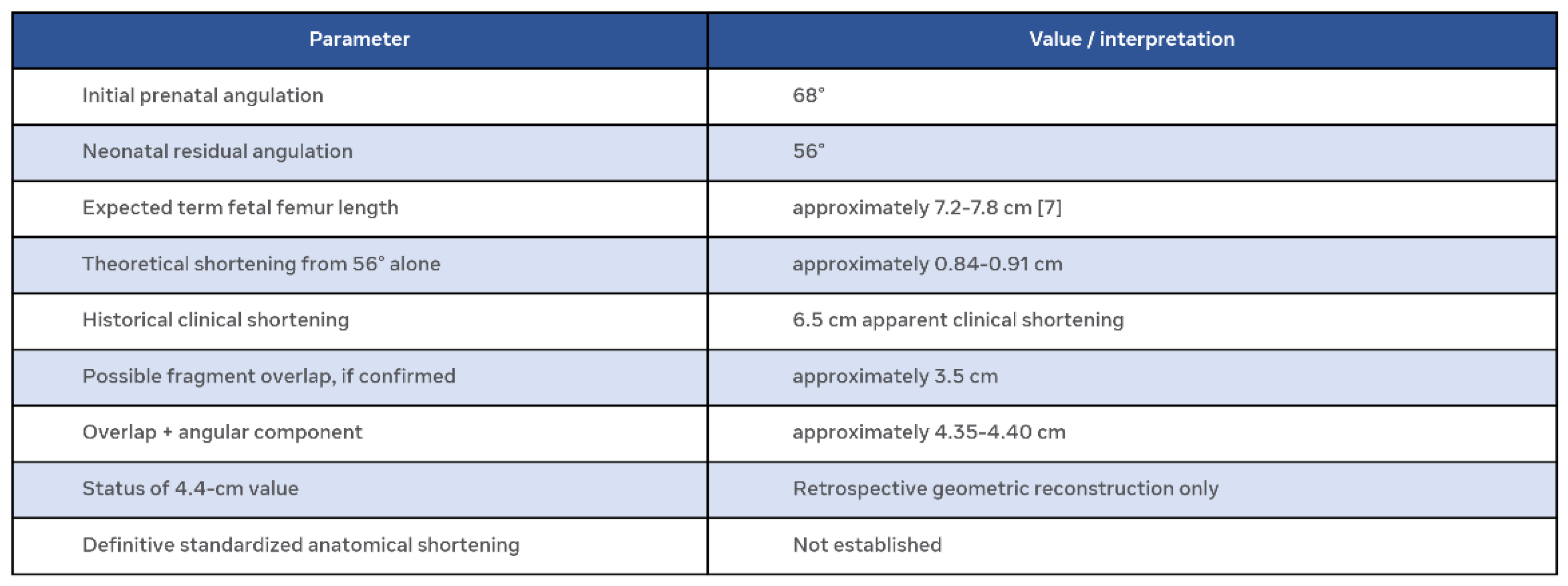
At 56°: 1 - cos(28°) ≈0.117. Thus, the angular deformity produces approximately 11.7% geometric shortening. Established fetal biometric relationships provide a term fetal femur length of approximately 7.2–7.8 cm. For L=7.2–7.8 cm: S≈0.84–0.91 cm. Therefore, the 56° angular deformity alone cannot explain a 6.5-cm apparent clinical shortening. The same calculation at the initial 68° deformity gives: 1 - cos(34°)≈0.171 which corresponds to approximately 1.1–1.3 cm for a femur in the expected late-third-trimester range. This analysis demonstrates that the historical apparent clinical shortening value cannot be interpreted as the direct geometric consequence of angulation alone.[7]
Longitudinal overlap, or overriding, is conceptually different from angular deformity.[10] Overriding should only be reported as true anatomical shortening if the original imaging or contemporaneous documentation demonstrates longitudinal overlap of the proximal and distal fracture fragments. Severe angulation alone is insufficient to establish the amount of true shortening. If the original images independently demonstrate approximately 3.5 cm of true longitudinal overlap, then an approximate combined geometric estimate would be: 3.5 cm + 0.85–0.90 cm ≈4.35–4.40 cm. However, this value should be described as a retrospective reconstruction rather than a measured true limb-length discrepancy unless the 3.5-cm overlap can be independently demonstrated on the original imaging. The manuscript therefore retains the historical value as “6.5 cm apparent clinical shortening” rather than converting it into a definitive 4.4-cm anatomical shortening.
4. Discussion
4.1. Rarity:
Intrauterine femoral fracture is exceptionally uncommon. Present case distinctive because fracture occurred immediately after documented maternal motor vehicle collision at 35 weeks and because serial observations captured complete sequence from acute fracture through prenatal callus formation, severe neonatal malunion, progressive remodeling, and normal long-term function.[3]
4.2. Prenatal Diagnosis:
Ultrasonography remains primary modality for prenatal diagnosis. Measurement reliability may decrease when anatomy does not conform to assumptions. Present case extreme example: femur not continuous straight structure but sharply angulated displaced fracture with two fragments potentially in different imaging planes. Relatively hypoechoic appearance of proximal fragment should be interpreted in context of orientation relative to ultrasound beam. This is limitation of imaging severely deformed bone rather than operator error.[4,5,8]
4.3. Ultrasound vs Mri and Ct:
Retrospective correction using fetal MRI inappropriate due to intermodality differences mean difference 4.6mm, poor agreement. 3D-CT may provide additional detail when dysplasia suspected.[11] Neither MRI nor CT should be used retrospectively to correct contemporaneous ultrasound measurement. If original ultrasound images available, most rigorous approach is blinded retrospective review by two experienced observers.[9]
4.4. Remodeling:
Wallace and Hoffman evaluated 28 children with united middle-third femoral shaft fractures and residual angular deformities 10°–26°.[12] Average correction ∼85%, ∼74% attributed to changes involving proximal and distal physes. These findings provide biological precedent but should not be extrapolated quantitatively. Their cohort had substantially smaller deformities than 56° neonatal malunion observed here. 74% physeal contribution refers to correction of overall interphyseal alignment. Complete correction of 56° deformity by 10 months suggests both fracture-site remodeling and growth-related physeal modulation contributed.
4.5. Growth Cartilage Memory and Wolff’s Law:
Wolff’s law describes the ability of bone to adapt its architecture and structure in response to the mechanical stresses to which it is subjected.[13] In this context, changes in mechanical loading may influence bone remodeling and tissue organization, thereby contributing to the adaptation of bone to functional demands.
Based on clinical observations concerning the behavior of the growth plate, G. Burnei proposed the concept of “growth plate memory,” subsequently formulated in 2021 as the “law of growth cartilage memory.”[14] According to the author’s formulation, “The law of growth cartilage memory and Wolff’s law act in interdependence.” This concept suggests that the physis may retain, to some extent, information regarding the genetically programmed growth trajectory and may tend to return toward this trajectory after removal of the deforming factor, provided that residual growth potential remains.
In support of this hypothesis, G. Burnei described the case of a newborn from a nomadic family in whom a femoral fracture healed in malalignment, with an angulation of approximately 90°.[14] The subsequent clinical course was characterized by an acceleration of growth in the affected segment, such that, after approximately three years, the limb-length discrepancy had been corrected. The author interpreted this outcome as a possible manifestation of physeal “memory,” proposing that this potential is greatest during the neonatal period, progressively decreases with age, and disappears following physiological closure of the growth plates.
However, this concept must be interpreted with caution. The evolution of the present case can be considered compatible with the hypothetical mechanism of growth cartilage memory, but it cannot, in itself, demonstrate the existence of such a mechanism. Similarly, the study of two clinical cases and several observations found in the evolution of other cases is insufficient to establish a biological law or to demonstrate a causal relationship.
A more cautious interpretation is that the observed phenomenon may be consistent with an interaction between growth-plate modulation and mechanically driven bone remodeling. Within this model, the growth plate may respond to local changes in loading and alignment, while bone remodeling, in accordance with the principles described by Wolff’s law, contributes to the progressive adaptation of bone shape and structure.
Therefore, G. Burnei’s concept may provide an interesting conceptual framework for interpreting phenomena involving the recovery of growth after deformities or injuries during the developmental period. Nevertheless, in the absence of sufficient experimental and clinical evidence, “growth plate memory” should currently be regarded as a biologically plausible hypothesis rather than a definitively established causal mechanism.
4.6. Why Conservative Management Was Successful:
At birth deformity severe enough that operative correction could have been considered if persistent functional impairment developed. However the newborn was asymptomatic, hip and knee motion preserved, fracture already consolidated. Subsequent rapid correction suggests biological remodeling potential exceptionally high. Present case should not be interpreted as evidence that all severely displaced fetal femoral fractures should be treated conservatively. Rather, illustrates that conservative management may be successful in selected cases when fracture is isolated, the infant clinically stable, limb motion and perfusion preserved, and close orthopedic follow-up available.
4.7. Significance of the 6.5-Cm Apparent Clinical Shortening:
Reported shortening requires particular attention because it was considerably greater than shortening that can be explained by angular deformity. Measurement was clinical rather than standardized radiographic; obtained with hip and knee in physiological flexion; performed without forced extension because of concern regarding consolidated callus; obtained in setting of severe angular deformity; potentially influenced by longitudinal fragment overlap. Angular geometry alone accounts for only ∼0.8–1.0 cm at 56° deformity for term fetal femur ∼7.2–7.8 cm. Consequently manuscript should not state newborn had true 6.5-cm femoral shortening. Likewise manuscript should not state true shortening was 4.4 cm unless original imaging independently demonstrates ∼3.5 cm of true longitudinal fragment overlap.
4.8. Long-Term Outcome:
12-year follow-up is particularly important. Complete angular correction and equal limb length achieved without surgical intervention, followed by normal independent walking and normal long-term functional outcome. Absence of residual limb-length discrepancy, gait abnormality, pain, or functional limitation at 12 years suggests initial severe malunion did not result in clinically detectable permanent sequelae. 12-year observation provides substantially longer follow-up than commonly available in reports of prenatal fetal fractures.
5. Limitations
First, single case cannot establish incidence, natural history, or mechanism. Second, original prenatal ultrasound obtained under non-standard geometric conditions because femur acutely fractured and severely angulated. Third, the 6.5-cm value was a historical clinical estimate of apparent clinical shortening and was not established by standardized radiographic limb-length measurement. It should be interpreted as apparent clinical shortening. Fourth, individual contributions of fragment overlap, angular deformity, and limb positioning cannot now be separated with certainty. Fifth, relative contributions of fracture-site remodeling and physeal growth cannot be quantified. If original ultrasound images available, blinded retrospective remeasurement by two experienced observers would strengthen quantitative component. Finally, absence of radiographic long-term measurements at every follow-up interval limits ability to construct precise quantitative remodeling curve. Principal strength is longitudinal clinical and imaging observation rather than formal morphometric analysis.
6. Conclusions
This case describes an exceptionally rare traumatic fetal femoral shaft fracture diagnosed at 35 weeks’ gestation, followed by prenatal callus formation, term delivery with severe neonatal malunion, and complete spontaneous remodeling within 10 months. Most important observation is not simply magnitude of initial deformity, but longitudinal demonstration that a severely angulated neonatal femoral malunion can undergo complete spontaneous correction during early growth without orthopedic intervention. The reported 6.5-cm apparent clinical shortening should be regarded as apparent clinical shortening, because angular deformity alone accounts for only approximately 0.8–1.0 cm and standardized measurement of true anatomical shortening was not available. If contemporaneous imaging independently demonstrates approximately 3.5 cm of longitudinal fragment overlap, a retrospective combined estimate of approximately 4.4 cm may be presented as a geometric reconstruction, but not as a measured true limb-length discrepancy. The remodeling observed is consistent with a combination of fracture-site remodeling and growth-related physeal modulation. The case is also compatible with broader concepts of mechanically driven skeletal adaptation and growth-cartilage-mediated correction, but it does not establish a causal mechanism or prove the proposed “growth cartilage memory” concept. This case therefore provides a rare longitudinal clinical example of the remarkable remodeling potential of the immature human femur following severe prenatal traumatic injury.
Table 2.
Quantitative interpretation of the neonatal deformity.
 |
Ethical statement
Written informed consent was obtained from the patient’s parents for publication of this case report, including anonymized clinical images and long-term follow-up data. The parents consented to the 12-year follow-up evaluation and to the use of the images for scientific and educational purposes. All personal identifiers have been removed to ensure anonymity.
References
- Jain, V.; Chari, R.; Maslovitz, S.; Farine, D. Guidelines for the management of a pregnant trauma patient. J. Obstet. Gynaecol. Can. 2015, 37, 553–574. [Google Scholar] [CrossRef] [PubMed]
- Appelbaum, R.D.; et al. Trauma in pregnancy: A systematic review, meta-analysis, and practice management guideline from the Eastern Association for the Surgery of Trauma. J. Trauma Acute Care Surg. 2025, 99, 298–309. [Google Scholar] [CrossRef] [PubMed]
- Senanayake, H.; Anandakumar, C.; De Silva, M.V.C. Mid-trimester fracture of femur in a normal fetus. J. Obstet. Gynaecol. Res. 2003, 29, 186–188. [Google Scholar] [CrossRef] [PubMed]
- Salomon, L.J.; Alfirevic, Z.; da Silva Costa, F.; Deter, R.L.; Figueras, F.; Ghi, T.; Glanc, P.; Khalil, A.; Lee, W.; Napolitano, R.; et al. ISUOG Practice Guidelines: ultrasound assessment of fetal biometry and growth. Ultrasound Obstet. Gynecol. 2019, 53, 715–723. [Google Scholar] [CrossRef] [PubMed]
- Krook, P.M.; Wawrukiewicz, A.S.; Hackethorn, J.C. Caveats in the sonographic determination of fetal femur length for estimation of gestational age. Radiology 1985, 154, 823–824. [Google Scholar] [CrossRef] [PubMed]
- Unger, S.; Ferreira, C.R.; Mortier, G.R.; Ali, H.; Bertola, D.R.; Calder, A.; Cohn, D.H.; Cormier-Daire, V.; Girisha, K.M.; Hall, C.; et al. Nosology of genetic skeletal disorders: 2023 revision. Am. J. Med. Genet. A 2023, 191, 1164–1209. [Google Scholar] [CrossRef] [PubMed]
- Hadlock, F.P.; Deter, R.L.; Harrist, R.B.; Park, S.K. Estimating fetal age: computer-assisted analysis of multiple fetal growth parameters. Radiology 1984, 152, 497–501. [Google Scholar] [CrossRef] [PubMed]
- Combs, C.A.; Amara, S.; Kline, C.; Ashimi Balogun, O.; Bowman, Z.S. Quantitative approach to quality review of prenatal ultrasound examinations: fetal biometry. J. Clin. Med. 2024, 13, 4860. [Google Scholar] [CrossRef] [PubMed]
- Parkar, A.P.; Olsen, Ø.E.; Gjelland, K.; Kiserud, T.; Rosendahl, K. Common fetal measurements: a comparison between ultrasound and magnetic resonance imaging. Acta Radiol. 2010, 51, 85–91. [Google Scholar] [CrossRef] [PubMed]
- Paley, D. Principles of Deformity Correction; Springer: Berlin/Heidelberg, Germany, 2002. [Google Scholar] [CrossRef]
- Waratani, M.; Ito, F.; Tanaka, Y.; Mabuchi, A.; Mori, T.; Kitawaki, J. Prenatal diagnosis of fetal skeletal dysplasia using 3-dimensional computed tomography: a prospective study. BMC Musculoskelet. Disord. 2020, 21, 662. [Google Scholar] [CrossRef] [PubMed]
- Wallace, M.E.; Hoffman, E.B. Remodelling of angular deformity after femoral shaft fractures in children. J. Bone Jt. Surg. Br. 1992, 74, 765–769. [Google Scholar] [CrossRef] [PubMed]
- Wolff, J. Das Gesetz der Transformation der Knochen; Hirschwald: Berlin, Germany, 1892. [Google Scholar]
- Burnei, G. Pediatric Orthopedics is Science, Art and Prediction. J. Surg. 2021, 6, 1423. [Google Scholar] [CrossRef]
Disclaimer/Publisher’s Note: The statements, opinions and data contained in all publications are solely those of the individual author(s) and contributor(s) and not of MDPI and/or the editor(s). MDPI and/or the editor(s) disclaim responsibility for any injury to people or property resulting from any ideas, methods, instructions or products referred to in the content. |
© 2026 by the authors. Licensee MDPI, Basel, Switzerland. This article is an open access article distributed under the terms and conditions of the Creative Commons Attribution (CC BY) license (http://creativecommons.org/licenses/by/4.0/).
Copyright: This open access article is published under a Creative Commons CC BY 4.0 license, which permit the free download, distribution, and reuse, provided that the author and preprint are cited in any reuse.