
Submitted:
30 September 2026
Posted:
30 September 2026
You are already at the latest version
Abstract
Background: Fetal long-bone fractures are rare and may result from maternal trauma, skeletal dysplasia, or, less commonly, apparently spontaneous events. Prenatal diagnosis is primarily based on ultrasonography, while the subsequent course of fetal fracture healing and long-term remodeling remains poorly documented.
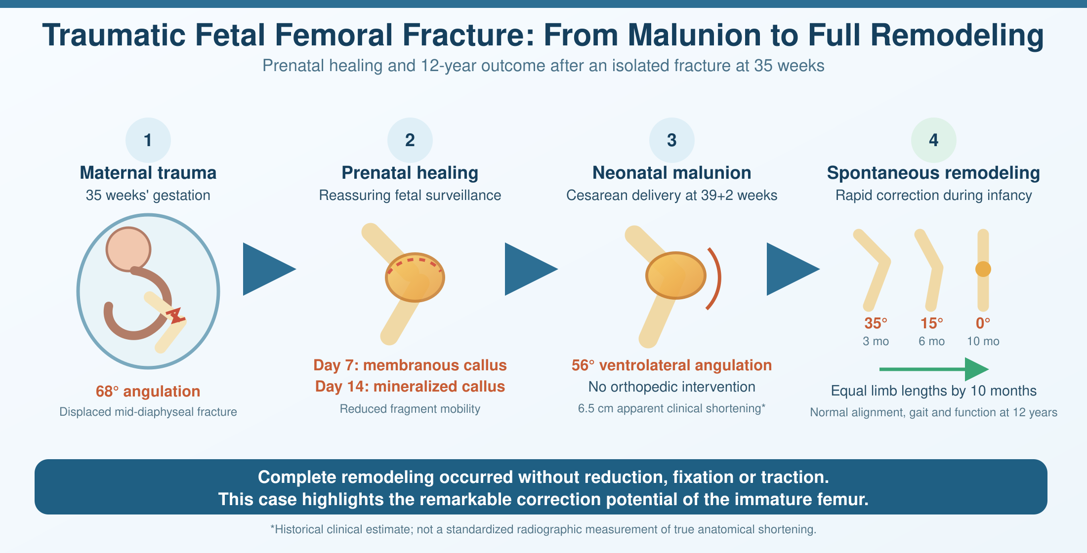
Case Presentation: A 35-week fetus sustained a displaced mid-diaphyseal fracture of the right femur following a maternal frontal motor vehicle collision. Initial ultrasonography demonstrated approximately 68° of interfragmentary angulation and surrounding soft-tissue edema. Maternal and fetal surveillance over the following 7 days showed no evidence of fetal compromise. Serial ultrasound examinations demonstrated progressive callus formation, with a globular membranous callus at day 7 and a prominent hyperechoic callus with peripheral vascularization and reduced fragment mobility at day 14. Delivery occurred by cesarean section at 39+2 weeks for an obstetric indication. The newborn weighed 3150 g and had Apgar scores of 9 and 10. The fracture was clinically consolidated but remained in severe malunion, with approximately 56° of ventrolateral angulation and a clinically estimated 6.5 cm apparent clinical shortening. No orthopedic intervention was undertaken. Progressive spontaneous correction was documented, with residual angulation of 35° at 3 months and 15° at 6 months. By 10 months, complete angular correction and limb-length equalization were documented. Independent walking was achieved at 12 months. At 12 years of age, the child had normal limb alignment, equal limb lengths, normal gait, and no functional sequelae.
Conclusions: This case demonstrates an unusual combination of prenatal fracture healing, severe neonatal malunion, and complete long-term spontaneous correction without orthopedic intervention. The reported 6.5-cm shortening should be interpreted as apparent clinical shortening rather than definitive anatomical shortening because it was not established by standardized radiographic limb-length measurement. Geometric analysis indicates that angular deformity alone could account for only approximately 0.8–1.0 cm of shortening at term. The subsequent course is consistent with substantial growth-related remodeling and provides a rare longitudinal example of the remodeling potential of the immature femur.
Keywords:
fetal femoral fracture
; fetal trauma
; prenatal diagnosis
; ultrasonography
; callus formation
; malunion
; spontaneous remodeling
; femoral shaft fracture
; physeal growth
; long-term follow-up
Copyright: This open access article is published under a Creative Commons CC BY 4.0 license, which permit the free download, distribution, and reuse, provided that the author and preprint are cited in any reuse.