
Submitted:
28 September 2026
Posted:
30 September 2026
You are already at the latest version
Abstract
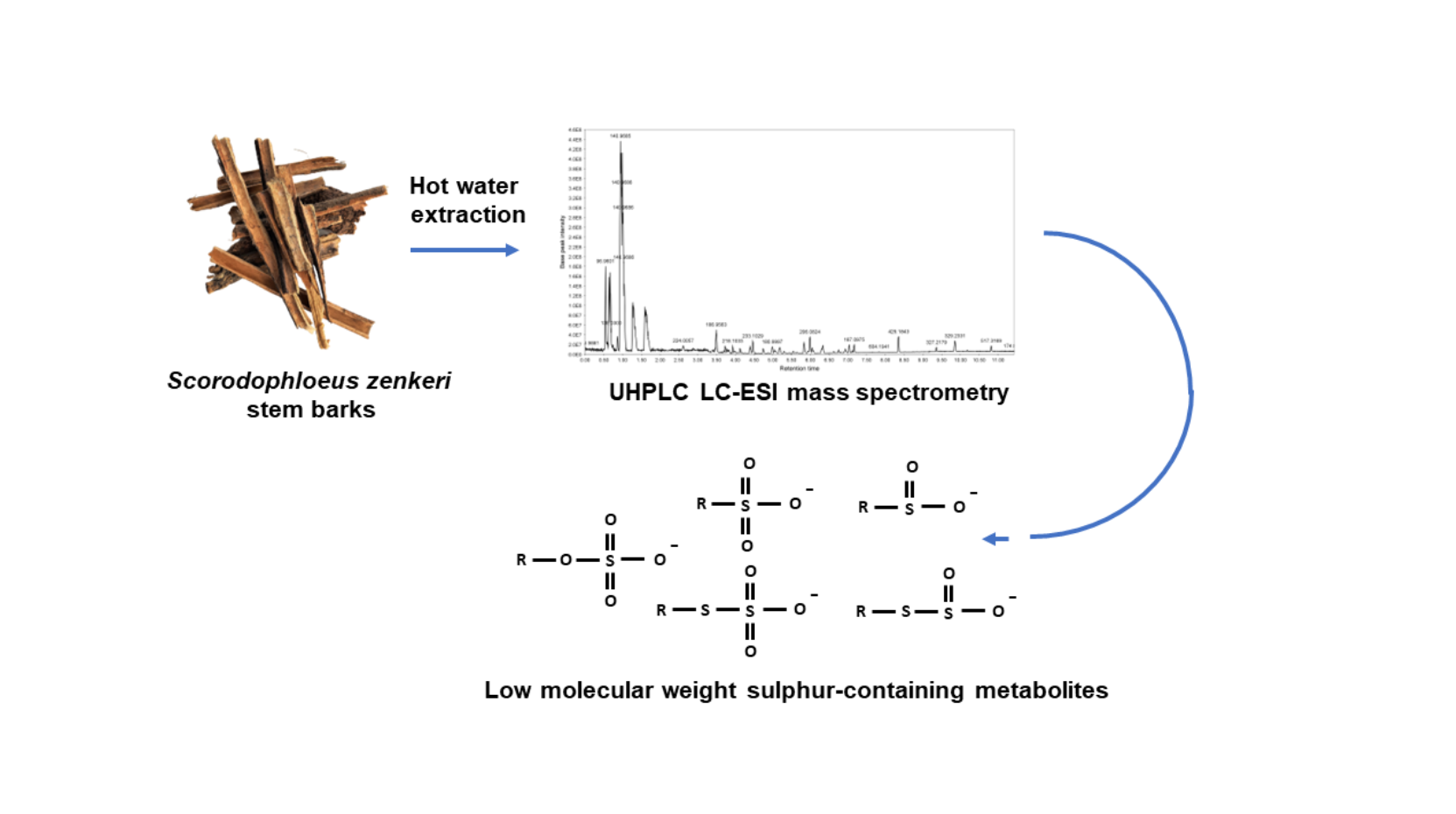
Scorodophloeus zenkeri is a tree endemic to Central Africa that gives out a garlic-like odour. Therefore, its bark, seeds and wood are commonly used as a condiment in traditional foods in Cameroon and Gabon. Moreover, the infusion of stem barks of S. zenkeri is used in traditional medicine in Gabon as a laxative. In the present study, we report on the phyto-chemical analysis by ultra-high performance liquid chromatography-tandem mass spec-trometry and by Fourier-transform ion cyclotron resonance mass spectrometry of the infu-sion of S. zenkeri barks. We identified numerous novel sulfur-containing metabolites, in-cluding sulfinic, sulfonic, sulfinosulfanyl, thiosulfuric acid derivatives and organosulfur metabolites, containing up to four sulfur atoms. Rules for the MS/MS fragmentation in negative ion mode of these unusual molecules are proposed.
Keywords:
mass spectrometry
; phytochemical analysis
; sulfur-containing molecules
; Scorodophloeus zenkeri
1. Introduction
Scorodophloeus zenkeri is a tree endemic to Central Africa. The infusion of its stem barks is widely used as a laxative in the traditional Gabonese medicine [1]. Mixed with chilli peppers and bitter eggplants, it is also used to treat colds and coughs [2]. S. zenkeri is also renowned for its garlic-like odour and therefore, its bark, seeds and wood are commonly used as a condiment in traditional foods in Cameroon and Gabon. It is usually admitted that this garlic-like odour comes from sulfur-containing molecules that are produced for instance by garlic (Allium sativum) or onion (Allium sepa) [3,4]. Similar sulfur-containing molecules were also reported in the fungus Marasmius alliaceus also known for its garlic-like odour [5]. The sulfur-containing molecules in these plants were identified as being mono-, di- and trisulfide and allyl isothiocyanate disulfide [3]. With regards to S. zenkeri, previous studies on the phytochemical analyses of hexane and dichloromethane extracts from their stem barks enabled the identification of organo-sulfur molecules that are composed of an alkane backbone with the insertion of sulfur atoms in carbon-carbon bounds. The main organo-sulfur compounds were metabolites ranging from trithiohexane to hexathioundecane [6] that have been previously identified in Marasmius alliaceus [5] or in alliaceous and cruciferous vegetables [7]. This indicates that the garlic-like odour of stem barks of S. zenkeri likely originates from its content in sulfur-containing molecules.
In the present study, we report on the phytochemical analysis of the infusion of S. zenkeri stem barks. We focussed on the characterization of water-soluble sulfur-containing metabolites by ultra-high performance liquid chromatography-tandem mass spectrometry (UHPLC ESI-MS/MS) and by Fourier-transform ion cyclotron resonance mass spectrometry (FT-ICR MS), and defined rules for their MS/MS fragmentation in negative ion mode, given their presumed involvement in the laxative activity of this plant extract.
2. Results
The metabolites of stem barks of Scorodophloeus zenkeri were extracted by infusion in boiling water and then were analysed by UHPLC ESI-MS/MS in negative (Figure S1) and positive ion modes. This enabled the annotation of many sulfur-containing metabolites (25 over 44 metabolites) containing up to four sulfur atoms. Other metabolites, including amino acids, aromatic acids and fatty acids were also detected and structurally annotated (Table S1) using databases (HMDB, MassBank, GNPS Databases). Among these metabolites, hydroxy diacids (C7, C9 and C10) and hydroxy stearic acids were putatively identified using literature data on hydroxylated fatty acids (Table S1) [8,9,10]. These compounds likely derive from the degradation of suberin contained in barks [11]. In addition, [M−H]− ion at m/z 517.3166 could correspond to a triterpenoid (C30H46O7) exhibiting two carboxylic groups. However, the MS/MS spectrum did not allow the detailed identification of its structure (Table S1). The structure of sulfur-containing metabolites was then proposed on the basis of their accurate mass measurements in negative ion mode and of their ESI-MS/MS fragmentation patterns. Among the sulfur-containing metabolites, sulfated polyhydroxy C18 mono and diacids were characterized by the occurrence in their MS/MS spectra of an intense ion at m/z 96.9601 assigned to HSO4- fragment ion (Figure 1A). Due to the lack of informative fragment ions, the structures of these fatty acids were not fully resolved but they also likely derive from the oxidation of C18 mono and diacid of suberin [11] (Table S2).
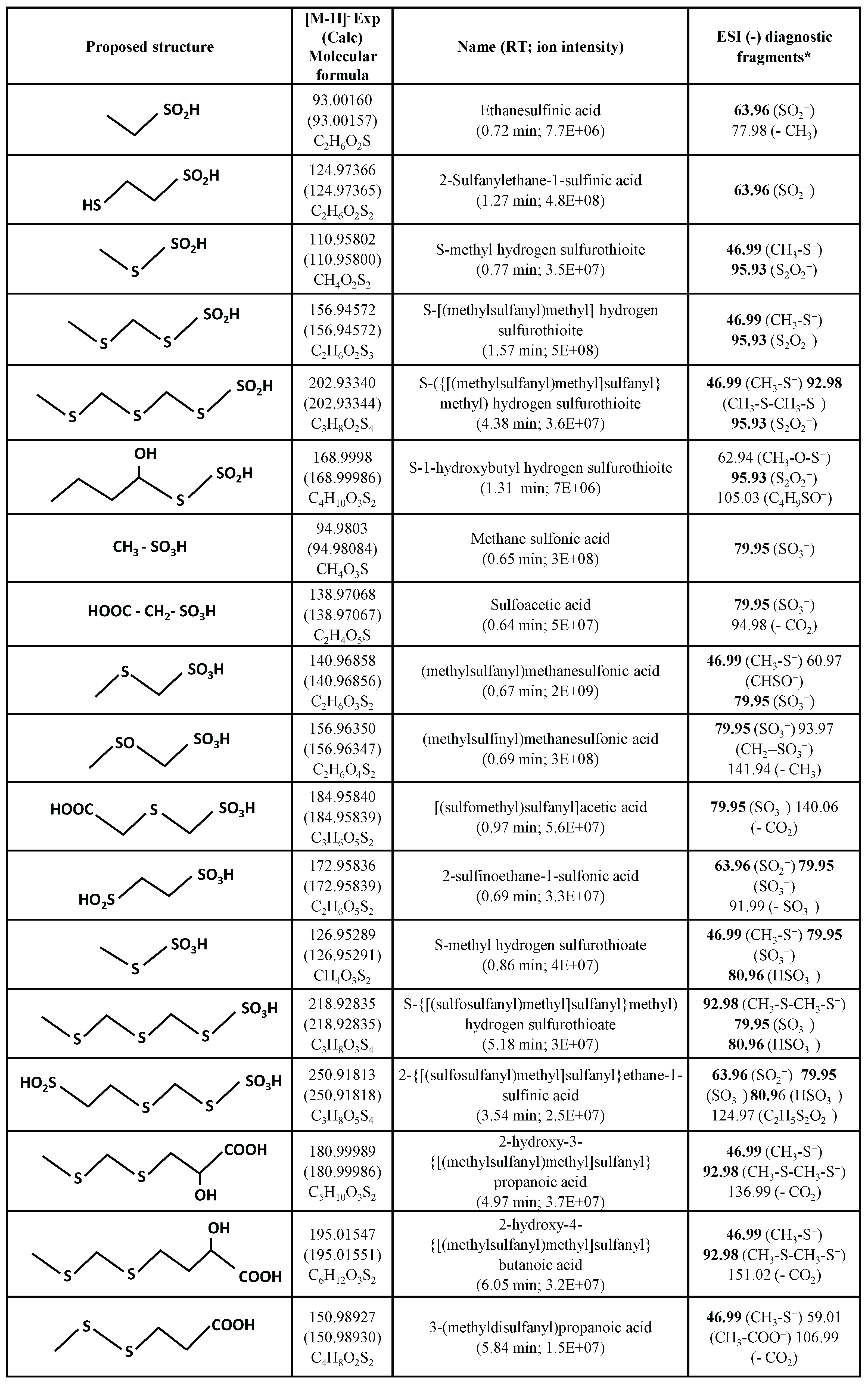
We then investigated low molecular weight sulfur-containing metabolites of the infusion of stem barks of S. zenkeri. Given the lack of published data on these compounds, we proposed putative structures based on the MS/MS data recorded on high-resolution mass spectrometers. Thus, UHPLC ESI-MS/MS analysis using an Orbitrap instrument, as well as direct introduction in negative ESI ion mode of the infusion using a Fourier-transform ion cyclotron resonance mass spectrometer (FT-ICR MS), suggested that these sulfur-containing metabolites were sulfinic and sulfonic derivatives, together with organosulfur compounds (Table 1). These sulfur-containing metabolites were named according to ChemSketch (https://chemsketch.fr.softonic.com) (Table 1).
It is worth noting that some of these metabolites contain many sulfur and oxygen atoms. This could lead to various isobaric combinations that were differentiated thanks to the very high resolution and high mass accuracy of FT-ICR MS. The metabolite molecular formula (i.e., elemental composition) was determined by accurate m/z measurement as well as isotopic pattern analysis. Indeed, sulfur-containing compounds show characteristic isotopic profiles linked to the natural abundance of the 32S, 33S and 34S isotopes, which are respectively 95.0%, 0.8% and 4.2 %. This is illustrated in Figure S2 for the isotopic peaks associated with the monoisotopic ion at m/z 140.96858 (C2H5O3S2–), which showed a fine structure where could clearly be distinguished the contribution of 13C from 33S and 34S. The relative intensities confirmed the presence of two S atoms.
The structure of low molecular weight sulfur-containing metabolites was proposed on the basis of the UHPLC ESI-MS/MS fragmentation patterns of [M−H]− ions. We deduced diagnostic fragment ions from these analyses that are specific for different sulfur-containing motifs found in these metabolites (Figure 1; Table 1). These fragmentation patterns have been observed in different metabolites sharing the same chemical motifs. As an illustration, Figure 2A-B show the ESI-MS/MS spectra of [M−H]− ions of ethane sulfinic acid (SO2− fragment ion at m/z 63.9624) and its sulfinosulfanyl derivative (S-methyl hydrogen sulfurothioite; S2O2− fragment ion at m/z 95.9343). With regards to sulfonic acids, fragmentation of their [M−H]− ions is characterized by the diagnostic fragment ion SO3− at m/z 79.9573 (Figure 1C and Figure 2C, Table 1). When this acidic function contains an additional sulfur atom, two main fragment ions from [M−H]− are detected for the resulting sulfinosulfanyl derivatives (S-methyl hydrogen sulfurothioate) at m/z 79.9573 and m/z 80.9651 corresponding to SO3− and HSO3− ions, respectively (Figure 1C and Figure 2D, Table 1). In addition, the SO2− fragment ion at m/z 63.9624 is also observed (Figure 1C and Figure 2D, Table 1). This MS/MS fragmentation is in accordance with data reported on S-propyl thiosulfuric acid isolated from onion (HMDB0041066).
Other diagnostic fragments observed in UHPLC ESI-MS/MS of [M−H]− ions were used for the structural identification of organosulfur molecules, such as fragment ions at m/z 46.9961 (CH3-S−) and m/z 92.9838 (CH3-S-CH2-S−) resulting from the cleavage of C-S bond of organosulfur backbones (Figure 1D and 2E-F, Table 1). Moreover, the occurrence of the latter fragment ion m/z 92.9838 assigned to CH3-S-CH2-S− enables the discrimination between metabolites containing non-consecutive or consecutive sulfur atoms. For instance, the structural characterization of organosulfur backbones in propanoic acid derivatives (Figure 2E and Figure 2F) is based on the occurrence of CH3-S-CH2-S− ion at m/z 92.9838 in only the ESI-MS/MS spectrum of the [M−H]− ion of the metabolite exhibiting sulfur atoms at the 4 and 6 positions on the backbone (Figure 2F) and not in the homologue metabolite with a disulfide at positions 4 and 5 (Figure 2E). It should be noted that in the MS/MS spectra of these two carboxylic acids, [M – H − CO2]− ions resulting from the loss of CO2 from [M − H]− ions were specific to the fragmentation of carboxylic acids in negative ion mode (Table 1).
It is worth noting that the diagnostic negative fragment ions represented in Figure 1 and assigned to sulfur-containing molecules are also observed in larger metabolites exhibiting both organosulfur and acidic motifs. This enables the structural identification of metabolites having multiple sulfur atoms inserted in different chemical groups. This can be illustrated by the UHPLC ESI-MS/MS spectra of sulfinosulfanyl and S-alkyl sulfuric acid derivatives, as well as sulfur-containing diacid derivatives (Figure 3). Fragment ion at m/z 92.9838 arises from the cleavage of C-S bonds of thioether or sulfide groups (R-S-R). Other negative fragment ions were assigned to sulfinosulfanyl (S-alkyl hydrogen sulfurothioite; R-SSO2H; S2O2− at m/z 95.9345), S-alkylthiosulfate (S-alkyl hydrogen sulfurothioate; R-SSO3H; SO3− at m/z 79.9573 and HSO3− at m/z 80.9651) and sulfinate (R-SO2H; SO2− at m/z 63.9624) motifs.
3. Discussion-Conclusion
Analysis by ultra-high performance liquid chromatography-tandem mass spectrometry and by Fourier-transform ion cyclotron resonance mass spectrometry of the infusion of S. zenkeri barks enabled the identification of novel sulfur-containing metabolites containing up to four sulfur atoms and reporter MS/MS fragment ions for their structural elucidation. It would be of interest to apply such an analytical strategy to other plants exhibiting a garlic-like odour, such as alliaceous and cruciferous vegetables.
4. Materials and Methods
4.1. Plant Material
The harvesting of stem barks of Scorodophloeus zenkeri was carried out in 2020 in Libreville, Gabon. A sample of this plant sample was deposited at the National Herbarium of Gabon where it was identified and preserved. The stem barks were dried out for two weeks at the Department of Traditional Medicine of the Institut de Pharmacopée et Médecines Traditionnelles (IPHAMETRA), Libreville, Gabon, and then reduced to a fine powder using a grinder. Five hundred grams of ground material were put in boiling water and then stirred for 15 min. The suspension was then filtered and the solution was frozen and freeze-dried to yield the infusion of stem bark of Scorodophloeus zenkeri.
4.2. Ultra-High Performance Liquid Chromatography-Tandem Mass Spectrometry (UHPLC ESI-MS/MS) Analysis
Five mg of the infusion of stem bark of Scorodophloeus zenkeri were dissolved in 1 mL of HPLC grade water and then filtrated through a 0.5 mL centrifugal filters Ultracel 10 kDa (Amicon) to remove high molecular weight compounds and impurities. Ultra-high performance liquid chromatography coupled to tandem mass spectrometry (UHPLC ESI-MS/MS) analysis was carried out using an UHPLC system (Vanquish, Thermo Scientific, San Jose, CA, USA) coupled to a quadrupole-Orbitrap mass spectrometer (Exploris 120, Thermo Scientific, San Jose, CA, USA). The chromatographic separations were performed using a C18 column (Acquity UPLC HSS T3 1.8 µm × 2.1 mm × 100 mm, Waters Corporation, Milford, MA, USA) with a prefilter of 0.2 µm, kept at 50°C during the analysis. An autosampler kept the samples at 6°C. The injection volume was 3 µL. The solvents used for gradient separation were 0.1 % (v/v) formic acid in water as mobile phase A and 0.1 % (v/v) formic acid in acetonitrile as mobile phase B. The flow rate was 0.4 mL/min. The elution gradient was first 1 % B for 1 min, then increased linearly to 100 % B over 20 min and then maintained at 100 % B for 8 min. Samples were analyzed in both negative and positive ion modes. The ESI source parameters were as follows: spray voltage 3500 V and 3000 V for positive and negative ion modes, respectively, sheath gas 35 (arbitrary unit), auxiliary gas 10 (arbitrary unit), sweep gas 2 (arbitrary unit), ion transfer tube 320°C and temperature of vaporizer 275°C. Data dependent acquisitions were carried out in both positive and negative ion modes. MS1 resolution was set at 60,000 with a standard automatic gain control (AGC) target, a maximum injection time set to auto, a microscan to 1, RF lens to 70 %, and a scan range from m/z 80 to 1200. EASY-IC internal standard (fluoranthene) was used. For MS/MS, resolution was set at 15,000 with a maximum injection time of 50 ms. The isolation window was of 2 m/z, dynamic exclusion was set at 4 s, mass tolerance was + 4 ppm and the precursor intensity threshold were set at 5.105 in positive mode and 1.105 in negative mode. The HCD collision energies were 15 %, 40 % and 60 % in both positive and negative ion modes. Data processing was carried out using MZmine 4.5 (version 4.5.0 [12,13]). Annotation was performed based on accurate mass measurements and ESI-MS/MS spectra according to the literature and databases.
4.3. Fourier-Transform Ion Cyclotron Resonance Mass Spectrometry (FT-ICR MS) Analysis
The filtered sample was diluted 100-fold in a 50/50 (v/v) water/methanol mixture (LC-MS grade, Carlo-Erba, Val de Reuil, France), and was then analysed by direct-infusion on a prototype tims MRMS instrument equipped with an 18 T superconducting magnet [14]. Ionization was performed using an electrospray ion source at a flow rate of 120 µL/h, capillary voltage of 4.5 kV, drying gas temperature of 200°C and drying gas flow rate of 4 L/min. Mass spectra were acquired using quadrupolar (2ω) detection over a mass range of m/z 80-700 with a data size of 8M points (transient signal of 0.5592 s). Key ion transmission in the mass spectrometer were: funnels 1 and 2 RF amplitude 150 Vp-p, multipole RF amplitude 150 Vp-p, quadrupole filter low mass set to m/z 80, collision cell RF amplitude 500 Vp-p. The transfer parameters to the ICR cell were: time-of-flight 0.6 ms, transfer hexapole RF frequency 6 MHz and amplitude 400 Vp-p. To obtain fine isotopic structures, 500 scans were summed each following a 300-ms accumulation in the collision cell of the instrument.
Supplementary Materials
The following supporting information can be downloaded at the website of this paper posted on Preprints.org.
Author Contributions
Conceptualization, E.L.M.E., L.E.M. and P.L.; methodology, E.L.M.E., S.O., O.P. and I.S.; validation, C.A., L.E.M, P.L. and C.L.B.; investigation, E.L.M.E., S.O., I.S., C.L.B. and O.P.; data curation, E.L.M., P.L., C.L.B. and H.L.; writing—original draft preparation, E.L.M.E., L.E.M. and P.L.; writing—review and editing, L.E.M. and P.L.;; supervision, L.E.M. and P.L.; project administration, L.E.M. and P.L.; funding acquisition, L.E.M. and P.L. All authors have read and agreed to the published version of the manuscript.
Funding
Please add: This work has been supported by IPHAMETRA, Libreville, Gabon and in France by the University of Rouen Normandy, INSA Rouen Normandy, the Centre National de la Recherche Scientifique (CNRS), SFR Normandie Végétal FED 4277, GDR CNRS Chemobiologie, European Regional Development Fund (ERDF), Labex SynOrg (ANR-11-LABX-0029), Carnot Institute I2C, the graduate school for research XL-Chem (ANR-18-EURE-0020 XL CHEM) and Region Normandie. Financial support from the IR INFRANALYTICS FR2054 for conducting the research is also gratefully acknowledged.
Data Availability Statement
Data will be made available on request.
Conflicts of Interest
“The authors declare no conflicts of interest.”.
Abbreviations
The following abbreviations are used in this manuscript:
| UHPLC ESI-MS/MS | Ultra-high performance liquid chromatography-tandem mass spectrometry |
| FT-ICR MS | Fourier-transform ion cyclotron resonance mass spectrometry |
References
- Mayindza Ekaghba, E.L.; Grenet, M.; Gandolfo, P.; Loutelier-Bourhis, C.; Schmitz, I.; Afonso, C.; Lerouge, P.; Mengome, L.E. Phytochemical analysis and antidiarrheal activity of stem bark decoctions of Pentadesma butyracea sabine (Clusiaceae). Molecules 2024, 29, 5789. [Google Scholar] [CrossRef]
- Raponda-Walker, A.; Sillans, R. Les Plantes utiles du Gabon; Lechevalier, Paul, Ed.; Encyclopédie Biologique 56: Ed. Paris, France, 1961; p. 624 pages. [Google Scholar]
- Iranshahi, M. A review of volatile sulfur-containing compounds from terrestrial plants: Biosynthesis, distribution and analytical methods. J. Essent. Oil Res. 2012, 24, 393–434. [Google Scholar] [CrossRef]
- Vecchi, V.; Sabbioni, G.; Altieri, M.T.; Breveglieri, G.; Costa, S.; Marchetti, F.; Gugel, I.; Vertuani, S.; Borgatti, M. Black garlic: Evolution of the chemical composition and broad biological activities. J. Agric. Food Chem. 2025, 73, 27933–27949. [Google Scholar] [CrossRef]
- Rapior, S.; Breheret, S.; Talou, T.; Bessière, J.M. Volatile flavor constituents of fresh Marasmius alliaceus (garlic Marasmius). J. Agric. Food Chem. 1997, 45, 820–825. [Google Scholar] [CrossRef]
- Kouokam, J.C.; Jahns, T.; Becker, H. Antimicrobial activity of the essential oil and some isolated sulfur-rich compounds from Scorodophloeus zenkeri. Planta Med. 2002, 68, 1082–1087. [Google Scholar] [CrossRef]
- Kasamatsu, S.; Kinno, A.; Hishiyama, J.I.; Akaike, T.; Ihara, H. Development of methods for quantitative determination of the total and reactive polysulfides: Reactive polysulfide profiling in vegetables. Food Chem. 2023, 413, 135610. [Google Scholar] [CrossRef]
- Yang, N.Y.; Yang, Y.F.; Li, K. Analysis of Hydroxy Fatty Acids from the Pollen of Brassica campestris L. var. oleifera DC. by UPLC-MS/MS. J. Pharmacol. 2013, 874875. [Google Scholar]
- Yuan, Z.X.; Rapoport, S.I.; Soldin, S.J.; Remaley, A.T.; Taha, A.Y.; Kellom, M.; Gu, J.; Sampson, M.; Ramsden, C.E. Identification and profiling of targeted oxidized linoleic acid metabolites in rat plasma by quadrupole time-of-flight mass spectrometry. Biomed. Chromatogr. 2013, 7, 422–432. [Google Scholar] [CrossRef]
- Kokotou, M.G.; Mantzourani, C.; Babaiti, R.; Kokotos, G. Study of the royal jelly free fatty acids by liquid chromatography-high resolution mass spectrometry (LC-HRMS). Metabolites 2020, 10, 40. [Google Scholar] [CrossRef]
- Philippe, G.; Sørensen, I.; Jiao, C.; Sun, X.; Fei, Z.; Domozych, D.S.; Rose, J.K.C. Cutin and suberin: assembly and origins of specialized lipidic cell wall scaffolds. Curr. Opin. Plant Biol. 2020, 55, 11–20. [Google Scholar] [CrossRef]
- Pluskal, T.; Korf, A.; Smirnovic, A.; Schmid, R.; Fallon, T.R.; Du, X.; Weng, J.K. Metabolomics data analysis using MZmine. New Developments in mass spectrometry. No. 8 Processing metabolomics and proteomics data with open software: A practical guide. In Royal Soc. Chem.; Winkler, Robert, Ed.; 2020; Volume 7, p. 232. [Google Scholar]
- Heuckeroth, S.; Damiani, T.; Smirnov, A.; Mokshyna, O.; Brungs, C.; Korf, A.; Smith, J.D.; Stincone, P.; Dreolin, N.; Nothias, L.F.; Hyötyläinen, T.; Orešič, M.; Karst, U.; Dorrestein, P.C.; Petras; Du, D.; van der Hooft, X.; Schmid, J.J.J.; Pluskal, R.T. Reproducible mass spectrometry data processing and compound annotation in MZmine 3. Nat. Protoc. 2024, 19, 2597–2641. [Google Scholar] [CrossRef]
- Wootton, C.A.; Maillard, J.; Theisen, A.; Brabeck, G.F.; Schat, C.L.; Rüger, C.P.; Afonso, C.; Giusti, P.A. Gated TIMS FTICR MS instrument to decipher isomeric content of complex organic mixtures. Anal. Chem. 2024, 96, 11343–11352. [Google Scholar] [CrossRef]
Figure 1.
Diagnostic ESI-MS/MS negative fragment ions of (A) sulfated metabolites (R-SO3H), (B) sulfinic acid (R-SO2H) and sulfinosulfanyl derivatives (S-alkyl hydrogen sulfurothioite; R-SSO2H), (C) sulfonic acid (R-SO3H) and S-alkylthiosulfuric acid (S-alkyl hydrogen sulfurothioate; R-SSO3H) and (D) organosulfur (R-S-R) metabolites.
Figure 1.
Diagnostic ESI-MS/MS negative fragment ions of (A) sulfated metabolites (R-SO3H), (B) sulfinic acid (R-SO2H) and sulfinosulfanyl derivatives (S-alkyl hydrogen sulfurothioite; R-SSO2H), (C) sulfonic acid (R-SO3H) and S-alkylthiosulfuric acid (S-alkyl hydrogen sulfurothioate; R-SSO3H) and (D) organosulfur (R-S-R) metabolites.
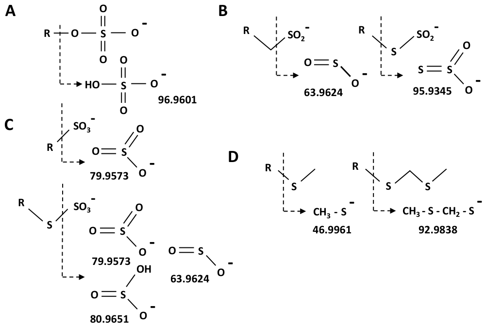
Figure 2.
UHPLC ESI-MS/MS spectra of [M−H]− ions of (A) ethane sulfinic acid at m/z 93.0016, (B) S-methyl hydrogen sulfurothioite at m/z 110.9581, (C) (methylsulfinyl)methanesulfonic acid at m/z 156.9635, (D) S-methyl hydrogen sulfurothioate at m/z 126.9530, (E) 3-(methyldisulfanyl)propanoic acid at m/z 150.9893 and (F) 2-hydroxy-3-{[(methylsulfanyl)methyl]sulfanyl}propanoic acid at m/z 180.9998. See Figure 1 for the structure of sulfur-containing fragment ions observed in negative ESI-MS/MS. Diamond: precursor ion.
Figure 2.
UHPLC ESI-MS/MS spectra of [M−H]− ions of (A) ethane sulfinic acid at m/z 93.0016, (B) S-methyl hydrogen sulfurothioite at m/z 110.9581, (C) (methylsulfinyl)methanesulfonic acid at m/z 156.9635, (D) S-methyl hydrogen sulfurothioate at m/z 126.9530, (E) 3-(methyldisulfanyl)propanoic acid at m/z 150.9893 and (F) 2-hydroxy-3-{[(methylsulfanyl)methyl]sulfanyl}propanoic acid at m/z 180.9998. See Figure 1 for the structure of sulfur-containing fragment ions observed in negative ESI-MS/MS. Diamond: precursor ion.
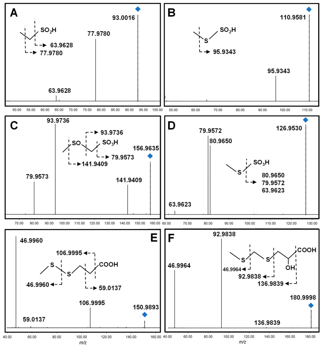
Figure 3.
UHPLC ESI-MS/MS spectra of [M−H]− ions of (A) S-({[(methylsulfanyl)methyl]sulfanyl}methyl) hydrogen sulfurothioite at m/z 202.9334, (B) S-{[(sulfosulfanyl)methyl]sulfanyl}methyl) hydrogen sulfurothioate at m/z 218.9283 and (C) 2-{[(sulfosulfanyl)methyl]sulfanyl}ethane-1-sulfinic acid at m/z 250.9181. See Figure 1 for structure of sulfur-containing fragment ions. Diamond: precursor ion. *: contamination ion.
Figure 3.
UHPLC ESI-MS/MS spectra of [M−H]− ions of (A) S-({[(methylsulfanyl)methyl]sulfanyl}methyl) hydrogen sulfurothioite at m/z 202.9334, (B) S-{[(sulfosulfanyl)methyl]sulfanyl}methyl) hydrogen sulfurothioate at m/z 218.9283 and (C) 2-{[(sulfosulfanyl)methyl]sulfanyl}ethane-1-sulfinic acid at m/z 250.9181. See Figure 1 for structure of sulfur-containing fragment ions. Diamond: precursor ion. *: contamination ion.
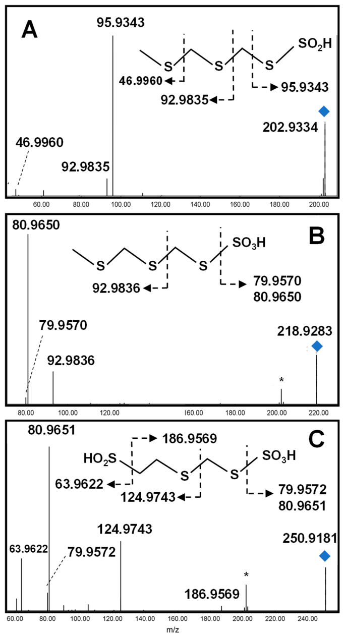

Table 1.
Proposed structures and ESI-MS/MS fragment ions in negative ion mode of sulfur-containing metabolites. * Fragments in bold are represented in Figure 1. Exp: experimental value determined by FT-ICR MS and proposed molecular formula. Calc: calculated value.
Table 1.
Proposed structures and ESI-MS/MS fragment ions in negative ion mode of sulfur-containing metabolites. * Fragments in bold are represented in Figure 1. Exp: experimental value determined by FT-ICR MS and proposed molecular formula. Calc: calculated value.
Disclaimer/Publisher’s Note: The statements, opinions and data contained in all publications are solely those of the individual author(s) and contributor(s) and not of MDPI and/or the editor(s). MDPI and/or the editor(s) disclaim responsibility for any injury to people or property resulting from any ideas, methods, instructions or products referred to in the content. |
© 2026 by the authors. Licensee MDPI, Basel, Switzerland. This article is an open access article distributed under the terms and conditions of the Creative Commons Attribution (CC BY) license.
Copyright: This open access article is published under a Creative Commons CC BY 4.0 license, which permit the free download, distribution, and reuse, provided that the author and preprint are cited in any reuse.