

Submitted:
28 September 2026
Posted:
29 September 2026
You are already at the latest version
Preprints on COVID-19 and SARS-CoV-2
Abstract
The severe acute respiratory syndrome coronavirus 2 (SARS-CoV-2)-induced global pandemic, caused by coronavirus disease 2019 (COVID-19), has taken a toll on millions of lives worldwide. Hence, there is an increasing need for accurate disease diagnosis to prevent rapid transmission and provide immediate treatment to patients having mild to severe symptoms of COVID-19 on an urgent basis. Immunoassays are among the several developed techniques for COVID-19 diagnosis. These are generally biochemical tests designed to identify the presence and level of a SARS-CoV-2-specific biomarker employing either antigens (Ags) or antibodies (Abs). Immunoassays rely on a competitive affinity reaction between the target immunological biomarker (Ag/Ab) and other (bio)molecules present in a patient’s sample for the limited binding sites on the immobilised capture molecule (Ab/Ag). Unlike conventional immunoassays, including quantitative/semi-quantitative serology tests that detect circulating anti-SARS-CoV-2 Abs using enzyme-linked immunosorbent assay (ELISA) and/or chemiluminescent immunoassay (ChLIA) techniques, rapid immunoassays, or rapid diagnostic tests (RDTs), are qualitative, simple-to-use assays that utilise an immunochromatographic (lateral flow) test format. Moreover, such tests can be conducted at or near the point of care (PoC) without requiring any high-throughput laboratory infrastructure. Based on the mechanism of detection, SARS-CoV-2 RDTs can be broadly classified into two types, namely (1) Ag tests, which directly identify the SARS-CoV-2 viral Ag(s), and (2) Ab tests that identify one or more variants of antiviral Abs secreted by the host immune system in response to viral infection. Although effective, both techniques have certain limitations. New developments in existing colourimetric lateral flow immunoassays (LFIAs) are underway to address these issues and challenges. Ag-detecting RDTs (Ag-RDTs) are expected to significantly broaden the testing domain, enabling more precise viral load estimation, cost-effective treatment for infected persons, and the formulation of adequate prophylactic measures. In contrast, serology tests [including Ab-detecting RDTs (Ab-RDTs)] could be used for serosurveillance of patients recovered from COVID-19.
Keywords:
SARS-CoV-2
; COVID-19 diagnosis
; immunoassays
; rapid diagnostic tests (RDTs)
; SARS-CoV-2-specific biomarkers
; serology tests
; lateral flow immunoassays (LFIAs)
; antigen-detecting rapid diagnostic tests (Ag-RDTs)
; antibody-detecting rapid diagnostic tests (Ab-RDTs)
1. Introduction
1.1. Brief Historical Overview of the COVID-19 Pandemic
On 31st December (2019), the World Health Organisation (WHO) first noted clustered cases of pneumonia of unknown origin in Wuhan city/district (Hubei province), People’s Republic of China (PRC). On 30th January 2020, WHO announced a Public Health Emergency of International Concern (PHEIC). The causative agent of this viral pneumonia was initially called 2019 novel coronavirus (2019-nCoV) (https://www.who.int/publications/i/item/diagnostic-testingfor-sars-cov-2) [1]. However, on 11th February 2020, the new coronavirus was officially termed severe acute respiratory syndrome coronavirus 2 (SARS-CoV-2), and the disease caused by the infectious viral particles was referred to as coronavirus disease 2019 (COVID-19). Based on the rapid spread of SARS-CoV-2 infection across the world, WHO evaluated that COVID-19 could be considered as a pandemic of global concern on 11th March (2020) (https://www.who.int/dg/speeches/detail/who-director-general-s-opening-remarks-at-the-media-briefing-on-covid-19---11-march-2020) [2]. The clinical manifestation of SARS-CoV-2 infection can range from an asymptomatic carrier state (i.e., with no detectable symptoms) to mild/severe disease phenotypes (requiring hospitalization and/or intensive/critical care with noninvasive/invasive ventilation support), which may last for several weeks with associated post-COVID-19 complications. Transmission and mortality rates have varied across countries to date, with the emergence of several new viral variants (https://www.who.int/publications/i/item/diagnostic-testingfor-sars-cov-2) [1].
1.2. Pivotal Role of Testing During Pandemic Response
Early diagnosis is an integral part of the first line of defence against COVID-19, enabling early detection of positive cases and their isolation to slow transmission, provide targeted treatment, and protect healthcare system operations. Although nucleic acid amplification technique-based clinical tests for patients with COVID-19 were rapidly developed at the onset of the COVID-19 pandemic, these molecular tests typically require advanced laboratory equipment and trained personnel. To date, a plethora of COVID-19 diagnostic tests have been conducted worldwide; however, the demand for timely, comprehensive, and precise testing continues to outrun supply (World Health Organisation, 2020) [3].
According to WHO recommendations, person(s) encountering probable COVID-19 suspect(s) (https://www.who.int/emergencies/diseases/novel-coronavirus-2019/technical-guidance-publications?publicationtypes=df113943-c6f4-42a5-914f-0a0736769008) [4] should be immediately tested in order to either confirm or negate SARS-CoV-2 infection. In situations where testing is not feasible, a potential COVID-19 suspect may instead be reported based on specific epidemiological criteria and clinical signs or symptoms (https://www.who.int/emergencies/diseases/novel-coronavirus-2019/technical-guidance-publications?publicationtypes=df113943-c6f4-42a5-914f-0a0736769008) [4]. One of the developed immunological assays that can be used for rapid diagnosis of COVID-19 is antigen (Ag) tests or Ag-detecting rapid diagnostic tests (Ag-RDTs). Ag-RDTs directly detect the presence of viral (SARS-CoV-2) Ag(s), usually nucleocapsid proteins (NPs) produced by replicating viral particles in patients' respiratory secretions. Thus, like molecular testing, Ag-RDTs help identify active COVID-19 infection (i.e., infection the patient currently has). However, Ag-RDTs do not provide information about prior infections. To overcome this challenge, antibody (Ab) or serology tests [including Ab-detecting rapid diagnostic tests (Ab-RDTs)], besides complementing molecular tests by presenting a more precise estimate of COVID-19 incidence, can potentially detect individuals with developed immunity against the disease (i.e., serosurveillance of patients who have recovered from COVID-19), since these tests identify biomarkers that are generated in response to the viral Ag by the host immune response (Ghaffari et al. 2020) [5]. SARS-CoV-2 Ag-RDTs are expected to greatly broaden access to testing (expeditiously) for suspected individuals, enabling more precise estimation of viral load in infected hosts and, subsequently, targeting prophylactic measures and appropriate treatments. On the other hand, the serological tests (including Ab-RDTs) could be used for serosurveillance of patients who have recovered from COVID-19.
1.3. Crucial Needs During Pandemic Response
Controlling active COVID-19 cases to prevent rapid disease transmission requires scaling up testing services and improving access to testing, mainly in decentralised settings. Restricted access to laboratory services, together with prolonged turnaround times, has shown that the capacity of nucleic acid amplification tests (NAATs), including real-time reverse transcriptase polymerase chain reaction (rRT-PCR), has been inadequate to meet demand in several nations, especially in low- and middle-income countries (LMICs). In LMICs, these restrictions have significantly reduced testing rates (~10 times lower) compared with high-income or developed nations (https://www.finddx.org/covid-19/test-tracker/) [6].
Timely, accurate, and comprehensive COVID-19 diagnosis and surveillance data are essential for an immediate COVID-19 public health (pandemic) response. According to WHO, it is necessary to possess (1) real-time, authentic testing data of suspected COVID-19 cases, (2) the nature and quarantine status of all confirmed positive COVID-19 cases, (3) the number of contacts per symptomatic or asymptomatic case along with completeness of contact tracing, and (4) the flexibility of healthcare facilities to handle positive cases of COVID-19 (https://www.who.int/publications/i/item/covid-19-strategy-update---14-april-2020) [7]. Real-time data for researchers and clinicians is the need of the hour. Therefore, countries across the globe need to join hands to prioritise and actively work towards achieving this goal. In addition, developed nations need to share know-how and/or supply raw materials for manufacturing testing kits for rapid disease diagnosis, along with ready-made, cost-effective, easy-to-use test kits, with developing and poor/underdeveloped nations.
2. Host Humoral Immune Response to SARS-CoV-2 Infection
In a host humoral immune response against a pathogen-induced infection, circulating B cells (B lymphocytes) secrete pathogen-specific immunoglobulins (antibodies, or Abs) into the blood, which eventually neutralise antigenic proteins of invading pathogens by forming Ag-Ab complexes, helping prevent further pathogenesis or disease spread. The activation and maturation of B cells into Ab-secreting plasma cells are triggered by a series of well-coordinated events involving phagocytosis (i.e., ingestion and digestion of viral particles) by Ag-presenting cells (APCs) such as macrophages, dendritic cells, etc., and presentation of viral peptides (i.e., virus-specific antigenic proteins) to helper T (TH) cells or T lymphocytes (Figure 1). Abs protect the host by binding to specific antigenic proteins on the virus, preventing fusion and subsequent entry into the host cell and facilitating recognition and inactivation by phagocytic cells (Janeway, 2001) [8]. In humans, at least three types of immunoglobulins, or Abs, have been used as target biomarkers in serological tests for COVID-19 diagnosis: immunoglobulin M (IgM), immunoglobulin G (IgG), and immunoglobulin A (IgA). Although the dynamics of the host immune response in the case of COVID-19 are yet to be fully explored, usually IgM Abs are secreted by the body’s immune cells in response to SARS-CoV-2 exposure during the initial stages of COVID-19. In contrast, IgG Abs, which are often most abundant in the blood, function more prominently in the later stages of SARS-CoV-2 infection and help establish long-lasting immunological memory (Azkur et al. 2020) [9]. Interestingly, while IgM and IgG Abs have been the principal candidates for COVID-19 serology-based tests, current research shows that IgA Abs, primarily found in the mucosal epithelium, may also play a significant role in the host immune response and subsequent disease progression (Yu et al. 2020) [10].
3. Developed Immunoassay Techniques for the Diagnosis of COVID-19
Precise diagnosis of a disease like COVID-19 can be based on biomarkers specific to the disease-causing microbe [SARS-CoV-2, an enveloped, single-stranded, positive-sense RNA (+ssRNA) virus in this case]. Protein molecules that are involved in the host immune response against the viral Ags [such as the spike (S) proteins of SARS-CoV-2 in this case] constitute another set of biomarkers used for disease diagnostics apart from the genetic content of the virus (e.g., the viral nucleic acid or RNA) that are generally used for molecular tests. This set of immune biomarkers primarily comprises Abs circulating in the host blood to combat the invading viral Ags or antigenic proteins, and serological tests using different immunological techniques have been developed for their detection. Serological tests are a traditional method for SARS-CoV-2 detection, like those used for other infectious viral pneumonias, alongside molecular tests and chest computed tomography (CT) scans. Serological tests typically target immunoglobulin molecules, or Abs, produced in response to the invading virus (Taleghani and Taghipour, 2020) [11].
Immunological assays, or immunoassays, are generally biochemical tests designed to detect both the presence and concentration of a disease-specific biomarker, using either Ags or Abs as immobilised capture reagents. The mechanism of identifying such immune biomarkers relies on a competitive affinity reaction between the target biomarker (Ag/Ab) and various other (bio)molecules present in the collected patient’s sample for limited binding sites that are available on the immobilised capture molecule (Ab/Ag) (Kellner et al. 1998) [12]. In general, immunoassay techniques developed for the diagnosis of COVID-19 either assess the presence of SARS-CoV-2 Ag(s) (referred to as Ag tests) or the presence of anti-SARS-CoV-2 Ab(s) secreted in response to SARS-CoV-2 infection (referred to as Ab or serology tests). Moreover, these immunoassays can be qualitative or quantitative. Recently, qualitative rapid detection techniques (rapid immunoassays) have been developed that include both Ag and Ab tests based on lateral flow immunochromatography. However, the timing of patient sample collection dictates the sampling results and, ultimately, the disease diagnosis and its current state for appropriate treatment. The following section provides an in-depth description of the immunoassay technologies (including both conventional and rapid immunoassays) developed to date.
3.1. Serology or Antibody Tests
Unlike molecular-based tests that identify the SARS-CoV-2 genome or ribonucleic acid (RNA), serological or serology tests (also called as Ab tests) detect the host immune response to SARS-CoV-2 exposure (i.e., the presence of circulating anti-SARS-CoV-2 Abs), thereby aiming to identify prior SARS-CoV-2 infection while also aiding in confirming the current infection’s presence in suspected cases. In medicine, serology refers to the scientific study of serum (and other body fluids), and in practice, it generally means the diagnostic identification of Abs present in the blood serum. Thus, COVID-19 serology-based tests look for Abs that specifically bind to SARS-CoV-2 antigenic proteins on the virus surface. These Abs are present only if a person has been infected with SARS-CoV-2 and recovered. Because the host immune response takes time to develop, serology-based tests provide a wider window for detecting COVID-19. Serological tests use blood samples that are collected either by drawing blood from a suspected individual’s arm or through a finger prick. These Ab tests, however, are not used for the diagnosis of active COVID-19 (i.e., a person with SARS-CoV-2 infection), since they do not directly detect the presence of the virus in patients, but rather provide proof of the body’s natural reflex against the invading virus by analysing specific products of the host immune response. Serology tests are generally used for screening individuals who are either asymptomatic or have recovered from the disease (Dutta et al. 2020) [13]; (Taleghani and Taghipour, 2021) [11].
Qualitative versus quantitative serology tests
A variety of serological tests are available for COVID-19 diagnosis, all of which identify Abs present in a person's blood serum.
- Qualitative serological tests typically provide a simple “yes”/“no” answer to any previous exposure of an individual to SARS-CoV-2 (i.e., the presence or absence of SARS-CoV-2-specific Abs). For example, rapid serology tests (RSTs), mainly using lateral flow assay (LFA) techniques, are qualitative and provide a positive or negative readout.
- Quantitative serological tests, including enzyme-linked immunosorbent assays (ELISAs) and chemiluminescent immunoassays (ChLIAs) together with their variants, provide more detailed information, such as the level of Abs in a patient’s sample.
The precision and performance of SARS-CoV-2-specific serology tests vary greatly across test types. In general, quantitative serology tests are more accurate than qualitative serology tests, partly because they generate more detailed data.
Currently used serology tests for COVID-19 diagnosis
- Rapid serology test (RST): This is predominantly a qualitative LFA, which is small, portable, and can be employed at the PoC. RST samples may include blood (from a finger prick), saliva, or nasal swab fluids. In COVID-19, these serology tests most often assess the presence or absence of the patient’s anti-viral Abs (i.e., IgM and/or IgG). In a few instances, it can be useful to measure baseline (before infection) IgM and IgG Ab titers (Figure 2). This is discussed in more detail in section Antibody-Detecting Rapid Diagnostic Tests.
- Enzyme-linked immunosorbent assay (ELISA): This is a qualitative/quantitative technique to detect the presence of Ags (in this case viral proteins) in biological samples, and in general, is a laboratory-based test. The test typically uses whole blood, serum or plasma samples from suspected individuals. The assay relies on the viral Ags of interest, such as S proteins, being immobilised on a solid surface/plate. Samples (i.e., whole blood, serum or plasma) from patients are then incubated with the previously prepared (protein-coated) plate. If the patient sample already has Abs for the viral Ag(s) on the plate, then Ab-Ag conjugates will be formed that can be detected by another set of secondary Abs (each secondary Ab is conjugated with a molecule for detection such as an enzyme or a fluorophore), which produce a colour or a fluorescence-based read-out. In the context of SARS-CoV-2, indirect ELISA is frequently used to detect IgM and IgG Abs in COVID-19 patients (Figure 3A). Among the variants of this method is an automated, highly sensitive digital sandwich ELISA (also known as digital ELISA or single molecule ELISA) that uses single molecule arrays (SiMoA) to detect single enzyme-linked immune complexes on paramagnetic microbeads sealed in arrays of femtoliter-sized wells. In a nutshell, the assay captures and detects every target viral antigenic protein molecule (Chang et al. 2012) [14]. In the future, the assay may be modified to capture and detect every target anti-viral Ab molecule (Figure 3B). Another modification of this technique is a recently developed (and yet to be authorised) rapid, inexpensive, quantitative magnetic bead-based chromogenic ELISA that detects anti-SARS-CoV-2 human IgM or IgG Abs using recombinant viral nucleocapsid protein (NP). This novel immunogenic method uses a low volume of serum, plasma, or whole blood samples with low Ab titer and returns results within 12 minutes, which can be read by visual inspection without requiring instrumentation. Interestingly, this method can be implemented for both PoC and high-throughput analysis (Huergo et al. 2021) [15].
- Chemiluminescent immunoassay (ChLIA): In general, this is a laboratory-based qualitative/ semi-quantitative technique to detect the presence of viral proteins in biological samples. The test uses the patient’s whole blood, serum or plasma samples. The assay relies on mixing the patient’s sample with known viral protein(s) [Ag(s)], reagents/buffers and secondary Abs, each conjugated with an enzyme for light-based luminescent read-out. If the patient’s sample already contains Abs for the viral Ag(s), Ab-Ag conjugates form, which are then detected by specific enzyme-labelled secondary Abs, inducing a chemical reaction that produces light (Figure 4A). The emitted light intensity is then used to quantify the amount of Abs present in each sample. The test can identify different types of Abs, viz. IgM, IgG, and IgA. A modification of this test is the chemiluminescent microparticle immunoassay (CMIA), which uses magnetic, protein/Ag-coated microparticles to detect Abs in a patient’s sample (Figure 4B).
-
Neutralisation assay (NA): This test depends on preformed Abs, obtained from patients capable of preventing further viral infections, in a laboratory set-up. The assay indicates whether patients have preformed Abs and, if so, whether these Abs are active and effective against the viral Ags, even though the patients have recovered from the infection. The test uses the patient’s whole blood, plasma or serum samples. Because NA depends on animal cell culture, SARS-CoV-2 is cultured in the laboratory (e.g., Vero E6 cells) for this assay. When the co-culture of viruses and mammalian cells is mixed with decreasing titers of the patient’s Abs, the number of Abs in the patient sample capable of blocking viral replication can be visualised and quantified. This blocking of viral multiplication can be mediated by Abs binding to a key viral entry protein on the virus surface, as shown in Figure 5.Figures 2’5 summarise the fundamental principles for detecting SARS-CoV-2-specific Ab(s) in the developed qualitative and quantitative/semi-quantitative serological tests.
Figure 2, Figure 3, Figure 4 and Figure 5 summarise the fundamental principles for detecting SARS-CoV-2-specific Ab(s) in the developed qualitative and quantitative/semi-quantitative serological tests.
Table 1 presents a brief overview of the different categories of serological tests commonly used to diagnose SARS-CoV-2 infection.
In general, a serological test measures two types of Abs, namely (1) pentameric IgM and (2) monomeric IgG. Serology tests also assess the occurrence and concentration of a third type of Ab: (3) monomeric/dimeric IgA. IgA is mostly found in mucous membranes lining the respiratory (bronchial) and digestive tracts. It can also be present in tears, saliva, colostrum, blood, the nasopharynx, and the genitourinary tract (Roda et al., 2021) [20]. In general, IgM (which constitutes around 5 – 8% of serum immunoglobulin) is the first Ab produced as a body’s primary immune response to numerous pathogens, and therefore, its presence is indicative of active or recent infection. To develop a more specific host immune response against a viral infection like COVID-19, the body produces IgG Ab, the most abundant class of serum immunoglobulin. IgA Ab, the second most abundant class of serum immunoglobulin, can promote an Ab-mediated immune response, further controlling and preventing microbial infections. Figure 6 displays the approximate concentration of anti-SARS-CoV-2 Abs such as IgM, IgA and IgG at different days after SARS-CoV-2 infection.
Although most serological tests assess IgG Ab because it is more pathogen-specific, IgG Ab may take longer to develop (sometimes up to 21 days) (CDC, 2020a) [21]. Therefore, based on the above details on IgM, IgG, and IgA Abs, serological tests are better suited for COVID-19 surveillance and can provide valuable information about people who are either fighting COVID-19 or have survived the disease. However, a potential drawback of serological tests is inaccuracy, since cross-reactivity between anti-SARS-CoV-2 Abs and Abs produced against other coronaviruses is possible. Lv et al. (2020) [22] demonstrated cross-reactivity between Abs against the SARS-CoV-2 S protein and the severe acute respiratory syndrome coronavirus (SARS-CoV or SARS-CoV-1) S protein. Several strategies can enhance the specificity of serological tests. For example, Abs targeting other SARS-CoV-2 epitopes can reduce the potential for cross-reactivity. Moreover, a dual selection technique can be employed to ameliorate the precision of serological tests (Garcia-Rodriguez et al., 2007) [23]. Furthermore, a few additional steps can be introduced before conducting the serological test. These include adding blocking reagents to either prevent further interaction of SARS-CoV-1 with Ab or increase Ab’s affinity for SARS-CoV-2 by making the milieu competitive for SARS-CoV-2 and SARS-CoV-1, thereby favouring its specific interaction with Ab. Finally, different artificial intelligence (AI) technologies, viz. machine learning (ML) and deep learning (a type of ML, also known as deep neural learning or deep neural network), can be applied for developing more effective Abs with enhanced affinities towards SARS-CoV-2 than SARS-CoV-1 (Klompus et al., 2020) [24].
3.2. Rapid Immunoassays or Rapid Diagnostic Tests
Rapid immunoassays or rapid diagnostic tests (RDTs) are biochemical assays recently developed to detect the presence or absence of a SARS-CoV-2-specific biomarker, using either Ags or Abs as immobilised capture reagents on a lateral flow strip. RDTs are simple-to-use, rapid assays that can be performed at or near the PoC, without requiring state-of-the-art laboratory infrastructure or costly equipment (World Health Organisation, 2020) [3]. In general, SARS-CoV-2 RDTs (developed for COVID-19 diagnosis) are of two broad types. They are as follows:
1) Ag tests, which directly measure/assess the presence of viral (SARS-CoV-2) antigen(s).
2) Ab tests, which identify a single or multiple types of antiviral (anti-SARS-CoV-2) Ab(s) produced by the host as an immediate humoral immune response against SARS-CoV-2 to combat the viral infection (World Health Organisation, 2020) [3].
Like conventional immunoassays (e.g., quantitative serology tests with long turnaround time), the basic mechanism of detection of such target biomarkers remains the same in RDTs (as mentioned in section 3); however, RDTs function based on the principle of lateral flow immunochromatography for fast and qualitative (but not quantitative) detection of active SARS-CoV-2 infection and/or post-COVID-19 serosurveillance in recovered patients.
3.2.1. Lateral Flow Immunoassays for Rapid Detection of Antigens and Antibodies
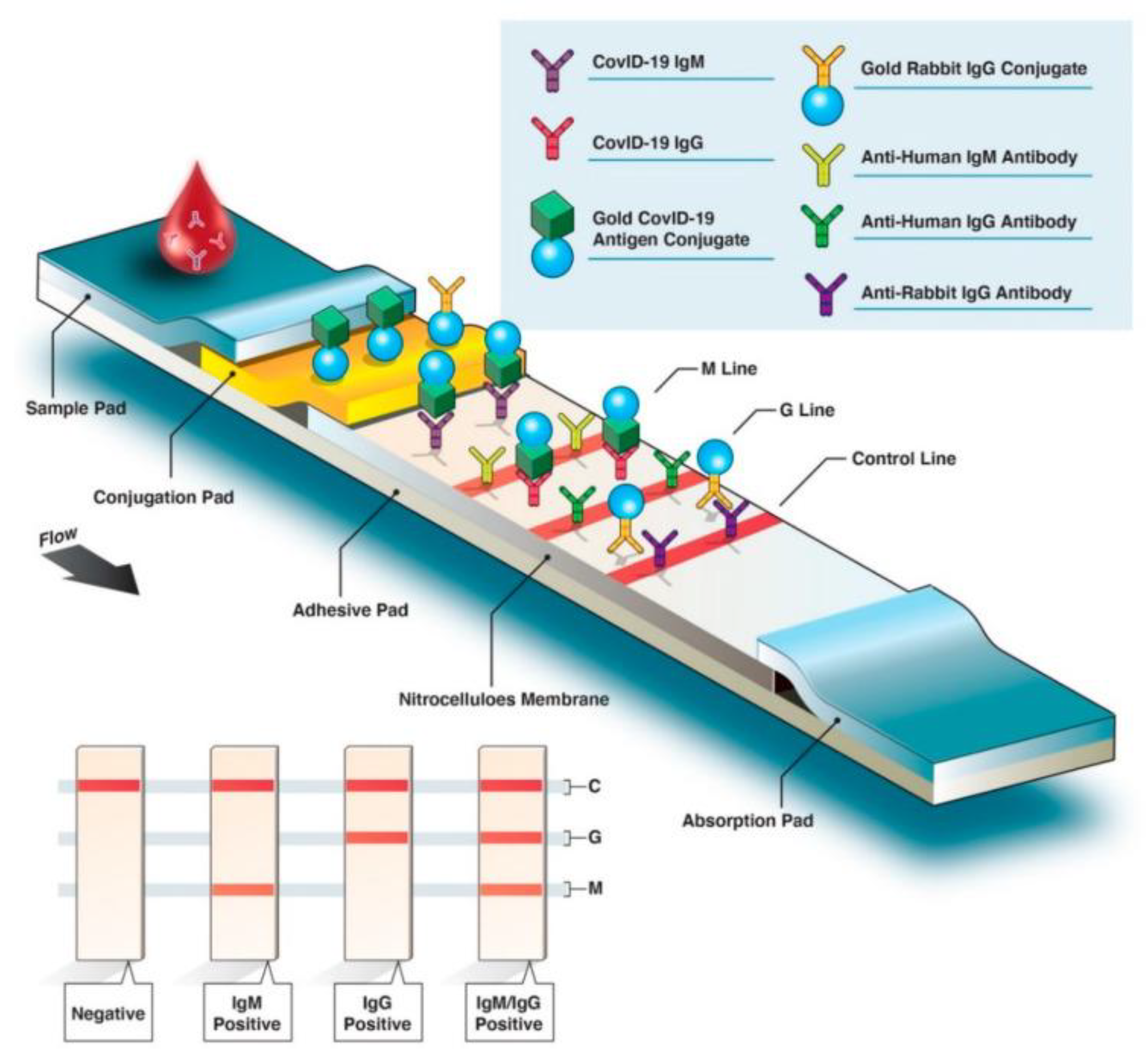
Lateral flow immunoassay (LFIA) [often referred to as LFA or lateral flow immunochromatographic assay (LFIcA)] is a promising technique for fast, accurate, and economical detection of SARS-CoV-2 infection; it can detect both SARS-CoV-2-specific Ags and Abs. LFIA is primarily a qualitative detection method that does not require any specific instrument, thereby making it a promising alternative for PoC diagnosis of COVID-19 (Fu et al., 2011) [87]. Figure 7 shows the basic structural design and experimental workflow of Ab versus Ag detection using colourimetric LFIA. The following sections discuss additional details, including the working principle, test-strip compartmentalisation, and reaction steps involved in Ag/Ab-based LFIAs.
Antigen-Detecting Rapid Diagnostic Tests
One of the immunoassay techniques that can be employed for rapid diagnosis of COVID-19 is Ag-detecting RDTs (Ag-RDTs) [otherwise referred to as Ag tests or rapid Ag tests (RATs)]. Ag-RDTs directly detect viral (SARS-CoV-2) Ags, usually nucleocapsid protein (NP), produced by replicating viral particles in patients' respiratory secretions. Thus, like molecular testing, Ag-RDTs help identify active SARS-CoV-2 infection (i.e., infection the patient currently has). However, Ag tests provide no information about prior infections. Most SARS-CoV-2 Ag-RDT samples are collected from the patient’s oral or nasal cavity using oropharyngeal, nasopharyngeal, or nasal swabs. Other sample types may also be used for Ag-RDTs once more clinical trial data become available (CDC, 2020c [89]; WHO, 2020 [3]).
Several factors determine the success and accuracy of Ag-RDTs, which include: (1) the time/duration from onset of viral infection, (2) the viral load in the sample collected from a person, (3) the quality as well as processing of the collected specimen, and (4) the specific formulation of the different reagents provided in the commercial diagnostic kits (WHO, 2020) [3].
Ag-RDTs for COVID-19 generally give positive results when patients are most infectious, with the highest viral load in their bodies (i.e., usually 1−3 days before the onset of disease symptoms and during the initial 5−7 days after the first appearance of disease symptoms). But once the patient clears the viral infection and recovers, results of Ag-RDTs will become negative (https://www.who.int/publications/i/item/antigen-detection-in-the-diagnosis-of-sars-cov-2infection-using-rapid-immunoassays) [90]. It is noteworthy that Ag-RDTs may give false negative results when viral concentration drops below the test’s detection limit, which is generally about real-time reverse transcriptase polymerase chain reaction (rRT-PCR) cycle threshold (Ct) values of <30−35. Therefore, a negative Ag-RDT result fails to completely rule out an active SARS-CoV-2 infection. In such cases, repeat testing or, ideally, more confirmatory testing using NAAT should be carried out whenever possible, especially for symptomatic patients. Moreover, negative Ag-RDT results should not mean that symptomatic or even asymptomatic contacts of suspected cases are removed from quarantine. Nevertheless, positive Ag-RDT outcomes in asymptomatic contacts can help rapidly expand contact-tracing efforts within communities (WHO, 2020) [3].
When performed in settings/locale where SARS-CoV-2 is prevalent, positive Ag-RDT results represent active SARS-CoV-2 infection (provided such Ag-RDTs have met WHO’s minimum performance criteria (WHO, 2020) [3].
Format of Ag-RDTs – Ag-RDTs utilise an easy-to-use immunochromatographic (lateral flow) format (as generally used for rapid testing of malaria and HIV) for detecting Ags from clinical specimens (https://www.who.int/publications/i/item/antigen-detection-in-the-diagnosis-of-sars-cov-2infection-using-rapid-immunoassays) [90]. Typically, RDTs comprise a nitrocellulose strip with a sample well enclosed in a plastic cassette. After combining the infected patient’s specimen with the test buffer, the mixture is loaded onto the sample well. Target Ags present in the mixture then bind to immobilised labelled Abs present on the nitrocellulose test strip and migrate together. They are eventually captured by Abs bound to the test (T) line, triggering a colourimetric reaction with a detectable colour change. On the basis of the test (and the specific labels used on the Abs), the change in colour can be monitored by the user with or without the help of a detector (https://www.who.int/publications/i/item/antigen-detection-in-the-diagnosis-of-sars-cov-2infection-using-rapid-immunoassays) [90]. The general format of Ag-RDTs is illustrated in Figure 8. COVID-19 RDTs usually yield results in ~10-30 minutes compared to the several hours needed for most NAATs (https://www.who.int/publications/i/item/antigen-detection-in-the-diagnosis-of-sars-cov-2infection-using-rapid-immunoassays) [90].
Interpreting the performance of Ag-RDTs – The gold standard for determining the performance of Ag-RDTs is the (1) sensitivity and (2) specificity of the detection test for identifying SARS-CoV-2 infection with reference to a NAAT standard (usually rRT-PCR). In this context, sensitivity and specificity are defined as:
(1) Sensitivity refers to the percentage of positive cases based on a NAAT reference standard, which are read as positive by the Ag-RDT under evaluation. It is calculated as (equation 1):
Sensitivity = [(true positives) ÷ (true positives + false negatives)] x 100
(2) Specificity refers to the percentage of negative cases based on a NAAT reference standard, which are read as negative by the Ag-RDT under evaluation. It is calculated as (equation 2):
Specificity = [(true negatives) ÷ (true negatives + false positives)] x 100
The predictive value of a test for a disease under consideration is greatly affected by the prevalence of the said disease in the population and can be either a positive predictive value (PPV) or a negative predictive value (NPV). Here, PPV indicates the probability that an individual with a positive test result actually has the infection, whereas NPV indicates the probability that an individual with a negative test result does not have the infection. The prevalence of the disease in the population should be estimated through surveillance to determine the PPVs and NPVs for Ag-RDTs and facilitate optimal interpretation of the final outcomes. The PPV and NPV can be calculated using the following formulae (equations 3 and 4) shown below:
PPV = [(true positives) ÷ (true positives + false positives)] x 100
NPV = [(true negatives) ÷ (true negatives + false negatives)] x 100
Additional information on how PPV and NPV can influence the interpretation of Ag-RDT results is available in the Annex of (https://www.who.int/publications/i/item/antigen-detection-in-the-diagnosis-of-sars-cov-2infection-using-rapid-immunoassays) [90].
Expectations from Ag-RDT performance – Several SARS-CoV-2-specific Ag-RDTs are either in development or have been commercialised recently. The FIND SARS-CoV-2 diagnostic pipeline tracker (https://www.finddx.org/covid-19/pipeline) [92] lists such SARS-CoV-2 Ag-RDTs.
Currently, evidence on the overall performance of these Ag-RDTs remains limited, but it is growing daily. Available raw data from independent upper respiratory tract (URT) samples show heterogeneous Ag-RDT performance (sensitivity ranging from 0–94%) compared with NAAT. Nevertheless, most of these RATs have demonstrated high specificity (i.e., >97%), and interestingly, the performance of a few commercial products in patient samples with higher viral loads (≥106 viral copies per mL or Ct values of ≤25-30) has exceeded the minimum performance requirements.
Unlike molecular tests, Ag-RDTs do not need any sophisticated laboratory equipment/instruments, highly skilled manpower/resources, or specific reagents. Therefore, these tests are inexpensive to mass-produce and administer. Moreover, Ag-RDTs provide results within 15–20 minutes, thereby making them ideal for mass screening in high-risk communities with large numbers of patients. Consequently, Ag-RDTs are the most potential candidates for PoC diagnostics.
Nonetheless, developing Ag-RDTs with high sensitivity and specificity involves several inherent challenges. They are as follows:
1. One of the primary challenges is the choice of the right Abs (Peeling et al., 2020) [93].
- For example, if an Ag-RDT for the SARS-CoV-2 S protein need to be developed successfully, then the ideal two Abs are required to possess a high degree of affinity towards the S protein at different sites, should not interfere with one another, and should not exhibit cross-reactivity with other S proteins of other strains of coronaviruses.
2. The second major obstacle is the signal intensity.
- Unlike most molecular-based tests, Ag-RDTs do not amplify the target biomolecule, resulting in lower sensitivity compared with molecular tests. Moreover, the ideal time to carry out these Ag-RDTs is when the disease burden (viral load) reaches its peak.
RATs have been developed to identify other infectious viruses, including HIV and influenza, and a few test kits have been commercially launched for COVID-19 diagnosis as well (Table 6). For example, Quidel Corporation (San Diego, CA, USA) developed a qualitative COVID-19 detection kit approved by the Food and Drug Administration (FDA) on 8th May, 2020 (Quidel Corporation, 2020) [94]. The kit uses an immunofluorescence-based lateral flow technique called Sofia 2 SARS Antigen Fluorescent Immunoassay, which uses a sandwich design to detect SARS-CoV-2 via the N protein. This PoC diagnostic kit with automated reading fetches results within 15 min without any cross-reactivity with other coronaviruses. The diagnostic kit can be used directly with nasal or nasopharyngeal swab samples from suspected individuals, or after adding viral transport media to the collected samples. The developed kit has a 12-month shelf life, making it suitable for transportation.
Antibody-Detecting Rapid Diagnostic Tests
Ab-detecting RDTs (Ab-RDTs) [also referred to as rapid Ab test or RST] detect specific products of the body’s immune response to the virus (i.e., the presence of specific anti-SARS-CoV-2 Abs). These rapid Ab tests are fairly accurate approximately 15−21 days after acquiring the infection (Deeks et al. 2020) [118]. LFIAs (at least for Ab-RDTs) detect the Abs generated in response to invading pathogenic microbes. In general, LFIA consists of four distinct pads (as illustrated in Figure 8), which include:
1) The sample pad – It receives the sample from the suspected individual.
2) The conjugation pad – It contains specific Ags and/or Abs (in case of Ag-RDTs mentioned in the previous section) conjugated with specific labels (typically colloidal gold, carbon nanoparticles, or latex).
3) The nitrocellulose membrane (attached to an adhesive pad) – It helps to draw the sample solution using capillary forces to the control and test lines.
4) The absorption pad – It collects the processed sample at the end (i.e., in the final phase).
Ab-RDTs are conducted using blood samples. Briefly, the collected blood sample is loaded onto the sample well present on the sample pad. Then, the diluent buffer is added to the loaded sample. The resulting mixture of sample and diluent flows through the sample pad by capillary action (lateral flow) and eventually reaches the conjugation pad, which contains viral Ags conjugated with specific label molecules or nanoparticles. Consequently, the target biomarker (SARS-CoV-2-specific Abs in this case) binds to its specific labelled viral Ags, forming immune complexes. Next, the conjugated immune complexes move toward the nitrocellulose membrane, where they encounter the three test lines: IgM, IgG, and control, in that order. The M line is the first line containing immobilised Abs that recognise human IgM. Any or all IgM Abs present in the sample will bind here, even though a visible colored line will be produced only for those human IgM Abs that are specific for the viral infection and form complexes with labelled Ags. The G line is the second line and contains immobilised Abs that recognise human IgG. As explained for the M line, any or all IgG Abs present in the sample will bind here. But again, only human IgG Abs conjugated with virus-specific labelled Ag complexes will be recognised, and accordingly a visible colored line will be produced. Finally, the Ab-Ag complexes reach the test line that contains immobilised capture reagents, including the target-biomarker-specific Abs, thereby capturing the target biomarker and conjugated Ag complexes. LFIA operates via a sandwich immunoassay that captures the target biomolecule and displays different coloured signals on the control and test lines, depending on the specific labels used. The last line the sample encounters is the control line, which contains immobilised Abs capable of recognising the control Abs (i.e., rabbit IgG). To act as a test control, a coloured line must always appear in the control line region to confirm that an appropriate sample volume has been loaded and that membrane wicking has occurred. The test results can usually be obtained after 10 mins. (Figure 12.9) (Augustine et al. 2020 [16]; Taleghani and Taghipour, 2021 [11]). Clinical studies by Xiang et al. (2020b) [119] have shown that LFIA has 82% sensitivity for detecting both anti-SARS-CoV-2 IgM and IgG Abs, and this can be further enhanced with novel nanoparticles.
Figure 9.
In-depth overview of a qualitative rapid diagnostic serological test for anti-SARS-CoV-2 antibody detection. Schematic diagram illustrating the mode of detection and the final appearance of the result for LFIA. Ab-detecting rapid diagnostic tests (Ab-RDTs) are usually based on LFIAs, in which host IgM/G Abs migrate across an adhesive pad (e.g., nitrocellulose membrane) and interact with immobilised SARS-CoV-2-specific Ags and secondary Abs (anti-human IgM/G Abs). Conjugated (gold-labelled) virus-specific Ag(s) on the conjugation pad bind(s) with the corresponding host Abs and form(s) immune complexes. As Ab–Ag complexes travel across the membrane using capillary forces, immobilised anti-SARS-CoV-2 IgM and anti-SARS-CoV-2 IgG Abs interact with fixed anti-IgM and anti-IgG secondary Abs on the M and G lines, respectively. If the blood sample from a
suspected individual does not contain SARS-CoV-2-specific Abs, then the M or G lines fail to appear in the final test results. In that case, only the control (C) line appears. A positive result denotes the presence of SARS-CoV-2 infection, while a negative result denotes the absence of SARS-CoV-2 infection (Source: Ghaffari et al. 2020 [5] with modifications; https://creativecommons.org/licenses/by/4.0/) [17].
Figure 9.
In-depth overview of a qualitative rapid diagnostic serological test for anti-SARS-CoV-2 antibody detection. Schematic diagram illustrating the mode of detection and the final appearance of the result for LFIA. Ab-detecting rapid diagnostic tests (Ab-RDTs) are usually based on LFIAs, in which host IgM/G Abs migrate across an adhesive pad (e.g., nitrocellulose membrane) and interact with immobilised SARS-CoV-2-specific Ags and secondary Abs (anti-human IgM/G Abs). Conjugated (gold-labelled) virus-specific Ag(s) on the conjugation pad bind(s) with the corresponding host Abs and form(s) immune complexes. As Ab–Ag complexes travel across the membrane using capillary forces, immobilised anti-SARS-CoV-2 IgM and anti-SARS-CoV-2 IgG Abs interact with fixed anti-IgM and anti-IgG secondary Abs on the M and G lines, respectively. If the blood sample from a
suspected individual does not contain SARS-CoV-2-specific Abs, then the M or G lines fail to appear in the final test results. In that case, only the control (C) line appears. A positive result denotes the presence of SARS-CoV-2 infection, while a negative result denotes the absence of SARS-CoV-2 infection (Source: Ghaffari et al. 2020 [5] with modifications; https://creativecommons.org/licenses/by/4.0/) [17].
For SARS-CoV-2 detection, different LFIAs are either in development or already developed (Xiang et al. 2020b) [119]. Because these tests primarily detect IgM and IgG Abs, they may produce false-negative results during the early stages of the disease. Integration of molecular tests with LFIAs can also be done, as the combination of Reverse Transcriptase Loop-Mediated Isothermal Amplification (RT-LAMP) and LFIAs was applied for Middle East respiratory syndrome coronavirus (MERS-CoV) detection; however, such a test combo displayed low sensitivity as well (Huang et al. 2018) [120]. Major challenges with LFIAs include determining the appropriate test timing and assay sensitivity. To overcome these problems, a promising strategy would be to develop an LFIA capable of detecting the virus instead of Abs and implementing a few of the signal amplification techniques such as application of carbon nanomaterials, dual sensitisers, organic molecules, and plasmonic nanoparticles, which would even respond to low levels of SARS-CoV-2 during the initial infection stages (Taleghani and Taghipour, 2021) [11].
Our understanding of Ab/host immune responses to SARS-CoV-2 is still evolving. Therefore, WHO suggests that Ab detection tests should not be used to determine active SARS-CoV-2 infections in clinical care or for contact tracing. Moreover, interpretation of Ab-RDT results must be done by an expert and is contingent on various factors/parameters such as (1) the timing of the disease, (2) clinical morbidity, (3) the epidemiology and disease prevalence within the specific setting, (4) the type of test employed, (5) the validation technique, and (6) the reliability of the test results [1]. The clinical significance of a positive Ab-RDT result is still under scrutiny. It is important to note that the presence of SARS-CoV-2-specific Abs does not guarantee or necessarily mean that these are neutralising Abs or that these would provide protective immunity. Nonetheless, Ab testing may be helpful for carrying out serosurveillance studies not only to support the investigation of the current pandemic but also to corroborate the retrospective assessment of the size of COVID-19 outbreak or the rate of viral attack (WHO, 2020) [3].
4. Benefits, Issues and Challenges
Each one of the developed immunoassay techniques (both conventional and rapid) for SARS-CoV-2 detection is appropriate for specific situations (prior or active SARS-CoV-2 infection), locations (laboratory-based/PoC-based) and/or specific target biomarkers (Ags/Abs), but each one has a few shortcomings, so there is an ongoing search for alternative/advanced detection methods with higher sensitivity, enhanced specificity, and lower time of detection. A comparative review of immunoassays for COVID-19 detection is available in Mohit et al. (2021).
Serology tests, including qualitative rapid Ab tests (Ab-RDTs), can detect individuals with prior infection as well as those currently suffering from COVID-19. Hence, serological tests provide better insight into the actual level of SARS-CoV-2 infection in the population. Additionally, these tests can determine the phase of SARS-CoV-2 infection based on the concentration of different Abs in the patient’s specimen. In contrast, serological tests do not detect the virus itself, but the Ab generated by the host immune system against it. Therefore, they might produce false-negative results during the initial stage of infection/disease, sharing the major drawback of the PCR-based detection methods (Taleghani and Taghipour, 2021) [11]. Similarly, the qualitative Ag-RDTs also have several benefits, issues and challenges. Table 7 lists the main advantages and disadvantages of qualitative RDTs.
5. Conclusions
The COVID-19 pandemic caused by severe acute respiratory syndrome coronavirus 2 (SARS-CoV-2) highlighted the critical importance of rapid, accurate, and accessible diagnostic technologies for effective disease management and infection control. Immunoassays, particularly rapid diagnostic tests (RDTs) based on immunochromatographic or lateral flow formats, have emerged as valuable tools for SARS-CoV-2 detection because of their simplicity, rapid turnaround time, and suitability for point-of-care applications.
In this manuscript, we provide an overview of the principles and applications of immunoassay-based approaches for SARS-CoV-2 diagnosis, with particular emphasis on rapid Ag- and Ab-detecting tests. We discuss the underlying immunological mechanisms, the distinctions between conventional laboratory-based immunoassays and rapid lateral flow assays, and the respective advantages and limitations of Ag-RDTs and Ab-RDTs. We also highlight recent developments in colourimetric lateral flow immunoassays to improve diagnostic performance, accessibility, and practical utility.
A key focus of the manuscript is the complementary role of these diagnostic approaches. Ag-RDTs can facilitate rapid identification of active infection and support timely clinical and public-health interventions, whereas Ab-based assays have important applications in evaluating the host immune response and conducting serological surveillance. We believe this perspective will interest researchers and professionals in diagnostic biotechnology, clinical microbiology, immunology, infectious diseases, and point-of-care diagnostics.
The review provides a concise synthesis of the current understanding of SARS-CoV-2 immunoassays while highlighting ongoing technological developments and the challenges that remain in achieving reliable, accessible, and rapid diagnostic testing.
6. Future Perspectives
Knowledge gained from previous viral epidemics/outbreaks caused by SARS-CoV and MERS-CoV has played a crucial role in enabling fast, precise COVID-19 diagnosis. Although NAAT remains the principal diagnostic method for COVID-19 (i.e., the first step toward limiting further viral spread), immunoassays have also garnered considerable attention because they are simple to perform and provide a better understanding of infection status than NAATs.
Of the plethora of serological tests in vogue now, partially owing to the current spike in mass seroprevalence/serosurveillance studies, it becomes necessary for both national and regional or state Governments to pursue combined efforts to independently validate the performance of the serological tests and collaborate with the pharmaceutical/drug industry for large-scale manufacturing and production of the diagnostic test kits. With the surge of COVID-19 cases and SARS-CoV-2’s inherent propensity to evolve and change, giving rise to new variants, current detection procedures must be accompanied by clear, informed guidelines.
Despite the war-footing response of the human population to the COVID-19 pandemic, a lacuna remains in understanding the immunological crosstalk between the virus and our immune system, which may negatively affect the use of various serological tests. It becomes imperative now to enrich our domain of knowledge with respect to certain facets of SARS-CoV-2, namely:
- Are there any serological tests to identify neutralising Abs for SARS-CoV-2, and if so, which ones?
- Are there any neutralising Abs cross-reactive to other variants of coronaviruses?
- Which SARS-CoV-2 Ags are optimal for neutralising Ab detection?
- Is there any correlation between anti-SARS-CoV-2 Abs and innate or adaptive immunity?
- After recovery, how long can the Abs generated in response to SARS-CoV-2 provide immunity to recovered individuals? Do these individuals become susceptible to a second attack of SARS-CoV-2?
COVID-19 outbreak has caused unprecedented economic loss. In the wake of the pandemic, restoring a new normal through return-to-work policies, along with the failure to use available diagnostic techniques due to uncertainty, may have profound consequences. Thus, at this juncture, it is important to understand the limits and potential of various serology tests to make a fruitful, informed decision based on a risk-benefit assessment in each case.
HIV and malaria programs have expanded the use of rapid immunoassays for several years, with considerable knowledge of test performance, operational quality assurance (QA) systems, and awareness campaigns targeting patients and providers. In contrast, to fight the COVID-19 outbreak, it is necessary to roll out SARS-CoV-2 Ag-RDTs urgently over weeks and months, ideally across the full spectrum of medical and community settings, as well as among health care workers. Such Ag-RDTs could be used in multiple ways, including managing individual cases, contact tracing, surveillance studies, and investigating further outbreaks. To achieve this goal safely and efficiently, a general understanding of Ag-RDT operational principles, along with their benefits and drawbacks, should be integrated with stringent protocols for utilisation, QA, and ease of communication. Moreover, when utilising SARS-CoV-2 Ag-RDTs, specific environmental and personal safety precautions are necessary to prevent respiratory pathogen transmission. In addition, these combined efforts must be carefully monitored to determine their impact on patients and public health.
The paper thoroughly summarises the various facets of the diverse immunoassay-techniques developed and implemented for detecting SARS-CoV-2 infection. It also provides a detailed, comparative assessment of various immunoassays, including their modes of operation, advantages, and disadvantages. Because early diagnosis and detection are the only fail-safe mechanisms for fighting SARS-CoV-2 infection, it is imperative to address the issues and challenges of existing techniques and develop more efficient, cost-effective methods for rapid, accurate detection in all suspected individuals during the early stages of infection, including asymptomatic COVID-19 carriers. In this context, we have showcased some of the most promising techniques already implemented, including LFIAs. Given the unprecedented nature of the pandemic, it can now only be hoped that these methods will address the emergency requirement of developing PoC and multiplex testing to a certain extent, and aid in arresting additional viral outbreaks by providing the test results more easily, more quickly, and in a more precise manner for hospitals as well as small healthcare facilities.
Author Contributions
P.M. conceived the idea and designed the manuscript. P.M. and M.B. wrote the manuscript. P.M., M.B., and A.M. edited the final manuscript. All three authors read and approved the final version of the manuscript.
Funding
No funding was available.
Data Availability Statement
Not applicable.
Conflicts of Interest
The authors declare no conflict of interest.
References
- Available online: https://www.who.int/publications/i/item/diagnostic-testingfor-sars-cov-2.
- Available online: https://www.who.int/dg/speeches/detail/who-director-general-s-opening-remarks-at-the-media-briefing-on-covid-19---11-march-2020.
- Diagnostic testing for SARS-CoV-2. In terim guidance; World Health Organization: Geneva, 11 September 2020; Available online: https://www.who.int/publications/i/item/diagnostic-testingfor-sars-cov-2 (accessed on 18 November 2020).
- Available online: https://www.who.int/emergencies/diseases/novel-coronavirus-2019/technical-guidance-publications?publicationtypes=df113943-c6f4-42a5-914f-0a0736769008.
- Ghaffari, A.; Meurant, R.; Ardakani, A. COVID-19 serological tests: how well do they actually perform? Diagnostics 2020, 10(7), 453. [Google Scholar] [CrossRef]
- Available online: https://www.finddx.org/covid-19/test-tracker/.
- Available online: https://www.who.int/publications/i/item/covid-19-strategy-update---14-april-2020.
- Janeway, C.A. Immunobiology, 5th edition: The Immune System in Health and Disease; Garland Publishing: New York, NY, USA, 2001. [Google Scholar]
- Azkur, A.K.; Akdis, M.; Azkur, D.; Sokolowska, M.; van de Veen, W.; Bruggen, M.C.; O’Mahony, L.; Gao, Y.; Nadeau, K.; Akdis, C.A. Immune response to SARS-CoV-2 and mechanisms of immunopathological changes in COVID-19. Allergy 2020. [Google Scholar] [CrossRef]
- Yu, H.Q.; Sun, B.Q.; Fang, Z.F.; Zhao, J.C.; Liu, X.Y.; Li, Y.M.; Sun, X.Z.; Liang, H.F.; Zhong, B.; Huang, Z.F.; et al. Distinct features of SARS-CoV-2-specific IgA response in COVID-19 patients. Eur. Respir. J. 2020. [Google Scholar] [CrossRef]
- Taleghani, N.; Taghipour, F. Diagnosis of COVID-19 for controlling the pandemic: A review of the state-of-the-art. Biosens. Bioelectron. 2020, 112830. [Google Scholar] [CrossRef]
- Kellner, R.J.; Mermet, J.M.; Otto, M.; and Widmer, H.M.; Born, J.L., 1998. , New York: Wiley-VCH. American Chemical Society (ACS). [CrossRef]
- Dutta, J.; Goswami, S.; Mitra, A. COVID-19 and Emerging Environmental Trends: A Way Forward; CRC Press, 2020. [Google Scholar]
- Chang, L.; Rissin, D.M.; Fournier, D.R.; Piech, T.; Patel, P.P.; Wilson, D.H.; Duffy, D.C. Single molecule enzyme-linked immunosorbent assays: theoretical considerations. J. Immunol. Methods 2012, 378(1-2), 102–115. [Google Scholar] [CrossRef]
- Huergo, L.F.; Selim, K.A.; Conzentino, M.S.; Gerhardt, E.C.; Santos, A.R.; Wagner, B.; Alford, J.T.; Deobald, N.; Pedrosa, F.O.; De Souza, E.M.; Nogueira, M.B. Magnetic bead-based immunoassay allows rapid, inexpensive, and quantitative detection of human SARS-CoV-2 antibodies. ACS Sens. 2021, 6(3), 703–708. [Google Scholar] [CrossRef]
- Augustine, R.; Das, S.; Hasan, A.; Abdul Salam, S.; Augustine, P.; Dalvi, Y. B., ...; Kevadiya, B. D. Rapid antibody-based COVID-19 mass surveillance: relevance, challenges, and prospects in a pandemic and Post-Pandemic world. J. Clin. Med. 2020, 9(10), 3372. [Google Scholar] [CrossRef]
- https://creativecommons.org/licenses/by/4.0/.
- BioRender.com
- Source
- Roda, A.; Cavalera, S.; Di Nardo, F.; Calabria, D.; Rosati, S.; Simoni, P.; Colitti, B.; Baggiani, C.; Roda, M.; Anfossi, L. Biosens. Bioelectron. 2021, 172, 112765. Available online: https://. [CrossRef]
- CDC. 2020a. Available online: https://www.cdc.gov/coronavirus/2019-ncov/lab/guidelines-clinical-speci (accessed on 14 June 2020).
- Lv, H.; Wu, N.C.; Tak-Yin Tsang, O.; Malik Peiris, J.; Wilson, I.A.; Mok, C.K.; Yuan, M.; APM Perera, R.; Shing Leung, W.; So, R.T.; Man Chun Chan, J.; Yip, G.K.; Shiu Hong Chik, T.; Wang, Y.; Yau Chung Choi, C.; Lin, Y.; Ng, W.W.; Zhao, J.; Poon, L.L. Cell Rep. 2020, 31, 107725. [CrossRef]
- Garcia-Rodriguez, C.; Levy, R.; Arndt, J.W.; Forsyth, C.M.; Razai, A.; Lou, J.; Geren, I.; Stevens, R.C.; Marks, J.D. Nat. Biotechnol. 2007, 25, 107–116.
- Klompus. 2020.
- Available online: https://www.fda.gov/media/141891/download (accessed on 30th June 2021).
- Available online: https://www.fda.gov/media/140297/download (accessed on 30th June 2021).
- Available online: https://www.fda.gov/media/144877/download (accessed on 30th June 2021).
- Available online: https://www.fda.gov/media/145080/download (accessed on 30th June 2021).
- Available online: https://www.fda.gov/media/138660/download (accessed on 30th June 2021).
- Available online: https://www.fda.gov/media/144562/download (accessed on 1st July 2021).
- Available online: https://www.fda.gov/media/144071/download (accessed on 1st July 2021).
- Available online: https://www.fda.gov/media/139410/download (accessed on 1st July 2021).
- Available online: https://www.fda.gov/media/142422/download (accessed on 1st July 2021).
- Available online: https://www.fda.gov/media/140443/download (accessed on 1st July 2021).
- Available online: https://www.fda.gov/media/140447/download (accessed on 2nd July 2021).
- Available online: https://www.fda.gov/media/139792/download (accessed on 2nd July 2021).
- Available online: https://www.fda.gov/media/141773/download (accessed on 2nd July 2021).
- Available online: https://www.fda.gov/media/141550/download (accessed on 2nd July 2021).
- Available online: https://www.fda.gov/media/140082/download (accessed on 2nd July 2021).
- Available online: https://www.fda.gov/media/140030/download (accessed on 3rd July 2021).
- Available online: https://www.fda.gov/media/138660/download (accessed on 3rd July 2021).
- Available online: https://www.fda.gov/media/138438/download (accessed on 3rd July 2021).
- Available online: https://www.fda.gov/media/136625/download (accessed on 3rd July 2021).
- Available online: https://www.fda.gov/media/144764/download (accessed on 4th July 2021).
- Available online: https://www.fda.gov/media/147368/download (accessed on 4th July 2021).
- Available online: https://www.fda.gov/media/139730/download (accessed on 4th July 2021).
- Available online: https://www.fda.gov/media/146901/download (accessed on 4th July 2021).
- Available online: https://www.fda.gov/media/145277/download (accessed on 4th July 2021).
- Available online: https://www.fda.gov/media/144010/download (accessed on 5th July 2021).
- Available online: https://www.fda.gov/media/143583/download (accessed on 5th July 2021).
- Available online: https://www.fda.gov/media/137493/download (accessed on 5th July 2021).
- Available online: https://www.fda.gov/media/143452/download (accessed on 5th July 2021).
- Available online: https://www.fda.gov/media/142809/download (accessed on 5th July 2021).
- Available online: https://www.fda.gov/media/142700/download (accessed on 6th July 2021).
- Available online: https://www.fda.gov/media/141777/download (accessed on 6th July 2021).
- Available online: https://www.fda.gov/media/140929/download (accessed on 6th July 2021).
- Available online: https://www.fda.gov/media/139053/download (accessed on 6th July 2021).
- Available online: https://www.fda.gov/media/138810/download (accessed on 6th July 2021).
- Available online: https://www.fda.gov/media/137609/download (accessed on 6th July 2021).
- Available online: https://www.fda.gov/media/137029/download (accessed on 6th July 2021).
- Available online: https://www.fda.gov/media/147808/download (accessed on 6th July 2021).
- Available online: https://www.fda.gov/media/136967/download (accessed on 6th July 2021).
- Available online: https://www.fda.gov/media/145944/download (accessed on 6th July 2021).
- Available online: https://www.fda.gov/media/142940/download (accessed on 6th July 2021).
- Available online: https://www.fda.gov/media/142911/download (accessed on 6th July 2021).
- Available online: https://www.fda.gov/media/139627/download (accessed on 7th July 2021).
- Available online: https://www.fda.gov/media/138753/download (accessed on 7th July 2021).
- Available online: https://www.fda.gov/media/138757/download (accessed on 7th July 2021).
- Available online: https://www.fda.gov/media/137363/download (accessed on 7th July 2021).
- Available online: https://www.fda.gov/media/137605/download (accessed on 7th July 2021).
- Available online: https://www.fda.gov/media/137359/download (accessed on 7th July 2021).
- Available online: https://www.fda.gov/media/142940/download (accessed on 8th July 2021).
- Available online: https://www.fda.gov/media/142552/download (accessed on 8th July 2021).
- Available online: https://www.fda.gov/media/137383/download (accessed on 8th July 2021).
- Available online: https://www.fda.gov/media/142233/download (accessed on 8th July 2021).
- Available online: https://www.fda.gov/media/142002/download (accessed on 8th July 2021).
- Available online: https://www.fda.gov/media/142006/download (accessed on 11th July 2021).
- Available online: https://www.fda.gov/media/141255/download (accessed on 11th July 2021).
- Available online: https://www.fda.gov/media/140699/download (accessed on 11th July 2021).
- Available online: https://www.fda.gov/media/138442/download (accessed on 11th July 2021).
- Available online: https://www.fda.gov/media/138442/download (accessed on 11th July 2021).
- Available online: https://www.fda.gov/media/140704/download (accessed on 13th July 2021).
- Available online: https://www.fda.gov/media/140704/download (accessed on 13th July 2021).
- Available online: https://www.fda.gov/media/139446/download (accessed on 13th July 2021).
- Available online: https://www.fda.gov/media/138629/download (accessed on 13th July 2021).
- Available online: https://www.fda.gov/media/143584/download (accessed on 21st August 2021).
- Fu, E.; Liang, T.; Houghtaling, J.; Ramachandran, S.; Ramsey, S.A.; Lutz, B.; Yager, P. Anal. Chem. 2011, 83, 7941–7946. [CrossRef]
- Jacobs, J.; Kühne, V.; Lunguya, O.; Affolabi, D.; Hardy, L.; Vandenberg, O. Implementing COVID-19 (SARS-CoV-2) Rapid Diagnostic Tests in Sub-Saharan Africa: A Review. Front. Med. 2020, 7, 557797. [Google Scholar] [CrossRef]
- CDC. 2020c. Available online: https://www.cdc.gov/coronavirus/2019-ncov/lab/resources/antigen-te (accessed on 11 July 2020).
- Available online: https://www.who.int/publications/i/item/antigen-detection-in-the-diagnosis-of-sars-cov-2infection-using-rapid-immunoassays.
- Campbell, Ian M. CC BY-SA 3.0. Available online: https://creativecommons.org/licenses/by-sa/3.0.
- Available online: https://www.finddx.org/covid-19/pipeline.
- Peeling, R.W.; Wedderburn, C.J.; Garcia, P.J.; Boeras, D.; Fongwen, N.; Nkengasong, J.; Sall, A.; Tanuri, A.; Heymann, D.L. Lancet Infect. Dis. 2020, 20(9), 245–249. [CrossRef]
- Quidel Coroporation. 2020.
- Available online: https://www.fda.gov/media/149906/download (accessed on 20th July 2021).
- Available online: https://www.fda.gov/media/149911/download (accessed on 20th July 2021).
- Available online: https://www.fda.gov/media/149918/download (accessed on 20th July 2021).
- Available online: https://www.fda.gov/media/149055/download (accessed on 20th July 2021).
- Available online: https://www.fda.gov/media/148353/download (accessed on 20th July 2021).
- Available online: https://www.fda.gov/media/141570/download (accessed on 20th July 2021).
- Available online: https://www.fda.gov/media/147694/download (accessed on 20th July 2021).
- Available online: https://www.fda.gov/media/147578/download (accessed on 20th July 2021).
- Available online: https://www.fda.gov/media/137885/download (accessed on 20th July 2021).
- Available online: https://www.fda.gov/media/139755/download (accessed on 20th July 2021).
- Available online: https://www.fda.gov/media/147265/download (accessed on 20th July 2021).
- Available online: https://www.fda.gov/media/147254/download (accessed on 20th July 2021).
- Available online: https://www.fda.gov/media/147259/download (accessed on 20th July 2021).
- Available online: https://www.fda.gov/media/147264/download (accessed on 20th July 2021).
- Available online: https://www.fda.gov/media/147016/download (accessed on 21st July 2021).
- Available online: https://www.fda.gov/media/146312/download (accessed on 21st July 2021).
- Available online: https://www.fda.gov/media/145697/download (accessed on 21st July 2021).
- Available online: https://www.fda.gov/media/142919/download (accessed on 21st July 2021).
- Available online: https://www.fda.gov/media/145073/download (accessed on 21st July 2021).
- Available online: https://www.fda.gov/media/144668/download (accessed on 21st July 2021).
- Available online: https://www.fda.gov/media/143270/download (accessed on 21st July 2021).
- Available online: https://www.fda.gov/media/142704/download (accessed on 21st July 2021).
- Available online: https://www.fda.gov/media/141304/download (accessed on 21st July 2021).
- Deeks, J.J.; Dinnes, J.; Takwoingi, Y.; Davenport, C.; Spijker, R.; Taylor-Phillips, S.; et al. Antibody tests for identification of current and past infection with SARS-CoV-2. Cochrane Database Syst. Rev. 2020, 6, CD013652. [Google Scholar] [CrossRef]
- Xiang, J.; Yan, M.; Li, H.; Liu, T.; Lin, C.; Huang, S.; Shen, C. medRxiv 2020.02.27.20028787. 2020b. [Google Scholar] [CrossRef]
- Huang, P.; Wang, H.; Cao, Z.; Jin, H.; Chi, H.; Zhao, J.; Yu, B.; Yan, F.; Hu, X.; Wu, F.; Jiao, C.; Hou, P.; Xu, S.; Zhao, Y.; Feng, N.; Wang, J.; Sun, W.; Wang, T.; Gao, Y.; Yang, S.; Xia, X. Front. Microbiol. 2018, 9, 1101. [CrossRef]
- Mohit, E.; Rostami, Z.; Vahidi, H. A comparative review of immunoassays for COVID-19 detection. Expert Rev. Clin. Immunol. 2021, 17(6), 573–599. [Google Scholar] [CrossRef]
Figure 1.
SARS-CoV-2-induced humoral immune response in humans during COVID-19. (1) SARS-CoV-2 enters the target host cell through specific interaction between the viral spike (S) protein and human angiotensin-converting enzyme 2 (ACE2). (2–3) After viral replication and their subsequent release from the lysed host cells, a subset of viral particles is phagocytosed/ingested and digested by Ag-presenting cells (APCs), including macrophages and/or dendritic cells. (4) Disintegrated viral Ag(s) or antigenic peptide(s) is(are) then presented to T helper (TH) cells. This, in turn, interacts with and activates circulating B cells. (5) Upon activation, B cells proliferate and differentiate into plasma (or memory) B cells with high-affinity SARS-CoV-2 Ag-binding receptors. Plasma cells secrete IgM, IgG, and IgA Abs that act as their viral-specific receptors. (6) Ab-mediated neutralisation of viral Ag(s) takes place when anti-SARS-CoV-2 Abs bind to viral Ag(s) and inhibit subsequent interaction and entry of the virus into human cells. (Source: Ghaffari et al. 2020) [5].
Figure 1.
SARS-CoV-2-induced humoral immune response in humans during COVID-19. (1) SARS-CoV-2 enters the target host cell through specific interaction between the viral spike (S) protein and human angiotensin-converting enzyme 2 (ACE2). (2–3) After viral replication and their subsequent release from the lysed host cells, a subset of viral particles is phagocytosed/ingested and digested by Ag-presenting cells (APCs), including macrophages and/or dendritic cells. (4) Disintegrated viral Ag(s) or antigenic peptide(s) is(are) then presented to T helper (TH) cells. This, in turn, interacts with and activates circulating B cells. (5) Upon activation, B cells proliferate and differentiate into plasma (or memory) B cells with high-affinity SARS-CoV-2 Ag-binding receptors. Plasma cells secrete IgM, IgG, and IgA Abs that act as their viral-specific receptors. (6) Ab-mediated neutralisation of viral Ag(s) takes place when anti-SARS-CoV-2 Abs bind to viral Ag(s) and inhibit subsequent interaction and entry of the virus into human cells. (Source: Ghaffari et al. 2020) [5].
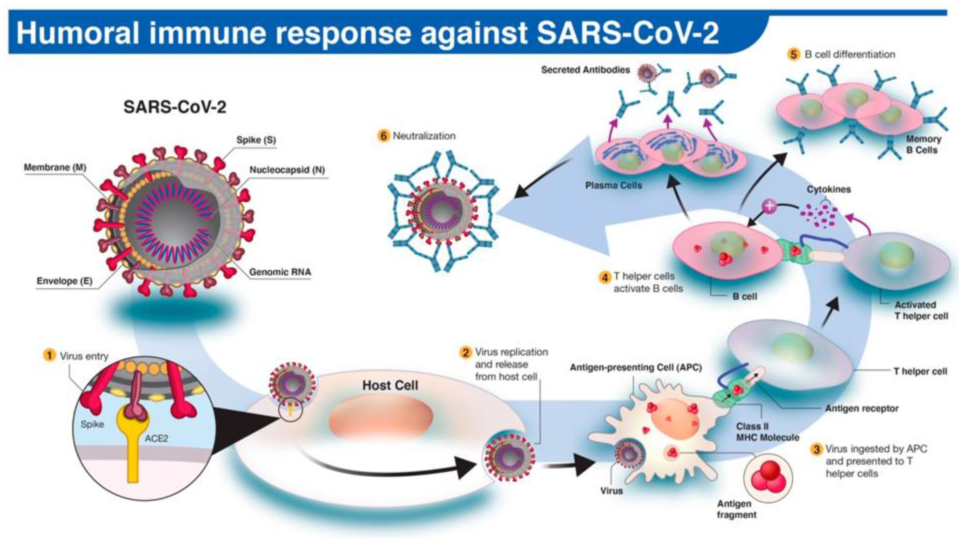
Figure 2.
Illustrative representation of the rapid serology test (RST) for the diagnosis of COVID-19. This figure depicts the basic steps in Ag-based detection of SARS-CoV-2 from a patient’s blood sample. The main working principle of SARS-CoV-2-specific Ab detection in RST on an immunochromatographic lateral flow format is as follows: After loading the collected blood sample, followed by addition of the sample diluent, the blood-diluent mixture moves across the lateral flow test strip, and Abs (IgM/IgG) present in the sample bind to labelled Ags that are immobilised on the test strip, forming Ag-Ab complexes. These immune complexes (together with labelled rabbit Abs) eventually reach the test (T) line (containing anti-human IgM/IgG Abs) and control (C) line (containing anti-rabbit IgG Abs), leading to a colour change in these lines (A–H). Figure 8 describes this immunoassay technique in detail. (Source: Augustine et al. 2020 [16]; https://creativecommons.org/licenses/by/4.0/) [17].
Figure 2.
Illustrative representation of the rapid serology test (RST) for the diagnosis of COVID-19. This figure depicts the basic steps in Ag-based detection of SARS-CoV-2 from a patient’s blood sample. The main working principle of SARS-CoV-2-specific Ab detection in RST on an immunochromatographic lateral flow format is as follows: After loading the collected blood sample, followed by addition of the sample diluent, the blood-diluent mixture moves across the lateral flow test strip, and Abs (IgM/IgG) present in the sample bind to labelled Ags that are immobilised on the test strip, forming Ag-Ab complexes. These immune complexes (together with labelled rabbit Abs) eventually reach the test (T) line (containing anti-human IgM/IgG Abs) and control (C) line (containing anti-rabbit IgG Abs), leading to a colour change in these lines (A–H). Figure 8 describes this immunoassay technique in detail. (Source: Augustine et al. 2020 [16]; https://creativecommons.org/licenses/by/4.0/) [17].
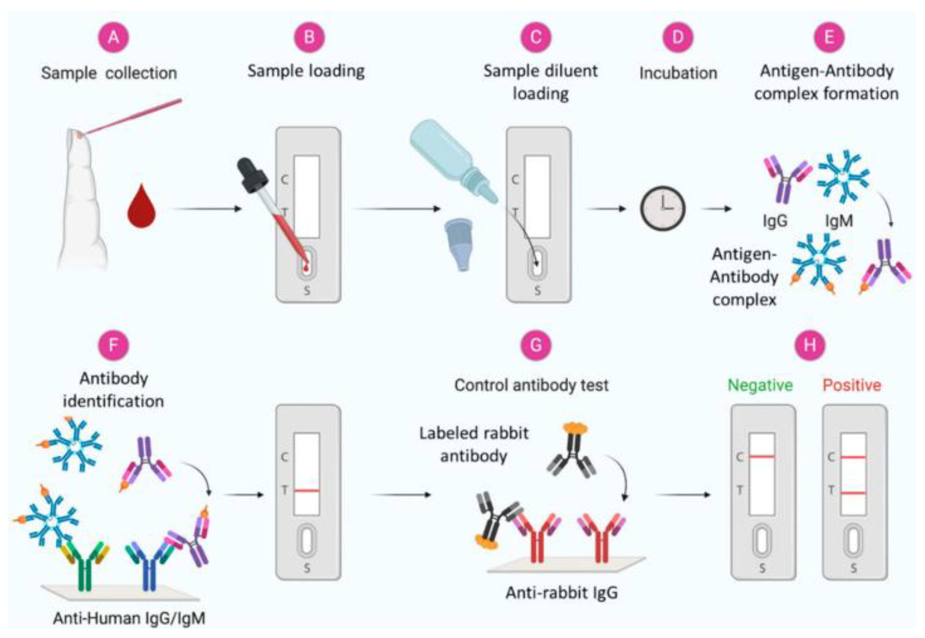
Figure 3.
Pictorial representation of the enzyme-linked immunosorbent assay (ELISA) and its variant for the diagnosis of COVID-19. A. Indirect ELISA: A simplified illustrative image of the 96-well ELISA plate and the general components of each well are shown here. (1) Patient sample [containing a few antibodies (Abs) specific to SARS-CoV-2 infection]. (2) The ELISA plate is coated with the antigen (Ag) of interest. In this case, the Ag should induce specific binding by the patient’s antiviral Abs, if they are present in the sample. (3) The patient’s serum is added to each well, and the ELISA plate is incubated. SARS-CoV-2 Ag-specific IgG (primary Ab) binds to the Ag coating the well, forming an Ab-Ag complex. (4) Anti-human IgG (secondary antibody) is then added to the well. This secondary Ab binds to the Ag-Ab immune complex. Anti-human IgG is tagged with an enzyme that reacts with its substrate when added, producing a coloured signal/readout in the final steps. (5) A chromogenic substrate for the enzyme is added, which results in a colour change corresponding to the Ab-Ag complex. Image created using BioRender.com [18]. B. Paramagnetic microbead-based sandwich single molecule or digital ELISA: A general illustration of this variant of ELISA for detecting single immune complexes in arrays of femtoliter-sized wells is presented here. (1) Capturing of single antigenic protein molecules on paramagnetic microbeads coated with capture Abs. (2) Labelling of captured antigenic proteins with a biotinylated detection Ab and then with an enzyme conjugate. (3) Loading of beads suspended in enzyme substrate into arrays of femtoliter wells for isolation and detection of single molecules (Source: [19]).
Figure 3.
Pictorial representation of the enzyme-linked immunosorbent assay (ELISA) and its variant for the diagnosis of COVID-19. A. Indirect ELISA: A simplified illustrative image of the 96-well ELISA plate and the general components of each well are shown here. (1) Patient sample [containing a few antibodies (Abs) specific to SARS-CoV-2 infection]. (2) The ELISA plate is coated with the antigen (Ag) of interest. In this case, the Ag should induce specific binding by the patient’s antiviral Abs, if they are present in the sample. (3) The patient’s serum is added to each well, and the ELISA plate is incubated. SARS-CoV-2 Ag-specific IgG (primary Ab) binds to the Ag coating the well, forming an Ab-Ag complex. (4) Anti-human IgG (secondary antibody) is then added to the well. This secondary Ab binds to the Ag-Ab immune complex. Anti-human IgG is tagged with an enzyme that reacts with its substrate when added, producing a coloured signal/readout in the final steps. (5) A chromogenic substrate for the enzyme is added, which results in a colour change corresponding to the Ab-Ag complex. Image created using BioRender.com [18]. B. Paramagnetic microbead-based sandwich single molecule or digital ELISA: A general illustration of this variant of ELISA for detecting single immune complexes in arrays of femtoliter-sized wells is presented here. (1) Capturing of single antigenic protein molecules on paramagnetic microbeads coated with capture Abs. (2) Labelling of captured antigenic proteins with a biotinylated detection Ab and then with an enzyme conjugate. (3) Loading of beads suspended in enzyme substrate into arrays of femtoliter wells for isolation and detection of single molecules (Source: [19]).
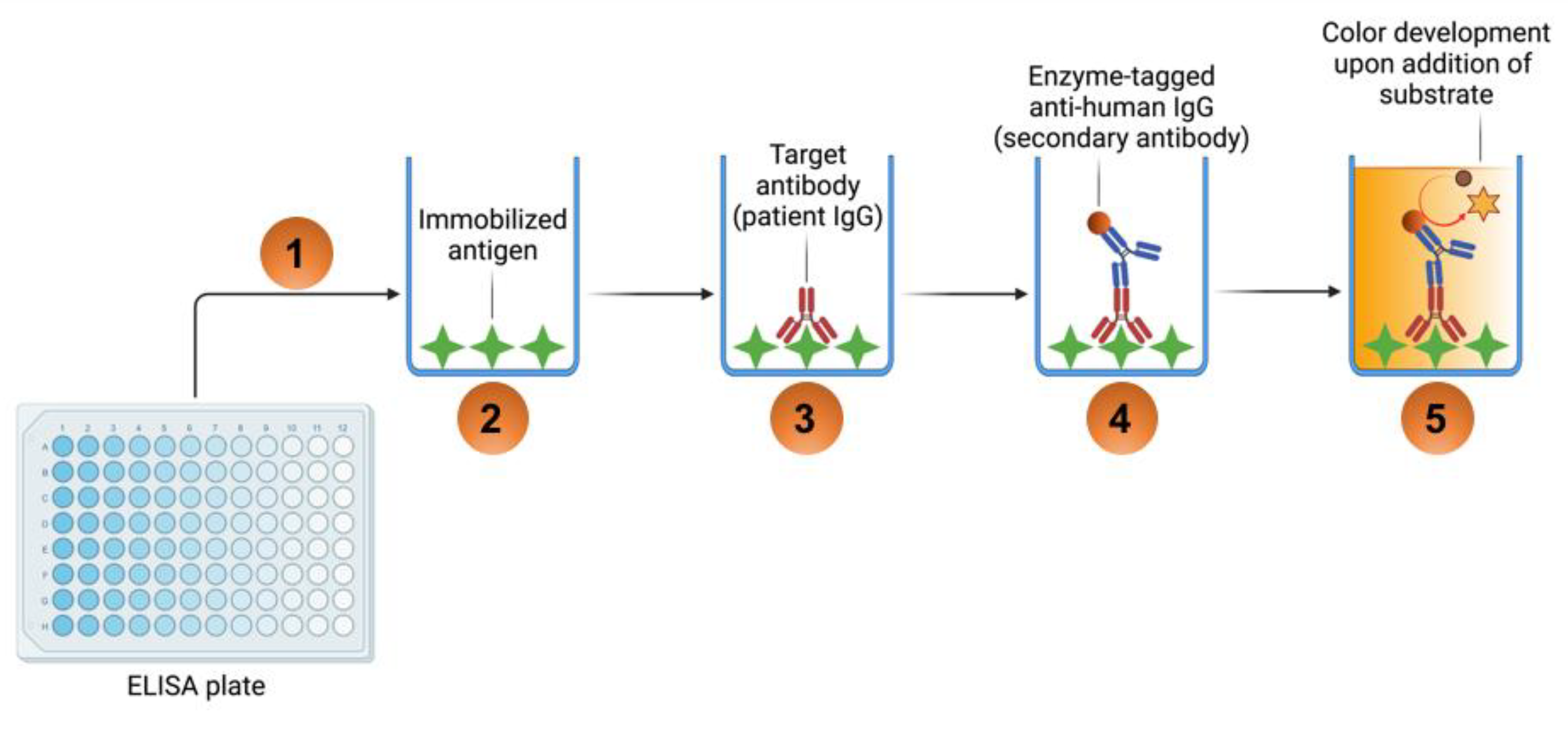
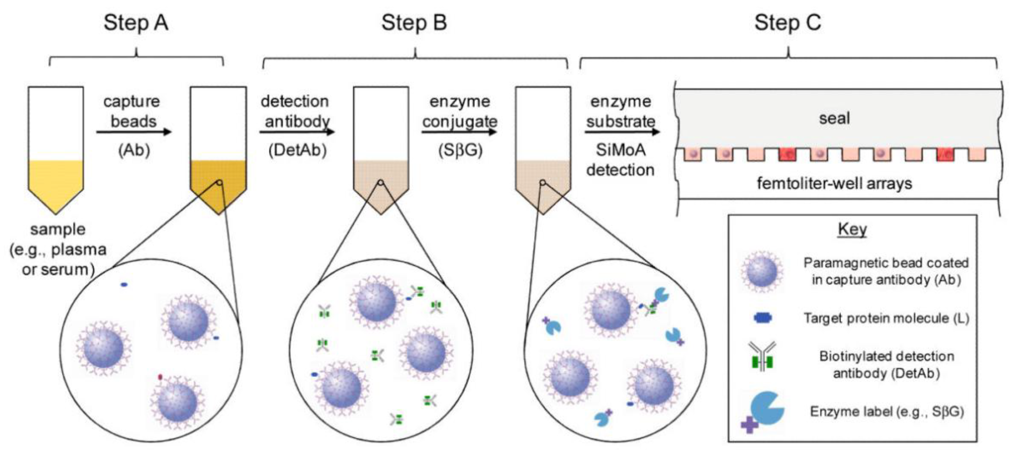
Figure 4.
Pictorial representation of the chemiluminescent immunoassay (ChLIA) and its variant for the diagnosis of COVID-19. A. ChLIA: A simplified illustration of the 96-well microtiter plate and the general composition of each well is shown here. (1) Patient sample [containing a few antibodies (Abs) specific to the SARS-CoV-2 infection]. (2) The 96-well microplate is coated with deactivated antigen (Ag) of interest. The coating Ag is chosen so it binds specific Abs, if present in the serum sample. (3) Patient’s serum is added to each well, and the microtiter plate is incubated. Ag-specific IgG will bind to the Ag coating the well, forming an Ab-Ag complex. (4) A secondary Ab is then added to the well. This Ab binds to the already formed Ag-Ab complex. The secondary Ab is tagged with an enzyme that enables luminescence readout in the final steps. (5) A substrate for the enzyme is added, which causes a chemical reaction with the tagged secondary Ab. The chemical reaction produces light, and the luminescence can be used to quantify the amount of Abs in the serum sample. B. Chemiluminescent microparticle immunoassay (CMIA): A basic illustration of this variant of ChLIA is depicted here. SARS-CoV-2-specific Ags are immobilised on the surface of each microparticle (contained in a well) to specifically bind to the target Abs after addition of the patient’s sample, and then enzyme-labelled secondary Abs are used to produce a detectable or quantifiable signal confirming the presence or concentration of target Abs, respectively. Image created using BioRender.com [18].
Figure 4.
Pictorial representation of the chemiluminescent immunoassay (ChLIA) and its variant for the diagnosis of COVID-19. A. ChLIA: A simplified illustration of the 96-well microtiter plate and the general composition of each well is shown here. (1) Patient sample [containing a few antibodies (Abs) specific to the SARS-CoV-2 infection]. (2) The 96-well microplate is coated with deactivated antigen (Ag) of interest. The coating Ag is chosen so it binds specific Abs, if present in the serum sample. (3) Patient’s serum is added to each well, and the microtiter plate is incubated. Ag-specific IgG will bind to the Ag coating the well, forming an Ab-Ag complex. (4) A secondary Ab is then added to the well. This Ab binds to the already formed Ag-Ab complex. The secondary Ab is tagged with an enzyme that enables luminescence readout in the final steps. (5) A substrate for the enzyme is added, which causes a chemical reaction with the tagged secondary Ab. The chemical reaction produces light, and the luminescence can be used to quantify the amount of Abs in the serum sample. B. Chemiluminescent microparticle immunoassay (CMIA): A basic illustration of this variant of ChLIA is depicted here. SARS-CoV-2-specific Ags are immobilised on the surface of each microparticle (contained in a well) to specifically bind to the target Abs after addition of the patient’s sample, and then enzyme-labelled secondary Abs are used to produce a detectable or quantifiable signal confirming the presence or concentration of target Abs, respectively. Image created using BioRender.com [18].
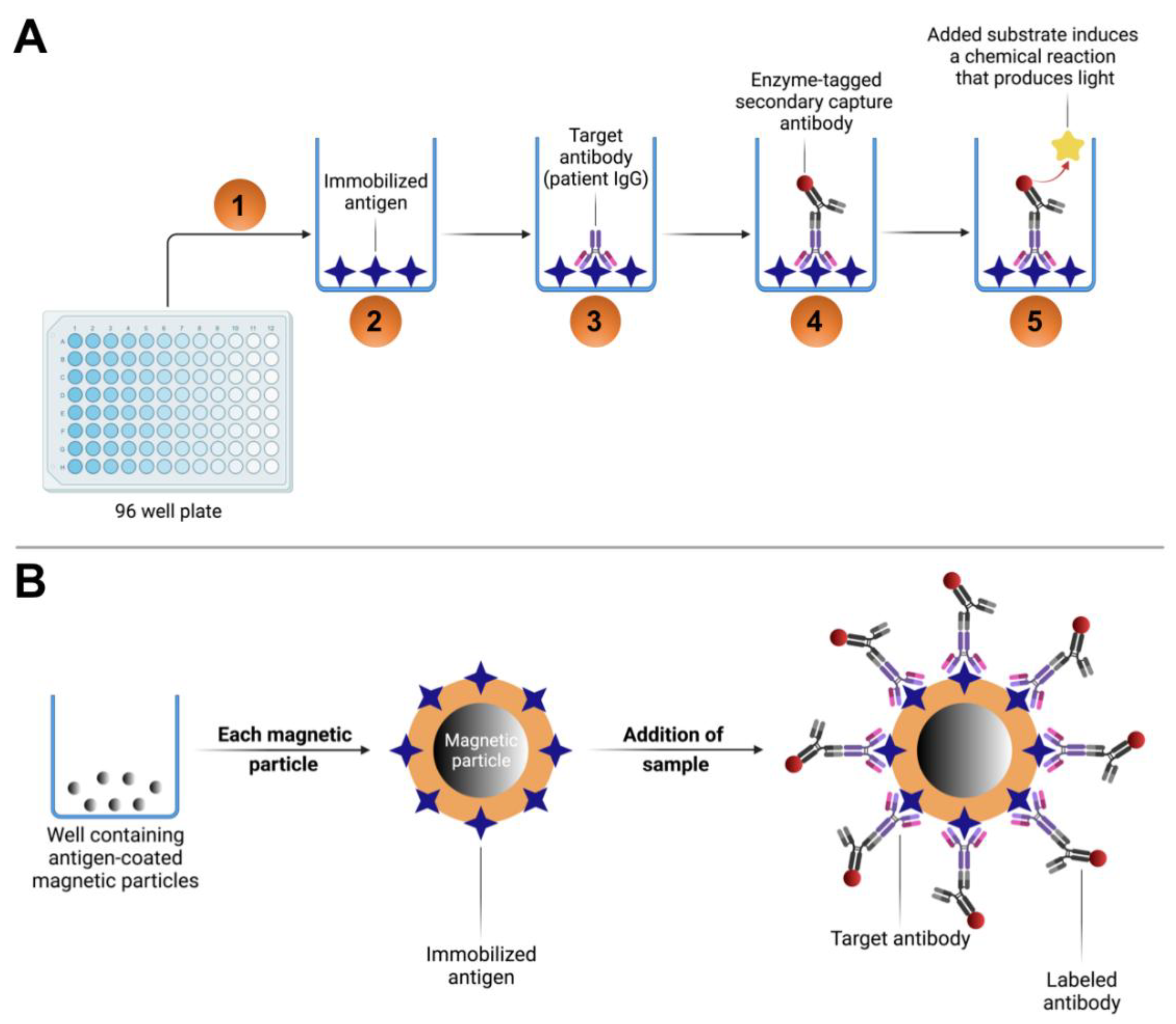
Figure 5.
Illustration of the neutralisation assay (NA) for the diagnosis of COVID-19. A schematic representation of a lateral flow test strip is shown at the bottom of each graphic, mimicking the actual line density. (A) Addition of non-COVID-19-immune plasma or serum fails to block the binding of the nanoshell-RBD conjugate to ACE2, which results in the nanoshell-RBD-ACE2 complex, thereby creating a visible green colored test (T) line. (B) Adding a moderate titer/concentration of neutralising Abs to the sample pad generates a weak green T line. (C) Addition of high titer/concentration (>1:640) of neutralising Abs completely blocks binding of nanoshell-RBD conjugate to ACE2, and no detectable line is observed at the T location on the lateral flow strip. The red control (C) line denotes binding of gold nanospheres conjugated with a mouse monoclonal Ab. Image created using BioRender.com [18].
Figure 5.
Illustration of the neutralisation assay (NA) for the diagnosis of COVID-19. A schematic representation of a lateral flow test strip is shown at the bottom of each graphic, mimicking the actual line density. (A) Addition of non-COVID-19-immune plasma or serum fails to block the binding of the nanoshell-RBD conjugate to ACE2, which results in the nanoshell-RBD-ACE2 complex, thereby creating a visible green colored test (T) line. (B) Adding a moderate titer/concentration of neutralising Abs to the sample pad generates a weak green T line. (C) Addition of high titer/concentration (>1:640) of neutralising Abs completely blocks binding of nanoshell-RBD conjugate to ACE2, and no detectable line is observed at the T location on the lateral flow strip. The red control (C) line denotes binding of gold nanospheres conjugated with a mouse monoclonal Ab. Image created using BioRender.com [18].
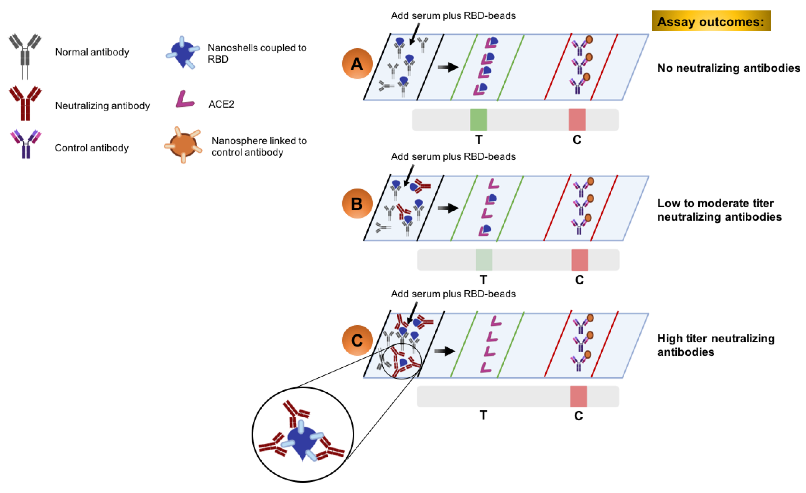
Figure 6.
Graphical illustration displaying the time kinetics of anti-SARS-CoV-2 Ab response in case of COVID-19. The figure shows relative host immunoglobulin (IgM, IgG, IgA) levels (produced against SARS-CoV-2) and viral (SARS-CoV-2) load at different phases of COVID-19. Ab-specific seroconversion takes place once the Ab reaches a detectable level in the blood. According to research studies, there may be 1–2 weeks with no visible symptoms and no detectable Abs in the patient’s blood (i.e., during the virus's incubation period). Then, IgM Abs are generated as a primary immune response against the virus, followed by the production of IgG Abs to develop a more specific host immune response against COVID-19. This image is for illustration only and does not depict the actual concentrations of the different antibodies. (Source: Ghaffari et al. 2020 [5] with modifications; https://creativecommons.org/licenses/by/4.0/) [17].
Figure 6.
Graphical illustration displaying the time kinetics of anti-SARS-CoV-2 Ab response in case of COVID-19. The figure shows relative host immunoglobulin (IgM, IgG, IgA) levels (produced against SARS-CoV-2) and viral (SARS-CoV-2) load at different phases of COVID-19. Ab-specific seroconversion takes place once the Ab reaches a detectable level in the blood. According to research studies, there may be 1–2 weeks with no visible symptoms and no detectable Abs in the patient’s blood (i.e., during the virus's incubation period). Then, IgM Abs are generated as a primary immune response against the virus, followed by the production of IgG Abs to develop a more specific host immune response against COVID-19. This image is for illustration only and does not depict the actual concentrations of the different antibodies. (Source: Ghaffari et al. 2020 [5] with modifications; https://creativecommons.org/licenses/by/4.0/) [17].
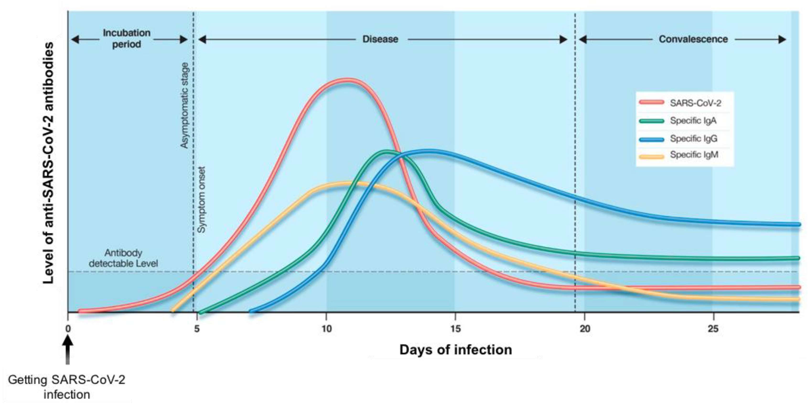
Figure 7.
Design and experimental workflow of antibody (Ab) versus antigen (Ag) detection using colourimetric lateral flow immunoassay (LFIA) (Source: Jacobs et al. 2020 [88]; https://creativecommons.org/licenses/by/4.0/) [17].
Figure 7.
Design and experimental workflow of antibody (Ab) versus antigen (Ag) detection using colourimetric lateral flow immunoassay (LFIA) (Source: Jacobs et al. 2020 [88]; https://creativecommons.org/licenses/by/4.0/) [17].
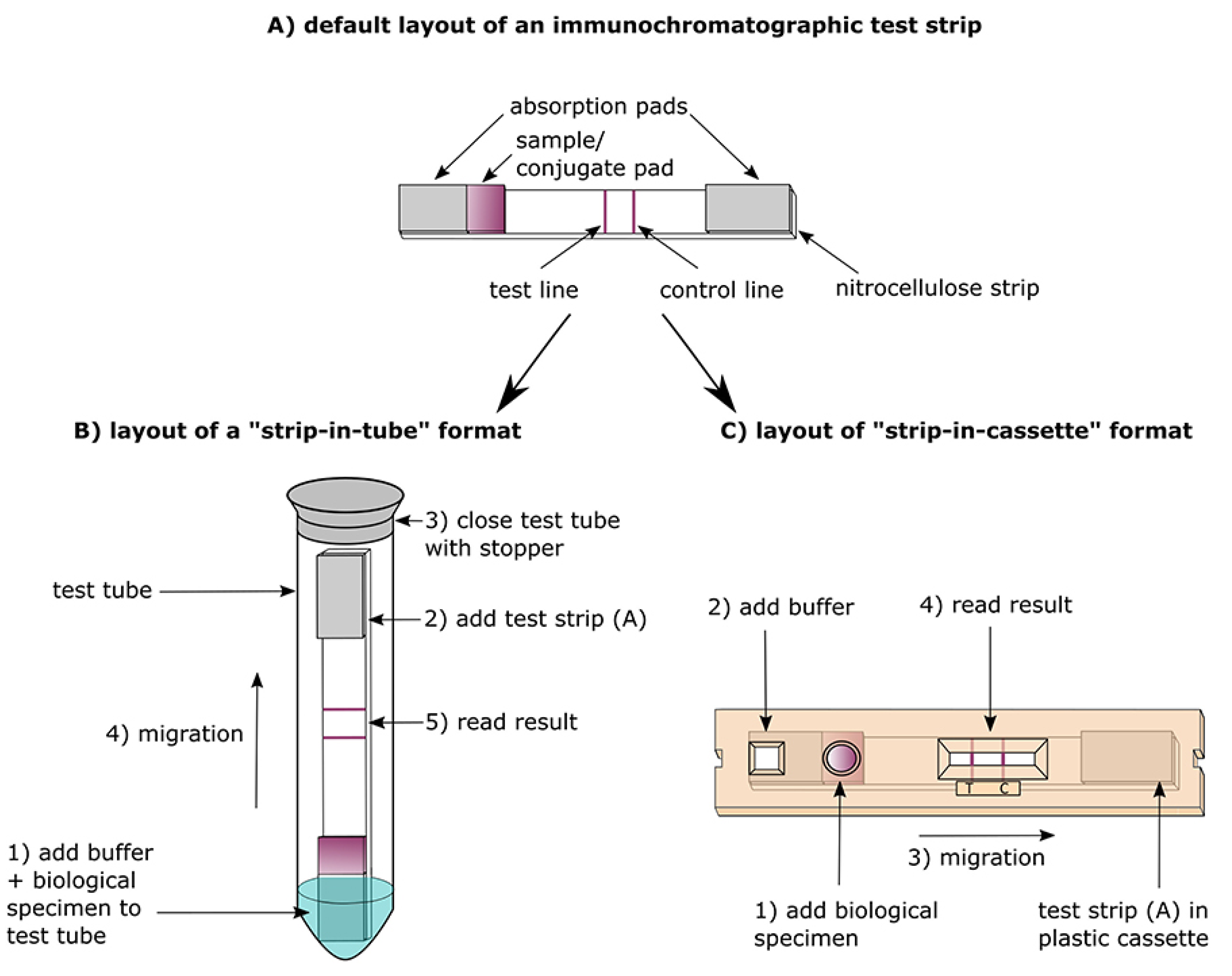
Figure 8.
Diagrammatic representation of a rapid antigen test (RAT) using an immunochromatographic (lateral flow) test format. Sample from a suspected individual is applied to the nitrocellulose test strip. If the sample contains Ag, it is bound by Abs linked to the detector molecules, as well as Abs immobilised in the test line further down the strip (Source: Ian M Campbell, CC BY-SA 3.0 https://creativecommons.org/licenses/by-sa/3.0, via Wikimedia Commons) [91].
Figure 8.
Diagrammatic representation of a rapid antigen test (RAT) using an immunochromatographic (lateral flow) test format. Sample from a suspected individual is applied to the nitrocellulose test strip. If the sample contains Ag, it is bound by Abs linked to the detector molecules, as well as Abs immobilised in the test line further down the strip (Source: Ian M Campbell, CC BY-SA 3.0 https://creativecommons.org/licenses/by-sa/3.0, via Wikimedia Commons) [91].
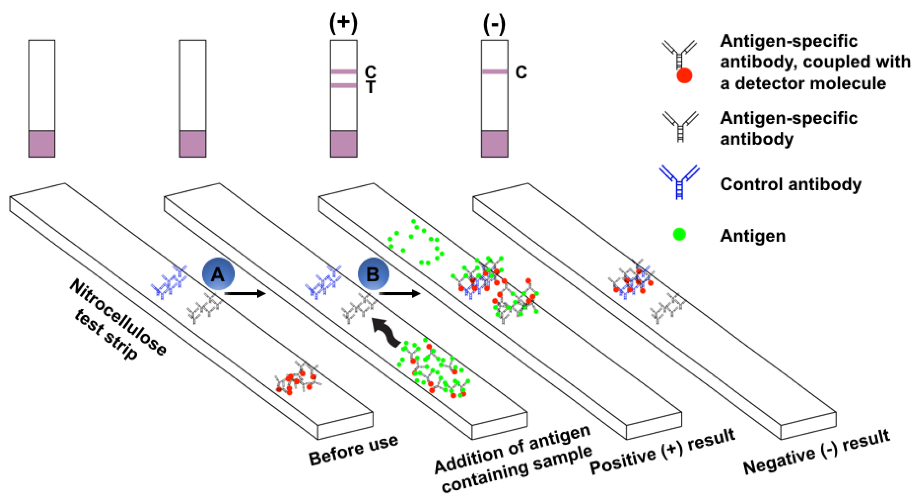

Table 1.
Comparative assessment of the four standard serological tests in prevalence for SARS-CoV-2 infection diagnosis.
Table 1.
Comparative assessment of the four standard serological tests in prevalence for SARS-CoV-2 infection diagnosis.
| Type of test | Type of sample(s) needed from patient | Turnaround time/Time required for fetching results | Advantages/Information provided by the test | Limitations of the test | Reference(s) |
| 1. Rapid serology test (RST) or Ab-detecting rapid diagnostic test (Ab-RDT) using lateral flow immunoassay (LFIA) | • Blood samples (obtained from patient’s finger by pricking with a sterile needle) • Saliva • Nasal swab fluids |
• ~10 – 30 minutes | • Detects the presence or absence of anti-SARS-CoV-2 Abs in patient’s serum (i.e., qualitative) • Portable test that can be performed at or near the PoC |
• Does not detect the level of Abs present in patient’s serum • Does not provide any information whether these Abs are able to inhibit viral growth |
• Dutta et al. 2020 [13] |
| 2. Enzyme-linked immunosorbent assay (ELISA) | • Whole blood, plasma or serum | • 2 – 5 hours | • Detects the presence or absence of anti-SARS-CoV-2 Abs in patient’s serum (i.e., qualitative) • Detects the level of Abs present in patient’s serum (i.e., quantitative) • Laboratory-based test |
• Does not provide any information whether these Abs are able to inhibit viral growth | • Dutta et al. 2020 [13] |
| 3. Chemiluminescent immunoassay (ChLIA) | • Whole blood, plasma or serum | • 1 – 2 hour(s) |
• Detects the presence or absence of anti-SARS-CoV-2 Abs in patient’s serum (i.e., qualitative) • Detects the level of Abs present in patient’s serum (i.e., quantitative) • Laboratory-based test |
• Does not provide any information whether these Abs are able to inhibit viral growth | • Dutta et al. 2020 [13] |
| 4. Neutralization assay (NA) | • Whole blood, plasma or serum | • 3 – 5 days | • Detects the presence of neutralizing/active Abs and their levels in patient’s serum capable of inhibiting viral growth in vitro, in a cell culture system (i.e., quantitative) • Laboratory-based test |
• May not detect Abs to viral proteins, which are not involved in viral replication | • Dutta et al. 2020 [13] |
Ab/Abs refers to antibody/antibodies; SARS-CoV-2 refers to severe acute respiratory syndrome coronavirus 2; PoC refers to point of care.
Table 2.
List of rapid serology tests (RSTs) that have been developed and authorised for use in SARS-CoV-2 infection diagnosis.
Table 2.
List of rapid serology tests (RSTs) that have been developed and authorised for use in SARS-CoV-2 infection diagnosis.
| Sl. no. | Name of the test (linked to IFU) | Manufacturer (linked to page) | Type of test | Sensitivity | Type of sample(s) needed from patient | Description and limitations of the test (if any) |
| 1. | SGTi-flex COVID-19 IgG [25] | Sugentech Inc. | RST | IgG: 41.2% (0-7 days), 91.7% (7-14 days), and 98.6% (15+ days) |
Human serum, plasma, or whole blood samples, including fingerstick whole blood samples |
|
| 2. | Rapid COVID-19 IgM/IgG Combo Test Kit [26] | Megna Health, Inc. | RST | IgM: 73.81%; IgG: 73.81%; Combined: 90.5% | Fingerstick whole blood samples |
|
| 3. | MidaSpot COVID-19 Antibody Combo Detection Kit [27] | Nirmidas Biotech, Inc. | RST | Combined: 76.9% (7-14 days), and 100% (15+ days) |
Serum, plasma, and fingerstick whole blood samples |
|
| 4. | RapCov Rapid COVID-19 Test [28] | ADVAITE, Inc. | RST | IgG: 90% | Fingerstick whole blood samples |
|
| 5. | RightSign COVID-19 IgG/IgM Rapid Test Cassette [29] | Hangzhou Laihe Biotech Co., Ltd | RST | IgM: 100% (0-6 days), 85.7% (7-14 days), and 99.25% (14+ days); IgG: 0% (0-6 days), 76% (7-14 days), and 98.5% (14+ days) |
Venous whole blood, serum or plasma, and fingerstick whole blood |
|
| 6. | ACON SARS-CoV-2 IgG/IgM Rapid Test [30] | ACON Laboratories, Inc. | RST | Combined: 100% (0-7 days), 100% (8-14 days), and 100% (15+ days) | Human serum, plasma or whole blood |
|
| 7. | Innovita 2019-nCoV Ab Test (Colloidal Gold) [31] | Innovita (Tangshan) Biological Technology Co., Ltd. | RST | Combined: 95.6% (8-14 days), and 100% (15+ days) | Serum or plasma or venous whole blood |
|
| 8. | LYHER Novel Coronavirus (2019-nCoV) IgM/IgG Antibody Combo Test Kit (Colloidal Gold) [32] | Hangzhou Laihe Biotech Co., Ltd | RST | IgM: 100% (0-6 days), 85.7% (7-14 days), and 99.25% (14+ days); IgG: 0% (0-6 days), 76% (7-14 days), and 98.5% (14+ days) |
Human serum, plasma or whole blood |
|
| 9. | Orawell IgM/IgG Rapid Test [33] | Jiangsu Well Biotech Co., Ltd. | RST | Combined: 97.1% | Human serum, and plasma |
|
| 10. | BIOTIME SARS-CoV-2 IgG/IgM Rapid Qualitative Test [34] | Xiamen Biotime Biotechnology Co., Ltd | RST | IgG: 46.38% (0-7 days), 67.65% (7-14 days), and 100% (14+ days); IgM: 55% (0-7 days), 94% (7-14 days), and 100% (14+ days) |
Potassium- EDTA plasma, Mixed serum or potassium- EDTA venous whole blood |
|
| 11. | CareStart COVID-19 IgM/IgG [35] | Access Bio, Inc. | RST | IgM: 89%; IgG: 96.7%; Combined: 98.44% | Human serum, plasma or whole blood samples, including fingerstick whole blood samples |
|
| 12. | Assure COVID-19 IgG/IgM Rapid Test Device [36] | Assure Biotech (Hangzhou Co, Ltd.) | RST | Combined: 95% | Venous whole blood |
|
| 13. | TBG SARS-CoV-2 IgG / IgM Rapid Test Kit [37] | TBG Biotechnology Corp. | RST | IgM: 87.5%; IgG: 96.4% |
Human serum, and plasma |
|
| 14. | Tell Me Fast Novel Coronavirus (COVID-19) IgG/IgM Antibody Test [38] | Biocan Diagnostics Inc. | RST | IgM: 90%; IgG: 93.3% |
Human serum or plasma or venous whole blood |
|
| 15. | Sienna-Clarity COVIBLOCK COVID-19 IgG/IgM Rapid Test Cassette [39] | Salofa Oy | RST | Combined: 100% | Human serum or plasma or venous whole blood |
|
| 16. | WANTAI SARS-CoV-2 Ab Rapid Test [40] | Beijing Wantai Biological Pharmacy Enterprise Co., Ltd. | RST | Total Abs: 94.7% | Human serum or plasma or venous whole blood |
|
| 17. | RightSign COVID-19 IgG/IgM Rapid Test Cassette [41] | Hangzhou Biotest Biotech Co., Ltd | RST | IgM: 96.3% (8-14 days), and 94.9% (15+ days); IgG: 91.6% (8-14 days), and 96.2% (15+ days) |
Human serum, plasma or whole blood |
|
| 18. | COVID-19 IgG/IgM Rapid Test Cassette (Whole Blood/Serum/Plasma) [42] | Healgen Scientific LLC | RST | IgM: 86.7 %; IgG: 96.7%; Combined: 96.7% |
Human serum or plasma or venous whole blood |
|
| 19. | qSARS-CoV-2 IgG/IgM Rapid Test [43] | Cellex Inc. | RST | Combined: 93.8% | Human serum, plasma or whole blood |
|
Ab/Abs: Antibody/antibodies; Ag/Ags: Antigen/antigens; NP: Nucleocapsid protein; RBD: Receptor-binding domain; RST: Rapid serology test; LFIcA: Lateral flow immunochromatographic assay; LFIA: Lateral flow immunoassay; SARS-CoV-2: Severe acute respiratory syndrome coronavirus 2.
Table 3.
List of enzyme-linked immunosorbent assays (ELISAs) that have been developed and authorised for use in SARS-CoV-2 infection diagnosis.
Table 3.
List of enzyme-linked immunosorbent assays (ELISAs) that have been developed and authorised for use in SARS-CoV-2 infection diagnosis.
| Sl. no. | Name of the test (linked to IFU) | Manufacturer (linked to page) | Type of test | Sensitivity | Type of sample(s) needed from patient | Description and limitations of the test (if any) |
| 1. | Simoa Semi-Quantitative SARS-CoV-2 IgG Antibody Test [44] | Quanterix Corporation | Paramagnetic microbead-based sandwich ELISA coupled with single molecule array (Simoa) | IgG: 45.2% (0-7 days), 87.5% (7-14 days), and 100% (15+ days) |
Human serum and dipotassium EDTA plasma |
|
| 2. | COVID-19 Self-Collected Antibody Test System [45] | Symbiotica, Inc. | Home collection, ELISA | IgG: 100% (8-14 days), and 100% (15+ days) | Human whole blood |
|
| 3. | SCoV-2 Detect IgM ELISA [46] | InBios International, Inc. | ELISA | IgM: 93% | Human serum |
|
| 4. | Access SARS-CoV-2 IgG II [47] | Beckman Coulter, Inc. | ELISA | IgG: 81.8% (0-7 days), 95.8% (8-14 days), and 98.9% (15+ days) | Human plasma |
|
| 5. | UBI SARS-CoV-2 ELISA [48] | United Biomedical, Inc. | ELISA | IgG: 38.5% (0-7 days), 79.5% (7-14 days), and 100% (15+ days) | Human serum, and plasma |
|
| 6. | COVID-SeroKlir, Kantaro Semi-Quantitative SARS-CoV-2 IgG Antibody Kit [49] | Kantaro Biosciences LLC | ELISA | IgG: 100% (0-7 days), 100% (7-14 days), and 98.87% (15+ days) | Human serum, and plasma |
|
| 7. | cPass SARS-CoV-2 Neutralization Antibody Detection Kit [50] | GenScript USA, Inc. | Modified ELISA | Neutralizing Abs: 100% | Human serum, and plasma |
|
| 8. | Platelia SARS-CoV-2 Total Ab assay [51] | Bio-Rad | Modified ELISA | Combined: 98% | Human serum, and plasma |
|
| 9. | Q-Plex SARS-CoV-2 Human IgG (4 Plex) [52] | Quansys Biosciences, Inc. | ELISA | IgG: 100% (0-7 days,), 100% (8-14 days), and 95.2% (15+days) | Human serum, and plasma |
|
| 10. | ZEUS ELISA SARS-CoV-2 IgG Test System [53] | ZEUS Scientific, Inc. | ELISA | IgG: 100% | Human serum, and plasma |
|
| 11. | OmniPATH COVID-19 Total Antibody ELISA Test [54] | Thermo Fisher Scientific | ELISA | Combined: 19% (0-7 days), 76.7% (8-14 days), and 100% (15+ days) | Human serum |
|
| 12. | COVID-19 ELISA pan-Ig Antibody Test [55] | University of Arizona Genetics Core for Clinical Services | ELISA | Combined: 97.5% | Human serum |
|
| 13. | WANTAI SARS-CoV-2 Ab ELISA [56] | Beijing Wantai Biological Pharmacy Enterprise Co., Ltd. | ELISA | Combined: 55.4% (0-7 days), 84.8% (7-14 days), and 98.7% (15+ days) | Human serum, and plasma |
|
| 14. | SARS-CoV-2 RBD IgG test [57] | Emory University | ELISA | IgG: 73% (0-7 days), 100% (8-14 days), and 100% (14+ days) | Human serum |
|
| 15. | SCoV-2 Detect IgG ELISA [58] | InBios International, Inc. | ELISA | IgG: 97.8% | Human serum |
|
| 16. | Anti-SARS-CoV-2 ELISA (IgG) [59] | Euroimmun AG | ELISA | IgG: 13.9% (0-10 days), 61.1% (11-20 days), and 100% (21+ days) | Human serum, and plasma |
|
| 17. | COVID-19 ELISA IgG Antibody Test [60] | Mount Sinai Laboratory | ELISA | IgG: 97.8% | Human serum |
|
ACE2: Angiotensin-converting enzyme 2; Abs: Antibodies; Ag/Ags: Antigen/antigens; DBS: Dried blood spot; ELISA: Enzyme-linked immunosorbent assay; NP: Nucleocapsid protein; RBD: Receptor-binding domain; SARS-CoV-2: Severe acute respiratory syndrome coronavirus 2.
Table 4.
List of chemiluminescent immunoassays (ChLIAs) that have been developed and authorised for use in SARS-CoV-2 infection diagnosis.
Table 4.
List of chemiluminescent immunoassays (ChLIAs) that have been developed and authorised for use in SARS-CoV-2 infection diagnosis.
| Sl. no. | Name of the test (linked to IFU) | Manufacturer (linked to page) | Type of test | Sensitivity | Type of sample(s) needed from patient | Description and limitations of the test (if any) |
| 1. | QUANTA Flash SARS-CoV-2 IgG [61] | Inova Diagnostics, Inc. | ChLIA | IgG: 66.7% (0-7 days), 61.5% (7-14 days), and 100% (15+ days) |
Human serum, and plasma |
|
| 2. | VITROS Immunodiagnostic Products Anti-SARS-CoV-2 Total Reagent Pack [62] | Ortho Clinical Diagnostics | ChLIA | Combined: 80% (0-8 days), and 100% (8+ days) | Human whole blood |
|
| 3. | IDS SARS-CoV-2 IgG [63] | Immunodiagnostic Systems Ltd | ChLIA | IgG: 60.9% (0-7 days), 80% (7-14 days), and 97.6% (15+ days) | Human serum, plasma or whole blood |
|
| 4. | (ARCHITECT and Alinity i systems) [64] | Abbott Laboratories Inc. | CMIA | IgG: 43% (3-7 days), 78.9% (8-14 days), and 100% (15+ days) | Human serum, and plasma |
|
| 5. | Access SARS-CoV-2 IgM [65] | Beckman Coulter | ChLIA | IgM: 91.7% (8-14 days), and 98.3% (15+ days) | Human serum, and plasma |
|
| 6. | Access SARS-CoV-2 IgG [66] | Beckman Coulter, Inc. | ChLIA | IgG: 75% (0-7 days), 95.3% (8-14 days), and 96.8% (15+ days) | Human serum, and plasma |
|
| 7. | Dimension Vista SARS-CoV-2 Total antibody assay (COV2T) [67] | Siemens Healthcare Diagnostics Inc. | ChLIA | Total Abs: 91.3% (7-13 days), and 100% (15+ days) | Human serum, and plasma |
|
| 8. | Dimension EXL SARS-CoV-2 Total antibody assay (CV2T) [68] | Siemens Healthcare Diagnostics Inc. | ChLIA | Total Abs: 91.3% (7-13 days), and 100% (15+ days) | Human serum, and plasma |
|
| 9. | VITROS Immunodiagnostic Products Anti-SARS-CoV-2 IgG Reagent Pack [69] | Ortho-Clinical Diagnostics, Inc. | ChLIA | IgG: 83% (12-15 days), and 100% (15+ days) | Human serum, and plasma |
|
| 10. | Elecsys Anti-SARS-CoV-2 [70] | Roche | EChLIA | 65.5% (0-6 days), 88.1% (7-13 days), and 100% (14+ days) | Human serum |
|
| 11. | LIAISON SARS-CoV-2 S1/S2 IgG [71] | DiaSorin Inc. | ChLIA | IgG: 90% (8-14 days), and 97% (15+ days) | Human serum, and plasma |
|
| 12. | AdviseDx SARS-CoV-2 IgM (ARCHITECT and Alinity i systems) [72] | Abbott Laboratories Inc. | CMIA | IgM: 95% | Human serum, and plasma |
|
| 13. | DiaSorin LIAISON SARS-CoV-2 IgM Assay [73] | DiaSorin, Inc. | ChLIA | IgM: 92.6% | Human serum, and plasma |
|
| 14. | SARS-CoV-2 IgG (ARCHITECT and Alinity i) [74] | Abbott Laboratories Inc. | CMIA | IgG: 0% (0-3 days), 25% (3-7 days), 86.4% (8-13 days), and 100% (14+ days) | Human serum, and plasma |
|
| 15. | MAGLUMI 2019-nCoV IgM/IgG [75] | Shenzhen New Industries Biomedical Engineering Co., Ltd. (SNIBE) | ChLIA | Combined: 43.8% (0-7 days), 93.4% (8-14 days), and 100% (15+ days) | Human serum |
|
| 16. | BioCheck SARS-CoV-2 IgM Antibody Test Kit [76] | BioCheck, Inc. | ChLIA | IgM: 93.7% (8-14 days), and 88.9% (15+ days) | Human serum |
|
| 17. | BioCheck SARS-CoV-2 IgG Antibody Test Kit [77] | BioCheck, Inc. | ChLIA | IgG: 100% | Human serum |
|
| 18. | Diazyme DZ-Lite SARS-CoV-2 IgM CLIA Kit [78] | Diazyme Laboratories, Inc. | ChLIA | IgM: 26.1% (0-7 days), 83.3% (7-14 days), and 94.4% (15+ days) | Human serum, and plasma |
|
| 19. | Diazyme DZ-Lite SARS-CoV-2 IgG CLIA Kit [79] | Diazyme Laboratories, Inc. | ChLIA | IgG: 92% | Human serum, and plasma |
|
| 20. | Atellica IM SARS-CoV-2 IgG (COV2G) [80] | Siemens Healthcare Diagnostics Inc. | ChLIA | IgG: 56% (0-6 days), 92% (7-14 days), and 100% (14+ days) | Human serum, and plasma |
|
| 21. | Atellica IM SARS-CoV-2 Total (COV2T) [81] | Siemens Healthcare Diagnostics Inc. | ChLIA | Total Abs: 60.7% (0-6 days post PCR positive), 97.5% (7-13 days post PCR positive), and 100% (14+ days post PCR positive) | Human serum, and plasma |
|
| 22. | ADVIA Centaur SARS-CoV-2 IgG (COV2G) [82] | Siemens Healthcare Diagnostics Inc. | ChLIA | IgG: 53.5% (0-6 days), 93.4% (7-14 days), and 100% (14+ days) | Human serum, and plasma |
|
| 23. | ADVIA Centaur SARS-CoV-2 Total (COV2T) [83] | Siemens Healthcare Diagnostics Inc. | ChLIA | Total Abs: 65.1% (0-6 days post PCR positive), 97.5% (7-13 days post PCR positive), and 100% (14+ days post PCR positive) | Human serum, and plasma |
|
| 24. | Babson Diagnostics aC19G1 [84] | Babson Diagnostics | ChLIA/indirect sandwich immunoassay | IgG: 66.7% (8-14 days), 100% (15+ days), and 100% (asymptomatic) | Human serum |
|
| 25. | Vibrant COVID-19 Ab Assay [85] | Vibrant America Clinical Labs | ChLIA | Combined: 98.1% | Human serum or DBS using fingerstick blood sample |
|
ACE2: Angiotensin-converting enzyme 2; Ab/Abs: Antibody/antibodies; Ag/Ags: Antigen/antigens; ChLIA: Chemiluminescent immunoassay; CMIA: Chemiluminescent microparticle immunoassay; DBS: Dried blood spot; NP: Nucleocapsid protein; RBD: Receptor-binding domain; RLU: Relative Light Unit; SARS-CoV-2: Severe acute respiratory syndrome coronavirus 2.
Table 5.
Summary of neutralisation assays (NA) that have been developed and authorised for use in SARS-CoV-2 infection diagnosis.
Table 5.
Summary of neutralisation assays (NA) that have been developed and authorised for use in SARS-CoV-2 infection diagnosis.
| Name of the test (linked to IFU) | Manufacturer (linked to page) | Type of test | Type of sample(s) needed from patient | Description and limitations of the test (if any) | |
| 1. | cPass SARS-CoV-2 Neutralization Antibody Detection Kit [86] | GenScript USA Inc. | NA | Human serum, and K2-EDTA plasma |
|
hACE2: Human ACE2 receptor protein; HRP: Horseradish peroxidase; HRP-RBD: Horseradish peroxidase (HRP) conjugated recombinant SARS-CoV-2 receptor-binding domain (RBD) fragment; NA: Neutralization assay; RBD: Receptor-binding domain; SARS-CoV-2: Severe acute respiratory syndrome coronavirus 2; SARS-CoV-2 RBD: Severe acute respiratory syndrome coronavirus 2-receptor-binding domain.
Table 6.
List of rapid Ag tests (RATs) that have been developed and authorised for use in SARS-CoV-2 infection diagnosis.
Table 6.
List of rapid Ag tests (RATs) that have been developed and authorised for use in SARS-CoV-2 infection diagnosis.
| Sl. no. | Name of the test (linked to IFU) | Manufacturer (linked to page) | Type of test | Sensitivity | Type of sample(s) needed from patient | Description and limitations of the test (if any) |
| 1. | InteliSwab COVID-19 Rapid Test Rx [95] | OraSure Technologies, Inc. | RAT | 84% | Anterior nasal sample |
|
| 2. | InteliSwab COVID-19 Rapid Test [96] | OraSure Technologies, Inc. | RAT | 84% | Anterior nasal sample |
|
| 3. | InteliSwab COVID-19 Rapid Test Pro [97] | OraSure Technologies, Inc. | RAT | 84% | Anterior nasal sample |
|
| 4. | Sienna-Clarity COVID-19 Antigen Rapid Test Cassette [98] | Salofa Oy | RAT | 87.5% | Direct nasopharyngeal swab |
|
| 5. | SCoV-2 Ag Detect Rapid Test [99] | InBios International, Inc. | RAT | 86.6% | Anterior nasal swab |
|
| 6. | BinaxNOW COVID-19 Ag Card [100] | Abbott Diagnostics Scarborough, Inc. | RAT | 84.6% | Anterior nasal swab |
|
| 7. | Celltrion DiaTrust COVID-19 Ag Rapid Test [101] | Celltrion USA, Inc. | RAT | 93.3% | Direct nasopharyngeal swab |
|
| 8. | Omnia SARS-CoV-2 Antigen Test [102] | Qorvo Biotechnologies, LLC. | RAT | 90% | Anterior nasal swab |
|
| 9. | Sofia SARS Antigen FIA [103] | Quidel Corporation | RAT | 97% | Anterior nasal swab |
|
| 10. | BD Veritor System for Rapid Detection of SARS-CoV-2 [104] | Becton, Dickinson and Company (BD) | RAT | 84% (symptomatic) | Anterior nasal swab |
|
| 11. | QuickVue At-Home OTC COVID-19 Test [105] | Quidel Corporation | RAT | 83.5% | Anterior nasal swab |
|
| 12. | BinaxNOW COVID-19 Antigen Self Test [106] | Abbott Diagnostics Scarborough, Inc. | RAT | 91.7% (symptomatic) | Anterior nasal swab |
|
| 13. | BinaxNOW COVID-19 Ag Card 2 Home Test [107] | Abbott Diagnostics Scarborough, Inc. | RAT | 91.7% (symptomatic) | Anterior nasal swab |
|
| 14. | BinaxNOW COVID-19 Ag 2 Card [108] | Abbott Diagnostics Scarborough, Inc. | RAT | 84.6% (symptomatic) | Anterior nasal swab |
|
| 15. | BD Veritor System for Rapid Detection of SARS-CoV-2 & Flu A+B [109] | Becton, Dickinson and Company | RAT | SARS-CoV-2: 87.6%; Flu-A: 100%; Flu-B: 100% |
Anterior nasal swab |
|
| 16. | QuickVue At-Home COVID-19 Test [110] | Quidel Corporation | RAT | 84.8% | Anterior nasal swab |
|
| 17. | Status COVID-19/Flu [111] | Princeton BioMeditech Corp. | RAT | SARS-CoV-2: 93.9%; Flu-A: 100%; Flu-B: 100% |
Anterior nasal swab |
|
| 18. | CareStart COVID-19 Antigen test [112] | Access Bio, Inc. | RAT | NP: 93.8%; Nasal: 87.2% | Anterior nasal swab |
|
| 19. | VITROS Immunodiagnostic Products SARS-CoV-2 Antigen Reagent Pack [113] | OrthoClinical Diagnostics | RAT | 80.0% | Anterior nasal swab |
|
| 20. | QuickVue SARS Antigen Test [114] | Quidel Corporation | RAT | 96.6% | Anterior nasal swab |
|
| 21. | Sampinute COVID-19 Antigen MIA [115] | Celltrion USA, Inc. | RAT | 94.4% | Anterior nasal swab |
|
| 22. | Sofia 2 Flu + SARS Antigen FIA [116] | Quidel Corporation | RAT | SARS-CoV-2: 95.2%; Flu A: 94.8%; Flu B: 78.5-90% |
Nasopharyngeal, and nasal swab |
|
| 23. | LumiraDx SARS-CoV-2 Ag Test [117] | LumiraDx UK Ltd. | RAT | 97.6% | Anterior nasal swab, and nasopharyngeal swab |
|
Ag: Antigen; ChLIA: Chemiluminescent immunoassay; MESIA: Magnetic force-assisted electrochemical sandwich immunoassay; NP: Nucleocapsid protein; RBD: Receptor-binding domain; RAT: Rapid antigen test; IFT: Immunofluorescence technology; LFIA: Lateral flow immunoassay; SARS-CoV-2: Severe acute respiratory syndrome coronavirus 2.
Table 7.
Pros and cons of the qualitative rapid diagnostic tests (RDTs) for SARS-CoV-2 infection.
| Test type | Pros | Cons | Reference |
| 1. Ag-RDTs |
• Identifies active SARS-CoV-2 infection • Applicable at the PoC (i.e., outside laboratories; inside hospitals) • Simple to conduct • Provide rapid results (usually under 30 minutes), thereby enabling fast implementation of prophylactic measures such as contact tracing, to control further spread of the infection • Cost-effective (less expensive compared to NAATs, for example, RT-PCR tests) |
• Variable sensitivity and specificity, usually lower compared to NAAT • Lower sensitivity means NPV is lower compared to NAAT, particularly in settings with high prevalence of COVID-19 • Confirmatory NAAT testing of Ag-RDT positives is recommended in all low-prevalence settings and for Ag-RDT negatives in high-prevalence settings. • Negative rapid Ag test results cannot be taken into consideration to remove a contact (symptomatic or asymptomatic) from quarantine |
• World Health Organization, 2020 [3] |
| 2. Ab-RDTs |
• Identifies past infection with SARS-CoV-2 • Applicable at the PoC (i.e., outside laboratories; inside hospitals) or in high-throughput formats in laboratories • Simple to conduct • Provide rapid results (usually under 30 minutes for PoC testing) • Cost-effective (less expensive compared to NAATs, for example, RT-PCR tests) |
• Clinical significance of a positive rapid Ab test is still under scrutiny • Positive rapid Ab test results do not assure presence of active/neutralizing Abs or protective immunity • Should not be implemented for the determination of active SARS-CoV-2 infections in clinical care or for the purpose of contact tracing • Interpretation of rapid Ab test results is contingent on several factors, including the timing of the disease, clinical morbidity, the epidemiology and disease prevalence within the setting, the type of test employed, the method of validation, and the reliability of the test results |
• World Health Organization, 2020 [3] |
Ab/Abs – Antibody/antibodies; Ab-RDTs – Antibody-detecting rapid diagnostic tests; Ag-RDTs – Antigen-detecting rapid diagnostic tests; NAAT – Nucleic acid amplification test; NPV – Negative predictive value; PoC – Point of care; RDTs – Rapid diagnostic tests; RT-PCR – Reverse Transcription Polymerase Chain Reaction; SARS-CoV-2 – Severe acute respiratory syndrome coronavirus 2.
Disclaimer/Publisher’s Note: The statements, opinions and data contained in all publications are solely those of the individual author(s) and contributor(s) and not of MDPI and/or the editor(s). MDPI and/or the editor(s) disclaim responsibility for any injury to people or property resulting from any ideas, methods, instructions or products referred to in the content. |
© 2026 by the authors. Licensee MDPI, Basel, Switzerland. This article is an open access article distributed under the terms and conditions of the Creative Commons Attribution (CC BY) license (http://creativecommons.org/licenses/by/4.0/).
Copyright: This open access article is published under a Creative Commons CC BY 4.0 license, which permit the free download, distribution, and reuse, provided that the author and preprint are cited in any reuse.