

Submitted:
28 September 2026
Posted:
29 September 2026
You are already at the latest version
Abstract
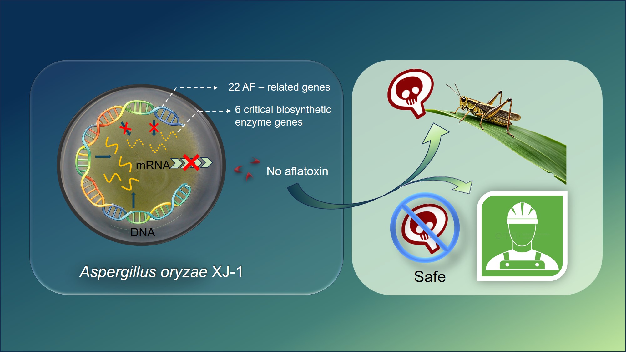
Aspergillus oryzae XJ-1 is a highly pathogenic fungus against locusts. However, whether it likes Aspergillus flavus to produce aflatoxin (AF), a serious toxin and not safety to mammals, is questioned for further development as a biological control agent. In this study, we first test the AF production from 4 strains: Aspergillus. oryzae XJ-1 (A.oryzae XJ-1), non-aflatoxigenic Aspergillus. oryzae (NAF A. oryzae) (CGMCC: 3.9544), the non-aflatoxigenic Aspergillus. flavus (NAF A. flavus) (CGMCC: 3.3950), and the aflatoxin-producing Aspergillus. flavus (AF A. flavus) (CGMCC: 3.4408) with high performance liquid chromatography-fluorescence detection (HPLC-FLD). Then transcriptome sequencing and bioinformation analysis, reverse transcription PCR (RT-PCR), and quantitative real-time reverse transcription PCR (RT-qPCR) were used to reveal the transcript expression of key genes in biosynthesis of AF in above 4 strains. The level of AF in A. oryzae XJ-1, like other two non-AF producing strains, was significantly lower than in AF A. flavus which produces AF. Although the 22 aflatoxin-related genes were present within the gene clusters of both strains, the transcript levels of six critical biosynthetic enzyme genes (avnA, verB, omtA, ordA, vbs, cypX) were significantly lower in A. oryzae XJ-1 than in AF A. flavus. Our findings indicate that the transcript suppression of the six key biosynthetic enzyme genes is the primary mechanism underlying the lack of AF production in A. oryzae XJ-1. This stable, non-aflatoxigenic phenotype supports the potential of A. oryzae XJ-1 as a safe, environmentally friendly biocontrol agent for locust management.Transcriptional suppression of six critical biosynthetic enzyme genes underlies the lack of aflatoxin production in Aspergillus oryzae XJ-1.
Keywords:
Aspergillus oryzae XJ-1
; locust
; aflatoxins
; transcriptome sequencing
; molecular mechanism
1. Introduction
Locusts (Locusta migratoria) are among the most destructive agricultural pests worldwide. Biopesticide is an effective approach to achieving sustainable control for locust. The ability of adults to rapidly form gregarious swarms can devastate crops over vast areas [1]. In 2015, we isolated and identified a new fungal pathogen, Aspergillus oryzae XJ-1, from a dead locust collected in the Chonghuer Grassland in Altay Prefecture, Xinjiang [2]. Subsequent studies revealed that this strain shows high virulence against locust nymphs and adults [2,3]. Given its high pathogenicity and environmentally friendly characteristics, A. oryzae represents a promising candidate for the development of effective biological agents.
A critical prerequisite for the safe deployment of Aspergillus strains in biocontrol is safe to mammals. Aflatoxins (AFs) are a class of secondary metabolites with high toxicity and carcinogenicity. These toxins are believed to associate with the development of liver cancer, posing severe risks to the health of both humans and livestock [4]. Among 16 known AF analogs, the most significant risk is posed by AFB1, which is a contaminant of agricultural products that poses a threat to both livestock and human health [4,5,6,7]. They are mainly produced by Aspergillus flavus and Aspergillus parasiticus [8,9]. In Ghana, researchers isolated many A. flavus strains from maize and peanuts and found that over 70% were aflatoxigenic. The prevalence of aflatoxin-producing A. flavus strains was significantly higher in dry and hot areas than in humid forest areas [10]. A. oryzae is a close phylogenetic relative of the well-known AF producers A. flavus and A. parasiticus, and it retains a homologous AF biosynthetic gene cluster [11,12]. The strains’ ability to produce aflatoxin varies in A. flavus, the percentage of non-aflatoxigenic A. flavus isolated from the soil might vary from less than 5% to more than 50% [13,14].
The biosynthesis and regulation of AFs are highly complex. The AF synthesis pathway involves a series of highly coordinated redox reactions, governed by an approximately 80-kb AF gene cluster containing at least 25 structural genes and two key regulatory genes (aflR and aflJ). AFs are synthesized from acetyl-CoA and malonyl-CoA, which are converted into the anthraquinone compound NOR (norsolorinic acid) by polyketide synthase. Subsequent enzymatic transitions lead to the formation of AFs, which are characterized by a coumarin core linked to a dihydrofuran ring [15,16,17]. And deletion of any one of the genes in the gene cluster or nonfunctional enzyme would lead to a non-aflatoxigenic phenotype [5,9]. And few reports showed that suppression of genes at transcript level in the AF biosynthetic pathway leads to non-AF production.
Here we for the first time report that A. oryzae XJ-1 is a non-aflatoxigenic phenotype and its molecular mechanism which underlies non-aflatoxin production. We first determine the non-production of AF by high performance liquid chromatography-fluorescence detection (HPLC-FLD) in comparison of A. oryzae XJ-1 and other three strains: non-aflatoxigenic (NAF) A. oryzae, NAF A. flavus, and aflatoxin-producing (AF) A. flavus. Then we demonstrate 6 key genes in AF biosynthetic pathway suppressed at transcript level in A. oryzae XJ-1 though these genes are presented in the genome by transcriptome analysis, PCR in gDNA, RT-PCR and RT-qPCR in cDNA indicating that suppression of the 6 genes in transcript level is the genetic basis for the lack of AF production in A. oryzae XJ-1.
2. Materials and Methods
2.1. Fungi
A. oryzae XJ-1 was obtained from the Institute of Plant Protection, Shandong Academy of Agricultural Sciences. NAF A. oryzae (CGMCC No. 3.9544), NAF A. flavus (CGMCC No. 3.3950), and AF A. flavus (CGMCC No. 3.4408) were obtained from the China General Microbiological Culture Collection Center. The original strains were stored long-term at -40 °C. After removal from the freezer, they were placed at room temperature for approximately 20 minutes before inoculation. The strains were cultivated in potato dextrose agar (PDA) liquid medium. All cultures were incubated at 28 °C in a rotary shaker at 150 rpm for 5 days. The resulting mycelia and culture broth were used for subsequent experiments.
2.2. AFB1 Assay with High Performance Liquid Chromatography-Fluorescence Detection (HPLC-FLD)
The culture supernatants were first filtered through filter paper and then passed through 0.22-μm nylon membrane filters. The filtrates were used for AFB1 analysis by high-performance liquid chromatography with fluorescence detection (HPLC-FLD). Because the initial injection solution exceeded the linear range of the calibration curve, the filtrates were diluted 100-fold with methanol/acetonitrile/water (17.5:17.5:65, v/v/v), which was identical to the mobile phase before HPLC analysis.
Chromatographic analysis was performed on a Waters 2695 HPLC system (Waters, Milford, MA, USA) equipped with a fluorescence detector and an Ultimate XB-C18 reversed-phase column (4.6 mm × 150 mm, 5 μm). The mobile phase was methanol/acetonitrile/water (17.5:17.5:65, v/v/v), the flow rate was 1.0 mL/min, the column temperature was 40 °C, and the injection volume was 20 μL. AFB1 was detected at an excitation wavelength of 360 nm and an emission wavelength of 440 nm.
Quantification was performed using the external standard method. Calibration standards were prepared at seven concentration levels of 0.1, 0.5, 2.0, 5.0, 10.0, 20.0, and 40.0 ng/mL, which defined the linear working range. The calibration curve was constructed by plotting peak area (Y) against AFB1 concentration (X, ng/mL), with the regression equation Y = 4.04×10⁴X + 2.21×10³ and a coefficient of determination (R²) of 0.9991. The AFB1 concentration in each culture filtrate was calculated from the regression equation and corrected by the 100-fold dilution factor. AFB1 standard solution (2 μg/mL in acetonitrile, analytical standard; Cat. No. 34029) was purchased from Sigma-Aldrich/Supelco (Merck KGaA, Darmstadt, Germany). All experiments were performed in triplicate.
2.3. DNA Extraction
After 50 mg Aspergillus mycelia were pulverized into a fine powder using liquid nitrogen, 500 μL of lysis buffer (100 mM Tris-HCl (pH 8.5), 250 mM NaCl, 25 mM Na2EDTA, and 2% SDS) was added to each sample. The mixtures were agitated and incubated in a water bath at 65 °C for 30 minutes, with periodic mixing. Following incubation, 200 μL of 10 M NH4AC was added, and the samples were chilled in an ice bath for 10 minutes. Samples were centrifuged at 4 °C at 12,000 rpm for 15 minutes, and the resulting supernatant was transferred to a 1.5 mL Eppendorf tube and supplemented with 300 μL of isopropanol and 50 μL of 3 M NaAC. After thorough mixing, the nucleic acids were precipitated at -20 °C for 20 minutes. The samples were then centrifuged at 12,000 rpm for 10 minutes. The resulting pellet was collected, washed twice with 1 mL 70% ethanol, and air-dried completely within a laminar flow hood. The purified DNA was then dissolved in double distilled H2O. The quality of genomic DNA (gDNA) extracted from A. oryzae XJ-1, NAF A. oryzae, NAF A. flavus, and AF A. flavus was assessed by agarose gel electrophoresis. The DNA concentration was quantified using a Nano Drop One C spectrophotometer (Thermo Fisher Scientific, Waltham, MA, USA).
2.4. RNA Extraction
RNA was extracted from Aspergillus mycelia using the Trizol method. Filtered, dried 50 mg mycelia were pulverized into a fine powder in a precooled mortar using liquid nitrogen. 1 mL Trizol (Thermo Fisher Scientific, Waltham, MA, USA) was then added immediately to the powder and mixed thoroughly. Samples were incubated at room temperature for 3 minutes, followed by centrifugation at 4 °C and 12,000 rpm for 10 minutes. The resulting supernatant was transferred to a 1.5 mL Eppendorf tube, and 200 μL chloroform was added. After mixing and standing at room temperature for 3 minutes, samples were centrifuged for 10 minutes. The RNA, partitioned into the upper organic phase, was collected to a 1.5 mL Eppendorf tube again. RNA was precipitated by adding 500 μL isopropanol and incubating the mixture at -20 °C for approximately 10 minutes, followed by centrifugation. The RNA pellet at the bottom of the Eppendorf tube was washed twice with 1 mL 75% ethanol, air-dried, and dissolved in RNase-free water. The RNA concentration was measured using a Nano Drop One C spectrophotometer (Thermo Fisher Scientific, Waltham, MA, USA), and the samples were stored at -80 °C.
2.5. Transcriptome Sequencing and Analysis
mRNA was purified from total RNA using poly-T oligo-attached magnetic beads. After fragmentation, first-strand cDNA was synthesized using random hexamer primers, followed by second-strand cDNA synthesis. The sequencing library was prepared via a series of steps, including end-repair, A-tailing, adapter ligation, size selection, amplification, and purification. Library quantification was performed using a Qubit® 2.0 Fluorometer (Thermo Fisher Scientific, Waltham, MA, USA) and real-time PCR for quantification, and the size distribution was characterized using an Agilent 2100 Bioanalyzer. After quality control, libraries were pooled based on an effective concentration of 1.5 nM and sequenced on the Illumina platform by Novogene (Beijing, China). Sequencing was performed using the “Sequencing by Synthesis” approach, where fluorescently labeled dNTPs, DNA polymerase, and adapter primers were added to the flow cell. The resulting fluorescence signals were captured and converted into sequencing data, which yielded sequence information for the target fragment. Clean reads were obtained by filtering raw data to remove low-quality reads, followed by assessment of sequencing error rates and the GC content distribution. Clean reads were aligned to the reference genome using Hisat2 (v2. 0. 5) software (Johns Hopkins University, USA). Novel transcripts were assembled using String Tie (Johns Hopkins University, USA) and annotated using the Pfam database (European Bioinformatics Institute, UK). FeatureCounts v1.5.0-p3 (The Walter and Eliza Hall Institute of Medical Research, Australia) was used to determine the number of reads mapped to each gene. Each sample was analyzed in triplicate. Expression levels of all genes were normalized, averaged, and transformed into log2(FPKM+1) values to generate a heatmap.
2.6. PCR and RT-PCR Analysis of Genes Involved in AF Biosynthesis
DNA and RNA were extracted from the four Aspergillus strains (A. oryzae XJ-1, NAF A. oryzae, NAF A. flavus, and AF A. flavus) using the liquid nitrogen pulverization method described in Section 2.3 and Section 2.4. For transcriptomic validation, 1 μg of total RNA was reverse-transcribed into cDNA using the HiScript III 1st Strand cDNA Synthesis Kit (+gDNA wiper) (Vazyme, Nanjing, China). The resulting cDNA samples were stored at -20 °C for subsequent analysis. Specific primers for AF biosynthesis-related genes were designed using DNAMAN V6.0.3.99 based on the sequencing data for AF synthesis-related genes. Because of the presence of introns, distinct band shifts were observed between gDNA and cDNA products during agarose gel electrophoresis for several genes. All primer sequences are shown in Supplementary Table S1. Specifically, primers avnA-forward and avnA-reverse were used to amplify avnA when gDNA was as template, similarly for primers verB-forward and verB-reverse, whereas primers avnA-1-forward and avnA-1-reverse, verB-1-forward and verB-1-reverse for cDNA as template.
2.7. Quantitative Real-Time PCR (RT-qPCR)
The templates for RT-qPCR were reverse-transcribed from total RNA samples using the HiScript® III 1st Strand cDNA Synthesis Kit (Vazyme, Nanjing, China), according to the manufacturer’s instructions. The gene-specific primers listed in Supplementary Table S1 were used for the analysis. The 2−△△CT method was used to quantify the relative expression levels of each gene, with the gene of AF A. flavus serving as the internal reference for normalization. The calculations were performed as follows: △CT (AF A. flavus) = CT (target gene in AF A. flavus) – CT (actin gene in AF A. flavus); △CT (A. oryzae XJ-1) = CT (target gene in A. oryzae XJ-1) – CT (actin gene in A. oryzae XJ-1); and △△CT (A. oryzae XJ-1) = △CT (A. oryzae XJ-1) - average △CT (AF A. flavus). △△CT (AF A. flavus) = △CT (AF A. flavus) - average △CT (AF A. flavus). The reaction mixtures were prepared using the ChamQ Universal SYBR qPCR Master Mix Kit (Vazyme, Nanjing, China), according to the manufacturer’s instructions. All assays were performed on an ABI 7500 Fast Real-Time PCR System (Thermo Fisher Scientific, Waltham, MA, USA) with the following cycling parameters: 95° C for 20 s, followed by 40 cycles of 95 °C for 3 s and 60 °C for 30 s. A final dissociation stage was included (95 °C for 15 s, 60 °C for 1 min) to ensure primer specificity. Each treatment was performed in triplicate.
2.8. Data Analysis
All data were processed using Origin 2024 software (OriginLab, Northampton, MA, USA). The significance of differences in the relative gene expression levels was evaluated using Student’s t-test.
3. Results
3.1. AFB1 Assay
AFB1 production by the four Aspergillus strains was determined using the HPLC-FLD method described above. No detectable AFB1 peak was observed in the culture filtrates of A. oryzae XJ-1, NAF A. oryzae, and NAF A. flavus under the present analytical conditions, whereas a distinct AFB1 peak was observed in that of the aflatoxin-producing A. flavus strain (Figure 1). After correction for the 100-fold dilution factor, the AFB1 concentrations in the three replicate culture filtrates of AF A. flavus were 1035.8, 1080.1, and 943 ng/mL, respectively, corresponding to a mean concentration of 1019.6 ± 40.4 ng/mL (n = 3). These results confirm that A. oryzae XJ-1 did not produce detectable AFB1, whereas the positive control strain AF A. flavus showed evident AFB1 production.
3.2. Transcriptome Sequencing Analysis
To determine the molecular basis for the lack of AFB1 production in A. oryzae XJ-1, we performed a comparative transcriptome analysis. An average of 6.54 Gb, 6.69 Gb, 6.36 Gb, and 6.62 Gb of clean data (per three replicates) were obtained for A. oryzae XJ-1, NAF A. oryzae, NAF A. flavus, and AF A. flavus, respectively. Using HISAT2, clean reads were aligned to the A. oryzae XJ-1 reference genome, yielding total mapping rates of 91.7% to 97.24% and unique mapping rates of 91.34% to 96.65%. avnA was not found from A. oryzae XJ-1 genome directedly. By ananlyzing the data of A. oryzae XJ-1 genome, avnA and verB was found to be fused into one big gene and was annotated as verB. The A. oryzae XJ-1 genome was as reference genome, avnA was all not found from transcriptome data of these four strains. Except for avnA, the information of other 21 genes among 22 genes (cypA, pksA, nor-1, fas-2, fas-1, aflR, aflJ, adhA, estA, norA, ver-1, verA, verB, avnA, avfA, omtB, omtA, ordA, vbs, cypX, moxY, ordB) involved in the AF biosynthetic pathway were screened and their normalized FPKM values were calculated. Based on the normalized FPKM values, the heatmap was generated. In A. oryzae XJ-1, NAF A. oryzae, and NAF A. flavus strains, most of these genes exhibited low or even undetectable transcript levels, whereas they were highly expressed in AF A. flavus (Figure 2). These results suggest that the disruption of AF biosynthesis in A. oryzae XJ-1 might be attributed to the lack of expression of some key biosynthetic genes.
3.3. Demonstration of AF Synthesis Genes
PCR and RT-PCR were conducted to analyze 22 AF synthesis-related genes including cypA, pksA, nor-1, fas-2, fas-1, aflR, aflJ, adhA, estA, norA, ver-1, verA, avfA, omtB, omtA, ordA, vbs, cypX, moxY, ordB, avnA and verB in Aspergillus strains (primer sequences are shown in Supplementary Table S1). 22 AF synthesis-related genes were all successfully amplified from gDNA of A. oryzae XJ-1 and AF A. flavus, indicating the structural integrity of the AF biosynthetic gene cluster in both genomes (Figure 3A and Figure 3B, Table 1). Additionally, avnA and verB were also successfully amplified from gDNA of NAF A. oryzae and NAF A. flavus (Figure 3B). 22 AF synthesis-related genes were all successfully amplified from cDNA of AF A. flavus, whereas six key biosynthetic genes avnA, verB, cypX, vbs, omtA, and ordA were absent in cDNA of A. oryzae XJ-1, indicating marked divergence of transcriptional profiles between these two strains (Figure 4A and Figure 4B, Table 1). Additionally, avnA and verB were also not amplified from gDNA of NAF A. oryzae and NAF A. flavus (Figure 4B). These results demonstrate that the non-aflatoxigenic phenotype of A. oryzae XJ-1 is driven by the targeted transcriptional suppression of key biosynthetic enzymes, consistent with the transcriptome sequencing data.
3.4. Validation of Six Genes with RT-qPCR
Six key AF biosynthesis genes (avnA, verB, omtA, ordA, vbs, cypX) were transcribed at extremely low level in A. oryzae XJ-1, which could not be successfully amplified from the cDNA of A. oryzae XJ-1 by RT-PCR. RT-qPCR analyses were performed to further quantify the magnitude of these transcript level differences between in A. oryzae XJ-1 and AF A. flavus. The transcript levels of these six genes were all significantly lower in A. oryzae XJ-1 than in AF A. flavus, with the transcript levels in the former near zero (Figure 5). This finding is highly consistent with previous transcriptome sequencing data and RT-PCR, demonstrating that AFB1 biosynthesis is disrupted by transcriptional suppression of six critical biosynthetic enzyme genes.
4. Discussion
A. oryzae XJ-1 is similar with the non-aflatoxigenic strains A. oryzae (CGMCC: 3.9544) and A. flavus (CGMCC: 3.3950), no AFB1 was detected in the supernatants after culture, whereas the concentration of AFB1 in the culture filtrate of aflatoxin-producing A. flavus (CGMCC: 3.4408) reached 1019.6 ± 40.4 ng/mL. The absence of single or multiple biosynthetic gene segments could lead to the disruption of the AF biosynthetic pathway [18,19]. Non-transcription of three biosynthetic genes aflD (nor-1), aflO (omtB), aflP (omtA) presented in the genome in strains A. flavus SRRC 242, A. flavus SRRC 243, A. flavus SRRC 285, A. flavus MAM03 also could result in non-producing aflatoxin, which is similar with our results [20]. Our results showed that six biosynthetic genes in the AF biosynthetic pathway suppressed at transcript level in A. oryzae XJ-1 resulted in non-producing aflatoxin though all of the AF biosynthetic genes were detected in the genome. The six genes are primarily involved in the middle to late stages of AF synthesis. And these stages are most frequently inactivated in other non-aflatoxigenic Aspergillus strains [19]. This kind of silencing likely disrupts the AF biosynthetic pathway at multiple key nodes (Supplementary Figure S1) and the multi-point transcriptional blockade provides a molecular explanation for the stable, non-toxic phenotype of this strain.
Six key AF biosynthesis genes avnA, cypX, vbs, verB, omtA, ordA are transcriptionally suppressed in A. oryzae XJ-1. The avnA gene encodes a cytochrome P450 monooxygenase responsible for the conversion of averantin (AVN) to 5-hydroxy-averantin (HAVN). The lack of avnA transcription would lead to the accumulation of AVN, effectively blocking the pathway at this initial committed step [21]. cypX also encodes a monooxygenase involved in the conversion of AVF to HVN. Its silencing further disrupts AF biosynthesis [22]. The silencing of vbs halts the conversion of versiconal hemiacetal acetate (VHA) to versicolorin B (VERB) [23,24,25], and the absence of verB transcripts prevents the desaturation of the bifuran ring required to convert VERB into versicolorin A (VERA). This transition is a key branch point affecting the synthesis of various AF subtypes [26,27]. The pathway was also blocked at its terminal steps. The omtA gene encodes the O-methyltransferase necessary for the conversion of sterigmatocystin (ST) to O-methylsterigmatocystin (OMST) [28], and ordA encodes the NADPH-dependent monooxygenase that catalyzes the conversion of OMST to the highly toxic AFB1 [29,30]. The concurrent silencing of omtA and ordA further affects late-stage synthesis. Unlike the partial deletions or single-point mutations observed in other non-aflatoxigenic strains [13,14,31], the simultaneous transcriptional silencing of six distinct AF biosynthetic enzymes in Aspergillus is rarely reported, which may be associated with the non-aflatoxigenic phenotype of A. oryzae XJ-1 and represent a new evolutionary mechanism for the loss of toxigenicity in this genus.
While this study establishes a link between the silencing of downstream biosynthetic genes and the non-aflatoxigenic phenotype, the upstream triggers for this blockade require further investigation. Loss of function in the AF pathway can occur at two regulatory levels: through the diminished transcription of aflR/aflJ or through the production of dysfunctional AflR or AflJ proteins. Previous research in A. oryzae has shown that even when aflJ and aflR genes are actively transcribed, specific amino-acid substitutions in AflJ can lead to protein-level inactivation. This inactivation, in turn, impairs the downstream transcription of key biosynthetic genes (e.g., avnA and vbs), ultimately halting AF production [20,32]. Given that we observed normal transcript levels for aflR and aflJ in A. oryzae XJ-1, our findings may be consistent with the latter mechanism. We speculate that post-transcriptional impairments or translational modifications might underlie the observed pathway silencing [13]. However, further experiments are needed to clarify the precise molecular nature of this regulation. We did not conduct a detailed sequence analysis of these two regulatory genes, nor did we assess global regulatory genes, such as LaeA and VeA, which are also essential for AF synthesis [33,34]. Other epigenetic factors, such as partial promoter deletions or DNA methylation, may also contribute to the observed transcriptional abnormalities [19,35]. DNA methylation is a known regulatory mechanism in the A. flavus genome. For example, the knockout of a key DNA methyltransferase has been shown to significantly upregulate the AF biosynthetic cluster, indicating that DNA methylation typically functions to suppress toxin gene transcription [36]. Similar epigenetic silencing mechanisms have also been documented in Aspergillus terreus and Aspergillus niger, where DNA methylation serves as a defense or homeostatic mechanism to prevent the overexpression of specific genes [37,38]. Future research may focus on clarifying whether these epigenetic or translational mechanisms drive the transcriptional silencing of key synthetic enzymes in A. oryzae XJ-1.
We characterized A. oryzae XJ-1 as a non-aflatoxigenic strain using both molecular and HPLC-FLD approaches. Given its role as a novel pathogen of locusts, these findings provide a robust scientific foundation for the development of this strain as a safe and environmentally friendly biocontrol agent for locust management.
5. Conclusions
(1) A. oryzae XJ-1 was confirmed to be non-aflatoxigenic, with no detectable AFB1 production.
(2) Comparative transcriptome sequencing revealed that most key AF biosynthetic genes in A. oryzae XJ-1 exhibit significantly lower expression levels compared with those in AF A. flavus.
(3) RT-PCR and RT-qPCR analyses confirmed that six genes encoding critical AF biosynthetic enzymes were the transcriptional suppression though they exist in the genome of A. oryzae XJ-1. This provides one of the underlying molecular mechanisms that the strain does not produce aflatoxin.
Supplementary Materials
The following supporting information can be downloaded at the website of this paper posted on Preprints.org, Figure S1: Diagram of Inhibition in AF biosynthetic pathway of Aspergillus oryzae XJ-1; Table S1: PCR primers used in this study.
Author Contributions
Yinwei You, Xiao Xu, and Long Zhang conceived and designed the research; Shaojing Hu, Fengen Wang, Jiaming Li, Jiawen Du, and Jiangtao Fu conducted the experiments; Shaojing Hu, Xiao Xu, and Yinwei You analyzed the data; Shaojing Hu and Xiao Xu wrote the manuscript. All authors read and approved the manuscript.
Funding
This work was supported by the Key R&D Program of Shandong Province, China (2023TSGC0029), the National Natural Science Foundation of China (No. 32472640), and the Agricultural Science & Technology Innovation Project of Shandong Academy of Agricultural Sciences (CXGC2025H09, CXGC2026G10[1]). The authors have reviewed and edited the output and take full responsibility for the content of this publication.
Data Availability Statement
The original contributions presented in this study are included in the article. Further inquiries can be directed to the corresponding author.
Conflicts of Interest
The authors declare no conflicts of interest..
References
- Zhang, L.; Lecoq, M.; Latchininsky, A.; Hunter, D. Locust and grasshopper management. Annu. Rev. Entomol. 2019, 64, 15–34. [Google Scholar] [CrossRef]
- Zhang, P.; You, Y.; Song, Y.; Wang, Y.; Zhang, L. First record of Aspergillus oryzae (Eurotiales: Trichocomaceae) as an entomopathogenic fungus of the locust, Locusta migratoria (Orthoptera: Acrididae). Biocontrol Sci. Techn. 2015, 25, 1285–1298. [Google Scholar] [CrossRef]
- You, Y.; An, Z.; Zhang, X.; Liu, H.; Yang, W.; Yang, M.; Wang, T.; Xie, X.; Zhang, L. Virulence of the fungal pathogen, Aspergillus oryzae XJ-1 to adult locusts (Orthoptera: Acrididae) in both laboratory and field trials. Pest Manag. Sci. 2023, 79, 3767–3772. [Google Scholar] [CrossRef]
- Georgianna, D. R.; Payne, G. A. Genetic regulation of aflatoxin biosynthesis: from gene to genome. Fungal Genet. Biol. 2009, 46, 113–125. [Google Scholar] [CrossRef]
- Yu, J.; Chang, P., K.; Ehrlich, K., C.; Cary, J., W.; Bhatnagar, D.; Cleveland, T. E.; Payne, G. A.; Linz, J. E.; Woloshuk, C. P.; Bennett, J. W. Clustered pathway genes in aflatoxin biosynthesis. Appl. Environ. Microbiol. 2004, 70, 1253–1262. [Google Scholar] [CrossRef]
- Schmidt-Heydt, M.; Abdel-Hadi, A.; Magan, N.; Geisen, R. Complex regulation of the aflatoxin biosynthesis gene cluster of Aspergillus flavus in relation to various combinations of water activity and temperature. Int. J. Food Microbiol. 2009, 135, 231–237. [Google Scholar] [CrossRef]
- Li, Y.; He, Y.; Li, X.; Fasoyin, O.E.; Hu, Y.; Liu, Y.; Yuan, J.; Zhuang, Z.; Wang, S. Histone Methyltransferase aflrmtA gene is involved in the morphogenesis, mycotoxin biosynthesis, and pathogenicity of Aspergillus flavus. Toxicon 2017, 27, 112–121. [Google Scholar] [CrossRef]
- Bhatnagar-Mathur, P.; Sunkara, S.; Bhatnagar-Panwar, M.; Waliyar, F.; Sharma, K. K. Biotechnological advances for combating Aspergillus flavus and aflatoxin contamination in crops. Plant Sci. 2015, 234, 119–132. [Google Scholar] [CrossRef]
- Ehrlich, K. C. Predicted roles of the uncharacterized clustered genes in aflatoxin biosynthesis. Toxins 2009, 1, 37–58. [Google Scholar] [CrossRef]
- Agbetiameh, D.; Ortega-Beltran, A.; Awuah, R. T.; Atehnkeng, J.; Cotty, P. J.; Bandyopadhyay, R. Prevalence of aflatoxin contamination in maize and groundnut in Ghana: population structure, distribution, and toxigenicity of the causal agents. Plant Dis. 2018, 102, 764–772. [Google Scholar] [CrossRef]
- Chang; P, K.; Ehrlich, K. C. What does genetic diversity of Aspergillus flavus tell us about Aspergillus oryzae? Int. J. Food Microbiol. 2010, 138(3), 189–199. [Google Scholar] [CrossRef]
- Jørgensen, T.R. Identification and toxigenic potential of the industrially important fungi, Aspergillus oryzae and Aspergillus sojae. J. Food Prot. 2007, 70, 2916–2934. [Google Scholar] [CrossRef]
- Chang, P., K.; Horn, B., W.; Dorner, J., W. Sequence breakpoints in the aflatoxin biosynthesis gene cluster and flanking regions in nonaflatoxigenic Aspergillus flavus isolates. Fungal Genet. Biol. 2005, 42, 914–923. [Google Scholar] [CrossRef]
- Yin, Y.; Lou, T.; Yan, L.; Michailides; T, J.; Ma, Z. Molecular characterization of toxigenic and atoxigenic Aspergillus flavus isolates, collected from peanut fields in China. J. Appl. Microbiol. 2009, 107, 1857–1865. [Google Scholar] [CrossRef]
- Yu, J.; Chang, P. K.; Cary, J. W.; Wright, M.; Bhatnagar, D.; Cleveland, T. E.; Payne, G. A.; Linz, J. E. Comparative mapping of aflatoxin pathway gene clusters in Aspergillus parasiticus and Aspergillus flavus. Appl. Environ. Microbiol. 1995, 61, 2365–2371. [Google Scholar] [CrossRef]
- Uka, V.; Cary, J., W.; Lebar, M., D.; Puel, O.; De; Saeger, S.; Diana, Di; Mavungu, J. Chemical repertoire and biosynthetic machinery of the Aspergillus flavus secondary metabolome: A review. Compr. Rev. Food. Sci. Food. Saf. 2020, 19, 2797–2842. [Google Scholar] [CrossRef]
- Chang; P, K. The Aspergillus parasiticus protein AFLJ interacts with the aflatoxin pathway-specific regulator AFLR. Mol. Genet. Genom. 2003, 268, 711–719. [Google Scholar] [CrossRef]
- Rao, K. R.; Vipin, A. V.; Venkateswaran, G. Molecular profile of non-aflatoxigenic phenotype in native strains of Aspergillus flavus. Arch. Microbiol. 2020, 202, 1143–1155. [Google Scholar] [CrossRef]
- Probst, C.; Callicott; K, A.; Cotty; P, J. Deadly strains of Kenyan Aspergillus are distinct from other aflatoxin producers. Eur. J. Plant Pathol. 2012, 132, 419–429. [Google Scholar] [CrossRef]
- Scherm, B.; Palomba, M.; Serra, D.; Marcello, A.; Migheli, Q. Detection of transcripts of the aflatoxin genes aflD, aflO, and aflP by reverse transcription-polymerase chain reaction allows differentiation of aflatoxin-producing and non-producing isolates of Aspergillus flavus and Aspergillus parasiticus. Int. J. Food Microbiol. 2005, 98, 201–210. [Google Scholar] [CrossRef]
- Cary, J., W.; Montalbano, B., G.; Ehrlich, K., C. Promoter elements involved in the expression of the Aspergillus parasiticus aflatoxin biosynthesis pathway gene avnA. Biochim. Biophys. Acta 2000, 1491, 7–12. [Google Scholar] [CrossRef]
- Wen, Y.; Hatabayashi, H.; Arai, H.; Kitamoto, H., K.; Yabe, K. Function of the cypX and moxY genes in aflatoxin biosynthesis in Aspergillus parasiticus. Appl. Environ. Microbiol. 2005, 71, 3192–3198. [Google Scholar] [CrossRef]
- McGuire, S., M.; Silva, J., C.; Casillas, E., G.; Townsend, C., A. Purification and characterization of versicolorin B synthase from Aspergillus parasiticus. Catalysis of the stereodifferentiating cyclization in aflatoxin biosynthesis essential to DNA interaction. Biochemistry 1996, 35, 11470–11486. [Google Scholar] [CrossRef]
- Silva, J. C.; Townsend; C, A. Heterologous expression, isolation, and characterization of versicolorin B synthase from Aspergillus parasiticus. A key enzyme in the aflatoxin B1 biosynthetic pathway. J. Biol. Chem. 1997, 272, 804–813. [Google Scholar] [CrossRef]
- Silva, J., C.; Minto, R., E.; Barry, C., E., 3rd; Holland, K. A.; Townsend, C.A. Isolation and characterization of the versicolorin B synthase gene from Aspergillus parasiticus. Expansion of the aflatoxin b1 biosynthetic gene cluster. J. Biol. Chem. 1996, 271, 13600–13608. [Google Scholar] [CrossRef]
- Bhatnagar, D.; Cleveland, T., E.; Kingston, D., G. Enzymological evidence for separate pathways for aflatoxin B1 and B2 biosynthesis. Biochemistry 1991, 30, 4343–4350. [Google Scholar] [CrossRef]
- Yabe, K.; Hamasaki, T. Stereochemistry during aflatoxin biosynthesis: cyclase reaction in the conversion of versiconal to versicolorin B and racemization of versiconal hemiacetal acetate. Appl. Environ. Microbiol. 1993, 59, 2493–2500. [Google Scholar] [CrossRef]
- Yu, J.; Chang, P., K.; Payne, G., A.; Cary, J., W.; Bhatnagar, D.; Cleveland, T. E. Comparison of the omtA genes encoding O-methyltransferases involved in aflatoxin biosynthesis from Aspergillus parasiticus and A. flavus. Gene 1995, 163, 121–125. [Google Scholar] [CrossRef]
- Yu, J.; Chang, P., K.; Ehrlich, K., C.; Cary, J., W.; Montalbano, B.; Dyer, J., M.; Bhatnagar, D.; Cleveland, T. E. Characterization of the critical amino acids of an Aspergillus parasiticus cytochrome P-450 monooxygenase encoded by ordA that is involved in the biosynthesis of aflatoxins B1, G1, B2, and G2. Appl. Environ. Microbiol. 1998, 64, 4834–4841. [Google Scholar] [CrossRef]
- Yabe, K.; Ando, Y.; Hamasaki, T. Biosynthetic relationship among aflatoxins B1, B2, G1, and G2. Appl. Environ. Microbiol. 1988, 54, 2101–2106. [Google Scholar] [CrossRef]
- Rodrigues, P.; Venâncio, A.; Kozakiewicz, Z.; Lima, N. A polyphasic approach to the identification of aflatoxigenic and non-aflatoxigenic strains of Aspergillus Section Flavi isolated from Portuguese almonds. Int. J. Food Microbiol. 2009, 129, 187–193. [Google Scholar] [CrossRef]
- Kiyota, T.; Hamada, R.; Sakamoto, K.; Iwashita, K.; Yamada, O.; Mikami, S. Aflatoxin non-productivity of Aspergillus oryzae caused by loss of function in the aflJ gene product. J. Biosci. Bioeng. 2011, 111, 512–517. [Google Scholar] [CrossRef]
- Amaike, S.; Keller, N., P. Distinct roles for VeA and LaeA in development and pathogenesis of Aspergillus flavus. Eukaryot. Cell 2009, 8, 1051–1060. [Google Scholar] [CrossRef]
- Calvo A, M. The VeA regulatory system and its role in morphological and chemical development in fungi. Fungal Genet. Biol. 2008, 45, 1053–1061. [Google Scholar] [CrossRef]
- Schamann, A.; Soukup, S., T.; Geisen, R.; Kulling, S.; Schmidt-Heydt, M. Comparative analysis of the genomes and aflatoxin production patterns of three species within the Aspergillus section Flavi reveals an undescribed chemotype and habitat-specific genetic traits. Commun. Biol. 2024, 7, 1134. [Google Scholar] [CrossRef]
- Yang, K.; Zhuang, Z.; Zhang, F.; Song, F.; Zhong, H.; Ran, F.; Yu, S.; Xu, G.; Lan, F.; Wang, S. Inhibition of aflatoxin metabolism and growth of Aspergillus flavus in liquid culture by a DNA methylation inhibitor. Food Addit. Contam. Part A-Chem. Anal. Control Expo. & Risk Assess. 2015, 32, 554–563. [Google Scholar] [CrossRef]
- Fisch, K. M.; Gillaspy, A. F.; Gipson, M.; Henrikson, J. C.; Hoover, A. R.; Jackson, L.; Najar, F. Z.; Wägele, H.; Cichewicz, R. H. Chemical induction of silent biosynthetic pathway transcription in Aspergillus niger. J. Ind. Microbiol. Biotechnol. 2009, 36, 1199–1213. [Google Scholar] [CrossRef]
- Meilus, M.; Castro-Prado, M.A. Gene inactivation in the parasexual cycle of duplication strains of Aspergillus nidulans. Cytologia 1998, 63, 395–403. [Google Scholar] [CrossRef]
Figure 1.
Chromatograms of the AFB1 standards and the supernatants of four strains. A, AFB1 standard (10 ng/mL); B, AFB1 standard (40 ng/mL); C, AF A. flavus; D, A. oryzae XJ-1; E, NAF A. oryzae; F, NAF A. flavus.
Figure 1.
Chromatograms of the AFB1 standards and the supernatants of four strains. A, AFB1 standard (10 ng/mL); B, AFB1 standard (40 ng/mL); C, AF A. flavus; D, A. oryzae XJ-1; E, NAF A. oryzae; F, NAF A. flavus.
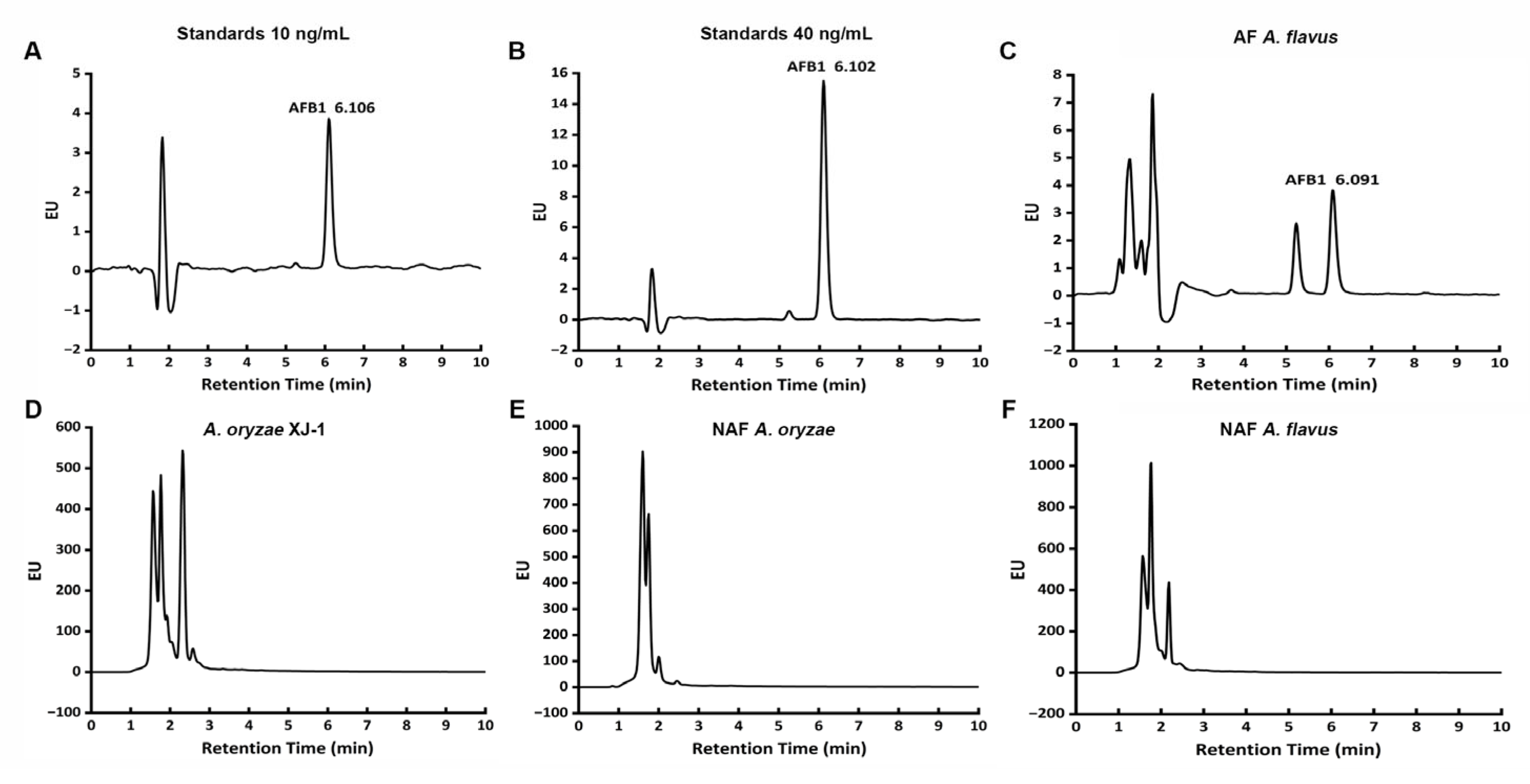
Figure 2.
Heatmap of AF synthesis-related genes from four Aspergillus strains. A. o XJ-1, A. oryzae XJ-1; NAF A. o, NAF A. oryzae; NAF A. f, NAF A. flavus; AF A. f, AF A. flavus.
Figure 2.
Heatmap of AF synthesis-related genes from four Aspergillus strains. A. o XJ-1, A. oryzae XJ-1; NAF A. o, NAF A. oryzae; NAF A. f, NAF A. flavus; AF A. f, AF A. flavus.
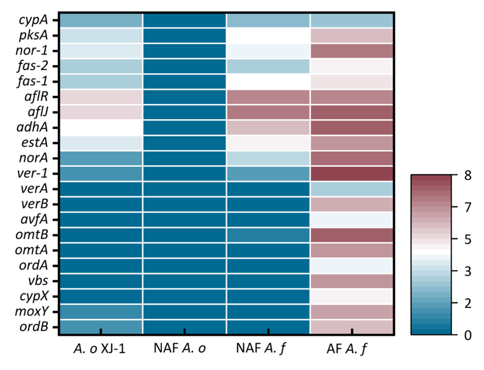
Figure 3.
Amplification of 22 AF synthesis-related genes from gDNA of Aspergillus strains. A, Amplification of 20 AF synthesis-related genes from gDNA of A. oryzae XJ-1 and NAF A. oryzae; B, Amplification of avnA and verB from gDNA of A. oryzae XJ-1, NAF A. oryzae, NAF A. flavus, AF A. flavus. A. o XJ-1, A. oryzae XJ-1; NAF A. o, NAF A. oryzae; NAF A. f, NAF A. flavus; AF A. f, AF A. flavus. M, DNA Marker; 1, cypA; 2, pksA; 3, nor-1; 4, fas-2; 5, fas-1; 6, aflR; 7, aflJ; 8, adhA; 9, estA; 10, norA; 11, ver-1; 12, verA; 13, avfA; 14, omtB; 15, omtA; 16, ordA; 17, vbs; 18, cypX; 19, moxY; 20, ordB.
Figure 3.
Amplification of 22 AF synthesis-related genes from gDNA of Aspergillus strains. A, Amplification of 20 AF synthesis-related genes from gDNA of A. oryzae XJ-1 and NAF A. oryzae; B, Amplification of avnA and verB from gDNA of A. oryzae XJ-1, NAF A. oryzae, NAF A. flavus, AF A. flavus. A. o XJ-1, A. oryzae XJ-1; NAF A. o, NAF A. oryzae; NAF A. f, NAF A. flavus; AF A. f, AF A. flavus. M, DNA Marker; 1, cypA; 2, pksA; 3, nor-1; 4, fas-2; 5, fas-1; 6, aflR; 7, aflJ; 8, adhA; 9, estA; 10, norA; 11, ver-1; 12, verA; 13, avfA; 14, omtB; 15, omtA; 16, ordA; 17, vbs; 18, cypX; 19, moxY; 20, ordB.
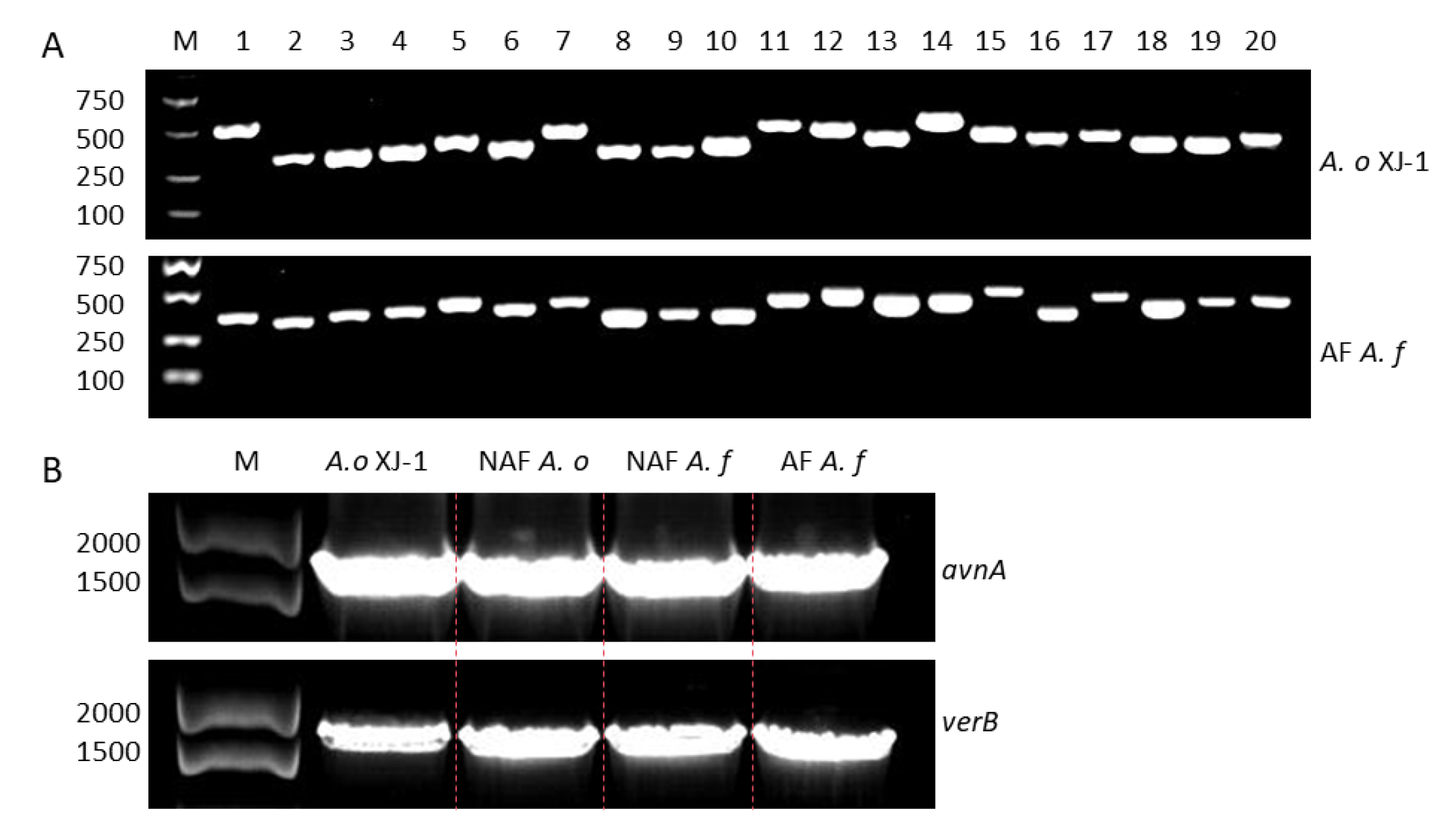
Figure 4.
Amplification of 22 AF synthesis-related genes from cDNA of Aspergillus strains. A, Amplification of 20 AF synthesis-related genes from cDNA of A. oryzae XJ-1 and NAF A. oryzae. B, Amplification of avnA and verB from cDNA of A. oryzae XJ-1, NAF A. oryzae, NAF A. flavus, AF A. flavus. A. o XJ-1, A. oryzae XJ-1; NAF A. o, NAF A. oryzae; NAF A. f, NAF A. flavus; AF A. f, AF A. flavus. M, DNA Marker; 1, cypA; 2, pksA; 3, nor-1; 4, fas-2; 5, fas-1; 6, aflR; 7, aflJ; 8, adhA; 9, estA; 10, norA; 11, ver-1; 12, verA; 13, avfA; 14, omtB; 15, omtA; 16, ordA; 17, vbs; 18, cypX; 19, moxY; 20, ordB.
Figure 4.
Amplification of 22 AF synthesis-related genes from cDNA of Aspergillus strains. A, Amplification of 20 AF synthesis-related genes from cDNA of A. oryzae XJ-1 and NAF A. oryzae. B, Amplification of avnA and verB from cDNA of A. oryzae XJ-1, NAF A. oryzae, NAF A. flavus, AF A. flavus. A. o XJ-1, A. oryzae XJ-1; NAF A. o, NAF A. oryzae; NAF A. f, NAF A. flavus; AF A. f, AF A. flavus. M, DNA Marker; 1, cypA; 2, pksA; 3, nor-1; 4, fas-2; 5, fas-1; 6, aflR; 7, aflJ; 8, adhA; 9, estA; 10, norA; 11, ver-1; 12, verA; 13, avfA; 14, omtB; 15, omtA; 16, ordA; 17, vbs; 18, cypX; 19, moxY; 20, ordB.
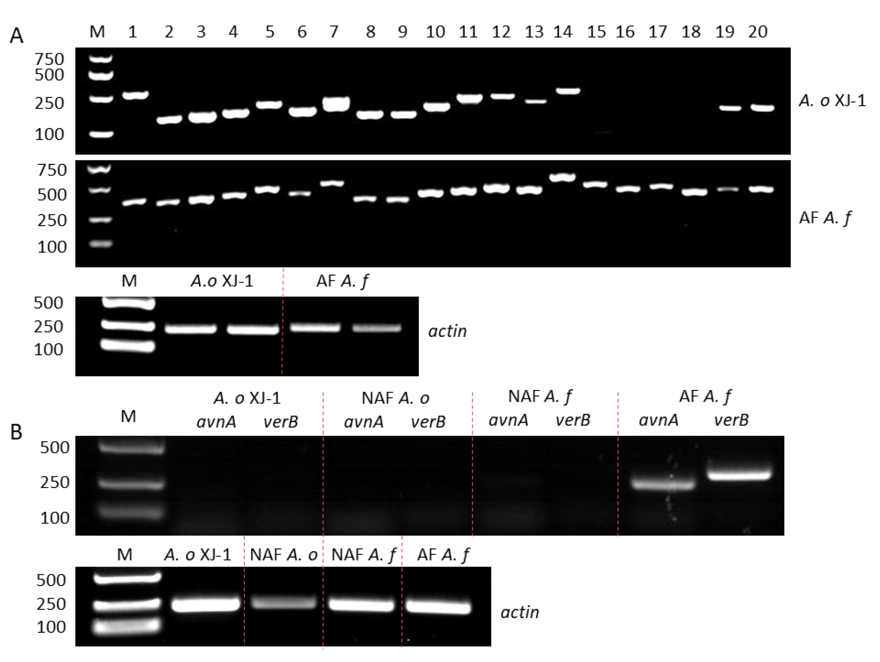
Figure 5.
Relative expression level of the six gene between A. oryzae XJ-1 and AF A. flavus. A. o XJ-1, A. oryzae XJ-1; AF A. f, AF A. flavus. A, avnA; B, cypX; C, vbs; D, verB; E, omtA; F, ordA. *, p < 0.05; **, p < 0.01; ***, p<0.001. Bar, SEM. Student’s t-test. n = 3.
Figure 5.
Relative expression level of the six gene between A. oryzae XJ-1 and AF A. flavus. A. o XJ-1, A. oryzae XJ-1; AF A. f, AF A. flavus. A, avnA; B, cypX; C, vbs; D, verB; E, omtA; F, ordA. *, p < 0.05; **, p < 0.01; ***, p<0.001. Bar, SEM. Student’s t-test. n = 3.
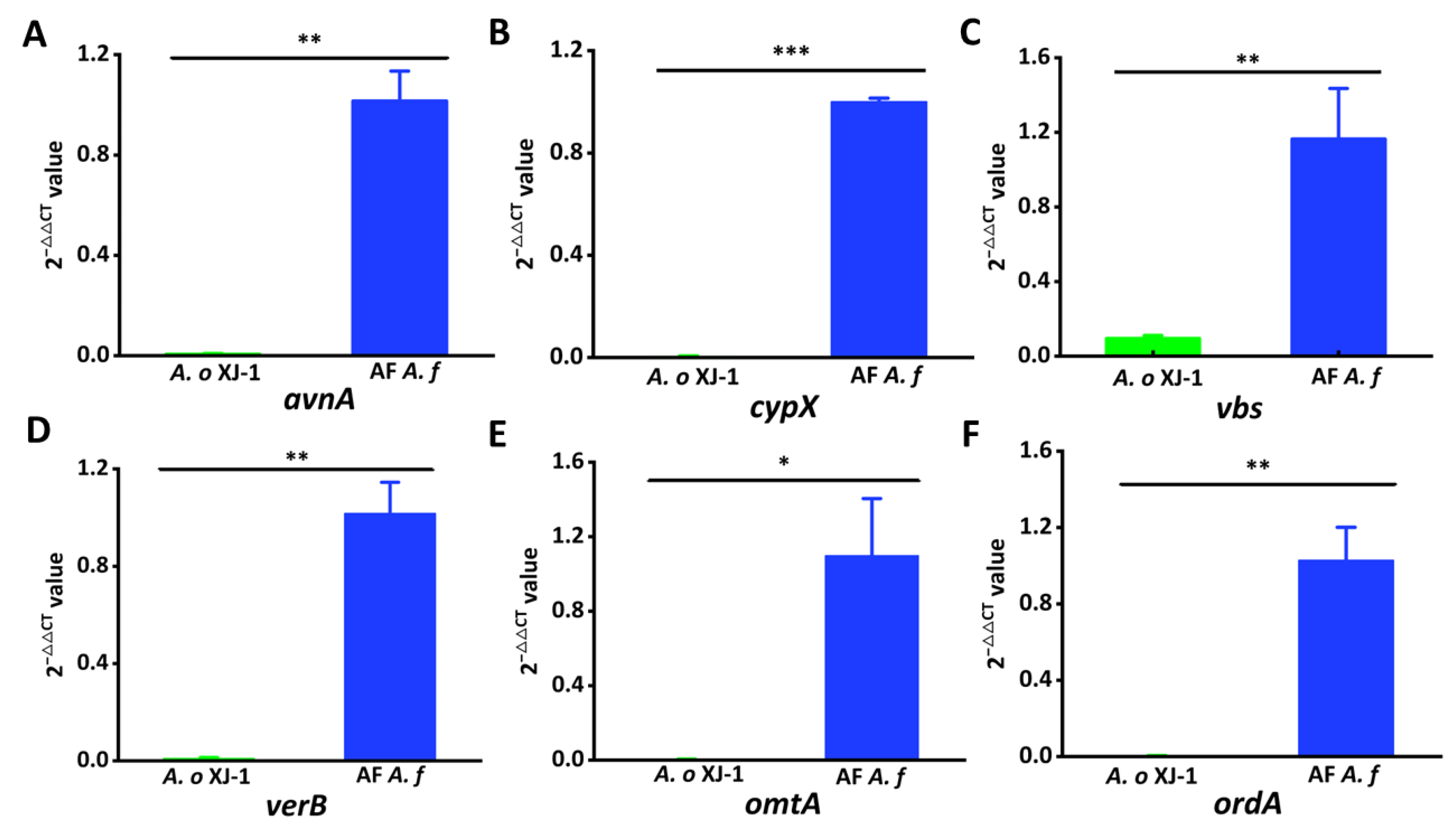

Table 1.
The amplification status of 22 AF synthesis-related genes in A. oryzae XJ-1 and AF A. flavus.
Table 1.
The amplification status of 22 AF synthesis-related genes in A. oryzae XJ-1 and AF A. flavus.
| Gene name | A. oryzaeXJ-1 | AF A. flavus | ||
| gDNA | cDNA | gDNA | cDNA | |
| cypA | √ | √ | √ | √ |
| pksA | √ | √ | √ | √ |
| nor-1 | √ | √ | √ | √ |
| fas-2 | √ | √ | √ | √ |
| fas-1 | √ | √ | √ | √ |
| aflR | √ | √ | √ | √ |
| aflJ | √ | √ | √ | √ |
| adhA | √ | √ | √ | √ |
| estA | √ | √ | √ | √ |
| norA | √ | √ | √ | √ |
| ver-1 | √ | √ | √ | √ |
| verA | √ | √ | √ | √ |
| avnA | √ | × | √ | √ |
| verB | √ | × | √ | √ |
| avfA | √ | √ | √ | √ |
| omtB | √ | √ | √ | √ |
| omtA | √ | × | √ | √ |
| ordA | √ | × | √ | √ |
| vbs | √ | × | √ | √ |
| cypX | √ | × | √ | √ |
| moxY | √ | √ | √ | √ |
| ordB | √ | √ | √ | √ |
Disclaimer/Publisher’s Note: The statements, opinions and data contained in all publications are solely those of the individual author(s) and contributor(s) and not of MDPI and/or the editor(s). MDPI and/or the editor(s) disclaim responsibility for any injury to people or property resulting from any ideas, methods, instructions or products referred to in the content. |
© 2026 by the authors. Licensee MDPI, Basel, Switzerland. This article is an open access article distributed under the terms and conditions of the Creative Commons Attribution (CC BY) license (http://creativecommons.org/licenses/by/4.0/).
Copyright: This open access article is published under a Creative Commons CC BY 4.0 license, which permit the free download, distribution, and reuse, provided that the author and preprint are cited in any reuse.