

Submitted:
27 September 2026
Posted:
29 September 2026
You are already at the latest version
Abstract
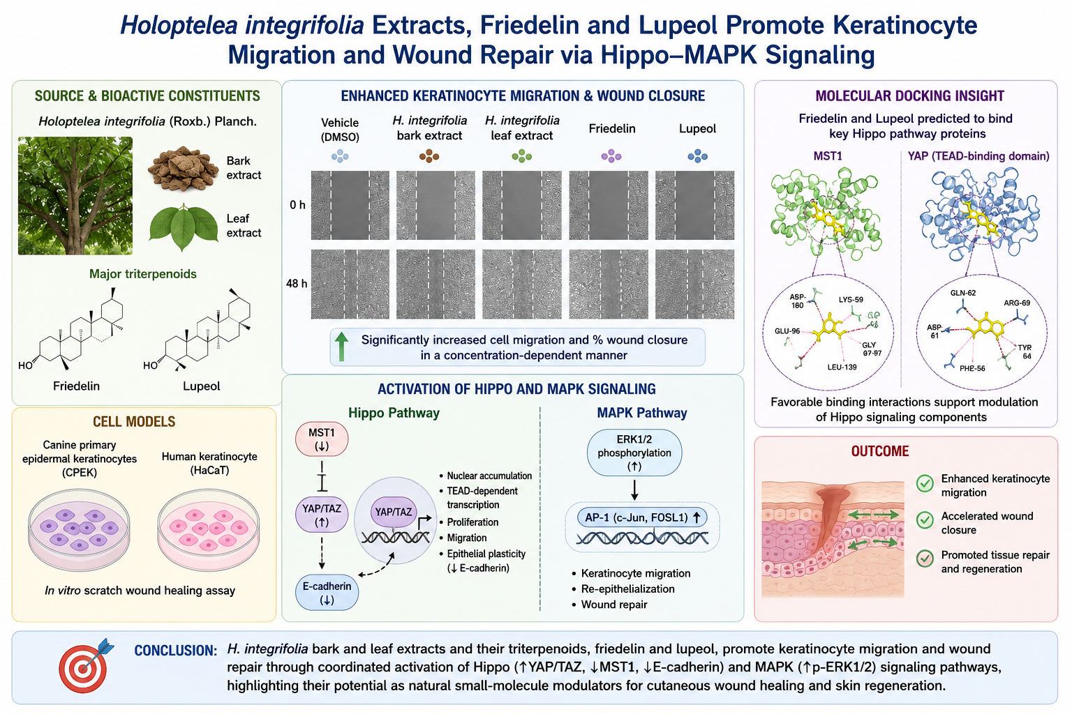
Cutaneous wound healing requires coordinated keratinocyte migration, proliferation, and epithelial remodeling. Natural products, particularly pentacyclic triterpenoids, have emerged as promising agents for tissue repair. Holoptelea integrifolia (Roxb.) Planch., a medicinal plant distributed across South and Southeast Asia and traditionally used for skin disorders and wounds, contains the bioactive triterpenoids friedelin and lupeol. However, the signaling mechanisms underlying the wound-repair effects of its extracts and bioactive compounds remain incompletely understood. This study investigated the wound-repair potential of H. integrifolia bark and leaf extracts and their major triterpenoids, friedelin and lupeol, using in vitro models and molecular analyses. Scratch assays in canine primary epidermal keratinocytes (CPEK) and human keratinocytes (HaCaT) showed that both extracts and triterpenoids significantly enhanced keratinocyte migration and accelerated wound closure in a concentration-dependent manner. Western blotting revealed increased Yes-associated protein (YAP) and transcriptional co-activator with PDZ-binding motif (TAZ) levels, accompanied by reduced mammalian sterile 20-like kinase 1 (MST1) and E-cadherin expression, supporting modulation of Hippo signaling toward a pro-migratory phenotype. Molecular docking predicted favorable interactions of friedelin and lupeol with MST1 and YAP. Collectively, these findings indicate that H. integrifolia extracts and their triterpenoids promote keratinocyte-mediated wound repair through coordinated modulation of Hippo and MAPK signaling, supporting friedelin and lupeol as natural candidates for cutaneous wound healing and skin regeneration.
Keywords:
Holoptelea integrifolia
; Indian elm
; wound healing
; inflammation
; friedelin
; lupeol
1. Introduction
Restoration of injured skin proceeds through a tightly regulated series of biological events, beginning with hemostatic control and inflammation, followed by cellular proliferation and ultimately remodeling of the repaired tissue. Among the cellular events critical to successful wound repair, keratinocyte migration and re-epithelialization represent key determinants of healing outcomes. Dysregulation of these processes underlies a range of clinically significant wound-healing impairments, including chronic wounds and delayed repair in diabetic or immunocompromised patients. The identification of bioactive compounds capable of modulating regenerative signaling in keratinocytes therefore represents an important avenue for the development of novel wound-healing therapeutics. At the molecular level, regenerative responses are governed by interconnected signaling networks, including Hippo signaling through its downstream effectors Yes-associated protein (YAP) and transcriptional coactivator with PDZ-binding motif (TAZ), the mitogen-activated protein kinase (MAPK) network, and phosphoinositide 3-kinase (PI3K)–AKT signaling associated with epithelial–mesenchymal transition (EMT)[1,2,3,4]. The Hippo pathway is a central regulator of epithelial homeostasis. In its active state, mammalian sterile 20-like kinases ½ (MST1/2) initiate a kinase cascade involving large tumor suppressor kinases 1/2 (LATS1/2), which subsequently phosphorylates YAP and TAZ. Phosphorylated YAP/TAZ are retained in the cytoplasm and targeted for degradation, limiting their availability for TEAD-mediated transcription. Upon pathway inactivation, nuclear accumulation of YAP/TAZ drives transcriptional programs associated with cellular proliferation and migration [1,2]. Pharmacological modulation of the Hippo pathway has emerged as an attractive therapeutic strategy in regenerative medicine; however, small-molecule activators capable of selectively engaging pro-repair YAP/TAZ signaling in keratinocytes remain scarce. Alongside Hippo signaling, MAPK pathway contributes to epidermal repair by engaging extracellular signal-regulated kinase 1/2 (ERK1/2) and downstream AP-1 transcription factors, including c-Jun and fos-related antigen 1 (FOSL1) [3,5]. Crosstalk between ERK–AP-1 and YAP–TEAD complexes further amplifies transcriptional programs required for efficient wound closure [6]. Additionally, PI3K–AKT signaling contributes to keratinocyte motility by upregulating EMT-associated transcription factors, most notably snail family transcriptional repressor 1 (SNAI1), and by downregulating E-cadherin expression [4,7]. Collectively, these observations suggest that coordinated modulation of the Hippo, MAPK, and AKT pathways is essential for efficient epidermal tissue repair [1,6]. The chemical diversity of natural products offers a valuable platform for identifying bioactive molecules with therapeutic potential. Among these, pentacyclic triterpenoids have gained particular interest because of their reported anti-inflammatory and tissue-regenerative properties [8]. Friedelin and lupeol, two naturally occurring pentacyclic triterpenoids isolated from Holoptelea integrifolia (Roxb.) Planch., have demonstrated wound-healing and anti-inflammatory properties in keratinocyte-based models; however, their precise molecular targets and mechanistic basis of action remain insufficiently characterized [8,9]. H. integrifolia, or Indian elm a large deciduous tree indigenous to the Indian subcontinent and Southeast Asia, has been extensively documented in Ayurvedic and traditional medicine systems for the management of wound-related cutaneous disorders, including chronic ulcers, eczema, and inflammatory skin conditions [9,10]. Ethnopharmacological records indicate that multiple plant organs, principally the bark, leaves, and stem, are employed therapeutically, either individually or in polyherbal formulations, reflecting a holistic and organ-inclusive approach to wound management [9,11]. Phytochemical investigations have established that the bioactive profile of H. integrifolia encompasses pentacyclic triterpenoids, most notably friedelin and lupeol, alongside phenolic acids, flavonoids, and tannins, with the bark and leaf fractions representing the principal repositories of pharmacologically relevant constituents [9,10]. Notwithstanding this ethnopharmacological foundation, systematic mechanistic investigation of H. integrifolia extracts and their triterpenoid constituents regarding to Hippo pathway modulation, YAP/TAZ transcriptional regulation, and direct molecular target engagement in keratinocyte-based wound-healing models has not been undertaken [9]. Previous investigations into H. integrifolia have provided initial evidence supporting its wound-healing potential across multiple plant parts, including the stem bark [8] and leaves [12]. Phytochemical profiling of the fresh stem bark extract by thin-layer and gas chromatography revealed friedelin and lupeol as its predominant bioactive constituents. In keratinocyte models, friedelin and lupeol accelerated closure of the scratched area and promoted cell migration, accompanied by increased matrix metalloproteinase-9 (MMP-9) production. Gene expression analyses further revealed that treatment with these compounds decreased mRNA levels of CDH1 and DSG1, markers associated with cell–cell adhesion, while increasing expression of KRT17, a gene involved in keratinocyte proliferation and homeostasis. Anti-inflammatory effects were also observed, as exposure to the n-hexane extract and its active constituents lowered the levels of several inflammatory markers, including cyclooxygenase-2 (COX-2), inducible nitric oxide synthase (iNOS), tumor necrosis factor-alpha (TNF-α), and interleukin-6 (IL-6). Further evidence from excision and incision models showed that ethanolic leaf and stem bark extracts facilitated wound repair, with treated animals reaching over 90% closure by day 14 compared with 62.99% in controls [12]. This improvement was accompanied by greater wound-breaking strength, increased hydroxyproline levels, enhanced collagen deposition, and histological evidence of tissue restoration. The extracts additionally exhibited antibacterial, antifungal, and DPPH radical-scavenging activities, with the stem bark extract generally displaying greater activity than the leaf extract. Despite this accumulating body of evidence, no mechanistic studies have been conducted on leaf extracts of H. integrifolia, and the upstream intracellular signaling pathways through which friedelin and lupeol exert their wound-healing effects, particularly with respect to the Hippo–YAP/TAZ and MAPK/ERK axes, remain uncharacterized. The present study therefore extends this earlier work by investigating the effects of friedelin and lupeol on defined signaling pathway components governing keratinocyte migration, with the aim of providing a mechanistic basis for their wound-healing activity across both stem bark and leaf-derived bioactive constituents. To investigate the molecular mechanisms underlying keratinocyte migration and wound repair, representative proteins involved in the Hippo signaling pathway and MAPK signaling cascade were selected for analysis in the present study. Scratch-based wound closure assays were performed in both canine primary keratinocyte (CPEK) and human immortalized keratinocyte (HaCaT) cell models to assess their effects on wound closure. The Hippo pathway plays a critical role in regulating cell proliferation, migration, and tissue regeneration, processes essential during cutaneous wound healing. YAP and TAZ were examined as key downstream effectors of Hippo signaling, as their nuclear activation promotes transcriptional programs that stimulate keratinocyte proliferation and migration during epithelial repair. To evaluate upstream regulation of the Hippo pathway, MST1 was included in the analysis, given its established role as a core Hippo kinase that negatively regulates YAP/TAZ activity. Additionally, E-cadherin (CDH1) was examined as a marker of cell–cell adhesion and epithelial phenotype, as its reduction is frequently associated with enhanced cellular migration and epithelial remodeling during wound healing. To further assess signaling pathways involved in cell migration, the ERK1/2 cascade was evaluated, with particular focus on phosphorylated ERK1/2 (p-ERK1/2) as an indicator of MAPK pathway activation, a pathway recognized for its importance in keratinocyte proliferation, migration, and re-epithelialization. In the present study, friedelin and lupeol, the principal pentacyclic triterpenoid constituents of H. integrifolia, were investigated as candidate small-molecule modulators of regenerative signaling networks in keratinocytes. Western blot analyses were conducted to examine protein expression of key Hippo pathway components, including YAP, TAZ, MST1, and E-cadherin, as well as MAPK pathway activation as reflected by p-ERK1/2 levels, in both CPEK and HaCaT cell models. Molecular docking analyses were additionally performed to explore the thermodynamic binding interactions of friedelin and lupeol at the active sites of MST1 and YAP, with the aim of providing a structural rationale for their observed biological effects. Taken together, this study seeks to elucidate the mechanistic basis by which these triterpenoids engage the interconnected Hippo–MAPK–AKT signaling axis governing keratinocyte-driven tissue repair, and to establish a structural and functional foundation for future medicinal chemistry optimization of the pentacyclic triterpenoid scaffold toward therapeutic applications in cutaneous wound healing.
2. Materials and Methods
2.1. Plant Material and Extraction
Bark and leaf materials of H. integrifolia (Roxb.) Planch. were collected from Lampang Province, Thailand, in 2025 and authenticated by Asst. Prof. Dr. Narin Printarakul, Department of Biology, Faculty of Science, Chiang Mai University, Chiang Mai, Thailand (voucher specimen no. HI004), with a voucher specimen deposited at Herbarium of the Department of Biology, Faculty of Science, Chiang Mai University (CMUB). Freshly collected plant materials were rinsed under running tap water and then left in the shade at ambient temperature until no visible moisture remained on the surface. The bark was coarsely ground using a mechanical grinder and subsequently subjected to maceration in n-hexane at a solid-to-solvent ratio of 1:20 (g/mL) for 24 h at room temperature with occasional agitation. For leaf extraction, whole dried leaves were extracted using the identical maceration procedure; this solvent system was specifically selected to minimize co-extraction of chlorophyll and polar pigment interference. Following vacuum filtration through Whatman No. 1 filter paper, the filtrates were evaporated at 45 °C under reduced pressure to obtain the crude extracts, which were subsequently transferred to amber glass vials and maintained at −20 °C until use.
2.2. Analysis of Friedelin and Lupeol Content Using Gas Chromatography (GC)
Quantitative chemical profiling of H. integrifolia bark and leaf extracts was performed by GC-FID, with friedelin and lupeol employed as authentic reference standards [8]. Individual stock solutions of friedelin and lupeol were prepared in n-hexane at 2000 μg/mL, followed by successive two-fold dilutions to generate external calibration curves. Extract samples were dissolved in hexane at a concentration of 100 mg/mL. For derivertization, a 100-μL portion of each extract solution was placed in a glass tube and dried at room temperature by nitrogen evaporation. The resulting residue was mixed with 100 μL of N,O-bis(trimethylsilyl)trifluoroacetamide (BSTFA) and incubated at 60 °C for 30 min before GC analysis. Samples were analyzed on an Agilent 7890B gas chromatograph coupled to a flame ionization detector (FID) (Agilent Technologies, Santa Clara, CA, USA). A DB-5MS capillary column (30 m × 0.250 mm i.d.; 0.50 μm film thickness; Agilent Technologies) was employed for chromatographic separation. Helium served as the mobile carrier at a fixed flow of 1 mL/min. A 1-μL sample volume was introduced at 250 °C using a 10:1 split injection. The solvent delay was set to 10 min. The GC oven was initially set to 80 °C without a holding period, after which the temperature was increased to 100 °C at 2 °C/min. Without an intermediate hold, the oven was further heated to 270 °C at the same rate and maintained at this temperature for 20 min. The complete chromatographic run required 115 min. The FID temperature was maintained at 270 °C throughout the analysis. The identities of lupeol and friedelin within the H. integrifolia extracts were confirmed by GC based on their characteristic retention times, verified against authentic reference standards run under identical chromatographic conditions. Quantification of friedelin and lupeol in the H. integrifolia extracts was performed by external standard calibration conducted in triplicate. For quantitative calibration, chromatographic peak areas were correlated with the corresponding analyte concentrations across the validated linear range.
2.3. Cell Cultures
The effects of friedelin, lupeol, and H. integrifolia bark and leaf extracts on keratinocyte wound closure were examined using a scratch assay in CPEK and HaCaT cells, with minor modifications to a previously described protocol. Cells were plated in 24-well plates at 1 × 10⁶ cells per well and allowed to develop a confluent monolayer. A cell-free gap was then generated by drawing a sterile 10-μL pipette tip across the monolayer. After scratching, loose cells were eliminated by two PBS washes, followed by immediate addition of serum-free medium containing the designated treatments. Friedelin and lupeol were tested at 0.125 and 0.25 μM, whereas the bark and leaf extracts were applied at 0.125, 0.25, and 0.5 μg/mL. An equivalent volume of dimethyl sulfoxide (DMSO) was included as the vehicle control. Each experiment included three independent biological replicates. Scratch areas were photographed at 0, 24, and 48 h using a Nikon Ti-U inverted microscope (Nikon Instruments, Tokyo, Japan) under 10× magnification. The remaining cell-free area was quantified at each time point using ImageJ version 1.54g (National Institutes of Health, USA) with TScratch software. Closure was normalized to the wound area measured immediately after scratching (0 h) and reported as a percentage using the following equation:
Wound closure (%) = [(Wound area at 0 h − Wound area at t h) / Wound area at 0 h] × 100.
2.4. Western Blot Analysis of Hippo Signaling Proteins in CPEK Cell Line
CPEK cells were plated in 6-well plates at 6 × 10⁵ cells per well and allowed to reach approximately 70–80% confluence before treatment. Cells were then exposed for 24 h to H. integrifolia bark or leaf extracts (0.125, 0.25, and 0.5 μg/mL) or to friedelin or lupeol (0.125 and 0.25 μM). An equivalent amount of 0.1% DMSO was applied to the vehicle-control group. After the 24-h exposure, the cells were rinsed twice with ice-cold PBS before protein extraction with radioimmunoprecipitation assay (RIPA) buffer (Enzyomics, catalog no. EBR001) containing a protease and phosphatase inhibitor cocktail (Sigma-Aldrich, catalog no. P8340). Cell lysates were clarified by centrifugation at 16,000 × g for 10 min at 4 °C, after which the supernatant fractions were recovered. Protein content was quantified with a bicinchoninic acid (BCA) assay kit (Thermo Scientific, catalog no. A55865) following the supplier’s protocol. For immunoblotting, 20 μg of total protein from each sample was separated on 10–12% SDS-polyacrylamide gels and subsequently transferred electrophoretically to nitrocellulose membranes (GVS Filter Technology). The membranes were blocked for 1 h at room temperature in Tris-buffered saline containing 0.1% Tween-20 (TBST) and 5% non-fat dry milk. Primary antibody incubation was carried out overnight at 4 °C using antibodies against TAZ (Santa Cruz Biotechnology, sc-518026; 1:1000), YAP (Santa Cruz Biotechnology, sc-101199; 1:1000), and E-cadherin (Cell Signaling Technology, 14472S; 1:1000). β-Actin (ACTB; Santa Cruz Biotechnology, sc-47778) served as the loading reference. After three TBST washes, the membranes were exposed for 1 h at room temperature to the appropriate HRP-conjugated secondary antibodies: anti-rabbit IgG (Thermo Scientific, 31460; 1:5000) or anti-mouse IgG (Cell Signaling Technology, 7076; 1:5000). Protein bands were detected with an enhanced chemiluminescence (ECL) reagent (Thermo Fisher Scientific, catalog no. 32106) and recorded using a UVITEC chemiluminescence imaging system (Cambridge, UK). Densitometric quantification was performed in ImageJ version 1.54g (National Institutes of Health, USA). The intensity of each target band was normalized against its corresponding β-actin signal, and the resulting values were expressed relative to the DMSO vehicle control. Results are reported as the mean ± standard error of the mean (SEM) from three independent experiments.
2.5. Western Blot Analysis of MST1, Hippo and MAPK Signaling Protein Expression in CPEK and HaCaT Cell Lines
CPEK and HaCaT cells were grown to approximately 70–80% confluence before exposure to H. integrifolia bark or leaf extracts (0.125, 0.25, and 0.5 μg/mL) or to friedelin or lupeol (0.125 and 0.25 μM). DMSO-treated cells were included as the vehicle control. Following treatment, cellular proteins were extracted with RIPA buffer containing protease and phosphatase inhibitors, and protein concentrations were measured by the BCA assay. Equivalent amounts of protein were resolved by SDS-PAGE and subsequently transferred to nitrocellulose membranes. After blocking with 5% non-fat milk in TBST, the membranes were exposed overnight at 4 °C to primary antibodies targeting MST1, YAP, TAZ, phosphorylated ERK1/2 (p-ERK1/2), total ERK1/2, and E-cadherin. Following washing, the appropriate HRP-conjugated secondary antibodies were applied, and immunoreactive signals were developed by enhanced chemiluminescence (ECL). β-Actin served as the loading reference. Densitometric values for individual target proteins were normalized against the corresponding β-actin signal and reported relative to the DMSO vehicle control. Results represent the mean ± SEM from three independent experiments.
2.6. Molecular Docking Analysis
To investigate potential molecular interactions between the isolated triterpenoid compounds and key wound-healing–related signaling proteins, molecular docking simulations were performed. Three-dimensional structures of ERK, MST1, and YAP were retrieved from the Protein Data Bank (PDB) using the accession codes 1MKP, 6YAT, and 3KYS, respectively. The chemical structures of the triterpenoid ligands friedelin and lupeol were retrieved from public chemical databases (NCBI:PubMED) and energy-minimized before docking. Prior to simulation, each protein structure was processed by eliminating crystallographic water molecules and co-crystallized ligands, followed by the addition of polar hydrogen atoms and assignment of atomic charges. Docking simulations were performed using a molecular docking (AutoDock Vina), through the PyRx virtual screening platform to predict ligand–protein binding conformations and calculate binding affinity values (kcal/mol). For each protein, the search space was centered on the binding pocket occupied by the co-crystallized ligand. Multiple docking runs were conducted to ensure reproducibility of the predicted binding modes. For comparison, the native ligand or reference inhibitor present in the crystal structure of each protein was also docked under identical conditions. Binding affinity values were calculated as the predicted free energy of binding, where more negative values indicate stronger predicted ligand–protein interactions. Results from repeated docking simulations are presented as mean ± standard deviation.
2.7. Analysis of Ligand–Protein Binding Interactions
To further characterize the predicted interactions between the triterpenoid compounds and target proteins, two-dimensional ligand–protein interaction analyses were performed based on the docking poses obtained from the molecular docking simulations. The best-ranked docking conformations of friedelin and lupeol with the selected protein targets, ERK (PDB ID: 1MKP), MST1 (PDB ID: 6YAT), and YAP (PDB ID: 3KYS) were selected for interaction analysis. The docked complexes were analyzed using ligand interaction visualization tools to determine which amino acid residues contribute to ligand recognition within the predicted binding pockets. Interactions evaluated included van der Waals contacts, hydrogen bonds, π–alkyl interactions, π–sulfur interactions, π–cation interactions, and steric contacts. The resulting interaction patterns were visualized as two-dimensional interaction diagrams, highlighting the residues surrounding the ligand-binding pocket and the types of intermolecular interactions formed between the ligand and protein residues. These analyses were performed to provide structural insights into the binding orientation and interaction patterns of friedelin and lupeol within the catalytic or regulatory pockets of the target proteins.
2.8. Evaluation of COX-2 Expression by Western Blot Analysis
To investigate the anti-inflammatory potential of the triterpenoid compounds and plant extracts, COX-2 protein expression was evaluated using Western blot analysis in LPS-stimulated cells. Cells were cultured under standard conditions and seeded into culture plates until reaching approximately 70–80% confluence. To induce an inflammatory response, cells were stimulated with lipopolysaccharide (LPS, 1 μg/mL). Following LPS stimulation, cells were treated with friedelin or lupeol at concentrations of 0.125, 0.5, and 2 mM, or with bark and leaf extracts of H. integrifolia at 0.125, 0.5, and 2 μg/mL. Cellular proteins were then extracted in RIPA buffer containing protease inhibitors, and their concentrations were quantified by BCA assay. Equivalent protein quantities were resolved by SDS-PAGE and transferred to nitrocellulose membranes. After blocking in TBST containing 5% non-fat milk, the membranes were exposed overnight at 4 °C to the primary antibody against COX-2. β-Actin served as the loading reference. Following washing, appropriate HRP-conjugated secondary antibodies were applied, and immunoreactive signals were developed using ECL. COX-2 band intensity was quantified by densitometry and normalized against the corresponding β-actin signal.
2.9. Statistical Analysis
Quantitative results are presented as the mean ± standard error of the mean (SEM) from at least three independent experiments, each conducted in triplicate. Data were analyzed with GraphPad Prism version 9. Differences among experimental groups were evaluated by one-way analysis of variance (ANOVA), with Tukey’s post hoc procedure applied for multiple-group comparisons. Statistical significance was defined at P < 0.05, with *P < 0.05 and P < 0.01 denoting the respective significance levels.
3. Results
3.1. GC-FID Quantification of Friedelin and Lupeol in H. integrifolia Bark and Leaf Extracts
Comparative chromatographic profiling of bark (100 mg) and leaf (100 mg) extracts revealed differential abundance of both triterpenoids across plant organs (Figure 1A and Figure 1B). For friedelin (tᵣ = 104 min), the calibration curve was linear over a concentration range of 31.25–2000 µg/mL, following the regression equation y = 0.7914x + 39.496, with a coefficient of determination (R²) of 0.9986 (Figure 1C). For lupeol (tᵣ = 97 min), linearity was established over a concentration range of 15.625–1000 µg/mL, with a regression equation of y = 1.4569x − 2.8033 and an R² of 0.9999 (Figure 1C). The R² values of ≥ 0.998 for both analytes confirmed excellent linearity and calibration reliability across the tested concentration ranges. Based on the respective calibration curves (Figure 1C), the quantified contents of friedelin and lupeol in the H. integrifolia bark extract were determined to be 27.85 and 3.25 µg/g FW while H. integrifolia leaf extract were determined to be 5.52 and 11.69 µg/g FW, respectively. The bark extract exhibited a higher relative abundance of friedelin compared with the leaf extract, whereas lupeol was more abundant in the leaf extract relative to the bark. These findings indicate organ-specific accumulation patterns of the two principal triterpenoid constituents in H. integrifolia and provide chromatographic justification for the comparative evaluation of bark and leaf extracts in subsequent bioactivity assessments.
3.2. Wound Healing Activity in CPEK and HaCaT Cells
3.2.1. Bark and Leaf Extracts of Holoptelea integrifolia Enhance Keratinocyte Migration in CPEK and HaCaT Cells
The effects of H. integrifolia bark and leaf extracts on keratinocyte wound closure were examined in CPEK and HaCaT cells. Each extract was tested at 0.125, 0.25, and 0.5 μg/mL, and changes in the scratched area were assessed over 48 h, with measurements taken at baseline (0 h), 24 h, and 48 h. Cells receiving DMSO alone were included as the vehicle control. Representative microscopic images (Figure 2A left panel) show progressive migration of CPEK keratinocytes into the scratched area over time following treatment with both bark and leaf extracts. Compared with the DMSO control, treatment with either extract significantly accelerated wound closure at 24 h and 48 h. Quantitative analysis (Figure 2A right panel) revealed that both extracts increased the percentage of wound healing area in a concentration-dependent manner, with significant differences observed relative to the control group (*P < 0.05 and **P < 0.001). At 24 h, bark extract treatments produced approximately 80–95% wound closure, compared with approximately 55–60% in the control group. Leaf extract treatment also enhanced migration, achieving approximately 75–90% wound closure at the same time point. By 48 h, keratinocyte migration further increased, with both bark and leaf extracts producing near-complete wound closure (~95–105%). The stimulatory effect appeared slightly stronger for bark extract, particularly at 0.25–0.5 μg/mL, although both extracts demonstrated comparable activity at later time points. Similar trends were observed in human HaCaT keratinocytes (Figure 2B). Treatment with bark and leaf extracts significantly enhanced wound closure compared with the control group. At 24 h, treated cells exhibited approximately 80–95% wound closure, whereas control cells remained below 60% closure. At 48 h, both extracts promoted substantial re-epithelialization, with wound closure reaching approximately 90–100% across the tested concentrations. The results indicate that the wound-healing activity of H. integrifolia extracts is conserved across both canine and human keratinocyte models. Collectively, these findings demonstrate that both bark and leaf extracts of H. integrifolia significantly stimulate keratinocyte migration, leading to accelerated wound closure in vitro in a concentration- and time-dependent manner. Near-complete re-epithelialization was achieved at 0.5 µg/mL in both cell lines at 48 h, with bark extract consistently producing marginally greater wound closure than leaf extract at equivalent concentrations across both models.
3.2.2. Friedelin and Lupeol Promote Wound Closure in CPEK and HaCaT Cells
The wound-healing activity of the triterpenoid compounds friedelin and lupeol was evaluated using a scratch assay in CPEK (Figure 3A) and HaCaT (Figure 3B) cell lines. Cells were treated with two concentrations of each compound (0.125 and 0.25 μM), and wound closure was monitored at 0, 24, and 48 h following scratch formation. Representative microscopic images show progressive migration of keratinocytes into the scratched area over time (left panel). Quantitative analysis (right panel) demonstrated that, at 24 h, the control group exhibited approximately 50% wound closure, whereas treatment with friedelin at 0.125 and 0.25 μM increased wound closure to approximately 82–88%, representing a significant improvement (P < 0.01). Lupeol also promoted wound closure, although its effect was slightly lower than that of friedelin. Wound closure reached approximately 83% at 0.125 μM and 75% at 0.25 μM, suggesting that the lower concentration produced greater migratory activity. At 48 h, the control group reached approximately 82–85% wound closure. Friedelin treatment further increased wound closure to approximately 96–98% at both concentrations, approaching complete closure. Lupeol-treated cells also exhibited enhanced wound closure (approximately 88–95%), although the higher concentration (0.25 μM) produced a slightly lower response than the lower concentration. These findings indicate that friedelin consistently stimulates canine keratinocyte migration, whereas lupeol demonstrates optimal activity at the lower concentration. In HaCaT cells, at 24 h, the control group showed approximately 30% wound closure. Friedelin treatment increased wound closure to approximately 50–55% at both concentrations, representing a significant improvement compared with the control (P < 0.01). Lupeol produced an even greater response, with wound closure increasing to approximately 65% at 0.125 μM and 72–75% at 0.25 μM, indicating stronger stimulation of keratinocyte migration than friedelin during the early phase of wound repair. At 48 h, wound closure in the control group increased to approximately 65%. Friedelin further enhanced wound closure to approximately 86% at 0.125 μM, whereas the response at 0.25 μM was slightly lower (approximately 75–78%). Lupeol maintained a consistently high level of wound closure, reaching approximately 82–84% at both concentrations. Although all treatments significantly accelerated wound healing compared with the control (P < 0.05), lupeol produced the most consistent enhancement of migration in HaCaT cells. Overall, these findings demonstrate that both triterpenoids promote keratinocyte-mediated wound healing in canine and human keratinocyte models. Friedelin exhibited the strongest activity in CPEK cells, whereas lupeol showed comparatively greater efficacy in HaCaT cells, suggesting that the two compounds may differentially regulate keratinocyte migration depending on cell origin. This cell type-dependent response indicates that friedelin and lupeol may activate distinct signaling pathways involved in epithelial wound repair.
3.3. Modulation of Hippo Pathway Effectors by H. integrifolia Extracts and Triterpenoid Constituents in CPEK Keratinocytes
To investigate the molecular mechanisms underlying the pro-migratory activity of H. integrifolia, the expression of Hippo signaling markers TAZ, YAP, and the epithelial adhesion protein E-cadherin was examined by Western blot analysis in CPEK keratinocytes (Figure 3). Treatment with bark and leaf extracts resulted in a dose-dependent increase in TAZ and YAP protein expression compared with the DMSO control (Figure 4A and Figure 4B). Bark extract produced a pronounced increase in TAZ levels across all tested concentrations, with the strongest induction observed at 0.5 μg/mL. Similarly, YAP expression was significantly elevated following treatment with both bark and leaf extracts. In contrast, expression of E-cadherin was reduced relative to the control group, suggesting modulation of epithelial cell adhesion consistent with enhanced migratory activity. The effects of the isolated triterpenoids friedelin and lupeol were subsequently evaluated (Figure 4C and Figure 4D). Both compounds significantly increased TAZ and YAP protein expression compared with DMSO-treated cells. Friedelin treatment produced a concentration-dependent increase in TAZ and YAP levels, whereas lupeol induced a similarly strong upregulation of both proteins at the tested concentrations. Consistent with the extract treatments, E-cadherin expression decreased following triterpenoid treatment, indicating a shift toward a pro-migratory cellular phenotype. Quantitative densitometric analysis confirmed that the observed changes were statistically significant relative to the control group. Overall, these results demonstrate that both H. integrifolia extracts and their triterpenoid constituents activate Hippo pathway effectors (TAZ and YAP) while reducing E-cadherin expression, supporting a mechanism in which these compounds promote keratinocyte migration through modulation of Hippo-associated signaling pathways.
3.4. Downregulation of MST1 Expression in CPEK Cells Following Treatment with H. integrifolia Extracts and Triterpenoid Compounds
To determine whether the wound-repair effects of H. integrifolia involve modulation of key regenerative signaling pathways, the expression of the upstream Hippo pathway kinase MST1 was examined in CPEK keratinocytes using Western blot analysis (Figure 5). Treatment with bark and leaf extracts resulted in a reduction in MST1 protein expression relative to the DMSO control (Figure 5A). Densitometric analysis revealed that bark extract at 0.25 and 0.5 μg/mL significantly decreased MST1 expression levels compared with control cells. A similar trend was observed with leaf extract treatment, where increasing concentrations were associated with progressive reduction of MST1 protein levels. Consistent with these findings, treatment with the isolated triterpenoids friedelin and lupeol also altered MST1 expression (Figure 5B). Friedelin produced a moderate reduction in MST1 levels at both tested concentrations, whereas lupeol treatment resulted in a more pronounced decrease, particularly at 0.125 μM, suggesting potential modulation of upstream Hippo signaling components. Quantitative densitometric analysis confirmed that several treatment conditions significantly decreased MST1 protein levels compared with the control group (*P < 0.05 and **P < 0.001). These findings suggest that both H. integrifolia extracts and their triterpenoid constituents influence Hippo pathway regulation by modulating MST1 expression, which may be associated with the increased keratinocyte migration observed during wound closure.
3.5. H. integrifolia Extracts and Triterpenoids Activate Hippo and ERK Signaling Pathways in HaCaT Cells
To determine how H. integrifolia influences signaling pathways involved in wound repair, the expression of Hippo signaling proteins (YAP and TAZ) and the MAPK signaling marker phosphorylated ERK1/2 (p-ERK1/2) was examined in HaCaT keratinocytes by Western blot analysis (Figure 6). Treatment with both bark and leaf extracts markedly upregulated YAP protein expression in a concentration-dependent manner (Figure 6A and Figure 6B). Compared with the DMSO-treated control, bark extract progressively increased YAP expression, with the highest level observed at 0.5 μg/mL. The leaf extract exhibited a stronger stimulatory effect than the bark extract, producing a robust increase in YAP expression at 0.5 μg/mL (**P < 0.001). Consistent with these findings, TAZ expression was significantly elevated following treatment with both extracts. The leaf extract induced greater TAZ expression than the bark extract, with maximal protein levels detected at 0.25 μg/mL (**P < 0.001), although a slight reduction was observed at the highest concentration while remaining substantially above the control. To determine whether ERK signaling was involved in the observed migratory response, the phosphorylation of ERK1/2 was evaluated. Both bark and leaf extracts significantly enhanced p-ERK1/2 levels in a concentration-dependent manner, whereas total ERK protein expression remained relatively unchanged, indicating activation of ERK signaling through phosphorylation rather than changes in ERK abundance. The leaf extract produced a greater increase in p-ERK1/2 than the bark extract, with maximal activation observed at 0.25 μg/mL (**P < 0.001). To identify the active constituents responsible for these effects, HaCaT cells were treated with the isolated triterpenoids friedelin and lupeol (Figure 6C and Figure 6D). Both compounds significantly increased YAP and TAZ protein expression in a dose-dependent manner. Lupeol exhibited a stronger stimulatory effect than friedelin, particularly at 0.25 μM, where the highest levels of both YAP and TAZ were observed (**P < 0.001). In contrast, friedelin induced a more pronounced increase in p-ERK1/2 phosphorylation than lupeol, suggesting that the two triterpenoids differentially regulate downstream signaling pathways involved in keratinocyte migration. Collectively, these findings demonstrate that H. integrifolia activates both the Hippo (YAP/TAZ) and ERK1/2 signaling pathways in HaCaT keratinocytes. The leaf extract exhibited greater activity than the bark extract, while the isolated triterpenoids differentially modulated intracellular signaling, with lupeol preferentially activating the Hippo pathway and friedelin more strongly enhancing ERK1/2 phosphorylation. These molecular changes are consistent with the accelerated keratinocyte migration observed in the scratch wound-healing assay and support the involvement of coordinated Hippo and ERK signaling in the wound-healing effects of H. integrifolia.
3.6. Predicted Interactions of Triterpenoids with Hippo and MAPK Signaling Proteins
To explore the potential molecular targets underlying the wound-healing activity of H. integrifolia triterpenoids, molecular docking simulations were performed between friedelin, lupeol, and key proteins involved in Hippo and MAPK signaling pathways, including ERK, MST1, and YAP (Figure 7). The docking results indicated that both triterpenoid compounds exhibited favorable binding affinities toward the selected protein targets. Friedelin showed predicted binding energies of approximately −6.5 kcal/mol with ERK, −7.6 kcal/mol with MST1, and −7.8 kcal/mol with YAP, suggesting moderate to strong interactions with these regulatory proteins. Similarly, lupeol demonstrated comparable or slightly stronger predicted interactions, with binding affinities of approximately −6.8 kcal/mol with ERK, −8.0 kcal/mol with MST1, and −7.1 kcal/mol with YAP. Among the evaluated targets, MST1 displayed the strongest predicted interaction with both triterpenoid compounds, indicating that MST1 may represent a potential upstream regulatory target involved in the observed biological effects. The predicted interaction with ERK further supports the activation of the MAPK signaling pathway, which plays an important role in keratinocyte migration and wound repair. When compared with the reference ligands docked to each protein, the triterpenoids showed binding affinities within a similar range, suggesting that these natural compounds may interact with functionally relevant regions of the proteins. Overall, the molecular docking analysis supports the experimental observations from the cellular assays, indicating that friedelin and lupeol may modulate wound-healing signaling pathways through interactions with proteins associated with the Hippo and MAPK pathways. These computational findings provide mechanistic insight into the potential molecular basis of the pro-migratory effects observed in keratinocyte models.
3.7. Predicted Binding Modes and Interaction Profiles of Triterpenoids
To gain structural insight into the predicted ligand–protein interactions, two-dimensional interaction diagrams were generated for the docked complexes of friedelin and lupeol with the target proteins YAP, ERK, and MST1 (Figure 8). Within the YAP binding pocket, both friedelin and lupeol were predicted to occupy a hydrophobic cavity surrounded by residues such as Leu, Val, Phe, and Pro, forming extensive van der Waals interactions with the surrounding amino acids. In addition, lupeol exhibited potential hydrogen-bond interactions with nearby polar residues, suggesting a slightly enhanced stabilization within the YAP binding site compared with friedelin. For the ERK protein, both triterpenoids were predicted to bind within the kinase pocket and interact with residues including Met, Tyr, Val, and Leu through predominantly hydrophobic contacts and van der Waals interactions. Additional interactions, including π-related contacts and water-mediated hydrogen bonds, were observed in certain docking poses, indicating potential stabilization of the ligand within the ERK catalytic region. In the case of MST1, both compounds demonstrated extensive hydrophobic interactions within the kinase domain binding pocket. Friedelin exhibited several van der Waals and π–alkyl interactions with residues such as Met, Val, Ile, and Pro, while lupeol showed similar hydrophobic interactions along with potential water-mediated hydrogen bonds involving nearby polar residues. Overall, the interaction analysis revealed that the binding of friedelin and lupeol is largely stabilized by hydrophobic and van der Waals interactions, consistent with the nonpolar pentacyclic triterpenoid scaffold of these molecules. The interaction patterns support the predicted binding affinities observed in the docking analysis and suggest that these compounds may interact with regulatory regions of Hippo and MAPK signaling proteins, potentially contributing to the modulation of keratinocyte migration and wound-healing processes observed in the cellular assays.
Both compounds exhibited favorable predicted binding affinities, with docking scores generally ranging from −6 to −8 kcal/mol, indicating energetically favorable interactions with the target proteins. Notably, friedelin and lupeol displayed stronger predicted binding toward MST1 and YAP compared with ERK, suggesting a preferential association with components of the Hippo signaling pathway. Interaction mapping indicated that ligand binding was predominantly mediated by hydrophobic and van der Waals interactions, consistent with the pentacyclic triterpenoid scaffold of these compounds. Within the YAP binding pocket, friedelin interacted mainly through hydrophobic contacts with surrounding residues, whereas lupeol additionally formed a conventional hydrogen bond, which may contribute to its slightly enhanced binding stability. Docking analysis against ERK indicated feasible but comparatively weaker binding interactions, characterized primarily by hydrophobic contacts with limited stabilizing interactions and localized steric constraints. In contrast, docking within the MST1 kinase pocket revealed favorable shape complementarity, where both friedelin and lupeol were stably accommodated through extensive hydrophobic interactions, consistent with their relatively strong docking scores for this target. Collectively, these docking results provide supportive in silico evidence that friedelin and lupeol can interact with Hippo pathway–associated proteins, particularly MST1 and YAP. When integrated with the experimental findings demonstrating modulation of MST1 and YAP/TAZ expression and enhanced keratinocyte migration in scratch assays, the docking analysis supports a mechanistic link between triterpenoid treatment and Hippo signaling–associated wound-healing responses.
3.8. Suppression of LPS-Induced COX-2 Expression
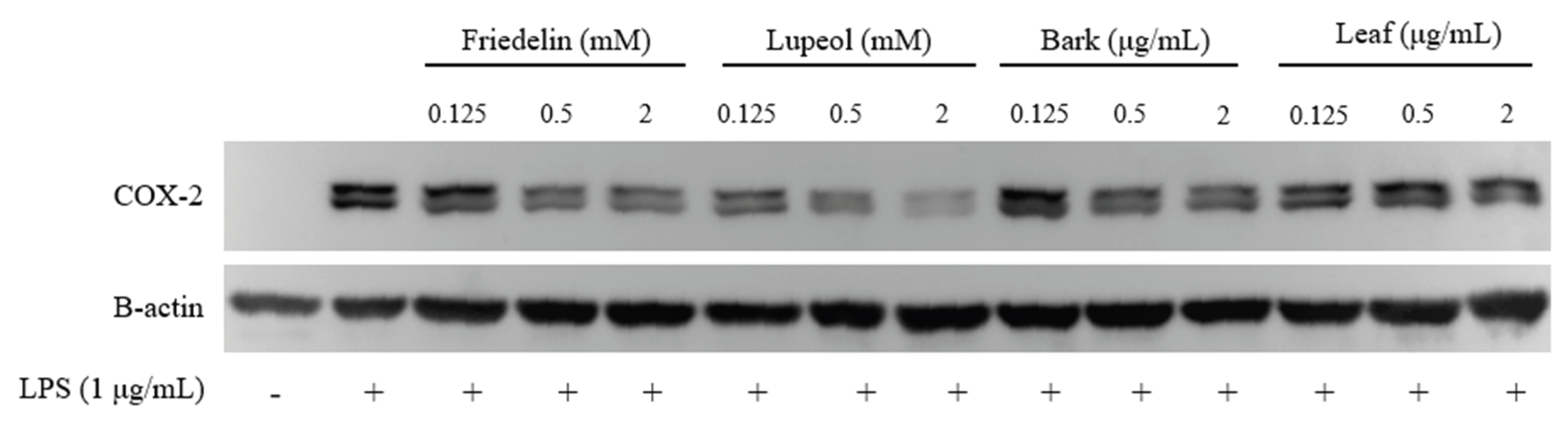
To evaluate the anti-inflammatory effects of friedelin, lupeol, and H. integrifolia extracts, COX-2 protein expression was examined in LPS-stimulated cells (Figure 9). As expected, LPS stimulation markedly increased COX-2 expression compared with the untreated control, confirming successful induction of an inflammatory response. Treatment with friedelin and lupeol resulted in a dose-dependent reduction of COX-2 protein levels, indicating suppression of inflammatory signaling. Similarly, treatment with bark and leaf extracts of H. integrifolia also reduced LPS-induced COX-2 expression, with higher concentrations producing stronger inhibitory effects. The reduction in COX-2 expression suggests that both the isolated triterpenoids and crude extracts possess anti-inflammatory activity. Overall, these results demonstrate that friedelin, lupeol, and H. integrifolia extracts attenuate LPS-induced COX-2 expression, supporting their potential role in modulating inflammatory responses associated with tissue injury and wound healing.
4. Discussion
Cutaneous wound healing requires the coordinated regulation of inflammatory responses, keratinocyte migration, and tissue remodeling. Keratinocytes are the principal epithelial cells of the epidermis and serve as the primary drivers of re-epithelialization during this process [13,14]. Upon injury, keratinocytes at the wound margin become activated and undergo a phenotypic transition characterized by cytoskeletal reorganization, detachment from the basement membrane, and directional migration across the provisional wound matrix [15]. This migratory response is mediated in part through interactions with fibrin–fibronectin clot components via specific integrin receptors, including α5β1 and αvβ6, enabling cell translocation into the wound bed [16]. Concurrently, keratinocytes upregulate the expression of pro-inflammatory cytokines and antimicrobial peptides, including IL-1α, IL-6, and IL-8, and engage in paracrine crosstalk with dermal fibroblasts and infiltrating macrophages through growth factors such as keratinocyte growth factor (KGF), thereby coordinating the broader wound repair response [17,18]. Re-epithelialization is typically initiated within 6–24 hours post-injury and proceeds through a coordinated sequence of migration, proliferation, and terminal differentiation to restore epidermal continuity [19]. Impairment of these processes, as observed in chronic wounds where elevated matrix metalloproteinase-9 (MMP-9) activity predominates, results in persistent epidermal defects and failure of wound closure [20].
To investigate the molecular mechanisms underlying the wound-healing activity of H. integrifolia, both canine primary keratinocytes (CPEK) and human immortalized keratinocytes (HaCaT) were employed as complementary experimental models. Although the Hippo signaling pathway is highly conserved across mammalian species, differences in antibody specificity, protein detectability, and signaling dynamics between species can influence the selection of appropriate molecular markers [21,22]. In the canine CPEK model, analysis was therefore focused on core components of the Hippo regulatory axis, specifically Mammalian sterile 20-like kinase 1 (MST1), Yes-associated protein (YAP), and Transcriptional co-activator with PDZ-binding motif (TAZ), to evaluate the upstream and downstream regulatory balance governing keratinocyte migration and epithelial repair [23,24]. In the human HaCaT model, analysis was extended to include phosphorylated ERK1/2 (p-ERK1/2) as a marker of MAPK pathway activation, given the well-established role of ERK signaling in promoting keratinocyte proliferation, cytoskeletal remodeling, and directed migration during re-epithelialization [25,26]. The complementary use of these two models therefore enabled a broader evaluation of the signaling mechanisms involved in keratinocyte-mediated wound repair while accounting for species-specific experimental considerations.
In the present study, extracts from H. integrifolia and the bioactive pentacyclic triterpenoids friedelin and lupeol demonstrated significant pro-migratory and anti-inflammatory activities in both keratinocyte models. The scratch wound assays revealed that both bark and leaf extracts significantly enhanced keratinocyte migration in canine CPEK and human HaCaT cell lines, with nearly complete wound closure observed within 48 h at relatively low concentrations, indicating strong pro-migratory activity. These findings are consistent with previous reports demonstrating that H. integrifolia extracts promote wound healing in keratinocyte models through increased MMP-9 production and modulation of cell adhesion molecule expression [8]. Similar pro-migratory effects were observed with the isolated triterpenoids friedelin and lupeol [27]. However, lupeol exhibited a biphasic response in which higher concentrations partially attenuated keratinocyte migration. This observation is consistent with prior in vitro studies demonstrating that lupeol at elevated concentrations reduces keratinocyte proliferation while continuing to enhance wound closure through p38 MAPK activation, suggesting concentration-dependent modulation of distinct signaling branches [28]. Such biphasic behavior may also reflect feedback regulation of pathway activity, a phenomenon previously documented for PI3K/Akt-dependent migratory responses [29]
Mechanistic investigation revealed that treatment with both plant extracts and isolated triterpenoids increased the expression of YAP and TAZ, the key transcriptional co-activators of the Hippo signaling pathway, while concurrently reducing expression of the upstream kinase MST1. Since MST1 functions as a negative regulator of YAP/TAZ through inhibitory phosphorylation of downstream LATS1/2 kinases, its downregulation likely relieves this suppressive control, facilitating nuclear accumulation of YAP/TAZ and activation of pro-migratory transcriptional programs [23,24]. This interpretation is consistent with established roles for YAP and TAZ in skin wound repair, where nuclear YAP activity is strongly induced in the thickened epithelial leading edge and wounded dermis following injury [30]. Furthermore, reduced YAP/TAZ signaling has been shown to impair epithelial repair, underscoring the critical importance of this pathway in re-epithelialization [31]. Reduced expression of E-cadherin in treated cells further supports a shift toward a migratory phenotype, consistent with the partial epithelial–mesenchymal transition that characterizes leading-edge keratinocytes during wound re-epithelialization [32]. Importantly, the activation of YAP/TAZ signaling was observed in both canine and human keratinocyte models, indicating that the pro-migratory effects of H. integrifolia extracts and triterpenoid compounds are conserved across species, supporting their potential relevance for translational wound-healing applications.
In addition to Hippo pathway modulation, activation of MAPK signaling was observed in HaCaT cells, as evidenced by increased ERK1/2 phosphorylation following treatment with extracts and triterpenoids. ERK signaling plays a well-established role in keratinocyte proliferation, migration, and MMP-1 expression during wound repair, and its abrogation causes profound defects in epidermal development and barrier function [25,26]. Notably, p38 MAPK has also been specifically implicated in collagen-driven keratinocyte migration and acts as a necessary mediator of directional cell movement across the wound bed [33]. The combined results from scratch wound assays and Western blot analyses therefore suggest that the pro-migratory activity of H. integrifolia extracts and their constituent triterpenoids involves simultaneous engagement of both ERK and p38 MAPK branches. Furthermore, crosstalk between ERK–AP-1 transcriptional complexes and YAP–TEAD transcriptional activity has been reported to synergistically enhance gene expression programs involved in tissue regeneration [34]. The simultaneous activation of Hippo and MAPK pathways observed in this study therefore provides a plausible mechanistic explanation for the enhanced keratinocyte migration detected in the wound-healing assays.
Inflammation represents another critical component of wound repair, as excessive or prolonged inflammatory signaling can impair keratinocyte migration and delay tissue regeneration [35,36]. In the present study, Western blot analysis demonstrated that friedelin, lupeol, and H. integrifolia extracts significantly suppressed LPS-induced COX-2 expression in keratinocytes. COX-2 is a key inducible enzyme responsible for prostaglandin synthesis and amplification of inflammatory signaling, and its sustained overexpression is associated with chronic wound environments characterized by impaired re-epithelialization [37,38]. Downregulation of COX-2 by these compounds is consistent with previously reported anti-inflammatory activities of friedelin, including inhibition of carrageenan-induced edema and suppression of pro-inflammatory cytokines such as TNF-α, IL-6, and IL-8 [39,40], as well as mechanistic evidence that friedelin inhibits the NF-κB pathway through promotion of p65 autophagic degradation [41]. The simultaneous suppression of inflammatory signaling and stimulation of regenerative Hippo and MAPK pathways suggests that these triterpenoids may act in concert to create a cellular microenvironment that is coordinately favorable for tissue repair.
Molecular docking simulations provided additional structural insights into the potential molecular targets of friedelin and lupeol. Both compounds exhibited favorable predicted binding affinities toward MST1, YAP, and ERK1/2, with docking scores generally ranging from approximately −6 to −8 kcal/mol. Notably, stronger predicted interactions were observed with MST1 and YAP compared with ERK1/2, suggesting preferential engagement with components of the Hippo signaling pathway. Ligand binding was predominantly stabilized by hydrophobic contacts and van der Waals interactions, consistent with the nonpolar pentacyclic triterpenoid scaffold characteristic of friedelin and lupeol [42,43]. These computational findings corroborate the experimental observations of MST1 downregulation and YAP/TAZ upregulation in treated keratinocytes, lending additional mechanistic support to the proposed mode of action.
Taken together, the experimental and computational findings support a mechanistic model in which H. integrifolia extracts and their constituent triterpenoids friedelin and lupeol promote cutaneous wound repair through the coordinated modulation of Hippo signaling, MAPK activation, and inflammatory regulation. Downregulation of MST1 and consequent activation of YAP/TAZ enhance transcriptional programs associated with keratinocyte migration and re-epithelialization, while ERK1/2 activation further amplifies proliferative and migratory responses. Concurrent suppression of COX-2-mediated inflammatory signaling reduces the pro-inflammatory burden within the wound microenvironment, collectively establishing conditions favorable for efficient tissue regeneration. These findings suggest that H. integrifolia and its bioactive triterpenoid constituents represent promising candidates for the development of plant-derived wound-healing therapeutics, with potential applicability across both veterinary and human clinical contexts.
5. Conclusion
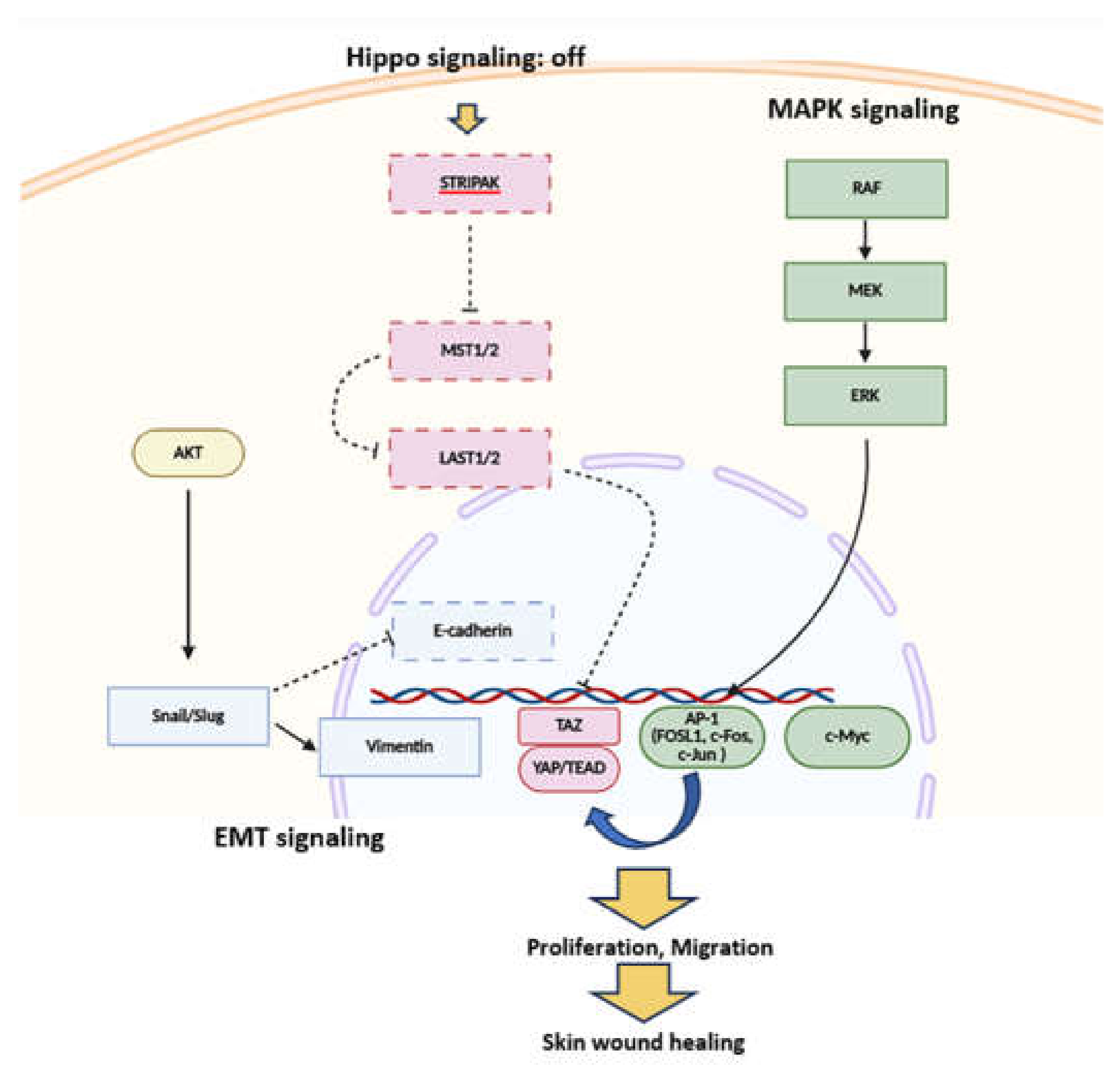
Based on the combined experimental and computational findings, a mechanistic model can be proposed to explain the wound-healing activity of the triterpenoid compounds friedelin and lupeol. The results suggest that these compounds modulate Hippo and MAPK signaling pathways, which are key regulators of keratinocyte proliferation and migration during skin repair. Docking analysis indicated that friedelin and lupeol exhibit favorable interactions with MST1 and YAP, suggesting that the triterpenoids may influence the Hippo signaling cascade. Reduced MST1 activity is associated with decreased phosphorylation of downstream kinases LATS1/2, leading to the activation and nuclear translocation of the transcriptional co-activators YAP and TAZ. Once in the nucleus, the YAP/TAZ–TEAD complex promotes transcription of genes involved in cell proliferation and migration. Simultaneously, activation of the MAPK pathway, particularly the RAF–MEK–ERK signaling axis, stimulates downstream transcription factors such as AP-1 (FOSL1, c-Fos, c-Jun) and c-Myc, further enhancing keratinocyte proliferation and motility. Crosstalk between Hippo signaling and MAPK signaling amplifies these responses. In addition, modulation of epithelial–mesenchymal transition (EMT)–related regulators, including Snail/Slug, vimentin, and E-cadherin, facilitates cytoskeletal remodeling and increased cellular motility. The coordinated activation of these pathways ultimately promotes keratinocyte proliferation, migration, and re-epithelialization, leading to accelerated skin wound healing. Collectively, these findings support a mechanism in which friedelin and lupeol enhance wound repair through coordinated regulation of Hippo signaling, MAPK signaling, and EMT-associated pathways, resulting in improved keratinocyte migration and tissue regeneration.
Figure 10.
Proposed mechanism illustrating the roles of Hippo, MAPK, and EMT pathways in keratinocyte-mediated wound healing. When Hippo signaling is active, the MST1/2–LATS1/2 kinase cascade phosphorylates YAP and TAZ, restricting their nuclear accumulation and consequently reducing TEAD-mediated transcription and proliferative activity. In response to epidermal damage, RAF–MEK–ERK signaling can engage AP-1 components, including FOSL1, c-Fos, and c-Jun, which may functionally interact with YAP–TEAD to regulate transcriptional programs that support tissue restoration. In parallel, PI3K–AKT signaling can influence EMT-associated regulators such as Snail/Slug, accompanied by reduced E-cadherin and increased vimentin expression. These molecular changes favor a more motile epithelial phenotype that can facilitate wound closure. The coordinated integration of these signaling pathways enhances keratinocyte proliferation, migration, survival, and EMT-associated remodeling, ultimately contributing to effective skin wound healing.
Acknowledgements
This research project was supported by Science and Technology Park, Chiang Mai University (STEP CMU), Innovation Management Office, Chiang Mai University (IMO, CMU), The Development and Promotion of Science and Technology Talents Project, the Thailand Science Research and Innovation (TSRI) Fundamental Fund fiscal year 2025 and the Research Institute for Veterinary Science and BK21 PLUS Program for Creative Veterinary Science Research Center, Seoul National University.
Declaration of Interest Statement
The authors declare no conflicts of interest. We acknowledge the use of ChatGPT (OpenAI) to improve the readability and language of this manuscript. The authors reviewed and edited the generated content, and take full responsibility for the final text.
References
- Dey, A.; Varelas, X.; Guan, K.L. Targeting the Hippo pathway in cancer, fibrosis, wound healing and regenerative medicine. Nat Rev Drug Discov. 2020, 19(7), 480-494. [CrossRef]
- Jia, J.; Li, C.; Yang, J.; Wang, X.; Li, R.; Luo, S.; Li, Z.; Liu, J.; Liu, Z.; Zheng, Y. Yes-Associated Protein Promotes the Abnormal Proliferation of Psoriatic Keratinocytes via an Amphiregulin-Dependent Pathway. Sci. Rep. 2018, 8, 14513. [CrossRef]
- Jin, L.; Zhou, S.; Zhao, S.; Long, J.; Huang, Z.; Zhou, J.; Zhang, Y. Early Short-Term Hypoxia Promotes Epidermal Cell Migration by Activating the CCL2–ERK1/2 Pathway during Wound Healing. Burns Trauma 2024, 12, tkae017. [CrossRef]
- Lamouille, S.; Xu, J.; Derynck, R. Molecular Mechanisms of Epithelial–Mesenchymal Transition. Nat. Rev. Mol. Cell Biol. 2014, 15, 178-196. [CrossRef]
- Zhou, H.; Zhang, S.; Jin, X.; Gong, P.; Zhao, S. The Electric Field Guided HaCaT Cell Migration Through the EGFR/p38 MAPK/Akt Pathway. Curr. Issues Mol. Biol. 2024, 47(1), 16. [CrossRef]
- Kim, J.Y.; Quan, T. Emerging Perspectives of YAP/TAZ in Human Skin Epidermal and Dermal Aging. Ann. Dermatol. 2024, 36(3), 135-144. [CrossRef]
- Cooper, F.; Overmiller, A.M.; Loder, A.; Brennan-Crispi, D.M.; McGuinn, K.P.; Marous, M.R.; Freeman, T.A.; Riobo-Del Galdo, N.A.; Siracusa, L.D.; Wahl, J.K., III. Enhancement of Cutaneous Wound Healing by Dsg2 Augmentation of uPAR Secretion. J. Invest. Dermatol. 2018, 138(11), 2470–2479. [CrossRef]
- Somwong, K.; Lertpatipanpong, P.; Nimlamool, W.; Panya, A.; Tragoolpua, Y.; Yongsawas, R.; Gritsanapan, W.; Pandith, H.; Baek, S.J. Effect of Holoptelea integrifolia (Roxb.) Planch. n-Hexane Extract and Its Bioactive Compounds on Wound Healing and Anti-Inflammatory Activity. Molecules 2022, 27(23), 8540. [CrossRef]
- Ganie, S.A.; Yadav, S.S. Holoptelea integrifolia (Roxb.) Planch: A Review of Its Ethnobotany, Pharmacology, and Phytochemistry. BioMed Res. Int. 2014, 401213. [CrossRef]
- Kumar, S.; Verma, S.; Verma, P. HPTLC Analysis of Methanolic Extract of Holoptelea integrifolia (Roxb.) Planch Leaves. Res. J. Pharm. Technol. 2024, 17(11), 5247–5253. [CrossRef]
- Kim, M.; Kim, J.G.; Kim, K.Y. Trichosanthes kirilowii Extract Promotes Wound Healing through Phosphorylation of ERK1/2 in Keratinocytes. Biomimetics (Basel) 2022, 7(4), 154. [CrossRef]
- Reddy, B.S.; Reddy, R.K.K.; Naidu, V.G.M.; Madhusudhana, K.; Agwane, S.B.; Ramakrishna, S.; Diwan, P.V. Evaluation of antimicrobial, antioxidant and wound-healing potentials of Holoptelea integrifolia. Journal of Ethnopharmacology 2008, 115(2), 249-256. [CrossRef]
- Eming, S.A.; Martin, P.; Tomic-Canic, M. Wound repair and regeneration: Mechanisms, signaling, and translation. Sci Transl Med. 2014, 6(265), 265sr6. [CrossRef]
- Pastar, I.; Stojadinovic, O.; Yin, N.C.; Ramirez, H.; Nusbaum, A.G.; Sawaya, A.; Patel, S.B.; Khalid, L.; Isseroff, R.R.; Tomic-Canic, M. Epithelialization in wound healing: A comprehensive review. Adv Wound Care. 2014, 3(7), 445–464. [CrossRef]
- Gurtner, G.C.; Werner, S.; Barrandon, Y.; Longaker, M.T. Wound repair and regeneration. Nature 2008, 453(7193), 314–321. [CrossRef]
- Larjava, H.; Salo, T.; Haapasalmi, K.; Kramer, R.H.; Heino, J. Expression of integrins and basement membrane components by wound keratinocytes. J Clin Invest. 1993, 92(3), 1425–1435. [CrossRef]
- Werner, S.; Grose, R. Regulation of wound healing by growth factors and cytokines. Physiol Rev. 2003, 83(3), 835–870. [CrossRef]
- Barrientos, S.; Stojadinovic, O.; Golinko, M.S.; Brem, H.; Tomic-Canic, M. Growth factors and cytokines in wound healing. Wound Repair Regen. 2008, 16(5), 585–601. [CrossRef] [PubMed]
- Reinke, J.M.; Sorg, H. Wound repair and regeneration. Eur Surg Res. 2012, 49(1), 35–43. [CrossRef]
- Trengove, N.J.; Stacey, M.C.; MacAuley, S.; Bennett, N.; Gibson, J.; Burslem, F.; Murphy, G.; Schultz, G. Analysis of the acute and chronic wound environments: The role of proteases and their inhibitors. Wound Repair Regen. 1999, 7(6), 442–452. [CrossRef]
- Rinn, J.L.; Wang, J.K.; Liu, H.; Montgomery, K.; van de Rijn, M.; Chang, H.Y. A systems biology approach to anatomic diversity of skin. J Invest Dermatol. 2008, 128(4), 776-782. [CrossRef]
- Watt, F.M.; Fujiwara, H. Cell–extracellular matrix interactions in normal and diseased skin. Cold Spring Harb Perspect Biol. 2011, 3(4), a005124. [CrossRef]
- Pocaterra, A.; Romani, P.; Dupont, S. YAP/TAZ functions and their regulation at a glance. J Cell Sci. 2020, 133(2), jcs230425. [CrossRef]
- Hong, W.; Guan, K.L. The YAP and TAZ transcription co-activators: Key downstream effectors of the mammalian Hippo pathway. Semin Cell Dev Biol. 2012, 23(7), 785–793. [CrossRef]
- Roux, P.P.; Blenis, J. ERK and p38 MAPK-activated protein kinases: A family of protein kinases with diverse biological functions. Microbiol Mol Biol Rev. 2004, 68(2), 320–344. [CrossRef]
- Scholl, F.A.; Dumesic, P.A.; Bhatt, D.L.; Khavari, P.A. Mek1/2 gene dosage determines tissue response to oncogenic Ras signaling in the skin. Oncogene 2009, 28(11), 1485–1495. [CrossRef]
- Saleem, M. Lupeol, a novel anti-inflammatory and anti-cancer dietary triterpene. Cancer Lett. 2009, 285(2), 109-15. [CrossRef]
- Pereira Beserra, F.; Xue, M.; Maia, G.L.A.; Leite Rozza, A.; Helena Pellizzon, C.; Jackson, C.J. Lupeol, a Pentacyclic Triterpene, Promotes Migration, Wound Closure, and Contractile Effect In Vitro: Possible Involvement of PI3K/Akt and p38/ERK/MAPK Pathways. Molecules 2018, 23(11), 2819. [CrossRef]
- Rodrik-Outmezguine, V.S.; Chandarlapaty, S.; Pagano, N.C.; Poulikakos, P.I.; Scaltriti, M.; Moskatel, E.; Baselga, J.; Guichard, S.; Rosen, N. mTOR kinase inhibition causes feedback-dependent biphasic regulation of AKT signaling. Cancer Discov. 2011, 1(3), 248-59. [CrossRef]
- Lee, M.J.; Byun, M.R.; Furutani-Seiki, M.; Hong, J.H.; Jung, H.S. YAP and TAZ regulate skin wound healing. J Invest Dermatol. 2014, 134(2), 518–525. [CrossRef]
- Pankratova, M.D.; Riabinin, A.A.; Butova, E.A.; Selivanovskiy, A.V.; Morgun, E.I.; Ulianov, S.V.; Vorotelyak, E.A.; Kalabusheva, E.P. YAP/TAZ Signalling Controls Epidermal Keratinocyte Fate. Int J Mol Sci. 2024, 25(23), 12903. [CrossRef]
- Savagner, P.; Kusewitt, D.F.; Carver, E.A.; Magnino, F.; Choi, C.; Gridley, T.; Hudson, L.G. Developmental transcription factor slug is required for effective re-epithelialization by adult keratinocytes. J Cell Physiol. 2005, 202(3), 858-66. [CrossRef]
- Nadelman, C.; Henry, G.; Fan, J.; Muellenhoff, M.; Medina, E.; Gratch, N.S.; Chen, M.; Han, J.; Woodley, D. The p38-MAPK/SAPK pathway is required for human keratinocyte migration on dermal collagen. J Invest Dermatol. 2001, 117(6), 1601-11. [CrossRef]
- Zanconato, F.; Forcato, M.; Battilana, G.; Azzolin, L.; Quaranta, E.; Bodega, B.; Rosato, A.; Bicciato, S.; Cordenonsi, M.; Piccolo, S. Genome-wide association between YAP/TAZ/TEAD and AP-1 at enhancers drives oncogenic growth. Nat Cell Biol. 2015, 17(9), 1218-27. [CrossRef]
- Eming, S.A.; Krieg, T.; Davidson, J.M. Inflammation in wound repair: Molecular and cellular mechanisms. J Invest Dermatol. 2007, 127(3), 514–525. [CrossRef]
- Wilgus, T.A. Immune cells in the healing skin wound: Influential players at each stage of repair. Pharmacol Res. 2008, 58(2), 112–116. [CrossRef]
- Seibert, K.; Zhang, Y.; Leahy, K.; Hauser, S.; Masferrer, J.; Perkins, W.; Lee, L.; Isakson, P. Pharmacological and biochemical demonstration of the role of cyclooxygenase 2 in inflammation and pain. Proc Natl Acad Sci USA. 1994, 91(25), 12013–12017. [CrossRef]
- Futagami, A.; Ishizaki, M.; Fukuda, Y.; Kawana, S.; Yamanaka, N. Wound healing involves induction of cyclooxygenase-2 expression in rat skin. Lab Invest. 2002, 82(11), 1503–1510. [CrossRef] [PubMed]
- Antonisamy, P.; Duraipandiyan, V.; Ignacimuthu, S. Anti-inflammatory, analgesic and antipyretic effects of friedelin isolated from Azima tetracantha Lam. in mouse and rat models. J Pharm Pharmacol. 2011, 63(8), 1070-7. [CrossRef]
- Ferrini, F.; Donati Zeppa, S.; Fraternale, D.; Carrabs, V.; Annibalini, G.; Verardo, G.; Gorassini, A.; Albertini, M. C.; Ismail, T.; Fimognari, C.; Sestili, P. Characterization of the biological activity of the ethanolic extract from the roots of Cannabis sativa L. grown in aeroponics. Antioxidants 2022, 11(5), 860. [CrossRef]
- Jiang, H.; Lin, X.; Liang, W.; Li, Y.; Yu, X. Friedelin alleviates the pathogenesis of collagenase-induced tendinopathy in mice by promoting the selective autophagic degradation of p65. Nutrients 2022, 14(8), 1673. [CrossRef]
- Lionta, E.; Spyrou, G.; Vassilatis, D.K.; Cournia, Z. Structure-based virtual screening for drug discovery: Principles, applications and recent advances. Curr Top Med Chem. 2014, 14(16), 1923–1938. [CrossRef]
- Daina, A.; Michielin, O.; Zoete, V. SwissTargetPrediction: Updated data and new features for efficient prediction of protein targets of small molecules. Nucleic Acids Res. 2019, 47(W1), W357–W364. [CrossRef]
Figure 1.
Gas chromatography–flame ionization detection (GC-FID) analysis of friedelin and lupeol in Holoptelea integrifolia (Roxb.) Planch. bark and leaf extracts. (A) Schematic overview of the extraction and GC analysis workflow from bark and leaf plant materials. (B) Representative GC-FID chromatograms of bark extract (left) and leaf extract (right), with labeled peaks corresponding to lupeol and friedelin identified by retention time matching. (C) External calibration curves for friedelin (tᵣ = 104 min; y = 0.7887x + 43.3140, R² = 0.9986) and lupeol (tᵣ = 97 min; y = 1.4569x − 2.8041, R² = 0.9999), demonstrating linearity over the validated concentration ranges. (D) GC-FID chromatograms of authentic reference standards of friedelin and lupeol with corresponding chemical structures, confirming compound identity by retention time under identical chromatographic conditions.
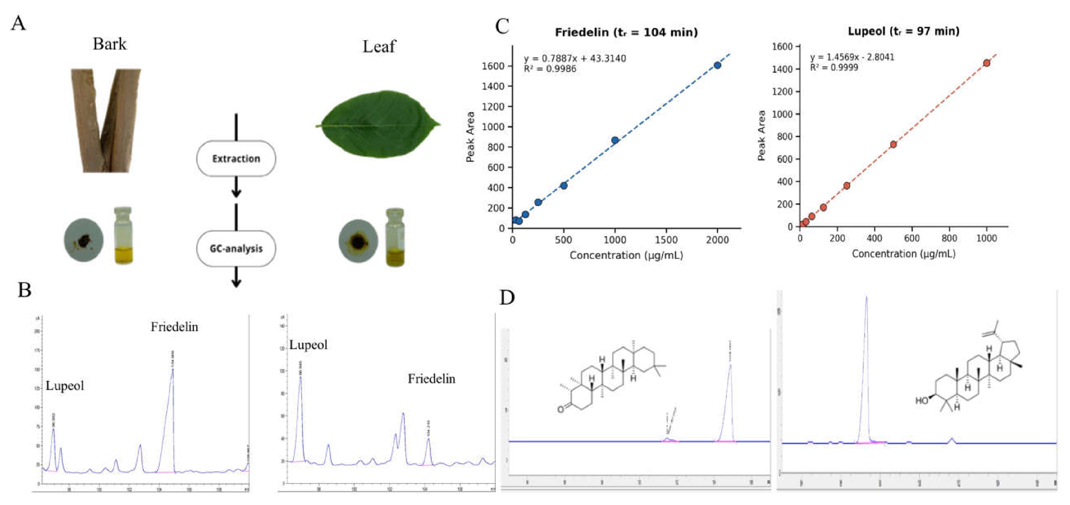
Figure 2.
Effects of H. integrifolia (Roxb.) Planch. bark and leaf extracts on wound closure in CPEK and HaCaT cells. Confluent monolayers were mechanically scratched to create a cell-free gap and subsequently exposed to bark or leaf extracts at 0.125, 0.25, and 0.5 µg/mL. Representative phase-contrast images of (A) CPEK cells and (B) HaCaT cells illustrate wound closure at 0, 24, and 48 h post-wounding. Red dashed lines demarcate the wound edges at each time point. (C) Bar graphs represent the percentage wound healing area of CPEK cells (left) and HaCaT cells (right) at 24 and 48 h, normalized to the initial wound area at 0 h. Values represent the mean ± SEM of three independent experiments. Differences from the DMSO vehicle control were considered significant at *P < 0.05 and **P < 0.001.
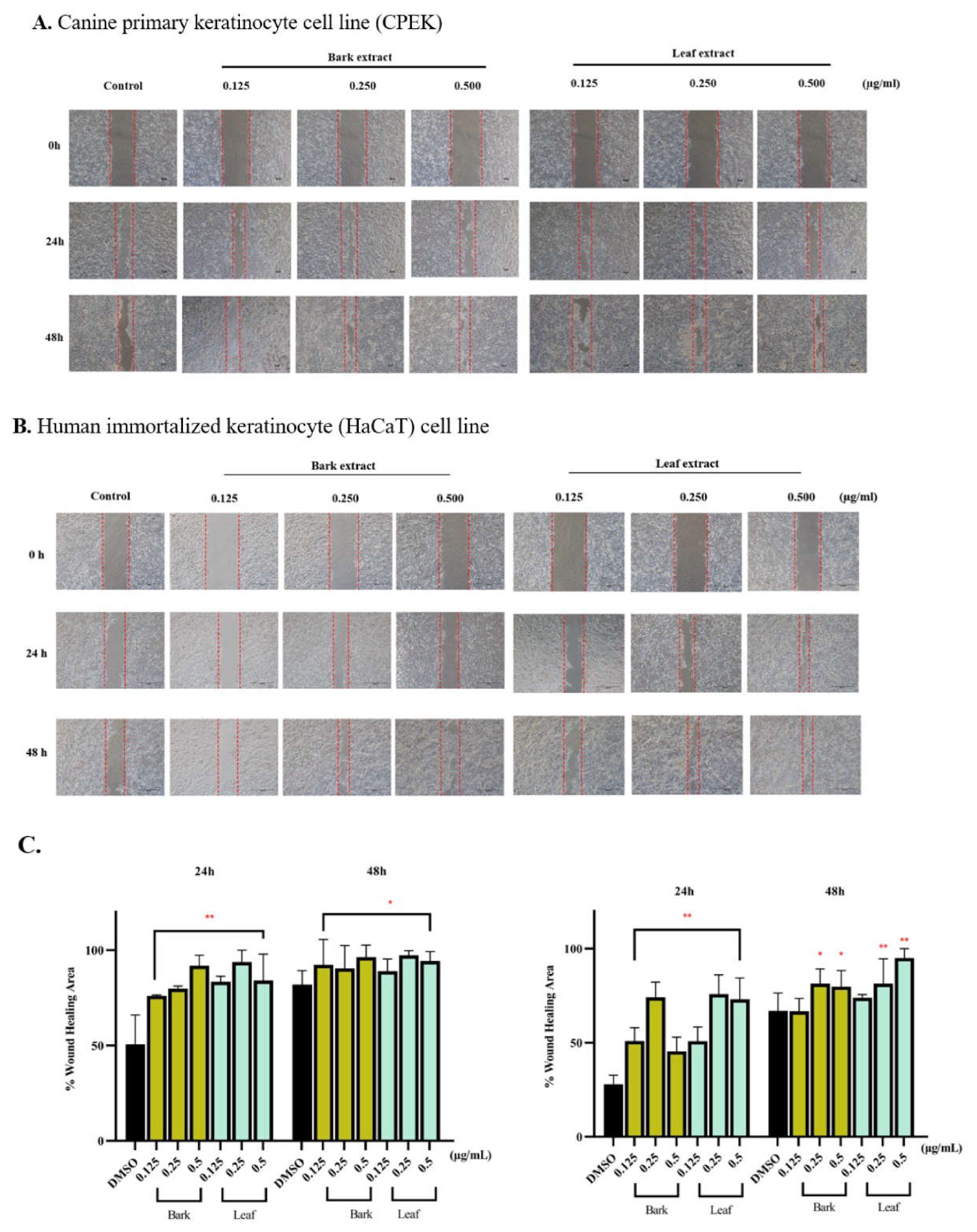
Figure 3.
Effects of friedelin and lupeol on wound closure in CPEK (A) and HaCaT (B) keratinocytes. Confluent cell monolayers were subjected to a linear scratch wound and treated with friedelin and lupeol at concentrations of 0.125 and 0.25 µM. Representative phase-contrast images of CPEK cells (A) and HaCaT cells (B) illustrate wound closure at 0, 24, and 48 h post-wounding. Red dashed lines demarcate the wound edges at each time point. Bar graphs represent the percentage wound healing area of CPEK cells and HaCaT cells at 24 and 48 h, normalized to the initial wound area at 0 h. Values represent mean ± SEM obtained from three independent experiments. Comparisons with the DMSO vehicle group are denoted as *P < 0.05 and **P < 0.001.
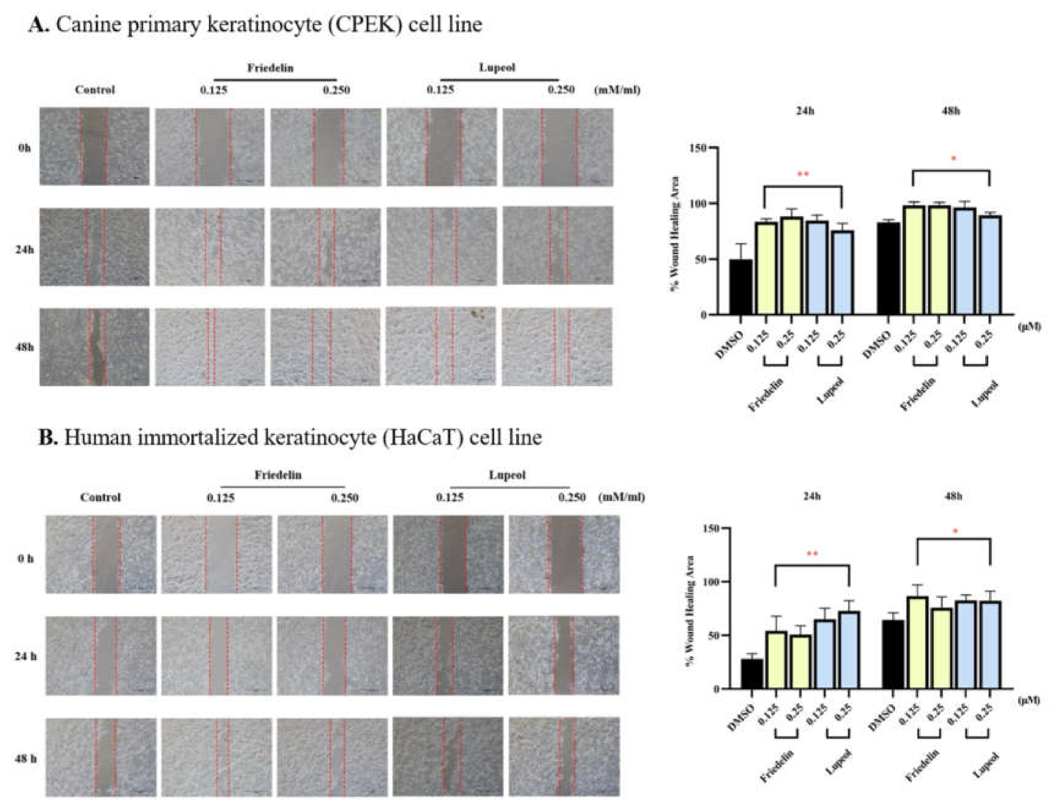
Figure 4.
Hippo signaling markers in CPEK cells treated with H. integrifolia (Roxb.) extracts and triterpenoid compounds. (A) Western blot analysis of TAZ, YAP, and E-cadherin in CPEK cells treated with bark and leaf extracts of H. integrifolia at 0.125, 0.25, and 0.5 µg/mL. β-Actin was used as the internal loading control. Bar graphs represent densitometric quantification of protein levels normalized to β-actin and expressed relative to the DMSO-treated control. (B) Effects of friedelin and lupeol on TAZ, YAP, and E-cadherin protein levels in CPEK cells. Cells were exposed to either triterpenoid at 0.125 or 0.25 µM, and protein abundance was assessed by Western blotting. Densitometric values were normalized to β-actin and expressed relative to the DMSO vehicle control. Results are shown as mean ± SEM from three independent experiments; *P < 0.05 and **P < 0.001 versus the DMSO-treated group.
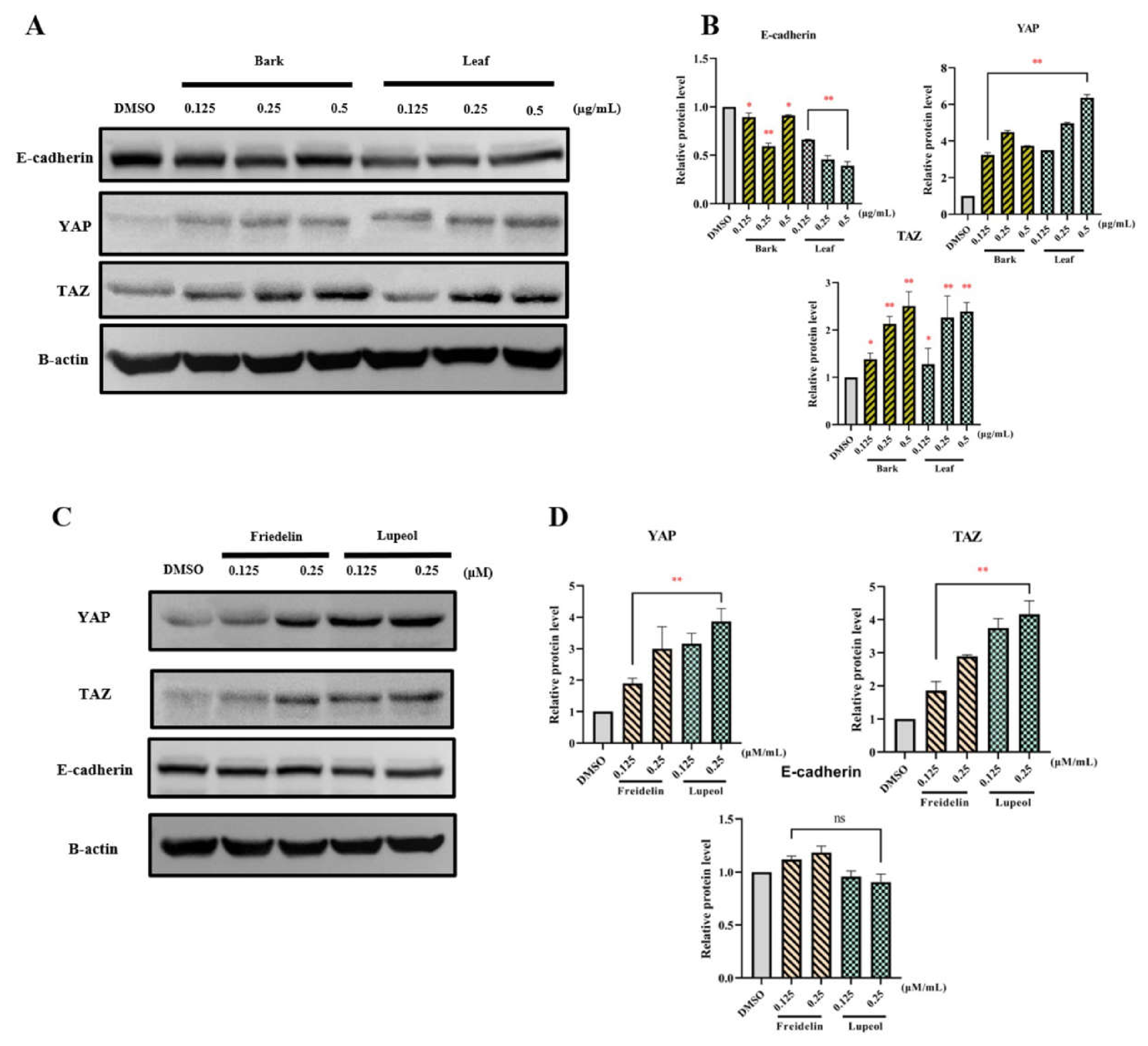
Figure 5.
Effects of H. integrifolia extracts and triterpenoids on MST1 protein levels in CPEK cells. (A) Cells were exposed to bark or leaf extracts at 0.125, 0.25, and 0.5 µg/mL, followed by Western blot assessment of MST1. (B) MST1 levels were examined following treatment with friedelin or lupeol at 0.125 and 0.25 µM. β-Actin served as the loading reference for densitometric analysis, and MST1 abundance was expressed relative to the DMSO vehicle control. Values represent mean ± SEM from three independent experiments; *P < 0.05 and **P < 0.001 versus the DMSO-treated group.
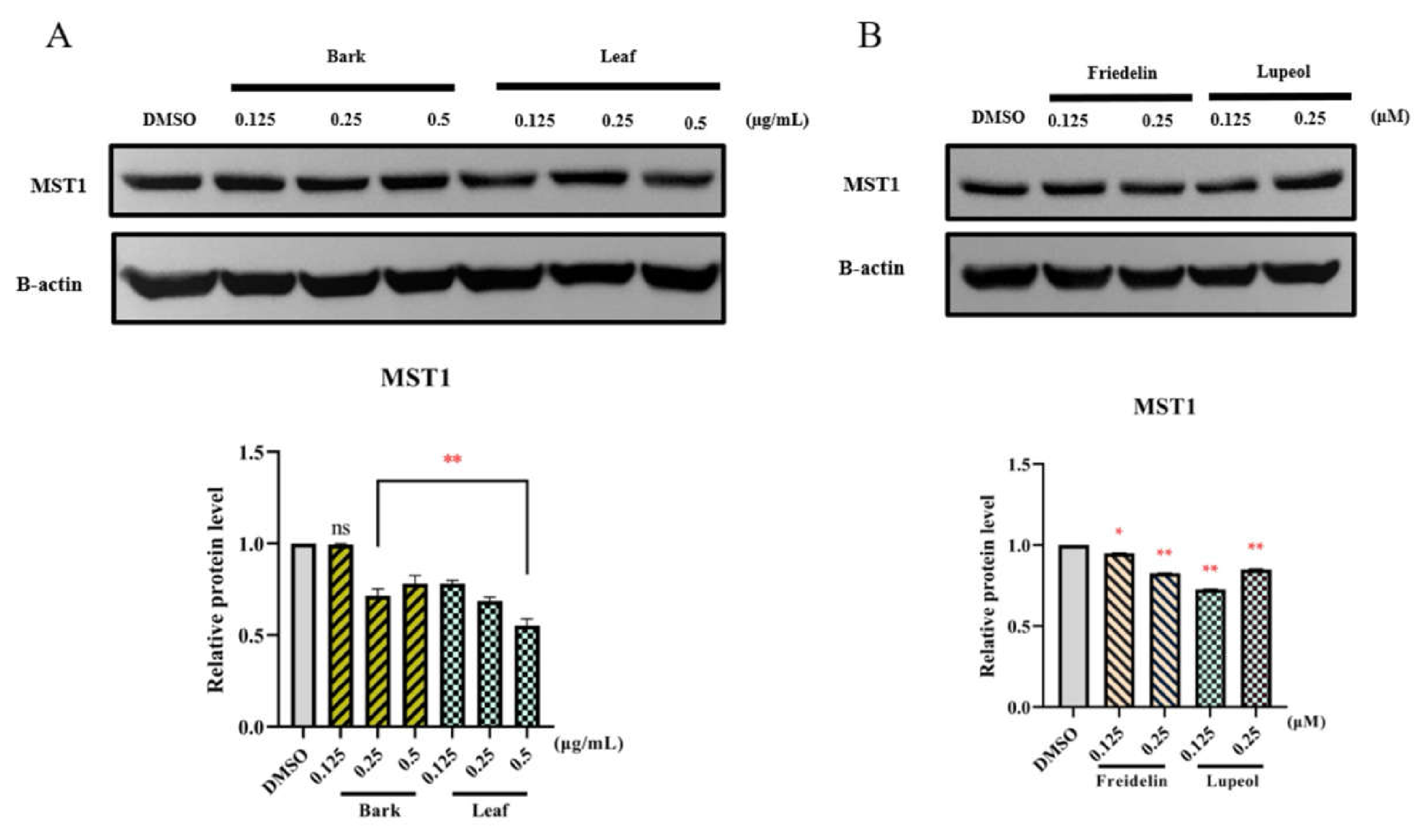
Figure 6.
Effects of H. integrifolia extracts and triterpenoids on Hippo signaling-related proteins in HaCaT cells. (A) TAZ, YAP, and E-cadherin protein levels were assessed after exposure to bark or leaf extracts at 0.125, 0.25, and 0.5 µg/mL. (B) The same proteins were examined following treatment with friedelin or lupeol at 0.125 and 0.25 µM. Protein abundance was determined by Western blotting, with β-actin serving as the loading reference for densitometric normalization. Relative expression was calculated against the DMSO vehicle control. Values represent mean ± SEM; *P < 0.05 and **P < 0.001 versus DMSO-treated cells.
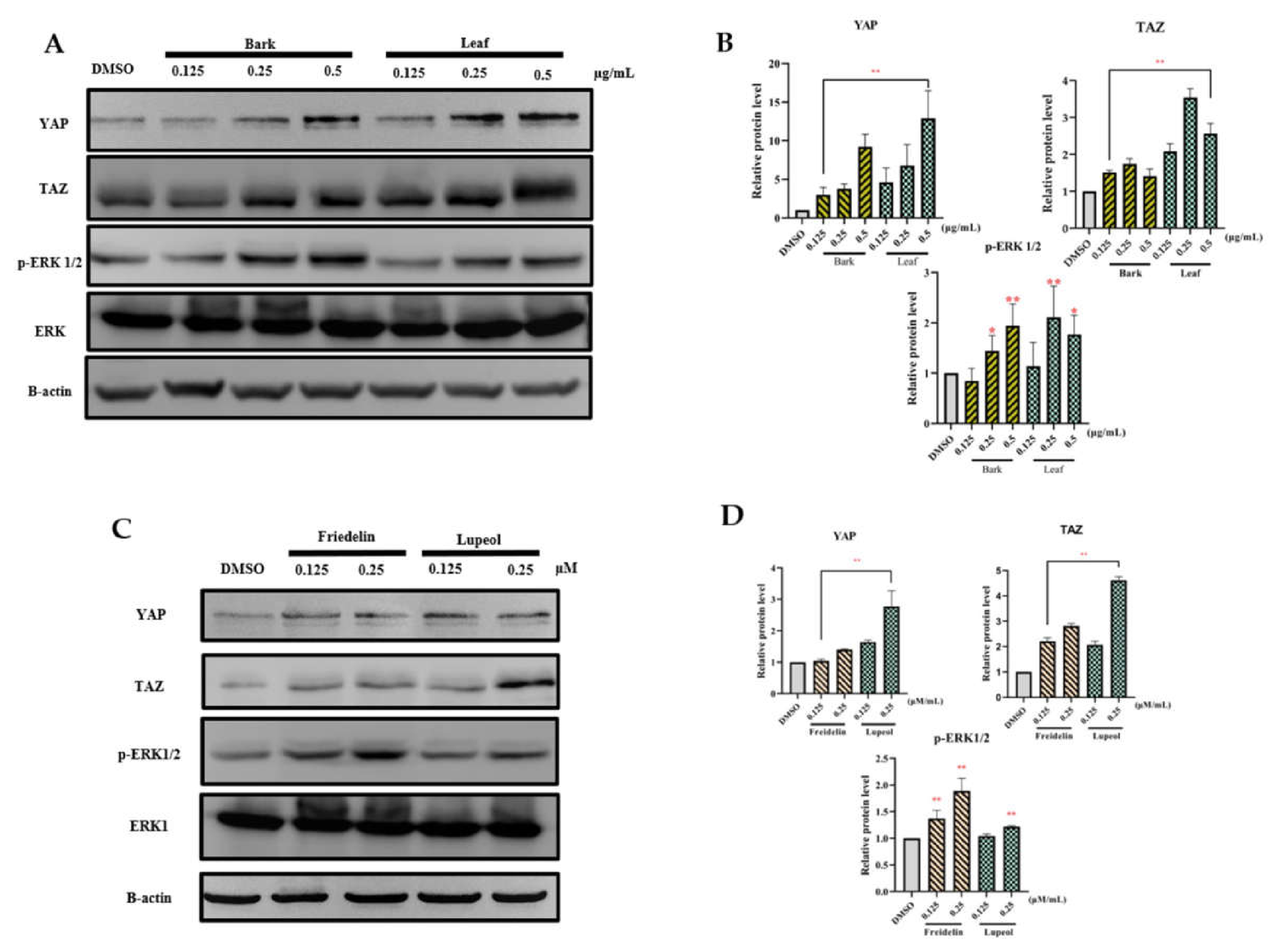
Figure 7.
Docking-derived binding affinities of friedelin and lupeol for proteins associated with Hippo and MAPK signaling. Friedelin and lupeol were docked against ERK (PDB ID: 1MKP), MST1 (PDB ID: 6YAT), and YAP (PDB ID: 3KYS), with the corresponding co-crystallized ligand of each target used as a reference. Predicted binding energies are reported in kcal/mol, with lower (more negative) values reflecting greater predicted binding affinity. Results represent the mean ± SD from repeated docking runs.
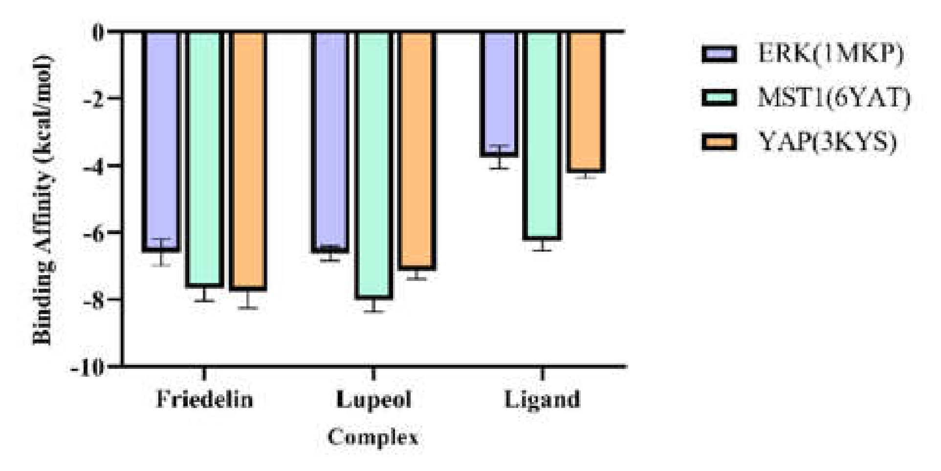
Figure 8.
Two-dimensional binding profiles of friedelin and lupeol with YAP (PDB ID: 3KYS), ERK (PDB ID: 1MKP), and MST1 (PDB ID: 6YAT). The diagrams depict the predicted orientation of each triterpenoid within the binding pocket and its contacts with surrounding amino acid residues. Interaction types are distinguished by color: light green circles denote van der Waals contacts; bright green dashed lines, conventional hydrogen bonds; cyan/blue dashed lines, water-mediated hydrogen bonds; pale green dashed lines, carbon–hydrogen bonds; purple dashed lines, π–sigma contacts; orange dashed lines, π–cation or π–sulfur contacts; pink dashed lines, π–alkyl contacts; and red dashed lines or highlighted regions, unfavorable steric contacts. These profiles provide a residue-level view of the predicted interactions of friedelin and lupeol with selected components of the Hippo and MAPK signaling pathways.
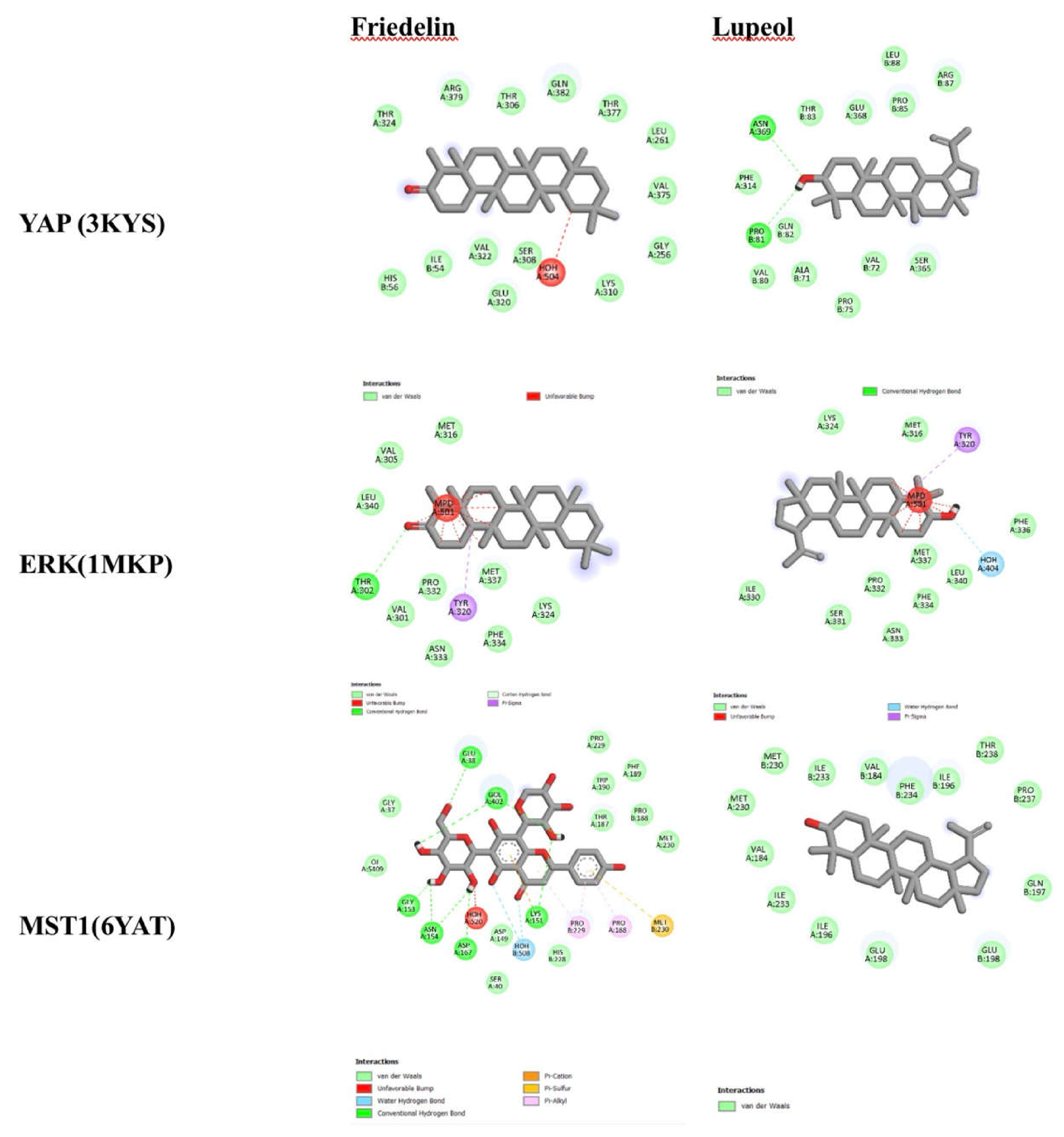
Figure 9.
Representative Western blot analysis of COX-2 protein expression in LPS-stimulated cells following treatment with friedelin, lupeol, and H. integrifolia bark and leaf extracts. Cells were pretreated with friedelin or lupeol (0.125, 0.5, and 2 mM) or H. integrifolia bark and leaf extracts (0.125, 0.5, and 2 μg/mL) prior to stimulation with LPS (1 μg/mL). COX-2 protein expression was analyzed by Western blotting, with β-actin used as the internal loading control. Representative immunoblots from three independent experiments are shown. LPS stimulation markedly increased COX-2 expression compared with the untreated control, whereas treatment with friedelin, lupeol, and H. integrifolia extracts reduced COX-2 expression in a concentration-dependent manner, indicating suppression of the inflammatory response.
Disclaimer/Publisher’s Note: The statements, opinions and data contained in all publications are solely those of the individual author(s) and contributor(s) and not of MDPI and/or the editor(s). MDPI and/or the editor(s) disclaim responsibility for any injury to people or property resulting from any ideas, methods, instructions or products referred to in the content. |
© 2026 by the authors. Licensee MDPI, Basel, Switzerland. This article is an open access article distributed under the terms and conditions of the Creative Commons Attribution (CC BY) license (http://creativecommons.org/licenses/by/4.0/).
Copyright: This open access article is published under a Creative Commons CC BY 4.0 license, which permit the free download, distribution, and reuse, provided that the author and preprint are cited in any reuse.