

Submitted:
26 September 2026
Posted:
29 September 2026
You are already at the latest version
Abstract
Abstract
Humans are continuously exposed to lysosomotropic xenobiotics, including cationic amphiphilic drugs, per- and polyfluoroalkyl substances (PFAS), redox-active heavy metals, and airborne particulates, through occupational, dietary, and environmental routes. Lysosomes are acidic organelles central to cellular degradation, nutrient sensing, and regulated cell death, and the stability of the lysosomal membrane is essential for cell viability, yet it is a preferential target for accumulation of these agents. This review synthesizes current understanding of the molecular mechanisms driving lysosomal membrane permeabilization (LMP) and lysosomal membrane rupture (LMR) following xenobiotic exposure, and their downstream consequences for human tissue health. We review lysosomal injury, namely intralysosomal ion trapping, metal‑catalyzed Fenton chemistry, and lipid peroxidation, which converges to generate oxidative membrane pores linked to respiratory, hepatic, renal, and neurodegenerative toxicity in exposed populations. We also examine cellular membrane‑repair systems, particularly the Endosomal Sorting Complex Required for Transport (ESCRT)-III machinery and ER–lysosome lipid transfer pathways, that act to restore lysosomal integrity and limit tissue injury. Failure of these protective mechanisms initiates distinct regulated cell death programs, including apoptosis, ferroptosis, and pyroptosis, that underlie chronic disease following sustained environmental exposure. Finally, we discuss the dual role of LMP in human health: both as a mediator of environmental toxicant‑induced injury and, more briefly, as an exploitable vulnerability in disease contexts such as drug-resistant cancer. By integrating findings from emerging non‑mammalian sentinel models and advanced imaging modalities, this review provides a framework for understanding how environmental lysosomal membrane dynamics translate into human health outcomes.
Keywords:
lysosome
; lysosomal membrane permeabilization (LMP)
; environmental xenobiotics
; PFAS
; heavy metals
; particulate matter
; Fenton chemistry
; ESCRT repair
; regulated cell death
; toxicant-induced injury
1. Introduction
Lysosomes are organelles that contain over 60 hydrolases capable of digesting proteins, nucleic acids, carbohydrates, and lipids. These enzymes are active in an acidic milieu, pH 4.5 - 5.0. This acidic environment is preserved by the activity of the vacuolar ATPase (v-ATPase) and counter-ion transport systems, including Na+/K+ leakage and Cl- import. [1,2].
Lysosomal digestion must take place within the lysosomal lumen to avoid degradation of the entire cell. As a result, the lysosomal membrane has a dualism of function. It provides structural support against mechanical stress and isolation of the lysosomal hydrolases and sequestered xenobiotics from the cytosol. Subtle alterations of this barrier can disrupt luminal pH, impair autophagic flux, and trigger signaling pathways resulting in adaptive repair or cell death [3].
Human exposure to lysosomotropic xenobiotics is neither rare nor confined to occupational settings. Population biomonitoring detects PFAS in nearly all serum samples tested, with US adult geometric mean concentrations of 9.72 ng/mL for PFOS and 3.07 ng/mL for PFOA [4], prompting the US Environmental Protection Agency (EPA) to set enforceable drinking-water limits of 4.0 parts per trillion for PFOA and PFOS and 10 parts per trillion for PFHxS, PFNA, and HFPO-DA in 2024 [5]. Redox-active metals face similarly stringent limits, with an EPA drinking-water maximum contaminant level of 0.005 mg/L for cadmium and an action level of 0.015 mg/L for lead [6], reflecting long-standing concern over their capacity to accumulate in tissue. Airborne particulate exposure is regulated even more broadly: the EPA tightened its annual fine particulate matter (PM2.5) standard from 12.0 to 9.0 μg/m3 in 2024 in direct response to evidence of harm at previously permitted levels [7]. These regulatory thresholds are set primarily from epidemiological and whole-organism toxicity data; the subcellular, lysosome-centered mechanisms reviewed here provide a complementary, mechanistic basis for understanding why exposures below acute toxicity thresholds can nonetheless produce cumulative tissue injury.
Lysosomes have been shown to be uniquely vulnerable to a variety of mechanical, oxidative, and metabolic stressors. Several reports demonstrate that environmental toxins, cationic amphiphilic drugs, heavy metals, nanoparticles, and organic pollutants preferentially accumulate within lysosomes. These agents can induce lysosomal membrane permeabilization (LMP), characterized by pore formation and cytosolic leakage, or lysosomal membrane rupture (LMR), defined by bilayer failure and uncontrolled enzyme release [3,8]. LMP and LMR are mechanistically and functionally distinct. Partial and transient LMP activates the intrinsic apoptotic pathway initiated by cathepsin release, whereas extensive membrane disruption results in necrosis, inflammation, or ferroptosis.[8,9].
Therefore, the severity and speed of lysosomal membrane damage ultimately
decide the cell's fate. Lysosomes are the primary sites of sequestration for
xenobiotics. The major routes by which xenobiotics accumulate within these organelles and initiate intralysosomal stress are summarized in Figure 1.
This makes lysosomes attractive targets for developing novel therapeutic strategies that exploit lysosomal vulnerabilities, particularly in cancer cells that expanded lysosomal compartments for proliferation and drug resistance [10]. Lysosomal damage does not always result in cell death. Cells possess membrane repair and regeneration networks that restore lysosomal integrity following injury. These pathways include ESCRT-III-mediated membrane sealing, lipid reinforcement, and organelle regeneration [11]. When these protective mechanisms fail, lysosomes are transformed from stress responders to inducers of cell death. This review examines how environmental xenobiotics disrupt lysosomal membranes, the processes that counteract this damage, and the consequences when repair fails.
2. Classes of Lysosomotropic Xenobiotics
Lysosomes are preferentially targeted by chemicals for two reasons, namely thermodynamics and compartment biology. The intralysosomal pH gradient drives protonation and trapping of lipophilic, weak bases that diffuse across the lysosomal membrane in its neutral form. The resulting ion-trapped species, which cannot freely exit, can cause concentrations within the lysosomal lumen to exceed cytosolic levels by two to three times [12]. Compounding this chemical sequestration is the lysosome’s role as the terminal destination for autophagic substances, including metalloprotein complexes and lipid droplets, which delivers additional redox-reactive substrates directly into the lumen.
Lysosomotropic xenobiotics fall into three operationally useful categories: weak-base cationic amphiphiles, redox-active and non-redox metals, and insoluble particulates. They differ considerably in structure but share the capacity to exploit lysosomal chemistry. Cationic amphiphilic drugs accumulate through classical ion trapping and disrupt lipid packing; redox-active metals, such as iron and copper, drive Fenton-type hydroxyl radical generation within the acidic lumen; and particulates impose sustained mechanical and oxidative stress that lysosomal hydrolases cannot resolve.
The lysosomotropic behavior of cationic amphiphilic drugs (CADs) is primarily based on their structure. CADs carry both a hydrophobic domain and a weakly basic amine, which means that at physiological pH they exist in their uncharged form. Passive diffusion carries them across the plasma membrane and into the lysosomal lumen, where the sharply acidic environment protonates the amine group. The charged species can no longer move out of the lysosome via passive transport, and so the drug accumulates. This is ion trapping, resulting in intralysosomal CAD concentrations that can reach levels two to three orders of magnitude above the cytosol [13].
Paradoxically, the lysosome’s initial success in isolating these potentially toxic compounds from cytosolic targets becomes its downfall. This protective sequestration is ultimately unsustainable; CADs progressively accumulate, they drive up osmotic pressure and place the lysosomal membrane under severe mechanical tension [14]. Consequently, the luminal pH destabilizes, autophagic flux fails, and the organelle transforms from a vital detoxification site into a cellular liability.
Cancer cells utilize similar mechanisms to evade therapy. By expanding lysosomal volume and upregulating transporters such as LAPTM4B and P-glycoprotein, malignant cells divert chemotherapeutic CADs, such as doxorubicin, sunitinib, tamoxifen, away from their intended nuclear or cytosolic targets. The drug reaches the cell but never reaches its mark, and the cell survives a dose that should have been lethal [14,15].
2.1. Weak-Base Sequestration and Ion Trapping
A large fraction of lysosomotropic agents are cationic amphiphilic drugs (CADs), characterized by hydrophobic regions and weakly basic amine groups. These compounds readily diffuse across cellular membranes in their unprotonated form and preferentially accumulate within acidic compartments such as lysosomes. Upon entry into the lysosomal lumen, protonation renders them membrane-impermeable, a phenomenon known as ion trapping [13]. As a result, CAD concentrations within lysosomes can exceed cytosolic concentrations by several orders of magnitude.
Although this sequestration may initially function as a protective detoxification mechanism by isolating xenobiotics from sensitive cytosolic targets, sustained accumulation induces osmotic swelling, elevation of intralysosomal pressure, destabilization of luminal pH, and progressive membrane tension [14]. Over time, these biophysical stresses promote lysosomal membrane permeabilization and impair autophagic flux. In cancer cells, ion trapping contributes directly to multidrug resistance by diverting chemotherapeutic agents away from their intended targets, such as nuclear DNA or cytosolic kinases. Expansion of the lysosomal compartment and upregulation of lysosomal transporters further reinforce this compartmentalized resistance mechanism, enabling malignant cells to survive otherwise lethal drug exposures [14,15].
2.2. Metal Sequestration and Redox Cycling
Lysosomes also serve as central hubs for intracellular metal handling, as they degrade metalloproteins, metal-binding complexes, and iron-storage proteins delivered through autophagy. This role places lysosomes at the intersection of metal detoxification and redox biology, rendering them especially vulnerable to metal-induced oxidative injury. Table 1 shows several metals that are sequestered in lysosomes.
2.2.1. Iron
Lysosomal iron accumulation depends heavily on ferritinophagy, a selective autophagic route driven by the cargo receptor NCOA4. Once inside the lysosomal lumen, Fe3+ is reduced to Fe2+ by metalloreductases, creating an unstable, highly reactive lysosomal iron pool. This localized Fe2+ acts as a powerful Fenton catalyst; it readily splits hydrogen peroxide to yield hydroxyl radicals, which are among the most destructive reactive oxygen species (ROS) known in biology [18]. These aggressive radicals target polyunsaturated fatty acid side chains directly within the lysosomal membrane. The resulting lipid peroxidation chain reaction creates oxidative pores and destabilizes the bilayer, ultimately causing lysosomal membrane permeabilization (LMP) and triggering both apoptotic and ferroptotic pathways. Compartmentalization is what makes this process so lethal. Rather than scattered toxicity, the lysosome provides a perfect environment: it aggregates labile iron via ferritinophagy, maintains the acidic pH necessary to stabilize Fe2+, and faces a constant barrage of oxidative inputs. These three variables actively synergize, turning the organelle into the primary subcellular site where the precise chemical thresholds for Fenton-driven membrane rupture are achieved [15,18]. In humans, iron overload of this kind is most often dietary or supplement-related, with additional relevance in hereditary hemochromatosis-adjacent physiology [6].
2.2.2. Copper
Copper overload does more than just generate localized ROS; it actively damages lysosomal structure. Excess copper destabilizes endoplasmic reticulum (ER)–lysosome contact sites, interfering with the lipid transfer and sterol homeostasis required for routine membrane repair. At the molecular level, copper impairs vesicular trafficking and blocks ER-to-lysosome lipid delivery by compromising adaptor proteins like LAPTM4B [17]. Stripped of these structural lipids, lysosomes lose their ability to heal oxidative lesions, making them unstable and prone to catastrophic rupture. Because this copper-induced lysosomal breakdown occurs in both mammalian and non-mammalian models, it represents a fundamentally conserved cytotoxic pathway [16]. Human copper exposure is predominantly occupational or arises from corroded plumbing; the EPA action level for lead-associated corrosion, a common co-contaminant with copper in plumbing, is 0.015 mg/L [6].
2.2.3. Cadmium
Rather than relying on Fenton chemistry like redox-active metals, cadmium compromises lysosomes through non-redox mechanisms. It gains entry bound to metallothioneins or within endocytosed protein aggregates, where it proceeds to disrupt protein folding, disable local antioxidant defenses, and incapacitate autophagic flux. This intra-lysosomal buildup soon culminates in LMP, spilling destructive cathepsins into the cytoplasm and driving apoptosis-like cell death [23]. Because hepatic and phagocytic lines are heavily reliant on these trafficking pathways, they exhibit a severe, tissue-specific vulnerability to this form of cadmium toxicity. In humans, cadmium accumulates from tobacco smoke and contaminated shellfish or leafy vegetables; the EPA drinking-water maximum contaminant level for cadmium is 0.005 mg/L [6].
2.3. Nanoparticles and Environmental Particulates
Both engineered nanoparticles and airborne fine particulate matter act as potent lysosomotropic stressors. After entering cells through diverse endocytic routes, such as phagocytosis, macropinocytosis, or clathrin-mediated pathways, these non-biodegradable particulates accumulate inside lysosomes, opposing enzymatic clearance. Their toxic potential is based on physical properties like size, shape, crystallinity, and surface charge. Sequestration leads to direct organelle failure via several mechanisms: mechanical membrane damage, localized ROS surges, altered luminal acidity, and disrupted autophagy. This toxicity manifests across varied particulate types; carbon quantum dots disturb cellular bioenergetics and alter basic lysosomal physiology [24], while vehicle emissions and urban air pollutants alter luminal pH to compromise hydrolase function and drive LMP [25]. Chronic exposure to these particles stalls autophagic clearance and ignites downstream inflammatory cascades, marking the lysosome as the primary casualty of nanoparticle toxicity [26].
3. PFAS Accumulation and Lysosomal Membrane Stress
Beyond standard xenobiotic threats, per- and polyfluoroalkyl substances (PFAS) present a unique danger to lysosomes. Instead of behaving like typical weak-base compounds, PFAS leverage their highly hydrophobic, fluorinated carbon backbones to embed directly into lipid membranes. This biophysical profile forces the progressive entrapment of PFAS within endolysosomal pathways [27]. Lacking an efficient clearance mechanism, the organelle suffers long-term retention that amplifies cellular distress, ultimately altering membrane architecture, locking down autophagic flux, and sensitizing the cell to secondary metabolic stresses [28].
3.1. Chain-Length Dependency of PFAS Toxicity
Fluorocarbon chain length acts as a primary gatekeeper for PFAS-induced lysotoxicity. Testing across hepatic models shows a clear trend: the longer the perfluorinated chain, the greater its lysosomal retention and subsequent cytotoxic impact. Long-chain variants exhibit a heightened affinity for lipid bilayers, anchoring themselves deeply within endolysosomal compartments [29]. This physical entrapment triggers heavy cellular pushback; for instance, HepaRG and HepG2 cells respond to long-chain PFAS by upregulating amino acid and xenobiotic transporters as part of a broader compensatory metabolic survival strategy [30,31]. This burden drops off significantly with shorter-chain analogs, which are cleared rapidly and leave lysosomal function largely intact [32].
3.2. Sequestration and Lipid Binding Within Lysosomes
Inside the cell, internalized PFAS show sharp co-localization with lysosomal markers. They cause direct structural damage by driving their fluorinated tails straight into the membrane, disrupting the bilayer architecture and crowding out resident phospholipids [33]. While classic weak-base compounds clear out relatively fast, PFAS display remarkably sluggish kinetics. The result is a state of perpetual membrane stress that eventually paralyzes autophagic degradation [29]. This ongoing sequestration effectively primes the organelle for failure, dramatically lowering the threshold required for oxidative stress to trigger LMP [34].
3.3. Oxidative Stress and Lysosome-Centered Toxicity
PFAS exposure has been linked to increased intracellular ROS generation, which exacerbates lysosomal vulnerability. Subcellular imaging studies demonstrate that PFAS-induced oxidative stress concentrates within lysosomal regions, where redox-active metal pools favor lipid peroxidation reactions [35,36]. ROS initiate peroxidation of polyunsaturated fatty acids in the limiting membrane, compromising lipid packing density and generating nanoscale oxidative pores [37,38]. As oxidative injury accumulates, cells transition from adaptive stress responses toward regulated cell death pathways [39].
4. Mechanisms Governing Lysosomal Membrane Repair
To survive oxidative, mechanical, and chemical challenges, lysosomes utilize an active, highly regulated array of repair mechanisms. Cells orchestrate these defenses in a hierarchical manner, deploying distinct pathways depending on the size and timeline of the injury—moving from the rapid closure of nanoscale punctures to total organelle regeneration.
4.1. ESCRT-III-Driven Sealing of Membrane Lesions
The cell counters minor lysosomal breaches using the Endosomal Sorting Complex Required for Transport (ESCRT) complex as its primary rapid responder [40]. Micro-disruptions trigger a localized flash of Ca2+ efflux, which instantly flags the damage site and docks the adaptor proteins ALIX and TSG101 [41]. These adaptors recruit ESCRT-III core components, driving the polymerization of subunits like CHMP4B into helical, curved filaments that draw the edges of nanoscale pores together until they seal [42]. To wrap up the repair sequence, the AAA-ATPase VPS4 drives the energy-dependent disassembly and recycling of these ESCRT-III complexes [43].
4.2. ER–Lysosome Contact Sites and Lipid-Mediated Membrane Reinforcement
Severe membrane breaches that overwhelm ESCRT-III capacity force the cell to engage a secondary, structural defense pathway operating at ER–lysosome contact zones [44]. These specialized junctions provide a highway for direct lipid transfer, allowing the ER to directly shore up the compromised lysosomal membrane. Mechanistically, ORP1L drives this process by moving cholesterol directly into the lysosomal bilayer to correct damage-induced lipid imbalances [45,46]. This rapid influx of lipids stabilizes the structural integrity of the broken membrane, creating a barrier that curtails cytosolic cathepsin leakage [47,48]. Crucially, impairing this ER-mediated lipid delivery strips the organelle of its resilience, sensitizing lysosomes to chemical injury and ensuring progression toward unmitigated rupture [49,50].
4.3. Lysosomal Regeneration and Organelle Renewal Pathways
Irreparable structural damage forces the cell to abandon patch-repair and instead trigger wholesale organelle regeneration. A major arm of this recovery is autophagic lysosomal reformation (ALR), which is controlled upstream by the Rab7 GTPase-activating protein TBC1D15 [51,52]. Under conditions of energetic stress, however, the cell relies on a distinct TECPR1-mediated pathway that pulls double duty by sprouting new membrane tubules directly out of the damaged lysosomal remnants [53]. Ultimately, the collapse or exhaustion of these homeostatic backup systems defines a terminal cellular threshold—the precise point where the broken lysosomes cease attempting self-repair and actively spark regulated cell death [54].
5. Downstream Execution Pathways of Lysosomal Injury
The structural kinetics of lysosomal injury act as the primary determinant of downstream cellular fate. The specific threshold of membrane permeabilization, coupled with the rate of luminal leakage, actively shapes the cellular microenvironment to favor either apoptotic, ferroptotic, necroptotic, or distinctly inflammatory modes of regulated cell death.
5.1. Cathepsin-Mediated Apoptosis
Partial and regulated LMP permits controlled release of cathepsins B and D into the cytosol [55]. One of the central mechanisms involves cathepsin-mediated cleavage of BID into truncated BID (tBID), which promotes mitochondrial outer membrane permeabilization (MOMP) and caspase activation [56]. Cathepsin B was shown to be responsible for initiating the mitochondrial apoptotic pathway and TNF-induced apoptosis in hepatocytes [57]. Cathepsins B and D, which are also active at neutral pH, trigger caspase-dependent and caspase-independent cell death both in physiological and pathological conditions [58,59] . Cathepsin D activates caspase-8 in mature neutrophils, resulting in Bax cleavage, activation of caspase-3, and apoptosis [60].
5.2. Lysosomal Iron Sequestration in Ferroptotic Signaling
Lysosomes acquire their iron burden through NCOA4-mediated ferritinophagy, a pathway that concentrates Fe2+ within the organelle's acidic interior. Under oxidative conditions, this localized iron surplus acts as a volatile substrate for Fenton chemistry, generating highly destructive hydroxyl radicals that systematically strip the lysosomal membrane via lipid peroxidation [19,20]. This progressive structural breakdown culminates in LMP. Once breached, the lysosome leaks Fe2+and lipid peroxides into the cytoplasm, drastically amplifying the cellular ferroptotic program [21]. This critical dependence is highlighted by pharmacological interventions: lowering lysosomal iron accumulation breaks this circuit, actively protecting cells against ferroptosis [61,62].
5.3. Necroptosis and Endolysosomal Collapse
Necroptosis directly impacts lysosomal stability when the executioner protein phospho-MLKL (pMLKL) docks onto membrane-bound phosphatidylinositol phosphates (PIPs) [63]. This interaction induces rapid, early LMP, neutralizing the organelle's proton gradient and driving structural decay. This breach allows cathepsins B and D to escape into the cytosol, transforming them from localized hydrolases into active drivers of cell death that accelerate necrosome assembly [64]. Mechanistically, these escaped cathepsins act as broad-spectrum proteases, clearing away the inhibitory regulators that would otherwise keep the necroptotic machinery in check [65]. The sudden, high-concentration flooding of the cytosol by these enzymes inflicts irreversible damage, stripping the cell of any capacity for homeostatic repair and locking in the death program.
6. Lysosomal Sequestration and Drug Resistance: Translational Relevance
The same sequestration mechanisms that concentrate environmental xenobiotics within lysosomes also trap a range of lipophilic, weak-base chemotherapeutics, including anthracyclines such as doxorubicin, tyrosine kinase inhibitors such as sunitinib, and lysosomotropic agents such as siramesine and triptolide [66,67,68,69,70,71,72,73,74,75]. Protonation and confinement within the acidic lumen sequesters these drugs away from their nuclear or mitochondrial targets, and tumor cells frequently upregulate transporters such as P-glycoprotein and LAPTM4B to reinforce this trapping, producing a clinically significant form of multidrug resistance [69,70,71]. Because hypertrophied tumor lysosomes are simultaneously more susceptible to LMP, this same fragility is being explored therapeutically, for example through triptolide-induced LMP in caspase-3-deficient breast cancer cells and combinatorial regimens pairing lysosomal destabilizers with BH3 mimetics [76,77,78,79,80,81,82,83,84,85,86]. A full treatment of lysosome-targeted oncology therapeutics is beyond the environmental-health focus of this review; we summarize it briefly here because it illustrates the dual, context-dependent significance of the same membrane vulnerability discussed throughout.
7. Nanoparticles and Environmental Particulates
Lysosomal networks are increasingly challenged by engineered nanoparticles and environmental particulates, which act as robust lysosomotropic stressors. Once compartmentalized, these xenobiotics exert continuous chemical and mechanical pressure alongside oxidative insult, often surpassing the cell's homeostatic defense mechanisms. This chronic overload frequently induces catastrophic lysosomal membrane failure, an event heavily linked to cascading oxidative damage, inflammatory signaling, secondary DNA lesions, and alternative pathways of programmed or unprogrammed cell death [87,88,89,90].
7.1. Cell-Type-Specific Lysosomal Vulnerability
Organelle-level responses to particulate stress vary significantly across different tissue types. While hepatocytes successfully counteract moderate nanoparticle accumulation by mobilizing TFEB–dependent autophagic pathways and expanding their lysosomal networks [91,92], immune cells do not fare as well. Professional phagocytes internalize these materials at far greater magnitudes, a process that disrupts luminal pH regulation and precipitates early-onset LMP [93].
7.2. Environmental Particulates and Lysosomal Dysfunction
Particulate matter from ambient pollution, particularly automotive exhaust, exhibits long-term persistence within lysosomes where it interferes with normal acidification. In respiratory epithelial cells, this particulate accumulation has been shown to elevate lysosomal pH and compromise structural integrity, thereby driving the onset of progressive LMP [94,95]. These subcellular findings are consistent with the trajectory of ambient air quality regulation: the EPA's 2024 downward revision of the annual PM2.5 standard from 12.0 to 9.0 μg/m3 reflects epidemiological evidence of health effects at concentrations previously considered acceptable, a pattern the lysosomal injury mechanisms above may help explain at the cellular level [7].
7.3. Sentinel Organisms and Lysosome-Related Organelles
To evaluate the sublethal effects of environmental pollutants, researchers frequently utilize non-mammalian sentinel models like Caenorhabditis elegans, which feature specialized lysosome-related organelles (LROs) [22,96,97]. Environmental toxins compromise these systems by altering LRO-mediated signaling and provoking immune responses. Notably, exposure to Bt-Cry1Ab in Spodoptera litura induces distinct morphological and ultrastructural abnormalities within the lysosomal compartment [98].
8. Adaptive Lysosomal Stress Responses
When subjected to persistent or low-grade stress, cellular systems mobilize a diverse array of protective feedback loops. These survival strategies encompass widespread changes in gene expression, targeted autophagic degradation, and alterations in membrane composition, which collectively preserve the architectural stability of the broader lysosomal network.
8.1. TFEB-Mediated Lysosomal Biogenesis and Functional Recovery
Transcription Factor EB (TFEB) acts as the principal controller of lysosomal biogenesis. When lysosomes experience functional stress, mTORC1 activity is inhibited, prompting TFEB dephosphorylation and its subsequent migration into the nucleus [99,100]. Once localized there, it activates the CLEAR gene network to stimulate the expression of various lysosomal hydrolases and membrane scaffolding proteins [101,102]. This TFEB-mediated expansion of the lysosomal network generally provides vital cytoprotection against cell death pathways [103]. Nonetheless, recent literature highlights a dual role for this system, demonstrating that certain cellular contexts can shift TFEB-dependent lysosomal remodeling toward a pro-apoptotic outcome [104].
8.2. Lysophagy: Selective Clearance of Damaged Lysosomes
Cells rely on lysophagy as a quality-control mechanism to eliminate severely damaged lysosomes that are beyond repair. The initiation of this pathway depends on the exposure of intraluminal glycan motifs to the cytosol, which prompts the immediate binding of galectins, particularly galectin-3 [105]. Once bound, these galectins serve as a beacon for autophagy receptors, facilitating the structural linkage of the damaged lysosome to the emerging autophagosomal membrane [106,107].
8.3. Microautophagy and Membrane Remodeling
Through microautophagy, lysosomes can directly invaginate their own limiting membranes to selectively isolate and degrade oxidized lipids or compromised membrane microdomains. This internal maintenance pathway relies heavily on phosphoinositide-mediated signaling governed by PIKfyve [108]. Additionally, components of the ESCRT machinery are recruited to physically shape and sever these membrane topologies during microautophagic internalization [109,110].
9. Therapeutic and Translational Implications
Contemporary research has recharacterized the lysosome from a simple degradative vacuole into a pivotal determinant of cell fate and drug sensitivity. Harnessing this organelle's structural vulnerabilities, specifically via the fine-tuning of lysosomal membrane permeability, presents an expanding, clinically actionable framework for counteracting both treatment-resistant malignancies and xenobiotic-induced toxicities.
9.1. Note on Oncology Applications
The same lysosomal fragility that underlies environmental toxicant injury is, in disease contexts such as drug-resistant cancer, deliberately exploited: lysosomotropic agents and aptamer-functionalized delivery platforms can trigger LMP-driven cell death in neoplastic cells, particularly when combined with BH3 mimetics that heighten mitochondrial priming [82,83,84,85,86]. This oncology application is noted briefly for context; it is not the focus of the present review.
9.2. Reinforcing Lysosomal Integrity in Degenerative Disease and Toxicology
While oncological therapies seek to compromise the lysosome, managing neurodegeneration and environmental toxicities requires strategies that reinforce its structural stability. Various pharmacological candidates engineered for this purpose are outlined in Table 2. Among these, agents that upregulate heat shock protein 70 (HSP70) exert strong cytoprotective effects by mitigating the lysosomal degradation typically brought on by intracellular oxidative stress [111,112]. Furthermore, facilitating the recruitment and kinetic efficiency of the ESCRT-III complex represents a promising therapeutic avenue to reinforce the limiting membrane against mechanical damage [113].
9.3. Emerging Biological Models and Imaging Technologies
Characterizing sublethal lysosomal injury under xenobiotic stress has been significantly accelerated by modern biological models and optical innovations. While non-mammalian sentinel organisms featuring LROs serve as highly sensitive platforms for screening systemic environmental toxicities [22,96,97,98], stimulated Raman scattering (SRS) microscopy provides a microscopic window into the organelle itself. This imaging modality allows investigators to monitor lipid peroxidation and real-time membrane remodeling within live-cell lysosomes without the artifacts often introduced by chemical labels [114,115].
9.4. Implications for Biomarkers and Environmental Risk Assessment
The mechanisms reviewed here point toward a practical translation from bench to environmental health practice. Because sublethal LMP precedes overt cytotoxicity, molecular readouts of intralysosomal stress, such as galectin puncta formation, TFEB nuclear translocation, or cathepsin leakage into the cytosol, are candidate biomarkers for detecting subclinical xenobiotic injury before it manifests as tissue-level disease [99,105]. Current regulatory exposure limits for PFAS, heavy metals, and particulate matter are derived largely from epidemiological endpoints and whole-organism toxicity studies [4,5,6,7]; incorporating lysosomal stress and repair-capacity markers into biomonitoring could offer a mechanistic complement to these limits, particularly for cumulative, low-dose exposures that fall below acute toxicity thresholds but may still exceed the repair capacity of chronically stressed tissue. Non-mammalian sentinel models and vibrational imaging modalities discussed above are well suited to generating the dose-response data such biomarker-informed thresholds would require [22,96,97,98,114,115].
10. Conclusions
Ultimately, the lysosomal limiting membrane functions as a critical regulatory checkpoint that determines cell survival or demise under chemical challenge. A diverse array of xenobiotics manipulate the unique intraluminal environment of this organelle, driving localized damage via mechanisms such as lysosomal ion trapping, metal-catalyzed Fenton reactions, and oxidative lipid peroxidation [116,117,118]. Crucially, lysosomal injury does not automatically trigger an irreversible death cascade. Instead, cells mobilize a sophisticated, hierarchical network of defense mechanisms, including immediate ESCRT-mediated membrane patching, endoplasmic reticulum-driven lipid donation, and TFEB-dependent organelle biogenesis, to restore cellular homeostasis [119]. The kinetic interplay between the rate of membrane damage and the efficiency of these repair loops determines whether lysosomal membrane permeabilization (LMP) remains a reversible stress state or escalates into catastrophic rupture and subsequent programmed cell death, a distinction with direct bearing on how chronic, low-dose environmental exposures translate into tissue-level disease over time. For environmental health practice, this framework suggests that lysosomal stress and repair markers merit consideration as candidate biomarkers of subclinical xenobiotic injury, and that exposure limits for lysosomotropic pollutants should account for cumulative, sublethal membrane stress rather than acute cytotoxicity alone. Viewing lysosomal membrane dynamics not merely as a consequence of toxicity, but as an active driver of cell fate, cements this organelle as a pivotal node connecting environmental exposure to human disease outcomes, with translational relevance extending from toxicology and risk assessment to, more narrowly, precision oncology.
Author Contributions
Conceptualization, R.H.; writing – original draft preparation, R.H.; writing – review and editing, R.H.; visualization, R.H.; supervision, R.H. The author has read and agreed to the published version of the manuscript.
Funding
This research received no external funding.
Institutional Review Board Statement
Not applicable.
Informed Consent Statement
Not applicable.
Data Availability Statement
No new data were created or analyzed in this study. Data sharing is not applicable to this article.
Acknowledgments
During the preparation of this manuscript, the authors used Google Gemini, Version 3.5 for the purposes of generating Figure 1. The authors have reviewed and edited the output and take full responsibility for the content of this publication.
Conflicts of Interest
The author declares no conflicts of interest.
References
- Wang, B.; Martini-Stoica, H.; Qi, C.; Lu, T.C.; Wang, S.; Xiong, W.; Qi, Y.; Xu, Y.; Sardiello, M.; Li, H.; et al. TFEB-vacuolar ATPase signaling regulates lysosomal function and microglial activation in tauopathy. Nat. Neurosci. 2024, 27, 48–62. [Google Scholar] [CrossRef] [PubMed]
- Astaburuaga, R.; Quintanar Haro, O.D.; Stauber, T.; Relógio, A. A Mathematical Model of Lysosomal Ion Homeostasis Points to Differential Effects of Cl⁻ Transport in Ca²⁺ Dynamics. Cells 2019, 8, 1263. [Google Scholar] [CrossRef] [PubMed]
- Ratto, E.; Chowdhury, S.R.; Siefert, N.S.; Schneider, M.; Wittmann, M.; Helm, D.; Palm, W. Direct control of lysosomal catabolic activity by mTORC1 through regulation of V-ATPase assembly. Nat. Commun. 2022, 13, 4848. [Google Scholar] [CrossRef] [PubMed]
- Gu, J.K.; Charles, L.E.; Lim, C.S.; Mnatsakanova, A.; Anderson, S.; Dzubak, L.; McCanlies, E. Serum Concentration of Selected Per- and Polyfluoroalkyl Substances (PFAS) by Industry and Occupational Groups Among US Adult Workers, NHANES 2005–2014. Am. J. Ind. Med. 2025, 68, 531–542. [Google Scholar] [CrossRef] [PubMed]
- U.S. Environmental Protection Agency. PFAS National Primary Drinking Water Regulation; Final Rule, 89 Fed. Reg. 32532. 26 April 2024. Available online: https://www.epa.gov/sdwa/and-polyfluoroalkyl-substances-pfas (accessed on 2026).
- U.S. Environmental Protection Agency. National Primary Drinking Water Regulations. Available online: https://www.epa.gov/ground-water-and-drinking-water/national-primary-drinking-water-regulations (accessed on 2026).
- U.S. Environmental Protection Agency. Final Rule to Strengthen the National Air Quality Health Standards for Fine Particulate Matter (PM2.5). 7 February 2024. Available online: https://www.epa.gov/system/files/documents/2024-02/pm-naaqs-overview.pdf (accessed on 2026).
- Wang, Y.; Xi, W.; Zhang, X.; Bi, X.; Liu, B.; Zheng, X.; Chi, X. CTSB promotes sepsis-induced acute kidney injury through activating mitochondrial apoptosis pathway. Front. Immunol. 2023, 13, 1053754. [Google Scholar] [CrossRef] [PubMed]
- Bonet-Ponce, L.; Beilina, A.; Williamson, C.D.; Lindberg, E.; Kluss, J.H.; Saez-Atienzar, S.; Landeck, N.; Kumaran, R.; Mamais, A.; Bleck, C.K.E.; et al. LRRK2 mediates tubulation and vesicle sorting from lysosomes. Sci. Adv. 2020, 6, eabb2454. [Google Scholar] [CrossRef] [PubMed]
- Jia, J.; Wang, F.; Bhujabal, Z.; Peters, R.; Mudd, M.; Duque, T.; Allers, L.; Javed, R.; Salemi, M.; Behrends, C.; et al. Membrane Atg8ylation, stress granule formation, and MTOR regulation during lysosomal damage. Autophagy 2023, 19, 1893–1895. [Google Scholar] [CrossRef] [PubMed]
- Tengberg, J.F.; Russo, F.; Benned-Jensen, T.; Nielsen, J. LRRK2 and RAB8A regulate cell death after lysosomal damage in macrophages through cholesterol-related pathways. Neurobiol. Dis. 2024, 202, 106728. [Google Scholar] [CrossRef] [PubMed]
- Ji, N.; Li, H.; Zhang, Y.; Li, Y.; Wang, P.; Chen, X.; Liu, Y.N.; Wang, J.Q.; Yang, Y.; Chen, Z.S.; et al. Lansoprazole (LPZ) reverses multidrug resistance (MDR) in cancer through impeding ATP-binding cassette (ABC) transporter-mediated chemotherapeutic drug efflux and lysosomal sequestration. Drug Resist. Updat. 2024, 76, 101100. [Google Scholar] [CrossRef] [PubMed]
- Dey, D.; Marciano, S.; Poryval, A.; Groborz, O.; Wohlrabova, L.; Slanina, T.; Schreiber, G. Reversing protonation of weakly basic drugs greatly enhances intracellular diffusion and decreases lysosomal sequestration. eLife 2024, 13, RP97255. [Google Scholar] [CrossRef] [PubMed]
- Zhitomirsky, B.; Assaraf, Y.G. Lysosomal sequestration of hydrophobic weak base chemotherapeutics triggers lysosomal biogenesis and lysosome-dependent cancer multidrug resistance. Oncotarget 2015, 6, 1143–1156. [Google Scholar] [CrossRef] [PubMed]
- Kim, M.S.; Kim, M.S. TRPML3-mediated lysosomal Ca²⁺ release enhances drug sequestration and biogenesis, promoting osimertinib resistance in non-small cell lung cancer. Oncol. Rep. 2025, 54, 113. [Google Scholar] [CrossRef] [PubMed]
- Huang, Y.; Su, D.; Cui, X.; Ding, Y.; Zhan, Q.; Hong, B.; Zhao, J.; Xu, H.; Cui, L.; Cheng, C.; et al. NEU1 Sustains Mitochondria-Lysosome Contacts to Trigger Lysosomal Cu²⁺/Cathepsin Escape and Mitochondrial lysis in Glioblastoma (NeuLysis). Neuro Oncol. 2026, 27, noag087. [Google Scholar] [CrossRef] [PubMed]
- Shang, H.; Dai, X.; Chen, J.; Xing, C.; Gao, X.; Cao, H.; Hu, G.; Li, H.; Hu, M.; Yang, F. LAPTM4B as a key regulator in the copper-induced endoplasmic reticulum-lysosome interplay disorder in duck liver and the protective role of baicalin. J. Anim. Sci. Biotechnol. 2025, 16, 121. [Google Scholar] [CrossRef] [PubMed]
- van der Made, J.J.A.; Landis, E.A.; Deans, G.T.; Lai, R.A.; Chandran, K. Synergistic lignin degradation between Phanerochaete chrysosporium and Fenton chemistry is mediated through iron cycling and ligninolytic enzyme induction. Sci. Total Environ. 2023, 905, 166767. [Google Scholar] [CrossRef] [PubMed]
- Davies, K.J.; Goldberg, A.L. Proteins damaged by oxygen radicals are rapidly degraded in extracts of red blood cells. J. Biol. Chem. 1987, 262, 8227–8234. [Google Scholar] [CrossRef]
- Zdolsek, J.M.; Svensson, I. Effect of reactive oxygen species on lysosomal membrane integrity. A study on a lysosomal fraction. Virchows Arch. B Cell Pathol. Incl. Mol. Pathol. 1993, 64, 401–406. [Google Scholar] [CrossRef] [PubMed]
- Saimoto, Y.; Kusakabe, D.; Morimoto, K.; Matsuoka, Y.; Kozakura, E.; Kato, N.; Tsunematsu, K.; Umeno, T.; Kiyotani, T.; Matsumoto, S.; et al. Lysosomal lipid peroxidation contributes to ferroptosis induction via lysosomal membrane permeabilization. Nat. Commun. 2025, 16, 3554. [Google Scholar] [CrossRef] [PubMed]
- Yang, Y.F.; Lin, Y.J.; Liao, C.M. Toxicity-based toxicokinetic/toxicodynamic assessment of bioaccumulation and nanotoxicity of zerovalent iron nanoparticles in Caenorhabditis elegans. Int. J. Nanomed. 2017, 12, 4607–4621. [Google Scholar] [CrossRef] [PubMed]
- Zhang, K.; Xiao, Q.; Jia, J.; Gao, Y.; Zhou, Y.; Zhou, X.X.; Yan, B. Mechanistic Insights into Cadmium Sulfide Nanoparticles-Induced Digestive Gland Damage in Corbicula fluminea: Comparison with Cadmium Ions. Environ. Sci. Technol. 2025, 59, 8451–8460. [Google Scholar] [CrossRef] [PubMed]
- Bai, Y.; Zhao, J.; Wang, S.; Lin, T.; Ye, F.; Zhao, S. Carbon Dots with Absorption Red-Shifting for Two-Photon Fluorescence Imaging of Tumor Tissue pH and Synergistic Phototherapy. ACS Appl. Mater. Interfaces 2021, 13, 35365–35375. [Google Scholar] [CrossRef] [PubMed]
- Nakahira, Y.; Otomo, D.; Okuda, T.; Onodera, A. Sub-Toxic Exposure to DEPs and PM₂.₅ Impairs Dendritic Cell Function Through Intracellular Particle Accumulation. J. Xenobiot. 2025, 15, 142. [Google Scholar] [CrossRef] [PubMed]
- Seydoux, E.; Rothen-Rutishauser, B.; Nita, I.M.; Balog, S.; Gazdhar, A.; Stumbles, P.A.; Petri-Fink, A.; Blank, F.; von Garnier, C. Size-dependent accumulation of particles in lysosomes modulates dendritic cell function through impaired antigen degradation. Int. J. Nanomed. 2014, 9, 3885–3902. [Google Scholar] [CrossRef] [PubMed]
- Wang, B.; Yao, Y.; Sun, H. In-vitro inhaled bioavailability of particle-bound per- and polyfluoroalkyl substances: affecting factors and risk assessment. Environ. Geochem. Health 2025, 47, 462. [Google Scholar] [CrossRef] [PubMed]
- Panella, M.; Rabadi, A.; Ceja-Vega, J.; Said, J.; Andersen, E.; Mitchell, J.; Ceja, J.; Lee, S. Membrane-Modifying Effects of Perfluoroalkyl Substances in Model Bacterial Membranes. ACS Omega 2025, 10, 39884–39897. [Google Scholar] [CrossRef] [PubMed]
- Amstutz, V.H.; Mircheva, A.; Cengo, A.; Dubois, L.J.; Sijm, D.T.H.M.; Vrolijk, M.F. A subcellular study on reactive oxygen species generation by PFAS in HepG2 cells. Sci. Rep. 2025, 15, 21914. [Google Scholar] [CrossRef] [PubMed]
- Hart, S.N.; Li, Y.; Nakamoto, K.; Subileau, E.A.; Steen, D.; Zhong, X.B. A comparison of whole genome gene expression profiles of HepaRG cells and HepG2 cells to primary human hepatocytes and human liver tissues. Drug Metab. Dispos. 2010, 38, 988–994. [Google Scholar] [CrossRef] [PubMed]
- Le Vee, M.; Jigorel, E.; Glaise, D.; Gripon, P.; Guguen-Guillouzo, C.; Fardel, O. Functional expression of sinusoidal and canalicular hepatic drug transporters in the differentiated human hepatoma HepaRG cell line. Eur. J. Pharm. Sci. 2006, 28, 109–117. [Google Scholar] [CrossRef] [PubMed]
- Wang, B.; Yao, Y.; Sun, H. In-vitro inhaled bioavailability of particle-bound per- and polyfluoroalkyl substances: affecting factors and risk assessment. Environ. Geochem. Health 2025, 47, 462. [Google Scholar] [CrossRef] [PubMed]
- Naumann, A.; Alesio, J.; Poonia, M.; Bothun, G.D. PFAS fluidize synthetic and bacterial lipid monolayers based on hydrophobicity and lipid charge. J. Environ. Chem. Eng. 2022, 10, 107351. [Google Scholar] [CrossRef] [PubMed]
- Sanchez Garcia, D.; Sjödin, M.; Hellstrandh, M.; Norinder, U.; Nikiforova, V.; Lindberg, J.; Wincent, E.; Bergman, Å.; Cotgreave, I.; Munic Kos, V. Cellular accumulation and lipid binding of perfluorinated alkylated substances (PFASs)—A comparison with lysosomotropic drugs. Chem. Biol. Interact. 2018, 281, 1–10. [Google Scholar] [CrossRef] [PubMed]
- Amstutz, V.H.; Mircheva, A.; Cengo, A.; Dubois, L.J.; Sijm, D.T.H.M.; Vrolijk, M.F. A subcellular study on reactive oxygen species generation by PFAS in HepG2 cells. Sci. Rep. 2025, 15, 21914. [Google Scholar] [CrossRef] [PubMed]
- Pascua-Maestro, R.; Diez-Hermano, S.; Lillo, C.; Ganfornina, M.D.; Sanchez, D. Protecting cells by protecting their vulnerable lysosomes: Identification of a new mechanism for preserving lysosomal functional integrity upon oxidative stress. PLoS Genet. 2017, 13, e1006603. [Google Scholar] [CrossRef] [PubMed]
- Yadav, D.K.; Kumar, S.; Choi, E.H.; Chaudhary, S.; Kim, M.H. Molecular dynamic simulations of oxidized skin lipid bilayer and permeability of reactive oxygen species. Sci. Rep. 2019, 9, 4496. [Google Scholar] [CrossRef] [PubMed]
- Yusupov, M.; Wende, K.; Kupsch, S.; Neyts, E.C.; Reuter, S.; Bogaerts, A. Effect of head group and lipid tail oxidation in the cell membrane revealed through integrated simulations and experiments. Sci. Rep. 2017, 7, 5761. [Google Scholar] [CrossRef] [PubMed]
- Kang, Q.; Chai, W.; Min, J.; Qu, X. Yin Yang 1 suppresses apoptosis and oxidative stress injury in SH-SY5Y cells by facilitating NR4A1 expression. J. Neurogenet. 2023, 37, 115–123. [Google Scholar] [CrossRef] [PubMed]
- Radulovic, M.; Wenzel, E.M.; Gilani, S.; Holland, L.K.; Lystad, A.H.; Phuyal, S.; Olkkonen, V.M.; Brech, A.; Jäättelä, M.; Maeda, K.; et al. Cholesterol transfer via endoplasmic reticulum contacts mediates lysosome damage repair. EMBO J. 2022, 41, e112677. [Google Scholar] [CrossRef] [PubMed]
- Zhang, L.; Tan, J. Effect of primary osteoblast-derived extracellular vesicles on osteoclast differentiation. Zhejiang Da Xue Xue Bao Yi Xue Ban. 2024, 53, 434–442. [Google Scholar] [CrossRef] [PubMed]
- Tran, P.T.; Chiramel, A.I.; Johansson, M.; Melik, W. Roles of ESCRT Proteins ALIX and CHMP4A and Their Interplay with Interferon-Stimulated Gene 15 during Tick-Borne Flavivirus Infection. J. Virol. 2022, 96, e0162421. [Google Scholar] [CrossRef] [PubMed]
- Nachmias, D.; Melnikov, N.; Zorea, A.; Sharon, M.; Yemini, R.; De-Picchoto, Y.; Tsirkas, I.; Aharoni, A.; Frohn, B.; Schwille, P.; et al. Asgard ESCRT-III and VPS4 reveal conserved chromatin binding properties of the ESCRT machinery. ISME J. 2023, 17, 117–129. [Google Scholar] [CrossRef] [PubMed]
- Wang, X.; Xu, P.; Bentley-DeSousa, A.; Hancock-Cerutti, W.; Cai, S.; Johnson, B.T.; Tonelli, F.; Shao, L.; Talaia, G.; Alessi, D.R.; et al. The bridge-like lipid transport protein VPS13C/PARK23 mediates ER-lysosome contacts following lysosome damage. Nat. Cell Biol. 2025, 27, 776–789. [Google Scholar] [CrossRef] [PubMed]
- Han, Y.; Liu, X.; Xu, L.; Wei, Z.; Gu, Y.; Ren, Y.; Hua, W.; Zhang, Y.; Liu, X.; Jiang, C.; et al. RILP Induces Cholesterol Accumulation in Lysosomes by Inhibiting Endoplasmic Reticulum-Endolysosome Interactions. Cells 2024, 13 1313. [Google Scholar] [CrossRef] [PubMed]
- Chen, L.; Zhang, J.; Xu, W.; Chen, J.; Tang, Y.; Xiong, S.; Li, Y.; Zhang, H.; Li, M.; Liu, Z. Cholesterol-rich lysosomes induced by respiratory syncytial virus promote viral replication by blocking autophagy flux. Nat. Commun. 2024, 15, 6311. [Google Scholar] [CrossRef] [PubMed]
- Davidson, J.W.; Jain, R.; Kizzar, T.; Geoghegan, G.; Nesbitt, D.J.; Cavanagh, A.; Abe, A.; Nyame, K.; Hunger, A.; Chao, X.; et al. Hepatic lipid remodeling in cold exposure uncovers direct regulation of bis(monoacylglycero)phosphate lipids by phospholipase A2 group XV. Cell Metab. 2025, 37, 1413–1425. [Google Scholar] [CrossRef] [PubMed]
- Chen, H.; Zhu, M.; Zhao, Z.; Jiang, Y.; Wang, H.; Liu, Y.; Chen, Y.; Li, H.; Xun, C.; Hui, H. Overcoming ELDR-mediated cancer cell survival in vitro and in vivo via sphingomyelin-driven TRPML1 inactivation and TFEB suppression through lysosomal HSP70 targeting. Pharmacol. Res. 2025, 222, 108050. [Google Scholar] [CrossRef] [PubMed]
- Zhang, H.; Kang, Y.; Zhao, T.; Huang, D.; Hu, X.; Di, J.; Zhang, Y.; Lu, Y.; Huang, M.; Li, H.; et al. The PI4K2A-OSBPL6/ORP6-PS axis mediates lysosomal membrane repair to restore neuronal lipid homeostasis and promote neuronal survival after spinal cord injury. Autophagy 2026, 22, 938–962. [Google Scholar] [CrossRef] [PubMed]
- Smith, E.M.; Chanaday, N.L.; Maday, S. Astrocytes mobilize a broader repertoire of lysosomal repair mechanisms than neurons. bioRxiv 2025. [Google Scholar] [CrossRef] [PubMed]
- Toifl, S.; Didusch, S.; Ehrenreiter, K.; Desideri, E.; Dorard, C.; Baccarini, M. RAF1 kinase contributes to autophagic lysosome reformation. Cell Rep. 2025, 44, 115490. [Google Scholar] [CrossRef] [PubMed]
- Bhattacharya, A.; Mukherjee, R.; Kuncha, S.K.; Brunstein, M.E.; Rathore, R.; Junek, S.; Münch, C.; Dikic, I. A lysosome membrane regeneration pathway depends on TBC1D15 and autophagic lysosomal reformation proteins. Nat. Cell Biol. 2023, 25, 685–698. [Google Scholar] [CrossRef] [PubMed]
- Corkery, D.P.; Castro-Gonzalez, S.; Knyazeva, A.; Herzog, L.K.; Wu, Y.W. An ATG12-ATG5-TECPR1 E3-like complex regulates unconventional LC3 lipidation at damaged lysosomes. EMBO Rep. 2023, 24, e56841. [Google Scholar] [CrossRef] [PubMed]
- Tian, Z.; Wu, Y.; Yi, B.; Li, L.; Liu, Y.; Zhang, H.; Li, A. ESCRT III-mediated lysosomal repair improve renal tubular cell injury in cisplatin-induced AKI. Autophagy 2025, 21, 1927–1944. [Google Scholar] [CrossRef] [PubMed]
- Appelqvist, H.; Johansson, A.C.; Linderoth, E.; Johansson, U.; Antonsson, B.; Steinfeld, R.; Kågedal, K.; Ollinger, K. Lysosome-mediated apoptosis is associated with cathepsin D-specific processing of bid at Phe24, Trp48, and Phe183. Ann. Clin. Lab. Sci. 2012, 42, 231–242. [Google Scholar] [PubMed]
- Song, L.; Chen, Y.; Li, W.; Liao, Y.; Zhu, J.; Cai, L.; Han, X.X. Insights into the Molecular Mechanism Underlying tBID-Triggered Cell Death Probed by In Situ Raman Spectroscopy. Nano Lett. 2025, 25, 17407–17415. [Google Scholar] [CrossRef] [PubMed]
- Guicciardi, M.E.; Miyoshi, H.; Bronk, S.F.; Gores, G.J. Cathepsin B knockout mice are resistant to tumor necrosis factor-alpha-mediated hepatocyte apoptosis and liver injury: Implications for therapeutic applications. Am. J. Pathol. 2001, 159, 2045–2054. [Google Scholar] [CrossRef] [PubMed]
- Boya, P.; Andreau, K.; Poncet, D.; Zamzami, N.; Perfettini, J.L.; Metivier, D.; Ojcius, D.M.; Jäättelä, M.; Kroemer, G. Lysosomal membrane permeabilization induces cell death in a mitochondrion-dependent fashion. J. Exp. Med. 2003, 197 1323–1334. [Google Scholar] [CrossRef] [PubMed]
- Boya, P.; Gonzalez-Polo, R.A.; Poncet, D.; Andreau, K.; Vieira, H.L.; Roumier, T.; Perfettini, J.L.; Kroemer, G. Mitochondrial membrane permeabilization is a critical step of lysosome-initiated apoptosis induced by hydroxychloroquine. Oncogene 2003, 22, 3927–3936. [Google Scholar] [CrossRef] [PubMed]
- Conus, S.; Perozzo, R.; Reinheckel, T.; Peters, C.; Scapozza, L.; Yousefi, S.; Simon, H.U. Caspase-8 is activated by cathepsin D initiating neutrophil apoptosis during the resolution of inflammation. J. Exp. Med. 2008, 205, 685–698. [Google Scholar] [CrossRef] [PubMed]
- Park, G.C.; Bang, S.Y.; Kim, J.M.; Shin, S.C.; Cheon, Y.I.; Park, H.; Suh, S.; Cho, J.H.; Sung, E.S.; Lee, M.; et al. Ferrostatin-1 Prevents Salivary Gland Dysfunction in an Ovariectomized Rat Model by Suppressing Mitophagy-Driven Ferroptosis. Antioxidants 2025, 14, 1058. [Google Scholar] [CrossRef] [PubMed]
- Jin, Y.; Wang, X.; Xu, X.; Zhou, X.; Wang, Q.; Zhang, L.; Li, L.; Jin, M.; Ni, H. Dimethyl malonate preserves brain and neurobehavioral phenotype following neonatal hypoxia-ischemia by inhibiting FTH1-mediated ferritinophagy. Redox Biol. 2025, 86, 103792. [Google Scholar] [CrossRef] [PubMed]
- Liu, M.; Lu, J.; Hu, J.; Chen, Y.; Deng, X.; Wang, J.; Zhang, S.; Guo, J.; Li, W.; Guan, S. Sodium sulfite triggered hepatic apoptosis, necroptosis, and pyroptosis by inducing mitochondrial damage in mice and AML-12 cells. J. Hazard. Mater. 2024, 467, 133719. [Google Scholar] [CrossRef] [PubMed]
- Das, A.; McDonald, D.G.; Dixon-Mah, Y.N.; Jacqmin, D.J.; Samant, V.N.; Vandergrift, W.A.; Lindhorst, S.M.; Cachia, D.; Varma, A.K.; Vanek, K.N.; et al. RIP1 and RIP3 complex regulates radiation-induced programmed necrosis in glioblastoma. Tumour Biol. 2016, 37, 7525–7534. [Google Scholar] [CrossRef] [PubMed]
- Tanzer, M.C.; Frauenstein, A.; Stafford, C.A.; Phulphagar, K.; Mann, M.; Meissner, F. Quantitative and Dynamic Catalogs of Proteins Released during Apoptotic and Necroptotic Cell Death. Cell Rep. 2020, 30, 1260–1270. [Google Scholar] [CrossRef] [PubMed]
- Kazmi, F.; Hensley, T.; Pope, C.; Funk, R.S.; Loewen, G.J.; Buckley, D.B.; Parkinson, A. Lysosomal sequestration (trapping) of lipophilic amine (cationic amphiphilic) drugs in immortalized human hepatocytes (Fa2N-4 cells). Drug Metab. Dispos. 2013, 41, 897–905. [Google Scholar] [CrossRef] [PubMed]
- Gotink, K.J.; Broxterman, H.J.; Labots, M.; de Haas, R.R.; Dekker, H.; Honeywell, R.J.; Rudek, M.A.; Beerrepoot, L.V.; Musters, R.J.; Jansen, G.; et al. Lysosomal sequestration of sunitinib: A novel mechanism of drug resistance. Clin. Cancer Res. 2011, 7, 7337–7346. [Google Scholar] [CrossRef] [PubMed]
- Adar, Y.; Stark, M.; Bram, E.E.; Nowak-Sliwinska, P.; van den Bergh, H.; Szewczyk, G.; Sarna, T.; Skladanowski, A.; Griffioen, A.W.; Assaraf, Y.G. Imidazoacridinone-dependent lysosomal photodestruction: A pharmacological Trojan horse approach to eradicate multidrug-resistant cancers. Cell Death Dis. 2012, 3 e293. [Google Scholar] [CrossRef] [PubMed]
- Jansen, G.; Barr, H.; Kathmann, I.; Bunni, M.A.; Priest, D.G.; Noordhuis, P.; Peters, G.J.; Assaraf, Y.G. Multiple mechanisms of resistance to polyglutamatable and lipophilic antifolates in mammalian cells: Role of increased folylpolyglutamylation, expanded folate pools, and intralysosomal drug sequestration. Mol. Pharmacol. 1999, 55, 761–769. [Google Scholar] [CrossRef]
- Li, Y.; Zhang, Q.; Tian, R.; Wang, Q.; Zhao, J.J.; Iglehart, J.D.; Wang, Z.C.; Richardson, A.L. Lysosomal transmembrane protein LAPTM4B promotes autophagy and tolerance to metabolic stress in cancer cells. Cancer Res. 2011, 71, 7481–7489. [Google Scholar] [CrossRef] [PubMed]
- Feng, X.; Ding, Y.; Huang, Y.; Qin, H.; Wang, X.; Zeng, Y. Inhibition of CRM1 reverses hypoxia-driven chemoresistance in acute myeloid leukemia via overcoming HIF-1α-mediated lysosomal sequestration. Front. Immunol. 2025, 16, 1710230. [Google Scholar] [CrossRef] [PubMed]
- Mlejnek, P.; Havlasek, J.; Pastvova, N.; Dolezel, P.; Dostalova, K. Lysosomal sequestration of weak base drugs, lysosomal biogenesis, and cell cycle alteration. Biomed. Pharmacother. 2022, 153, 113328. [Google Scholar] [CrossRef] [PubMed]
- Zhang, H.; Meng, L.; Yin, L.; Fan, T.; Yu, L.; Han, S.; Wang, L.; Liang, W.; Yang, X.; Sun, S. ClC-3 silencing mediates lysosomal acidification arrest and autophagy inhibition to sensitize chemo-photothermal therapy. Int. J. Pharm. 2022, 628, 122297. [Google Scholar] [CrossRef] [PubMed]
- Ruzickova, E.; Skoupa, N.; Dolezel, P.; Smith, D.A.; Mlejnek, P. The Lysosomal Sequestration of Tyrosine Kinase Inhibitors and Drug Resistance. Biomolecules 2019, 9, 675. [Google Scholar] [CrossRef] [PubMed]
- Fu, D.; Zhou, J.; Zhu, W.S.; Manley, P.W.; Wang, Y.K.; Hood, T.; Wylie, A.; Xie, X.S. Imaging the intracellular distribution of tyrosine kinase inhibitors in living cells with quantitative hyperspectral stimulated Raman scattering. Nat. Chem. 2014, 6, 614–622. [Google Scholar] [CrossRef] [PubMed]
- Yong, J.; Villalta, J.E.; Vu, N.; Kukurugya, M.A.; Olsson, N.; López, M.P.; Lazzari-Dean, J.R.; Hake, K.; McAllister, F.E.; Bennett, B.D.; et al. Impairment of lipid homeostasis causes lysosomal accumulation of endogenous protein aggregates through ESCRT disruption. eLife 2024, 12, RP86194. [Google Scholar] [CrossRef] [PubMed]
- Baehr, C.M.; Zhang, L.; Wu, Y.; Domokos, A.; Xiao, W.; Wang, L.; Lam, K.S. Transformable amyloid-beta mimetic peptide amphiphiles for lysosomal disruption in non-small cell lung cancer. Biomaterials 2021, 277, 121078. [Google Scholar] [CrossRef] [PubMed]
- Shah, H.; Stankov, M.; Panayotova-Dimitrova, D.; Yazdi, A.; Budida, R.; Klusmann, J.H.; Behrens, G.M.N. Autolysosomal activation combined with lysosomal destabilization efficiently targets myeloid leukemia cells for cell death. Front. Oncol. 2023, 3, 999738. [Google Scholar] [CrossRef] [PubMed]
- Duan, L.; Jiang, J.; Tu, S.; Li, C.; Dong, Y.; Li, Y.; Zhang, W.; Chen, X. Inhibition of PLA2G15 Alleviates Palmitic Acid-Induced Lysosomal Membrane Permeabilization in Human Nucleus Pulposus Cells During Intervertebral Disc Degeneration. JOR Spine 2026, 9, e70178. [Google Scholar] [CrossRef] [PubMed]
- Owa, C.; Messina, M.E.; Halaby, R. Triptolide induces lysosomal-mediated programmed cell death in MCF-7 breast cancer cells. Int. J. Womens Health 2013, 5, 557–569. [Google Scholar] [CrossRef] [PubMed]
- Manivannan, M.S.; Yang, X.; Patel, N.; Peters, A.; Johnston, J.B.; Gibson, S.B. Lysosome-Disrupting Agents in Combination with Venetoclax Increase Apoptotic Response in Primary Chronic Lymphocytic Leukemia (CLL) Cells Mediated by Lysosomal Cathepsin D Release and Inhibition of Autophagy. Cells 2024, 13, 1041. [Google Scholar] [CrossRef] [PubMed]
- Villamil Giraldo, A.M.; Eriksson, I.; Wennmalm, S.; Fyrner, T.; Ederth, T.; Öllinger, K. Interactions of the Lysosomotropic Detergent O-Methyl-Serine Dodecylamide Hydrochloride (MSDH) with Lipid Bilayer Membranes—Implications for Cell Toxicity. Int. J. Mol. Sci. 2020, 21, 3136. [Google Scholar] [CrossRef] [PubMed]
- Ostenfeld, M.S.; Høyer-Hansen, M.; Bastholm, L.; Fehrenbacher, N.; Olsen, O.D.; Groth-Pedersen, L.; Puustinen, P.; Kirkegaard-Sørensen, T.; Nylandsted, J.; Farkas, T.; et al. Anti-cancer agent siramesine is a lysosomotropic detergent that induces cytoprotective autophagosome accumulation. Autophagy 2008, 4, 487–499. [Google Scholar] [CrossRef] [PubMed]
- Chen, Y.; Yang, J.; Wang, C.; et al. Aptamer-functionalized triptolide with release controllability as a promising targeted therapy against triple-negative breast cancer. J. Exp. Clin. Cancer Res. 2024, 43, 207. [Google Scholar] [CrossRef] [PubMed]
- López, J.; Llop-Hernández, À.; Verdura, S.; et al. Mitochondrial priming and response to BH3 mimetics in "one-two punch" senogenic-senolytic strategies. Cell Death Discov. 2025, 11, 91. [Google Scholar] [CrossRef] [PubMed]
- Saxena, K.; Hung, S.H.; Ryu, E.; et al. BH3 mimetics augment cytotoxic T cell killing of acute myeloid leukemia via mitochondrial apoptotic mechanism. Cell Death Discov. 2025, 11, 120. [Google Scholar] [CrossRef] [PubMed]
- Miu, B.A.; Voinea, I.C.; Diamandescu, L.; Dinischiotu, A. MRC-5 Human Lung Fibroblasts Alleviate the Genotoxic Effect of Fe-N Co-Doped Titanium Dioxide Nanoparticles through an OGG1/2-Dependent Reparatory Mechanism. Int. J. Mol. Sci. 2023, 24, 6401. [Google Scholar] [CrossRef] [PubMed]
- Gorbacheva, V.I.; Syrocheva, A.O.; Kolesova, E.P. Albumin-Phthalocyanine Nanoconjugates as Platforms for Enhanced Photodynamic Cancer Therapy. Int. J. Mol. Sci. 2025, 26, 11559. [Google Scholar] [CrossRef] [PubMed]
- Albright, J.M.; Sydor, M.J.; Shannahan, J.; Ferreira, C.R.; Holian, A. Imipramine Treatment Alters Sphingomyelin, Cholesterol, and Glycerophospholipid Metabolism in Isolated Macrophage Lysosomes. Biomolecules 2023, 13, 1732. [Google Scholar] [CrossRef] [PubMed]
- Zheng, Z.; Zuo, W.; Ye, R.; Grunberger, J.W.; Khurana, N.; Xu, X.; Ghandehari, H.; Chen, F. Silica Nanoparticles Promote Apoptosis in Ovarian Granulosa Cells via Autophagy Dysfunction. Int. J. Mol. Sci. 2023, 24, 5189. [Google Scholar] [CrossRef] [PubMed]
- L'homme, L.; Sermikli, B.P.; Haas, J.T.; Fleury, S.; Quemener, S.; Guinot, V.; Barreby, E.; Esser, N.; Caiazzo, R.; Verkindt, H.; et al. Adipose tissue macrophage infiltration and hepatocyte stress increase GDF-15 throughout development of obesity to MASH. Nat. Commun. 2024, 15, 7173. [Google Scholar] [CrossRef] [PubMed]
- Thomes, P.G.; Trambly, C.S.; Fox, H.S.; Tuma, D.J.; Donohue, T.M., Jr. Acute and Chronic Ethanol Administration Differentially Modulate Hepatic Autophagy and Transcription Factor EB. Alcohol Clin. Exp. Res. 2015, 39, 2354–2363. [Google Scholar] [CrossRef] [PubMed]
- Ye, T.; Wang, C.; Yan, J.; Qin, Z.; Qin, W.; Ma, Y.; Wan, Q.; Lu, W.; Zhang, M.; Tay, F.R.; et al. Lysosomal destabilization: A missing link between pathological calcification and osteoarthritis. Bioact. Mater. 2023, 34, 37–50. [Google Scholar] [CrossRef] [PubMed]
- Onodera, A.; Shimomura, T.; Ochi, H.; Sunada, R.; Fukutomi, E.; Hidaka, K.; Kawai, Y. The Cellular Accumulation of Vehicle Exhaust Particulates Changes the Acidic pH Environment of Lysosomes in BEAS-2B Airway Epithelial Cells. J. Xenobiot. 2023, 13, 653–661. [Google Scholar] [CrossRef] [PubMed]
- Lancaster, C.E.; Fountain, A.; Dayam, R.M.; Somerville, E.; Sheth, J.; Jacobelli, V.; Somerville, A.; Terebiznik, M.R.; Botelho, R.J. Phagosome resolution regenerates lysosomes and maintains the degradative capacity in phagocytes. J. Cell Biol. 2021, 220, e202005072. [Google Scholar] [CrossRef] [PubMed]
- Luo, X.; Xu, S.; Yang, Y.; Zhang, Y.; Wang, S.; Chen, S.; Xu, A.; Wu, L. A novel method for assessing the toxicity of silver nanoparticles in Caenorhabditis elegans. Chemosphere 2017, 168, 648–657. [Google Scholar] [CrossRef] [PubMed]
- Wang, Q.; Zhu, Y.; Song, B.; Fu, R.; Zhou, Y. The In Vivo Toxicity Assessments of Water-Dispersed Fluorescent Silicon Nanoparticles in Caenorhabditis elegans. Int. J. Environ. Res. Public Health 2022, 19, 4101. [Google Scholar] [CrossRef] [PubMed]
- Wang, Y.J.; Shen, Y.Q.; Xiao, Y.D.; Yang, X.; Hao, S.P.; Liu, J.Q.; Yang, X.X.; Mita, K.; Xu, Y.J. New Insight on the Sublethal Effect of Bt-Cry1Ab in Spodoptera litura (Fabricius): Tissular Distribution of Cry1Ab, Ultrastructural Alterations and the Lysosomal Response. Insects 2025, 16, 10. [Google Scholar] [CrossRef] [PubMed]
- Roczniak-Ferguson, A.; Petit, C.S.; Froehlich, F.; Qian, S.; Ky, J.; Angarola, B.; Walther, T.C.; Ferguson, S.M. The transcription factor TFEB links mTORC1 signaling to transcriptional control of lysosome homeostasis. Sci. Signal. 2012, 5, ra42. [Google Scholar] [CrossRef] [PubMed]
- Zhitomirsky, B.; Yunaev, A.; Kreiserman, R.; et al. Lysosomotropic drugs activate TFEB via lysosomal membrane fluidization and consequent inhibition of mTORC1 activity. Cell Death Dis. 2018, 9, 1191. [Google Scholar] [CrossRef] [PubMed]
- Palmieri, M.; Impey, S.; Kang, H.; di Ronza, A.; Pelz, C.; Sardiello, M.; Ballabio, A. Characterization of the CLEAR network reveals an integrated control of cellular clearance pathways. Hum. Mol. Genet. 2011, 20, 3852–3866. [Google Scholar] [CrossRef] [PubMed]
- Sardiello, M.; Palmieri, M.; di Ronza, A.; Medina, D.L.; Valenza, M.; Gennarino, V.A.; Di Malta, C.; Donaudy, F.; Embrione, V.; Polishchuk, R.S.; et al. A gene network regulating lysosomal biogenesis and function. Science 2009, 325, 473–477. [Google Scholar] [CrossRef] [PubMed]
- Pan, H.Y.; Alamri, A.H.; Valapala, M. Nutrient deprivation and lysosomal stress induce activation of TFEB in retinal pigment epithelial cells. Cell Mol. Biol. Lett. 2019, 24, 33. [Google Scholar] [CrossRef] [PubMed]
- Yang, C.B.; Liu, J.; Tong, B.C.; Wang, Z.Y.; Zhu, Z.; Su, C.F.; Sreenivasmurthy, S.G.; Wu, J.X.; Iyaswamy, A.; Krishnamoorthi, S.; et al. TFEB, a master regulator of autophagy and biogenesis, unexpectedly promotes apoptosis in response to the cyclopentenone prostaglandin 15d-PGJ2. Acta Pharmacol. Sin. 2022, 43, 1251–1263. [Google Scholar] [CrossRef] [PubMed]
- Jia, J.; Claude-Taupin, A.; Gu, Y.; Choi, S.W.; Peters, R.; Bissa, B.; Mudd, M.H.; Allers, L.; Pallikkuth, S.; Lidke, K.A.; et al. Galectin-3 Coordinates a Cellular System for Lysosomal Repair and Removal. Dev. Cell 2020, 52, 69–87. [Google Scholar] [CrossRef] [PubMed]
- Thurston, T.L.; Wandel, M.P.; von Muhlinen, N.; Foeglein, A.; Randow, F. Galectin 8 targets damaged vesicles for autophagy to defend cells against bacterial invasion. Nature 2012, 482, 414–418. [Google Scholar] [CrossRef] [PubMed]
- Weng, I.C.; Chen, H.L.; Lo, T.H.; Lin, W.H.; Chen, H.Y.; Hsu, D.K.; Liu, F.T. Cytosolic galectin-3 and -8 regulate antibacterial autophagy through differential recognition of host glycans on damaged phagosomes. Glycobiology 2018, 28, 392–405. [Google Scholar] [CrossRef] [PubMed]
- Klein, A.D.; Petruzzi, K.L.; Lee, C.; Overholtzer, M. Stress-induced microautophagy is coordinated with lysosome biogenesis and regulated by PIKfyve. Mol. Biol. Cell 2024, 35, ar70. [Google Scholar] [CrossRef] [PubMed]
- Schäfer, J.A.; Schessner, J.P.; Bircham, P.W.; Tsuji, T.; Funaya, C.; Pajonk, O.; Schaeff, K.; Ruffini, G.; Papagiannidis, D.; Knop, M.; et al. ESCRT machinery mediates selective microautophagy of endoplasmic reticulum in yeast. EMBO J. 2020, 39, e102586. [Google Scholar] [CrossRef] [PubMed]
- Li, J.; Hochstrasser, M. Selective microautophagy of proteasomes is initiated by ESCRT-0 and is promoted by proteasome ubiquitylation. J. Cell Sci. 2022, 135, jcs259393. [Google Scholar] [CrossRef] [PubMed]
- Nylandsted, J.; Gyrd-Hansen, M.; Danielewicz, A.; Fehrenbacher, N.; Lademann, U.; Høyer-Hansen, M.; Weber, E.; Multhoff, G.; Rohde, M.; Jäättelä, M. Heat shock protein 70 promotes cell survival by inhibiting lysosomal membrane permeabilization. J. Exp. Med. 2004, 200, 425–435. [Google Scholar] [CrossRef] [PubMed]
- Doulias, P.T.; Kotoglou, P.; Tenopoulou, M.; Keramisanou, D.; Tzavaras, T.; Brunk, U.; Galaris, D.; Angelidis, C. Involvement of heat shock protein-70 in the mechanism of hydrogen peroxide-induced DNA damage: The role of lysosomes and iron. Free Radic. Biol. Med. 2007, 42, 567–577. [Google Scholar] [CrossRef] [PubMed]
- Scheffer, L.; Sreetama, S.; Sharma, N.; et al. Mechanism of Ca²⁺-triggered ESCRT assembly and regulation of cell membrane repair. Nat. Commun. 2014, 5, 5646. [Google Scholar] [CrossRef] [PubMed]
- Sepp, K.; Lee, M.; Bluntzer, M.T.J.; Helgason, G.V.; Hulme, A.N.; Brunton, V.G. Utilizing Stimulated Raman Scattering Microscopy To Study Intracellular Distribution of Label-Free Ponatinib in Live Cells. J. Med. Chem. 2020, 63, 2028–2034. [Google Scholar] [CrossRef] [PubMed]
- Zhang, C. Coherent Raman scattering microscopy of lipid droplets in cells and tissues. J. Raman Spectrosc. 2023, 54, 988–1000. [Google Scholar] [CrossRef] [PubMed]
- Takahashi, K.; Morita, Y.; Udagawa, S.; Higashi, E.; Nakajima, M.; Miyamoto, Y.; Oshida, K. Lysosomal trapping of 4-dimethylamino-1-{3-(1-methyl-1H-imidazole-2-yl)propanoyl}piperidine, a hydrophilic and weakly basic amine, in human aortic vascular smooth muscle cells. Toxicol. In Vitro 2023, 91, 105614. [Google Scholar] [CrossRef] [PubMed]
- Chen, Z.; Wang, Y.; Li, Z.; Chen, M.; Li, Y.; Lu, C.; Lin, Z.; Zheng, H.; Chen, L.; Zhang, Q. Improving ferroptosis-mediated immunotherapy for colorectal cancer through lysosome-targeted photodynamic therapy. Mater. Today Bio 2025, 31, 101552. [Google Scholar] [CrossRef] [PubMed]
- Wang, M.; Deng, Q.; Zhang, W.; Qi, Q. Oxidative lipid damage by naringenin selectively sensitizes chronic myeloid leukemia cell lines and patient samples to Bcr-Abl tyrosine kinase inhibitors. Biochem. Biophys. Res. Commun. 2024, 733, 150653. [Google Scholar] [CrossRef] [PubMed]
- Penfield, L.; Shankar, R.; Szentgyörgyi, E.; Laffitte, A.; Mauro, M.S.; Audhya, A.; Müller-Reichert, T.; Bahmanyar, S. Regulated lipid synthesis and LEM2/CHMP7 jointly control nuclear envelope closure. J. Cell Biol. 2020, 219, e201908179. [Google Scholar] [CrossRef] [PubMed]
- Du, K.; Chen, H.; Pan, Z.; Zhao, M.; Cheng, S.; Luo, Y.; Zhang, W.; Li, D. Small-molecule Activation of TFEB Alleviates Niemann-Pick Disease Type C via Promoting Lysosomal Exocytosis and Biogenesis. eLife 2024, 13, RP103137. [Google Scholar] [CrossRef] [PubMed]
Figure 1.
Major pathways of xenobiotic accumulation, intralysosomal stress, and therapeutic implications. This schematic overview illustrates how environmental pollutants, heavy metals, and industrial chemicals cross the lysosomal membrane, induce oxidative damage, and ultimately dictate cell fate. Hydrophobic weak bases enter the lysosome via simple diffusion across the lipid bilayer. Once they encounter the highly acidic lumen (~4.5-5.0), these molecules become protonated and trapped, a process that triggers osmotic swelling and compromises normal organelle function. Concurrently, heavy metals like Cd2+, Cu2+, and Fe2+, alongside persistent organic pollutants, gain entry through receptor-mediated endocytosis, macroautophagy, or specialized membrane transporters. Per- and polyfluoroalkyl substances (PFAS) take a distinct route, partitioning directly into the lysosomal lipid bilayer and altering membrane fluidity independent of pH trapping. The abnormal accumulation of these redox-active transition metals fuels localized Fenton chemical reactions. This generates highly reactive hydroxyl radicals (⋅OH ) that rapidly deplete internal glutathione pools and cause widespread lipid peroxidation across the lysosomal limiting membrane. Chronic lipid peroxidation weakens the structural integrity of the membrane, leading to lysosomal membrane permeabilization (LMP) and the subsequent leakage of luminal cathepsins into the neutral cytosol. While the cell can rapidly manage minor, localized membrane tears using the ESCRT-III machinery and lipid-transfer proteins at ER-lysosome contact sites to seal the breach, massive or unmitigated LMP completely overwhelms these endogenous repair networks. In parallel, TFEB-driven activation of the CLEAR gene network expands lysosomal biogenesis, while galectin-3-flagged lysophagy selectively clears organelles that are damaged beyond repair, together restoring the broader lysosomal pool even when an individual organelle cannot be salvaged. This structural failure activates downstream cytosolic pathways that culminate in apoptotic or necrotic cell death. On a broader scale, understanding these destructive pathways highlights a critical dual paradigm in environmental health: while xenobiotic-induced LMP acts as a primary driver of tissue toxicity, deliberately exploiting this inherent lysosomal fragility offers a viable therapeutic strategy to selectively bypass death resistance in cancer cells. The lower panel summarizes how this same vulnerability is being translated into lysosome-targeted strategies, spanning direct LMP-inducing agents, combinatorial regimens that potentiate mitochondrial priming, and approaches that reverse lysosomal drug sequestration to overcome multidrug resistance. Color-coded panels distinguish xenobiotic exposure classes, intralysosomal stress responses, the central LMP/LMR event, protective repair outcomes, cell-death pathways, and therapeutic strategies, as indicated in the legend at the bottom of the figure.
Figure 1.
Major pathways of xenobiotic accumulation, intralysosomal stress, and therapeutic implications. This schematic overview illustrates how environmental pollutants, heavy metals, and industrial chemicals cross the lysosomal membrane, induce oxidative damage, and ultimately dictate cell fate. Hydrophobic weak bases enter the lysosome via simple diffusion across the lipid bilayer. Once they encounter the highly acidic lumen (~4.5-5.0), these molecules become protonated and trapped, a process that triggers osmotic swelling and compromises normal organelle function. Concurrently, heavy metals like Cd2+, Cu2+, and Fe2+, alongside persistent organic pollutants, gain entry through receptor-mediated endocytosis, macroautophagy, or specialized membrane transporters. Per- and polyfluoroalkyl substances (PFAS) take a distinct route, partitioning directly into the lysosomal lipid bilayer and altering membrane fluidity independent of pH trapping. The abnormal accumulation of these redox-active transition metals fuels localized Fenton chemical reactions. This generates highly reactive hydroxyl radicals (⋅OH ) that rapidly deplete internal glutathione pools and cause widespread lipid peroxidation across the lysosomal limiting membrane. Chronic lipid peroxidation weakens the structural integrity of the membrane, leading to lysosomal membrane permeabilization (LMP) and the subsequent leakage of luminal cathepsins into the neutral cytosol. While the cell can rapidly manage minor, localized membrane tears using the ESCRT-III machinery and lipid-transfer proteins at ER-lysosome contact sites to seal the breach, massive or unmitigated LMP completely overwhelms these endogenous repair networks. In parallel, TFEB-driven activation of the CLEAR gene network expands lysosomal biogenesis, while galectin-3-flagged lysophagy selectively clears organelles that are damaged beyond repair, together restoring the broader lysosomal pool even when an individual organelle cannot be salvaged. This structural failure activates downstream cytosolic pathways that culminate in apoptotic or necrotic cell death. On a broader scale, understanding these destructive pathways highlights a critical dual paradigm in environmental health: while xenobiotic-induced LMP acts as a primary driver of tissue toxicity, deliberately exploiting this inherent lysosomal fragility offers a viable therapeutic strategy to selectively bypass death resistance in cancer cells. The lower panel summarizes how this same vulnerability is being translated into lysosome-targeted strategies, spanning direct LMP-inducing agents, combinatorial regimens that potentiate mitochondrial priming, and approaches that reverse lysosomal drug sequestration to overcome multidrug resistance. Color-coded panels distinguish xenobiotic exposure classes, intralysosomal stress responses, the central LMP/LMR event, protective repair outcomes, cell-death pathways, and therapeutic strategies, as indicated in the legend at the bottom of the figure.
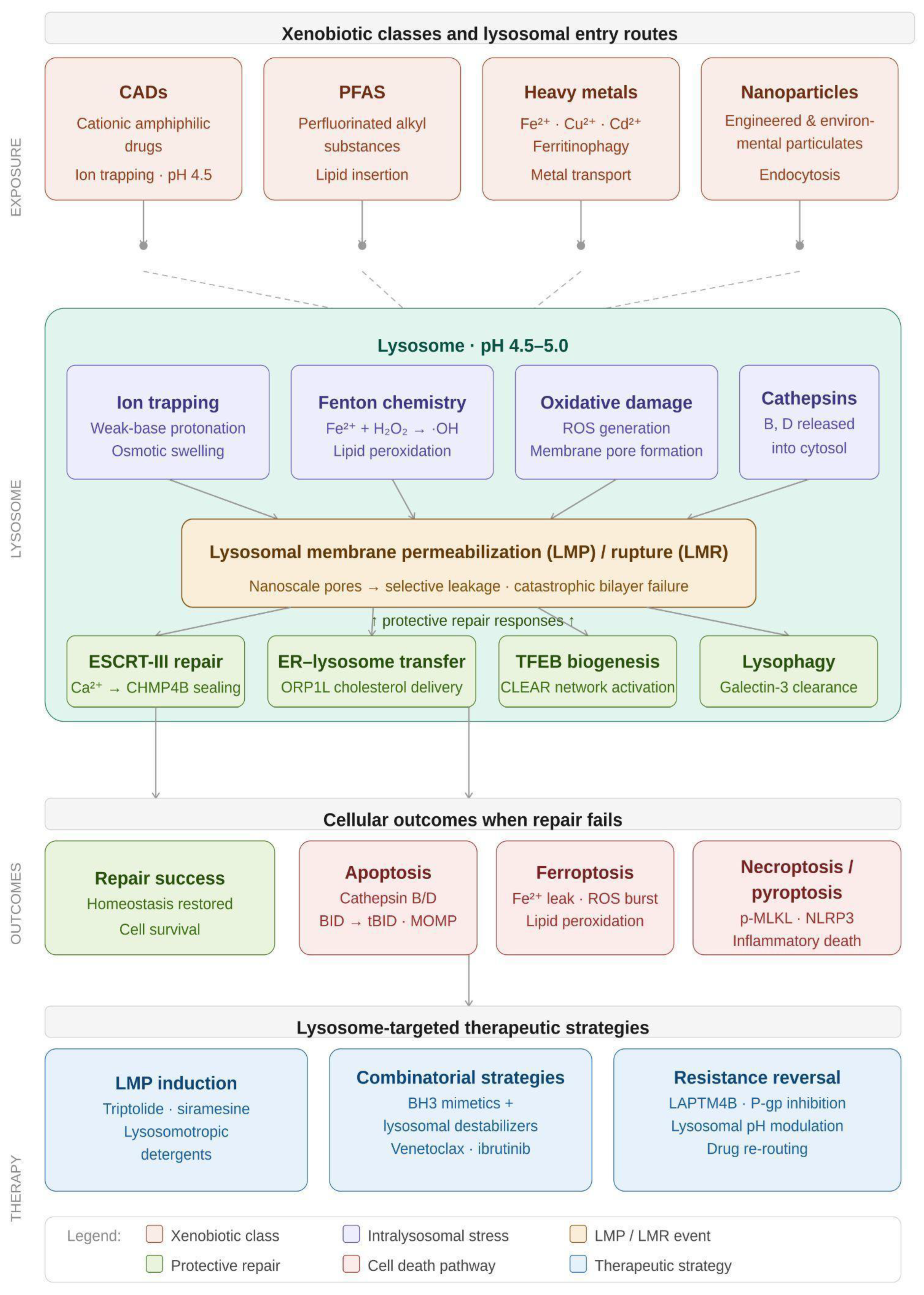

Table 1.
Heavy Metal Sequestration and Redox Toxicity.
| Heavy Metal | Primary Transporter | Mechanism of Interaction | Toxicological Consequence | References |
| Cadmium (Cd) | ZnT2 / DMT1 | Cd–metallothionein complex endocytosis | LMP; autophagy disruption | [1,3,16] |
| Copper (Cu) | DMT1 | Disrupts ER–lysosome contact sites | Impaired lipid replenishment | [16,17] |
| Iron (Fe) | NCOA4 (cargo-mediated) | Ferritinophagy-derived Fe2+ pool | Fenton chemistry; lipid peroxidation | [18,19,20,21,22] |
Table 2.
Therapeutic Strategies and Emerging Biological Models for Lysosomal Stress.
| Strategy / Model | Mechanism of Action | Application / Significance | References |
| ESCRT-III enhancers | Ca2+-dependent recruitment | Rapid sealing of nanolesions | [113] |
| HSP70–ASM axis | Enzyme stabilization | Destabilization in cancer | [111,112] |
| C. elegans | LRO signaling | Sentinel model of stress-induced immunity | [22,96,97] |
| S. litura | Bt-Cry1Ab toxin sequestration | Environmental pesticide toxicity model | [98] |
| Vibrational spectroscopy | Stimulated Raman scattering | Real-time lipid peroxidation mapping | [114,115] |
Disclaimer/Publisher’s Note: The statements, opinions and data contained in all publications are solely those of the individual author(s) and contributor(s) and not of MDPI and/or the editor(s). MDPI and/or the editor(s) disclaim responsibility for any injury to people or property resulting from any ideas, methods, instructions or products referred to in the content. |
© 2026 by the author. Licensee MDPI, Basel, Switzerland. This article is an open access article distributed under the terms and conditions of the Creative Commons Attribution (CC BY) license.
Copyright: This open access article is published under a Creative Commons CC BY 4.0 license, which permit the free download, distribution, and reuse, provided that the author and preprint are cited in any reuse.