

Submitted:
25 September 2026
Posted:
28 September 2026
You are already at the latest version
Abstract
Topoisomerases are a family of enzymes that relieve torsional strain in DNA. Type II topoisomerases utilize a temporary, enzyme-bound double-stranded break to relax, decatenate, or unknot DNA segments. Anticancer agents can disrupt topoisomerase II function and lead cell death, but there is a risk of off target toxicity including secondary leukemia or cardiotoxicity. Humans have two isoforms: topoisomerase IIa (TOP2A) and topoisomerase IIb (TOP2B), and current therapeutics cannot distinguish between these enzymes. We are interested in targeting TOP2A, which is more involved in replication and mitosis, and we hypothesize that avoiding TOP2B may reduce off-target toxicity. Both isoforms share much of their sequence and structure, but they also have intrinsically disordered C-terminal domains (CTD) that are very different in terms of primary structure. We hypothesized that new therapeutics could be designed to target this region of the enzyme. Therefore, we employed AlphaFold3 (AF3) to explore the possible folding conformations of the CTD of TOP2B similar to our previous study of TOP2A. AF3 predicts folding of TOP2B CTD when post-translational modifications and DNA are added to the structure prediction. We find that with both TOP2A and TOP2B, AF3 predicts increased helical structure within the CTD upon the addition of DNA to the model. We explored the AF3 predictions and utilized Edison Scientific to analyze the AF3 outputs. We identified regions within TOP2A that do not align with TOP2B and also are predicted to change secondary structure in the presence of DNA. We hypothesize that these regions may be of interest to explore for impact on TOP2A function with the potential for developing future therapeutics.
Keywords:
topoisomerase II
; AlphaFold 3
; DNA
; structural biology
; anticancer
1. Introduction
The preservation and maintenance of DNA is inherently crucial for the proper functioning of cellular life. The double helical structure of DNA presents both advantages and challenges for cells. While the structure promotes stability and facilitates replication, the twisting structure must be unwound by helicase activity, which results in the formation of supercoils and tangles that must be resolved [1]. A family of enzymes called topoisomerases resolve supercoils and tangles using temporary single- (type I topoisomerases) and double-stranded (type II topoisomerases) DNA breaks [1].
While type I topoisomerases can resolve supercoiling through relaxation, type II topoisomerases can additionally unknot DNA interlinks (catenanes) formed during replication [1]. There are additional subfamilies of topoisomerases that have been discussed elsewhere [1,2]. Notably, topoisomerases are the targets of several widely used therapeutic agents. While bacterial topoisomerases are targeted to treat infections, the eukaryotic topoisomerases are targeted for cancer therapy [3,4,5].
The focus of this current study is the eukaryotic type IIA enzyme topoisomerase II. Mammals have two isoforms of topoisomerase II (α and β) that are expressed from separate genes and are independently regulated [2]. Topoisomerase IIα (TOP2A) is primarily active during replication and mitosis while topoisomerase IIβ (TOP2B) is mostly active during transcription [2]. Anticancer agents such as etoposide and doxorubicin are used to target TOP2 for cancer treatment, but these drugs cannot differentiate between TOP2A and TOP2B [3]. Off target toxicities including cardio toxicity and secondary cancers may result from the impact of anticancer drugs on TOP2B [3]. It has been a focus for several years to explore the differences between TOP2A and TOP2B to identify regions that could be used to differentiate between the isoforms [3,6].
Both isoforms work in the same general manner by binding to helix-helix crossovers, making a temporary, enzyme-bound double-stranded break in one DNA segment, passing the other segment through the break, and then ligating the broken segment before releasing both DNA segments. The catalytic cycle is diagrammed in Figure 1. The details of this mechanism have been outlined elsewhere [3]. While the mechanism is the same for both isoforms, the selection of substrate appears to differ with TOP2A preferring to bind and relax positive supercoils while TOP2B can relax positive and negative supercoils with similar efficiency [7]. To better understand this preference, researchers have examined the similarities and differences in the protein sequences of TOP2A and TOP2B. While the core domains of the enzyme are essentially the same including the ATPase, transducer, TOPRIM metal-binding, DNA cleavage/ligation, and C-Gate domains, the C-terminal domain (CTD) is an Intrinsically Disordered Region (IDR) that is highly divergent between TOP2A and TOP2B and is poorly understood [6].
The CTDs of TOP2A and TOP2B have been studied by various mutagenic approaches including swapping the tails, deletions, point mutations, and mutating segments or groups of amino acids [8,9,10,11,12,13,14,15,16,17,18,19,20,21,22]. The evidence indicates that the CTD does play a role in interacting with DNA, affects catalytic activity, and influences the selection of DNA substrates based upon conformation. Recent studies also highlight the ability of the CTD to participate in phase separation, which appears to influence chromatin compaction [23,24]. In spite of this evidence, the flexible nature of the CTD for both isoforms has made it difficult to study.
Within the last few years, major developments in computational protein folding have enhanced analysis of proteins with and without X-ray crystallographic or cryo-electron microscopy structure data. One of these key developments is the AlphaFold 3 (AF3) algorithm [25,26,27]. AF3 can accurately predict a protein’s folding, and it allows users to input one or more amino acids sequences, DNA or RNA sequences, ions, and post-translational modifications (PTMs). AF3 will generate predicted folds based on the uploaded components along with confidence scores for the fold data.
Previous reports have indicated that AlphaFold algorithms should be able to predict intrinsically disordered domains and conditionally folded regions [28,29]. For example, Ruff and Pappu discussed how AF can determine which regions of proteins will be disordered through low confidence predictions [29]. AlphaFold2 has been used to study a variety of proteins that have intrinsically disordered regions [30,31,32]. As the algorithm has advanced, researchers have also explored AlphaFold Multimer and AlphaFold3 [33,34,35,36]. The need for supplemental computational analysis of intrinsically disordered proteins was highlighted, but AF was shown to provide a foundation for modeling ordered and disordered regions within a protein. Dao et al. aimed to see if AF3 could generate biologically meaningful features within the disordered regions of proteins [34]. Their work showed that AF3 could distinguish ordered and disordered regions, and provide predictions used for structural hypotheses for systems containing substantial disorder. AF3 was unable to represent the full dynamic conformational ensembles that characterize IDPs, but the researchers showed that AF3 can give reliable predictions to capture aspects of this disordered region. Zheng used AF3 to study IDPs that lacked stable tertiary structure [37]. Zheng found that AF3 gave useful information regarding potential interaction sites and transient structural features within disordered regions, but that these generations should be interpreted as potential states for IDPs rather than definitive generations on their structure. Brotzakis, Zhang, and Vendruscolo wanted to broaden the scope of AF3 predictions beyond static structures by incorporating ensemble-based descriptions of protein behavior [38]. They highlighted that disordered proteins exist as dynamic populations of conformations, and the authors demonstrated how AlphaFold-derived information can be integrated with ensemble modeling approaches to better capture structural heterogeneity. They emphasized that AF3 can be used as a reliable starting point to capture the variety within disordered regions. Escédi, et. al applied AF3 to the binding of DNA to single-strand DNA binding protein (SSB) through interactions with the IDR of SSB [39].
We previously applied AF3 to TOP2A and found that under specific circumstances it can predict folding of the CTD [35]. While the confidence levels for the CTD regions were low, the predictions provided a basis for experimentation and analysis of the CTD. AF3 results predict that the CTD folds in the presence of phosphorylation on specific amino acids combined with the presence of DNA substrates [35]. Importantly, the structures generated had varying degrees of plausibility and some structures may reflect actual conformations and provide testable structures for additional study.
To extend this work, we applied AF3 to study TOP2B’s CTD. As described below, we found that the CTD of TOP2B also folded given specific combinations of phosphorylation sites on the CTD and DNA substrates. While the confidence intervals remain low and the AF3 models highly variable, this data provides a new basis for experimental analysis and promotes further exploration of the CTD. We also utilized Edison Scientific AI agent platform to analyze the data and provide summary comparison data among the TOP2B and TOP2A models [40,41]. We report these data and also provide some ideas for future studies to validate the computational data.
2. Materials and Methods
The original sequence of WT Human TOP2B (residues 1–1626) was found using the Uniprot database (Q02880). This sequence was then uploaded into AF3 using the AlphaFold Server (https://alphafoldserver.com/). Once uploaded, the option for two copies was selected in order to the homodimeric structure. An important note is that the protein sequence for TOP2B, Q02880, was the only one entered, and no other settings were changed. All folding was accomplished through AlphaFold Server cloud-based folding. For the sequences with post translational modifications (PTMs), the specific sites for phosphoserine and phosphothreonine were manually added to the sequence based upon information from PhosphositePlus (https://www.phosphosite.org/). DNA sequences were derived from pBR322. The quality of the predicted models was assessed by comparing the Predicted Local Distance Difference Test (plDDT) values across the sequence, calculating RMSD values, and examining panels and overlays of the 5 predicted structures. Analysis of the data was performed using the Analyze function within Edison Scientific (https://platform.edisonscientific.com/). Edison Scientific generated output tables used in this manuscript. Pymol 3.1 was used for image generation. Edison Scientific full reports and outputs are available in Supplemental Information. AF3 outputs have been deposited at: [url]. Generative AI was not used to write this manuscript.
3. Results
3.1. Post-Translational Modifications Allow Folding of the TOP2B CTD
In our previous study with TOP2A, we utilized a set of phosphorylations identified in the literature [35,42]. In order to identify phosphorylation sites for TOP2B, we utilized PhosphositePlus and generated a list of the phosphorylation sites with the most literature evidence (Supplemental Table 1) [43]. From this list, we identified 35 positions that had seven or more references in the literature, and these sites are denoted in Figure 2A.
Using AlphaFold Server, we entered the protein sequence for human TOP2B (Q02880), and allowed AF3 to fold TOP2B as a dimer without modifications. As seen in Figure 2B, the core of the protein folds into ordered domains, but the C-terminus remains a floppy looping structure. This is consistent with the fact that this region is intrinsically disordered. Further, this matches what we observed previously with TOP2A [35]. We added PTMs at positions denoted in Figure 2A and repeated the folding with AF3. Figure 2C shows the predicted folding of TOP2B in the presence of the PTMs. The CTD appears to wrap around the N-terminal ATPase domain and also protrude into the upper DNA gate region. While this conformation would not allow for DNA binding, this was also observed with TOP2A in the absence of DNA [35].
The results in Figure 2C represent one of five models generated by AF3 during processing. Each model showed variations in the CTD with relatively little movement of the core domains of the protein. Figure 3A shows Models 0-4 generated by AF3 for TOP2B with the 35 phosphorylation sites on the CTD. While the lower portion of the prediction remains fairly consistent, the upper portions show a wide range of variability indicating that these predictions are not deterministic (Figure 3B). In most cases, the CTD does wrap around into the DNA binding pockets. Only Model 0 has portions of the CTD wrapping around the outside of the ATPase domain on the upper portion of the enzyme. While these results indicate that there is a predicted fold, the blocking of the DNA gate cannot occur while DNA is bound. Therefore, we also used AF3 to predict folding in the presence of DNA.
3.2. DNA Influences the CTD Folding Predicted by AF3
AF3 allows the input of DNA sequences into the folding prediction. In our previous work, the TOP2A CTD was predicted to fold differently in the presence of DNA. Specifically, we found that the TOP2A CTD moved from inside the upper gate region to around the outside of the DNA gate region when DNA was present, and the DNA segments occupied the DNA gate and lower gate regions [35]. When two DNA duplex fragments of 50-bp and 75-bp are included in the folding with TOP2B, AF3 prediction placed one segment in the active site region and a second segment in the lower gate segment (Figure 4). The active site segment is bent significantly as is seen in crystal structure and cryo-EM data [44,45,46,47]. Interestingly, in Model 0, the second segment is in the lower gate rather than the upper gate, as observed previously with TOP2A [35]. This represents a post-strand passage structure where the DNA has already passed through the DNA gate. Also of interest is the location of the CTD. AF3 predicts that the DNA moves out of the upper DNA gate segment. In Model 0, the CTD is predicted to interact significantly with the ATPase domain and wrap around on top of the ATPase. While there is substantial predicted interaction with the DNA, there is also a long segment that interacts with the ATPase domain.
When a 350-bp DNA duplex was modeled, the CTD retained a tight conformation near the DNA active site region (Figure 5). There was no predicted interaction of the CTD with the ATPase domain in this model. While this conformation may reflect a potentially realistic conformation, the DNA wraps around the enzyme multiple times and is found in the active site as well as the upper and lower gates. In the standard model of the catalytic cycle, the DNA cannot occupy both the upper and lower gates simultaneously since the transport segment would be present in one site or the other. Nevertheless, this result suggests that those regions are both places where DNA can preferentially bind.
3.3. Secondary Structure Stability Varies Across the CTD
CTD conformations found in the predictions exhibited variability depending upon conditions, as described above. To look at this further, we assessed the predicted secondary structure of each amino acid across the CTD. As seen in Figure 6, we have mapped the secondary structure of TOP2B CTD without DNA but in the presence of the phosphorylations mentioned above across all five models generated in the prediction. AF3 predictions indicate that looping structure tends to predominate over helices and very rare sites have strand-like nature.
In contrast to the above results, secondary structure in the presence of DNA is predicted to be more helical in nature. AF3 secondary structure predictions in the presence of 50-bp and 75-bp DNA segments are shown in Figure 7. DNA is predicted to cause a large shift in secondary structure from looping to helical. While there is variability across the five models, there is an overall trend toward more helical nature. Similar predictions are observed for folding with TOP2B in the presence of 350-bp of DNA (Supplementary Figure S1).
In order to assess these shifts and identify sites and segments that change secondary structure across predictions, we analyzed the model outputs from AF3 using Edison Scientific to assess trends in the data (see Supplemental Data). AF3 predicted that secondary structures of specific regions of the CTD will remain constant regardless of the presence or absence of DNA folded with the protein (Table 1). All regions with consistent secondary structure were between 1200 and 1262, and this portion of the protein is adjacent to the region that is not intrinsically disordered. Three of these regions are predicted to be helical and one is a loop ranging between 7-9 amino acids. While these regions maintained secondary structure, the actual three dimensional conformation of these segments is not necessarily consistent across the different predictions (presence or absence of DNA) and among models of a given prediction. One position within this region was phosphorylated in the models as shown in Table 1.
Other regions of the CTD are predicted to change upon addition of DNA to the folding model (Table 2). These regions were spread across the CTD, but were found more concentrated between positions 1200-1500. While the three dimensional conformations are not consistent among various models, the secondary structure went from loop to helix upon addition of DNA to the predictions. These segments cover up to ~25 amino acids.
Additional analysis indicates that a few regions were only predicted to be helical under in one of the three conditions (no DNA, 50-bp/75-bp DNA duplexes, or 350-bp DNA duplex). While Table 2 indicates that large regions are predicted to be helical in the presence of DNA, other regions only become helical in the presence of a long DNA segment (Supplemental Table S1) or only in the presence of the 50-bp/75-bp duplex (Supplemental Table S2). Further, there are locations that are predicted to lose helical nature in the presence of DNA (Supplemental Table S3). These findings suggest that the presence of DNA is not simply imposing helical nature on all positions in the CTD. These segments range from 3-16 amino acids.
3.4. Comparisons of AF3 Predictions for TOP2A Indicate Common Themes
We compared the AF3 folding prediction for TOP2B with the prediction for human TOP2A (using Uniprot P11388). AF3 was used to fold TOP2A as a dimer with CTD PTMs but without any DNA, as reported previously [ref]. As seen with TOP2B, AF3 predicted that the CTD would wrap around the N-terminal ATPase domain and protrude into the upper DNA gate region. As seen in Figure 8, the overall structure of the dimers are consistent, and in these models the CTDs are predicted to interact largely with the ATPase domain and the upper gate region. The conformations in this state would preclude DNA binding, but it is possible that this could represent a resting state in the absence of DNA.
While we previously published data from TOP2A, we did not assess the patterns mentioned above across all models from each AF3 folding output. Similar to results with TOP2B, the predictions for TOP2A secondary structure demonstrated that looping structures predominated in the absence of DNA (Figure 9). Further, the results shift toward more helical structure predicted when DNA is added. Figure 10 and Figure S2 show the secondary structure predictions for TOP2A models with 50-bp and 75-bp and 350-bp DNA fragments, respectively. Results are very similar to what was observed with TOP2B. The general trend is toward more helical secondary structure predicted in the presence of DNA.
3.5. Areas of Focus in the CTD of TOP2A
In order to identify whether there could be distinct areas of TOP2A that could be used to target the CTD therapeutically, we explored whether there were regions of TOP2A that did not align with TOP2B but also were predicted to move during binding of DNA. To do this, we used Edison Scientific to align the CTDs in both TOP2A and TOP2B (data not shown). Using this alignment and the secondary structure maps, we were able to identify regions in TOP2A that do not align with regions in TOP2B. Among these regions were patches of sequence that were predicted to have changes in secondary structure upon binding to DNA (Table 3). These regions include residues 1203–1207, 1241–1244 1248–1250, 1282–1287, and 1441–1446. All of these regions were predicted to shift from “Loop” to “Helix” in secondary structure with the addition of DNA to the folding model. It should be noted that the length of the DNA did not influence this secondary structure change at least with the sizes that were used in these predictions.
Interestingly, one region of the CTD for TOP2A overlapped parts of the CTD for TOP2B that caused conformational changes. Residues 1363–1366 for TOP2A partially overlapped residues 1365-1378 for TOP2B. In both TOP2A and TOP2B, these regions are predicted to change from “Loop” to “Helix” when DNA was incorporated either as a longer fragment or two shorter fragments.
When these regions are visualized in the three dimensional structure, some are closely clustered while others are spread apart (Figure 11). While 1203-1207 is away from the others, we found that 1241-1244, 1248-1250, and 1282-1287 cluster in a region just below the gate segment. Sitting between the transport segment and the gate segment of DNA are alpha helices that include positions 1363-1366 and 1441-1446 in close proximity.
4. Discussion
Computational folding of proteins has opened a new dimension for studying proteins and protein-protein interactions. Many proteins that lack biochemical structure data can now be explored computationally. AlphaFold through the AF3 algorithm on AlphaFold Server provides researchers the ability to enter protein sequence information, post-translational modifications, ions, and DNA or RNA sequences [25,35].
Previously, we applied this tool to studying TOP2A in order to explore the CTD [35]. The CTDs of TOP2A and TOP2B are considered intrinsically disordered, and no crystallographic or cryo-electron microscopy data are available for these regions. The CTDs of TOP2A and TOP2B are the regions with the greatest sequence difference between the isoforms [6]. Further, the CTD has a critical role in regulating enzyme activity, protein interactions, and localization. As a result, we are interested in exploring the CTD to map out function and potential structural conformations that may be biologically relevant. We hypothesize that the CTD could be targeted therapeutically to disrupt enzyme function, and that such therapeutics could be isoform specific for TOP2A and avoid TOP2B, which could mitigate off-target toxicity in cancer chemotherapy [3].
As seen with TOP2A, the TOP2B CTD is predicted by AF3 to fold when PTMs are added to the CTD. Similar to our study of TOP2A, the PTM sites used in this study were identified based upon literature evidence using Phosphosite and covered a range across the CTD. TOP2B CTD did fold with PTMs with or without DNA. The predicted folding was variable across the models generated by AF3. For instance, the CTD is predicted to fold around or inside the ATPase domain in the absence of DNA, which in some models occludes the upper DNA gate region.
The addition of DNA demonstrated that AF3 generates TOP2B:DNA interactions that appear biologically relevant with the DNA in the active site and within the upper or lower gate regions. With DNA present in the prediction, the CTD is predicted to fold outside of the upper gate region and wrap around the outside of the DNA cleavage/ligation domain. The presence of a single, long DNA duplex of 350 bp shows that AF3 does allow unrealistic scenarios since the DNA was found to occupy the upper and lower gate regions simultaneously while also threading through the active site. While these are all regions DNA can occupy, the ability of DNA to occupy the upper and lower gate regions simultaneously has not been demonstrated since these positions should be alternately occupied by the DNA segment being moved from the upper gate to the lower gate during strand passage. Nevertheless, the ability of AF3 to recognize these regions as DNA binding regions underscores the realistic nature of the overall prediction.
Through secondary structure analysis provided by the Analyze and Kosmos functions from Edison Scientific, we observed that some regions of the TOP2B CTD do not change secondary structure in the presence or absence of DNA [41]. All these regions are between residues 1200-1300 in TOP2B. Interestingly, other regions reliably shifted from looping secondary structure to helical in the presence of DNA. These regions are found across the CTD and span regions up to 25 residues long. DNA does not force a helical structure as some additional regions only became helical only with either the longer or shorter DNA duplexes but not in both scenarios. There are additional short segments that are predicted to be more loop-like in the presence of DNA. Similar results are seen with TOP2A secondary structure.
Comparisons between TOP2A and TOP2B show many similarities in the AF3 analyses including the CTD tending to fold into the active site and upper gate region in the absence of DNA. This may reflect the charged regions found along the CTD, which include both positive and negative charge regions. The DNA binding regions of the upper and lower gates have positive charges to help bind to the DNA during strand passage. In the computational folding predictions without DNA, the upper gate region in particular seems accessible to the CTD and may stabilize the negative charges added by the PTMs.
In both TOP2A and TOP2B structures, DNA modeled in these structures is predicted to bind in relevant locations including the DNA cleavage/ligation region (DNA gate) and the upper and lower gate regions, representing the pre- and post-strand passage conformations, respectively. In the case of the 350-bp fragment, the DNA is long enough to wrap back through the enzyme residing in all three binding sites. While this conformation is not biologically plausible since the occupation of the upper and lower gates should be alternate, it does underscore the fact that AF3 is able to identify relevant binding regions for DNA. This data also provides insight into which amino acids in the CTD may interact with the DNA substrates, which is helpful from an experimental perspective in exploring the CTD for functional purposes.
To take the analysis a step further, we identified regions of TOP2A that were predicted to change secondary structure upon binding of DNA but also did not align with the CTD of TOP2B. Among these were several regions spread across the CTD but with a concentration between positions 1200-1300. These positions provide a starting point for further exploration for the potential development of CTD-targeting agents that could be specific for TOP2A. It is possible that targeting these regions could disrupt enzyme-DNA interactions or catalytic activity. Additional studies will be needed to explore these possibilities.
Based upon the data above, we hypothesize that AF3 can generate models that can be used for testing the function and interactions of the CTDs of TOP2A and TOP2B. As such, this data should provide a foundation for beginning to explore isoform-specific targeting of these enzymes by potential therapeutic agents.
Supplementary Materials
The following supporting information can be downloaded at the website of this paper posted on Preprints.org. Figure S1: Helical secondary structure in the TOP2B CTD is predicted to predominate in the presence of a 350-bp DNA segment; Figure S2. Helical secondary structure in the TOP2A CTD is predicted to predominate in the presence of a 350-bp DNA segment; Table S1: TOP2B Segments that become Helical with 350 bp of DNA; Table S2: Regions of TOP2B that become Helical when shorter DNA fragments are present; Table S3: Regions of TOP2B Predicted to be less Helical with DNA Present.
Author Contributions
Conceptualization, J.E.D.; methodology, J.E.D., X.L.F.; investigation, X.L.F.; resources, J.E.D.; data curation, X.L.F.; writing—original draft preparation, X.L.F. and J.E.D.; writing—review and editing, X.L.F., J.E.D.; visualization, X.L.F., J.E.D.; project administration, J.E.D.; funding acquisition, J.E.D. All authors have read and agreed to the published version of the manuscript.
Funding
This research was funded by the Biological, Chemical, and Health Sciences Department (FHU), Center of Science and Culture, and Heintzman Research Fund.
Institutional Review Board Statement
Not applicable.
Informed Consent Statement
Not applicable.
Data Availability Statement
Raw AF3 data files are available at: https://doi.org/10.17605/OSF.IO/ZVMJP.
Acknowledgments
During the preparation of this manuscript/study, the authors used Edison Scientific Analyze and Kosmos functions for the purposes of analyzing the AlphaFold3 output files and generating summary secondary structure maps and tables analyzing the secondary structure features. The authors have reviewed and edited the output and take full responsibility for the content of this publication.
Conflicts of Interest
The authors declare no conflicts of interest.
Abbreviations
The following abbreviations are used in this manuscript:
| AF3 | AlphaFold3 |
| CTD | Carboxy-Terminal Domain or C-Terminal Domain |
| TOP2 | Topoisomerase II |
| TOP2A | Topoisomerase IIa |
| TOP2B | Topoisomerase IIb |
| PTM | Post-translational modification |
| bp | Base pair |
References
- Pommier, Y.; Nussenzweig, A.; Takeda, S.; Austin, C. Human topoisomerases and their roles in genome stability and organization. Nat. Rev. Mol. Cell Biol. 2022, 23, 407–427. [Google Scholar] [CrossRef] [PubMed]
- Pommier, Y.; Sun, Y.; Huang, S.N.; Nitiss, J.L. Roles of eukaryotic topoisomerases in transcription, replication and genomic stability. Nat. Rev. Mol. Cell Biol. 2016, 17, 703–721. [Google Scholar] [CrossRef] [PubMed]
- Murphy, M.B.; Mercer, S.L.; Deweese, J.E. Inhibitors and Poisons of Mammalian Type II Topoisomerases. In Advances in Molecular Toxicology; Fishbein, J.C., Heilman, J., Eds.; Academic Press: Cambridge, MA, 2017; Volume 11, pp. 203–240. [Google Scholar]
- Aldred, K.J.; Kerns, R.J.; Osheroff, N. Mechanism of quinolone action and resistance. Biochemistry 2014, 53, 1565–1574. [Google Scholar] [CrossRef] [PubMed]
- O'Donnell, A.C.; Berger, J.M. Structural Mechanisms of Topoisomerase-Targeting Drugs. Annu Rev. Biochem 2025, 94, 223–251. [Google Scholar] [CrossRef] [PubMed]
- Hoang, K.G.; Menzie, R.A.; Rhoades, J.H.; Fief, C.A.; Deweese, J.E. Reviewing the Modification, Interactions, and Regulation of the C-terminal Domain of Topoisomerase IIα as a Prospect for Future Therapeutic Targeting. EC Pharmacol. Toxicol. 2020, 8, 27–43. [Google Scholar]
- Jian, J.Y.; Osheroff, N. Telling Your Right Hand from Your Left: The Effects of DNA Supercoil Handedness on the Actions of Type II Topoisomerases. Int. J. Mol. Sci. 2023, 24. [Google Scholar] [CrossRef] [PubMed]
- Adachi, N.; Miyaike, M.; Kato, S.; Kanamaru, R.; Koyama, H.; Kikuchi, A. Cellular distribution of mammalian DNA topoisomerase II is determined by its catalytically dispensible C-terminal domain. Nucleic Acids Res. 1997, 25, 3135–3142. [Google Scholar] [CrossRef] [PubMed]
- Mirski, S.E.; Gerlach, J.H.; Cole, S.P. Sequence determinants of nuclear localization in the alpha and beta isoforms of human topoisomerase II. Exp. Cell Res. 1999, 251, 329–339. [Google Scholar] [CrossRef] [PubMed]
- Mirski, S.E.; Bielawski, J.C.; Cole, S.P. Identification of functional nuclear export sequences in human topoisomerase IIalpha and beta. Biochem Biophys. Res. Commun. 2003, 306, 905–911. [Google Scholar] [CrossRef] [PubMed]
- Dickey, J.S.; Osheroff, N. Impact of the C-terminal domain of topoisomerase IIα on the DNA cleavage activity of the human enzyme. Biochemistry 2005, 44, 11546–11554. [Google Scholar] [CrossRef] [PubMed]
- McClendon, A.K.; Osheroff, N. The geometry of DNA supercoils modulates topoisomerase-mediated DNA cleavage and enzyme response to anticancer drugs. Biochemistry 2006, 45, 3040–3050. [Google Scholar] [CrossRef] [PubMed]
- Linka, R.M.; Porter, A.C.; Volkov, A.; Mielke, C.; Boege, F.; Christensen, M.O. C-terminal regions of topoisomerase IIα and IIβ determine isoform-specific functioning of the enzymes in vivo. Nucleic Acids Res. 2007, 35, 3810–3822. [Google Scholar] [CrossRef] [PubMed]
- McClendon, A.K.; Gentry, A.C.; Dickey, J.S.; Brinch, M.; Bendsen, S.; Andersen, A.H.; Osheroff, N. Bimodal recognition of DNA geometry by human topoisomerase II alpha: preferential relaxation of positively supercoiled DNA requires elements in the C-terminal domain. Biochemistry 2008, 47, 13169–13178. [Google Scholar] [CrossRef] [PubMed]
- Meczes, E.L.; Gilroy, K.L.; West, K.L.; Austin, C.A. The impact of the human DNA topoisomerase II C-terminal domain on activity. PLoS ONE 2008, 3, e1754. [Google Scholar] [CrossRef] [PubMed]
- Gilroy, K.L.; Austin, C.A. The impact of the C-terminal domain on the interaction of human DNA topoisomerase II alpha and beta with DNA. PLoS ONE 2011, 6, e14693. [Google Scholar] [CrossRef] [PubMed]
- Lane, A.B.; Gimenez-Abian, J.F.; Clarke, D.J. A novel chromatin tether domain controls topoisomerase IIalpha dynamics and mitotic chromosome formation. J. Cell Biol. 2013, 203, 471–486. [Google Scholar] [CrossRef] [PubMed]
- Clarke, D.J.; Azuma, Y. Non-Catalytic Roles of the Topoisomerase IIalpha C-Terminal Domain. Int. J. Mol. Sci. 2017, 18. [Google Scholar] [CrossRef] [PubMed]
- Kozuki, T.; Chikamori, K.; Surleac, M.D.; Micluta, M.A.; Petrescu, A.J.; Norris, E.J.; Elson, P.; Hoeltge, G.A.; Grabowski, D.R.; Porter, A.C.G.; et al. Roles of the C-terminal domains of topoisomerase IIalpha and topoisomerase IIbeta in regulation of the decatenation checkpoint. Nucleic Acids Res. 2017, 45, 5995–6010. [Google Scholar] [CrossRef] [PubMed]
- Dougherty, A.C.; Hawaz, M.G.; Hoang, K.G.; Trac, J.; Keck, J.M.; Ayes, C.; Deweese, J.E. Exploration of the Role of the C-Terminal Domain of Human DNA Topoisomerase IIalpha in Catalytic Activity. ACS Omega 2021, 6, 25892–25903. [Google Scholar] [CrossRef] [PubMed]
- Endsley, C.E.; Moore, K.A.; Townsley, T.D.; Durston, K.K.; Deweese, J.E. Bioinformatic Analysis of Topoisomerase IIalpha Reveals Interdomain Interdependencies and Critical C-Terminal Domain Residues. Int. J. Mol. Sci. 2024, 25. [Google Scholar] [CrossRef] [PubMed]
- Chang, J.W.; O'Brian, A.K.; Thomas, A.J.; Hardin, M.R.; Latham, B.D.; Ngabonziza, D.; Simpson, L.G.; Wade, B.D.; Kuhnhenrich, L.; Thompson, N.M.; et al. Mutagenesis of Intrinsically Disordered Domain Impacts Topoisomerase IIalpha Catalytic Activity. Int. J. Mol. Sci. 2025, 26. [Google Scholar] [CrossRef] [PubMed]
- Wu, M.; Beck, C.; Lee, J.H.; Fulbright, R.M.; Jeong, J.; Inman, J.T.; Woodhouse, M.V.; Berger, J.M.; Wang, M.D. Human Topoisomerase IIα Promotes Chromatin Condensation Via a Phase Transition. bioRxiv 2024, 2024.2010.2015.618281. [CrossRef] [PubMed]
- Jeong, J.; Lee, J.H.; Carcamo, C.C.; Parker, M.W.; Berger, J.M. DNA-Stimulated Liquid-Liquid phase separation by eukaryotic topoisomerase ii modulates catalytic function. Elife 2022, 11. [Google Scholar] [CrossRef] [PubMed]
- Jumper, J.; Evans, R.; Pritzel, A.; Green, T.; Figurnov, M.; Ronneberger, O.; Tunyasuvunakool, K.; Bates, R.; Zidek, A.; Potapenko, A.; et al. Highly accurate protein structure prediction with AlphaFold. Nature 2021, 596, 583–589. [Google Scholar] [CrossRef] [PubMed]
- Krokidis, M.G.; Koumadorakis, D.E.; Lazaros, K.; Ivantsik, O.; Exarchos, T.P.; Vrahatis, A.G.; Kotsiantis, S.; Vlamos, P. AlphaFold3: An Overview of Applications and Performance Insights. Int. J. Mol. Sci. 2025, 26, 3671. [Google Scholar] [CrossRef] [PubMed]
- Peng, C.; Ni, W.; Liu, Q.; Hu, G.; Zheng, W. A comprehensive benchmarking of the AlphaFold3 for predicting biomacromolecules and their interactions. Brief. Bioinform. 2025, 26. [Google Scholar] [CrossRef] [PubMed]
- Piovesan, D.; Monzon, A.M.; Tosatto, S.C.E. Intrinsic protein disorder and conditional folding in AlphaFoldDB. Protein Sci. 2022, 31, e4466. [Google Scholar] [CrossRef] [PubMed]
- Ruff, K.M.; Pappu, R.V. AlphaFold and Implications for Intrinsically Disordered Proteins. J. Mol. Biol. 2021, 433, 167208. [Google Scholar] [CrossRef] [PubMed]
- Alderson, T.R.; Pritišanac, I.; Kolarić, Đ.; Moses, A.M.; Forman-Kay, J.D. Systematic identification of conditionally folded intrinsically disordered regions by AlphaFold2. Proc. Natl. Acad. Sci. 2023, 120, e2304302120. [Google Scholar] [CrossRef] [PubMed]
- Anbo, H.; Sakuma, K.; Fukuchi, S.; Ota, M. How AlphaFold2 Predicts Conditionally Folding Regions Annotated in an Intrinsically Disordered Protein Database, IDEAL. Biology 2023, 12, 182. [Google Scholar] [CrossRef] [PubMed]
- Zhao, B.; Ghadermarzi, S.; Kurgan, L. Comparative evaluation of AlphaFold2 and disorder predictors for prediction of intrinsic disorder, disorder content and fully disordered proteins. Comput. Struct. Biotechnol. J. 2023, 21, 3248–3258. [Google Scholar] [CrossRef] [PubMed]
- Cortes, M., Jr.; Sun, X.; Anusha; Batchelder-Schwab, E.J.; Li, J.; Siraj, N.; Jampana, R.; Zhang, Y.; Bai, Y.; Mao, C. AlphaFold 3 modeling of DNA nanomotifs: is it reliable? Nanoscale Horiz. 2025, 10, 1428–1435. [Google Scholar] [CrossRef] [PubMed]
- Dao, T.M.; Ghent, S.; Uversky, V.N.; Rahman, T. AlphaFold3 and Intrinsically Disordered Proteins: Reliable Monomer Prediction, Unpredictable Multimer Performance. bioRxiv 2025, 2005.691730. [Google Scholar] [CrossRef]
- Nartey, C.M.; Deweese, J.E. Evaluating AlphaFold 3 Folding of the Intrinsically Disordered Human DNA Topoisomerase IIa C-Terminal Domain. DNA 2025, 5, 46. [Google Scholar] [CrossRef]
- Omidi, A.; Møller, M.H.; Malhis, N.; Bui, J.M.; Gsponer, J. AlphaFold-Multimer accurately captures interactions and dynamics of intrinsically disordered protein regions. Proc. Natl. Acad. Sci. 2024, 121, e2406407121. [Google Scholar] [CrossRef] [PubMed]
- Zheng, S. Navigating the unstructured by evaluating alphafold’s efficacy in predicting missing residues and structural disorder in proteins. PLoS ONE 2025, 20, e0313812. [Google Scholar] [CrossRef] [PubMed]
- Brotzakis, Z.F.; Zhang, S.; Murtada, M.H.; Vendruscolo, M. AlphaFold prediction of structural ensembles of disordered proteins. Nat. Commun. 2025, 16, 1632. [Google Scholar] [CrossRef] [PubMed]
- Ecsédi, P.; Érfalvy, D.; Kovács, Zoltán J.; Katran, V.; Pálinkás, J.; Cervenak, M.; Pancsa, R.; Harami, Gábor M.; Smeller, L.; Kovács, M. Selective engineering of condensation properties of single-stranded DNA binding (SSB) protein via its intrinsically disordered linker region. Nucleic Acids Res. 2025, 53. [Google Scholar] [CrossRef] [PubMed]
- Ghareeb, A.E.; Chang, B.; Mitchener, L.; Yiu, A.; Szostkiewicz, C.J.; Shved, D.; Gyimesi, G.J.; Laurent, J.M.; Wright, S.M.; Razzak, M.T.; et al. A multi-agent system for automating scientific discovery. Nature 2026, 655, 497–505. [Google Scholar] [CrossRef]
- Mitchener, L.; Yiu, A.; Chang, B.; Bourdenx, M.; Nadolski, T.; Sulovari, A.; Landsness, E.C.; Barabasi, D.L.; Narayanan, S.; Evans, N. Kosmos: An ai scientist for autonomous discovery. arXiv 2025, arXiv:2511.02824. [Google Scholar]
- Bedez, C.; Lotz, C.; Batisse, C.; Broeck, A.V.; Stote, R.H.; Howard, E.; Pradeau-Aubreton, K.; Ruff, M.; Lamour, V. Post-translational modifications in DNA topoisomerase 2alpha highlight the role of a eukaryote-specific residue in the ATPase domain. Sci. Rep. 2018, 8, 9272. [Google Scholar] [CrossRef] [PubMed]
- Hornbeck, P.V.; Zhang, B.; Murray, B.; Kornhauser, J.M.; Latham, V.; Skrzypek, E. PhosphoSitePlus, 2014: mutations, PTMs and recalibrations. Nucleic Acids Res. 2015, 43, D512–520. [Google Scholar] [CrossRef] [PubMed]
- Dong, K.C.; Berger, J.M. Structural basis for gate-DNA recognition and bending by type IIA topoisomerases. Nature 2007, 450, 1201–1205. [Google Scholar] [CrossRef] [PubMed]
- Schmidt, B.H.; Burgin, A.B.; Deweese, J.E.; Osheroff, N.; Berger, J.M. A novel and unified two-metal mechanism for DNA cleavage by type II and IA topoisomerases. Nature 2010, 465, 641–644. [Google Scholar] [CrossRef] [PubMed]
- Schmidt, B.H.; Osheroff, N.; Berger, J.M. Structure of a topoisomerase II-DNA-nucleotide complex reveals a new control mechanism for ATPase activity. Nat. Struct. Mol. Biol. 2012, 19, 1147–1154. [Google Scholar] [CrossRef] [PubMed]
- Vanden Broeck, A.; Lotz, C.; Drillien, R.; Haas, L.; Bedez, C.; Lamour, V. Structural basis for allosteric regulation of Human Topoisomerase IIalpha. Nat. Commun. 2021, 12, 2962. [Google Scholar] [CrossRef] [PubMed]
Figure 1.
Catalytic cycle of Topoisomerase II. TOP2 is depicted at left. Two DNA helices (maroon and gold) are bound by the enzyme (1a, 1b), while one segment is cleaved. The gate segment (maroon) is separated and the transport segment (gold) is moved through the temporary break (2,3). The gate segment is ligated (3) and the transport segment released (4). The enzyme then releases the gate segment (5) and resets.
Figure 1.
Catalytic cycle of Topoisomerase II. TOP2 is depicted at left. Two DNA helices (maroon and gold) are bound by the enzyme (1a, 1b), while one segment is cleaved. The gate segment (maroon) is separated and the transport segment (gold) is moved through the temporary break (2,3). The gate segment is ligated (3) and the transport segment released (4). The enzyme then releases the gate segment (5) and resets.
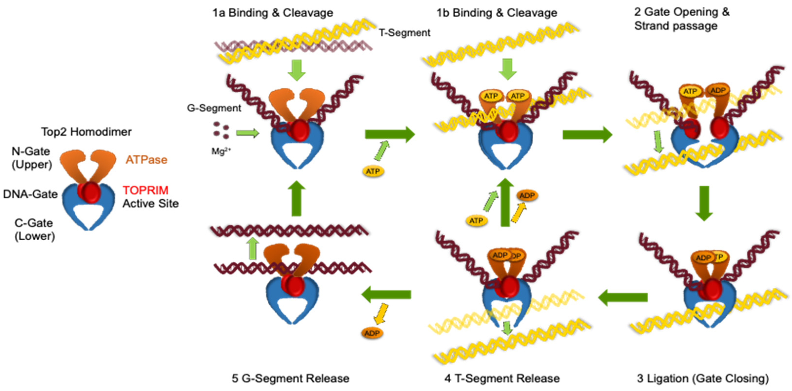
Figure 2.
AlphaFold 3 (AF3) Predicts Folding of TOP2B without PTMs vs with PTMs. A) Map of TOP2B with major domains labeled. ATPase in green, Transducer in orange, TOPRIM in purple, and DNA binding and C-gate in blue. CTD is in maroon and gold. Phosphorylation sites are denoted in blue under the CTD. B) AF3’s Predicted generation of human TOP2B without PTMs (Model 0). C) AF3’s Predicted generation of human TOP2B with the 35 PTMs (Model 0) shown from two angles. PTM sites denoted in cyan. Structure images generated using Pymol 3.1.
Figure 2.
AlphaFold 3 (AF3) Predicts Folding of TOP2B without PTMs vs with PTMs. A) Map of TOP2B with major domains labeled. ATPase in green, Transducer in orange, TOPRIM in purple, and DNA binding and C-gate in blue. CTD is in maroon and gold. Phosphorylation sites are denoted in blue under the CTD. B) AF3’s Predicted generation of human TOP2B without PTMs (Model 0). C) AF3’s Predicted generation of human TOP2B with the 35 PTMs (Model 0) shown from two angles. PTM sites denoted in cyan. Structure images generated using Pymol 3.1.
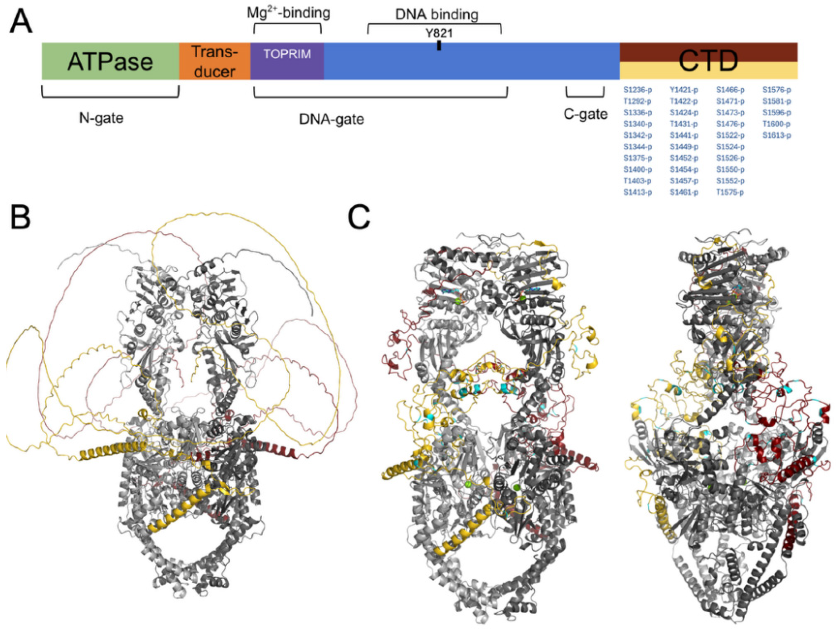
Figure 3.
AF3 Shows Variation between Predicted Folds. A) Separate representations of the CTD of Models 0-4 generated by AF3 for TOP2B with 35 phosphorylation sites in the CTD. The ATPase and core cleavage/ligation domains of TOP2B are not shown for clarity. B) An alignment of all five CTD models from the AF3 folding of TOP2B with 35 phosphorylation sites. Alignment was generated using Pymol 3.1.
Figure 3.
AF3 Shows Variation between Predicted Folds. A) Separate representations of the CTD of Models 0-4 generated by AF3 for TOP2B with 35 phosphorylation sites in the CTD. The ATPase and core cleavage/ligation domains of TOP2B are not shown for clarity. B) An alignment of all five CTD models from the AF3 folding of TOP2B with 35 phosphorylation sites. Alignment was generated using Pymol 3.1.
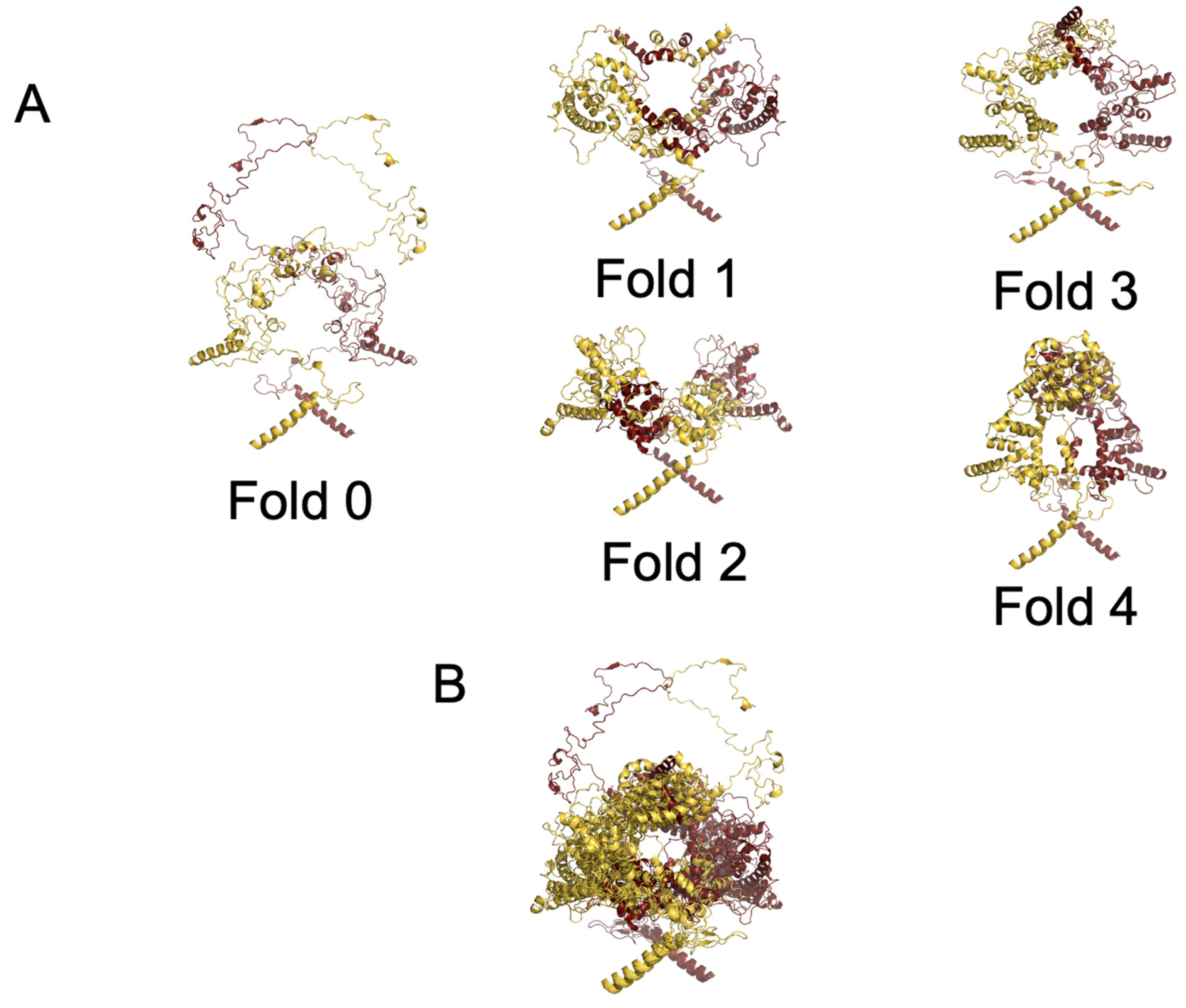
Figure 4.
AF3 Predicts Folding of TOP2B with PTMs and 50 BP and 75 BP DNA. Two views of AF3’s predicted folding TOP2B with the 35 PTMs and two short segments of duplex DNA (50 BP and 75 BP). DNA is seen in the active site and in the lower DNA gate, which represents a post-strand passage state. Images generated from AF3 Model 0 using Pymol 3.1.
Figure 4.
AF3 Predicts Folding of TOP2B with PTMs and 50 BP and 75 BP DNA. Two views of AF3’s predicted folding TOP2B with the 35 PTMs and two short segments of duplex DNA (50 BP and 75 BP). DNA is seen in the active site and in the lower DNA gate, which represents a post-strand passage state. Images generated from AF3 Model 0 using Pymol 3.1.
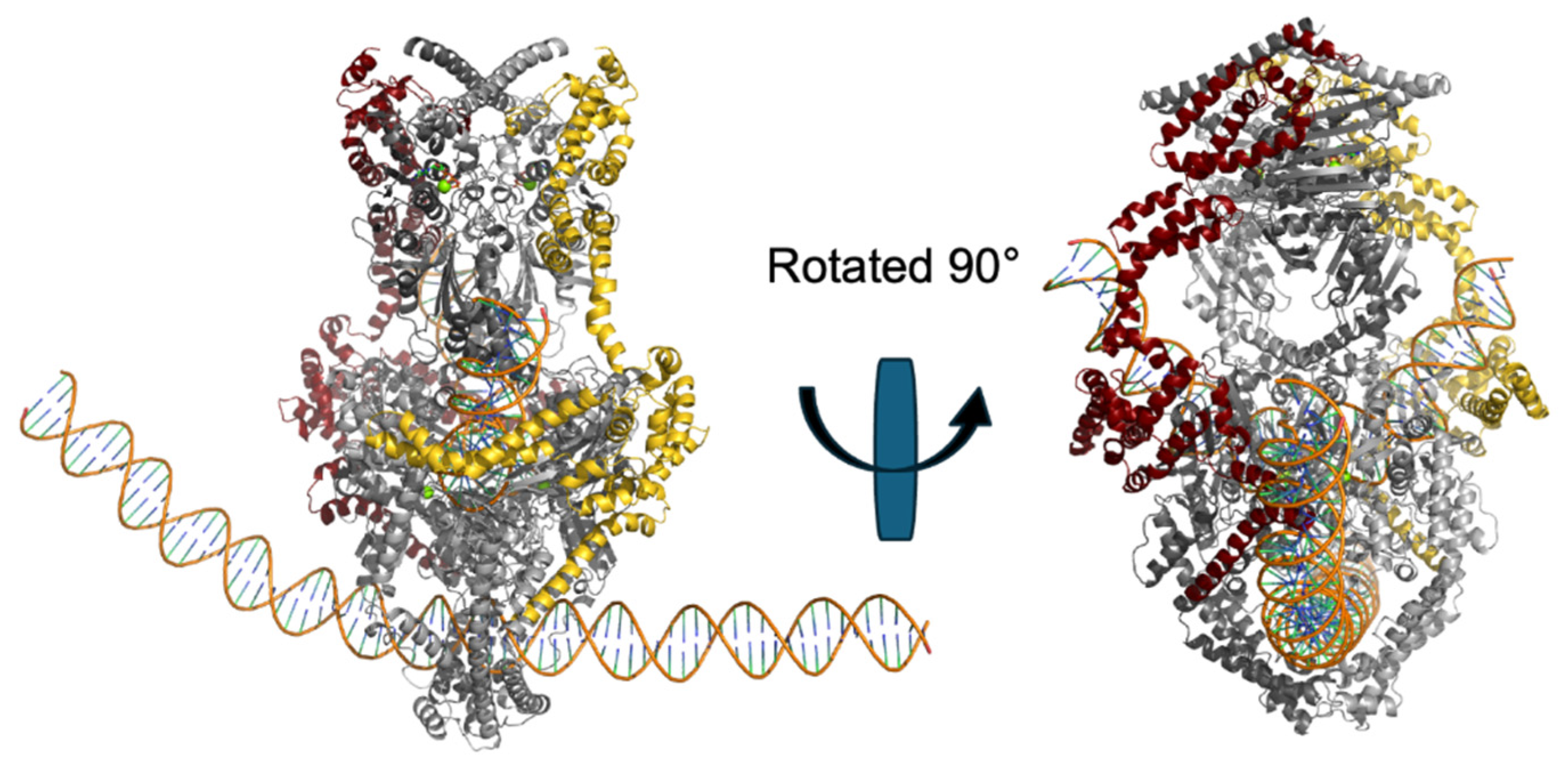
Figure 5.
AF3 Predicts Folding between TOP2B with PTMs and 350 BP DNA. Two views AF3’s predicted folding of TOP2B with 35 PTMs and a single, 350 bp segment of dsDNA (350 BP). AF3 predicted that the CTD will be more compact and concentrated around the sides of the TOP2B. The DNA segment passes through the active site in addition to the upper and lower gates, which would not occur in an actual DNA cleavage event. Images generated from AF3 Model 0 using Pymol 3.1.
Figure 5.
AF3 Predicts Folding between TOP2B with PTMs and 350 BP DNA. Two views AF3’s predicted folding of TOP2B with 35 PTMs and a single, 350 bp segment of dsDNA (350 BP). AF3 predicted that the CTD will be more compact and concentrated around the sides of the TOP2B. The DNA segment passes through the active site in addition to the upper and lower gates, which would not occur in an actual DNA cleavage event. Images generated from AF3 Model 0 using Pymol 3.1.
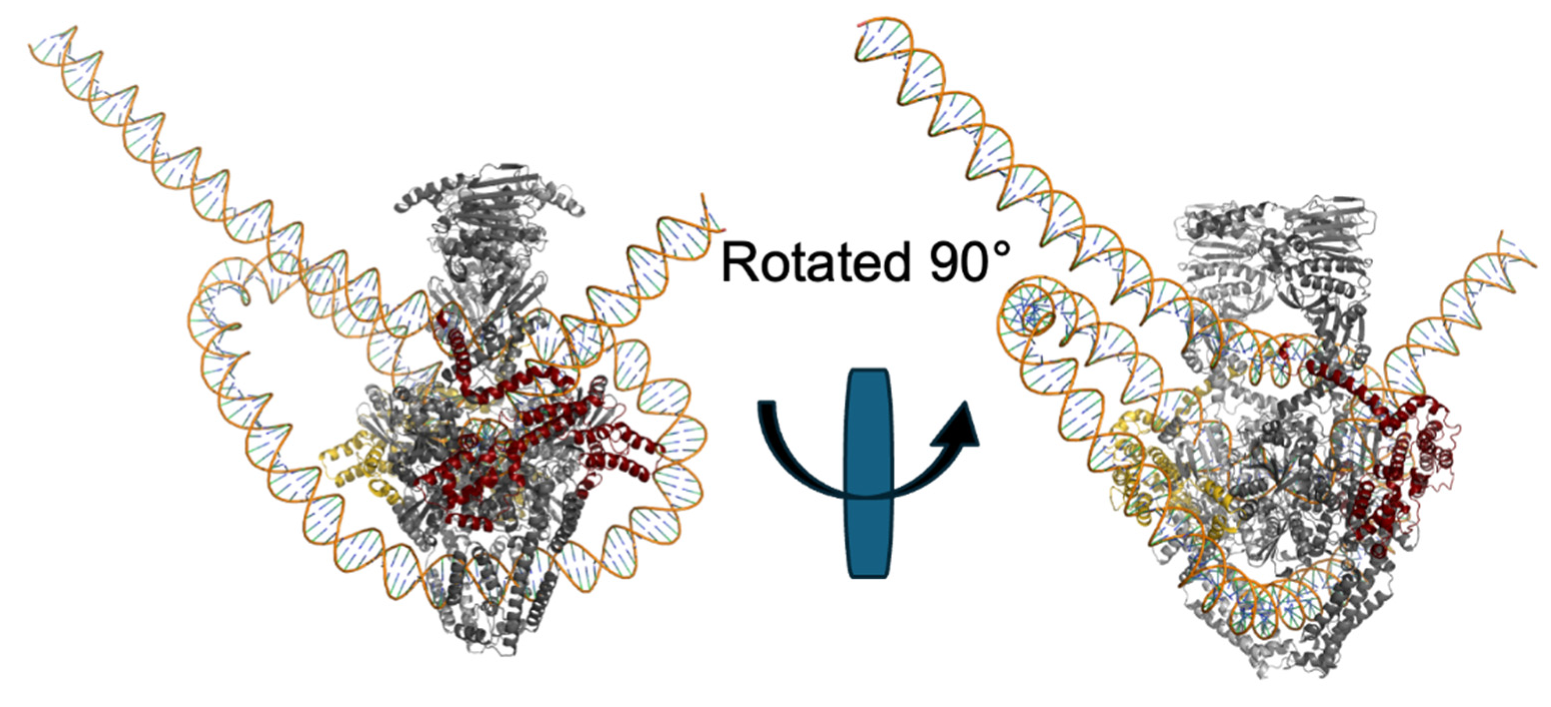
Figure 6.
Looping secondary structure in the TOP2B CTD is predicted to predominate in the absence of DNA. Five models of the TOP2B CTD with PTMs are mapped by sequence position and secondary structure as Helix (H, maroon), Loop (L, gold), or Strand (S, blue). Map generated by Edison Scientific Analyze function.
Figure 6.
Looping secondary structure in the TOP2B CTD is predicted to predominate in the absence of DNA. Five models of the TOP2B CTD with PTMs are mapped by sequence position and secondary structure as Helix (H, maroon), Loop (L, gold), or Strand (S, blue). Map generated by Edison Scientific Analyze function.
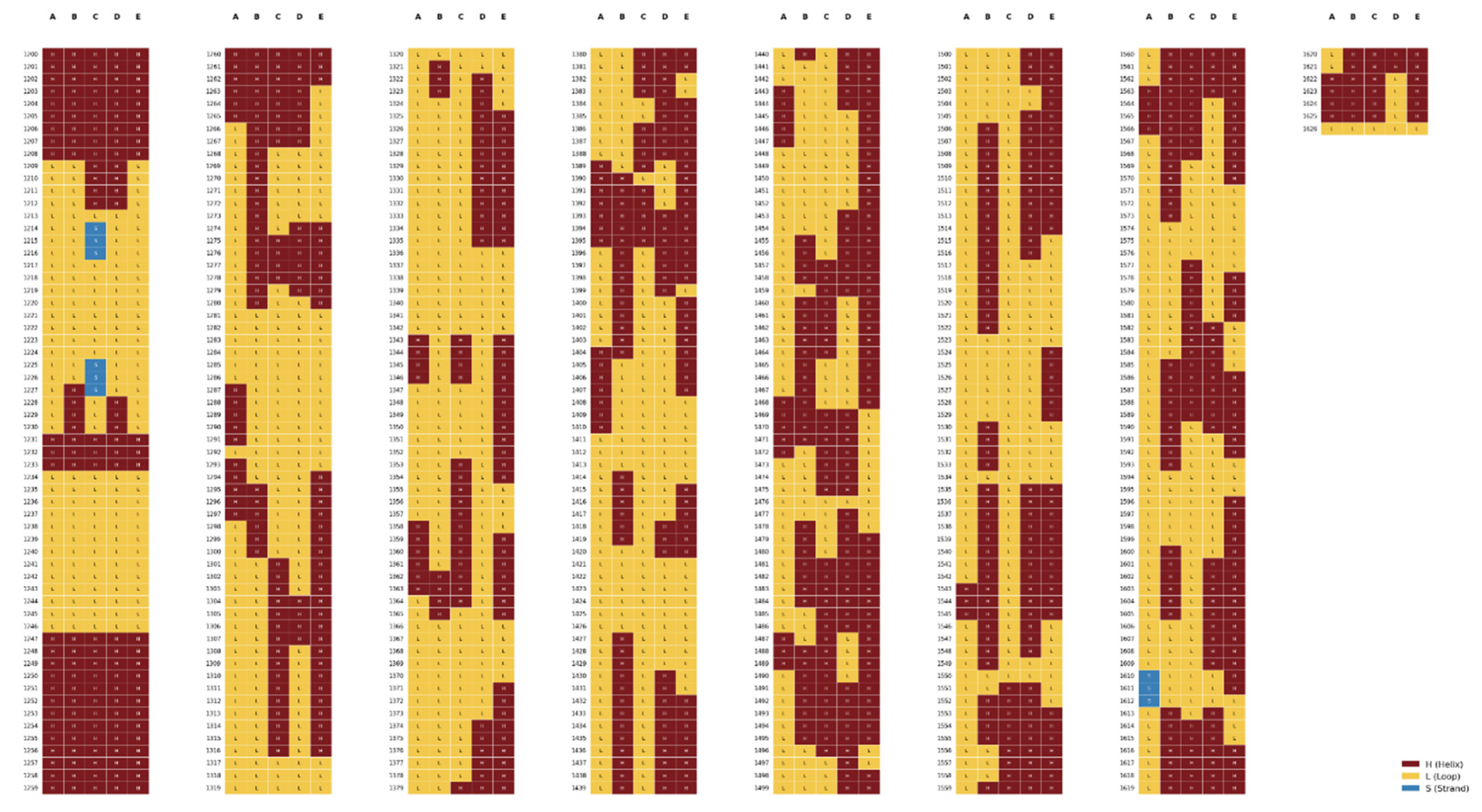
Figure 7.
Helical secondary structure in the TOP2B CTD is predicted to predominate in the presence of DNA segments. Five models of the TOP2B CTD with PTMs and 50-bp and 75-bp DNA fragments are mapped by sequence position and secondary structure as Helix (H, maroon), Loop (L, gold), or Strand (S, blue). Map generated by Edison Scientific Analyze function.
Figure 7.
Helical secondary structure in the TOP2B CTD is predicted to predominate in the presence of DNA segments. Five models of the TOP2B CTD with PTMs and 50-bp and 75-bp DNA fragments are mapped by sequence position and secondary structure as Helix (H, maroon), Loop (L, gold), or Strand (S, blue). Map generated by Edison Scientific Analyze function.
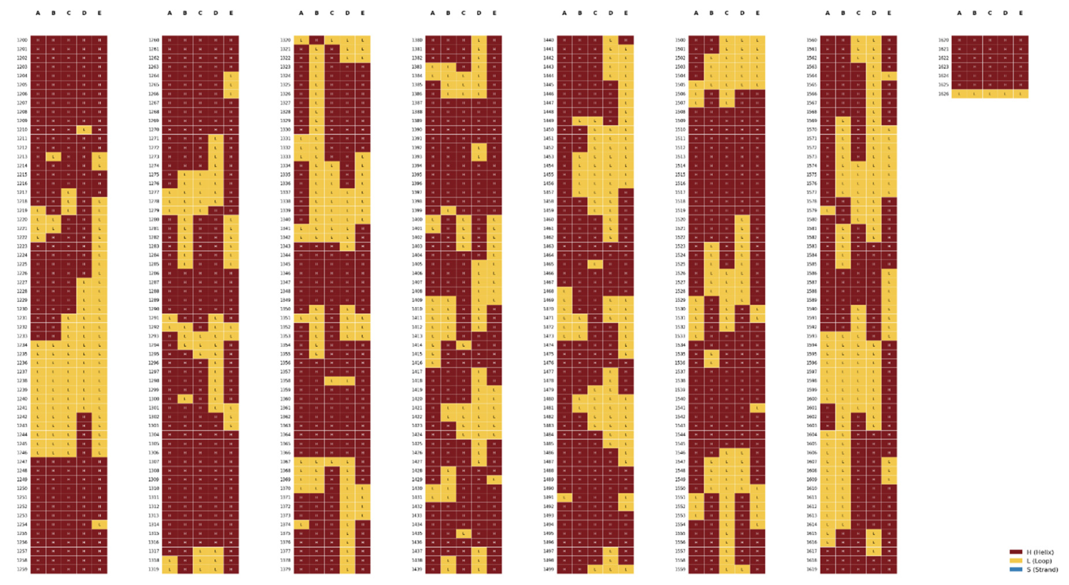
Figure 8.
Comparison of TOP2A and TOP2B predicted folds with AF3. Depicted are the structures of TOP2A (left) and TOP2B (right). The images are merged and aligned in the center. AF3 predictions were made with PTMs but no DNA. Images were generated and aligned using Pymol 3.1.
Figure 8.
Comparison of TOP2A and TOP2B predicted folds with AF3. Depicted are the structures of TOP2A (left) and TOP2B (right). The images are merged and aligned in the center. AF3 predictions were made with PTMs but no DNA. Images were generated and aligned using Pymol 3.1.
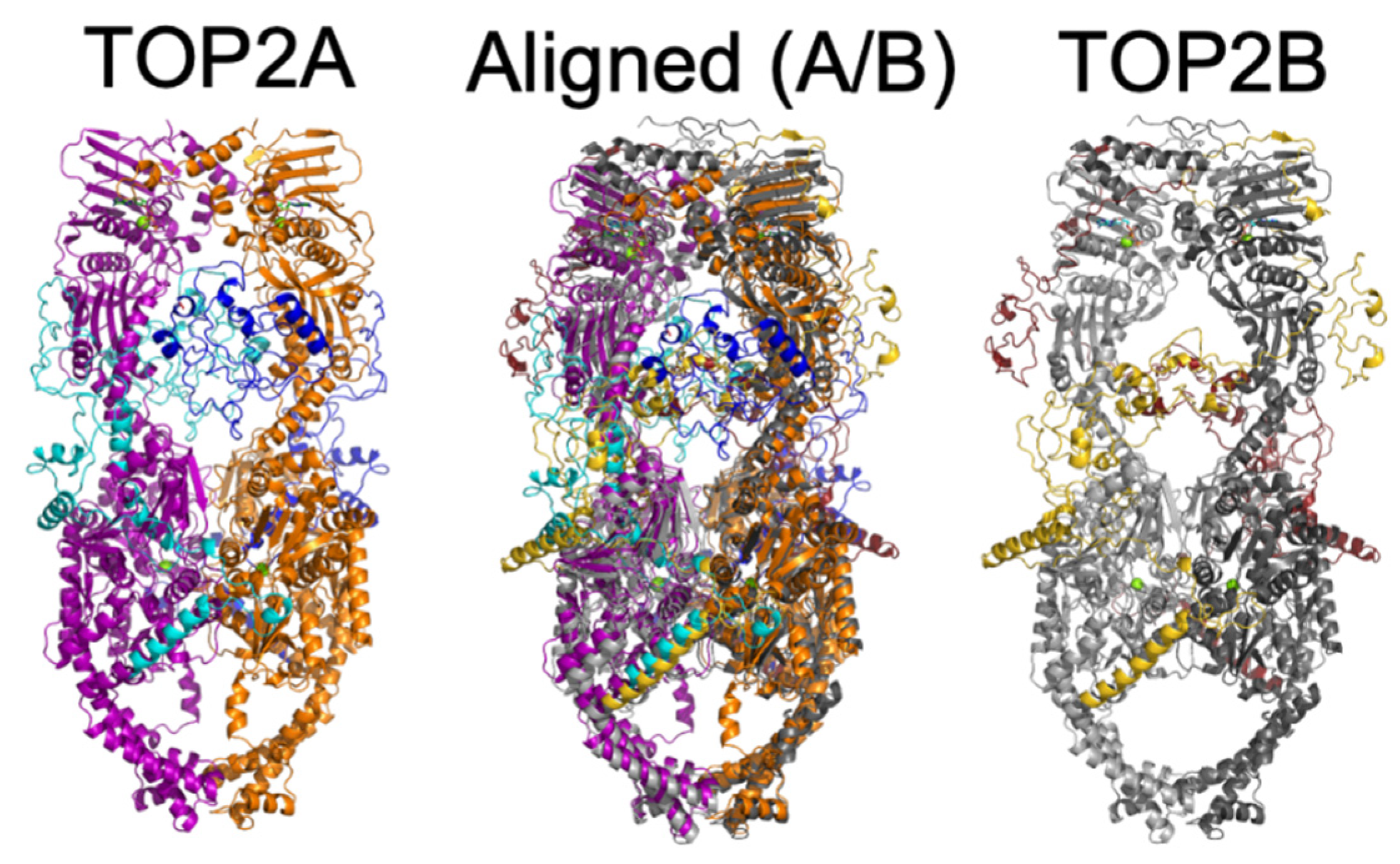
Figure 9.
Looping secondary structure in the TOP2A CTD is predicted to predominate in the absence of DNA segments. Five models of the TOP2B CTD with PTMs and no DNA fragments are mapped by sequence position and secondary structure as Helix (H, maroon), Loop (L, gold), or Strand (S, blue). Map generated by Edison Scientific Analyze Function.
Figure 9.
Looping secondary structure in the TOP2A CTD is predicted to predominate in the absence of DNA segments. Five models of the TOP2B CTD with PTMs and no DNA fragments are mapped by sequence position and secondary structure as Helix (H, maroon), Loop (L, gold), or Strand (S, blue). Map generated by Edison Scientific Analyze Function.
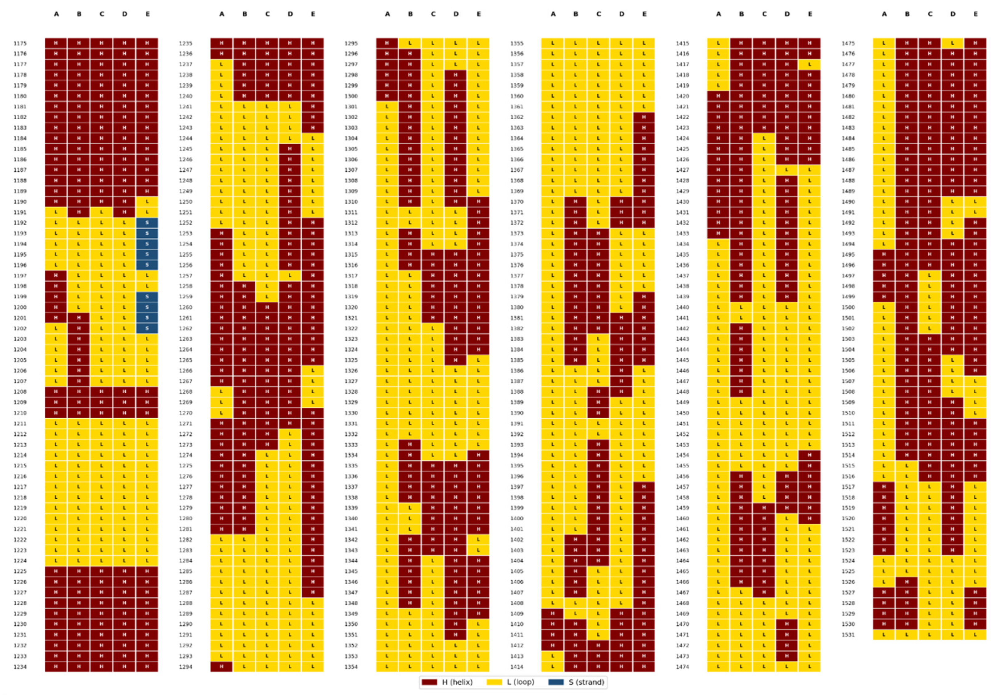
Figure 10.
Helical secondary structure in the TOP2A CTD is predicted to predominate in the presence of DNA segments. Five models of the TOP2A CTD with PTMs and 50-bp and 75-bp DNA fragments are mapped by sequence position and secondary structure as Helix (H, maroon), Loop (L, gold), or Strand (S, blue). Map generated by Edison Scientific Analyze function.
Figure 10.
Helical secondary structure in the TOP2A CTD is predicted to predominate in the presence of DNA segments. Five models of the TOP2A CTD with PTMs and 50-bp and 75-bp DNA fragments are mapped by sequence position and secondary structure as Helix (H, maroon), Loop (L, gold), or Strand (S, blue). Map generated by Edison Scientific Analyze function.
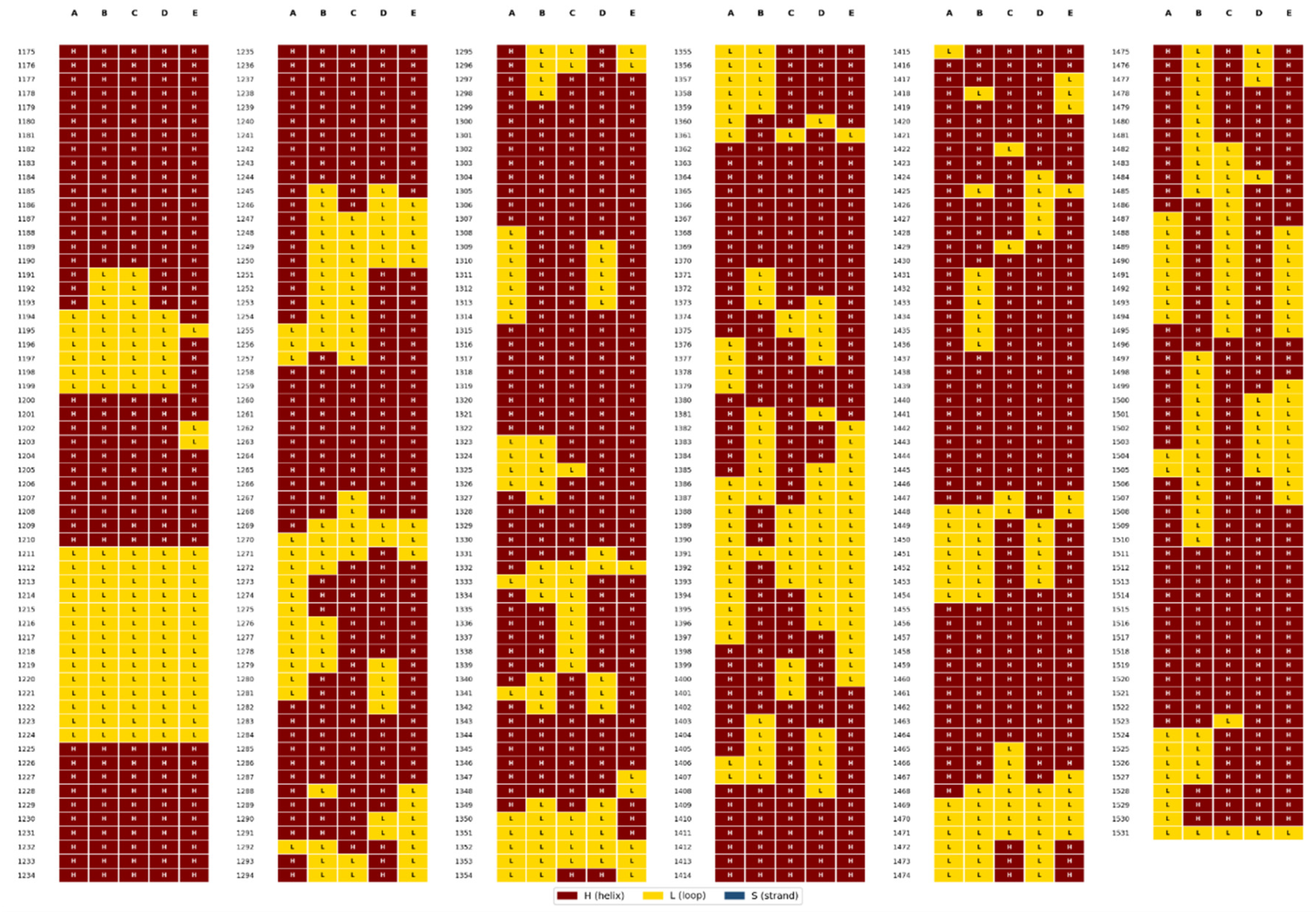
Figure 11.
Map of TOP2A CTD Regions Predicted to Respond to DNA Binding. Two views of the TOP2A CTD with PTMs and 50-bp and 75-bp DNA fragments. DNA (green) is shown. CTD is in gold with sequence regions from Table 3 shown in maroon. Image generated using Pymol 3.1.
Figure 11.
Map of TOP2A CTD Regions Predicted to Respond to DNA Binding. Two views of the TOP2A CTD with PTMs and 50-bp and 75-bp DNA fragments. DNA (green) is shown. CTD is in gold with sequence regions from Table 3 shown in maroon. Image generated using Pymol 3.1.
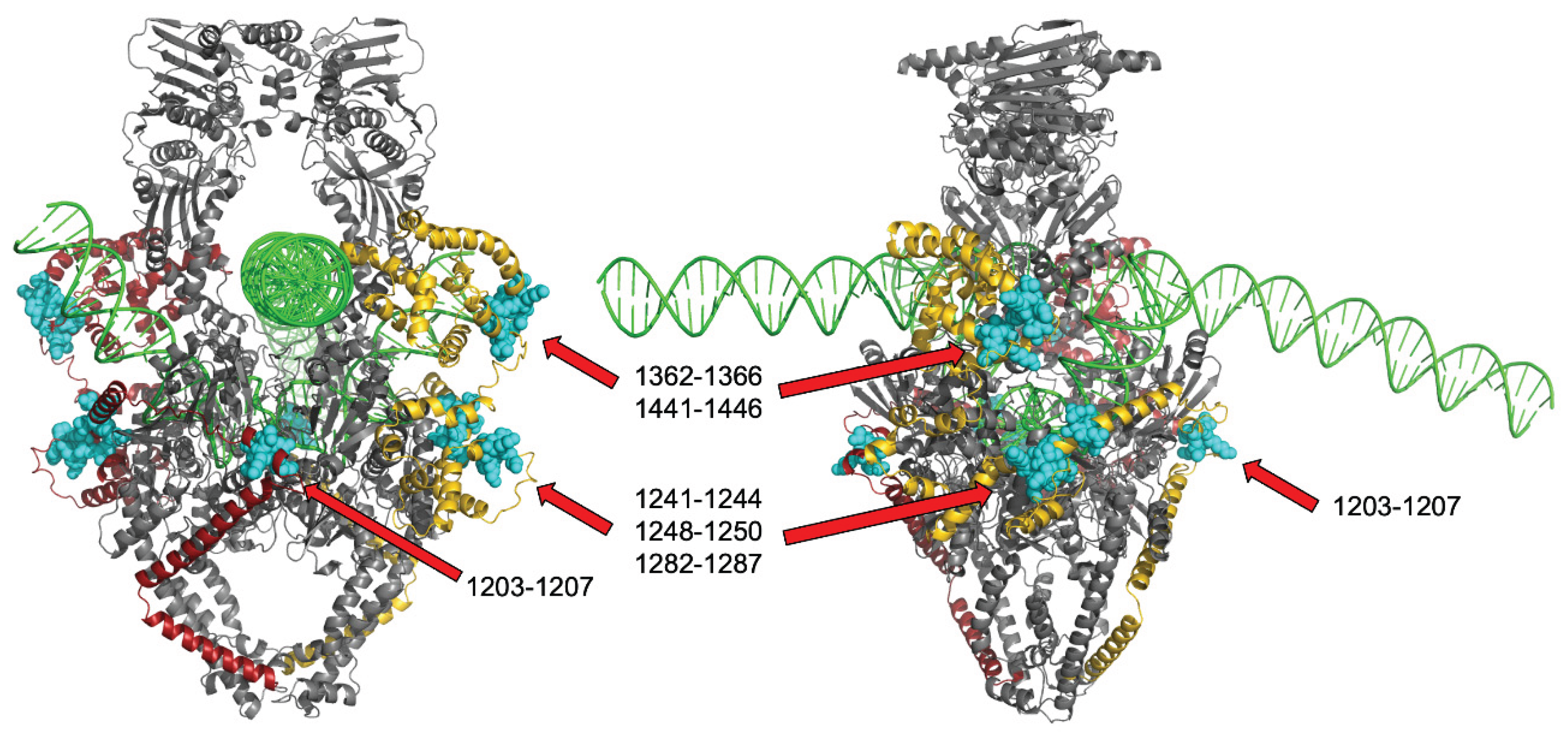

Table 1.
Regions of the TOP2B CTD that Retain Secondary Structure Across Conditions.
| Sequence | Start | End | Length | SS |
|---|---|---|---|---|
| ESNEREDVL | 1200 | 1208 | 9 | H |
| MPS*PYGRR | 1234 | 1241 | 8 | L |
| TAMKADA | 1247 | 1253 | 7 | H |
| KKLLKKKK | 1255 | 1262 | 8 | H |
* Phosphoserine.
Table 2.
Regions of the TOP2B CTD that Differ in Secondary Structure when DNA Is Modeled.
| Sequence | Start | End | Length | noDNA→50/75bp→350bp |
|---|---|---|---|---|
| AGMSGKAIKG | 1209 | 1218 | 10 | L → H → H |
| GKPKVKKLQLEET | 1221 | 1233 | 13 | L → H → H |
| AAVKVE | 1268 | 1273 | 6 | L → H → H |
| DEEFSGAPVEGAGEEALTPS | 1275 | 1294 | 20 | L → H → H |
| NKGPKP | 1298 | 1303 | 6 | L → H → H |
| KEPGTRVRKTPTSSGKPSAKKVKKR | 1308 | 1332 | 25 | L → H → H |
| DLEETEPVVIPRDS | 1345 | 1358 | 14 | L → H → H |
| AERPKYTFDFS*EEE | 1365 | 1378 | 14 | L → H → H |
| DGEDEFVPS*DGLDKDE | 1405 | 1420 | 16 | L → H → H |
| PGKSKAT | 1425 | 1431 | 7 | L → H → H |
| DS*N | 1465 | 1467 | 3 | L → H → H |
| VFS*PS | 1474 | 1478 | 5 | L → H → H |
| KVVEAVNS*DS*DSE | 1515 | 1527 | 13 | L → H → H |
| EVKYFAES* | 1606 | 1613 | 8 | L → H → H |
* Phosphoserine.
Table 3.
Regions of the TOP2B CTD that Retain Secondary Structure Across Conditions.
| Sequence | Start | End | Length | noDNA→50/75bp→350bp |
|---|---|---|---|---|
| KKTQM | 1203 | 1207 | 5 | L → H → H |
| NENT* | 1241 | 1244 | 4 | L → H → H |
| PQE | 1248 | 1250 | 3 | L → H → H |
| FKPIKK | 1282 | 1287 | 6 | L → H → H |
| KLS*N^ | 1363 | 1366 | 4 | L → H → H |
| TKRPDA | 1441 | 1446 | 6 | L → H → H |
* Phosphoserine, ^Partially overlaps a similar region in TOP2B.
Disclaimer/Publisher’s Note: The statements, opinions and data contained in all publications are solely those of the individual author(s) and contributor(s) and not of MDPI and/or the editor(s). MDPI and/or the editor(s) disclaim responsibility for any injury to people or property resulting from any ideas, methods, instructions or products referred to in the content. |
© 2026 by the authors. Licensee MDPI, Basel, Switzerland. This article is an open access article distributed under the terms and conditions of the Creative Commons Attribution (CC BY) license (http://creativecommons.org/licenses/by/4.0/).
Copyright: This open access article is published under a Creative Commons CC BY 4.0 license, which permit the free download, distribution, and reuse, provided that the author and preprint are cited in any reuse.